Article

Control of Aerosolised Type A Influenza Virus H1N1 and a Coronavirus with Vapours Containing Catmint Essential Oil

1
School of Optometry and Vision Science, University of New South Wales, Kensington, NSW 2052, Australia
2
Vapour Science, O’Connor, Canberra, ACT 2602, Australia
*
Author to whom correspondence should be addressed.
Hygiene 2025, 5(2), 15; https://doi.org/10.3390/hygiene5020015
Submission received: 9 January 2025 / Revised: 14 March 2025 / Accepted: 1 April 2025 / Published: 6 April 2025

Abstract

:
Background: Respiratory viruses spread through airborne droplets and aerosols, causing highly contagious acute respiratory syndromes in humans. This study evaluated the antiviral potential of vapours of catmint-oil-based formulations against respiratory viruses. Methods: The antiviral activity of formulations with or without catmint oil (CO) in solution or in aerosolised form was determined against influenza virus H1N1 ATCC VR-1469 and mouse hepatitis virus (MHV-1) ATCC/VR261. In solution, both viruses were exposed to CO formulations for 2–3 h. In aerosolised form, H1N1 was exposed to formulations for 2 min in a closed cylinder and MHV-1 for 10 min in a booth. The antiviral effect of the formulations was evaluated by growing H1N1 in a Madin–Darby canine kidney (MDCK; ATCC-CRL-2936) and MHV-1 in A9 ATCC/CCL 1.4 cells using TCID50 and a plaque assay, respectively. Transmission electron microscopy (TEM) was conducted to investigate the mode of action of the formulations. Results: In solution, the formulation containing hydrogenated CO (HCO), bromelain, N-acetylcysteine and Tween 20 (Formulation (1)) reduced the viability of H1N1 by 2.6 ± 0.07 log10 (p = 0.025) and MHV-1 by 4.5 ± 0.14 log10 (p = 0.014) within 2–3 h. In vapourised form, Formulation (1) produced similar antiviral effects against H1N1, reducing it by 3.00 ± 0.07 log10 (p = 0.002) within 2 min, and Formulation (1) produced a 3.00 ± 0.07 log10 reduction of MHV-1 (p < 0.001) within 10 min (the minimum time needed to detect infective viral particles in the experimental set-ups). Formulation (3) (without bromelain) reduced H1N1 by 1.57 ± 0.14 log10 (p = 0.008) after 2 min and MHV-1 by 1.3 ± 0.04 log10 (p = 0.057) after 10 min. In the absence of catmint oil (Formulation (4)) or in the absence of catmint oil and bromelain (Formulation (5)), there were only slight reductions in the viability of aerosolised H1N1 (1.00 ± 0.14 log10, p = 0.046; <1 log10, p = 0.966, respectively) and MHV-1 (1.07 ± 0.02 log10, p = 0.013; 0.16 ± 0.03 log10, p = 0.910, respectively). The TEM analysis showed that the formulation disrupted the H1N1 envelopes and caused a reduction in size of the viral particles. Conclusions: The catmint-oil-based formulations reduced the H1N1 and MHV-1 by disrupting the vial envelopes.

1. Introduction

Influenza viruses (Orthomyxoviridae) and coronaviruses are etiological agents causing highly contagious acute respiratory syndromes in humans and animals and posing significant public health problems worldwide. Both viruses can spread from person to person or animal to animal through airborne droplets and aerosols [1,2,3,4] and can also be transmitted through contact with contaminated surfaces [5,6]. Their spread can be controlled by disinfecting objects and cleaning and sanitising indoor air, as well as vaccination, which can also reduce the spread via aerosols [7]. However, as influenza and coronavirus continually mutate, the available vaccines may not be appropriate to control all the varieties that may be circulating [8]. Therefore, exploring control strategies that target the viruses directly in aerosols may be useful to help stop their spread.
Coronaviruses have been responsible for pandemics, such as the coronavirus disease 2019 (COVID-19) caused by severe acute respiratory syndrome coronavirus 2 (SARS-CoV-2). Besides SARS-CoV-2, other beta coronaviruses such as SARS-CoV and MERS-CoV have been linked to severe acute respiratory syndrome in humans. A mouse beta coronavirus known as murine hepatitis virus (MHV-1) is commonly used as a substitute for human coronaviruses. The Therapeutic Goods Administration (TGA) of Australia recommends MHV-1 as a surrogate (source: TGA, https://www.tga.gov.au/products/other-therapeutic-goods/disinfectantssterilants/surrogate-viruses-use-disinfectant-efficacy-tests-justify-claims-against-covid-19 accessed on 6 August 2024). Phylogenetic studies have shown that SARS-CoV and SARS-CoV-2 belong to beta coronavirus lineage b, MERS-CoV to lineage c, and MHV-1 to lineage a. This categorisation makes MHV-1 more closely related to the SARS-associated coronaviruses compared to other surrogates like human coronavirus 229E, feline coronavirus, or transmissible gastroenteritis virus, which are alpha coronaviruses from different lineages. Influenza viruses are primarily classified into four types: A, B, C, and D [9]. Of these, types A and B cause most seasonal flu infections in humans. Influenza A viruses are further categorised based on two surface proteins, hemagglutinin (H) and neuraminidase (N), with subtypes like H1N1 and H3N2 causing significant illness in humans [10]. Pandemics arise when a novel influenza A virus emerges, infects humans, and spreads widely due to little to no pre-existing immunity in the population. Notable influenza pandemics include the 1918 Spanish flu (H1N1), the 1957 Asian flu (H2N2), the 1968 Hong Kong flu (H3N2), and the 2009 H1N1 pandemic, each causing widespread illness and mortality [9]. Currently, there are limited strategies for controlling coronaviruses or influenza viruses in aerosolised form.
Essential oils (EOs) derived from plants have various health applications. EOs are complex mixtures of volatile compounds, such as monoterpenes, sesquiterpenes and phenylpropenes, which can impart antimicrobial activity to EOs [11,12]. Various EOs have been tested for their antimicrobial, antifungal, and antiviral activities. EOs from Pinus densiflora [13], Trigonella hamosa [14], Thalictrum species [15], Prunella vulgaris [16], Salvia dentata [17] and several others [18,19,20] have been shown to reduce the infectivity of influenza viruses in laboratory experiments. EOs have the potential to prevent and mitigate infections with viruses such as coronavirus, influenza virus, human immunodeficiency virus (HIV), human herpesviruses (HSV1 and HSV2), avian influenza, and yellow fever virus [21,22,23]. Aerosols of a mixture of tea tree oil, eucalyptus oil and lemon myrtle were able to inactivate bacteriophage MS2 in airborne form [24]. Similarly, sprays of a mixture of essential oils with oleoresins applied two hours prior to infection with avian coronavirus decreased the signs and symptoms and reduced the viral titre in chickens [25]. Most of these evaluations of antiviral activity have been performed when the EOs were suspended in liquid or by measuring the effects of EOs on the neuraminidase or haemagglutination proteins of influenza viruses. There are very limited data on the control of influenza viruses in aerosolised form with vapours of EOs.
Catmint oil (CO) is an EO derived from catmint (genus Nepeta). It has been used to alleviate the symptoms of disease caused by severe acute respiratory syndrome coronavirus (SARS-CoV-2) [26]. Catmint derived from Nepeta nuda has antiviral activity against human alpha herpesvirus type 1 [27] and herpes simplex virus [28] when tested in solution. However, as far as the authors can determine, there are no studies examining the direct anti-influenza and anti-coronavirus activity of catmint oil, either in solution or in aerosol. Bromelain is derived from the pineapple plant (Ananas comosus) and is known to have antiviral activity against Semliki Forest virus, Sindbis virus, mouse gastrointestinal coronavirus, hemagglutinating encephalomyelitis virus, and influenza virus H1N1 [29,30,31]. Acetylcysteine is an antioxidant and can cause destabilisation of viruses by reducing the disulfide bonds in spike and envelope proteins [32]. Polysorbate 20 (Tween 20), a non-ionic surfactant, has also been shown to have antiviral activities by solubilising viral lipid envelopes [33]. The antiviral effect of these compounds alone or in combination in vaporised form against aerosols of respiratory viruses has not been determined. Therefore, this study evaluates the antiviral potential of hydrogenated or non-hydrogenated catmint oil (HCO or NHCO)-based formulations containing bromelain, acetylcysteine and Tween 20 against H1N1 and MHV-1 in aerosolised form. The null hypothesis tested was that vapours of formulations containing catmint oil would not be able to reduce the infectivity of influenza virus or coronavirus.

2. Materials and Methods

2.1. Viruses and Cell Lines

Influenza virus H1N1 ATCC VR-1469 and mouse hepatitis virus (MHV-1) ATCC/VR261 stocks were prepared prior to testing by growing them in a Madin–Darby canine kidney (MDCK; ATCC-CRL-2936) and A9 ATCC/CCL 1.4 cells, respectively, in Dulbecco’s minimum essential medium (DMEM) containing 10% foetal bovine serum (FBS) and antibiotics (streptomycin sulphate and penicillin G) at 37 °C in a humidified incubator with 5% CO2. For H1N1 growth, the DMEM was supplemented with tosylamide phenylethyl chloromethyl ketone-treated trypsin (2 μg/mL) (Sigma Aldrich, St Louis MO, USA). Cytopathic effects were visually evident after 3–4 days, and the viruses were then harvested by centrifugation (2000× g for 20 min), aliquoted in 1.5 mL centrifuge tubes and stored at −80 °C. The aliquots were then used in subsequent experiments. The titres of H1N1 and MHV-1 were determined by TCID50 or plaque assays, respectively, as described below.

2.2. Preparation of Catmint Oil Formulations

The following formulations were prepared in per litre of sterile Milli-Q water (sterilised by autoclavation) by dissolving. Formulation (1): hydrogenated catmint (HCO) (0.5 g), bromelain (0.5 g), N-acetylcysteine (0.025 g), Tween 20 (1 g). Formulation (2): non-hydrogenated (NHCO) catmint oil (0.5 g), bromelain (0.5 g), N-acetylcysteine (0.025 g), Tween 20 (1 g). Formulation (3): HCO (0.5 g), + NAC (0.025 g), + Tween 20 (1 g). Formulation (4): bromelain (0.5 g), N-acetylcysteine (0.025 g), Tween 20 (1 g). Formulation (5): N-acetylcysteine (0.025 g), Tween 20 (1 g). The concentrations of the active ingredients were based on previous reports of the antiviral or other antimicrobial activities when used in solutions. For example, 1% Tween 20 has been used to remove the hemagglutinin and neuraminidase proteins from influenza viral particles [34], the minimum inhibitory concentration of catmint oil against a variety of bacteria and fungi is ≤1% (v/v) [35] or ≤1 mg/mL [36], and bromelain at 0.25 g/L or the combination of bromelain and N-acetylcysteine (BromAC) at 0.25 g/L + 20 mg/mL is active against SARS-CoV-2 [32,37].

2.3. Antiviral Activity

2.3.1. In Solution

Initial studies demonstrated that the HCO-based formulation dissolved into the DMEM containing 7.5% BSA, as a neutraliser, had no cytotoxic effect on the MDCK or A9 cells. The antiviral activity of the catmint oil formulations was assessed by incubating H1N1 virus with Formulation (1). Specifically, 1 mL of H1N1 or MHV-1 solution (1 × 10⁶-7 pfu/mL) was mixed with 9 mL of Formulation (1) at room temperature for 2–3 h. After the incubation period, the solution was diluted (10-fold serial dilution) in DMEM containing 7.5% (w/v) bovine serum albumin (BSA), which acted as a neutraliser for the active ingredients. Then, 100 µL of the dilutions were inoculated into a 96-well plate, which had already been seeded with MDCK cells (1 × 10⁴ cells/mL) to achieve approximately 80–90% confluency. The virus-inoculated plate was then incubated for 1 h, followed by the addition of 100 µL of fresh DMEM. After further incubation for 3–4 days, the antiviral efficacy of the disinfectant was assessed by determining the TCID50 (50% Tissue Culture Infective Dose) details, as outlined below.

2.3.2. In Aerosolised Form

An aliquot (4–5 mL) of H1N1 was aerosolised in a closed cylinder (Figure 1A) in a biosafety cabinet for 2 min at a rate of 2.5 mL per minute using aerosoliser A (containing 100 mL viruses), which generated average aerosol sizes of 3.0 to 5.0 µm. Similarly, 20–25 mL MHV-1 was aerosolised for 10 min in a closed booth with an area of 3 m2 (Figure 1B), but for 10 min. Preliminary studies had shown that the minimum times required to detect sufficient infective aerosolised viral particles of both viruses within these experimental set-ups after collection with an Anderson impactor were 2 min and 10 min, respectively. Simultaneously, all the formulations were separately aerosolised at the same rate into the same booth or cylinder from the other aerosoliser B (Figure 1A,B). The ultrasonically generated vapours of the formulations were allowed to interact for 2 min or 10 min with the H1N1 and MHV-1 aerosols, respectively, followed by collection for 5 min and 10 min, respectively, using a vacuum pump attached to a 6-stage Anderson impactor containing plates containing 2% agar. The viruses were exposed to aerosolised sterile Milli-Q water as a negative control. Subsequently, the H1N1 and MHV-1 particles were scraped from the agar plates into 1.5 mL of DMEM and further diluted for quantification by determination of the TCID50 and plaque assay, respectively.

2.4. Determination of TCID50

A modified TCID50 method was employed to quantify the H1N1 particles following treatment with the different formulations. Initially, 100 µL of the virus treated with the different formulation vapours, or with sterile Milli-Q water vapours (as control), were inoculated into a 96-well plate that had been pre-seeded with MDCK cells (1 × 10⁵ cells/mL) and had reached approximately 80–90% confluency. The plates were then incubated at 37 °C in a 5% CO2 atmosphere for 1 h, with gentle shaking occurring at 15 min intervals. Subsequently, each well received 100 µL of fresh DMEM and was further incubated at 37 °C in a 5% CO2 environment for 3–4 days. After the incubation period, all the wells of the plate were washed with phosphate buffer saline (PBS, NaCl 8 g/L, KCl 0.2 g/L, Na2HPO4 1.4 g/L, KH2PO4 0.24 g/L; pH 7.2) and then stained with 1% crystal violet. Wells exhibiting the loss of MDCK cell monolayers due to the cytopathic effect (CPE) of the virus were considered positive, while wells that retained the MDCK cell monolayer without displaying any signs of CPE were deemed negative. The difference in the viral particle count resulting from the activity of the formulations, compared to the Milli-Q water control aerosols, was determined using the Reed and Muench formula in conjunction with the TCID50 protocol.

2.5. Plaque Assay

For titration of infectious MHV-1, A9 cells were seeded in 12-well tissue culture plates at 5–10 × 105 cells per well and allowed to adhere overnight at 37 °C in 5% CO2. After incubation, the culture medium was removed, and the cells were washed with 1X phosphate-buffered saline. Subsequently, 100 µL of serially diluted virus was inoculated into a well containing A9 cells that had reached approximately 80–90% confluency. The plates were then incubated at 37 °C in a 5% CO2 atmosphere for 1 h, with gentle shaking occurring at 15 min intervals. After incubation, the cells were overlaid with 1% agarose (Sigma-Aldrich, Castle Hill, NSW, Australia) and further incubated for 72 h. Following incubation, the cells were fixed with 4% formaldehyde for 2–3 h. The number of viral plaque-forming units (PFUs) from each sample was quantified after staining the cells with 1% crystal violet (Sigma-Aldrich). The reduction in PFUs for each disinfectant compared to the negative control (water) was calculated [38].

2.6. Transmission Electron Microscopy

Transmission electron microscopy was conducted to investigate the mechanism of action of these disinfectant formulations on H1N1. Briefly, H1N1 was exposed to Formulations (1) and (2) for 2 h at 37 °C. Following treatment, 10 µL of the treated mixture was applied to a glow-discharged carbon-coated 200 mesh copper grid and allowed to evaporate for 5 min. The samples were subsequently stained with 1% phosphotungstic acid (pH 6.5) for 30 s to enhance the contrast. After air-drying, the grids were examined using an FEI Tecnai G2 20 transmission electron microscope [39].

2.7. Statistical Analysis

The quantitative data of the virus titres were expressed as the mean and SD values of the log10 reduction value for two separate experiments performed in triplicate. Statistical analysis was performed using Welch’s correction and the unpaired t-test for H1N1 and one-way ANOVA with Tukey’s test for MHV-1 with GraphPad Prism 8.0.2 (GraphPad Software, La Jolla, CA, USA). Significant differences were accepted at p < 0.05.

3. Results

3.1. Effect of Formulation (1) in Solution

The HCO-based Formulation (1) demonstrated excellent antiviral activity against H1N1 and MHV-1 in solution. After 2–3 h of incubation, Formulation (1) achieved a 2.6 ± 0.07 log10 reduction in H1N1 virus particles compared to the PBS negative control (p = 0.025; Figure 2. Similarly, for MHV-1, Formulation (1) resulted in a 4.5 ± 0.14 log10 reduction in infective particles after the same incubation period (p = 0.014; Figure 2). In the control (PBS-treated) samples, there were viable virus particles that induced infection and produced cytopathic effects in MDCK cells for H1N1 and A9 cells for MHV-1.

3.2. In Aerosol Form

The Formulation (1) vapours significantly affected the viability of aerosolised H1N1 during the 2 min of interaction, resulting in a 3.00 ± 0.14 log10 reduction in infective virus particles (p = 0.002; Table 1). Additionally, after vaporising Formulation (2) and allowing it to interact with aerosolised H1N1 for 2 min, a reduction of 2.34 ± 0.24 log10 in infective virus particles was observed (p = 0.025). Both Formulation (1) (HCO-based) and Formulation (2) (NHCO-based) exhibited comparable antiviral effects against aerosolised viruses in vaporised form (p > 0.05). Formulation (3), which omitted the bromelain, produced a 1.57 ± 0.14 log10 reduction in the viability of infective particles (p = 0.008). The vapours of Formulation (4), which contained bromelain and N-acetylcysteine only, and Formulation (5), which contained only N-acetylcysteine with Tween 20, did not significantly impact the viability of aerosolised H1N1 (p = 0.046 and p = 0.966, respectively).
Given that both Formulation (1) (HCO-based) and Formulation (2) (NHCO-based) produced similar antiviral effects against aerosolised H1N1, only Formulation (1) was tested against MHV-1. After 10 min of interaction, the vapours from the HCO-based Formulation (1) resulted in more than a 3.10 ± 0.04 log10 reduction in infective virus particles (p < 0.001; Table 2). Formulation (3), which included HCO, NAC, and Tween 20, produced a 1.30 ± 0.04 log10 reduction after 10 min of interaction with MHV. In contrast, Formulation (4), which did not contain HCO or NHCO, demonstrated slight antiviral activity, reducing the viability of MHV 1.07 ± 0.02 by 1 log10 after 10 min (p = 0.013). Formulation (5), containing acetylcysteine along with Tween 20, did not significantly affect the reduction in viability of aerosolised MHV-1, as the number of aerosolised viral particles remained unchanged when plated on A9 cells compared to the water-treated control (p = 0.910).

3.3. Transmission Electron Microscopy

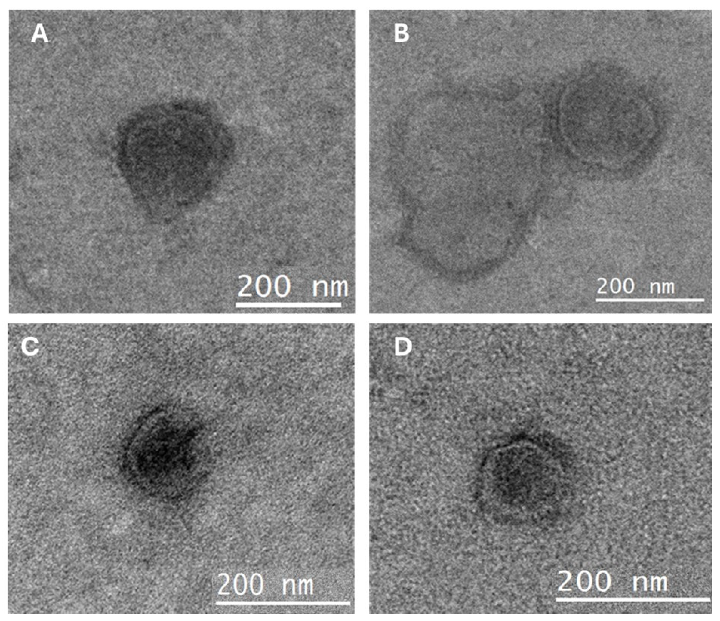
The TEM images of the HCO- or NHCO-based formulations and control-treated viral particles are shown in Figure 3. Ultrastructural analysis showed that both formulations directly affected the viral envelope. Figure 3C,D show disrupted viral envelopes (a clear area outside of the virion) after treatment with the HCO- and NHCO-based formulations, respectively. In comparison, in the control samples, the virus particles were well ordered, round, with an area of darker shading outside of virions (Figure 3A,B). Moreover, both formulations also caused a reduction in the size of the viral particle, presumably due to the decomposition of the viral envelope (Figure 3C,D).

4. Discussion

This study represents the first time that catmint oil vapours demonstrated an antiviral effect on aerosolised H1N1 virus and coronavirus particles and effectively neutralised them in the air. The observed antiviral effect appears to be attributed to the action of essential oils (EOs) on the viral envelope, as evidenced by the reduction in the virion particle size from the loss of the viral envelope of H1N1 during the TEM analysis. Whilst some previous studies have shown that compounds, mostly when used singly, of the formulations can have antiviral activity in solution [32,37,40], the current study demonstrated the ability of combinations in vapour form to reduce the infectivity of a strain of influenza virus and norovirus. This is important as respiratory viruses primarily spread through aerosols or direct/indirect contact, such as touching contaminated objects, with nasal and throat secretions of infected individuals [1,41,42,43]; therefore, the ability of antiviral compounds to prevent the subsequent infectivity of the virus from aerosols might be important to reduce the spread of the disease, thereby enhancing their practical application.
In the present study, vapours of both the HCO- or NHCO-based formulations showed excellent antiviral activity against H1N1 aerosols and reduced their infectivity by decreasing the viability by >2.5 log10 PFU/mL in 2 min. Similarly, the HCO-based formulation vapours inactivated aerosolised MHV-1 by >3 log10 PFU/mL within 10 min. Earlier studies have shown that electrolysed saline containing sodium hypochlorite or mouth washes can inactivate bioaerosols of the equine arteritis virus, and feline calicivirus, respiratory syncytial virus A, parainfluenza 4, influenza B, rhinovirus/enterovirus, SARS-CoV-2 and SARS-CoV-2 generated from saliva [44,45]. In a previous study, tea tree and bergamot oil vapours inactivated 2.35 log10 H1N1 viruses in 30 min [46]. The differences observed in the virus inactivation timeline may be due to the use of dried viruses in the previous study [46] compared to aerosolised viruses in the current study. It is hypothesised that viruses in aerosolised form or in solution can be inactivated more rapidly by disinfectants than those dried on surfaces. An earlier study using a gel containing various essential oils (such as eucalyptol, myrcene, limonene, terpinene, and cymene) achieved only a 0.3 log10 reduction in MHV-1 infectious virions in 10 min [47]. This difference in the efficacy of essential oil formulations may be attributed to the gel having to release the essential oils via evaporation, rather than as in the current study, where the catmint oil formulations were aerosolised. There is also likely to be a contribution of the different essential oils, with catmint possibly having a greater antiviral activity. This should be followed up in future research.
The current data suggest that the catmint oil was the predominant antiviral in the formulation, as Formulation (3), which did not contain bromelain but contained catmint and two other ingredients, produced better levels of viral reductions compared to Formulation (4), which contained bromelain and the other two ingredients. The number of H1N1 viruses in the control samples remained consistent, except for the samples used against Formulation (1), where a higher initial number of viruses were used for the test and control. Formula (1) was therefore tested against an approximately 1 log10 PFU/mL greater number of viruses than the other formulas. Whilst this difference resulted in a greater statistical difference for its effect on H1N1, it does not diminish the effect of catmint oil on H1N1.
The TEM analysis indicated that Formulations (1) and (2) directly impacted the virions, disrupting the lipid bilayers of H1N1. Due to their lipophilic nature, essential oils (EOs) can integrate into the lipid bilayer of viral envelopes [48]. The mechanism of action of EOs seems similar to quaternary ammonium compounds, which kill viruses by damaging their envelopes [49]. Additionally, EO vapours may act through non-specific intercalation within the viral lipid bilayer, leading to the disorganisation and removal of surface glycoproteins such as hemagglutinin (HA) and neuraminidase (NA), ultimately compromising the viral envelope. The antiviral efficacy of EOs against H1N1 and MHV-1 can likely be attributed to active components such as nepetalactone, geraniol, α-pinene, sabinene, β-pinene, 1-cyclohexen-1-yl-methyl ketone, triplal, thymol, nepetalactone caryophyllene, α-humulene, dodecenol, spathulenol, and dimethyl-2-undecane, which have been detected in catmint oil [11,50,51]. The current study demonstrated that the antiviral effect of Formulations (1) and (2) could be enhanced by the addition of bromelain. Bromelain is known to have a number of enzymes that have a variety of functions, including proteases [52] and glycosidases [31]. Bromelain can inactivate H1N1 virus by cleavage of the hemagglutinin (HA) protein at the C-terminus of the HA2 region [53], and SARS-CoV-2 by breaking the glycosidic linkages between the spike glycoprotein and envelope protein of SARS-CoV-2 [32]. Probably, due to the differences in the modes of action of catmint essential oil and bromelain, they acted synergistically against both viruses. Further research is needed to confirm the interaction of catmint essential oil and bromelain against these viruses.
The other components of the formulation, acetylcysteine and Tween 20, as Formulation (5) contained only these components, had no antiviral effect. Previous research has demonstrated that acetylcysteine can inactivate viruses by reducing the disulfide bonds between spike and envelope proteins [32]. Tween 20 can damage viral membranes. The difference in activity may stem, in the case of acetylcysteine, from the earlier study investigating the direct interaction between this and viral proteins, while the recent study assessed its antiviral effect in vaporised form. The previous studies investigating membrane/envelop interactions with Tween 20 and viruses used different viruses (West Nile virus), which may have different membrane structures [33], or demonstrated that the effect was on the host cell rather than directly on the virus [54].
The electronic diffuser used in this study produced aerosols ranging from 2 to 5 μm in size. This specific size range is crucial for evaluating the antiviral effects against aerosolised virus particles [55]. Infectious virus particles and viral RNA have been detected in aerosol particles smaller than 5 μm [55,56], which can remain airborne for extended periods and reach deep into the lungs. Aerosols of this size are implicated in airborne viruses, including the influenza virus [57] and SARS-CoV [58]. The present study demonstrated that vapours of the catmint oil formulations could inactivate the aerosols of these viruses of a size < 5 µm. This size range, observed for virus-containing aerosols, can also be applied to other instruments; for example, those used in dental procedures, after confirming the number of viruses, because those procedures might have a low number of viruses [59].
Whilst the current evidence suggests that EOs may have potential in reducing and controlling the transmission of airborne viruses, it does have certain limitations. The laboratory conditions do not reflect all the conditions in which human or animals are likely to be exposed to the viruses, and using MHV-1, a non-human pathogen, may not precisely reflect clinical conditions. It remains uncertain whether the same concentration of EOs would be effective in larger spaces. Additionally, this study focused on controlling the aerosols of enveloped viruses, raising the possibility that non-enveloped viruses may remain unaffected by EOs and persist in the air as infectious agents [60]. Therefore, future studies should investigate the impact of EOs on non-enveloped viruses under similar conditions, as well as viruses in other clinically relevant conditions.

5. Conclusions

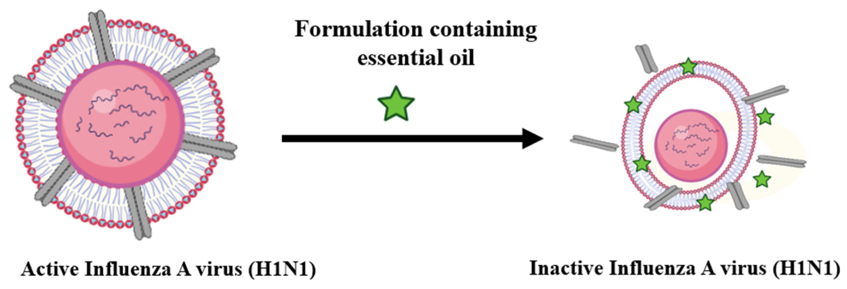
Both hydrogenated and non-hydrogenated catmint-oil-based formulations reduced the number of viral particles of type A influenza virus H1N1 and mouse hepatitis virus (MHV-1; a surrogate of coronavirus) in solution and vaporised form. The catmint essential oils and bromelain acted together against H1N1 and killed it by disorganising and disrupting the viral envelope and are likely to act similarly against MHV-1. The possible mechanism of envelope disruption is explained in Figure 4. Whether catmint essential oil and bromelain act synergistically against these viruses needs further investigation.

Author Contributions

M.Y. designed the study, performed the experiments, analysed the data and wrote the manuscript. M.D.P.W. planned the project and edited the article. J.I. and P.v.B. developed the experimental framework and edited the article. All authors have read and agreed to the published version of the manuscript.

Funding

No funding was received for conducting this study.

Institutional Review Board Statement

Not applicable.

Informed Consent Statement

Not applicable.

Data Availability Statement

All data are available in the manuscript.

Acknowledgments

The authors are grateful to the Mark Wainwright Analytical Centre at the University of New South Wales, Australia, for the help with the electron microscopy. The authors are also grateful to Umme Laila Urmi for the help in performing the transmission electron microscopy.

Conflicts of Interest

This work is original, has not been published before and is not being considered for publication elsewhere. Part of this manuscript has been presented as a poster presentation at the OPTIONS XII for the Control of Influenza annual meeting in 2024. There are no conflicts of interests for any of the authors that could have influenced the results of this work. The Vapour Science Canberra holds the provisional patent for the catmint oil formulation.

Abbreviations

The following abbreviations are used in this manuscript:
HCOHydrogenated catmint oil
NHCONon-hydrogenated catmint oil
TEMTransmission electron microscopy
TCID50Tissue culture infection dose
MHV-1 Murine hepatitis virus
TGATherapeutic Goods Administration

References

  1. Cowling, B.J.; Ip, D.K.; Fang, V.J.; Suntarattiwong, P.; Olsen, S.J.; Levy, J.; Uyeki, T.M.; Leung, G.M.; Malik Peiris, J.; Chotpitayasunondh, T. Aerosol transmission is an important mode of influenza A virus spread. Nat. Commun. 2013, 4, 1935. [Google Scholar] [PubMed]
  2. Koster, F. The experimental aerosol transmission of influenza virus. Future Virol. 2013, 8, 969–981. [Google Scholar]
  3. Corzo, C.A.; Culhane, M.; Dee, S.; Morrison, R.B.; Torremorell, M. Airborne detection and quantification of swine influenza a virus in air samples collected inside, outside and downwind from swine barns. PLoS ONE 2013, 8, e71444. [Google Scholar] [CrossRef]
  4. Zhang, H.; Li, X.; Ma, R.; Li, X.; Zhou, Y.; Dong, H.; Li, X.; Li, Q.; Zhang, M.; Liu, Z.; et al. Airborne spread and infection of a novel swine-origin influenza A (H1N1) virus. Virol. J. 2013, 10, 204. [Google Scholar] [CrossRef] [PubMed]
  5. Tellier, R. Review of aerosol transmission of influenza A virus. Emerg. Infect. Dis. 2006, 12, 1657. [Google Scholar]
  6. Neira, V.; Rabinowitz, P.; Rendahl, A.; Paccha, B.; Gibbs, S.G.; Torremorell, M. Characterization of Viral Load, Viability and Persistence of Influenza A Virus in Air and on Surfaces of Swine Production Facilities. PLoS ONE 2016, 11, e0146616. [Google Scholar] [CrossRef]
  7. Andraud, M.; Herve, S.; Gorin, S.; Barbier, N.; Queguiner, S.; Paboeuf, F.; Simon, G.; Rose, N. Evaluation of early single dose vaccination on swine influenza A virus transmission in piglets: From experimental data to mechanistic modelling. Vaccine 2023, 41, 3119–3127. [Google Scholar] [CrossRef]
  8. Konishi, T. Mutations in SARS-CoV-2 are on the increase against the acquired immunity. PLoS ONE 2022, 17, e0271305. [Google Scholar] [CrossRef]
  9. Krammer, F.; Smith, G.J.D.; Fouchier, R.A.M.; Peiris, M.; Kedzierska, K.; Doherty, P.C.; Palese, P.; Shaw, M.L.; Treanor, J.; Webster, R.G.; et al. Influenza. Nat. Rev. Dis. Primers 2018, 4, 3. [Google Scholar] [CrossRef]
  10. Nuwarda, R.F.; Alharbi, A.A.; Kayser, V. An Overview of Influenza Viruses and Vaccines. Vaccines 2021, 9, 1032. [Google Scholar] [CrossRef]
  11. Najar, B.; Nardi, V.; Stincarelli, M.A.; Patrissi, S.; Pistelli, L.; Giannecchini, S. Screening of the essential oil effects on human H1N1 influenza virus infection: An in vitro study in MDCK cells. Nat. Prod. Res. 2022, 36, 3149–3152. [Google Scholar] [CrossRef]
  12. Vimalanathan, S.; Hudson, J. Anti-influenza virus activity of essential oils and vapors. Am. J. Essent. Oils Nat. Prod. 2014, 2, 47–53. [Google Scholar]
  13. Oh, Y.J.; Kim, Y.S.; Kim, J.W.; Kim, D.W. Antibacterial and Antiviral Properties of Pinus densiflora Essential Oil. Foods 2023, 12, 4279. [Google Scholar] [CrossRef] [PubMed]
  14. Rao, H.; Ahmad, S.; Aati, H.Y.; Basit, A.; Ahmad, I.; Ghalloo, B.A.; Shehzad, M.N.; Nazar, R.; Zeeshan, M.; Nasim, M.J.; et al. Phytochemical screening, biological evaluation, and molecular docking studies of aerial parts of Trigonella hamosa (branched Fenugreek). Arab. J. Chem. 2023, 16, 104795. [Google Scholar]
  15. Singh, H.; Singh, D.; Lekhak, M.M. Ethnobotany, botany, phytochemistry and ethnopharmacology of the genus Thalictrum L. (Ranunculaceae): A review. J. Ethnopharmacol. 2023, 305, 115950. [Google Scholar] [CrossRef]
  16. Tang, Y.Q.N.; Deng, J.; Li, L.; Yan, J.; Lin, L.M.; Li, Y.M.; Lin, Y.; Xia, B.H. Essential oil from Prunella vulgaris L. as a valuable source of bioactive constituents: In vitro anti-bacterial, anti-viral, immunoregulatory, anti inflammatory, and chemical profiles. South Afr. J. Bot. 2022, 151, 614–627. [Google Scholar]
  17. Najar, B.; Mecacci, G.; Nardi, V.; Cervelli, C.; Nardoni, S.; Mancianti, F.; Ebani, V.V.; Giannecchini, S.; Pistelli, L. Volatiles and Antifungal-Antibacterial-Antiviral Activity of South African Salvia spp. Essential Oils Cultivated in Uniform Conditions. Molecules 2021, 26, 2826. [Google Scholar] [CrossRef]
  18. Oriola, A.O.; Oyedeji, A.O. Essential Oils and Their Compounds as Potential Anti-Influenza Agents. Molecules 2022, 27, 7797. [Google Scholar] [CrossRef]
  19. Wu, S.; Patel, K.B.; Booth, L.J.; Metcalf, J.P.; Lin, H.K.; Wu, W. Protective essential oil attenuates influenza virus infection: An in vitro study in MDCK cells. BMC Complement. Altern. Med. 2010, 10, 69. [Google Scholar] [CrossRef]
  20. Mao, X.; Gu, S.; Sang, H.; Ye, Y.; Li, J.; Teng, Y.; Zhang, F.; Ma, Q.; Jiang, P.; Yang, Z.; et al. Essential Oil-Rich Chinese Formula Luofushan-Baicao Oil Inhibits the Infection of Influenza A Virus through the Regulation of NF-kappaB P65 and IRF3 Activation. Evid. Based Complement. Altern. Med. 2021, 2021, 5547424. [Google Scholar] [CrossRef]
  21. Astani, A.; Reichling, J.; Schnitzler, P. Comparative study on the antiviral activity of selected monoterpenes derived from essential oils. Phytotherapy Research. Phytother Res. 2010, 24, 673–679. [Google Scholar] [CrossRef] [PubMed]
  22. Schnitzler, P. Essential oils for the treatment of herpes simplex virus infections. Chemotherapy 2019, 64, 1–7. [Google Scholar] [CrossRef]
  23. Rouf, R.; Uddin, S.J.; Sarker, D.K.; Islam, M.T.; Ali, E.S.; Shilpi, J.A.; Nahar, L.; Tiralongo, E.; Sarker, S.D. Antiviral potential of garlic (Allium sativum) and its organosulfur compounds: A systematic update of pre-clinical and clinical data. Trends Food Sci. Technol. 2020, 104, 219–234. [Google Scholar] [CrossRef]
  24. Mirskaya, E.; Agranovski, I.E. Control of airborne microorganisms by essential oils released by VaxiPod. Atmosphere 2021, 12, 1418. [Google Scholar] [CrossRef]
  25. Jackwood, M.; Rosenbloom, R.; Petteruti, M.; Hilt, D.; McCall, A.; Williams, S. Avian coronavirus infectious bronchitis virus susceptibility to botanical oleoresins and essential oils in vitro and in vivo. Virus Res. 2010, 149, 86–94. [Google Scholar] [CrossRef] [PubMed]
  26. Khan, T.; Khan, M.A.; Ullah, N.; Nadhman, A. Therapeutic potential of medicinal plants against COVID-19: The role of antiviral medicinal metabolites. Biocatal. Agric. Biotechnol. 2021, 31, 101890. [Google Scholar] [CrossRef]
  27. Petrova, D.; Gasic, U.; Yocheva, L.; Hinkov, A.; Yordanova, Z.; Chaneva, G.; Mantovska, D.; Paunov, M.; Ivanova, L.; Rogova, M.; et al. Catmint (Nepeta nuda L.) Phylogenetics and Metabolic Responses in Variable Growth Conditions. Front. Plant Sci. 2022, 13, 866777. [Google Scholar] [CrossRef] [PubMed]
  28. Hinkov, A.; Angelova, P.; Marchev, A.; Hodzhev, Y.; Tsvetkov, Y.; Dragolova, D.; Todorov, D.; Shishkova, K.; Kapchina-Toteva, V.; Blundell, R.; et al. Nepeta nuda ssp. nuda L. water extract: Inhibition of replication of some strains of human alpha herpes virus (genus simplex virus) in vitro, mode of action and NMR-based metabolomics. J. Herb. Med. 2020, 21, 100334. [Google Scholar] [CrossRef]
  29. Greig, A.; Bouillant, A. Binding effects of concanavalin A on a coronavirus. Can. J. Comp. Med. 1977, 41, 122. [Google Scholar]
  30. Schlegel, A.; Schaller, J.; Jentsch, P.; Kempf, C. Semliki Forest virus core protein fragmentation: Its possible role in nucleocapsid disassembly. Biosci. Rep. 1993, 13, 333–347. [Google Scholar] [CrossRef]
  31. Amini, A.; Masoumi-Moghaddam, S.; Morris, D.L. Utility of Bromelain and N-Acetylcysteine in Treatment of Peritoneal Dissemination of Gastrointestinal Mucin-Producing Malignancies; Springer: Berlin/Heidelberg, Germany, 2016. [Google Scholar]
  32. Akhter, J.; Quéromès, G.; Pillai, K.; Kepenekian, V.; Badar, S.; Mekkawy, A.H.; Frobert, E.; Valle, S.J.; Morris, D.L. The Combination of Bromelain and Acetylcysteine (BromAc) Synergistically Inactivates SARS-CoV-2. Viruses 2021, 13, 425. [Google Scholar] [CrossRef]
  33. Mayo, D.R.; Beckwith, W.H., 3rd. Inactivation of West Nile virus during serologic testing and transport. J. Clin. Microbiol. 2002, 40, 3044–3046. [Google Scholar] [CrossRef] [PubMed]
  34. Webster, R.G.; Darlington, R.W. Disruption of myxoviruses with Tween 20 and isolation of biologically active hemagglutinin and neuraminidase subunits. J. Virol. 1969, 4, 182–187. [Google Scholar] [CrossRef] [PubMed]
  35. Suschkea, U.; Sporera, F.; Schneelea, J.; Geissb, H.K.; Reichlinga, J. Antibacterial and Cytotoxic Activity of Nepeta cataria L., N. cataria var. citriodora (Beck.) Balb. and Melissa officinalis L. Essential Oils. Nat. Prod. Commun. 2007, 2, 1277–1286. [Google Scholar]
  36. Borlace, G.N.; Singh, R.; Seubsasana, S.; Chantaranothai, P.; Thongkham, E.; Aiemsaard, J. Antimicrobial effects of catnip (Nepeta cataria L.) essential oil against canine skin infection pathogens. Vet. World 2024, 17, 585–592. [Google Scholar] [CrossRef] [PubMed]
  37. Sagar, S.; Rathinavel, A.K.; Lutz, W.E.; Struble, L.R.; Khurana, S.; Schnaubelt, A.T.; Mishra, N.K.; Guda, C.; Palermo, N.Y.; Broadhurst, M.J.; et al. Bromelain inhibits SARS-CoV-2 infection via targeting ACE-2, TMPRSS2, and spike protein. Clin. Transl. Med. 2021, 11, e281. [Google Scholar] [CrossRef]
  38. Yasir, M.; Vijay, K.A.; Willcox, M. Antiviral effect of multipurpose contact lens disinfecting solutions against coronavirus. Contact Lens Anterior Eye 2022, 45, 101513. [Google Scholar] [CrossRef]
  39. Urmi, U.L.; Attard, S.; Vijay, A.K.; Willcox, M.D.; Kumar, N.; Islam, S.; Kuppusamy, R. Antiviral Activity of Anthranilamide Peptidomimetics against Herpes Simplex Virus 1 and a Coronavirus. Antibiotics 2023, 12, 1436. [Google Scholar]
  40. Lelešius, R.; Karpovaitė, A.; Mickienė, R.; Drevinskas, T.; Tiso, N.; Ragažinskienė, O.; Kubilienė, L.; Maruška, A.; Šalomskas, A. In vitro antiviral activity of fifteen plant extracts against avian infectious bronchitis virus. BMC Vet. Res. 2019, 15, 178. [Google Scholar]
  41. Jiang, X.; Zhao, C.; Chen, Y.; Gao, X.; Zhang, Q.; Chen, Z.; Li, C.; Zhao, X.; Liu, Z.; Huang, W.; et al. Probable Evidence of Aerosol Transmission of SARS-COV-2 in a COVID-19 Outbreak of a High-Rise Building. Environ. Health Insights 2023, 17, 11786302231188269. [Google Scholar] [CrossRef]
  42. Jayaweera, M.; Perera, H.; Gunawardana, B.; Manatunge, J. Transmission of COVID-19 virus by droplets and aerosols: A critical review on the unresolved dichotomy. Environ. Res. 2020, 188, 109819. [Google Scholar] [CrossRef] [PubMed]
  43. Sezer, B. The imminent danger related to measles virus infection from the perspective of paediatric dentistry. Eur. Arch. Paediatr. Dent. 2024, 25, 605–606. [Google Scholar] [PubMed]
  44. Shan, H.; Yan, L.Y.; Prasanna, N.; Hung, C.K.; Yi, L.J.K.; Ngai, H.F.; Colman, M. Effectiveness of Preprocedural Mouthwashes: A Triple-Blind Randomised Controlled Clinical Trial. Int. Dent. J. 2024, 75, 868–876. [Google Scholar] [CrossRef]
  45. Munjaković, H.; Mikuletič, T.; Zayed, N.; Kolenc, M.; Manevski, D.; Triglav, T.; Steyer, A.; Teughels, W.; Seme, K.; Fidler, A.; et al. Electrolyzed Saline Prevents Virus Transmission in Dental Procedures: An In Vitro Study. J. Dent. Res. 2025, 104, 211–220. [Google Scholar] [CrossRef]
  46. Madia, V.N.; Toscanelli, W.; De Vita, D.; De Angelis, M.; Messore, A.; Ialongo, D.; Scipione, L.; Tudino, V.; D’Auria, F.D.; Di Santo, R. Ultrastructural damages to H1N1 influenza virus caused by vapor essential oils. Molecules 2022, 27, 3718. [Google Scholar] [CrossRef]
  47. Kalaiselvan, P.; Yasir, M.; Kuppusamy, R.; Willcox, M.; Vijay, A.K. Ability of Essential Oil Vapours to Reduce Numbers of Culturable Aerosolised Coronavirus, Bacteria and Fungi. Antibiotics 2022, 11, 393. [Google Scholar] [CrossRef] [PubMed]
  48. Asif, M.; Saleem, M.; Saadullah, M.; Yaseen, H.S.; Al Zarzour, R. COVID-19 and therapy with essential oils having antiviral, anti-inflammatory, and immunomodulatory properties. Inflammopharmacology 2020, 28, 1153–1161. [Google Scholar]
  49. Graves, C.; Ghaltakhchyan, N.; Ngo, T.Q.; Liu, C.; Babikow, E.; Shoji, A.; Bocklage, C.; Sang, Y.; Phillips, S.T.; Bowman, N.; et al. A Cetylpyridinium Chloride Oral Rinse Reduces Salivary Viral Load in Randomized Controlled Trials. JDR Clin. Trans. Res. 2024. [Google Scholar] [CrossRef]
  50. Said-Al Ahl, H.; Naguib, N.Y.; Hussein, M.S. Evaluation growth and essential oil content of catmint and lemon catnip plants as new cultivated medicinal plants in Egypt. Ann. Agric. Sci. 2018, 63, 201–205. [Google Scholar] [CrossRef]
  51. Zomorodian, K.; Saharkhiz, M.J.; Shariati, S.; Pakshir, K.; Rahimi, M.J.; Khashei, R. Chemical Composition and Antimicrobial Activities of Essential Oils from Nepeta cataria L. against Common Causes of Food-Borne Infections. ISRN Pharm. 2012, 2012, 591953. [Google Scholar] [CrossRef]
  52. Kansakar, U.; Trimarco, V.; Manzi, M.V.; Cervi, E.; Mone, P.; Santulli, G. Exploring the Therapeutic Potential of Bromelain: Applications, Benefits, and Mechanisms. Nutrients 2024, 16, 2060. [Google Scholar] [CrossRef]
  53. Wang, W.; Suguitan Jr, A.L.; Zengel, J.; Chen, Z.; Jin, H. Generation of recombinant pandemic H1N1 influenza virus with the HA cleavable by bromelain and identification of the residues influencing HA bromelain cleavage. Vaccine 2012, 30, 872–878. [Google Scholar] [CrossRef] [PubMed]
  54. Hua, T.; Zhang, X.; Tang, B.; Chang, C.; Liu, G.; Feng, L.; Yu, Y.; Zhang, D.; Hou, J. Tween-20 transiently changes the surface morphology of PK-15 cells and improves PCV2 infection. BMC Vet. Res. 2018, 14, 138. [Google Scholar] [CrossRef]
  55. Pyankov, O.V.; Usachev, E.V.; Pyankova, O.; Agranovski, I.E. Inactivation of Airborne Influenza Virus by Tea Tree and Eucalyptus Oils. Aerosol Sci. Technol. 2012, 46, 1295–1302. [Google Scholar] [CrossRef]
  56. Lindsley, W.G.; Blachere, F.M.; Thewlis, R.E.; Vishnu, A.; Davis, K.A.; Cao, G.; Palmer, J.E.; Clark, K.E.; Fisher, M.A.; Khakoo, R. Measurements of airborne influenza virus in aerosol particles from human coughs. PLoS ONE 2010, 5, e15100. [Google Scholar]
  57. Alford, R.H.; Kasel, J.A.; Gerone, P.J.; Knight, V. Human influenza resulting from aerosol inhalation. Proc. Soc. Exp. Biol. Med. 1966, 122, 800–804. [Google Scholar] [CrossRef]
  58. Tang, J.W.; Wilson, P.; Shetty, N.; Noakes, C.J. Aerosol-Transmitted Infections-a New Consideration for Public Health and Infection Control Teams. Curr. Treat. Options Infect. Dis. 2015, 7, 176–201. [Google Scholar] [CrossRef]
  59. Malmgren, R.; Välimaa, H.; Oksanen, L.; Sanmark, E.; Nikuri, P.; Heikkilä, P.; Hakala, J.; Ahola, A.; Yli-Urpo, S.; Palomäki, V.; et al. High-volume evacuation mitigates viral aerosol spread in dental procedures. Sci. Rep. 2023, 13, 18984. [Google Scholar] [CrossRef]
  60. Firquet, S.; Beaujard, S.; Lobert, P.-E.; Sané, F.; Caloone, D.; Izard, D.; Hober, D. Survival of enveloped and non-enveloped viruses on inanimate surfaces. Microbes Environ. 2015, 30, 140–144. [Google Scholar]
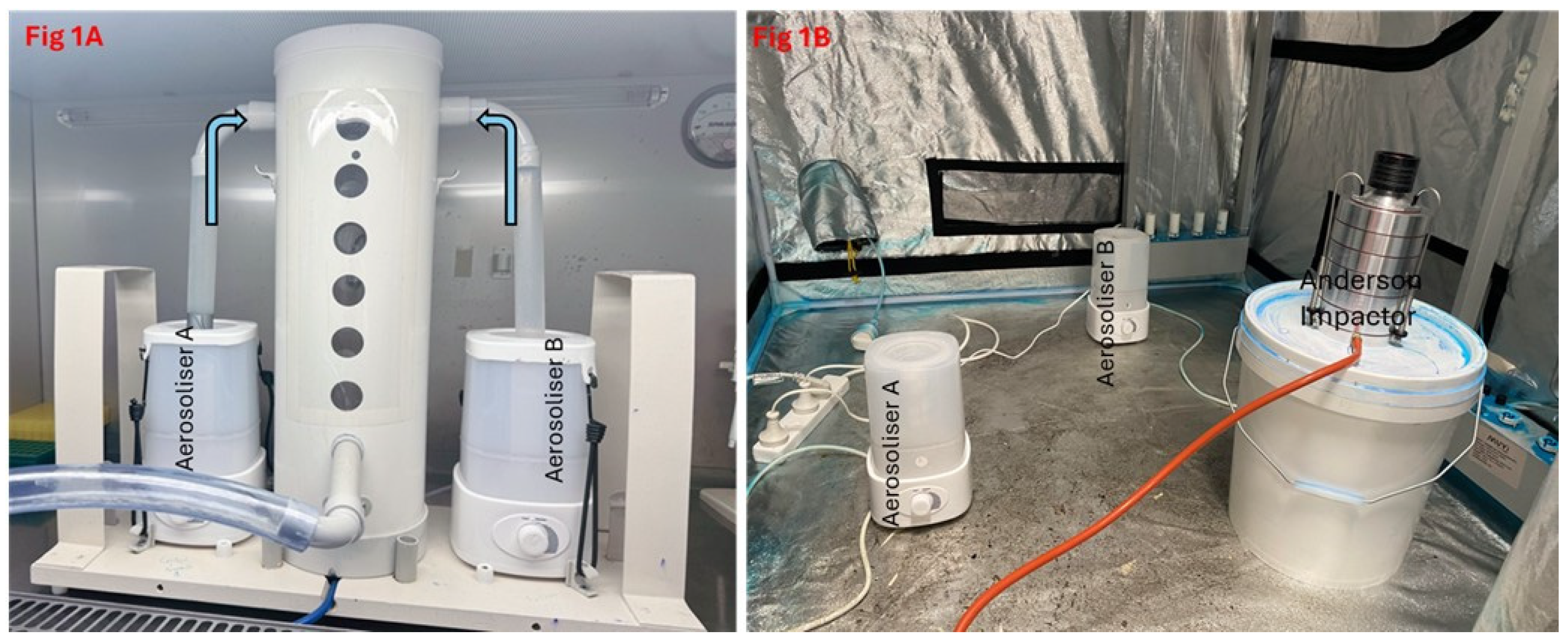
Figure 1. Aerosolisation apparatus for H1N1 and MHV-1 (A) and disinfectants (B). Aerosols of the viruses and disinfectants were generated through aerosoliser A and aerosoliser B, respectively, and interacted in a cubicle cylinder and booth.
Hygiene 05 00015 g001
Figure 2. Reduction in the number of H1N1 and MHV-1 infectious viral particles after treatment with Formulation (1). Error bars represents the means (±SD) of two independent experiments performed in triplicate. * represents p = 0.025 and ** represents p = 0.014.
Hygiene 05 00015 g002
Figure 3. Transmission electron micrographs of H1N1 virus after interaction with disinfectant: (A,B) PBS-treated control sample shows an intact H1N1 virus envelope (the roundness of the viral particle and the darker shading demonstrating an intact viral envelope), and (C,D) viruses after exposure to the disinfectant HCO and NHCO, respectively, demonstrate disruption of the lipid envelope and reduction in viral size due to the loss of the viral envelope.
Hygiene 05 00015 g003
Figure 4. Damage caused by the catmint essential oil formulations to the H1N1 envelope.
Hygiene 05 00015 g004
Table 1. Reduction in the number of aerosolised H1N1 particles after treatment with vapourised disinfectant containing vapours of different formulations. Data represent two independent experiments performed in triplicate (±SD).
DisinfectantsNumber of Aerosolised Viruses Log10 (PFU/mL)Water Control/
Number of Aerosolised Viruses Log10 (PFU/mL)
Reduction of Viruses (Log10)p Values
HCO + Bromelain + NAC + Tween 20 (1)2.33 ± 0.145.33 ± 0.143.00 ± 0.140.002
NHCO + Bromelain + NAC + Tween 20 (2)2.00 ± 0.284.34 ± 0.132.34 ± 0.240.025
HCO + NAC + Tween 20 (3)2.60 ± 1.144.17 ± 0.141.57 ± 0.140.008
Bromelain + NAC + Tween 20 (4)3.33 ± 0.144.33 ± 0.141.00 ± 0.140.046
NAC + Tween 20 (5)4.33 ± 0.284.33 ± 0.130.00 ± 0.000.966
HCO = hydrogenated catmint oil; NHCO = non-hydrogenated catmint oil; NAC = N-acetylcysteine.
Table 2. Reduction in the number of aerosolised MHV-1 particles after treatment with vapourised disinfectant formulations. Data represent two independent experiments performed in triplicate (±SD).
DisinfectantsNumber of Aerosolised Viruses Log10 (PFU/mL)Water Control/
Number of Aerosolised Viruses Log10 (PFU/mL)
Reduction of Viruses (Log10)p Values
HCO + Bromelain + NAC + Tween 20 (1)0.00 ± 0.003.10 ± 0.043.10 ± 0.04<0.001
HCO + NAC + Tween 20 (3)1.80 ± 0.421.30 ± 0.040.057
Bromelain + NAC + Tween 20 (4)2.03 ± 0.051.07 ± 0.020.013
NAC + Tween 20 (5)2.94 ± 0.050.16 ± 0.030.910
Disclaimer/Publisher’s Note: The statements, opinions and data contained in all publications are solely those of the individual author(s) and contributor(s) and not of MDPI and/or the editor(s). MDPI and/or the editor(s) disclaim responsibility for any injury to people or property resulting from any ideas, methods, instructions or products referred to in the content.