Article

Members of the Order Mastogloiales Sensu Cox Belong to the Different Evolutionary Lineages of Diatoms: Phylogenetic Resolutions and Descriptions of New Types of Pore Occlusions

1
K.A. Timiryazev Institute of Plant Physiology RAS, IPP RAS, 35 Botanicheskaya St., Moscow 127276, Russia
2
Faculty of Biology, M.V. Lomonosov Moscow State University, Leninskie Gory 1, Building 12, Moscow 119234, Russia
3
Papanin Institute for Biology of Inland Waters RAS, IBIW RAS, Borok 152742, Russia
4
Department of Biology, School of Arts and Sciences, National University of Mongolia, Ulaanbaatar 14201, Mongolia
*
Author to whom correspondence should be addressed.
Phycology 2025, 5(4), 68; https://doi.org/10.3390/phycology5040068
Submission received: 24 September 2025 / Revised: 9 October 2025 / Accepted: 23 October 2025 / Published: 1 November 2025

Abstract

The study focuses on the phylogeny and systematics of the order Mastogloiales sensu Cox. Results of a two-gene (18S rRNA and rbcL) molecular analysis on the order demonstrate that genera Aneumastus, Decussiphycus, Mastogloia, and Stigmagloia form a closely related phylogenetic group, while Achnanthes and Craspedostauros belong to a different evolutionary lineage. Heterogeneity is also expressed in the difference in morphological features, including the structure of pore occlusions. Combining molecular and morphological data, we hereby amend the description of the order Mastogloiales and describe new types of pore occlusions, typical for the order. Using our material from freshwater and saline waterbodies in China, Indonesia, Mongolia and Vietnam, we illustrate the diversity and ultrastucture of pore occlusions. As a part of morphological analysis, Mastogloia recta is studied with SEM and TEM for the first time. In addition, our study reveals a new species of the genus Aneumastus from Mongolia—Aneumastus khovsgolensis sp. nov.—which was subjected to molecular and morphological investigations using light and scanning electron microscopy. This new species is compared to similar taxa of the Aneumastus tusculus-group.

1. Introduction

Order Mastogloiales D.G. Mann was described in Round et al. [1]. Originally, the order included a single family—Mastogloiaceae Mereschkowsky. D.G. Mann—with genera Mastogloia Thwaites ex W. Smith and Aneumastus D.G. Mann & Stickle [1]. Mastogloia is a species-rich genus of predominantly marine taxa, characterized by unusual morphology [2]. The valves of Mastogloia are equipped with specialized valvocopulae—partecta (partectal rings). Partectal rings are constructed of partecta (compartments), pores (which excrete mucus), partectal ducts (channels connecting partectal pores to partecta), and lacunae (apical gaps in silica) [3]. Striae in Mastogloia are uni- or biseriate, often changing arrangement at different parts of the valve. Areolae in striae are covered with unique pore occlusions, traditionally associated with cribra or volae sensu Round et al. [1].
The morphology of species in Aneumastus, a close relative of Mastogloia, is equally complex. Aneumastus was assigned to the order Mastogloiales in Round et al. [1] on the basis of the sinuous morphology of the raphe, presence of valvocopulae, and H-shaped arrangement of plastids. Striae of Aneumastus are uniseriate throughout the valve or biseriate towards the valve margin [1]. Notably, Round et al. [1] characterized areolae in Aneumastus as “complex, opening into deep pits internally via sieves of small pores: occluded externally by flaps of silica borne by the areola walls”. No information on pore occlusion ultrastructure was provided therein. Later on, Lange-Bertalot [4] mentioned that areolae in Aneumastus are possibly occluded by cribra, rather than hymenes.
Later, Decussiphycus Guiry & K. Gandhi (=Decussata (Patrick) Lange-Bertalot nom. inval.), was added to the family [5]. Edlund et al. [5] were the first to focus on the protoplast organization and chloroplast morphology of the genus. Their study showed plastids to be H-shaped in girdle view, with a pyrenoid in the plastid bridge and four arms in the transapical cross-section extending along the valvar plane [5]. Interestingly, Decussiphycus differs from other genera of Mastogloiales by simple areolae, linear thickening of the internal proximal raphe ends and no chambering on the valvocopulae [5]. Most importantly, Edlund et al. [5] characterized the areolae of Decussiphycus as internally covered by circular convex hymenes. This discovery implied that pore occlusions in the genus were hymenate, but not cribrate, as was suggested for Mastogloia and Aneumastus [1,4,6].
The uncertainty about the structure of the pore occlusions resulted into a dispute about the phylogeny of Mastogloiales. Kociolek & Stoermer [7] proposed a relationship between the Mastogloiales and some monoraphid taxa, such as Cocconeis Ehrenberg. This proposal was challenged by Cox & Williams [8]. It was concluded that Aneumastus and Mastogloia were closely related to Craspedostauros E.J.Cox, a genus of stauros-bearing biraphid diatoms [9]. A second study by Cox & Williams [10] also stated that Aneumastus and Mastogloia formed a monophyletic group with Achnanthes Bory and Craspedostauros. The two studies by Cox & Williams [8,10] utilized a cladistic analysis of protoplast characters and valve features, e.g., raphe morphology, striae arrangement, structure of areolae, and pore occlusions. The results supported Cleve’s theory concerning the heterogeneity of monoraphid lineage [11]. Based on these conclusions, Cox [12] amended the diagnosis of Mastogloiales to include Achnanthes within the order.
According to Cox [12], the order Mastogloiales unites species with two fore and aft chloroplasts, usually more or less H-shaped in the girdle and valve views, indented below the raphe and connected by a central pyrenoid. In some taxa a stauros or a fascia is present. External raphe fissures may be sinuous, but internal raphe fissures are always straight. The girdle is represented by open porous bands, with one or more rows of cribrate pores. All genera of Mastogloiales sensu Cox have areolae that are occluded by cribra, but not hymenes [12].
Cox & Williams [10] emphasized the vital role of protoplast characters for morphological analysis but suggested that frustule characters should discriminate taxa within narrow phylogenetic groups. With regard to this point of view, Cox [12] united monoraphid (Achnanthes) and biraphid (Aneumastus, Craspedostauros, Mastogloia) genera within the order Mastogloiales, neglecting the discrepancies in raphe systems. All data on the diatom systematics was summarized in “Syllabus of Plant Families” [13]. In the new system, Mastogloiales comprised two families—Achnanthaceae Kützing, with Achnanthes, and Mastogloiaceae, with Aneumastus, Craspedoustauros, Mastogloia, and Decussiphycus [13].
In fact, Achnanthes was among the first diatom genera to be studied with molecular methods [14] and later on became a subject of multiple molecular phylogenetic studies [15,16,17,18,19,20,21,22]. All these investigations demonstrated close relationships between Achnanthes and genera of canal-raphe diatoms, e.g., Nitzschia Hassall, Bacillaria Gmelin, Tryblionella W. Smith, and Hantzschia Grunow, thus contradicting Cox’s ideas on the composition of Mastogloiales [12].
Later on, molecular data yielded a different relationship for Craspedostauros, another representative of Mastogloiales sensu Cox [23]. Ashworth et al. [23] deduced that Craspedostauros is closely related to Achnanthes, Staurotropis Paddock, and other canal-raphe diatoms, while Mastogloia appeared more closely related to Phaeodactylum K. Bohlin. However, in their study, Mastogloia was represented by only two uncultured strains, and the AchnanthesPhaeodactylum clade lacked statistical support. No molecular data was available for either Decussiphycus or Aneumastus [23]. Over time, more species of Mastogloia [24,25,26,27] and Aneumastus [28,29] have been studied with molecular data and, more recently, molecular data has become available for Decussiphycus [30]. Results from these analyses yield a monophyletic group of Aneumastus, Mastogloia, and Decussiphycus separate from the Achnanthes-Craspedostauros clade.
Moreover, the molecular analysis by Mironov et al. [30] disproved the affinity of Tetramphora Mereschkowsky to Mastogloiales, which was suggested by Stepanek & Kociolek [31], who resurrected Tetramphora in their study. Unexpectedly, the authors neglected the essential differences in valve structure between Mastogloia and Tetramphora but favored the similarities in plastids arrangement (two chloroplasts per cell, with two plates connected by a pyrenoid-containing isthmus). The study relied primarily on Mereschkowsky’s study of chloroplast morphology across major groups of diatoms [32]. However, even the phylograms constructed by Stepanek & Kociolek [31] show little support for the bond between Tetramphora and Mastogloiales sensu Round et al. [1].
Unlike Tetramphora, taxonomic proximity of genera Mastoneis Cleve and Paramastogloia Lobban to Mastogloiales is not particularly questionable. Mastoneis has been monotypic since its description by Cleve [33], and its only species, Mastoneis biformis (Grunow) Cleve, has been recently investigated by Lobban [34]. He utilized light microscopy (LM) and scanning electron microscopy (SEM) to amend the diagnosis of Mastoneis, pointing out the diagnostic features of the genus—unperforated valvocopula, absence of partecta on the valvocopula and presence of internal transapical costae (at every second or third interstria). Paramastogloia was erected therein, too, with a single marine species—Paramastogloia cubana Lobban. The genus is distinguished by the absence of several features—partecta, perforations of valvocopula, and transapical costae [34]. However, molecular data on these genera is wanting.
Apart from the issues addressed above, the polymorphism of Mastogloia is actually a major issue of systematics. A remarkable number of morphological studies on different species-groups of Mastogloia have emphasized this problem [2,35,36,37,38,39,40,41,42,43,44,45,46,47,48,49,50,51]. The same studies indicated that the presence of partecta in Mastogloia is the synapomorphy of the genus. Based on this concept, Lobban & Navarro [52] formed a new genus, Mastogloiopsis Lobban & J.N. Navarro, which included marine species deprived of partecta in the genus. Recently conducted precise SEM analysis of the genus’s only species—Mastogloiopsis biseriata Lobban & J.N. Navarro—illustrated laminar valves, densely perforated valvocopula, and transapical costae located at every virga [34].
The process of division of Mastogloia was initiated by Kezlya et al. [53], who erected a new genus Stigmagloia Glushchenko, Kezlya, Kapustin & Kulikovskiy. The representatives of the genus do bear partecta on their frustules but are also distinguished from the species of Mastogloia by the unique structure of stigma. Molecular analysis performed therein ensured the position of Stigmagloia within Mastogloiales, but, at the same time, demonstrated the distinct polyphyly of Mastogloia. Similar molecular genetic results were acquired by Mironov et al. [30].
Thus, the current situation with the systematics of Mastogloiales can be characterized as follows: the order is relatively well-studied from the molecular perspective, i.e., molecular data is currently available for Aneumastus, Mastogloia, Decussiphycus, Stigmagloia, Achnanthes, Craspedostauros, and Tetramphora. Nevertheless, some issues of phylogeny remain unsolved, such as the polymorphism of Mastogloia, the most species-rich genus of the order [54]. Features of valve morphology, foremost pore occlusions, require detailed studies, too. The accumulated taxonomic problems can be tackled with the help of a polyphasic approach. The current study utilizes both molecular and morphological analyses to re-assess the taxonomic composition of Mastogloiales. As a result, a new, amended diagnosis of the order is provided herein. Based on detailed investigation of valve ultrastructure using SEM, we resolve the structure of pore occlusions among the genera of the order and propose new terms—colanderus, colanderus-directus, colanderus-obliquus, colanderus-bifurcus, and spongia. These structures are visualized by examples from representatives of Mastogloiales, using material from China, Indonesia, Mongolia, and Vietnam. New types of occlusions are compared against the cribra, which are illustrated using the newly cultured strain of Achnanthes sp. from Mongolia. Morphological findings are supported by the results of two-gene (18S rRNA and rbcL) molecular analysis, which focused on the genera of Mastogloiales sensu Cox—Aneumastus, Decussiphycus, Mastogloia, Achnanthes, and Craspedostauros—and allegedly related genera, such as Tetramphora, Druehlago Lobban & M.P. Ashworth, and Nagumoea Kociolek & Witkowski. The molecular dataset was supplemented with two newly acquired strains of an unknown species of Aneumastus from Mongolia. The latter species is hereby described as Aneumastus khovsgolensis Glushchenko, Mironov, Genkal, Nergui & Kulikovskiy sp. nov. Morphological features of the new species were compared against the most similar taxa of the genus.

2. Materials and Methods

2.1. Sample Collection, Strain Isolation, and Culturing

During this study, we analyzed 7 samples collected from fresh and saline waterbodies in Mongolia, Vietnam, Indonesia, and China. For freshwater samples, conductivity, pH, and temperature measurements were performed using the Hanna Combo (HI 98129) device (Hanna Instruments Ltd., Bedfordshire, UK). Salinity and water temperature in saline waterbodies were measured by the Atago PAL-06S seawater refractometer (Atago Co., Ltd., Tokyo, Japan). For strain isolation, a subsample of each sample was added to WC liquid medium [55]. Monoclonal strains were established by micropipetting single cells under a Zeiss Axio Vert. A1 (ZEISS, Oberkochen, Germany) inverted microscope (with × 20 objective). Cultures were maintained in WC liquid medium in Petri dishes at 23 °C with a 12:12 h alternating light/dark photoperiod. Acquired samples and strains are currently deposited in the Culture and Barcode Collection of Microalgae and Cyanobacteria “Algabank” (CBMC) at the K. A. Timiryazev Institute of Plant Physiology RAS, Moscow, Russia. Information about the studied samples and isolated strains is summarized in Table 1.

2.2. Slide Preparation and Microscopy

Samples for LM and SEM investigations were processed by means of a standard procedure involving treatment with 10% HCl and concentrated hydrogen peroxide (37%). Afterwards, the samples were washed with deionized water 5 times at 12 h intervals. The suspensions were decanted and filled to 100 mL with deionized water. Suspensions were left for drying on coverslips at room temperature. Permanent samples were prepared using Naphrax® (refractive index = 1.73). LM was conducted under a Zeiss Axio Scope A1 microscope equipped with an oil immersion objective (×100, n.a. 1.4, differential interference contrast) and a mounted Zeiss Axiocam ERc 5s camera. Slides for SEM were prepared using suspensions from cleaned samples, which were mounted onto aluminum stubs and dried for 24 h at room temperature. The dried stubs were sputter-coated with 50 nm of Au by means of an Eiko IB 3 apparatus (Eiko Engineering, Tokyo, Japan). For SEM investigation, we utilized a JEOL JSM-6510LV field emission scanning electron microscope (JEOL, Tokyo, Japan), and a TEM–JEOL JEM-1011 (JEOL, Tokyo, Japan) transmission electron microscope with 80 kV accelerating voltage. TEM slides were prepared by pipetting the sample suspension onto standard TEM copper grids covered with formvar. The copper grids were left to dry at room temperature for 24 h. Permanent slides are currently housed in Diatom Herbarium (HD) at the K. A. Timiryazev Institute of Plant Physiology RAS, Moscow, Russia.

2.3. DNA Extraction and Amplification

DNA extracting procedures were performed by Chelex100 Chelating Resin (Bio-Rad Laboratories, Hercules, CA, USA) according to the 2.2. protocol. Amplification of 18S rDNA was conducted with D512for and D978rev primers [56]. For amplification of rbcL, dp7- [57] and rbcL404+ [58] primers were utilized. Polymerase chain reaction (PCR) was conducted using ScreenMix (Evrogen, Moscow, Russia). During 18S rDNA amplification, the following PCR algorithms were employed: initial denaturation (95 °C, 5 min), 35 cycles of denaturation (94 °C, 30 s), annealing (52 °C, 30 s), elongation (72 °C, 50 s), final extension (72 °C, 7 min), maintenance (12 °C). rbcL amplification was performed according to the following algorithm: initial denaturation (94 °C, 5 min), 44 cycles of denaturation (94 °C, 50 s), annealing (53 °C, 50 s), elongation (72 °C, 80 s), final extension (72 °C, 10 min), maintenance (12 °C). PCR products were visualized in agarose gel (1.0%) by electrophoresis using SYBRTM Safe stain (Life Technologies, Carlsbad, CA, USA). DNA sequences were read by a Genetic Analyzer 3500 instrument (Applied Biosystems, Waltham, MA, USA).

2.4. Phylogram Construction

The obtained sequences were manually edited in Ridom TraceEdit (Ridom© GmbH, Münster, Germany) and assembled in a dataset using MEGA7 software (Pennsylvania State University, Pennsylvania, PA, USA [59]). The datasets for 18S rDNA and rbcL consisted of 140 sequences each, selected from different diatom lineages, including the available sequences for the targeted genera Aneumastus, Mastogloia, Stigmagloia, Decussiphycus, Achnanthes, Craspedostauros, Druehlago, and Nagumoea. Four strains of centric diatoms were chosen as the outgroup. The 18S rDNA and rbcL datasets were aligned separately by the G-INS-I algorithm in Mafft ver.7 software (RIMD, Osaka, Japan [60]). Afterwards, unpaired regions were removed, and the aligned sequences were united into a single concatenated matrix. The total matrix length was 1267 nucleotides (883 for rbcL and 384 for 18S rDNA). BI analysis was performed using the BEAST ver.1.10.1 program (BEAST Developers, Auckland, New Zealand [61]). In the process of BI analysis, the Yule process tree prior speciation model and GTR+G+I substitution model were applied. A total of 10 MCMC analyses were employed for 10 million generations (burn-in 1 million generations). The resulting data was analyzed using Tracer ver. 1.7.1 software (MCMC Trace Analysis Tool, Edinburgh, UK [61]), and an initial 10% of trees were removed. Tree topology robustness was estimated by RAxML rapid bootstrapping and a subsequent ML search of the concatenated alignment via raxmlGUI 2.0 software [62]. The ML analysis was performed with 1000 replicas, GTR substitution matrix, and gamma substitution rates. The best resulting trees from the BI and ML analyses were visualized in FigTree ver. 1.4.4 (University of Edinburgh, Edinburgh, UK). The final phylogram was edited in Adobe Photoshop CC ver.19.0 (Adobe, San Jose, CA, USA). The input and resulting data for molecular analyses are attached as supplementary files below.

2.5. Taxonomy and Terminology

During the construction of the final molecular phylogram, species names were modified to meet the latest taxonomic novelties (according to the Algaebase [54]). In the process of morphological analysis, valve terminology follows Paddock & Kemp [3,63], Lange-Bertalot [4], Edlund et al. [5], Stancheva & Temniskova [64], Cox [65], Kulikovskiy et al. [22], and Maltsev et al. [29].

3. Results

3.1. Molecular Analysis

The combined phylogram from the BI and ML analyses is represented in Figure 1. The constructed tree includes all major groups of pennate diatoms. The overall topology demonstrates that Mastogloiales sensu Cox is polyphyletic, breaking into three distinct clades (see Figure 1, highlighted in grey).
Firstly, the “ANCD” clade comprises the Achnanthes, Nagumoea, and Craspedostauros strains alongside Druehlago cf. cuneata Lobban & Ashworth. Neither BI nor ML provide statistical support for the “ANCD” clade (LB = 18, PP = 0.69). This case is likely explained by the poor molecular representation of Achnanthes: we included nine strains of Achnanthes, with only two being identified to the species level. Unfortunately, referencing molecular database for this species-rich genus (280 taxa according to Algaebase [54]) is still scarce. In fact, among 248 sequences deposited to GenBank, 54 are labeled as Achnanthes sp. and the rest of sequences designate only 9 species. However, individual genus-level clades within the “ANCD” clade demonstrate better resolution: LB = 83, PP = 1 for the Achnanthes clade; LB = 100, PP = 1 for the Nagumoea clade; and LB = 76, PP = 1 for the Craspedostauros-Druehlago clade. Once again, our results reveal that the “ANCD” clade is allied to Bacillariaceae Ehrenberg rather than to Mastogloia, Aneumastus, and Decussiphycus. In particular, a superclade of “ANCD” and canal-raphid diatoms (Tryblionella W. Smith, Nitzschia Hassall, Hantzschia Grunow, Bacillaria J.F. Gmelin, Denticula Kützing, Cymbellonitzschia Hustedt, Cylindrotheca Rabenhorst) is strongly supported by BI (PP = 1). Notably, Staurotropis T.B.B. Paddock formed a branch among nitzschoid diatoms with maximum support from BI and ML. Thus, our phylogram reassures the fact that stauros-bearing diatoms are heterogenic and share little relation with Mastogloiales [23].
On the contrary, Aneumastus, Mastogloia, Stigmagloia, Decussiphycus, and Tetramphora belong to a different superclade, comprising mostly biraphid diatoms, joined by monoraphids of Achnanthidiaceae D.G. Mann (Achnanthidium Kützing, Psammothidium Bukhtiyarova & Round, Lemnicola Round & Basson, Pauliella Round & Basson) and canal-raphids (Rhopalodia O. Müller, Epithemia Kützing, Surirella Turpin). The BI support for this superclade is maximal (PP = 1), but the ML support is relatively low (LB = 57).
Within this part of the phylogram, another distinct clade can be distinguished. It unites seven strains of Aneumastus (including the newly acquired strains CBMC338mnp and CBMC529mnp, see Figure 1, highlighted in bold), Stigmagloia lobbanii Glushchenko, Kezlya, Kapustin & Kulikovskiy strain SVN638, Decussiphycus sinensis Glushchenko, Maltsev, Mironov, Liu & Kulikovskiy strain Ca68, and six strains of Mastogloia. The latter genus is hereby demonstrated as polyphyletic, which has already been supposed in previous studies [53,66]. Stigmagloia lobbanii forms a branch with Mastogloia recta Hustedt with mediocre statistical support (LB = 57, PP = 1), and Decussiphycus sinensis branches together with Mastogloia fimbriata (T. Brightwell) Grunow, although statistical support is lacking.
Our analysis reveals a certain degree of variability between the two strains of Aneumastus khovsgolensis sp. nov., e.g., PP = 0.87. However, the results of ML (LB = 99) and the data from the morphological analysis, as discussed below, prove that both of the strains belong to the same species. Expectedly, the closest ally of Aneumastus khovsgolensis sp. nov. is Aneumastus mongolotusculus Maltsev, Andreeva & Kulikovskiy, another species of Aneumastus from Mongolia. In our study, the presence of this well-supported (LB = 77, PP = 1) clade serves as key evidence to indicate the discrepancy between Achnanthes-like genera and genera of Mastogloiales.
Additionally, our findings are in agreement with previous studies on the phylogeny of Tetramphora [25,31]. In our phylogram, the genus is placed in a sister clade in relation to Mastogloiales, together with Amphipleura Kützing, Berkeleya Greville, Climaconeis Grunow, and Phaeodactylum Bohlin.

3.2. Morphological Analysis of Pore Occlusions

The currently accepted concept of diatom systematics [22,67,68,69,70,71] underscores the role of a polyphasic approach to the phylogenetic differentiation of species, genera, and taxa of the higher levels. The use of this approach implies that molecular analysis should be combined with the study of the essential and reliable morphological features of the valve. Pore occlusions are traditionally understood as a particular example of a “natural” valve feature [72,73]. Consequently, the detection of differences in the structure of the pore occlusions allows us to assume a phylogenetic separation of the analyzed taxa. Our morphological comparison of the pore occlusions of Mastogloiales against those of Achnanthes complements the molecular data discussed above. With the help of pore occlusions, we reaffirm the disparity between Achnanthes and the AneumastusDecussiphycusMastogloiaStigmagloia union.
In order to demonstrate the morphological variability of morphological features, specifically pore occlusions across the genera of Mastogloiales (and its disputed allies), we acquired LM, SEM, and TEM microphotographs for representatives of genera Aneumastus, Decussiphycus, Mastogloia, Stigmagloia, and Achnanthes (Figure 2, Figure 3, Figure 4, Figure 5, Figure 6, Figure 7, Figure 8, Figure 9, Figure 10, Figure 11, Figure 12 and Figure 13). As a result, we propose new terms for pore occlusions. The definitions of the terms are presented herein, with references to illustrations. The ultrastructure, variability, and distribution of the newly described occlusions is elaborated within the Discussion.
Colanderus (pl. colandera; from the eng. “colander”) is a type of occlusion covering a pseudolocula in a cavitate valve from the inside, represented by one or more small perforations in a flat or sometimes domed silica flap. Depending on the arrangement of outer and inner openings of the pseudolocula, the occlusion can be situated directly below the outer aperture, obliquely, or asymmetrically. Therefore, three sub-types of colandera can be distinguished:
  • colanderus-directus (pl. colanderus-directa)—This is a type of occlusion covering a pseudolocula in a cavitate valve (on its inner side), consisting of a flat or domed silica flap, with four–six (sometimes only one or more than four) openings. The occlusion is situated directly below the outer opening of the pseudolocula. Typical for Mastogloia danseyi (Thwaites) Thwaites ex W. Smith (generitype), Mastogloia angulata F.W. Lewis, Mastogloia cebuensis A. Mann, Mastogloia gibbosa Brun, Mastogloia lacustris (Grunow) Grunow, Mastogloia latecostata Hustedt, Mastogloia peracuta Janisch, Mastogloia sturdyi ([3] (Figures 44 and 46–49); [12] (Figures 53–57); [49] (Figure 27); [63] (Figures 1 and 52)), and Mastogloia recta (Figure 4). Also typical for Paramastogloia ([34] (Figure 2) {Paramastogloia cubana—generitype}), and Mastoneis ([34] (Figure 3), {Mastoneis biformis–generitype});
  • colanderus-obliquus (pl. colanderus-obliqua)—This is a type of occlusion covering a pseudolocula in a cavitate valve (on its inner side), consisting of a silica flap with one or more perforations. The occlusion is situated asymmetrically in relation to the outer aperture, not lying directly under, due to the oblique shape of the cavity. The occlusion is sometimes bordered by four papillae. Typical for Mastogloia emarginata Hustedt, Mastogloia lineata Cleve & Grove, Mastogloia rimosa Cleve ([3] (Figures 40–43)) and Stigmagloia ([53] (Figures 24–29 and 36–38) {Stigmagloia lobbanii—generitype}; Figure 5);
  • colanderus-bifurcus (pl. colanderus-bifurca)—This is a type of occlusion covering a pseudolocula in a cavitate valve (on its inner side), with four, sometimes two, or six–eight openings arranged in two groups and separated by an interrupting layer of silica. Wide silica interruptions alternate with narrow junctions so that the wide interruption lies below the outer aperture. Each group of openings proceeds to different, adjacent outer apertures of pseudoloculi. In the transverse section, the silica interruption between the openings looks mushroom-shaped, and the cavity looks bifurcated. Narrow junctions are sometimes equipped with T-shaped costae. Typical for Mastogloia chersonensis A.W.F. Schmidt, Mastogloia corallum Paddock & Kemp, Mastogloia goesii (Cleve) Cleve, Mastogloia elegans Lewis, Mastogloia labuensis var. lanceolata Hustedt, Mastogloia neomauritiana Paddock & Kemp, Masogloia umbra Paddock & Kemp ([3] (Figures 45, 50 and 51); [63] (Figures 6, 16, 24, 33 and 42)), and Aneumastus ([4] (Plate 114, Figures 2 and 4) {Aneumastus tusculus (Ehrenberg) D.G. Mann & Stickle—generitype}; [29] (Figures 18,19, 22 and 23); [51] (Figures 16–19); Figure 2);
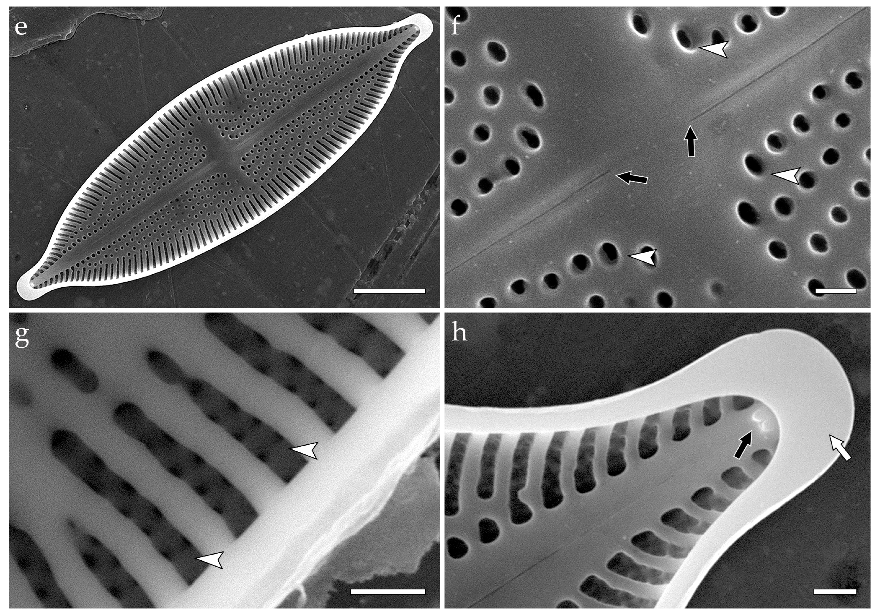
A different type of pore occlusion is unique to the genus Decussiphycus—spongia (pl. spongiae)—covering an areola in a laminar valve, located closer to the inner surface of the valve, and represented by structureless, homogenous silica flaps with no perforations visible in SEM or TEM ([5] (Figures 43 and 44) {Decussiphycus placenta (Ehrenberg) Guiry & Gandhi—generitype}; [30] (Figures 5C,D and 6C,D); [64] (Figures 14–15, 18 and 20); Figure 3).
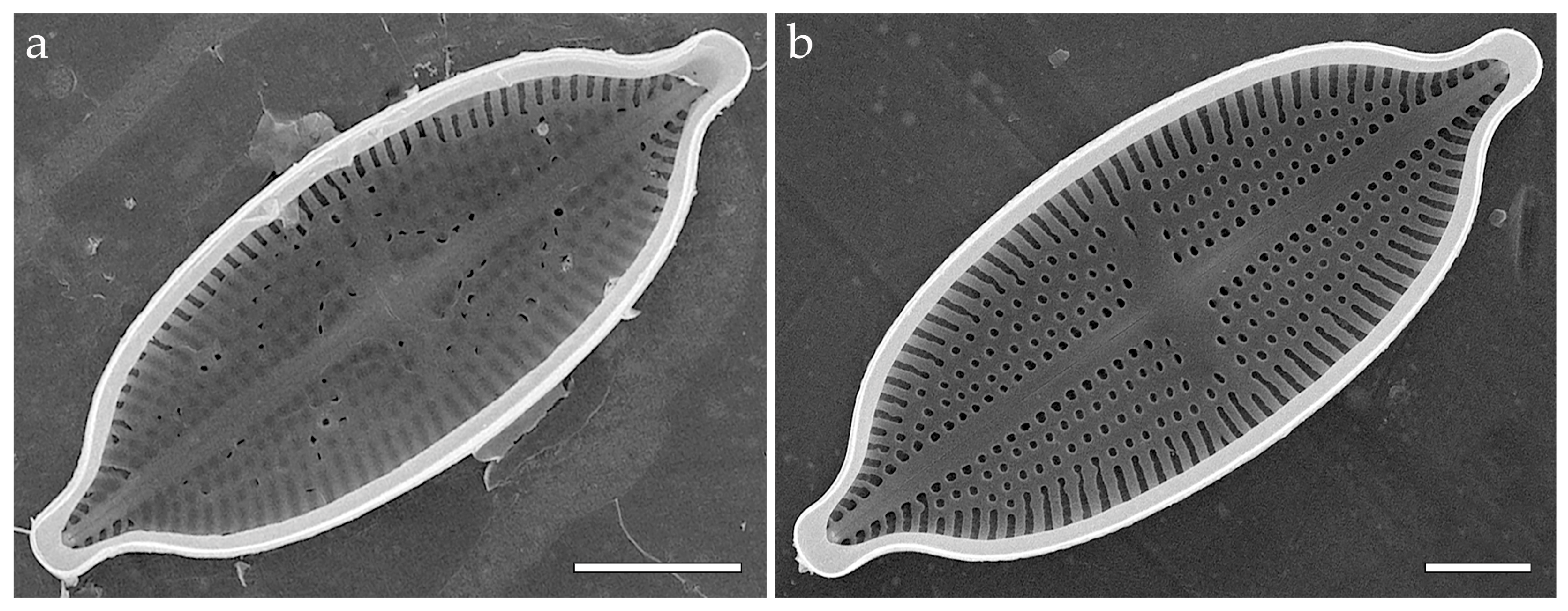
Specimens of Achnanthes sp. from strain CBMC102mns (Figure 6) demonstrate distinct areolae occluded by cribra rather than colandera of any type. In contrast to cribra, colandera and spongiae are constructed of a homogeneous flap of silica, with no (in case of spongiae) or multiple, usually four (in case of colandera), perforations. These perforations are small and arranged circumferentially (if multiple). Cribra of Achnanthes bear larger pores, formed as a result of irregular anastomosing of siliceous formations. Cribrate occlusions are attached to areolar walls with several, three–six, outgrowths ([65] (Figures 1 and 2); Figure 6c–e).
In the Discussion, we focus on previous studies, which investigated the fine structure of pore occlusions across Mastogloiales sensu Cox. We also explain the advantages of the proposed classification of pore occlusions and discuss the possibility of further work in this area. The elaborated classification is adopted in the following morphological descriptions of Aneumastus mongolotusculus, Decussiphycus sinensis, Mastogloia recta, Stigmagloia lobbanii, Achnanthes sp., and the newly proposed species Aneumastus khovsgolensis sp. nov.

3.3. Morphological Characteristics of Selected Species from the Order Mastogloiales

3.3.1. Aneumastus mongolotusculus (Figure 2)

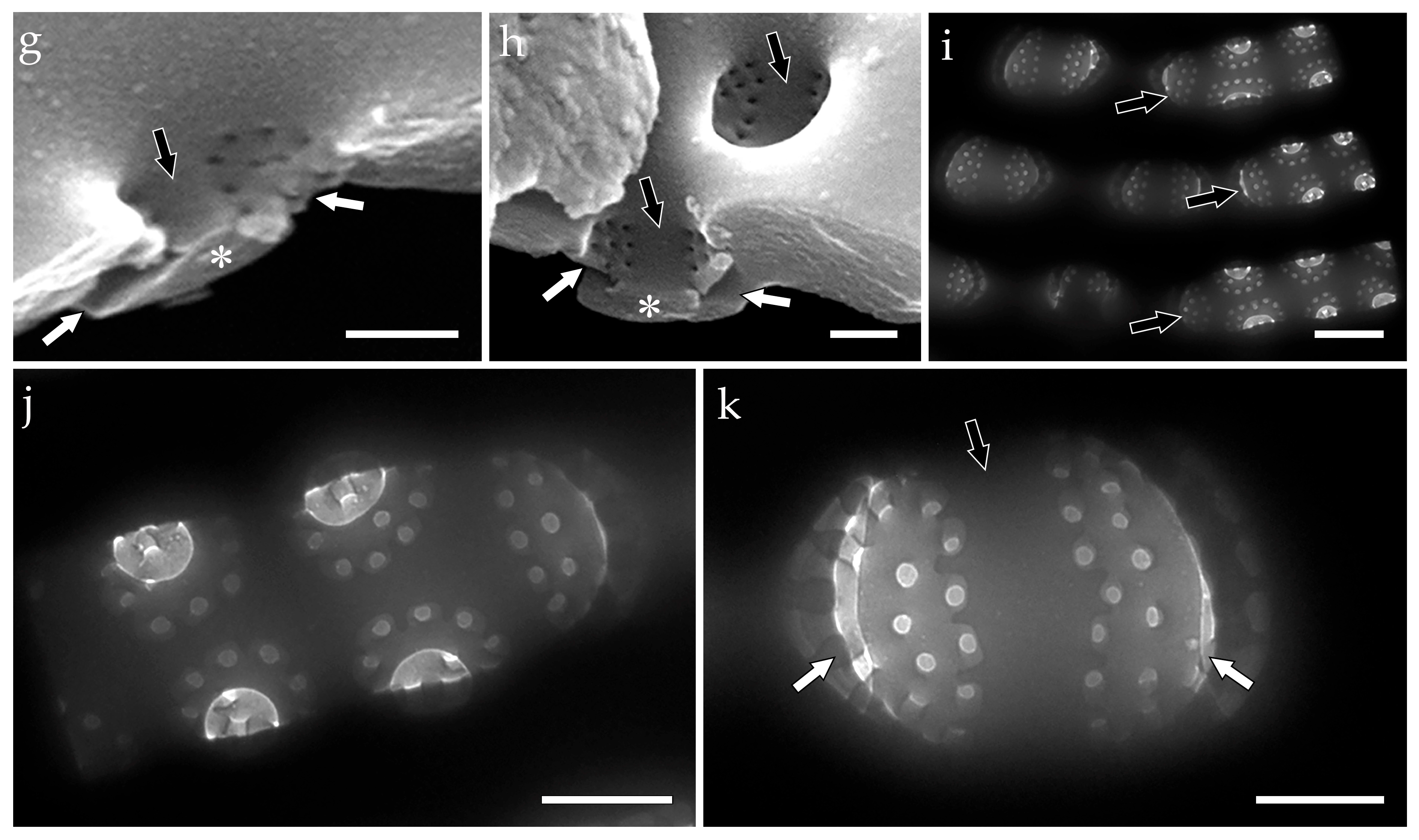
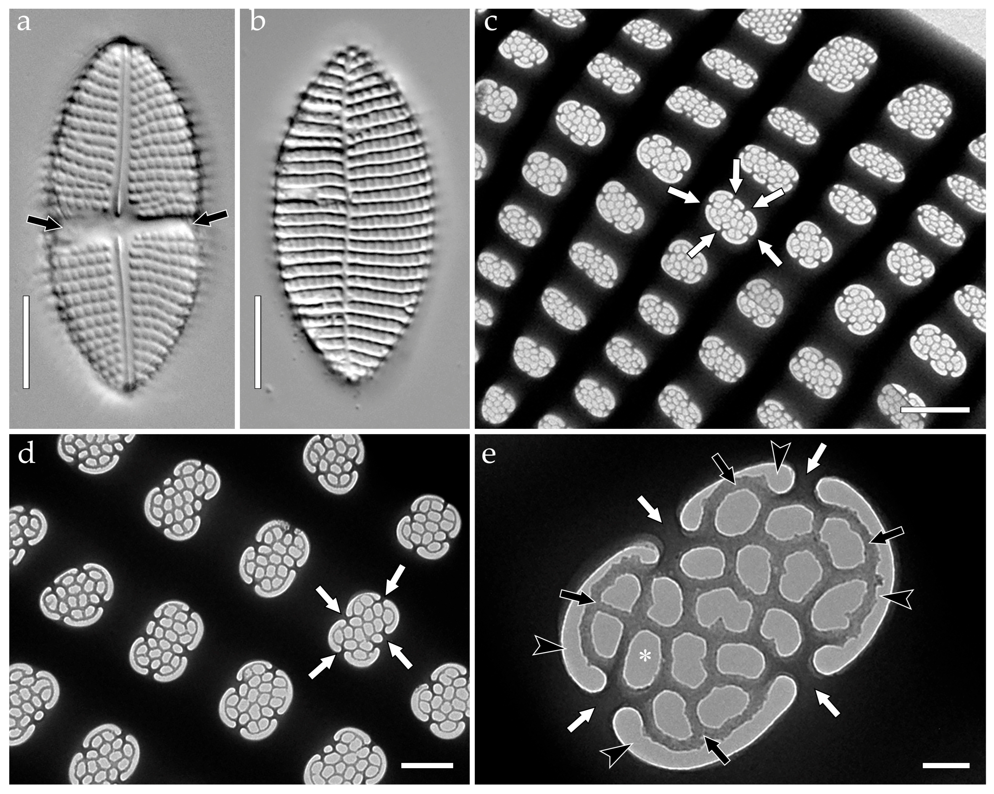
In LM (Figure 2a,b), valves broadly linear-elliptic, apices distinctly protracted, capitate to subcapitate. Valve dimensions (specimens in culture): length 46–50 μm, width 16–17 μm. Axial area narrow, linear; central area transapically enlarged, more or less oval, bordered by 2–3 shortened striae on each side. Raphe moderately undulate, with distinct circular proximal fissures. Striae radiate (11–12/10 μm), pseudoloculi sparsely arranged (8–9/10 μm). Transition from uni- to biseriate striae is discernible in LM. Externally, in SEM (Figure 2c), hourglass-shaped pseudoloculi are visible. External openings of pseudoloculi are represented by shallow rectangular depressions, which are elongated to varying degrees in the transapical direction (Figure 2c: white arrows). The shallow openings bifurcate into two canals that appear triangular in the external view (Figure 2c: black arrows). Internally, in SEM (Figure 2d–h) and TEM (Figure 2i–k), pseudoloculi are organized in uniseriate striae that become biseriate near the valve margin (Figure 2d,i: black arrows). Careful focusing reveals that pseudoloculi are occluded by colanderus-bifurca from the inside. The occlusions are represented by two silica flaps with small perforations (Figure 2e,g–k: white arrows), separated by a silica interruption (Figure 2e,g–k: black arrows). Each group includes ca. 10–12 perforations. Side view of the broken valves (Figure 2f–h) reveals that silica interruption and overhanging walls of the adjacent external pseudoloculi openings together construct a mushroom-shaped formation (Figure 2g,h: asterisks).

3.3.2. Decussiphycus sinensis (Figure 3)

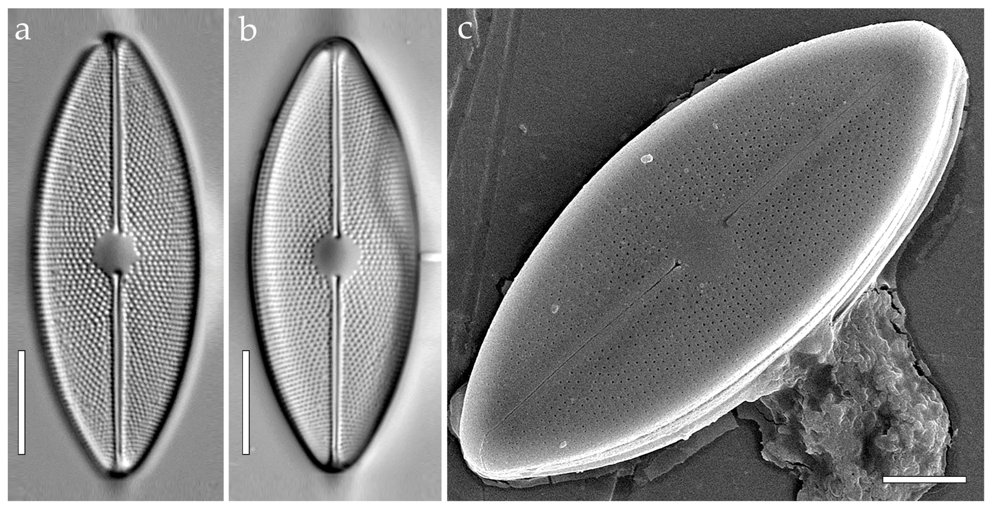
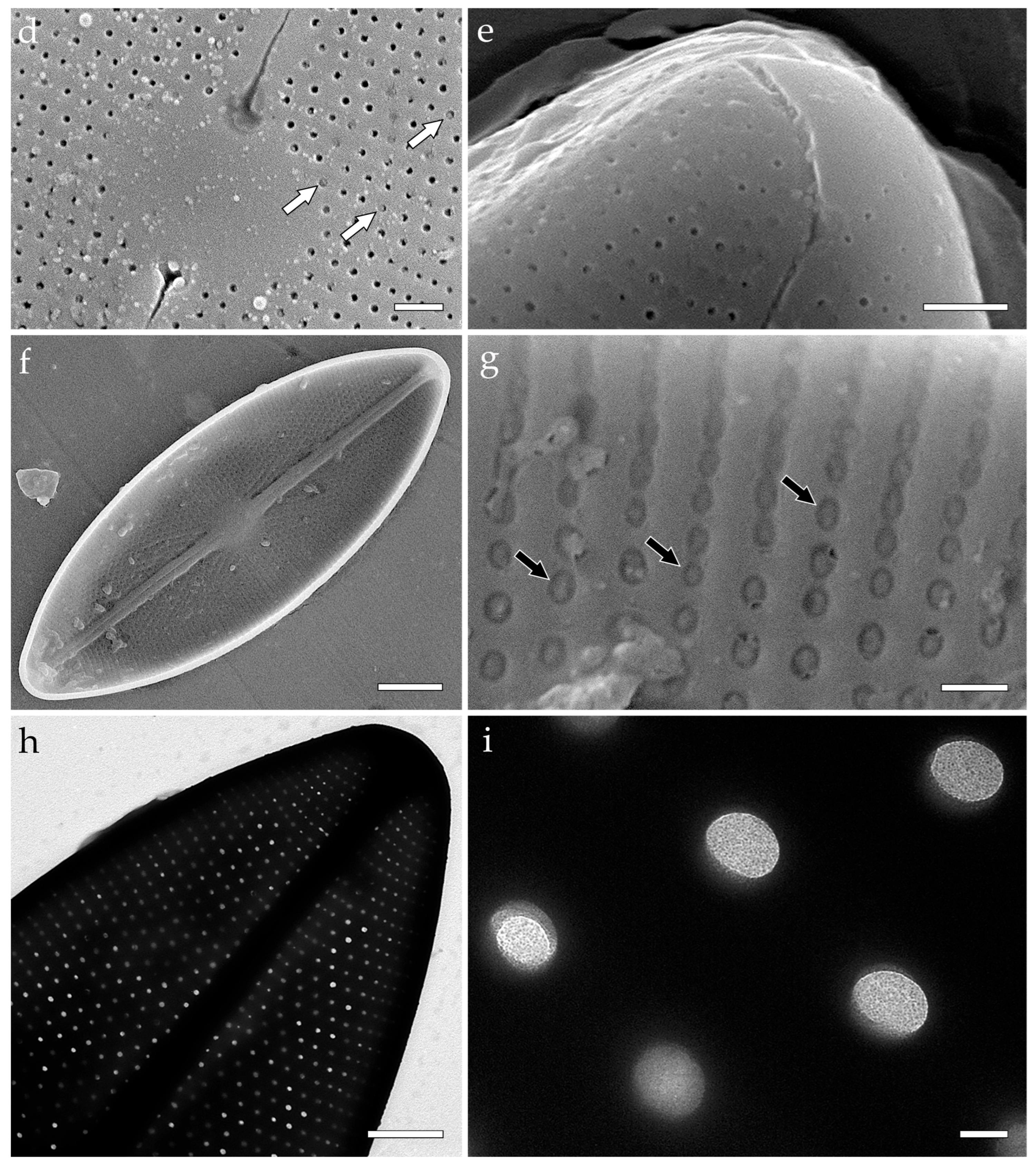
In LM (Figure 3a,b), Valves elliptic or linear-elliptic, apices unprotracted, broadly rounded. Valve dimensions (specimens in culture): length 42–46 μm, width 14–18 μm. Axial area narrow, linear; central area transapically enlarged, oval to nearly circular. Raphe filiform to slightly undulate. Striae decussate, arranged in quincunx, 22–23/10 µm. Areolae density—18–20/10 µm. Externally, in SEM (Figure 3c–e), areolar openings are round, with internal occlusions (spongiae) visible from the outside (Figure 3d: white arrows). Internally, in SEM (Figure 3f,g), areolae are slightly elongated in transapical direction. Spongiae are represented by unperforated, homogenous silica flaps (Figure 3g: black arrows). The flaps are somewhat convex inwards the valve. In TEM (Figure 3h,i), spongiae appear to be structureless, as well.

3.3.3. Mastogloia recta (Figure 4)

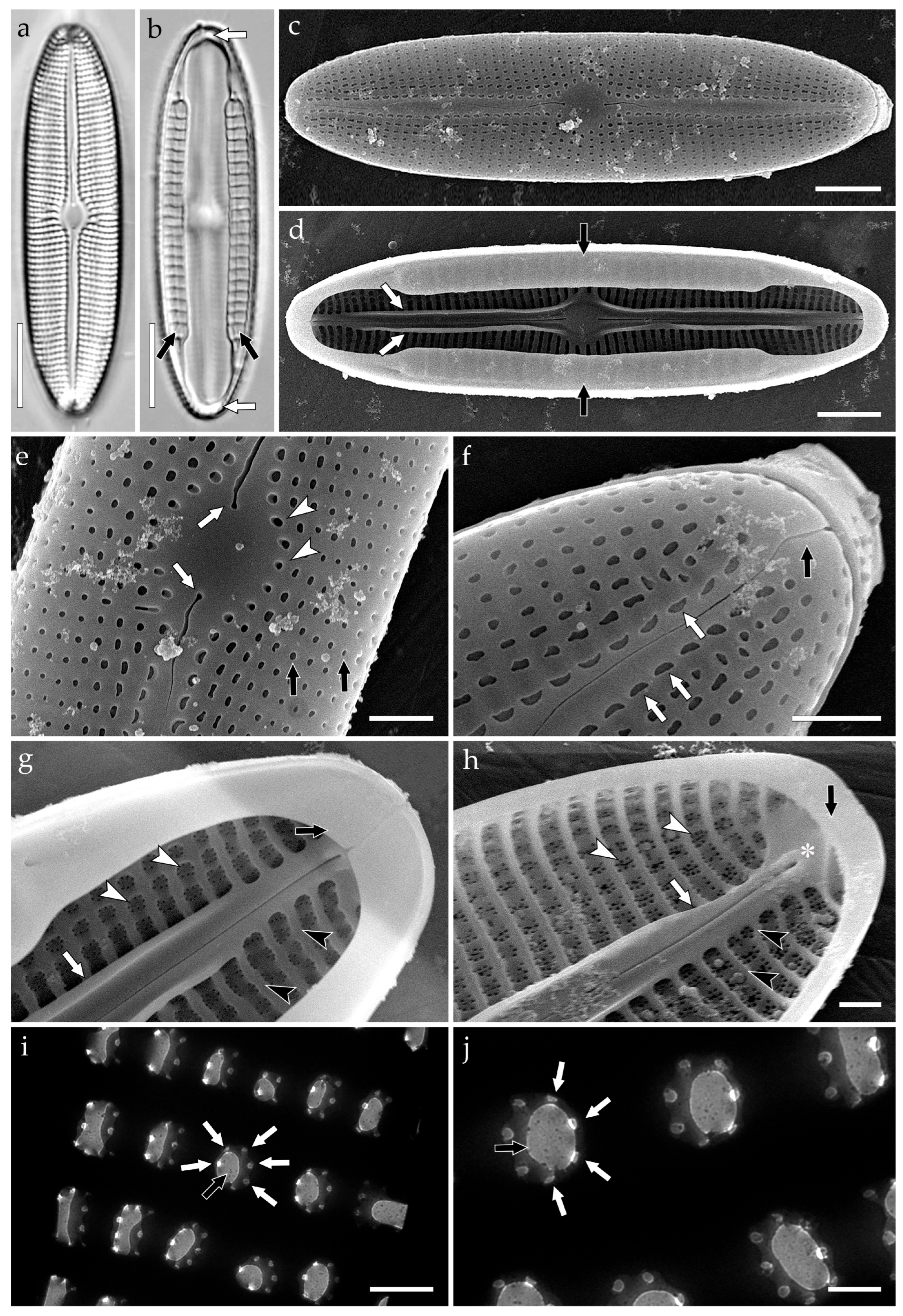
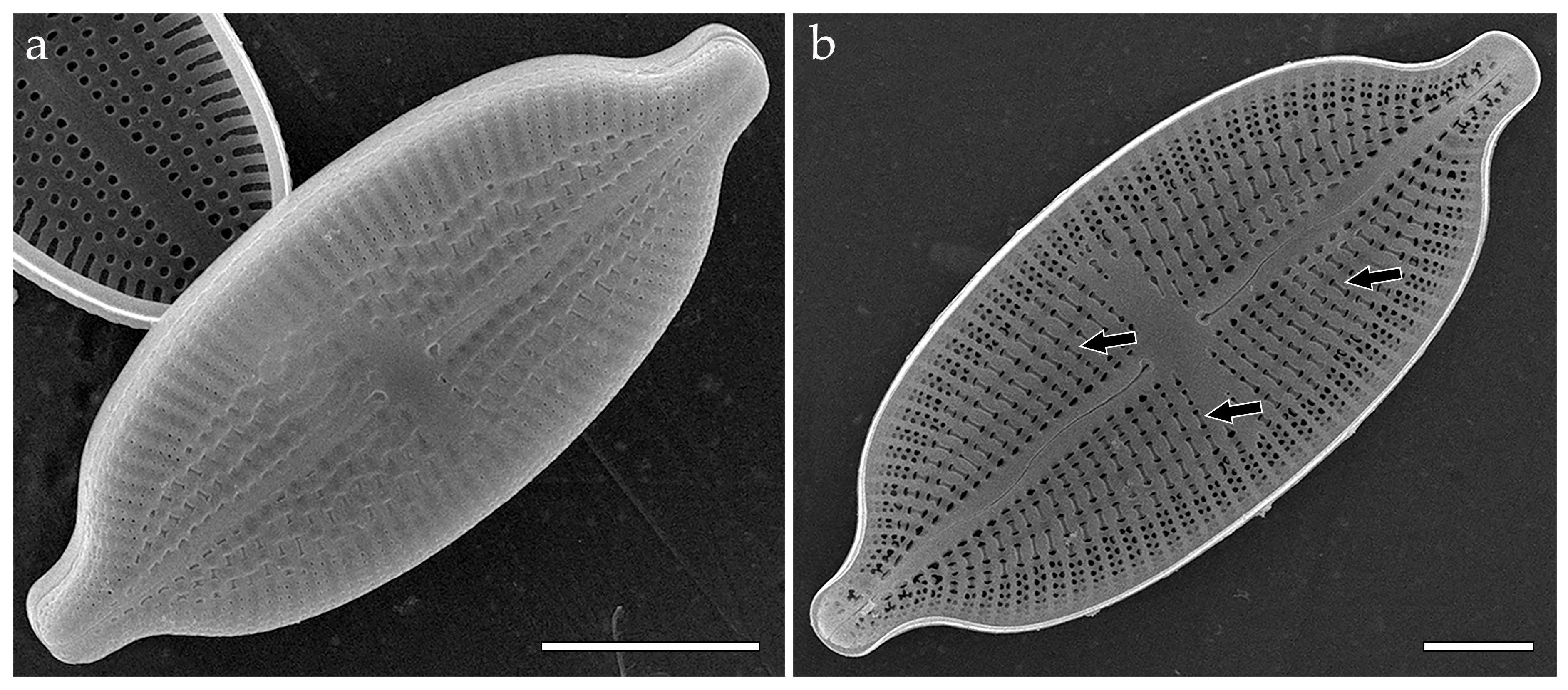
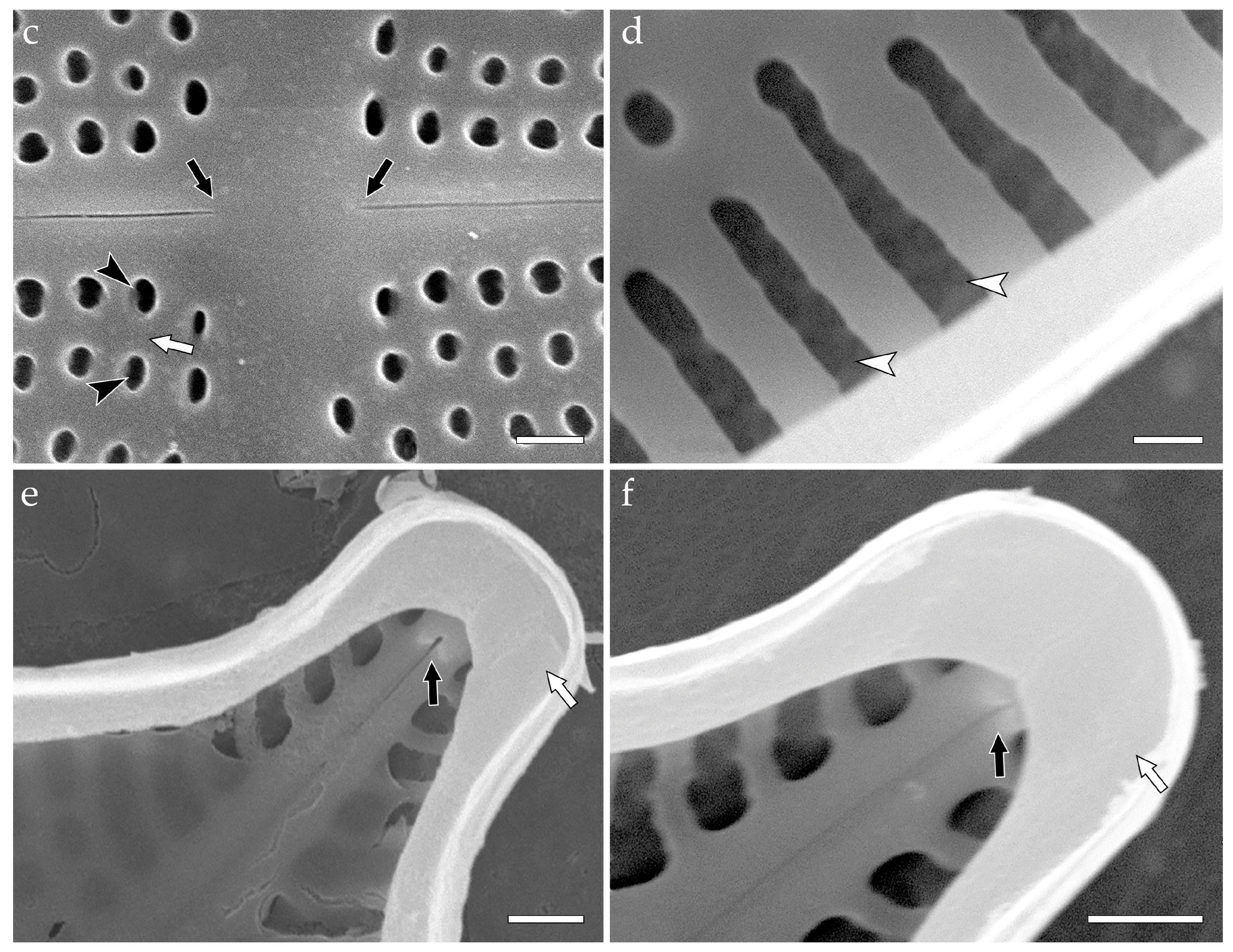
In LM (Figure 4a,b), valves linear-elliptic, with moderately tapering, obtusely rounded apices. Valve dimensions (specimens in culture): length 44–48 μm, width ca. 12 μm. Axial area narrow, linear; central area slightly enlarged, oval to rhomboid (apically elongated). Raphe moderately undulate, with visibly expanded proximal fissures. Striae uniseriate, radiate, or linear in the mid-portion of the valve (13–14/10 μm). Density of pseudoloculi—18–21/10 μm. A deeper focus reveals the rows of partecta on each side of the valvocopula (Figure 4b: black arrows). Pseudosepta are visible at each valve pole in LM (Figure 4b: white arrows) and SEM (Figure 4g,h: black arrows). In SEM, overall view on the valves (Figure 4c,d) also reveals well-developed partecta (Figure 4d: black arrows). On the inner side of the valve, raphe fissures lie in a gutter (sensu Paddock & Kemp [3]) with raised edges (Figure 4d: white arrows). Proximally, the raphe terminates with teardrop-shaped, unilaterally deflected fissures (Figure 4e: white arrows). Distal raphe fissures unilaterally deflected as well, terminating onto the valve margin (Figure 4f: black arrow). Externally, in SEM, pseudoloculi are predominantly round (Figure 4e: black arrows), becoming more transapically elongated, somewhat angled near the central nodule (Figure 4e: white arrowheads). Towards the poles, pseudoloculi adjacent to the raphosternum appear to be semilunar (Figure 4f: white arrows). Internally, SEM imaging of the apices demonstrates that gutter beside the raphe becomes more shallow (Figure 4g,h: white arrows). The raphe terminates with small helictoglossae (Figure 4h: white asterisk). Internal openings of pseudoloculi are occluded by colanderus-directa. Occlusions are formed of silica flaps with 6–10 circumferentially arranged perforations (Figure 4g: white arrowheads). As a result of corrosion, more perforations in the central portion are revealed (Figure 4h: white arrowheads). Uniseriate rows of pseudoloculi are separated by elevated interstriae (Figure 4g,h: black arrowheads). In TEM, perforations of colanderus-directa are arranged in a circle (Figure 4i,j: white arrows), with silica flap located centrally (Figure 4i,j: black arrows).

3.3.4. Stigmagloia lobbanii (Figure 5)

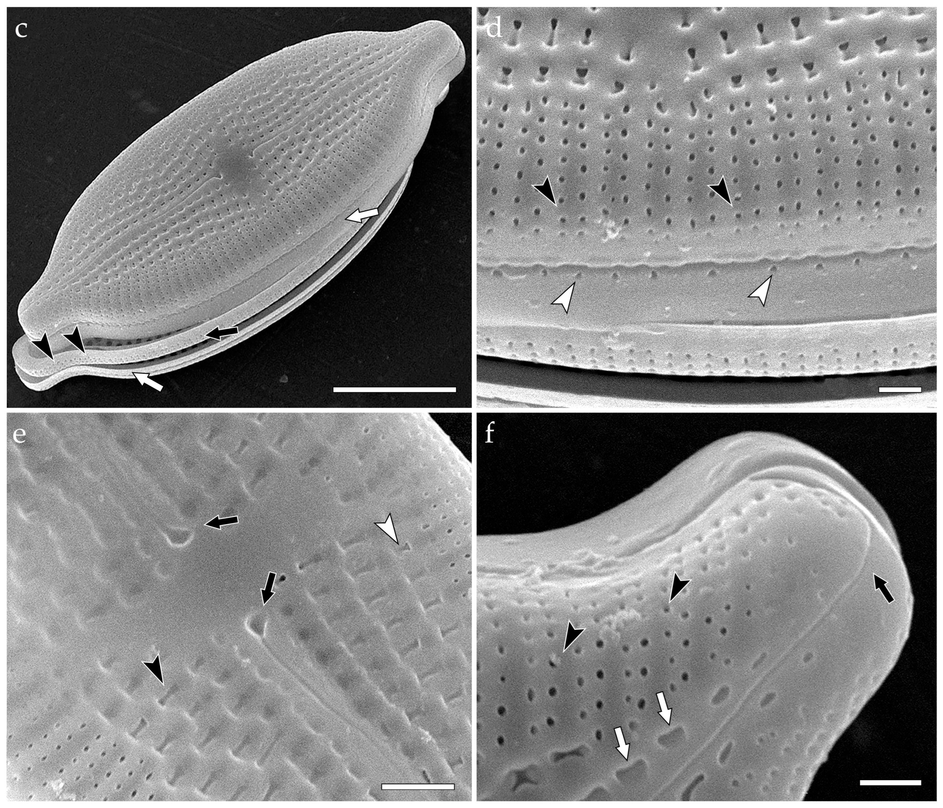
In LM (Figure 5a,b), valves elliptic to elliptic-lanceolate, slightly triundulate. Apices narrow, rostrate. Valve dimensions (specimens in culture): length 28–29 μm, width ca. 14 μm. Axial area narrow, central area small, apically elongated. Raphe moderately lateral. Striae predominantly parallel, more radiate towards the apices (23–24/10 μm). Pseudoloculi 14–16/10 μm, appear as rectangular due to the oblique arrangement of the outer and inner apertures. Next to the central nodule, between the striae, a stigma is located. The stigma is discernible in LM, TEM, and SEM (Figure 5a–c,e,f,h: white arrows), comprising a crater-like depression with numerous tentacular outgrowths. Moreover, TEM (Figure 5c–e) reveals the oblique position of the canal between the outer and inner openings of the pseudoloculi. The canal is angled at ca. 30°, so that the outer aperture is located closer to the raphe (Figure 5d,e: black arrowheads) than the inner aperture (Figure 5d,e: white arrowheads). External SEM view (Figure 5f,g) demonstrates that the raphe is displaced near the proximal fissures (Figure 5f: black arrows), while the distal fissures are gradually and unilaterally curved (Figure 5f: black arrowheads). Again, focus on the pseudoloculi shows their oblique arrangement (Figure 5g: black arrowheads). Internally, in SEM (Figure 5h,i), pseudosepta are notable at both apices of the valve (Figure 5h: black arrowheads). Internal valve surface is furnished with perforations of colanderus-obliqua, typically 4 per pseudolocula (Figure 5i: white arrowheads). The occlusions are not visible in external SEM view due to the obliquity of pseudolocular canals.

3.3.5. Achnanthes sp. (Figure 6)

In LM (Figure 6a,b), valves linear-elliptic. Apices unprotracted, acute. Valve dimensions (specimens in culture): length 36–38 μm, width ca. 17 μm. Raphe valve with narrow axial area and fascia. Raphe moderately lateral, with gradually curving branches. Proximal raphe ends slightly expanded. Striae linear in the central portion of the valve, radiate towards the apices (8–9/10 μm at the raphe valve, 7–8/10 μm at the rapheless valve). Areolae clearly discernible in LM, 8–9/10 μm. In TEM (Figure 6c–e), areolae are occluded with cribra. The cribra are flat, comprising intertwined net of silica formations (Figure 6e: black arrows). The net is connected to the wall of areola by 4 or 5 outgrowths (Figure 6c–e: white arrows). The peripheral perforations in cribra are always slender, reniform (Figure 6e: black arrowheads), while the rest of perforations are roundish (Figure 6e: asterisk).

3.4. Description of the New Species

Aneumastus khovsgolensis Glushchenko, Mironov, Genkal, Nergui & Kulikovskiy sp. nov. (Figure 7, Figure 8, Figure 9, Figure 10, Figure 11 and Figure 12).
Holotype: Slide 10206 in Diatom Herbarium (HD) in K.A. Timiryazev Institute of Plant Physiology RAS, Moscow, Russia, represented here by Figure 8c.
Type: Mongolia. Khovsgol aimak, Lake Khovsgol, southeastern shore, epilithon, washing from silty stones, 50.62592 °N, 100.50087 °E, leg. A. Mironov, 24.07.2024. Slide 10206 from oxidized culture strain CBMC529mnp, isolated from sample Mon59.
Reference strains: CBMC338mnp (isolated from sample Mon58), CBMC529mnp (isolated from sample Mon59), deposited in the Culture and Barcode Collection of Microalgae and Cyanobacteria “Algabank” (CBMC).
Representative specimens: Strains CBMC338mnp (slide 10105), CBMC529mnp (slide 10206); samples Mon58 (slide 09487), Mon59 (slide 09488).
Sequence data: GenBank accession numbers PV933319 (strain CBMC338mnp, 18S rRNA gene sequence, V4 region); PV942668 (strain CBMC338mnp, rbcL gene sequence); PV933320 (strain CBMC529mnp, 18S rRNA gene sequence, V4 region); PV942669 (strain CBMC529mnp, rbcL gene sequence).
Etymology: The specific epithet refers to Lake Khovsgol, the species type locality.
Distribution: So far, the species is known only in its type locality.
Ecology: The species was located in probes of shallow benthos and epilithon, at a temperature of 17.1–17.6 °C, pH = 8.30–8.51, and medium conductivity (210–248 µS/cm).
Description:
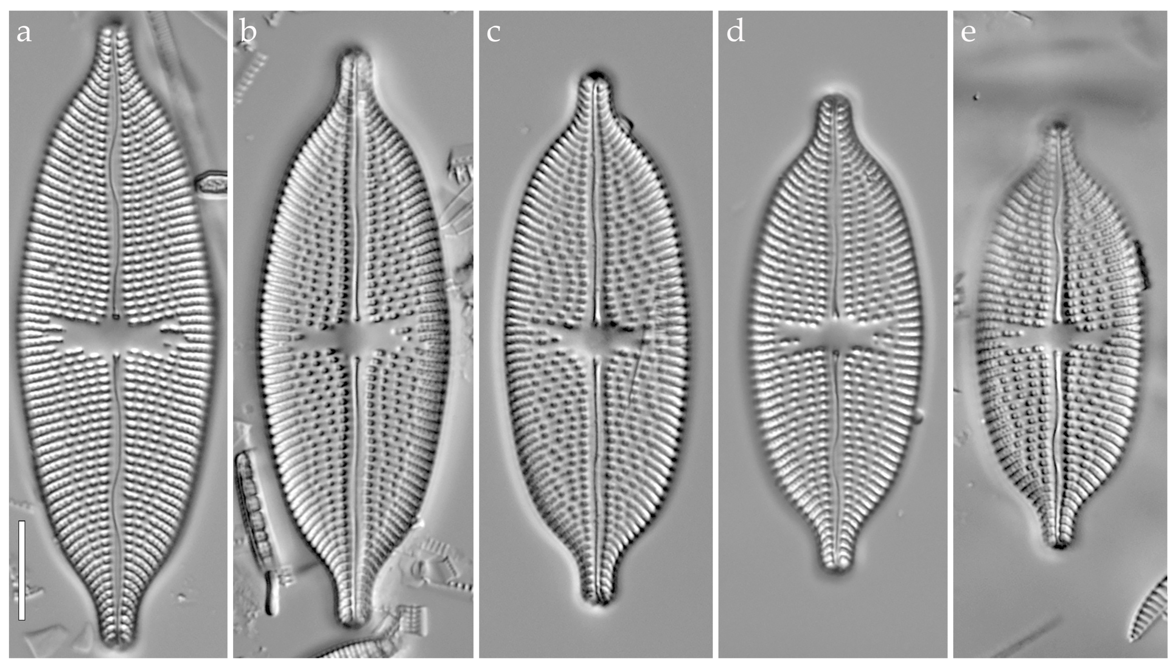
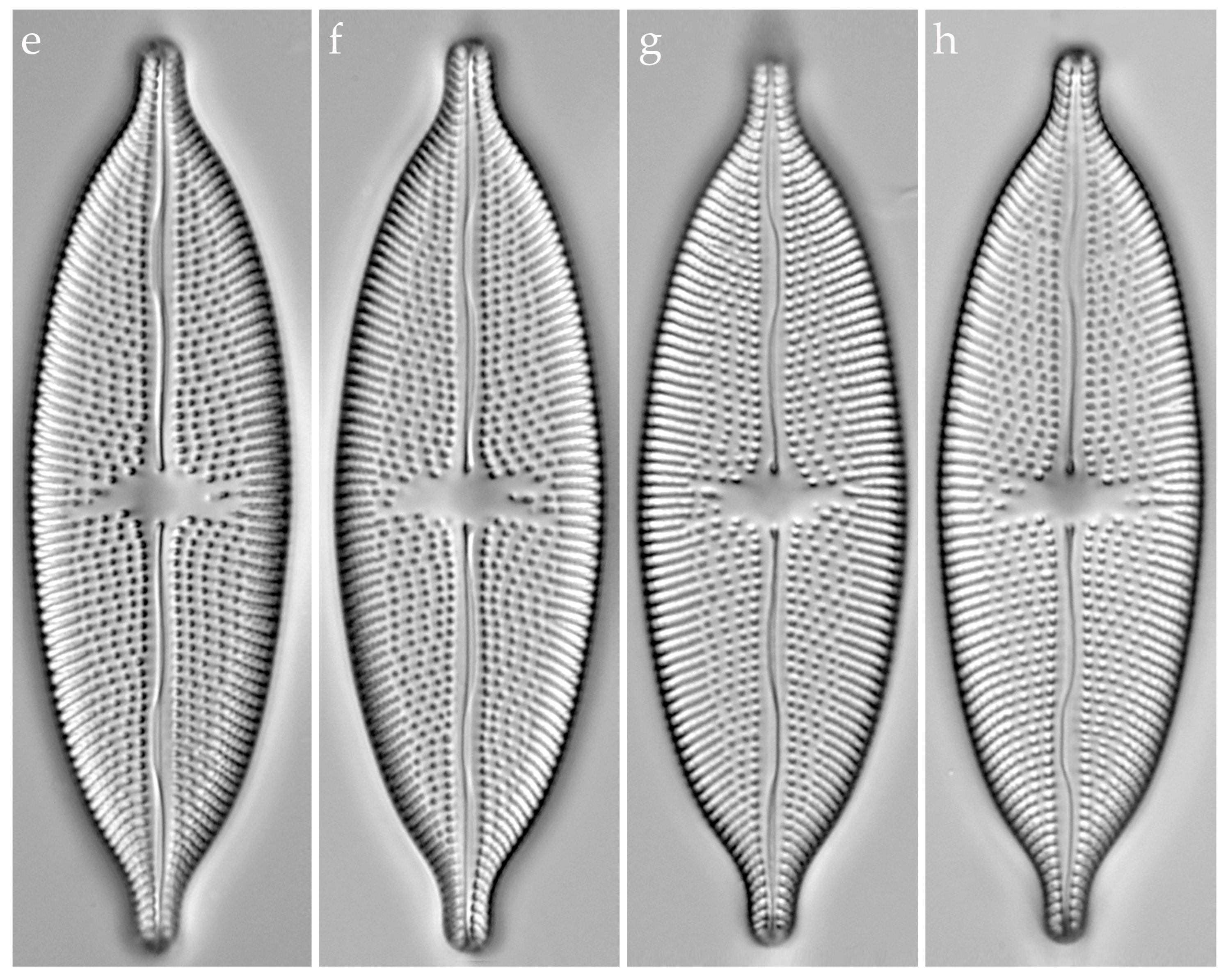
LM (Figure 7, Figure 8, Figure 10 and Figure 11): Valves linear-elliptic to elliptic. Valve apices obtusely rostrate. Valve dimensions: length 37.6–72.4 μm, width 15.8–21.0 μm. Raphe weakly lateral, with two moderate undulations. Proximal raphe ends expanded, teardrop-shaped; distal ends undiscernible in LM. Axial area hardly expressed, nearly linear, narrowing towards the apices. Central area irregular, mostly transapically enlarged, delimited by 3–5 striae on each side. Each valve is equipped with striae of two types—uniseriate (near the axial area) and biseriate (near the margin). Striae radiate, 12–13/10 μm. In uniseriate rows, pseudoloculi density ca. 8/10 μm.
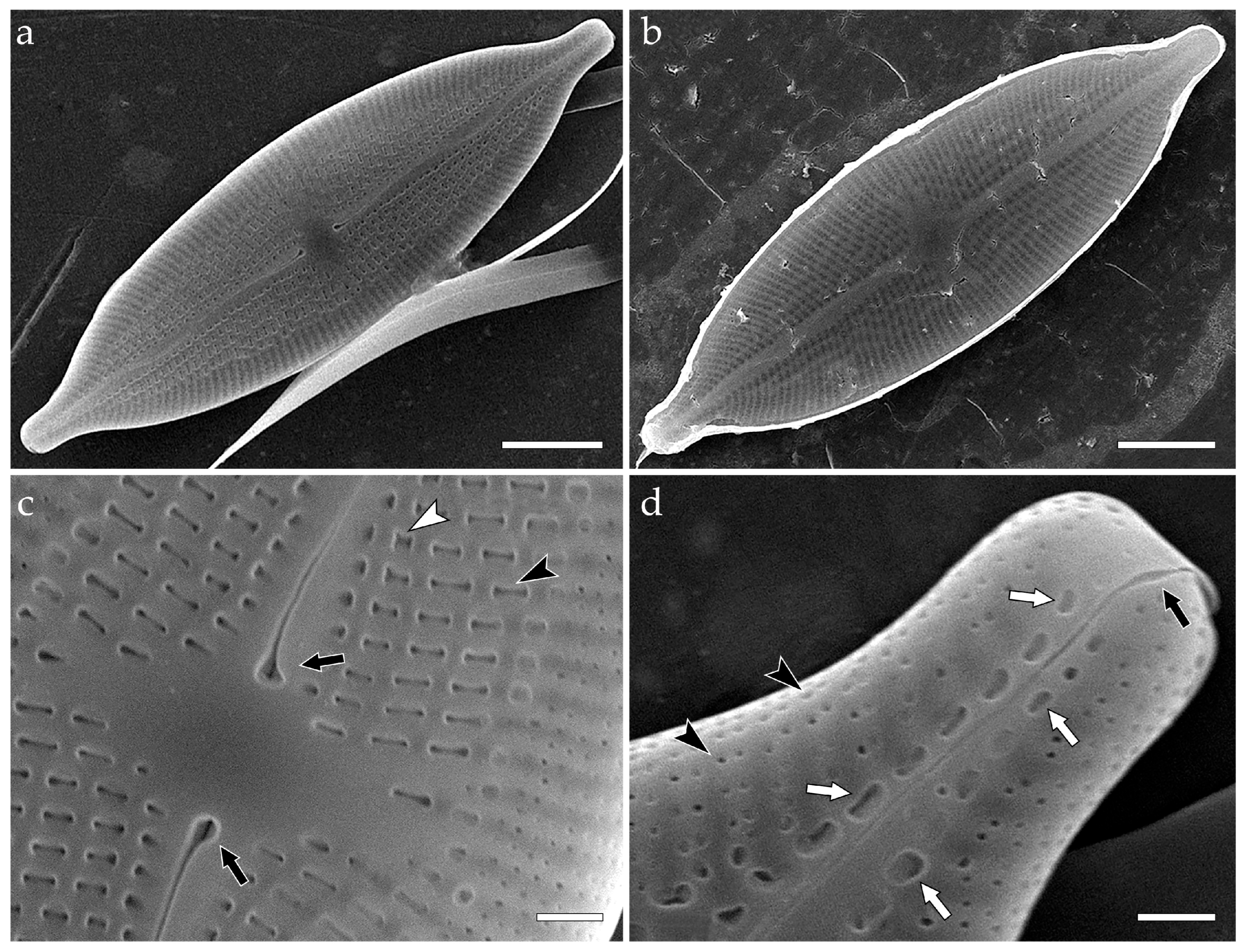
SEM, external view (Figure 9a–d and Figure 12): Valve face is flat. Raphe distinctly undulating. Proximal raphe fissures expanded, unilaterally deflected (Figure 9c and Figure 12e: black arrows). Distal raphe fissures gradually curved, continuing onto the mantle (Figure 9d and Figure 12f: black arrows). Distally, the fissures are curved in the same direction in relation to each other, but oppositely in relation to the proximal fissures. Striae differentiated into two types. Near the axial area, striae are uniseriate, comprising pseudoloculi. The external openings of pseudoloculi are slightly depressed, transapically elongated to various extents, thus appearing as X-shaped (Figure 9c and Figure 12e: white arrowheads) to bone-shaped (Figure 9c and Figure 12e: black arrowheads). Near the apices, pseudoloculi adjacent to the axial area are semilunar-shaped (Figure 9d and Figure 12f: white arrows). Mid-valve, striae become biseriate and retain this organization onto the valve margin. External openings of pseudoloculi in biseriate striae are small, more or less round (Figure 9d and Figure 12d,f: black arrowheads). Density of pseudoloculi in biseriate striae—18–24/10 μm. The cingulum comprises two valvocopulae (Figure 12c: white arrows) with an additional band between them (Figure 12c: black arrow). Each valvocopula is equipped with a single row of perforations (density–10–12/10 μm) (Figure 12d: white arrowheads). The intercalary band bears multiple fine perforations (density—ca. 50/10 μm) (Figure 12c: black arrowheads). Note that corrosion reveals the bifurcation of pseudoloculi (Figure 12b: black arrows), i.e., the presence of colanderus-bifurca on the inside of the valve.
SEM, internal view (Figure 9e–h and Figure 13): Internally, raphe branches are not raised above the valve surface. The raphe is straight, filiform. Proximal fissures straight, not expanded (Figure 9f and Figure 13c: black arrows); distal ends also straight, terminating with small helictoglossae (Figure 9h and Figure 13e,f: black arrows). Each valve pole bears a distinct pseudoseptum (Figure 9h and Figure 13e,f: white arrows). Inner openings of pseudoloculi adjacent to the axial area are crater-like, round, or transapically elongated (Figure 9f: white arrowheads). Towards the valve margin, inner openings of pseudoloculi lie at the bottom of a joint elongated foramina (Figure 9g and Figure 13d: white arrowheads). Each occlusion (colanderus-bifurcus) is represented by two openings (Figure 13c: black arrowheads) separated by a silica interruption (Figure 13c: white arrow).
Comments: Cultured specimens correspond to wild populations, i.e., valve dimensions overlap (see Table 2) and the outlines, apices, raphe structure, and striae arrangement match, too. Notably, clones from strain CBMC529mnp are characterized by bigger valves (length > 68 μm, width > 20 μm), while clones from strain CBMC338mnp are smaller (length < 44 μm, width < 18 μm). However, SEM (Figure 9, Figure 12 and Figure 13) demonstrates the uniformity of both wild and cultured specimens.

4. Discussion

4.1. Comparison of Aneumastus khovsgolensis sp. nov. with Similar Taxa

Morphologically, Aneumastus khovsgolensis sp. nov. belongs to the Aneumastus tusculus group (“Group 2” sensu Lange-Bertalot [4]), which unites the species with uniseriate (near the axial area) and biseriate (towards the margin) striation. In the recent years, the group has significantly expanded [51,74,75] and nowadays comprises A. tusculus, A. balticus Lange-Bertalot, A. macedonicus Levkov, A. rostratus (Hustedt) Lange-Bertalot, A. mongolotusculus, A. laosica Glushchenko, Kulikovskiy & Kociolek, and A. visovacensis Udovič & Levkov. Hereby, we provide a comparison of A. khovsgolensis sp. nov. with its two most similar species—A. tusculus and A. mongolotusculus.
Material type of A. tusculus is fossil and hitherto has been illustrated only in LM ([4] (Plate 113, Figures 1–4)). A. khovsgolensis sp. nov. can be distinguished from the fossil specimens of A. tusculus by valve outlines more abruptly narrowing towards the apices. In material type of A. tusculus, the narrowing of apices is more gradual, somewhat shoulder-like ([4] (Plate 113, Figures 1–4)). The valve apices in A. khovsgolensis sp. nov. are narrower (3.2–3.6 μm in smaller specimens, 3.6–3.8 μm in bigger specimens) than in the fossil species (3.9–4.5 μm). Moreover, central area in the new species is generally smaller than in A. tusculus ([4] (Plate 113, Figures 1–4)) and striae are more densely arranged (12–13/10 μm in A. khovsgolensis sp. nov. vs. 10/10 μm in A. tusculus).
It is worth mentioning that smaller specimens of A. khovsgolensis sp. nov. resemble A. mongolotusculus, another species discovered in Lake Khovsgol [29]. They possess valves of comparable sizes which demonstrate overlapping dimensions of width: 16–19 μm in A. mongolotusculus and 15.8–21.0 μm in A. khovsgolensis sp. nov. Their striae densities are also similar: 12–13/10 μm in A. khovsgolensis sp. nov., 11–12/10 μm in A. mongolotusculus. However, the two taxa are easily differentiated in LM by the shape and width of their valve apices. In A. mongolotusculus, the apices are more constricted and slender ([29] (Figures 1–8)), 2.9–3.2 μm wide, while in A. khovsgolensis sp. nov. the apices are evidently wider, 3.2–3.6 μm.
The morphological comparison of the new species to A. tusculus and A. mongolotusculus is summarized in Table 3.

4.2. Phylogenetic Resolution of the Order Mastogloiales

4.2.1. Molecular Phylogenetics of Mastogloiales

As explained in the introduction, diatoms of Mastogloiales sensu Cox have been a subject to numerous extensive molecular investigations [22,23,25,28,29,30,31,53,66,76,77,78]. These studies invariably demonstrated that representatives of the order belong to different evolutionary lineages. However, it seems that the heterogeneity of the order was ignored by some authors in favor of morphological similarities. For example, Stepanek & Kociolek [31] referred to Mereschkowsky [32] when comparing Tetramphora to Mastogloia and suggested that “it seems appropriate to place Tetramphora within the Mastogloiales” due to the similar arrangement of plastids. Despite that, molecular analysis conducted by Stepanek & Kociolek [31] shows little support for the Mastogloia-Tetramphora clade: PP = 0.57, LB = 41 at the 18S rRNA+rbcL+psbC phylogram; LB = 25 at the 18S rRNA+rbcL phylogram LB = 25. Unexpectedly, the subsequent analysis by Sabir et al. [25], which focused on the molecular phylogeny of Phaeodactylum tricornutum Bohlin, gave more support for the Mastogloia-Tetramphora association (LB = 99). This phenomenon is likely explained by the insufficiency of molecular referencing databases, which, if elaborated, could have provided better resolution for phylogenetic relationships between Mastogloia and other diatoms.
The heterogeneity of Mastogloiales was more evident in Ashworth et al. [23], whose study revealed an evident segregation between the Achnanthes-Craspedostauros clade and Mastogloia. In contrast to Stepanek & Kociolek [31], Ashworth et al. [23] denied the homology between the two fore and aft plastids of Achnanthes and Craspedostauros and Mastogloia, thus rejecting Cox’s [13] concept of Mastogloiales. At the time, Tetramphora was not included in the analysis, and Mastogloia was represented by only two strains. Similarly to the previous diatomists [25,31], Ashworth et al. [23] faced the problem of the lack of molecular genetic data on the order.
This issue has remained unsolved up to date, despite many efforts [25,26,27,28,29]. The situation has recently changed thanks to the innovations introduced by Kezlya et al. [53], who erected the genus Stigmagloia with molecular evidence, and Mironov et al. [30], who described Decussiphycus sinensis on the basis of molecular and morphological data. Hence, the current state of molecular phylogenetic research on Mastogloiales indicates that this taxonomic group is limited to four genera—Mastogloia, Decussiphycus, Aneumastus, and Stigmagloia—all supported by molecular references.

4.2.2. Morphological Characteristic of Mastogloiales with Particular Reference to the Structure of Pore Occlusions

The question of the morphological diagnosis of Mastogloiales, however, is still controversial, primarily due to the immense variability of valve features within Mastogloia, the most species-rich genus of the order. Interestingly, Round et al. [1] characterized Mastogloia as possessing a “natural but extraordinary variable in areola structure”, thus, in fact, anticipating future studies demonstrating the artificiality of the genus. Prior to Round et al. [1], numerous studies highlighted the polymorphism of Mastogloia [35,36,37,38,39,40,41]. SEM analyses revealed the complexity of areolae, which, at the time, were characterized as locular [39,40]. The structure of areolae was refined by Stephens & Gibson [36] based on the Mastogloia Ellipticae group. Two types of locular areolae were distinguished—simple loculi (in Mastogloia binotata (Grunow) Cleve, Mastogloia crucicula (Grunow) Cleve, Mastogloia ovalis A.W.F. Schmidt and Mastogloia splendida (Gregory) H. Pergallo), and loculi with rings (in Mastogloia cribrosa Grunow and Mastogloia fimbriata). Later, the Undulatae, Apiculatae, Lanceolatae, Paradoxae, and Inaequales groups were studied [37,38]. The authors postulated that species of the Apiculatae and Lanceolatae groups possessed narrow loculi with inclined locular tubes “in a second plane above the locular bands, crossing obliquely toward the outer valve margins”. A similar type of locular areolae, with rudimentary tubes, was identified in the Inaequales group and Mastogloia cyclops Voigt, a member of the Undulatae group. The latter species has been recently transferred to the genus Stigmagloia due to the presence of stigma [53]. Stephens & Gibson [36,37,38] also mentioned that species of Mastogloia may differ in valve construction, i.e., Mastogloia lanceolata Thwaites ex W. Smith, Mastogloia paradoxa Grunow, and M. cyclops possess “a single layer interrupted by puncta” (i.e., laminar valves), while the valves of Mastogloia apiculata W. Smith are more complex and two-layered, with “raised transapical silicious ribs, between which is a layer of silica perforated by double rows of puncta” (cavitate valves). These assumptions, in fact, corresponded to Hendey’s concept of single-layered (laminar) and multi-layered (cavitate) valves [79].
Apart from areolae and striae, diatomists investigated other identifying features of Mastogloia. Over the years, extensive morphological data on the structure of their valve surface, partecta (and accompanying formations), girdle, and valvocopula was collected [41,42,43,44,45]). Perhaps the most extensive analysis was carried out by Paddock & Kemp [3]. In the latter research, the authors described several valve patterns (double and triple zonality), modifications of the central area (elongated, reduced or transverse areolae, stigmata), valve adornments (marginal ridges, conopea, and pseudoconopea), papillae, pseudosepta, etc. The immense variety of morphological features, both general and ultrastructural, means that the only synapomorphy of the genus might be the partecta. This fact raises doubts about the monophyly of Mastogloia and could become a justification for future studies to disband the genus into several limited groups.
The main part of the analysis by Paddock & Kemp [3,63] was devoted to the investigation of areolar morphology. Therein, areolae of multiple Mastogloia species were described as pseudolocular, rather than locular, due to the presence of large external openings and internal occlusions. Pseudoloculi were visualized by Paddock & Kemp [63] based on the broken valves of M. goesii, M. elegans, and four more marine species introduced therein. In the study, two types of areolae were illustrated:
  • M. elegans-type (also typical for M. goesii, M. corallum, M. neomauritiana, M. umbra)—a pseudoloculus with a single large external opening corresponding to four small internal openings, grouped by pairs ([3] (Figures 1, 6, 16, 24, 33 and 42));
  • Mastogloia sturdyi-type—a loculus with a single large external opening and four small internal openings, perforating “in pairs into different loculi, to either side of the walls of the loculi” ([3] (Figure 52));
Paddock & Kemp significantly expanded the focus of their research by conducting a detailed SEM survey on 150+ species of Mastogloia to illustrate the immense diversity of the morphological features of their valves, valvocopulae, and girdle [3]. Areolar diversity was investigated as well. Paddock & Kemp [3] adopted the concept of laminar and cavitate valves in Mastogloia to distinguish the types of areolae as follows:
  • M. ovalis-type—a round areola in a laminar valve, occluded by a single, unilateral, tongue-shaped outgrowth. The outgrowth consists of a narrow base and a nearly round spatula; thus, areola appears horseshoe-shaped ([3] (Figure 31); [48] (Figures 21–28));
  • M. crucicula-type—a round areola in a laminar valve, occluded by a single, unilateral outgrowth. The outgrowth is semicircular, with a wide base ([3] (Figure 32); [48] (Figures 9–14));
  • M. fimbriata-type—a round areola in a laminar valve, occluded by a silica disk, which is supported by multiple thin, radially arranged struts ([2] (Figure 3); [3] (Figure 33));
  • Mastogloia binotata var. ovata Voigt-type—a round areola in a laminar valve, occluded by rota. The rota comprise a bagel-shaped central formation, connected to the wall of the areola by two (rarely, three) opposite struts ([2] Figure 1–8); [3] (Figure 34));
  • Mastogloia cocconeiformis Grunow-type—a loculus (sensu Paddock & Kemp) in a cavitate valve with a single external and a single internal opening, occluded by a velum-like structure amidst the two openings. The velum-like structure is “saucer-shaped”, lying at an acute angle in relation to the inner opening ([2] (Figure 1); [3] (Figure 35));
  • Mastogloia frickei Hustedt-type—an alveola (sensu Paddock & Kemp) in a cavitate valve, with a single lateral external opening and several squarish internal openings, which are arranged in a transverse row ([3] (Figure 36));
  • Mastogloia biocellata (Grunow) G. Novarino & A.R. Muftah-type—an alveola (sensu Paddock & Kemp) in a cavitate valve, with a single lateral external opening and a single squarish internal opening ([3] (Figure 37));
  • Mastogloia sp. 1-type—an alveolus in a cavitate valve, with a single round external opening and small internal openings. A pair of internal openings corresponds to an external opening, lying “directly below” the outer aperture. Two layers of the valve are firmly attached to each other ([3] (Figure 38));
  • Mastogloia sp. 2-type—a pseudoloculus in a cavitate valve, with a single internal and a single internal opening. Both openings are more or less round, one directly under another ([3] (Figure 39));
  • Mastogloia lineata-type—a pseudoloculus in a cavitate valve, with a single, large, rectangular external opening and one or two small, round internal openings. The inner openings are situated asymmetrically in relation to the outer aperture, not lying directly under (due to the shape of the cavity) ([3] (Figure 40));
  • Mastogloia sp. 3-type—a pseudoloculus in a cavitate valve, with a single, slit-like external opening and one or two small, round internal openings. The inner openings are situated asymmetrically in relation to the outer aperture, not lying directly under (due to the shape of the cavity) ([3] (Figure 41));
  • Mastogloia emarginata-type—a pseudoloculus in a cavitate valve, with a single, large, round external opening and multiple small internal openings. The inner openings are situated asymmetrically in relation to the outer aperture, not lying directly under (due to the shape of the cavity). The walls of the pseudoloculus are “overhanging” ([3] (Figure 42));
  • Mastogloia rimosa-type—a pseudoloculus in a cavitate valve, with a crescent, somewhat sunk external opening and a single round internal opening. The inner opening is situated asymmetrically in relation to the outer aperture, not lying directly under (due to the shape of the cavity). The internal opening is surrounded by four papillae ([3] (Figure 43));
  • Mastogloia peracuta-type—a pseudoloculus in a cavitate valve, with a large, round external opening and one, sometimes two, minute internal openings. Pseudolocular walls are equally developed ([3] (Figure 44));
  • M. elegans-type—a pseudoloculus in a cavitate valve, with a single, large, round or elongated external opening and usually four small internal openings. Inner openings are grouped in pairs, proceeding to different (adjacent) external openings. ([3] (Figure 45));
  • Mastogloia cebuensis-type—a pseudoloculus in a cavitate valve, with a single, large, round or rectangular opening and (usually) six internal openings. External and internal openings correspond to the same pseudoloculus ([3] (Figure 46));
  • Mastogloia latecostata-type—a pseudoloculus in a cavitate valve, with a large quadrate external opening and small internal openings, which are arranged in pairs. Each internal opening in a pair corresponds to adjacent (not the same) external apertures. Pseudolocular walls are asymmetrically developed ([3] (Figure 47));
  • Mastogloia gibbosa-type—a pseudoloculus in a cavitate valve, with a large, round or elongated external opening and four, six, or more small internal openings. External and internal openings correspond to the same pseudoloculus ([3] (Figure 48));
  • Mastogloia angulata-type—a pseudoloculus in a cavitate valve, with a large, round external opening and multiple small internal openings, forming a domed velum-like structure ([3] (Figure 49));
  • Mastogloia labuensis var. lanceolata-type—a pseudoloculus in a cavitate valve, organized the same way as M. elegans-type, but equipped with a costa beneath the transapical wall of pseudolocula ([3] (Figure 50));
  • Mastogloia chersonensis-type—a pseudoloculus in a cavitate valve, organized the same way as Mastogloia labuensis var. lanceolata-type, but differentiated by transapical shape of external opening, greater number of inner openings (six–eight vs. four–six) and thicker interstriae with costae ([3] (Figure 51));
This list of areola types, suggested by Paddock & Kemp [3,63], is obviously superfluous and, moreover, contains several inaccuracies. For example, descriptions of M. elegans-type areolae are different in the paper from 1988 and the later study in 1990: in the first case, it is postulated that pairs of inner areolae correspond to the pseudolocular opening, situated directly above ([63] (Figures 6, 16, 24, 33 and 42)), while in the second case, pairs of inner areolae correspond to different (adjacent) external openings [3]. In fact, Paddock & Kemp ([63] (Figure 52)) illustrate inner openings lying directly below in the pseudoloculi of M. sturdyi, and other figures demonstrate another type of areola for the remaining studied species ([63] (Figure 52)). In addition, several types of areolae suggested in Paddock & Kemp [3] seem to represent variations of the same type, thus being a result of species-level divergence. A particular example of the classification’s excessiveness is the lack of differentiating features between M. gibbosa-type, M. angulata-type, and M. cebuensis-type. Areolae of the three types are represented by pseudoloculi, with a large, round external opening and four–six or more small internal openings, situated directly under the outer aperture. M. angulata-type is only slightly different due to the domed structure of the internal occlusion.
Here, we suggest that several types of areolae should be synonymized, which would make the classification applicable for both species-level identification and phylogenetic resolution of Mastogloia and other genera of the order. Because of that, a new, unified term colanderus is proposed to describe the structure of pore occlusions among the species of Mastogloia with pseudoloculate valves, as well as the representatives of other genera of Mastogloailes. In fact, colandera are variable across different taxa of specific and generic ranks, which are discussed below. As was mentioned above, colandera can be differentiated into three types:
  • colanderus-directus, situated directly below the outer pseudolocula aperture ([3] (Figures 44 and 46–49); [12] (Figures 53–57); [49] (Figure 27); [63] (Figures 1 and 52); Figure 4e–i);
  • colanderus-obliquus, situated asymmetrically to the outer pseudolocula aperture ([3] (Figures 40–43); Figure 5d,e,g,i);
  • colanderus-bifurcus, forming as a result of pseudolocula branching and thus consisting of two groups of pores separated by a silica interruption ([3] (Figures 6, 16, 24, 33 and 42); Figure 2c–k).
It is worth mentioning that the proposed terms are typical not for a single species but rather serve as characteristic features for subgeneric groups. This approach allows, on the one hand, to identify differences between individual species, and, on the other, to avoid the introduction of new terms for each newly proposed species of Mastogloia or other genera of Mastogloiales. In order to comply with this rule, we do not propose new terms for such types of occlusions as M. ovalis-type, M. crucicula-type, M. fimbriata-type, M. binotata var. ovata-type, and M. cocconeiformis-type, which, in fact, are very distinct and peculiar, but have not been illustrated as single species as yet. Undoubtedly, the data from further morphological research will help clarify the overall picture of the diversity of pore occlusions within the genus. The results of future SEM and TEM investigations could serve as a basis for the separation of the new genera from Mastogloia. Until then, it is impossible to propose a comprehensive classification of the pore occlusions of 300+ species of Mastogloia.
However, the suggested differentiation of pore occlusions into three kinds can be helpful for identification of species-groups within Mastogloia and related genera of the order. It is important to mention that one of the proposed terms, colanderus-directus, characterizes the pore occlusions of M. danseyi, a species type of the genus Mastogloia. The structure of the pseudoloculi and occlusions in this species type were illustrated by Cox ([12] (Figures 50–57)) using the material type of W. Smith (SEM stub EJC170). The “direct” pseudoloculi (with inner occlusions) of M. danseyi were also demonstrated in Pavlov et al. ([50] (Figure 29)). In fact, similar types of colandera can be found in the related genera Stigmagloia and Aneumastus, which are less species-rich, and Paramastogloia and Mastoneis, both monotypic:
  • Stigmagloia—pseudoloculi are occluded with colanderus-obliqua, i.e., in each pseudolocula external opening is slightly transapically elongated, angled, internal opening small, round, horizontally placed ([53] (Figures 24–29 and 36–38); Figure 5d,e,g,i);
  • Aneumastus—pseudoloculi are occluded with colanderus-bifurca, i.e., in each pseudolocula external opening is somewhat hourglass-shaped, represented by a shallow rectangular depression. The depression is narrow to wide in the same valve or in different species. On the inside, small round perforations are organized in two groups (each with more than six perforations) separated by an apically elongated silica interruption. Each group of perforations within a single pair corresponds to different outer openings. In the transverse section, the silica interruption between the openings looks mushroom-shaped (formed by the silica interruption and two overhanging walls of the adjacent external apertures), and the cavity looks bifurcated ([4] (Plate 114, Figures 2 and 4); [29] (Figures 18,19, 22 and 23); [51] (Figures 16–19); Figure 2c–k;
  • Paramastogloia and Mastoneis—pseudoloculi are occluded with colanderus-directa, i.e., in each pseudolocula is a large, circular to rectangular external opening and four inner openings that are small and round. The external opening lies directly above the group of four inner openings. Within the foursome, the openings are situated equidistantly or grouped in pairs. The silica flap in between the inner openings is flat ([34] (Figures 2 and 3)).
Regarding the structure of striae and pore occlusions, Decussiphycus is quite dissimilar to the rest of the genera of Mastogloiales. In particular, in this genus, striae are composed of areolae, rather than loculi or pseudoloculi [5,64]. Externally, areolar openings are round, while the inner openings are also round or slightly transverse. Occlusions are situated internally, represented by homogenous silica flaps with no perforations visible in SEM or TEM. The flaps are convex inwards of the valve, flattening towards the raphosternum. Henceforth, it is appropriate to characterize ocllusions in Decussiphycus with a different term, spongia. This type of occlusion was already illustrated for the type species, Decussiphycus placenta ([5] (Figures 43 and 44)), as well as Decussiphycus hexagonus (Torka) Guiry & Gandhi ([5] (Figures 57 and 58); [64] (Figures 14,15, 18 and 20)) and Decussiphycus sinensis ([30] (Figures 5 and 6)). Based on the latter species, we visualize the ultrastructure of the spongiae with SEM and TEM (Figure 3d,g,i). Apparently, the concept of pore occlusions in Decussiphycus may be subject to modification in the near future. It is of particular interest whether morphological connections can be found between the areolae in Decussiphycus and species of Mastogloia with laminar valves, e.g., M. crucicula, M. fimbriata, and M. ovalis.
Hitherto, it is difficult to suggest a lineage of evolution of pore occlusions within the order Mastogloiales. Morphological data, which is still wanting, reveals remarkable dissimilarities between occlusions of pseudoloculi, in cavitate valves (which are classified herein), and occlusions of areolae in laminar valves. Moreover, certain pore occlusions, such as M. binotata var. ovata-type, which resembles rota sensu Cox [65], are very dissimilar from the other types of occlusions. Because of that, visualization of homology between different pore occlusions is hindered.
Regardless of this issue, our classification of pore occlusions demonstrates a crucial disparity between the colandera or spongiae and cribra of Craspedostauros ([9] (Figures 33–45)) and Achnanthes ([12] (Figures 19–23 and 28–35); Figure 6c–e). Both our SEM and TEM images (Figure 6c–e) and schemes from Cox ([65] (Figures 1 and 2)) reveal that the colandera and spongiae are each composed of a uniform silica sheet. Spongiae lack perforations, while colandera typically exhibit four minute, circularly arranged pores. Unlike these, cribra feature larger pores created by an irregular, interconnected network of silica. Furthermore, cribra are connected to areolar walls by a number of outgrowths, usually ranging from 3 to 6.
Thus, investigation of occlusions and other ultrastructural valve features (stigmata, partecta, conopea, etc.) in Mastogloia and related genera is a key to understanding the phylogenetic relationships within the Mastogloiales, and among other diatom lineages. As for today, two ways of morphological investigations can be implied. Firstly, further study of the diversity of pore occlusions within Mastogloia could help bind the heteromorphy of these structures to the polymorphism and alleged polyphyly of the genus. Consequently, new genera can be split off from Mastogloia. Additionally, such studies could contribute to the validation of genera Mastogloiopsis, which has already been separated from Mastogloia based on the absence of partecta [52]. The structure of pore occlusions in this genus remains unclassified, due to the laminar structure of its valves ([52] (Figures 23 and 26–29); [34] (Figure 5)). Secondly, comparative analysis of pore occlusions can be expanded beyond the order Mastogloiales, thus facilitating a universal classification, applicable for taxonomic studies and reconstruction of phylogeny. Particularly, this approach would be helpful in solving the issue of the genus Tetramphora, a formerly alleged ally of Mastogloiales [31] as its taxonomic position remains arguable.

4.2.3. The Emended Description of the Order Mastogloiales

The proposed novelties of the classification of pore occlusions are not inclusive for the immense species diversity of the order Mastogloiales. At the same time, these innovations are helpful to ascertain close phylogenetic connections between the four genera—Aneumastus, Mastogloia, Stigmagloia, and Decussiphycus. In addition, proximities of Mastogloiopsis, Paramastogloia, and Mastoneis, all demonstrating Mastogloia-like features of morphology, are re-assured. Based on our conclusions, we are able to finalize the dispute about the taxonomic entity of Mastogloiales by proposing an amended diagnosis of the order, which includes neither Achnanthes nor Craspedostauros.
Mastogloiales D.G. Mann emend. Mironov, Kulikovskiy, Glushchenko & Maltsev
Type genus: Mastogloia Thwaites ex W. Smith
Subordinate taxa:
Family: Mastogloiaceae Mereschkowsky
Genera: Aneumastus D.G. Mann & Stickle
Decussiphycus Guiry & K. Gandhi
Mastogloia Thwaites ex W. Smith
Mastogloiopsis Lobban & J.N. Navarro
Mastoneis Cleve
Paramastogloia Lobban
Stigmagloia Glushchenko, Kezlya, Kapustin & Kulikovskiy
Diagnosis: Cells solitary. Protoplast typically with two chloroplasts, separated by the transapical cytoplasmic bridge. Each chloroplast consists of four lobes, interconnected by an isthmus. Chloroplasts offset towards the different cell poles, in girdle view, H-shaped. Valves biraphid, isopolar, consisting of a single silica layer (laminar) or two silica layers (cavitate). Externally, raphe is straight, lateral, or reverse-lateral. Internally, raphe is straight. Central raphe fissures externally straight or unilaterally deflected, lying in shallow depressions; internally typically straight. Distal raphe fissures externally straight (terminating at the valve face) or unilaterally deflected (terminating at the valve margin); internally straight, equipped with helictoglossae. Striae uni- or biseriate, sometimes becoming multiseriate near the valve margin. Striae composed of areolae (in laminar valves), loculi or pseudoloculi (in cavitate valves). Occlusions are situated internally, i.e., convex inwards in laminar valves or covering the inner openings of loculi/pseudoloculi in cavitate valves. Occlusions are represented by colandera (of different sub-types) or spongiae. Girdle represented by multiple (usually three–four) open, generally perforated girdle bands. Valvocopula simple or more complex (partecta), equipped with perforations of different structures and usually bear pseudosepta.

Supplementary Materials

The following supporting information can be downloaded at: https://www.mdpi.com/article/10.3390/phycology5040068/s1, File S1: Information about strain numbers and available GenBank accessions for analyzed sequences; File S2: Alignment of rbcL and 18S rRNA genes used for molecular phylogenetic analyses in this study; File S3: The Bayesian phylogenetic topology for rbcL and 18S rRNA genes tree.

Author Contributions

Conceptualization, A.M., A.G., and M.K.; methodology, Y.M.; software, Y.M.; validation, Y.M. and M.K.; formal analysis, A.M., A.G., S.G., and E.K.; investigation, A.M. and A.G.; resources, M.K.; data curation, S.N.; writing—original draft preparation, A.M. and A.G.; writing—review and editing, Y.M., S.N., and M.K.; visualization, A.M. and A.G.; supervision, S.N. and M.K.; project administration, M.K.; funding acquisition, M.K. All authors have read and agreed to the published version of the manuscript.

Funding

This publication is based on research carried out with financial support by Russian Science Foundation (project number 24-44-03001, https://rscf.ru/project/24-44-03001/ accessed on 6 July 2025) for sample collection, cultivation and molecular analysis and the NSF of Mongolia (2024/206) for the work of S.N., percentage contribution is 90%. LM and SEM was performed with financial support by state assignment of the Ministry of Science and Higher Education of the Russian Federation (theme 122042700045-3), percentage contribution is 10%.

Institutional Review Board Statement

Not applicable.

Informed Consent Statement

Not applicable.

Data Availability Statement

Samples and genetic resources for the current study were obtained and utilized for analysis under the permission from the Mongolian National Chamber of Commerce and Industry (permission MNRU42085–A0134549). Samples collected during this study and prepared samples are deposited in Diatom Herbarium (HD) in K. A. Timiryazev Institute of Plant Physiology RAS, Moscow, Russia. Strains analyzed herein are housed at Culture and Barcode Collection of Microalgae and Cyanobacteria “Algabank” (CBMC) in K. A. Timiryazev Institute of Plant Physiology RAS, Moscow, Russia. The sequences obtained during the current study are available in the NCBI SRA database: GenBank accession PV933318 for 18S rRNA gene of the strain CBMC102mns; GenBank accession PV933319 for 18S rRNA gene of the strain CBMC338mnp; GenBank accession PV933320 for 18S rRNA gene of the strain CBMC529mnp; GenBank accession PV942667 for rbcL gene of the strain CBMC102mns; GenBank accession PV942668 for rbcL gene of the strain CBMC338mnp; GenBank accession PV942669 for rbcL gene of the strain CBMC529mnp. Molecular datasets and results of performed molecular analyses can be accessed in Supplementary files. Additional data requests should be addressed to A. Mironov.

Conflicts of Interest

The authors declare no conflicts of interest.

Abbreviations

The following abbreviations are used in this manuscript:
LMLight microscopy
SEMScanning electron microscopy
TEMTransmitting electron microscopy
rbcLGene-encoding large subunit of RuBisCO
RuBisCORibulose-1,5-bisphosphate carboxylase/oxygenase
18S rRNASmall subunit of the rRNA
DICDifferential interference contrast
PCRPolymerase chain reaction
BIBayesian inference
MLMaximum likelihood
GTRGeneral time reversible
LBLikelihood bootstrap
PPPosterior probabilities

References

  1. Round, F.E.; Crawford, R.M.; Mann, D.G. The Diatoms. Biology & Morphology of the Genera; Cambridge University Press: Cambridge, UK, 1990; pp. 1–747. [Google Scholar]
  2. Pennesi, C.; Poulin, M.; Totti, C. Phylogenetic relationships and biogeography of the diatom genus Mastogloia (Bacillariophyceae): Revision of the section ellipticae including the description of new taxa. Protist 2016, 167, 148–173. [Google Scholar] [CrossRef]
  3. Paddock, T.B.B.; Kemp, K.D. An illustrated survey of the morphological features of the diatom genus Mastogloia. Diatom Res. 1990, 5, 73–103. [Google Scholar] [CrossRef]
  4. Lange-Bertalot, H. Navicula sensu Stricto, 10 Genera Separated from Navicula sensu Lato, Frustulia. Diatoms of Europe; A.R.G. Gantner Verlag K.G.: Ruggell, Liechtenstein, 2001; Volume 2, pp. 1–526. [Google Scholar]
  5. Edlund, M.B.; Brant, L.A.; Levkov, Z.; Nakov, T. An emended description of Decussata (Patrick) Lange-Bertalot & Metzeltin that includes protoplast organization and detailed valve and cingulum ultrastructure. Diatom Res. 2006, 21, 269–280. [Google Scholar] [CrossRef]
  6. Spaulding, S.A.; Akbulut, A.; Kociolek, J.P. A new diatom species of Aneumastus D.G. Mann & Stickle from Central Turkey. Diatom Res. 2003, 18, 149–160. [Google Scholar] [CrossRef]
  7. Kociolek, J.P.; Stoermer, E.F. Observations on North American Gomphoneis (Bacillariophyceae). II. Descriptions and ultrastructure of two new species. Trans. Am. Microsc. Soc. 1986, 105, 141–151. [Google Scholar] [CrossRef]
  8. Cox, E.J.; Williams, D.M. Systematics of naviculoid diatoms: The interrelationships of some taxa with a stauros. European J. Phycol. 2000, 35, 273–282. [Google Scholar] [CrossRef]
  9. Cox, E. Craspedostauros gen. nov., a new diatom genus for some unusual marine raphid species previously placed in Stauroneis Ehrenberg and Stauronella Mereschkowsky. Eur. J. Phycol. 1999, 34, 131–147. [Google Scholar] [CrossRef]
  10. Cox, E.J.; Williams, D.M. Systematics of naviculoid diatoms (Bacillariophyta): A preliminary analysis of protoplast and frustule characters for family and order level classification. Syst. Biodivers. 2006, 4, 385–399. [Google Scholar] [CrossRef]
  11. Cleve, P.T. Synopsis of the naviculoid diatoms. Part II. In Kongliga Svenska Vetenskapsak Akademiens Handlingar; R. Swedish Academy of Sciences: Stockholm, Sweden, 1895; Volume 27, pp. 1–219. [Google Scholar]
  12. Cox, E.J. Achnanthes sensu stricto belongs with genera of the Mastogloiales rather than with other monoraphid diatoms (Bacillariophyta). Eur. J. Phycol. 2006, 41, 67–81. [Google Scholar] [CrossRef]
  13. Cox, E.J. Coscinodiscophyceae, Mediophyceae, Fragilariophyceae, Bacillariophyceae (Diatoms). In Syllabus of Plant Families—A. Engler’s Syllabus der Pflanzenfamilien Part 2/1: Photoautotrophic Eukaryotic Algae. Glaucocystophyta, Cryptophyta, Dinophyta/Dinozoa, Haptophyta, Heterokontophyta/Ochrophyta, Chlorarachniophyta/Cercozoa, Euglenophyta/Euglenozoa, Chlorophyta, Streptophyta p.p., 13th ed.; Frey, W., Ed.; Borntraeger Science Publishers: Stuttgart, Germany, 2015; pp. 64–103. [Google Scholar]
  14. Medlin, L.K.; Kaczmarska, I. Evolution of the diatoms: V. Morphological and cytological support for the major clades and a taxonomic revision. Phycologia 2004, 43, 245–270. [Google Scholar] [CrossRef]
  15. Sorhannus, U. Diatom phylogenetics inferred based on direct optimization of nuclear-encoded SSU rRNA sequences. Cladistics 2004, 20, 487–497. [Google Scholar] [CrossRef] [PubMed]
  16. Bruder, K.; Medlin, L. Molecular assessment of phylogenetic relationships in selected species/genera in the naviculoid diatoms (Bacillariophyta). I. The genus Placoneis. Nova Hedwig. 2007, 85, 331. [Google Scholar] [CrossRef]
  17. Bruder, K.; Medlin, L.K. Morphological and molecular investigations of naviculoid diatoms. II. Selected genera and families. Diatom Res. 2008, 23, 283–329. [Google Scholar] [CrossRef]
  18. Bruder, K.; Medlin, L.K. Morphological and molecular investigations of naviculoid diatoms. III. Hippodonta and Navicula ss. Diatom Res. 2008, 23, 331–347. [Google Scholar] [CrossRef]
  19. Bruder, K.; Sato, S.; Medlin, L.K. Morphological and molecular investigations of naviculoid diatoms IV. Pinnularia vs. Caloneis. Diatom 2008, 24, 8–24. [Google Scholar]
  20. Rimet, F.; Kermarrec, L.; Bouchez, A.; Hoffmann, L.; Ector, L.; Medlin, L.K. Molecular phylogeny of the family Bacillariaceae based on 18S rDNA sequences: Focus on freshwater Nitzschia of the section Lanceolatae. Diatom Res. 2011, 26, 273–291. [Google Scholar] [CrossRef]
  21. Kociolek, J.P.; Stepanek, J.G.; Lowe, R.L.; Johansen, J.R.; Sherwood, A.R. Molecular data show the enigmatic cave-dwelling diatom Diprora (Bacillariophyceae) to be a raphid diatom. Eur. J. Phycol. 2013, 48, 474–484. [Google Scholar] [CrossRef]
  22. Kulikovskiy, M.S.; Andreeva, S.A.; Gusev, E.S.; Kuznetsova, I.V.; Annenkova, N.V. Molecular phylogeny of monoraphid diatoms and raphe significance in evolution and taxonomy. Biol. Bull. 2016, 43, 398–407. [Google Scholar] [CrossRef]
  23. Ashworth, M.P.; Lobban, C.S.; Witkowski, A.; Theriot, E.C.; Sabir, M.J.; Baeshen, M.N.; Hajarah, N.H.; Baeshen, N.A.; Sabir, J.S.; Jansen, R.K. Molecular and morphological investigations of the stauros-bearing, raphid pennate diatoms (Bacillariophyceae): Craspedostauros EJ Cox, and Staurotropis TBB Paddock, and their relationship to the rest of the Mastogloiales. Protist 2017, 168, 48–70. [Google Scholar] [CrossRef]
  24. Ashworth, M.P.; Nakov, T.; Theriot, E.C. Revisiting Ross and Sims (1971): Toward a molecular phylogeny of the Biddulphiaceae and Eupodiscaceae (Bacillariophyceae). J. Phycol. 2013, 49, 1207–1222. [Google Scholar] [CrossRef]
  25. Sabir, J.S.; Theriot, E.C.; Manning, S.R.; Al-Malki, A.L.; Khiyami, M.A.; Al-Ghamdi, A.K.; Sabir, M.J.; Romanovicz, D.K.; Hajrah, N.H.; Omri, A.E.; et al. Phylogenetic analysis and a review of the history of the accidental phytoplankter, Phaeodactylum tricornutum Bohlin (Bacillariophyta). PLoS ONE 2018, 13, e0196744. [Google Scholar] [CrossRef]
  26. Lobban, C.S.; Tharngan, B.G.; Ashworth, M.P. Four new Licmophora species (Licmophorales), with a review of valve characters and exploration of cingulum characters, including a new septum type. Diatom Res. 2018, 33, 187217. [Google Scholar] [CrossRef]
  27. Lobban, C.S.; Ashworth, M.P.; Camacho, T.; Lam, D.W.; Theriot, E.C. Revision of Ardissoneaceae (Bacillariophyta, Mediophyceae) from Micronesian populations, with descriptions of two new genera, Ardissoneopsis and Grunowago, and new species in Ardissonea, Synedrosphenia and Climacosphenia. PhytoKeys 2022, 208, 103. [Google Scholar] [CrossRef] [PubMed]
  28. Stelbrink, B.; Jovanovska, E.; Levkov, Z.; Ognjanova-Rumenova, N.; Wilke, T.; Albrecht, C. Diatoms do radiate: Evidence for a freshwater species flock. J. Evol. Biol. 2018, 31, 1969–1975. [Google Scholar] [CrossRef]
  29. Maltsev, Y.; Andreeva, S.; Podunay, J.; Kulikovskiy, M. Description of Aneumastus mongolotusculus sp. nov. (Bacillariophyceae, Mastogloiales) from Lake Hovsgol (Mongolia) on the basis of molecular and morphological investigations. Nova Hedwig. 2019, 148, 21–33. [Google Scholar] [CrossRef]
  30. Mironov, A.; Glushchenko, A.; Kezlya, E.; Maltsev, Y.; Iurmanov, A.; Liu, Y.; Kulikovskiy, M. Decussiphycus sinensis sp. nov. (Bacillariophyceae, Mastogloiales)—A new species described from China, with comments on phylogenetic position of the genus. PhytoKeys 2025, 254, 1–19. [Google Scholar] [CrossRef]
  31. Stepanek, J.G.; Kociolek, J.P. Re-examination of Mereschkowsky’s genus Tetramphora (Bacillariophyta) and its separation from Amphora. Diatom Res. 2016, 31, 123–148. [Google Scholar] [CrossRef]
  32. Mereschkowsky, C. Les types de l’endochrome chez les diatomées. Bot. Zap. 1903, 21, 1–106. [Google Scholar]
  33. Cleve, P.T. Synopsis of the naviculoid diatoms. Part I. Kongliga Svenska Vetenskapsak Akademiens Handlingar; R. Swedish Academy of Sciences: Stockholm, Sweden, 1894; Volume 26, pp. 1–194. [Google Scholar]
  34. Lobban, C.S. Marine apartectal (chamberless) Mastogloiaceae (Diatomeae: Bacillariales): Paramastogloia cubana gen. nov., sp. nov., new observations and emended diagnosis of Mastoneis, and comparison with Mastogloiopsis. Taxonomy 2025, 5, 24. [Google Scholar] [CrossRef]
  35. Stoermer, E.F. Polymorphism in Mastogloia 1. J. Phycol. 1967, 3, 73–77. [Google Scholar] [CrossRef] [PubMed]
  36. Stephens, F.C.; Gibson, R.A. Ultrastructural studies on some Mastogloia (Bacillariophyceae) species belonging to the group Ellipticae. Bot. Mar. 1979, 32, 499–509. [Google Scholar] [CrossRef]
  37. Stephens, F.C.; Gibson, R.A. Ultrastructural studies of some Mastogloia (Bacillariophyceae) species belonging to the groups Undulatae, Apiculatae, Lanceolatae and Paradoxae. Phycologia 1980, 19, 143–152. [Google Scholar] [CrossRef]
  38. Stephens, F.C.; Gibson, R.A. Ultrastructure Studies on Some Mastogloia Species of the Group Inaequales (Bacillariophyceae) 1. J. Phycol. 1980, 16, 354–363. [Google Scholar] [CrossRef]
  39. Yohn, T.A.; Gibson, R.A. Marine diatoms of the Bahamas. II. Mastogloia Thw. ex Wm. Sm. species of the groups Decussatae and Ellipticae. Bot. Mar. 1982, 35, 41–53. [Google Scholar] [CrossRef]
  40. Yohn, T.A.; Gibson, R.A. Marine diatoms of the Bahamas III. Mastogloia Thw. ex Wm. Sm. species of the groups Inaequales, Lanceolatae, Sulcatae and Undulatae. Bot. Mar. 1982, 35, 277–288. [Google Scholar] [CrossRef]
  41. Simonsen, R. On some diatoms of the genus Mastogloia. Beih. Zur Nova Hedwig. 1990, 100, 121–142. [Google Scholar]
  42. Novarino, G. A note on the internal construction of the partectal ring of Mastogloia lanceolata. Diatom Res. 1987, 2, 213–217. [Google Scholar] [CrossRef]
  43. Novarino, G. Observations on the frustule architecture of Mastogloia smithii, with particular reference to the valvocopulae and its integration with the valve. Diatom Res. 1990, 5, 373–385. [Google Scholar] [CrossRef]
  44. Novarino, G.; Muftah, A.R. A description of Mastogloia biocellata stat. nov. from Qatari coastal waters. Diatom Res. 1991, 6, 337–344. [Google Scholar] [CrossRef]
  45. Novarino, G.; Muftah, A.R. Observations on the variability of the number of partecta in five species of Mastogloia. Diatom Res. 1992, 7, 103–108. [Google Scholar] [CrossRef]
  46. Pennesi, C.; Poulin, M.; De Stefano, M.; Romagnoli, T.; Totti, C. New insights to the ultrastructure of some marine Mastogloia species section Sulcatae (Bacillariophyceae), including M. neoborneensis sp. nov. Phycologia 2011, 50, 548–562. [Google Scholar] [CrossRef]
  47. Pennesi, C.; Poulin, M.; De Stefano, M.; Romagnoli, T.; Totti, C. Revision of the genus Mastogloia, section sulcatae through electron microscopy. In Proceedings of the Twenty-Second International Diatom Symposium, Abstracts, Ghent, Belgium, 26–31 August 2012. [Google Scholar]
  48. Pennesi, C.; Poulin, M.; Hinz, F.; Romagnoli, T.; De Stefano, M.; Totti, C. Comparison of two new species of Mastogloia (Bacillariophyceae) with other small members of section Ellipticae. Phytotaxa 2013, 126, 1–21. [Google Scholar] [CrossRef]
  49. Lee, S.S.; Gaiser, E.E.; Van De Vijver, B.; Edlund, M.B.; Spaulding, S.A. Morphology and typification of Mastogloia smithii and M. lacustris, with descriptions of two new species from the Florida Everglades and the Caribbean region. Diatom Res. 2014, 29, 325–350. [Google Scholar] [CrossRef]
  50. Pavlov, A.; Jovanovska, E.; Wetzel, C.E.; Ector, L.; Levkov, Z. Freshwater Mastogloia (Bacillariophyceae) taxa from Macedonia, with a description of the epizoic M. sterijovskii sp. nov. Diatom Res. 2016, 31, 85–112. [Google Scholar] [CrossRef]
  51. Udovič, M.G.; Šušnjara, M.; Kulaš, A.; Goreta, G.; Arapov, J.; Levkov, Z. One new species of Aneumastus DG Mann et Stickle (Bacillariophyceae) from Krka River, Croatia. Fottea Olomouc 2023, 23, 21–29. [Google Scholar] [CrossRef]
  52. Lobban, C.S.; Navarro, J.N. Mastogloiopsis biseriata, gen. et sp. nov., a diatom without partecta, is very similar to the Marginulatae group of Mastogloia. Nova Hedwig. 2012, 94, 251. [Google Scholar] [CrossRef]
  53. Kezlya, E.; Glushchenko, A.; Kapustin, D.; Maltsev, Y.; Doan-Nhu, H.; Kulikovskiy, M. Stigmagloia lobbanii gen. et sp. nov. (Bacillariophyceae, Mastogloiales), a new stigma-bearing diatom genus separated from Mastogloia. Phytotaxa 2024, 677, 49–65. [Google Scholar] [CrossRef]
  54. AlgaeBase. World-Wide Electronic Publication, University of Galway. Available online: https://www.algaebase.org/ (accessed on 24 September 2025).
  55. Guillard, R.R.L.; Lorenzen, C.J. Yellow-green algae with chlorophyllide c1,2. J. Phycol. 1972, 8, 10–14. [Google Scholar] [CrossRef]
  56. Zimmermann, J.; Jahn, R.; Gemeinholzer, B. Barcoding diatoms: Evaluation of the V4 subregion on the 18S rRNA gene, including new primers and protocols. Org. Divers. Evol. 2011, 11, 173–192. [Google Scholar] [CrossRef]
  57. Daugbjerg, N.; Andersen, R.A. A molecular phylogeny of the heterokont algae based on analyses of chloroplast-encoded rbcL sequence data. J. Phycol. 1997, 33, 1031–1041. [Google Scholar] [CrossRef]
  58. Ruck, E.C.; Theriot, E.C. Origin and evolution of the canal raphe system in diatoms. Protist 2011, 162, 723–737. [Google Scholar] [CrossRef]
  59. Kumar, S.; Stecher, G.; Tamura, K. MEGA7: Molecular evolutionary genetics analysis version 7.0 for bigger datasets. Mol. Biol. Evol. 2016, 33, 1870–1874. [Google Scholar] [CrossRef]
  60. Katoh, K.; Toh, H. Parallelization of the MAFFT multiple sequence alignment program. Bioinformatics 2010, 26, 1899–1900. [Google Scholar] [CrossRef]
  61. Drummond, A.J.; Rambaut, A. BEAST: Bayesian evolutionary analysis by sampling trees. BMC Evol. Biol. 2007, 7, 214. [Google Scholar] [CrossRef] [PubMed]
  62. Edler, D.; Klein, J.; Antonelli, A.; Silvestro, D. raxmlGUI 2.0: A graphical interface and toolkit for phylogenetic analyses using RAxML. Methods Ecol. Evol. 2021, 12, 373–377. [Google Scholar] [CrossRef]
  63. Paddock, T.B.B.; Kemp, K.D. A description of some new species of the genus Mastogloia with further observations on M. elegans and M. goessii. Diatom Res. 1988, 3, 109–121. [Google Scholar] [CrossRef]
  64. Stancheva, R.; Temniskova, D. Observations on Decussata hexagona (Torka) Lange-Bertalot (Bacillariophyta) from Holocene sediments in Bulgaria. Nova Hedwig. 2016, 82, 237–246. [Google Scholar] [CrossRef]
  65. Cox, E.J. Pore occlusions in raphid diatoms-a reassessment of their structure and terminology, with particular reference to members of the Cymbellales. Diatom 2004, 20, 33–46. [Google Scholar]
  66. Thomas, E.W.; Stepanek, J.G.; Kociolek, J.P. Historical and current perspectives on the systematics of the ‘enigmatic’ diatom genus Rhoicosphenia (Bacillariophyta), with single and multi-molecular marker and morphological analyses and discussion on the monophyly of ‘monoraphid’ diatoms. PLoS ONE 2016, 11, e0152797. [Google Scholar] [CrossRef]
  67. Kulikovskiy, M.; Maltsev, Y.; Andreeva, S.; Glushchenko, A.; Gusev, E.; Podunay, Y.; Ludwig, T.V.; Tusset, E.; Kociolek, J.P. Description of a new diatom genus Dorofeyukea gen. nov. with remarks on phylogeny of the family Stauroneidaceae. J. Phycol. 2019, 55, 173–185. [Google Scholar] [CrossRef]
  68. Kulikovskiy, M.; Glushchenko, A.; Kezlya, E.; Kuznetsova, I.; Kociolek, J.P.; Maltsev, Y. The genus Pinnularia Ehrenberg (Bacillariophyta) from the Transbaikal area (Russia, Siberia): Description of seven new species on the basis of morphology and molecular data with discussion of the phylogenetic position of Caloneis. Plants 2023, 12, 3552. [Google Scholar] [CrossRef]
  69. Li, J.; Mironov, A.; Jiang, Y.; Liang, J.; Kociolek, J.P.; Maltsev, Y.; Fan, Y.; Liu, Y.; Kulikovskiy, M. Molecular and morphological investigations of two new species in Qinia and Cymbella (Bacillariophyceae: Cymbellales) from China. PLoS ONE 2014, 19, e0314880. [Google Scholar] [CrossRef] [PubMed]
  70. Mironov, A.; Glushchenko, A.; Maltsev, Y.; Genkal, S.; Kuznetsova, I.; Kociolek, J.P.; Liu, Y.; Kulikovskiy, M. Reassessment of pore occlusion in some diatom taxa with re-evaluation of Placoneis Mereschkowsky (Bacillariophyceae: Cymbellales) and description of two new genera. PeerJ 2024, 12, e17278. [Google Scholar] [CrossRef] [PubMed]
  71. Mironov, A.; Maltsev, Y.; Kociolek, J.P.; Kezlya, E.; Liu, Y.; Kulikovskiy, M. Description of Encyonemataceae fam. nov. and Witkowskiaceae fam. nov. (Bacillariophyceae, Cymbellales) based on molecular and morphological analyses. Sci. Rep. 2024, 14, 31045. [Google Scholar] [CrossRef]
  72. Mann, D.G. An ontogenetic approach to diatom systematics. In Proceedings of the 7th International Diatom Symposium, Philadelphia, PA, USA, 22–27 August 1982. [Google Scholar]
  73. Mann, D.G. The species concept in diatoms. Phycologia 1999, 38, 437–495. [Google Scholar] [CrossRef]
  74. Levkov, Z.; Krstic, S.; Metzeltin, D.; Nakov, T. Diatoms of Lakes Prespa and Ohrid, about 500 taxa from ancient lake system. Iconogr. Diatomol. 2007, 16, 1–613. [Google Scholar]
  75. Glushchenko, A.; Kulikovskiy, M.; Okhapkin, A.; Kociolek, J.P. Aneumastus laosica sp. nov. and A. genkalii sp. Nov.—Two New Diatom Species from Laos (Southeast Asia) with Comments on the Biogeography of the Genus. Cryptogam. Algol. 2017, 38, 183–199. [Google Scholar] [CrossRef]
  76. Trentin, R.; Moschin, E.; Duarte Lopes, A.; Schiaparelli, S.; Custódio, L.; Moro, I. Molecular, morphological and chemical diversity of two new species of Antarctic Diatoms, Craspedostauros ineffabilis sp. nov. and Craspedostauros zucchellii sp. nov. J. Mar. Sci. Eng. 2022, 10, 1656. [Google Scholar] [CrossRef]
  77. Sugawara, K.; Suzuki, H.; Kamiya, M.; Osada, K.; Witkowski, A. Morphology and molecular phylogeny of the marine diatom genus Nagumoea (Bacillariophyceae) from Japan. Phycol. Res. 2023, 71, 182–192. [Google Scholar] [CrossRef]
  78. Sugawara, K.; Kamiya, M.; Osada, K.; Suzuki, H. Molecular phylogeny of the marine diatom genus Druehlago (Bacillariophyceae) from Japan. Phycol. Res. 2024, 72, 231–237. [Google Scholar] [CrossRef]
  79. Hendey, N.I. The structure of the diatom cell wall as revealed by the electron microscope. J. Quekett Microsc. Club 1959, 5, 147–175. [Google Scholar]
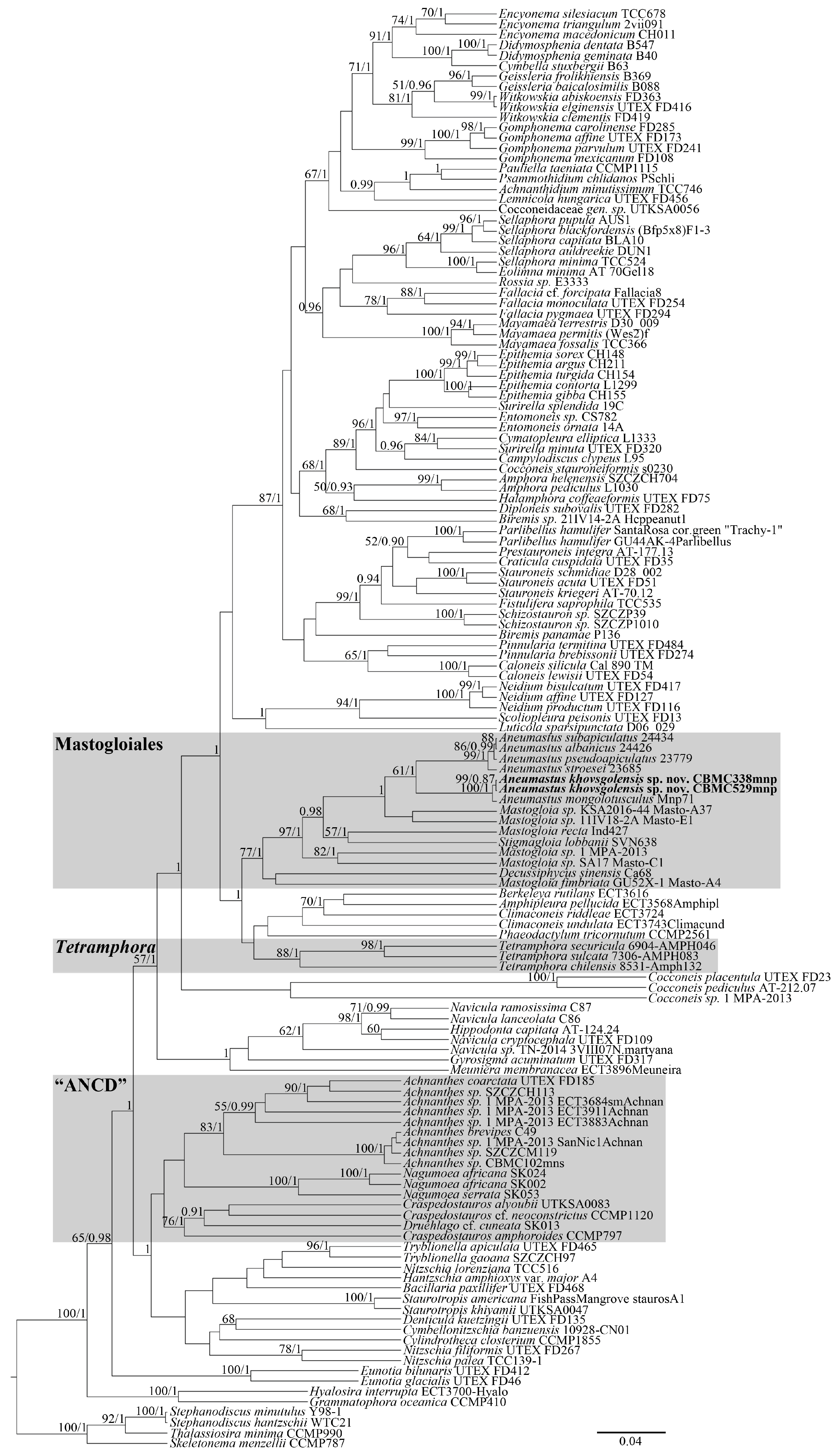
Figure 1. Phylogeny of Mastogloiales sensu Cox and its allies based on BI and ML analyses (rbcL and 18S rRNA genes). Values of LB (from ML analysis) below 50 and values of PP (from BI analysis) below 0.90 are hidden. Strain numbers are indicated for all sequences. GenBank accessions are hidden (see File S1).
Phycology 05 00068 g001
Figure 2. Ultrastructural morphology of Aneumastus mongolotusculus. Slide 02937 from strain Mnp71. (a,b) LM. (c) SEM, external view, pseudoloculi. White arrows show the rectangular external openings of pseudoloculi and black arrows show the triangular canals. (d) SEM, internal view, striae with pseudoloculi. Black arrows show the transition to biseriate arrangement of striae. (e) SEM, internal view on colanderus-bifurcus. White arrows show the two perforated silica flaps and the black arrow shows the silica interruption in between. (fh) SEM, internal view on the broken valves. White arrows show the two perforated silica flaps, the black arrow shows the silica interruption, and asterisks mark the mushroom-shaped formation. (i,j) TEM, internal view on striae. Black arrows show the transition to biseriate arrangement of striae. (k) TEM, internal view on colanderus-bifurcus. White arrows show the two perforated silica flaps and the black arrow shows the silica interruption in between. Scale bars = 10 μm (a,b), 0.5 μm (c), 1 μm (d), 0.2 μm (eh,j,k), 0.5 μm (i).
Phycology 05 00068 g002aPhycology 05 00068 g002b
Figure 3. Ultrastructural morphology of Decussiphycus sinensis. Slide 09153 from strain Ca68. (a,b) LM. (c) SEM, external view, the whole valve. (d) SEM, external view, structure of striae near the central nodule. White arrows show areolae with more or less corroded spongiae. (e) SEM, external view, valve apex. (f) SEM, internal view, the whole valve. (g) SEM, internal view, ultrastructure of areolae. Black arrows show the homogenous flaps of spongiae. (h) TEM, part of the valve. (i) TEM, view on areolae occluded by homogenous spongiae. Note the convexity of spongiae inwards the valve. Scale bars = 10 μm (a,b), 5 μm (c,f), 1 μm (d,e), 0.5 μm (g), 2 μm (h), 0.1 μm (i).
Phycology 05 00068 g003aPhycology 05 00068 g003b
Figure 4. Ultrastructural morphology of Mastogloia recta. Slide 04185 from strain Ind427. (a) LM, focus on the valve. (b) LM, focus on the valvocopula. White arrows show the pseudosepta and black arrows show the partecta. (c) SEM, external view, the whole valve. (d) SEM, internal view, the whole valve. White arrows show the gutter beside the raphosternum and black arrows show the partecta. (e) SEM, external view, central part of the valve. White arrows show the proximal raphe fissures, black arrows show round pseudoloculi, and white arrowheads show the slightly angled pseudoloculi. (f) SEM, external view, valve apex. The black arrow shows the distal raphe fissure and white arrows show the semilunar pseudoloculi. (g,h) SEM, internal view, valve apex. The black arrow shows the pseudoseptum, the white arrow shows the shallow gutter, black arrowheads show the interstriae, white arrowheads show the colanderus-directa, and the asterisk shows the helictoglossa. (i,j) TEM, ultrastructure of pseudoloculi. Black arrows show the silica flaps and white arrows show the perforations of occlusions. Scale bars = 10 μm (a,b), 5 μm (c,d), 2 μm (eg), 1 μm (h), 0.5 μm (i), 0.2 μm (j).
Phycology 05 00068 g004
Figure 5. Ultrastructural morphology of Stigmagloia lobbanii. Slide 09255 from strain SVN638. (a,b) LM. The white arrow shows the stigma. (c) TEM, the whole valve. The white arrow shows the stigma. (d) TEM, structure of striae. Black and white arrowheads show the outer and inner openings of pseudoloculi, respectively. Note that the outer openings are vague due to the focus. (e) Ultrastructure of pseudoloculi. The white arrow shows the stigma and black arrowheads show the oblique pseudolocular canals. (f) SEM, external view, the whole valve. Black arrows show the proximal raphe fissures, black arrowheads show the distal raphe fissures, and the white arrow shows the stigma. (g) SEM, external view, focus on the pseudoloculi. The white arrow shows the stigma and black arrowheads show the oblique pseudolocular canals. (h) SEM, internal view, the whole valve. The white arrow shows the stigma and black arrowheads show the pseudosepta. (i) SEM, internal view, focus on the pseudoloculi. White arrowheads show the perforations of colanderus-obliqua. Scale bars = 10 μm (a,b), 2 μm (c), 1 μm (d), 0.5 μm (e,g,i), 5 μm (f,h).
Phycology 05 00068 g005
Figure 6. Ultrastructural morphology of Achnanthes sp. Slide 03319 from strain CBMC102mns. (a) LM, raphe valve. Black arrows show the fascia. (b) LM, rapheless valve. (c,d) TEM, structure of striae. White arrows show the connections between the nets of cribra and the walls of areolae. (e) Ultrastructure of areolae. Black arrows show the intertwined formations of cribra, white arrows show the connective outgrowths, black arrowheads show the peripheral reniform perforations, and white asterisk shows the roundish perforations. Scale bars = 10 μm (a,b), 1 μm (c), 0.5 μm (d), 0.1 μm (e).
Phycology 05 00068 g006
Figure 7. Aneumastus khovsgolensis sp. nov. Slide 09488 from sample Mon59. LM. (ae) Size diminution series. Scale bar = 10 μm.
Phycology 05 00068 g007
Figure 8. Aneumastus khovsgolensis sp. nov. Slide 10206 from strain CBMC529mnp. LM. (ah) Clone series. (c) Holotype specimen. Scale bar = 10 μm.
Phycology 05 00068 g008aPhycology 05 00068 g008b
Figure 9. Aneumastus khovsgolensis sp. nov. Slide 10206 from strain CBMC529mnp. SEM. (a,b) The whole valve, externally. (c) Central area, externally. Black arrows show the proximal raphe fissures and arrowheads show the X-shaped (white) and bone-shaped (black) openings of pseudoloculi. (d) Valve end, externally. The black arrow shows the distal raphe fissure, white arrows show the semilunar-shaped openings of pseudoloculi, and black arrowheads show the biseriate striae. (e) The whole valve, internally. (f) Central area, internally. Black arrows show the proximal raphe fissures and white arrowheads show openings of pseudoloculi adjacent to the axial area. (g) Valve margin, internally. White arrowheads show the biseriate striae. (h) Valve end, internally. The black arrow shows the distal raphe fissure with helictoglossa and the white arrow shows the pseudoseptum. Scale bars = 10 μm (a,b,e), 1 μm (c,fh), 2 μm (d).
Phycology 05 00068 g009aPhycology 05 00068 g009b
Figure 10. Aneumastus khovsgolensis sp. nov. Slide 09487 from sample Mon58. LM. (ae) Size diminution series. Scale bar = 10 μm.
Phycology 05 00068 g010
Figure 11. Aneumastus khovsgolensis sp. nov. Slide 10105 from strain CBMC338mnp. LM. (al) Clone series. Scale bar = 10 μm.
Phycology 05 00068 g011
Figure 12. Aneumastus khovsgolensis sp. nov. Slide 10105 from strain CBMC338mnp. SEM, external view. (a,b) The whole valve, valve face view. Note the corrosion of pseudoloculi (black arrows). (c) The whole valve, semi-girdle view. The black arrow shows the intercalary band, white arrows show the valvocopulae, and black arrowheads show the perforations at the intercalary band. (d) Middle part of the valve, semi-girdle view. The black arrowheads show the biseriate striae and white arrowheads show the perforations at the valvocopula. (e) Central area. Black arrows show the proximal raphe fissures and arrowheads show the X-shaped (white) and bone-shaped (black) openings of pseudoloculi. (f) Valve end. The black arrow shows the distal raphe fissure, white arrows show the semilunar-shaped openings of pseudoloculi, and black arrowheads show the biseriate striae. Scale bars = 10 μm (a,c), 5 μm (b), 1 μm (d,f), 2 μm (e).
Phycology 05 00068 g012aPhycology 05 00068 g012b
Figure 13. Aneumastus khovsgolensis sp. nov. Slide 10105 from strain CBMC338mnp. SEM, internal view. (a,b) The whole valve. (c) Central area. Black arrows show the proximal raphe fissures, white arrows show the silica interruption, and black arrowheads show the two inner openings of the same pseudolocula. Altogether, the two openings and the interruption form a colanderus-bifurcus. (d) Valve margin, white arrowheads show the biseriate striae. (e,f) Valve end. The black arrow shows the distal raphe fissure with helictoglossa and white arrows show the pseudosepta. Scale bars = 10 μm (a), 5 μm (b), 1 μm (c,e,f), 0.5 μm (d).
Phycology 05 00068 g013aPhycology 05 00068 g013b
Table 1. List of studied samples.
Samples from Fresh Waterbodies
Sample №Slide №Strain №LocalityCoordinatesSubstratumt., °CpHµS/cmDate of Collection
Mon5809487 (wild), 10105 (strain)CBMC338mnpSoutheastern shore of Lake Khovsgol, Khovsgol aimak, Mongolia50.62634 °N, 100.50109 °EBenthos17.18.3024824 July 2024
Mon5909488 (wild), 10206 (strain)CBMC529mnpSoutheastern shore of Lake Khovsgol, Khovsgol aimak, Mongolia50.62592 °N, 100.50087 °EEpilithon, washing from silty stones17.68.5121024 July 2024
Mn26702937Mnp71Eastern shore of Lake Khovsgol, Khovsgol aimak, Mongolia50.80500 °N, 100.43972 °EBenthos219.5417819 July 2015
THHN 201404309153Ca68Unnamed stream, northern slope Wuzhishan Mountain, Hainan Province, China18.98150 °N, 109.68540 °EEpilithon, washing from silty stones26.77.646012 July 2014
I27704185Ind427Mahalona River, Sulawesi Island, Indonesia2.65694 °S, 121.52957 °EBenthos18.88.7918424 September 2015
Samples from saline waterbodies
Sample №Slide №Strain №LocalityCoordinatesSubstratumt., °CSalinity, ‰Date of collection
Mn18303319CBMC102mnsNortheastern shore of Lake Oigon, Zavkhan aimak, Mongolia49.21344 °N, 96.64444 °EPhytoplankton23.33914 July 2015
NTs6509255SVN638Shore of South China Sea, Khánh Hòa Province, Nha Trang, Vietnam12.20755 °N, 109.21541 °EEpilithon29.2343 April 2018
Table 2. Comparison of valve dimensions in wild and cultured populations of A. khovsgolensis sp. nov.
Type of PopulationSampleSlideLengthWidthStriae (/10 μm)Uniseriate Pseudoloculi (/10 μm)
Wild
Wild
Cultured
Mon580948737.6–62.015.8–19.4128
Mon590948843.5–63.817.0–20.3128
Mon581010541.6–43.716.5–17.312–138
CulturedMon591020668.5–72.420.3–21.0128
Table 3. Comparison Aneumastus khovsgolensis sp. nov. to most similar species.
A. khovsgolensis sp. nov.A. tusculus (Type)A. mongolotusculus
Valve outlineLinear-elliptic to elliptic, abruptly narrowing towards the apicesBroadly linear-elliptic, gradually narrowing towards the apicesBroadly linear-elliptic
Morphology of apicesObtusely rostrateMore or less rostrate, shoulder-likeAbruptly protracted, more or less capitate
Valve apices width, µm3.2–3.83.9–4.5 *2.9–3.2
Valve length, µm37.6–72.451–67 *42–61
Valve width, µm15.8–21.019–22 *16–19
Striae density, in 10 µm12–1310 *11–12
ReferenceThis study[4][29]
* Counted from referenced source.
Disclaimer/Publisher’s Note: The statements, opinions and data contained in all publications are solely those of the individual author(s) and contributor(s) and not of MDPI and/or the editor(s). MDPI and/or the editor(s) disclaim responsibility for any injury to people or property resulting from any ideas, methods, instructions or products referred to in the content.