Article

Effects of Artesunate on the Growth and Chlorophyll Fluorescence of the Cyanobacterium Microcystis aeruginosa

1
Chongqing Landscape and Gardening Research Institute, Chongqing 401329, China
2
Chongqing Institute of Geology and Mineral Resources, Chongqing 401120, China
3
Chongqing Key Laboratory of Germplasm Innovation and Utilization of Native Plants, Chongqing 401120, China
4
College of Life Science, Linyi University, Linyi 276000, China
*
Author to whom correspondence should be addressed.
These authors contributed equally to this work.
Phycology 2025, 5(4), 63; https://doi.org/10.3390/phycology5040063
Submission received: 30 July 2025 / Revised: 23 September 2025 / Accepted: 24 September 2025 / Published: 20 October 2025
(This article belongs to the Collection Harmful Microalgae)

Abstract

Microcystis aeruginosa, a kind of cyanobacterium, can lead to water blooms under specific conditions and it is harmful to human and ecological security due to the toxins produced by certain strains. Artemisinin, which is derived from Artemisia annua Linn, has a strong allelopathic effect on algae. Artesunate is a water-soluble derivative of artemisinin. We investigated the effect of artesunate on M. aeruginosa, including growth and key photosynthetic parameters (Fv/Fm, φPSII). Our findings demonstrate that artesunate inhibits the growth of M. aeruginosa by damaging the photosynthetic center of photosystem II (PS II), and this inhibitory effect is enhanced with increasing concentration. At the concentration of 200 mol/L, the maximum inhibition rate was 41.62% for FACHB-315 and 43.19% for FACHB-927 after 96 h. After 24 h of exposure, the φPSII of the two strains decreased significantly (p < 0.01). These results could inform further studies on the use of artesunate to control cyanobacterial growth in water bodies and provide theoretical support for the application of artemisinin derivatives in treating water blooms.

1. Introduction

While cyanobacteria play a crucial role in aquatic ecosystems, the incidence of harmful algal blooms involving cyanobacteria (cyanoHABs) has risen markedly worldwide over recent decades. Driven by ongoing eutrophication, elevated atmospheric CO2 levels, and global warming, these blooms are predicted to increase in frequency and intensity. Although numerous attempts have been made to control or prevent cyanoHABs, most interventions have proven either ineffective or only temporarily successful [1]. In freshwater systems—including lakes, ponds and rivers, Microcystis aeruginosa has emerged as the predominant species responsible for cyanoHABs since the 1980s [1,2,3]. Even with continuous efforts, effective management of Microcystis blooms remains challenging. Physical methods to eliminate Microcystis are generally associated with high operational costs, whereas chemical methods may lead to secondary environmental contamination due to the persistent toxic chemicals [4,5] Consequently, there is growing scientific interest in biological control methods as a sustainable and environmentally benign alternative to mitigate the adverse effects of cyanoHABs [4,6,7].
Artemisinin, a colorless crystalline compound, is derived from the aerial parts of Artemisia annua Linn with significant antimalarial properties [8,9,10]. Artemisinin and its derivatives demonstrate high efficacy against Plasmodium, with an inhibition rate of 100% [11]. Notably, these compounds also exert a strong allelopathic effect on algae, with the inhibition rate exceeding 90% for M.aeruginosa [12]. Derivatives such as dihydroartemisinin inhibit the growth of M.aeruginosa by destroying the photosynthetic reaction center of photosynthetic system II [13]. Allelopathy—defined as the biochemical inhibition of one organism by another through the release of secondary metabolites—plays a key role in plant defense and interspecies competition within ecological communities. Extensive research over the past few decades has revealed that artemisinin and its derivatives exhibit broad-spectrum inhibitory activity across diverse taxa, including bacteria, fungi, insects, plants, and particularly algae. A substantial body of evidence highlights the potent allelopathic (or toxic) effects of artemisinin and its derivatives, primarily documented in relation to insects and plants, as summarized in Table 1.
However, as a water-soluble derivative of artemisinin, the ecological and practical implications of artesunate’s algicidal effects remain inadequately understood. While its capacity to eliminate or inhibit cyanobacterial growth is well established, there exists a critical gap in our knowledge regarding its influence on microcystin production and the comparative susceptibility of toxigenic versus non-toxigenic strains of species such as M. aeruginosa.
Microcystins are potent hepatotoxins produced by certain cyanobacterial strains, posing significant risks to aquatic life, livestock, wildlife and human health through contaminated water supplies. The stress induced by algicidal agents like artemisinin can paradoxically stimulate increased toxin production (e.g., as a defense mechanism or due to cell lysis that releases intracellular toxins) in surviving toxigenic cells prior to any substantial population decline, potentially exacerbating the toxin burden in the water. Conversely, it may selectively suppress toxigenic strains.
Understanding whether exposure to artesunate results in elevated microcystin levels, favors the proliferation of toxigenic strains, or differentially affects the growth and survival rates of toxigenic versus non-toxigenic phenotypes is crucial for assessing its environmental safety and suitability as an agent for bloom control. This introduction explores the established breadth of artemisinin’s allelopathic activity, while highlighting its particularly strong effects on cyanobacteria such as M. aeruginosa. It also underscores a significant research gap concerning the differential impacts of artemisinin exposure on toxin production and resistance among toxigenic versus non-toxigenic algal strains.
The aims of the present study were (1) to examine the algicidal capability of artesunate, (2) to clarify the algicidal mode of chlorophyll fluorescence parameter variations in M. aeruginosa, and (3) to provide potential ideas and methods for the prevention and rapid emergency management of cyanobacterial blooms.

2. Materials and Methods

2.1. Algae Cultures

M. aeruginosa (toxigenic strain FACHB-315 and non-toxigenic strain FACHB-927) were purchased from the Freshwater Algae Culture Collection, Institute of Hydrobiology, Chinese Academy of Sciences (Wuhan, China) and cultivated using BG11 medium in 1 L autoclaved conical flasks. When cells are in the exponential growth phase, they usually have a good state. Therefore, we establish the standard curve for cell density by measuring the OD value at a wavelength of 680 nm using the optical density method. Then, in this study, cells in the exponential growth period were determined by counting 1 mL samples in triplicate fixed with 2% (v/v) Lugol’s solution. All the reagents and solvents were of at least analytical grade except as noted. All the flasks and the culture medium were sterilized at 121 °C for 30 min. The flasks were incubated at 25 ± 1 °C under a 12 h light:12 h dark photoperiod with irradiance of 58 μmol photons∙m−2∙s−1 supplied by white fluorescent lights.

2.2. Experimental Conditions

M. aeruginosa in the exponential growth phase was harvested and cultured in 300 mL BG11 medium in 500 mL flasks. Various doses of artesunate (obtained from Shanghai Jingke Industrial Co., Ltd., Shanghai, China) were added to the cultures at concentrations of 0, 10, 50, 100, and 200 μmol/L. The selection of concentration is based on previous preliminary experiments and a series of studies on the physiological effects of artesunate concentration on M. aeruginosa [12,26]. Controls were prepared by inoculating M. aeruginosa into culture medium without the addition of artesunate. Three replicates were made in the experiment. The BG11 medium, pipettes and conical flasks used in the experiment were sterilized by high-pressure steam. Before using them on the clean workbench, they were irradiated with ultraviolet rays for 30 min. Algae inoculation and other operations were completed inside the ultra-clean workbench to ensure that all the operations were carried out in axenic conditions. Measurements were carried out by a water chlorophyll fluorescence meter (AquaPen-C 100, Photon Systems Instruments (PSI), Brno, The Czech Republic) at 0, 12, 24, 48, 72, and 96 h after the onset of different treatments. In chlorophyll fluorescence parameters, the maximum effective quantum yield of photosystem II (PSII) (Fv/Fm) was monitored when M. aeruginosa was incubated with artesunate at 12, 24, 48, 72, and 96 h.

2.3. Measurements of Cell Density

Cell density was determined by measuring the optical density at 680 nm (OD680) with a visible spectrophotometer (722S; Shanghai Jingke Industrial Co., Ltd., Shanghai, China) to represent relative cyanobacteria biomass. The inhibition ratio (IR) was used to express the effects of artesunate, and its calculation is as follows:
IR (%) = (1 − T/C) × 100%
T and C: Cell density of the treatment group and control.

2.4. Determination of Chlorophyll Fluorescence Parameters

Fv/Fm represents the efficiency of the primary conversion efficiency of light energy of PSII or the potential activity of PSII. Fv/Fm and φPSII were measured by the FMS-2 chlorophyll fluorescence meter (Fv/Fm is the maximum photochemical efficiency when the PSII reaction center is completely open under dark adaptation, which reflects the maximum light energy conversion efficiency of the PSII reaction center. φPSII reflects the actual photochemical efficiency of the PSII reaction center when it is partially closed under light. Every 24 h, the fluorescence parameters of each group were measured and plotted. In this study, the most representative photosynthetic parameters for photosystem II (Fv/Fm, φPSII) were used.

2.5. Statistical Analysis

The software SPSS Statistics 22 (SPSS, IBM, New York, NY, USA) was used to perform one-way analysis of variance (ANOVA) to determine statistical significance, with the significance level of 0.05 and 0.01. Means were separated by conducting Duncan’s multiple range test, where there was a significant observable difference. Data shown in this study were presented as means ± standard deviation (SD). All figures were drawn using Origin 9.0. Detailed methods: the data of each group at 0, 12, 24, 48, 72, and 96 h were analyzed. Firstly, variables were input by column in SPSS software, and one-way ANOVA was performed for mean comparison, and the mean and standard deviation were output in descriptive statistics. Then set the significance level to 0.05 and 0.01 in the options. Subsequently, the mean and error bar data output by SPSS were saved to Excel. Import the data to Origin 9.0, select the chart type for plotting, and add error bars.

3. Results

3.1. Effects of Artesunate on M. aeruginosa Growth

The changes in OD value of M. aeruginosa at each concentration level of artesunate are shown in Figure 1. The cell number of the control group exhibited a substantial increase, indicating that cells were undergoing exponential growth during the experiment. Specifically, exposure to artesunate inhibited cell growth in both FACHB-315 and FACHB-927. The inhibition rate reached its peak after approximately 96 h of exposure to 200 μmol/L artesunate, reaching 41.62% for FACHB-315 and 43.19% for FACHB-927. At the concentrations of 100 and 200 μmol/L, significant inhibition of cell density was observed in FACHB-315 just 24 h after the artesunate treatment initiation. Furthermore, as treatment duration increased, the inhibitory effects intensified. In contrast, cell growth in FACHB-927 was only inhibited at a concentration of 200 μmol/L. The results suggest that exposure time is a critical factor in sustaining the inhibitory effects on M. aeruginosa growth. Notably, the non-toxigenic strain FACHB-927 demonstrated greater resistance to artesunate compared to the toxigenic strain FACHB-315. Similarly, studies exist on other factors, such as amoxicillin and sulfonamide antibiotics affecting M. aeruginosa, their research indicated that amoxicillin was more toxic to the toxin-producing strains than to non-toxin-producing strains [27,28].

3.2. Effects of Artesunate on Chlorophyll Fluorescence Parameters

3.2.1. Effects of Artesunate on Fv/Fm

Our study utilized the most representative photosynthetic parameters for photosystem II (Fv/Fm and φPSII). The parameter Fv/Fm reflects the maximum light energy conversion efficiency of PSII, and it remains relatively stable under non-stress conditions but significantly decreases under stress conditions [29,30]. Therefore, by measuring Fv/Fm values, we can assess the photosynthesis conditions of M. aeruginosa.
The effects of different concentrations of artesunate on the values of Fv/Fm in M. aeruginosa after treatment were investigated. The results are shown in Figure 2. Exposure to varying concentrations of artesunate resulted in significant inhibition of the Fv/Fm parameters across all tested cases. In each case, artesunate resulted in significant inhibition (p < 0.01), and the inhibition effects were more obvious with exposure time. Specifically, the level of inhibition was enhanced as the concentration of artesunate increased, reaching its most notable effect at the highest concentration tested (200 μmol/L) for both FACHB-315 and FACHB-927. The maximum quantum yield of photosystem II (Fv/Fm) exhibited a significant decrease (p < 0.01) after 96 h of exposure to 200 μmol/L, dropping from 0.654 to 0.424 in FACHB-315 and from 1.03 to 0.459 in FACHB-927, respectively. Concentrations of artesunate ranging from 50 to 100 μmol/L demonstrated similar inhibitory effects on Fv/Fm in both FACHB-315 and FACHB-927.

3.2.2. Change in φPSII

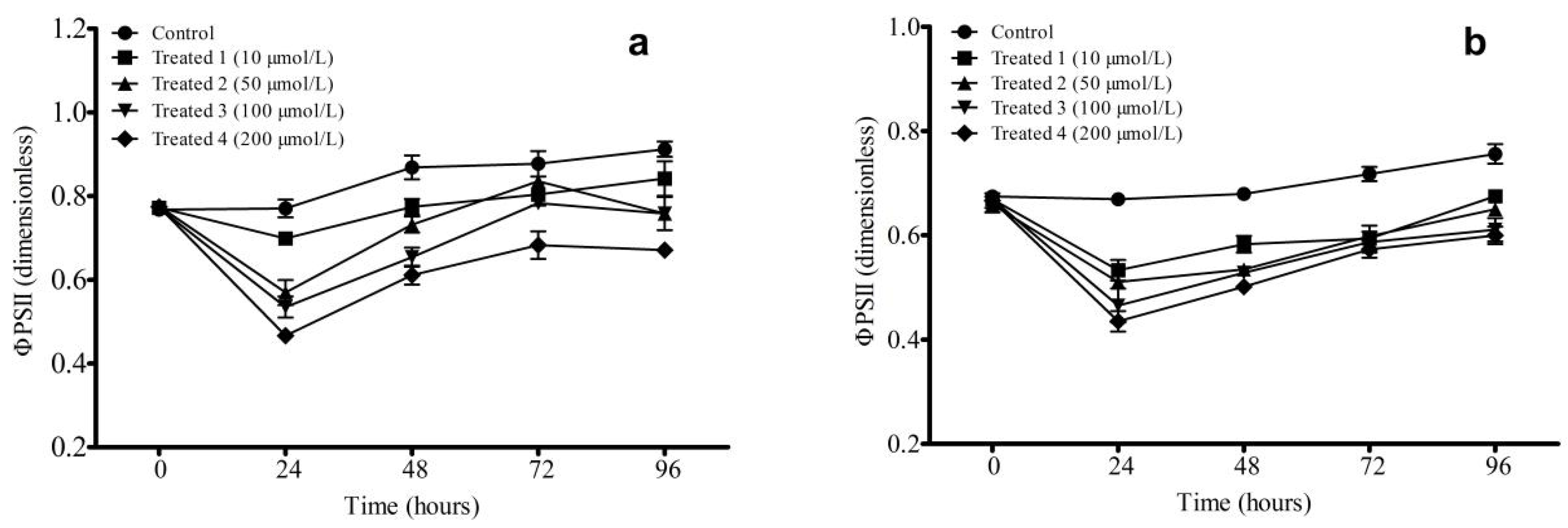
The variable φPSII represents the quantum efficiency of photosynthetic electron transfer. Figure 3 illustrates the impact of artesunate on φPSII when conducted at different concentrations. As depicted in Figure 3a,b, the φPSII for all treatments rapidly decreased following exposure to artesunate, but gradually levels up with extended exposure time. The maximum φPSII decreased as artesunate concentration increased. Figure 3a,b further indicate that φPSII levels among groups treated with artesunate experienced a rapid decline within the first 24 h post-exposure, and then gradually levels up with increasing exposure time from 24 h to 72 h. At 96 h, the value of φPSII decreases with the increase in artesunate concentration. The minimum observed value for φPSII occurred at 24 h for both FACHB-315 and FACHB-927, indicating that artesunate exerts an influence on the cyanobacterium M. aeruginosa. Moreover, the higher the concentration, the more obvious the inhibition effect. Although all treatment groups displayed lower φPSII values compared to control throughout experimentation, there was a gradual increase noted correlating with prolonged culture times. Further investigations are warranted through additional experiments to ascertain whether artesunate affects other microalgal species similarly.

4. Discussion

4.1. Differential Inhibition of Toxigenic and Non-Toxigenic Strains

Our research findings demonstrate that artesunate significantly inhibits the growth of M. aeruginosa in a concentration- and time-dependent manner. At concentrations exceeding 100 μmol/L, artesunate strongly suppressed the cell density of M. aeruginosa FACHB-315 (a toxigenic strain), whereas its impact on the non-toxigenic strain FACHB-927 was less marked. Cyanobacteria are capable of synthesizing diverse secondary metabolites, of which peptides account for approximately 65% [31]. Among these, microcystins, which are cyclic heptapeptides produced by toxic strains of M. aeruginosa, are significant secondary metabolites. Microcystins are recognized for their influence on interspecies competition and stress responses, potentially modulating the sensitivity of cyanobacteria to external inhibitors. Previous studies have shown that algal toxins might play a role in modulating the activity of antioxidant enzymes such as superoxide dismutase [32,33]. Upon encountering M. aeruginosa, artesunate can initiate the production of reactive oxygen species (ROS) within cyanobacteria, such as superoxide anions, hydroxyl radicals, hydrogen peroxide, and nitric oxide. This process leads to oxidative stress, which in turn causes membrane lipid peroxidation, protein oxidation, enzyme inactivation, and nucleic acid destruction. At the same time, artesunate may also interfere with the cell division, energy conversion, and signal transduction pathways in M. aeruginosa, thereby slowing down the rate of cell growth. In the future, it is very necessary to delve deeper into the toxicity mechanism of microalgae exposed to artesunate using omics techniques.
Additionally, the resistance variability among different algal species to artemisinin and its derivatives has been documented. Researcheres found that M. aeruginosa displayed differing levels of susceptibility to artemisinin, with inhibition rates exceeding 90% under certain conditions [12]. In their study, Ni et al. not only used pure artemisinin but also used artemisinin sustained-release granules, with a concentration range from 1 to 20 g/L. In our study, the maximum concentration of artesunate was 200 μmol/L. Their concentration is significantly higher than that observed in our experiment, so their inhibition rate is higher. A comparable observation was made, they reported that dihydroartemisinin (DHA) effectively inhibits M. aeruginosa by impairing Photosystem II (PSII), a critical component of the photosynthetic apparatus [13]. These findings suggest that the algicidal properties of artemisinin derivatives are strain-specific and influenced by the physiological and biochemical characteristics of the target organisms.

4.2. Time-Dependent Weakening of Inhibitory Effects

Our study revealed that artesunate caused more significant inhibitions, with the effects becoming more pronounced as the exposure time increased. Time effect has also been verified in other physiological experiments, indicating the existence of toxicity-time relationships. For example, the growth inhibition of metals and organic matter can be more effectively mitigated over time, reinforcing the idea that the duration of exposure plays a critical role in the efficacy of inhibitory agents [34,35]. However, the φPSII of groups treated with artesunate experiences a swift decline within 24 h of exposure, followed by a gradual increase with increasing exposure time from 24 h to 72 h. A consistent finding across various studies is the transient nature of artemisinin’s algicidal activity. While initial exposure leads to rapid growth inhibition, the effect weakens over time. Several mechanisms could account for this phenomenon. The first potential explanation is the degradation and transformation of active compounds. Artemisinin and its derivatives are prone to environmental degradation, especially under light and microbial activity [36] In aquatic environments, the gradual breakdown of artesunate reduces its bioavailability and lessens its inhibitory effect over time. The second explanation may be the metabolic adaptation and recovery of algal. Cyanobacteria have strong stress-response mechanisms such as enhanced antioxidant defenses and DNA repair pathways. Chia et al. (2016) proposed that M. aeruginosa can recover from the adverse effects of artemisinin by upregulating protein biosynthesis and repairing damaged cellular components. The presence of oxidative stress and the fluctuating levels of (ROS) are crucial factors in this process [37]. Artemisinin produces ROS, initially causing oxidative damage to chloroplasts and other organelles [38]. However, prolonged exposure can trigger the activation of algal antioxidant systems (e.g., superoxide dismutase and catalase), which mitigate ROS-induced damage and enable a partial recovery of photosynthetic activity.

4.3. May Mechanisms of Artemisinin’s Algicidal Action

The inhibitory effects of artesunate on M. aeruginosa may be mediated by multiple pathways. The most well-documented mechanism is likely the impairment of PSII, which results in decreased Fv/Fm value and compromised energy production [13]. In particular, DHA, which binds to the D1 protein in PSII, inhibiting electron transport and causing photoinhibition. Artesunate may destroy the stability of the plasma membrane and intracellular organelles, including mitochondria and endoplasmic reticulum. Wang et al. (2015) proposed that heme iron, a by-product of hemoglobin digestion in malarial parasites, activates artemisinin in a similar way in cyanobacteria, leading to iron-dependent free radical formation and subsequent oxidative damage [39]. Additionally, the interactions of allelopathy may also serve as an important explanation mechanism. Artemisinin exhibits allelopathic properties, suppressing competing phytoplankton species. Jessing et al. (2009) demonstrated its broad-spectrum toxicity against green algae (Pseudokirchneriella subcapitata) and duckweed (Lemna minor), with EC50 values as low as 0.19–0.24 mg/L [25]. Dihydroartemisinin (DHA) induces oxidative stress mainly by producing ROS, thus interfering with the structure and function of cell membranes. Theoretically, compared with the non-toxigenic strain FACHB-927, the toxigenic strain FACHB-315 of M. aeruginosa may have a stronger antioxidant defense system and higher tolerance to oxidative stress induced by DHA. Non-toxigenic strains may be more susceptible to oxidative damage from DHA, leading to more pronounced growth inhibition. However, similar to other environmental stressors, artesunate may stimulate the production of strains to increase the synthesis and release of microcystins, and this response may consume a large amount of energy and metabolic resources. Non-toxigenic strains do not show this metabolic redistribution due to they lack the ability to produce toxin. From Figure 2, we can also find that the decrease in photosynthetic activity (Fv/Fm) of the toxigenic strain is smaller than that of the non-toxigenic strain, indicating that the expression of photosynthesis-related genes in the non-toxigenic strain is more easily affected. Although artemisinin derivatives are expected to be natural algaecides, their application in large-scale water bodies needs to be carefully considered.

5. Conclusions

Our study shows that artesunate may have potential as an algicide, as it appears to reduce the Fv/Fm value and inhibit the growth of M. aeruginosa to some extent. For the toxigenic strain, artesunate concentration about twice as high as that of non-toxic strains is needed to achieve the same OD inhibition. For two strains, the effect of reducing quantum efficiency fades after 96 h. Moreover, the short-term artesunate stress has a stronger inhibitory effect on the toxic strain FACHB-315 than the non-toxic strain FACHB-927, and its underlying mechanism is unclear. However, before its application, it is necessary to further study the feasibility and the impact on whole aquatic ecosystems.

Author Contributions

Conceptualization, H.W. and A.Y.; methodology, H.W.; software, H.W.; validation, W.W., Y.H. and W.N.; formal analysis, W.N.; investigation, W.N.; resources, A.Y.; data curation, A.Y.; writing—original draft preparation, H.W.; writing—review and editing, A.Y.; visualization, W.N.; supervision, A.Y.; project administration, H.W.; funding acquisition, A.Y. All authors have read and agreed to the published version of the manuscript.

Funding

This research was funded by the Program for Shandong Provincial Natural Science Foundation of China (ZR2018PC003), the Scientific Research Project of Chongqing City Administration Bureau (No. CGK 2024-12, No. CGK 2024-13), the Youth Innovation Team Plan of Colleges and Universities in Shandong Province (2024KJG059), and the Performance Incentive and Guidance Special Project for Chongqing Scientific Research Institutes (CSTB2023JXJL-YFX0062). The APC was funded by No. CGK 2024-12.

Data Availability Statement

Research data acquisition can be obtained by directly contacting the first author or the corresponding author.

Acknowledgments

We gratefully acknowledge the above financial support. We wish to thank the participants and volunteers and express special thanks to the manuscript review expert and journal editors in our study.

Conflicts of Interest

The authors declare no conflicts of interest.

References

  1. Huisman, J.; Codd, G.A.; Paerl, H.W.; Ibelings, B.W.; Verspagen, J.M.H.; Visser, P.M. Cyanobacterial blooms. Nat. Rev. Microbiol. 2018, 16, 471–483. [Google Scholar] [CrossRef]
  2. Ho, J.C.; Michalak, A.M.; Pahlevan, N. Widespread global increase in intense lake phytoplankton blooms since the 1980s. Nature 2019, 574, 667–670. [Google Scholar] [CrossRef]
  3. Paerl, H.W.; Barnard, M.A. Mitigating the global expansion of harmful cyanobacterial blooms: Moving targets in a human-and climatically-altered world. Harmful Algae 2020, 96, 101845. [Google Scholar] [CrossRef] [PubMed]
  4. Kibuye, F.A.; Zamyadi, A.; Wert, E.C. A critical review on operation and performance of source water control strategies for cyanobacterial blooms: Part I- chemical control methods. Harmful Algae 2021, 109, 102099. [Google Scholar] [CrossRef] [PubMed]
  5. Han, Z.X.; Xu, C.; Shu, W.Q. Progress of research on potential chemoprotectants against microcystins. J. Environ. Health 2010, 27, 169–171. [Google Scholar]
  6. Pal, M.; Yesankar, P.J.; Dwivedi, A.; Qureshi, A. Biotic control of harmful algal blooms (HABs): Abrief review. J. Environ. Manag. 2020, 268, 110687. [Google Scholar] [CrossRef] [PubMed]
  7. Song, L.; Jia, Y.; Qin, B.; Li, R.; Carmichael, W.W.; Gan, N.; Xu, H.; Shan, K.; Sukenik, A. Harmful cyanobacterial blooms: Biological traits, mechanisms, risks, and control strategies. Annu. Rev. Environ. Resour. 2023, 48, 123–147. [Google Scholar] [CrossRef]
  8. Klayman, D.L. Qinghaosu (artemisinin): An antimalarial drug from China. Science 1985, 228, 1049–1055. [Google Scholar] [CrossRef]
  9. Tu, Y.Y. The discovery of artemisinin (qinghaosu) and gifts from Chinese medicine. Nat. Med. 2011, 17, 1217–1220. [Google Scholar] [CrossRef]
  10. Zhang, T.J.; Wang, Y.F.; Liu, D. Historical story on natural medicinal chemistry: Artemisinin-a milestone of traditional Chinese medicine study. Chin. Tradit. Herb. Drugs 2016, 19, 3351–3361. [Google Scholar]
  11. Portugaliza, H.P.; Miyazaki, S.; Geurten, F.J.A.; Pell, C.; Rosanas, U.A.; Janse, C.J.; Cortés, A. Artemisinin exposure at the ring or trophozoite stage impacts Plasmodium falciparum sexual conversion differently. Elife 2020, 9, e60058. [Google Scholar] [CrossRef] [PubMed]
  12. Ni, L.; Li, D.; Hu, S. Effects of artemisinin sustained-release granules on mixed alga growth and microcystins production and release. Environ. Sci. Pollut. Res. 2015, 22, 18637–18644. [Google Scholar] [CrossRef]
  13. Wang, S.; Xu, Z. Effects of dihydroartemisinin and artemether on the growth, chlorophyll fluorescence, and extracellular alkaline phosphatase activity of the cyanobacterium Microcystis aeruginosa. PLoS ONE 2016, 11, e0164178. [Google Scholar] [CrossRef]
  14. Brisibe, E.A.; Adugbo, S.E.; Ekanem, U.; Brisibe, F.; Figueira, G.M. Controlling bruchid pests of stored cowpea seeds with dried leaves of Artemisia annua and two other common botanicals. Afr. J. Biotechnol. 2011, 10, 9586–9592. [Google Scholar] [CrossRef]
  15. Khosravi, R.; Sendi, J.J.; Ghadamyari, M.; Yezdani, E. Effect of sweet wormwood Artemisia annua crude leaf extracts on some biological and physiological characteristics of the lesser mulberry pyralid Glyphodes pyloalis. J. Insect Sci. 2011, 11, 156. [Google Scholar] [CrossRef]
  16. Zibaee, A.; Bandani, A.R. A study on the toxicity of a medical plant, Artemisia annua L. (Asteracea) extracts to the sunn pest, Eurygaster integriceps puton (Hemiptera scutelleridae). J. Plant Prot. Res. 2010, 50, 79–85. [Google Scholar] [CrossRef]
  17. Shekari, M.; Sendi, J.J.; Etebari, K.; Zibaee, A.; Shadparvar, A. Effects of Artemisia annua L. (Asteracea) on nutritional physiology and enzyme activities of elm leaf beetle, Xanthogaleruca luteola Mull. (Coleoptera: Chrysomellidae). Pestic. Biochem. Physiol. 2008, 91, 66–74. [Google Scholar] [CrossRef]
  18. Hasheminia, S.M.; Sendi, J.J.; Jahromi, K.T.; Moharramipour, S. The effects of Artemisia annua L. and Achillea millefolium L. crude leaf extracts on the toxicity, development, feeding efficiency and chemical activities of small cabbage Pieris rapae L. (Lepidoptera: Pieridae). Pestic. Biochem. Physiol. 2011, 99, 244–249. [Google Scholar] [CrossRef]
  19. Maggi, M.E.; Mangeaud, A.; Carpinella, M.C.; Ferrayoli, C.G.; Valladares, G.R.; Palacios, S.M. Laboratory evaluation of Artemisia annua L. extract and artemisinin activity against Epilachna paenulata and Spodoptera eridania. J. Chem. Ecol. 2005, 31, 1527–1536. [Google Scholar] [CrossRef] [PubMed]
  20. Durden, K.; Sellars, S.; Cowell, B.; Brown, J.J.; Pszczolkowski, M.A. Artemisia annua extracts, artemisinin and 1,8-cineole, prevent fruit infestation by a major, cosmopolitan pest of apples. Pharm. Biol. 2011, 49, 563–568. [Google Scholar] [CrossRef]
  21. Duke, S.O.; Vaughn, K.C.; Croom, E.M.; ElSohly, H.N. Artemisinin, a constituent of annual wormwood (Artemisia annua), is a selective phytotoxin. Weed Sci. 1987, 35, 499–505. [Google Scholar] [CrossRef]
  22. Lydon, J.; Teasdale, J.R.; Chen, P.K. Allelopathic activity of annual wormwood (Artemisia annua) and the role of artemisinin. Weed Sci. 1997, 45, 807–811. [Google Scholar] [CrossRef]
  23. Delabays, N.; Slacanin, I.; Bohren, C. Herbicidal potential of artemisinin and allelopathic properties of Artemisia annua L: From the laboratory to the field. J. Plant Dis. Protect. 2008, 21, 317–322. [Google Scholar]
  24. Panamanik, R.C.; Chikkaswamy, B.K.; Roy, D.G.; Panamanik, A.; Kumar, V. Effects of biochemicals of Artemisia annua in plants. J. Phytol. Res. 2008, 21, 11–18. [Google Scholar]
  25. Jessing, K.K.; Cedergreen, N.; Jensen, J.; Hansen, H.C. Degradation and ecotoxicity of the biomedical drug artemisinin in soil. Environ. Toxicol. Chem. 2009, 28, 701–710. [Google Scholar] [CrossRef]
  26. Ni, L.; Acharya, K.; Ren, G. Preparation and characterization of anti-algal sustained-release granules and their inhibitory effects on algae. Chemosphere 2013, 91, 608–615. [Google Scholar] [CrossRef] [PubMed]
  27. Yisa, A.G.; Chia, M.A.; Sha’aba, R.I.; Gauje, B.; Gadzama, I.M.K.; Oniye, S.J. Risk assessment of the antibiotic amoxicillin on non-toxin-producing strains and toxin-producing strains of Microcystis. Environ. Sci. Pollut. Res. 2023, 30, 56398–56409. [Google Scholar] [CrossRef] [PubMed]
  28. Chen, J.X.; Qin, B.; Han, Y.H.; Yang, S.Y. Sulfamethoxazole and sulfaisoxazole stress on growth, photosynthesis, cell structure and oxidation system in Microcystis aeruginosa. Algal Res. 2025, 87, 103880. [Google Scholar] [CrossRef]
  29. Endo, H.; Moriyama, H.; Okumura, Y. Photoinhibition and photoprotective responses of a brown marine macroalga acclimated to different light and nutrient regimes. Antioxidants 2023, 12, 357. [Google Scholar] [CrossRef]
  30. Lin, S.Y.; Chen, P.A.; Zhuang, B.W. The stomatal conductance and Fv/Fm as the indicators of stress tolerance of avocado seedlings under short-term waterlogging. Agronomy 2022, 12, 1084. [Google Scholar] [CrossRef]
  31. He, Y.C.; Chen, Y.; Tao, H.M.; Zhou, X.F.; Liu, J.; Liu, Y.H.; Yang, B. Secondary metabolites from cyanobacteria: Source, chemistry, bioactivities, biosynthesis and total synthesis. Phytochem. Rev. 2025, 24, 483–525. [Google Scholar] [CrossRef]
  32. Babica, P.; Bláha, L.; Marsálek, B. Exploring the natural role of microcystins—A review of effects on photoautotrophic organisms. J. Phycol. 2010, 42, 9–20. [Google Scholar] [CrossRef]
  33. Fu, C.J.; Wang, X.Y.; Yu, J.; Cui, H.; Hou, S.N.; Zhu, H. From winter dormancy to spring bloom: Regulatory mechanisms in Microcystis aeruginosa post-overwintering recovery. Water Res. 2024, 269, 122807. [Google Scholar] [CrossRef]
  34. Li, T.; Wang, Y.; Qin, N.; Zhao, W.; Huang, H. Time dependence effect of metal toxicology and application in WQC derivation of main water basins in China. Ecol. Indic. 2025, 170, 113049. [Google Scholar] [CrossRef]
  35. Kathol, M.; Azme, A.; Saifur, S.; Aich, N.; Saha, R. Unique adaptations of a photosynthetic microbe Rhodopseudomonas palustristo the toxicological effects of perfluorooctanoic acid. Env. Sci. Adv. 2025, 4, 1191–1197. [Google Scholar] [CrossRef]
  36. Zhou, L.H.; Zheng, T.L.; Wang, X.; Ye, J.L.; Tian, Y.; Hong, H.S. Effect of five chinese traditional medicines on the biological activity of a red-tide causing alga-Alexandrium tamarense. Harmful Algae 2007, 6, 354–360. [Google Scholar] [CrossRef]
  37. Chia, M.A.; Micheline, K.C.A.; Lorenzi, A.S.; Bittencourt-Oliveira, M.D.C. Does anatoxin-a influence the physiology of Microcystis aeruginosa and Acutodesmus acuminatus under different light and nitrogen conditions? Environ. Sci. Pollut. Res. Int. 2016, 23, 23092–23102. [Google Scholar] [CrossRef] [PubMed]
  38. Mikula, P.; Zezulka, S.; Jancula, D.; Marsalek, B. Metabolic activity and membrane integrity changes in Microcystis aeruginosa—New findings on hydrogen peroxide toxicity in cyanobacteria. Eur. J. Phycol. 2012, 47, 195–206. [Google Scholar] [CrossRef]
  39. Wang, J.; Zhang, C.J.; Chia, W.N.; Loh, C.C.Y.; Li, Z.; Lee, Y.M. Haem-activated promiscuous targeting of artemisinin in Plasmodium falciparum. Nat. Commun. 2015, 6, 10111–10122. [Google Scholar] [CrossRef] [PubMed]
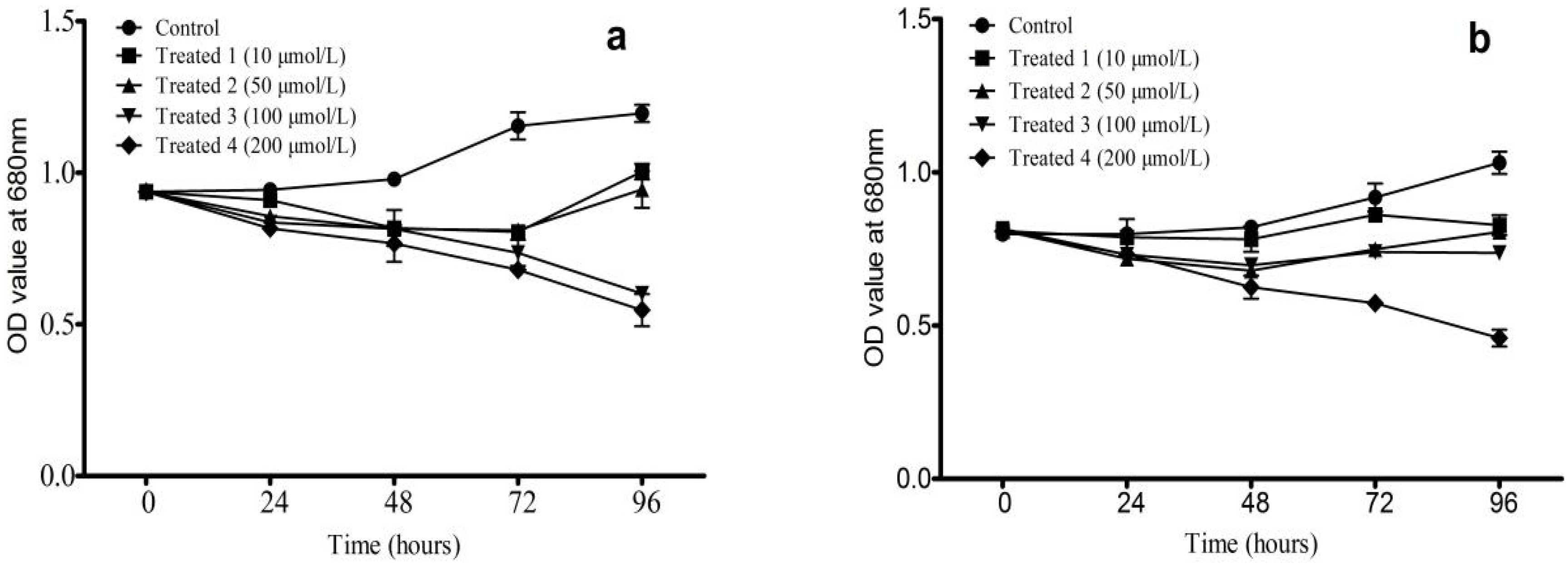
Figure 1. Effects of Artesunate on OD value of M. aeruginosa. ((a) FACHB-315; (b) FACHB-927).
Phycology 05 00063 g001
Figure 2. Effects of artesunate on Fv/Fm of M. aeruginosa ((a) FACHB-315; (b) FACHB-927). * p < 0.05; ** p < 0.01; Statistically significant differences when compared to the control.
Phycology 05 00063 g002
Figure 3. Effects of artesunate on φPS II of M. aeruginosa ((a) FACHB-315; (b) FACHB-927).
Phycology 05 00063 g003
Table 1. Toxicity effects (insect and plant) of artemisinin or extracts of Artemisia annua, where the effect could be due to artemisinin itself, some of the other bioactive compounds in A. annua, or synergy between artemisinin and some of the other bioactive compounds in A. annua.
Species ClassificationSpecies and StageEndpointEffect Formulation or
Concentration
Estimated or Given
Artemisinin Concentrations
Source
InsectCowpea bruchid, Callosobruchus maculatusSignificantly increased mortality2.5 g leaves/250 g cowpea
seeds
0.1–4 μg/g[14]
Lesser mulberry pyralid, Glyphodes pyloalis 4th instar larvaeLC50 by topical application0.33 mg leaf/mL0.033–1.32 mg/L[15]
Sunn pest (Eurygaster Integriceps)LC5032% (24 h) and 17%
(48 h), methanolic
extract
9.6–380 μg/mL (24 h) and 50–200 mg/L (48 h)[16]
Elm leaf Beetle, Xanthogaleruca luteola Mull.LC5048% (24 h) and 44%
(48 h), methanolic
extract
14.4–576 μg/mL (24 h) and 13.2–528 mg/L (48 h)[17]
Small white Pieris rapae, 3th
instar larvae
LC509.4% extract2.8–113 mg/L[18]
Epilachna paenulataComplete feeding rejection with ethanol extract of A. annua1.5 mg/cm236 μg/cm2[19]
Codling moth, Cydia pomonellaFeeding deterrent at p < 0.05, extract1 g/L0.2 mg/L[20]
PlantSorghum, Sorghum bicolor L.GerminationNo effect up to
9.3 μg/mL
No effect up to
56 μg/Petri dish
[21]
Soybean, Glycine maxGrowth inhibition, 25%0.73% dry leaf
material in soil
3.3 mg/kg soil[22]
Potato, Solanum tuberosumRoot growth inhibition, 100%material in soil32.5 kg/ha[23]
Barley, Hordeum vulgareGermination inhibition, 22%100 mg/L600 μg/Petri dish[24]
Redroot pigweed, Amaranthus
retroflexus L. dish
Growth inhibition9.3 mg/L56 μg/Petri[21]
Freshwater algae, Pseudokirchneriella subcapitataRelative growth rate, EC500.24 mg/L [25]
Duckweed, Lemna minorRelative growth rate, EC500.19 mg/L [25]
Microalgae Scenedesmus obliquusGrowth inhibition12–20 mg/L [12]
Microalgae Microcystis aeruginosaGrowth inhibition8–12 mg/L [12]
Disclaimer/Publisher’s Note: The statements, opinions and data contained in all publications are solely those of the individual author(s) and contributor(s) and not of MDPI and/or the editor(s). MDPI and/or the editor(s) disclaim responsibility for any injury to people or property resulting from any ideas, methods, instructions or products referred to in the content.