Article

A Novel Approach Towards RSM-Based Optimization of LED-Illuminated Mychonastes homosphaera Culture, Emphasizing Input Energy: An Industrial Perspective of Microalgae Cultivation

by
Doljit Borah
1,*,
Khalifa S. H. Eldiehy
2,
Khalid A. AL-Hothaly
3 and
Dhanapati Deka
1
1
Biomass Conversion Laboratory, Department of Energy, Tezpur University, Tezpur 784028, Assam, India
2
Department of Botany and Microbiology, Faculty of Science, Al-Azhar University, Assiut 71524, Egypt
3
Department of Biotechnology, Faculty of Science, Taif University, Taif 1099, Saudi Arabia
*
Author to whom correspondence should be addressed.
Phycology 2025, 5(4), 62; https://doi.org/10.3390/phycology5040062
Submission received: 22 September 2025 / Revised: 9 October 2025 / Accepted: 10 October 2025 / Published: 18 October 2025
(This article belongs to the Special Issue Development of Algal Biotechnology)

Abstract

The growing demand for sustainable bioprocesses highlights microalgae as a promising source of renewable feedstock. However, high energy use for artificial lighting limits the commercial viability of photobioreactor systems. This study proposes an energy-optimized framework for cultivating Mychonastes homosphaera using LED illumination. The optimization was performed using Response Surface Methodology (RSM) with a Face-Centered Central Composite Design (FCCCD) to assess the interactive effects of light intensity, duration, airflow rate, and nitrogen concentration on biomass and lipid productivity. The performance of LED wavelengths was compared for biomass, lipid productivity, and energy consumption. RSM models were statistically significant (p < 0.01), and ANOVA had a high coefficient of determination (R2) for all LEDs. Maximum biomass productivity was 512.0 ± 12.23 mg L−1 day−1 with cool-white, followed by pink (401.33 ± 10.48), blue (342.66 ± 3.53), and red (189.6 ± 1.36). Cool-white consumed the least energy (228.6 Wh day−1) to produce the maximum biomass, compared to blue (235.05 Wh day−1), pink (240.0 Wh day−1), and red (240.0 Wh day−1). Lipid content was highest under red (22.84%), followed by pink (17.39%), blue (15.82%), and cool-white (8.96%). However, lipid productivity was highest under pink (69.8 mg L−1 day−1), followed by blue (54.2 mg L−1 day−1), cool-white (45.86 mg L−1 day−1), and red (43.3 mg L−1 day−1).

1. Introduction

The importance and advantages of microalgae biomass as a feedstock capable of replacing petroleum have been known for many decades [1]. Microalgae have garnered significant scientific interest as a promising means for carbon sequestration and a potential contributor to achieving net-zero emissions targets [2]. Additionally, microalgae have the potential to replace petroleum-based fuels and chemicals [3]. However, economic sustainability is the bottleneck for commercial acceptance of microalgae [4]. Even after decades of research and development, economically sustainable microalgae production to sustain the biofuel industry has not yet been feasible [5]. Having a high lipid content is one of the essential characteristics in microalgae biomass that makes it suitable for the biofuel industry, as the lipid is converted into biodiesel [6]. With the existing microalgae culture technologies, it is difficult to achieve economic sustainability [7].
Recent advancements in semiconductor technology, particularly in light-emitting diodes (LEDs), have opened new possibilities for improving energy efficiency in photobioreactors [8]. Researchers have incorporated optoelectronics in microalgae culture using semiconductor technologies [8,9]. With technological advances, state-of-the-art semiconductor technologies, such as solar photovoltaics, LEDs, and microcontrollers, have become more efficient, reliable, cost-effective, and easily optimizable [10,11], making them highly suitable for illuminating microalgae culture. The ease of optimization of LEDs in terms of wavelength, intensity, and duration makes them an appropriate choice for manipulating and optimizing the microalgae growth environment, which in turn affects the growth and composition of the microalgae [12]. Light intensity, wavelength, and photoperiod have a remarkable effect on microalgae metabolism [8]. These parameters also significantly impact the production of biomass, lipid, and other metabolites from microalgae.
Though increasing light intensity increases biomass content, lipid content may vary based on the microalgae species. For example, Desmodesmus sp. and Tetradesmus obliquus (Chlorophyta) (formerly Scenedesmus obliquus) show an increase in lipid content with an increase in light intensity compared to other microalgae species [13]. Scenedesmus quadricauda (Chlorophyta) thrived best at 500 µmol photons m−2 s−1 and a 660 nm wavelength. Increasing light intensity to 1000 µmol photons m−2 s−1 caused photon damage, whereas lower light intensities, such as 100 µmol photons m−2 s−1, slowed or stopped growth [14]. In addition to light conditions, lipid accumulation in microalgae is also influenced by other environmental and physical factors, including nutrient availability, cultivation mode, and carrier characteristics, which together determine the efficiency of metabolic pathways and lipid biosynthesis [15,16,17]. Overall, growth conditions exert a major influence on microalgal metabolism, and their effects are highly species-dependent [18]. Therefore, cultivation parameters must be carefully optimized for each specific strain to maximize growth and metabolite production.
However, it should be noted that an input power level-based performance study of different LEDs is more useful than comparing the performance of different LEDs in microalgae culture based on illumination intensity. This is because energy cost parameters are more important in commercial production than light intensity. Choosing the best LED based on equal light intensities may misinterpret commercial applications, as the power consumption of different LEDs to emit equal light intensity varies [19].
In this study, we propose a novel, energy-centered optimization framework for LED-illuminated cultivation of Mychonastes homosphaera (formerly Chlorella homosphaera). This microalga was selected due to its high growth rate [20] and lipid accumulation potential [20]. M. homosphaera has already been applied in wastewater treatment [21], heavy metal recovery [22], and biofuel application [15]. However, little work has been done on optimizing M. homosphaera culture based on optical parameters such as light intensity, light wavelength, and light duration. Additionally, studies on the effects of single parameters, such as carbon sources [23], magnetic field stress [24], and nitrogen content [25], on the growth and metabolism of M. homosphaera have been reported, but few studies have investigated the complex interactive effects of multiple parameters. The experimental design was developed using Response Surface Methodology (RSM) and the Face-Centered Central Composite Design (FCCCD) approach to model the interactive influence of four key parameters—light intensity, photoperiod, airflow rate, and nitrogen concentration—on biomass and lipid productivity. Importantly, this research compares the performance of cool-white, pink, blue, and red LEDs based on their input electrical power, providing an energy-normalized assessment of their suitability for industrial-scale cultivation.
The innovation of this work lies in its integration of optoelectronic efficiency and biological productivity within a single optimization framework. By linking energy input directly to biomass and lipid output, this study provides a quantitative foundation for energy-efficient photobioreactor design. The outcomes contribute to a practical understanding of how illumination energy and cultivation parameters can be optimized simultaneously to enhance both productivity and techno-economic feasibility. This approach offers a scalable strategy for industrial microalgae cultivation, potentially reducing the cost barriers that currently hinder commercial biofuel and bioproduct manufacturing.

2. Methodology

2.1. Experimental Setup Design and Development

An experimental system was designed to control culture conditions for optimization experiments efficiently. The setup included a microalgal culture chamber (MCC), which was designed to accommodate four 500 mL Erlenmeyer flasks. The MCC was lined with LED strips (Crimson LED Strips, Goldmedal Electricals, Mumbai, India) and equipped with a light intensity sensor (BH1750, Rohm Semiconductor, Tianjin, China) and a temperature sensor (DS18B20, Maxim Integrated, China) attached. Four air pumps (AP-208, Venus Aqua, Jiangmen, China) supplied filtered air (0.22 µm Syringe Filter, Whatman, Buckinghamshire, UK) to the culture media. The culture’s air flow is controlled by adjusting a generic airflow valve and measuring the flow with a mass flow meter (3.2 SLPM, Alicat Scientific, Tucson, AZ, USA).
The experiment’s light intensity, light duration, and air flow duration were controlled by a system based on an Arduino Nano microcontroller board. Detailed information regarding the experimental setup is available in the manuscript by Doljit et al. [19]. Four experimental setups were created with cool-white, pink, blue, and red LED strips. A Compact CCD Spectrometer (CCS200/M, ThorLabs, Newton, NJ, USA) was used to measure the LED light spectrum, as described by Doljit et al. [19]. Light intensity was measured with a quantum sensor (MQ-510, Apogee Instruments, Logan, UT, USA), and electrical input power was measured using a digital multimeter (15B+, Fluke, Everett, WA, USA).
The electrical energy consumption (Wh day−1) of each LED illumination system was determined by direct measurement using a calibrated Fluke 15B+ digital multimeter connected to the LED unit. Both voltage and current were recorded for different LED intensities throughout the illumination range using the Arduino-based controller. The instantaneous electrical power (W) was obtained as the product of measured voltage (V) and current (A), and total daily energy consumption (Wh day−1) was calculated by integrating the average power over the 24 h operating period.

2.2. Microalgae Strain and Culture Conditions

The microalgae strain (Mychonastes homosphaera) was obtained from the Department of Biotechnology, Guwahati University, India. Prior to the experiments, the microalgae strain was initially cultured in a 500 mL Erlenmeyer flask, and the culture volume was gradually increased. It was then grown in a 20 L airlift photobioreactor (PBR) using BG11 medium [26]. The culture was illuminated with LED tubes (20 W cool-day LED tube light, Bajaj, Mumbai, India) at an intensity of 100 µmol photons m−2 s−1 and maintained at 30 ± 1 °C. For the experiments, a specific volume of the culture was taken from the PBR, centrifuged (Heraeus Biofuge Primo centrifuge, Thermo Fisher Scientific, Hanau, Germany) at 5000 rpm for 10 min. The supernatant was discarded, and the microalgae pellets were collected and washed with phosphate-buffer solution. The washed microalgae were then inoculated into 300 mL of BG-11 medium without a nitrogen source. The initial concentration of the culture was adjusted (initial absorbance of 0.1 at OD560) by controlling the quantity of the added BG11 medium and measuring optical density (OD560) [27] using a spectrophotometer (UV-1700, Shimadzu, Kyoto, Japan). For the experiments, the desired quantities of nitrogen, as per the experimental requirements, were added and cultured in a 500 mL volume Erlenmeyer flask for five days [28].

Control of Kinetic Processes

To minimize the influence of kinetic variability on the optimization results, all experiments were conducted under controlled and reproducible growth conditions. The initial inoculum density of M. homosphaera was standardized (OD560 = 0.1) for all runs to ensure that cultures started from the same physiological state. Each experiment was conducted for a fixed duration of five days, which corresponded to the late exponential phase as confirmed by preliminary kinetic growth studies. By terminating the culture before the onset of the stationary phase, the responses primarily reflected the active growth kinetics rather than nutrient depletion effects. Temperature (30 ± 1 °C) and pH (maintained near 7.0 ± 0.2) were continuously monitored to avoid metabolic fluctuations arising from thermal or pH drift. The airflow rate provided sufficient gas exchange to maintain dissolved CO2 availability and prevent oxygen accumulation, ensuring uniform photosynthetic activity. The LED illumination system, controlled by an Arduino-based microcontroller, precisely maintained the photoperiod and intensity to minimize photon flux fluctuations. These measures ensured that kinetic parameters influencing growth and lipid accumulation were consistently managed, allowing the RSM analysis to capture the effects of the controlled input variables (light, nitrogen, and aeration) rather than uncontrolled biological variations.

2.3. Experimental Design

To model and optimize the relationship between the independent parameters and the resultant response in LED-based microalgae culture experimentation, a statistical tool known, Response Surface Methodology (RSM), was employed. Face-Centered Central Composite Design (FCCCD) approach of RSM was used with the independent variables, nitrogen concentration (as sodium nitrate), light intensity, light duration, and air flow rate, to model and optimize for the highest possible resultant responses, i.e., biomass and lipid productivity of M. homosphaera. FCCCD was chosen for this experiment as it requires only three levels (high, low, and midpoint) of each experimental variable, making it relatively simple and minimizing the probability of error [29]. The highest and lowest values of nitrogen concentration and light duration were chosen based on the data from preliminary investigations conducted to study the effect of these parameters. The highest and lowest values of air flow and light intensity were determined by the maximum values achievable with the developed experimental setup.
To compare the LEDs based on their power consumption, the light intensities were expressed as percentages of the input power. A 10 W power level, being the maximum applicable in the developed setup, was considered 100% intensity, while 2 W represented the lowest power (20% intensity), and so on. The values of the independent variables used for modeling the parameters are provided in Table 1.
The experiment was designed using the Design-Expert software (version 13, Stat-Ease Inc., Minneapolis, MN, USA). The design consisted of 30 runs, including six center points, eight axial points, and sixteen factorial points, as shown in Table 2. To overcome the limitations of running a maximum of 4 experiments in the developed experimental setups at a time and to improve experimental efficiency, the experiments were divided into eleven groups (I–XI), as shown in Table 2, similar to the approach adopted by Hosseini et al. [30] to address the limited number of available bioreactors.
The selection of the experimental variables and their respective limits was established through a combination of preliminary trials, and equipment operational constraints. The lower and upper limits were defined to ensure both physiological tolerance of M. homosphaera and the technical feasibility of the experimental setup.
For light duration, the range of 0–24 h was selected to encompass conditions from complete darkness to continuous illumination, as continuous light has previously been reported to enhance growth in chlorophytes. For light intensity, the levels were expressed as a percentage of LED input power (0–100%) to reflect realistic operating conditions of the developed programmable illumination system. Preliminary tests showed that intensities below 20% resulted in negligible growth, whereas input powers above 100% was not practically feasible.
The airflow rate range (0–1.6 L min−1) was determined based on the aeration capacity of the areators. The nitrogen concentration range (0–10 g L−1) was selected to cover conditions from nitrogen limitation to saturation, as reported in previous studies on chlorophyte species.
The experimental variables were coded as per Equation (1):
y j   =   Y j Y j 0 Δ Y j
where yj, Yj, and yj0 represent the coded value, experimental value, and actual experimental value at the central point of the jth experimental variable, respectively, and ∆Yj indicates the step change in the value.

2.4. Biomass Productivity

The microalgal biomass productivity was evaluated by first determining the dry cell weight (DCW) of the culture. To determine the DCW, 25 mL of the algal culture was filtered using a pre-dried and pre-weighed glass fiber filter (Whatman GF/A, 1.6 µm pore size, 110 mm diameter). The filtered cells were dried at 80 °C until a constant weight was achieved. The dry cell weight was then determined gravimetrically. After determining the DCW, biomass productivity was calculated using Equation (2).
Biomass   productivity   ( mg   L 1   day 1 ) =   W e n d W s t a r t Δ T × V × 1000
where Wend and Wstart are the biomass dry weight at the end and start of the experiment, ∆T is the duration of the experiment, and V is the volume of culture used to calculate the dry weight, which is 25 mL in the current experiment.

2.5. Lipid Productivity

Lipid productivity was determined by first extracting the lipids using the Bligh and Dyer method [31]. The lipid productivity was then calculated using Equation (3).
Lipid   productivity   ( mg   L 1   day 1 ) =   W t l W t e Δ T × V × 1000
where Wtl and Wte are the weight of the test tube with lipid and the weight of the empty test tube, ∆T is the duration of the experiments, and V is the volume of the microalgae culture used to extract the lipid, which is 50 mL in the current scenario.

2.6. Statistical Analysis

The experimental data were analyzed, and the responses were predicted using the response surface regression procedure, based on the second-order quadratic model shown in Equation (4)
y = β o + β i x i + β ii x i 2 +   i < j β i j x i x j
where ‘y’ indicates the response, ‘βo’ indicates the graph intercept, ‘βi’, ‘βii’, and ‘βij’ are the mean values of linear, quadratic, and interaction constant coefficients, respectively, and ‘xi’ and ‘xj’ are the coded input variables. The output responses for biomass and lipid were both set as desired, with maximum desirability. The overall desirability (D) was calculated using Equation (5).
D = (d1y1 × d2y2)1/2
where d1 and d2 are desirability of the responses y1 and y2 [16].

3. Results and Discussion

The present study focuses on the benefits of LED illumination in microalgae cultivation. Furthermore, it aims to optimize the parameters influencing LED-based growth of M. homosphaera, while considering an industrial perspective by accounting for the energy input required to power the LEDs.

3.1. Effect of Input Parameters on Biomass Production

This study investigated the effects of light intensity, light duration, airflow rate, and nitrogen concentration on biomass production. The ANOVA results presented in Table 3 indicate a significant quadratic model (p < 0.001). Meanwhile, Table 4 compares the model predictions with the experimental outcomes. The F-values (30.86, 53.28, 13.66, and 52.95) demonstrate model fit for cool-white, pink, blue, and red LEDs, respectively, with probabilities < 0.001. The analysis indicates that light duration and intensity have a significant influence on biomass production (p < 0.001) in all four LED illuminations.
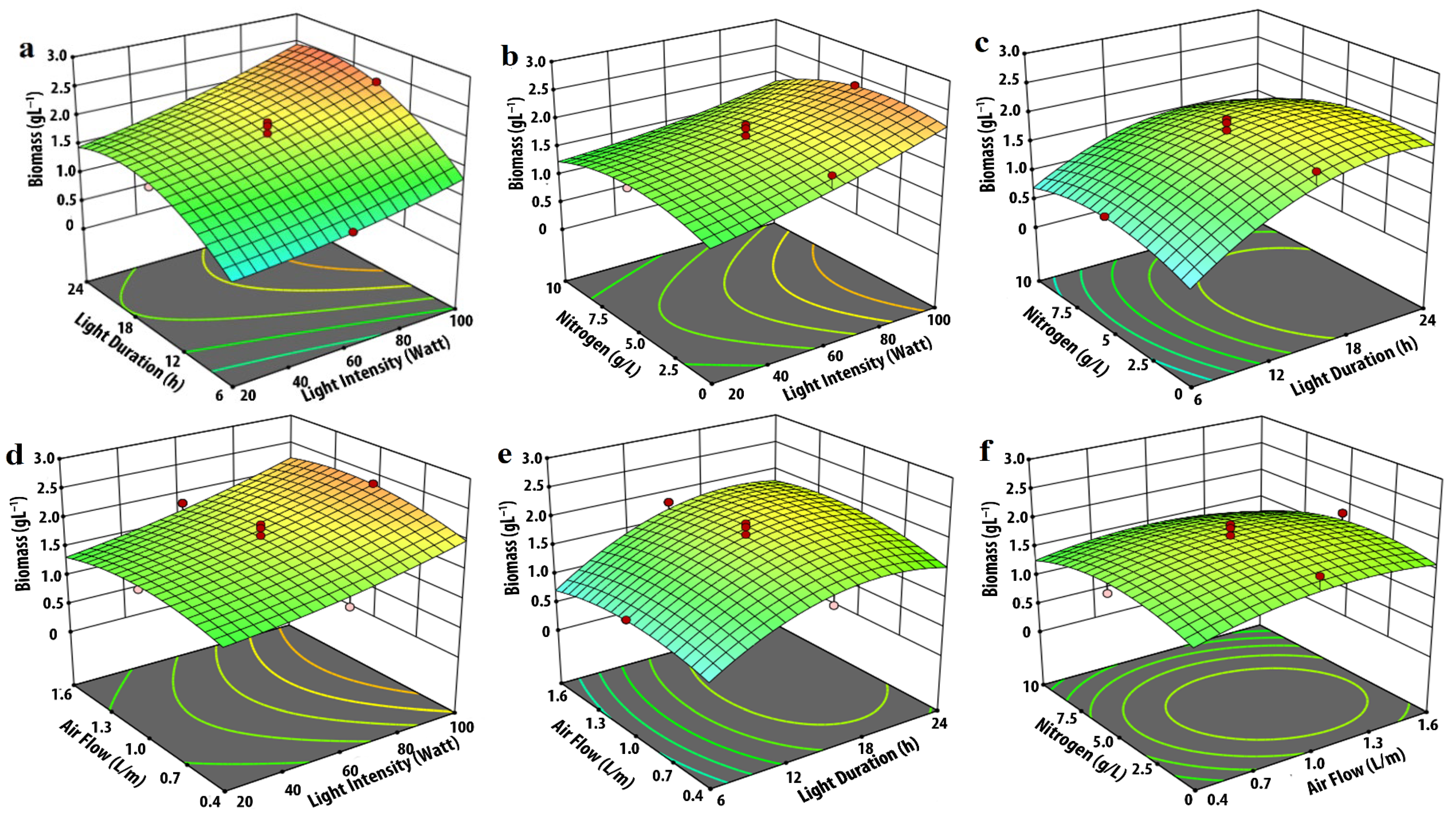
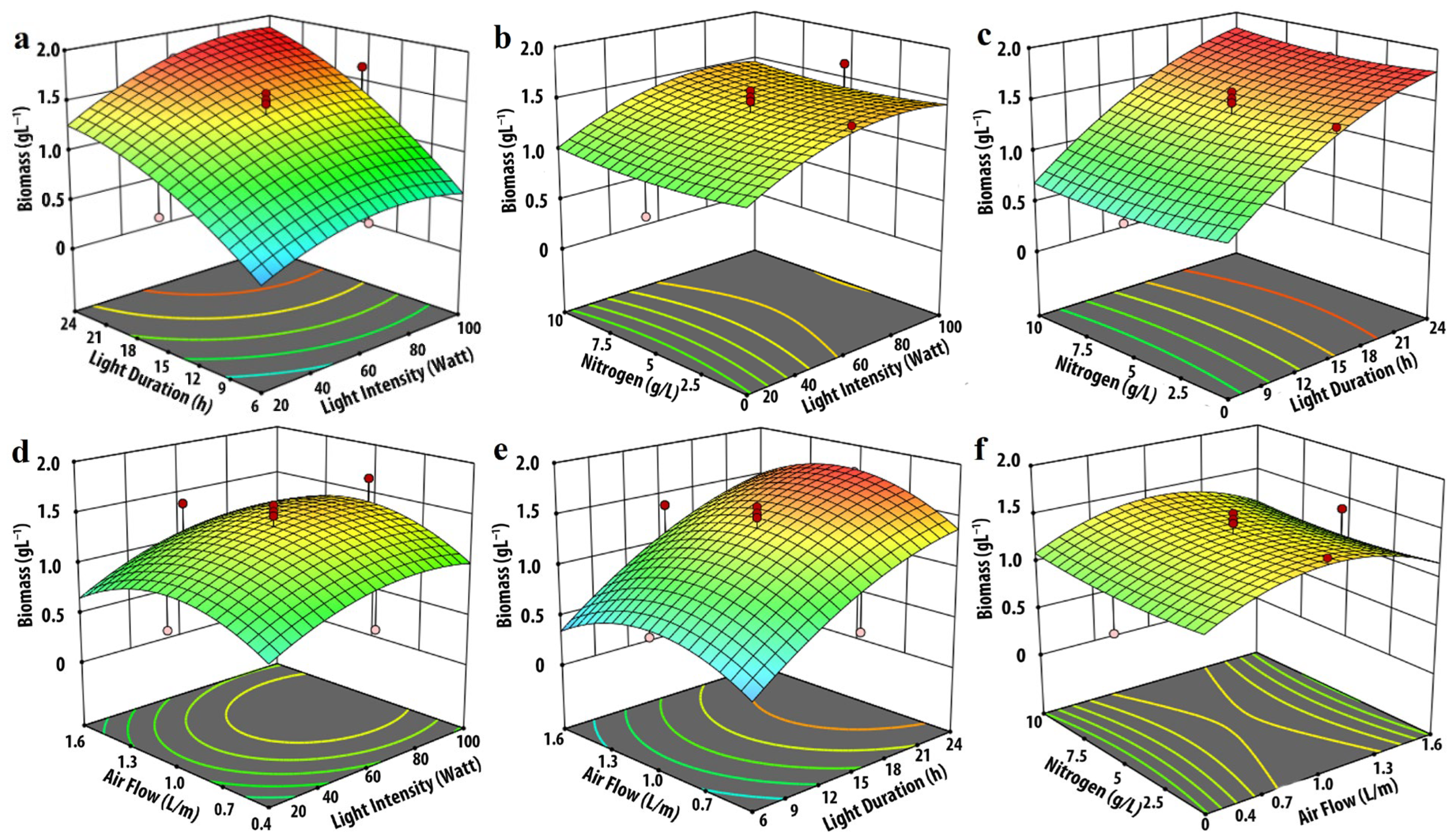
Under cool-white, pink, and red illumination, biomass productivity increased with light intensity, reaching a maximum at 100% intensity (Figure 1a,b,d, Figure 2a,b,d and Figure 4a,b,d). Under blue illumination, however, biomass productivity reached its maximum at 98% intensity, as shown in Figure 3a,b,d after which a slight decline was observed with further increases in intensity. The increase in biomass productivity with increasing light intensity is supported by the findings of Iasimone et al. [32], which highlighted the increase in biomass productivity with an increase in light intensity of a consortia of microalgae cultivated using wastewater. Similar findings were also reported by Nzayisenga et al. [13], where the biomass of Desmodesmus sp., Chlorella vulgaris, Ettlia pseudoalveolaris, and Tetradesmus obliquus increased with higher light intensities. Generally, microalgal growth continues to rise with increasing light intensity up to a certain threshold known as the saturation light intensity. Beyond this level, further increases in light intensity lead to a decline in growth [33]. The saturation light intensity exhibits variability based on both the specific microalgae species and the characteristics of the lighting [34]. The findings of this study suggest that cool-white, pink, and red LED illuminations have the potential to enhance biomass productivity. However, the existing limits of the experimental settings prevent the intensity from being increased beyond 100%, thereby hindering the realization of this potential.
Along with the light intensity, light duration plays a significant role in the growth and metabolism of microalgae [35]. As evident from the current findings, apart from cool-white LED illumination (Figure 1a,c,e), biomass productivity increased with illumination duration in all three (pink, blue, and red) LED illuminations, as shown in Figure 2a,c,e, Figure 3a,c,e and Figure 4a,c,e, giving maximum productivity under continuous illumination. In cool-white LED illumination, biomass increased with illumination time to a peak at 22.8 h, after which it declined. Microalgae can be cultivated under continuous illumination [36], light–dark illumination cycle [37], or under pulsating illumination [38]. The biochemical composition of microalgae is affected by the changes in the duration of the dark cycle. Depending on the growth environment and microalgae strain, different photoperiods are proven to be beneficial [39]. In certain cases, continuous illumination produces better results [40], as evident in the present scenario with pink, blue, and red LED illuminations. In certain cases, continuous illumination can cause photoinhibition, and thus a brief dark cycle is necessary to repair the damage to the photosystem [41].
Maintaining nitrogen content at its optimum concentration is crucial, as excessive nitrogen concentration can cause growth inhibition [42], while insufficient or nitrogen-depleted conditions result in low biomass productivity [43]. Nitrogen enhances biomass growth and affects the production of other metabolites. The present study showed similar findings. In cool-white and pink LED-illuminated culture (as shown in Figure 1b,c,f and Figure 2b,c,f), biomass growth gradually increased with increasing nitrogen concentration, reaching a maximum at 1.61 g L−1 and 1.03 g L−1 for cool-white and pink LED illumination, respectively, and then decreased. The optimized maximum nitrogen content of 0.1 g L−1 in blue and red LED lighting resulted in maximum biomass productivity.
As evident from the ANOVA analysis in Table 3, the influence of airflow on biomass growth was minimal in the present study. Airflow rates of 1.3, 1.44, 1.49, and 0.4 L min−1 for cool-white, pink, blue, and red LED illumination resulted in maximum biomass productivity. Further insight into the effect of airflow on biomass growth is presented in the following optimization section of this article.

3.2. Effect of Input Parameters on Lipid Production

The effects of different input parameters on lipid productivity were modeled. The quadratic model, analyzed with ANOVA (Table 5), was found to be statistically significant (p < 0.001). The F-values (67.49, 47.42, 26.51, and 33.28) for the models corresponding to cool-white, pink, blue, and red LEDs, respectively, with probabilities < 0.001, confirmed a good model fit. Lipid productivity in the cultures increased with increasing light duration. The highest lipid productivities of 384, 276, 239, and 232 mg L−1 were recorded under 24 h of illumination for pink, blue, cool-white, and red LED lighting, respectively, as shown in Table 4 (experiment number 29 for pink, cool-white, and blue, and experiment number 27 for red).
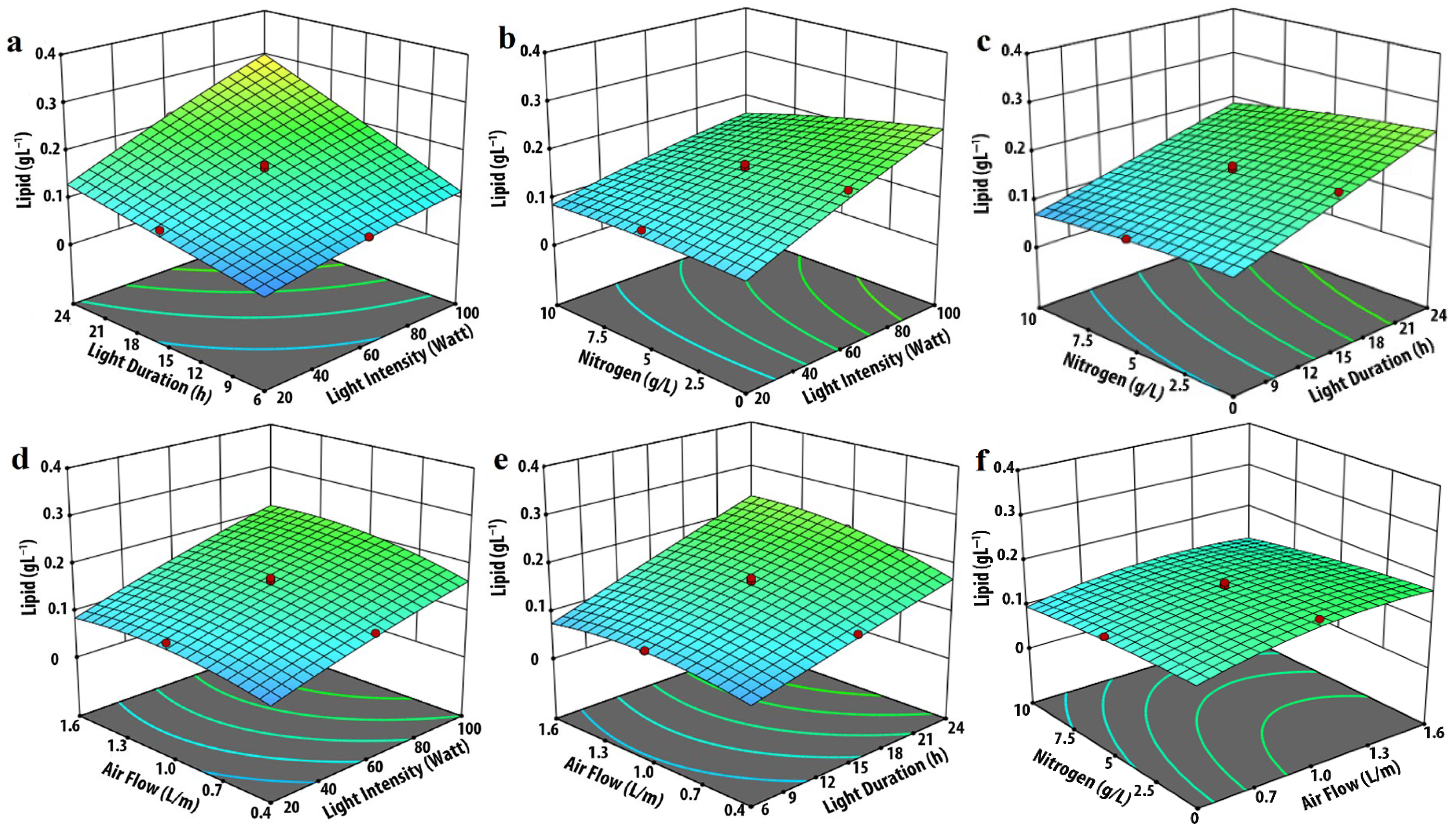
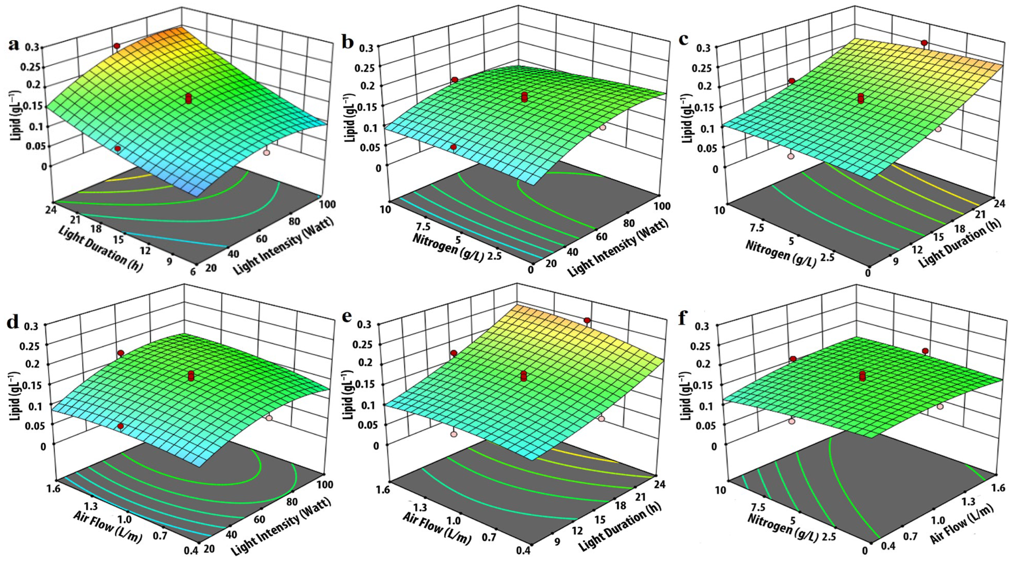
Light intensity played a similar role to that of light duration in influencing lipid production. The optimized maximum lipid production was observed at 100% light intensity for cool-white, pink, and red LEDs (Figure 5a,b,d, Figure 6a,b,d and Figure 8a,b,d), and at 98% intensity for blue LED illumination (Figure 7a,b,d). Lipid production under blue LED lighting declined with further increases in intensity, as shown in Figure 7a.
Nitrogen is a crucial macronutrient that significantly influences the growth and composition of microalgae, particularly their lipid content [44]. Low nitrogen content favors lipid accumulation [32]. The responses of the current investigation support the same, where lipid production in microalgae is significantly affected by nitrogen concentration. As shown in Figure 5, Figure 6, Figure 7 and Figure 8, lipid production was highest under nitrogen-limiting conditions. The maximum lipid production for all four LED illuminations (Table 4) was recorded at a nitrogen concentration of 0.1 g L−1.
Initially, lipid production in all LEDs increased with increasing airflow, reached a maximum at an optimal level, and declined with further increases in airflow rate. The optimized airflow rates for maximum lipid production were estimated to be 1.3, 1.43, 1.48, and 0.4 L min−1 for cool-white, pink, blue, and red LED illumination, respectively.
The effects of nitrogen and airflow on lipid productivity are further discussed in the optimization section that follows.

3.3. Modeling and Validation

The experimental design parameters were analyzed using Design-Expert software and fitted into quadratic polynomial models. High regression coefficients (R2 values) of 0.97, 0.98, 0.93, and 0.98 for biomass productivity under cool-white (Y1), pink (Y2), blue (Y3), and red (Y4) LED illumination were obtained. Similarly, for lipid productivity, R2 values of 0.98, 0.98, 0.96, and 0.97 were achieved under cool-white (Y5), pink (Y6), blue (Y7), and red (Y8) illumination, respectively. The quadratic model equations (Equations (6)–(13)) obtained are presented as follows:
Y1 = − 1.31 − 0.01A + 0.20B + 1.03C + 0.20D + 0.01AB + 0.01AC − 0.01AD + 0.01BC − 0.01BD − 0.03CD + 0.01A2 − 0.01B2 − 0.59C2 − 0.01D2
Y2 = − 0.92 + 0.01A + 0.08B + 0.98C + 0.32D + 0.01AB − 0.01AC − 0.01AD − 0.01BC − 0.01BD − 0.01CD + 1.31A2 − 0.01B2 − 0.41C2 − 0.03D2
Y3 = − 1.19 + 0.01A + 0.10B + 1.64C − 0.03D + 0.01AB + 0.01AC + 2.18AD + 0.01BC + 0.01BD − 0.01CD − 0.01A2 − 0.01B2 − 0.87C2 + 0.01D2
Y4 = − 0.40 + 0.01A + 0.03B + 0.33C + 0.01D + 0.01AB − 0.01AC − 9.94AD + 0.01BC − 0.01BD + 0.01CD − 6.22A2 − 0.01B2 − 0.18C2 − 0.0D2
Y5 = 0.16 + 0.04A + 0.04B + 0.01C − 0.01D + 0.01AB + 0.01AC − 0.01AD + 0.01BC − 0.01BD + 0.01CD − 0.02A2 + 0.01B2 − 0.03C2 − 0.01D2
Y6 = − 0.05 + 0.01A + 0.01B + 0.09C + 0.01C + 7.65D + 0.01AB − 0.01AC + 0.01BC − 0.01BD − 0.01CD − 3.85A2 − 1.44B2 − 0.06C2 − 0.01D2
Y7 = 0.02 + 0.01A − 0.01B + 0.01C + 0.01D + 4.49AB + 0.01AC − 4.83AD + 0.01BC − 0.01BD + 0.01CD − 2.06A2 + 0.01B2 − 0.03C2 − 0.01D2
Y8 = −0.08 + 0.01A + 0.01B − 0.04C − 0.01D + 2.85AB − 0.01AC − 4.17AD − 0.01BC − 0.01BD + 0.01CD − 1.77A2 − 0.01B2 + 0.01C2 + 0.01D2
The lack-of-fit tests for all models were found to be non-significant, as shown in Table 3 and Table 5. ANOVA results for all eight developed models were statistically significant (p < 0.0001). These results indicate that the developed models exhibit a strong fit and correlation between the input parameters and their corresponding responses, and could efficiently explain the experimental data.
In addition to the coefficient of determination (R2), the adjusted R2 values were also considered to evaluate the predictive strength of the models after accounting for the number of variables. The adjusted R2 values ranged from 0.93 to 0.98 for biomass productivity and from 0.96 to 0.98 for lipid productivity under different LED illuminations, indicating a strong agreement between predicted and experimental results, with no evidence of model overfitting.
The lack-of-fit tests for all models were statistically non-significant (p > 0.05) as reported in Table 3 and Table 5, indicating that the models adequately describe the experimental data within the studied range. Collectively, the high R2 and adjusted R2 values, the non-significant lack-of-fit, and the random distribution of residuals confirm that the developed quadratic models are statistically robust, accurate, and suitable for predictive optimization of M. homosphaera culture parameters.

3.4. Optimization

The optimum conditions for achieving maximum biomass and lipid production within the range of the input variables were predicted based on the developed model using Design-Expert software. The predicted values corresponding to the maximum responses with the highest desirability were selected, and the conditions were validated experimentally. The optimum parameters for attaining the highest biomass and lipid content under the four LED illuminations are summarized in Table 6.
All the experiments were conducted using the developed setup under real operating conditions to ensure reproducibility. The experimental outcomes were in close agreement with the model predictions, showing mean deviations of 0.86–3.55% for biomass and 1.66–5.63% for lipid productivity, confirming that the developed models can be reliably applied to real cultivation systems.
Furthermore, a sensitivity analysis was performed around the optimized conditions to evaluate the system’s tolerance limits. Each parameter (light intensity, duration, airflow, and nitrogen concentration) was varied by ±10% from its optimal level while keeping other parameters constant. The responses showed less than 5% deviation in biomass and lipid productivity within this range, indicating that the optimized conditions are robust and tolerant to minor variations that may occur in large-scale or industrial photobioreactor operations. These results demonstrate that the developed optimization framework is not only statistically significant but also practically stable and reproducible under real operating constraints.
The optimum conditions predicted were validated experimentally by conducting experiments in triplicate and comparing the mean results of the experiments with the predicted responses. The biomass and lipid production obtained under the optimized conditions were also compared with those from microalgae cultures grown under normal (non-optimized) conditions reported previously [19]. The responses of biomass and lipid production, obtained theoretically (predicted), experimentally, and under normal conditions, are presented in Table 6. The errors in the predicted and experimental values for the biomass were 3.21%, 0.86%, 3.55% and 2.48% and for lipid were 4.23%, 2.01%, 1.66% and 5.63% for cool-white, pink, blue, and red LED illumination, respectively, indicating that the developed model accurately predicts the growth performance of M. homosphaera.
Biomass production under optimized conditions increased by 1.30, 1.07, 1.01, and 1.13-fold compared with normal conditions for cool-white, pink, blue, and red LED illumination, respectively. Similarly, lipid production increased by 1.50, 1.81, 1.57, and 1.64-fold under cool-white, pink, blue, and red LED illumination, respectively. Thus, the developed models successfully optimized the culture conditions, leading to increased biomass and lipid production.
This is evident with the blue LED illumination, where biomass productivity peaked at 98% illumination, resulting in a biomass productivity of 342.66 ± 3.53 mg L−1 day−1. Further increases in light intensity reduced biomass productivity. In the case of cool-white, pink, and red LED illumination, peak biomass productivity of 512.0 ± 12.23, 401.33 ± 10.48, and 189.6 ± 1.36 mg L−1 day−1 was achieved at 100% intensity, making cool-white the most preferred LED illumination in terms of biomass productivity.
Furthermore, as observed in Table 6, cool-white LED illumination yielded the highest biomass productivity with a light–dark cycle of 22.8:1.2 h. The introduction of a dark period in an artificially illuminated microalgae culture has a major advantage, i.e., it saves energy. In this case, cool-white LED not only achieved superior biomass productivity but also consumed less energy compared with the other three LED illuminations. Thus, in terms of energy consumption, cool-white LED (228.6 Wh day−1) is the most favorable option, compared with pink (240.0 Wh day−1), blue (235.05 Wh day−1), and red (240.0 Wh day−1) LEDs for biomass production of M. homosphaera.
Considering lipid productivity, the most preferred LED illumination to achieve the highest lipid productivity in M. homosphaera is pink LED illumination, resulting in lipid productivity of 69.8 ± 1.1 mg L−1 day−1, followed by blue (54.2 ± 1.01 mg L−1 day−1), cool-white (45.86 ± 1.73 mg L−1 day−1), and red (43.3 ± 1.59 mg L−1 day−1) LED illumination.
As derived from Table 6, nitrogen-limiting conditions (0.1 g L−1) combined with red LED illumination result in the highest lipid content (22.84%) among cool-white (8.96%), pink (17.39%), and blue (15.82%) LED illuminations. Nevertheless, biomass productivity under red LED illumination was the lowest among all treatments, resulting in a minimum daily lipid productivity of 43.3 ± 1.59 mg L−1 day−1. This discrepancy can be attributed to a physiological trade-off between biomass and lipid accumulation. Under stress conditions (e.g., high light intensity or suboptimal spectral quality), cells divert carbon and energy from growth toward the formation of storage lipids [45], thereby increasing the lipid content but reducing overall biomass yield. These findings emphasize that maximizing lipid content alone does not ensure high lipid productivity; instead, a balance between biomass growth and lipid accumulation is crucial. This balance appears optimal under pink LED illumination, particularly when combined with adequate nitrogen availability.
The pink LED illumination, when combined with a nitrogen concentration of 1.02 g L−1, demonstrates the highest daily lipid productivity of 69.8 ± 1.1 mg L−1 day−1. Therefore, it can be concluded that the pink LED illumination with the optimal nitrogen content outperforms the other three LED options in terms of lipid productivity. Similar findings were reported by Markou [46], who observed the highest productivity of Arthrospira platensis under pink LED illumination. The advantage of pink LED can be attributed to the fact that the pink color is a combination of two primary color spectrums, red and blue [19], where the blue component is known to support biomass growth [47], whilst the red component encourages lipid productivity [48]. This is supported by the findings of Sirisuk et al. [12], where maximum productivity of Isochrysis galbana, and Phaeodactylum tricornutum was obtained using a 50:50 mixture of blue and red LED illumination.
Carbon, comprising over 50% of microalgae biomass [49], is the most important element required during microalgae cultivation. In the photoautotrophic mode of cultivation, CO2 is the most preferred source of carbon [49]. Typically, CO2 is supplied by bubbling ambient air through the culture media, with or without supplementary CO2. When ambient air is provided without CO2 supplementation, the airflow rate plays a crucial role in microalgae growth, as low airflow limits the available CO2, while excessive airflow can induce harmful shear stress, potentially damaging the microalgae culture [49]. The desired level of CO2 in the system depends on carbon fixation rates, which are influenced by factors affecting microalgae growth, such as the properties of the growth light, nitrogen concentration, temperature, and pH [50]. The experimental findings demonstrate a clear correlation between different LED illuminations (cool-white, pink, blue, and red) and their respective optimal airflow rates. Notably, cool-white (512.0 ± 12.23 mg L−1 day−1), pink (401.33 ± 10.48 mg L−1 day−1), and blue (342.66 ± 3.53 mg L−1 day−1) LEDs yielded significantly higher biomass compared to red (189.6 ± 1.36 mg L−1 day−1) LEDs, corresponding to a lower optimal airflow rate (0.40 L min−1) for the red LED illumination. In order to conduct a comprehensive analysis of the CO2 requirement and afterwards ascertain the airflow rate, it will be necessary to employ more advanced experimental methodologies. These refined approaches will be implemented in future investigations.

4. Industrial Perspective

The modular LED illumination system developed for this study can be readily scaled using parallel arrays with programmable control, enabling uniform light distribution while maintaining precise energy input monitoring. In contrast to conventional fluorescent or metal-halide lamps, the optimized LED setup offers longer operational lifetime (>50,000 h), lower thermal output, and real-time tunability—all of which are advantageous for continuous or semi-continuous industrial cultivation.
From a scalability perspective, the principal constraints include light attenuation, mass-transfer efficiency, and thermal management in dense cultures. However, the response-surface models established here provide quantitative guidance for balancing these parameters during scale-up by correlating illumination energy, aeration, and nutrient load with productivity. Integrating this framework with emerging biofilm or hybrid photobioreactors could further enhance energy efficiency by minimizing mixing energy losses and facilitating simplified harvesting.
Although a full techno-economic assessment was beyond the scope of this laboratory study, preliminary cost estimation suggests that the optimized LED regime could lower electricity cost per kilogram of biomass by roughly 20–25%, highlighting a clear industrial advantage. Future work will combine this empirical model with process-simulation-based cost analysis to develop a comprehensive AMPH-scale bioreactor model suitable for commercial application.

5. Conclusions

A comparative analysis of LED-illuminated microalgae cultures was conducted using the RSM and FCCCD approaches across different LEDs, with an emphasis on input energy and the commercial viability of the process. The objective was to evaluate the energy requirements of each LED and their effects on biomass and lipid production in M. homosphaera. Among the four LEDs, the cool-white LED yielded the highest biomass (512.0 ± 12.23 mg L−1 day−1) while consuming the least energy (228.6 Wh day−1). In contrast, pink LED achieved the highest lipid productivity (69.8 ± 1.1 mg L−1 day−1) at a nitrogen concentration of 1.02 g L−1, with an energy consumption of 240.0 Wh day−1.

Author Contributions

Conceptualization, D.B. and K.S.H.E.; Methodology, D.B. and K.S.H.E.; Software, D.B., K.A.A.-H. and D.D.; Validation, D.B. and K.S.H.E.; Formal Analysis, D.B., K.S.H.E. and D.D.; Investigation, D.B. and K.S.H.E.; Resources D.B. and D.D.; Data Curation, D.B. and D.D.; Writing—Original Draft Preparation, D.B. and K.S.H.E.; Writing—Review and Editing, D.B., K.S.H.E. and K.A.A.-H.; Visualization, D.B.; Supervision, D.D.; Funding Acquisition, K.A.A.-H. All authors have read and agreed to the published version of the manuscript.

Funding

This research received no external funding.

Data Availability Statement

Data is contained within the article. The original contributions presented in this study are included in the article. Further inquiries can be directed to the corresponding author.

Acknowledgments

The author, Khalid A. Al-Hothaly, would like to extend their sincere appreciation to the Deanship of Scientific Research, Taif University, Saudi Arabia, for funding this work.

Conflicts of Interest

The authors declare no conflicts of interest.

References

  1. Milano, J.; Ong, H.C.; Masjuki, H.; Chong, W.; Lam, M.K.; Loh, P.K.; Vellayan, V. Microalgae biofuels as an alternative to fossil fuel for power generation. Renew. Sustain. Energy Rev. 2016, 58, 180–197. [Google Scholar] [CrossRef]
  2. Dias, R.R.; Deprá, M.C.; Zepka, L.Q.; Jacob-Lopes, E. Roadmap to net-zero carbon emissions in commercial microalgae-based products: Environmental sustainability and carbon offset costs. J. Appl. Phycol. 2022, 34, 1255–1268. [Google Scholar] [CrossRef]
  3. González-Balderas, R.; Felix, M.; Bengoechea, C.; Ledesma, M.O.; Guerrero, A.; Velasquez-Orta, S. Development of composites based on residual microalgae biomass cultivated in wastewater. Eur. Polym. J. 2021, 160, 110766. [Google Scholar] [CrossRef]
  4. Rizwan, M.; Mujtaba, G.; Memon, S.A.; Lee, K.; Rashid, N.J.R. Exploring the potential of microalgae for new biotechnology applications and beyond: A review. Renew. Sustain. Energy Rev. 2018, 92, 394–404. [Google Scholar] [CrossRef]
  5. Moreno-Garcia, L.; Adjallé, K.; Barnabé, S.; Raghavan, G. Microalgae biomass production for a biorefinery system: Recent advances and the way towards sustainability. Renew. Sustain. Energy Rev. 2017, 76, 493–506. [Google Scholar] [CrossRef]
  6. Sharma, K.K.; Schuhmann, H.; Schenk, P.M. High lipid induction in microalgae for biodiesel production. Energies 2012, 5, 1532–1553. [Google Scholar] [CrossRef]
  7. Calijuri, M.L.; Silva, T.A.; Magalhães, I.B.; de Paula Pereira, A.S.A.; Marangon, B.B.; de Assis, L.R.; Lorentz, J.F. Bioproducts from microalgae biomass: Technology, sustainability, challenges and opportunities. Chemosphere 2022, 305, 135508. [Google Scholar] [CrossRef]
  8. Glemser, M.; Heining, M.; Schmidt, J.; Becker, A.; Garbe, D.; Buchholz, R.; Brück, T. Application of light-emitting diodes (LEDs) in cultivation of phototrophic microalgae: Current state and perspectives. Appl. Microbiol. Biotechnol. 2016, 100, 1077–1088. [Google Scholar] [CrossRef]
  9. Khalili, A.; Najafpour, G.D.; Amini, G.; Samkhaniyani, F. Influence of nutrients and LED light intensities on biomass production of microalgae Chlorella vulgaris. Biotechnol. Bioprocess Eng. 2015, 20, 284–290. [Google Scholar] [CrossRef]
  10. Pandey, A.K.; Tyagi, V.; Jeyraj, A.; Selvaraj, L.; Rahim, N.; Tyagi, S. Recent advances in solar photovoltaic systems for emerging trends and advanced applications. Renew. Sustain. Energy Rev. 2016, 53, 859–884. [Google Scholar] [CrossRef]
  11. Shubbak, M.H. Advances in solar photovoltaics: Technology review and patent trends. Renew. Sustain. Energy Rev. 2019, 115, 109383. [Google Scholar] [CrossRef]
  12. Sirisuk, P.; Ra, C.-H.; Jeong, G.-T.; Kim, S.-K. Effects of wavelength mixing ratio and photoperiod on microalgal biomass and lipid production in a two-phase culture system using LED illumination. Bioresour. Technol. 2018, 253, 175–181. [Google Scholar] [CrossRef] [PubMed]
  13. Nzayisenga, J.C.; Farge, X.; Groll, S.L.; Sellstedt, A. Effects of light intensity on growth and lipid production in microalgae grown in wastewater. Biotechnol. Biofuels 2020, 13, 4. [Google Scholar] [CrossRef]
  14. Fettah, N.; Derakhshandeh, M.; Tezcan Un, U.; Mahmoudi, L.J. Effect of light on growth of green microalgae Scenedesmus quadricauda: Influence of light intensity, light wavelength and photoperiods. Int. J. Energy Environ. Eng. 2022, 13, 703–712. [Google Scholar] [CrossRef]
  15. Eldiehy, K.S.; Daimary, N.; Borah, D.; Mandal, M.; Deka, D. Biodiesel Production from Chlorella homosphaera by Two-Step Catalytic Conversion Using Waste Radish Leaves as a Source for Heterogeneous Catalyst. Appl. Biochem. Biotechnol. 2023, 195, 4347–4367. [Google Scholar] [CrossRef]
  16. Eldiehy, K.S.; Bardhan, P.; Borah, D.; Rather, M.A.; Chutia, H.; Bhagya Raj, G.V.; Mandal, M.; Deka, D. Optimization of nutrient composition for enhanced microalgal biomass and macromolecules using RSM: An integrated approach towards improving microalgal biodiesel feasibility. J. Appl. Phycol. 2022, 34, 2869–2882. [Google Scholar] [CrossRef]
  17. Lu, R.; Hong, B.; Wang, Y.; Cui, X.; Liu, C.; Liu, Y.; Wu, X.; Ruan, R.; Zhang, Q. Microalgal biofilm cultivation on lignocellulosic based bio-carriers: Effects of material physical characteristics on microalgal biomass production and composition. Chem. Eng. J. 2025, 510, 161656. [Google Scholar] [CrossRef]
  18. Barsanti, L.; Gualtieri, P. Is exploitation of microalgae economically and energetically sustainable? Algal Res. 2018, 31, 107–115. [Google Scholar] [CrossRef]
  19. Borah, D.; Eldiehy, K.S.; Hatiboruah, D.; Mandal, M.; Deka, D. An Integrated Approach for Simultaneous Monitoring and Data Acquisition on the Culture of Green Microalga Chlorella homosphaera Using Different LED Illumination. BioEnergy Res. 2023, 16, 601–610. [Google Scholar] [CrossRef]
  20. Sandani, W.P.; Premaratne, M.; Ariyadasa, T.U.; Premachandra, J.K. Novel strategy for microalgae cell disruption and wet lipid extraction by employing electro-Fenton process with sacrificial steel anode. Bioresour. Technol. 2022, 343, 126110. [Google Scholar] [CrossRef] [PubMed]
  21. Rempel, A.; Gutkoski, J.P.; Nazari, M.T.; Biolchi, G.N.; Biduski, B.; Treichel, H.; Colla, L.M. Microalgae growth with a high concentration of emerging pollutants and phytotoxicity evaluation of cultivation wastewater. J. Water Process Eng. 2022, 46, 102616. [Google Scholar] [CrossRef]
  22. da Costa, A.C.A.; Leite, S.G.F. Metals biosorption by sodium alginate immobilized Chlorella homosphaera cells. Biotechnol. Lett. 1991, 13, 559–562. [Google Scholar] [CrossRef]
  23. de Freitas, B.C.B.; Brächer, E.H.; de Morais, E.G.; Atala, D.I.P.; de Morais, M.G.; Costa, J.A.V. Cultivation of different microalgae with pentose as carbon source and the effects on the carbohydrate content. Environ. Technol. 2019, 40, 1062–1070. [Google Scholar] [CrossRef]
  24. Costa, S.S.; Peres, B.P.; Machado, B.R.; Costa, J.A.V.; Santos, L.O. Increased lipid synthesis in the culture of Chlorella homosphaera with magnetic fields application. Bioresour. Technol. 2020, 315, 123880. [Google Scholar] [CrossRef]
  25. Margarites, A.C.; Volpato, N.; Araújo, E.; Cardoso, L.G.; Bertolin, T.E.; Colla, L.M.; Costa, J.A.V. Spirulina platensis is more efficient than Chlorella homosphaera in carbohydrate productivity. Environ. Technol. 2017, 38, 2209–2216. [Google Scholar] [CrossRef] [PubMed]
  26. Rippka, R.; Deruelles, J.; Waterbury, J.B.; Herdman, M.; Stanier, R.Y. Generic assignments, strain histories and properties of pure cultures of cyanobacteria. Microbiology 1979, 111, 1–61. [Google Scholar] [CrossRef]
  27. Li, S.; Song, C.; Li, M.; Chen, Y.; Lei, Z.; Zhang, Z. Effect of different nitrogen ratio on the performance of CO2 absorption and microalgae conversion (CAMC) hybrid system. Bioresour. Technol. 2020, 306, 123126. [Google Scholar] [CrossRef]
  28. Peng, H.; De-Bashan, L.E.; Higgins, B.T. Azospirillum brasilense reduces oxidative stress in the green microalgae Chlorella sorokiniana under different stressors. J. Biotechnol. 2021, 325, 179–185. [Google Scholar] [CrossRef]
  29. Zhang, Z.; Bai, X. Comparison about the three central composite designs with simulation. In Proceedings of the International Conference on Advanced Computer Control, Singapore, 22–24 January 2009; pp. 163–167. [Google Scholar]
  30. Hosseini, N.S.; Shang, H.; Scott, J.A. Optimization of microalgae-sourced lipids production for biodiesel in a top-lit gas-lift bioreactor using response surface methodology. Energy 2018, 146, 47–56. [Google Scholar] [CrossRef]
  31. Bligh, E.G.; Dyer, W.J. A rapid method of total lipid extraction and purification. Can. J. Biochem. Physiol. 1959, 37, 911–917. [Google Scholar] [CrossRef]
  32. Iasimone, F.; Panico, A.; De Felice, V.; Fantasma, F.; Iorizzi, M.; Pirozzi, F. Effect of light intensity and nutrients supply on microalgae cultivated in urban wastewater: Biomass production, lipids accumulation and settleability characteristics. J. Environ. Manag. 2018, 223, 1078–1085. [Google Scholar] [CrossRef] [PubMed]
  33. Wahidin, S.; Idris, A.; Shaleh, S.R.M. The influence of light intensity and photoperiod on the growth and lipid content of microalgae Nannochloropsis sp. Bioresour. Technol. 2013, 129, 7–11. [Google Scholar] [CrossRef]
  34. Bialevich, V.; Zachleder, V.; Bišová, K. The effect of variable light source and light intensity on the growth of three algal species. Cells 2022, 11, 1293. [Google Scholar] [CrossRef]
  35. Maltsev, Y.; Maltseva, K.; Kulikovskiy, M.; Maltseva, S. Influence of light conditions on microalgae growth and content of lipids, carotenoids, and fatty acid composition. Biology 2021, 10, 1060. [Google Scholar] [CrossRef]
  36. Wu, L.F.; Chen, P.C.; Huang, A.P.; Lee, C.M. The feasibility of biodiesel production by microalgae using industrial wastewater. Bioresour. Technol. 2012, 113, 14–18. [Google Scholar] [CrossRef]
  37. Amini Khoeyi, Z.; Seyfabadi, J.; Ramezanpour, Z. Effect of light intensity and photoperiod on biomass and fatty acid composition of the microalgae, Chlorella vulgaris. Aquac. Int. 2012, 20, 41–49. [Google Scholar] [CrossRef]
  38. Borella, L.; Diotto, D.; Barbera, E.; Fiorimonte, D.; Sforza, E.; Trivellin, N. Application of flashing blue-red LED to boost microalgae biomass productivity and energy efficiency in continuous photobioreactors. Energy 2022, 259, 125087. [Google Scholar] [CrossRef]
  39. Krzemińska, I.; Pawlik-Skowrońska, B.; Trzcińska, M.; Tys, J. Influence of photoperiods on the growth rate and biomass productivity of green microalgae. Bioprocess Biosyst. Eng. 2014, 37, 735–741. [Google Scholar] [CrossRef]
  40. Wong, Y.; Ho, Y.; Ho, K.; Lai, Y.; Tsang, P.; Chow, K.; Yau, Y.; Choi, M.; Hosseini, R. Effects of Light Intensity, Illumination Cycles on Microalgae Haematococcus Pluvialis for Production of Astaxanthin. J. Mar. Biol. Aquac. 2016, 2, 1–6. [Google Scholar] [CrossRef]
  41. Yustinadiar, N.; Manurung, R.; Suantika, G. Enhanced biomass productivity of microalgae Nannochloropsis sp. in an airlift photobioreactor using low-frequency flashing light with blue LED. Bioresour. Bioprocess. 2020, 7, 43. [Google Scholar] [CrossRef]
  42. Chong, C.C.; Cheng, Y.W.; Ishak, S.; Lam, M.K.; Lim, J.W.; Tan, I.S.; Show, P.L.; Lee, K.T. Anaerobic digestate as a low-cost nutrient source for sustainable microalgae cultivation: A way forward through waste valorization approach. Sci. Total Environ. 2022, 803, 150070. [Google Scholar] [CrossRef] [PubMed]
  43. Huang, X.; Huang, Z.; Wen, W.; Yan, J. Effects of nitrogen supplementation of the culture medium on the growth, total lipid content and fatty acid profiles of three microalgae (Tetraselmis subcordiformis, Nannochloropsis oculata and Pavlova viridis). J. Appl. Phycol. 2013, 25, 129–137. [Google Scholar] [CrossRef]
  44. Zarrinmehr, M.J.; Farhadian, O.; Heyrati, F.P.; Keramat, J.; Koutra, E.; Kornaros, M.; Daneshvar, E. Effect of nitrogen concentration on the growth rate and biochemical composition of the microalga, Isochrysis galbana. Egypt. J. Aquat. Res. 2020, 46, 153–158. [Google Scholar] [CrossRef]
  45. Eldiehy, K.S.H.; Das, V.; Borah, D.; Mandal, M.; Deka, D. Strategic approaches to enhance lipid accumulation in microalgae for bioprospecting. In Algal Biorefinery; Dalai, A., Goud, V., Nanda, S., Borugadda, V., Eds.; Routledge: London, UK, 2021; pp. 50–84. [Google Scholar]
  46. Markou, G. Effect of various colors of light-emitting diodes (LEDs) on the biomass composition of Arthrospira platensis cultivated in semi-continuous mode. Appl. Biochem. Biotechnol. 2014, 172, 2758–2768. [Google Scholar] [CrossRef]
  47. Jung, J.-H.; Sirisuk, P.; Ra, C.H.; Kim, J.-M.; Jeong, G.-T.; Kim, S.-K. Effects of green LED light and three stresses on biomass and lipid accumulation with two-phase culture of microalgae. Process Biochem. 2019, 77, 93–99. [Google Scholar] [CrossRef]
  48. Kim, S.H.; Sunwoo, I.Y.; Hong, H.J.; Awah, C.C.; Jeong, G.-T.; Kim, S.-K. Lipid and unsaturated fatty acid productions from three microalgae using nitrate and light-emitting diodes with complementary LED wavelength in a two-phase culture system. Bioprocess Biosyst. Eng. 2019, 42, 1517–1526. [Google Scholar] [CrossRef] [PubMed]
  49. López-Rosales, L.; García-Camacho, F.; Sánchez-Mirón, A.; Contreras-Gómez, A.; Molina-Grima, E. Modeling shear-sensitive dinoflagellate microalgae growth in bubble column photobioreactors. Bioresour. Technol. 2017, 245, 250–257. [Google Scholar] [CrossRef]
  50. Morales, M.; Sánchez, L.; Revah, S. The impact of environmental factors on carbon dioxide fixation by microalgae. FEMS Microbiol. Lett. 2018, 365, fnx262. [Google Scholar] [CrossRef] [PubMed]
Figure 1. Effect of different parameters on the biomass growth cultured using Cool-white LED illumination.
Phycology 05 00062 g001aPhycology 05 00062 g001b
Figure 2. Effect of different parameters on the biomass growth cultured using Pink LED illumination.
Phycology 05 00062 g002
Figure 3. Effect of different parameters on the biomass growth cultured using Blue LED illumination.
Phycology 05 00062 g003aPhycology 05 00062 g003b
Figure 4. Effect of different parameters on the biomass growth cultured using Red LED illumination.
Phycology 05 00062 g004
Figure 5. Effect of different parameters on the lipid production for microalgae cultured using Cool-white LED illumination.
Phycology 05 00062 g005
Figure 6. Effect of different parameters on the lipid production for microalgae cultured using Pink LED illumination.
Phycology 05 00062 g006
Figure 7. Effect of different parameters on the lipid production for microalgae cultured using Blue LED illumination.
Phycology 05 00062 g007
Figure 8. Effect of different parameters on the lipid production for microalgae cultured using Red LED illumination.
Phycology 05 00062 g008
Table 1. Actual and coded values of the independent variables of M. homosphaera.
Experimental VariablesCodeCoded Levels
−101
Light duration (h)A0624
Light intensity (%)B020100
Airflow (L min−1)C00.41.6
Nitrogen content (g L−1)D00.110.0
Table 2. Four factor FCCCD-based experimental design.
Four Factor FCCCD Experimental Design
Exp. No.Intensity
(%)
Duration
(h)
Air
(L min−1)
Nitrogen
(g L−1)
Group
12060.40.1I
22060.410
32061.60.1
42061.610
5201515.05II
620240.40.1III
720240.410
820241.60.1
920241.610
1060615.05IV
1160150.45.05V
12601510.1
13601515.05
14601515.05
15601515.05VI
16601515.05
17601515.05VII
18601515.05
196015110
2060151.65.05
21602415.05VIII
2210060.40.1IX
2310060.410
2410061.60.1
2510061.610
261001515.05X
27100240.40.1XI
28100240.410
29100241.60.1
30100241.610
Table 3. ANOVA analysis of biomass production using the four LED illuminations.
Biomass Production
SourcedfCool White LEDPink LEDBlue LEDRed LED
F-Valuep-Value
Prob > F
F-Valuep-Value
Prob > F
F-Valuep-Value
Prob > F
F-Valuep-Value
Prob > F
Model1430.86<0.0001Sig.53.28<0.0001Sig.13.66<0.0001Sig.52.95<0.0001Sig.
A-LI195.17<0.0001 117.77<0.0001 18.690.0006 250.37<0.0001
B-LD1135.60<0.0001 213.48<0.0001 117.78<0.0001 283.07<0.0001
C-AF11.960.1815 0.02610.8739 0.37020.5520 0.20360.6583
D-Nitro17.730.0140 12.440.0030 0.00170.9681 2.520.1333
AB16.110.0259 26.890.0001 2.770.1170 104.83<0.0001
AC12.270.1526 2.100.1675 0.37540.5492 0.10450.7510
AD14.640.0479 1.050.3215 0.00650.9369 1.750.2059
BC13.350.0871 0.14730.7066 0.34460.5659 1.040.3239
BD112.440.0030 9.360.0079 0.25300.6223 0.94030.3476
CD13.300.0895 0.22120.6449 0.01390.9077 0.01920.8917
A210.55430.4681 0.07040.7943 2.050.1728 7.250.0167
B2118.100.0007 2.280.1522 1.640.2196 4.010.0636
C214.140.0600 3.520.0803 5.590.0320 2.950.1062
D217.260.0167 81.17<0.0001 0.18660.6719 0.01230.9133
Lack of Fit101.510.3397N. Sig.3.160.1083N. Sig.2.100.2135N. Sig.1.210.4405N. Sig.
R20.96640.98030.92730.9802
LI = Light Intensity, LD = Light Duration, AF = Air flow, Nitro = Nitrogen, Sig. = significant, N. Sig. = not significant.
Table 4. Predicted and experimental results of biomass and lipid productivity obtained in the RSM experiments.
Experimental ParametersBiomass Production (g L−1)Lipid Production (mg L−1)
WhitePinkBlueRedWhitePinkBlueRed
NoInt.Dur.AirNitro.Exp.Pred.Exp.Pred.Exp.Pred.Exp.Pred.Exp.Pred.Exp.Pred.Exp.Pred.Exp.Pred.
12060.40.10.0580.1320.0420.0580.1460.1440.0290.00826.040.018.019.052.064.09.018.0
22060.4100.6420.5430.0680.0210.1340.0900.0310.02751.049.022.041.062.059.018.018.0
32061.60.10.1180.1150.0940.0790.1260.0890.030.00535.027.038.025.045.023.07.01.0
42061.6100.2010.2200.0880.0980.1020.0100.0420.02356.053.026.028.050.062.034.049.0
5201515.051.4941.5511.0661.2860.6380.9700.1360.239112.0106.0107.094.0114.099.066.047.0
620240.40.11.1220.9910.7060.7071.0420.9450.180.173116.0108.072.081.0144.0142.0124.0124.0
720240.4100.7540.8080.4220.3961.1860.9990.1380.13590.086.092.076.0116.0109.084.085.0
820241.60.11.1341.2830.8420.7950.9961.0160.2120.222114.0121.0124.0142.0132.0146.078.086.0
920241.6100.9140.7930.4210.4240.9381.0450.2180.192108.0116.0122.0118.0146.0156.0106.097.0
1060615.050.9660.9540.8841.0330.5980.6600.1970.251122.0124.094.091.096.0114.056.039.0
1160150.45.051.4221.5931.2581.4460.6591.0330.5260.491116.0120.0127.0120.0126.0139.0122.0115.0
12601510.11.9021.6900.8180.9841.4941.4380.5780.579175.0169.0186.0173.0154.0164.0134.0129.0
13601515.052.0811.8621.5261.5891.4821.3790.5450.561170.0161.0166.0158.0168.0160.092.0102.0
14601515.051.8541.8621.7421.5891.4831.3790.5140.561166.0161.0154.0158.0148.0160.0108.0102.0
15601515.051.8941.8621.7221.5891.5261.3790.6560.561155.0161.0150.0158.0162.0160.098.0102.0
16601515.052.021.8621.6981.5891.5871.3790.5940.561160.0161.0172.0158.0176.0160.094.0102.0
17601515.051.7181.8621.6421.5891.3181.3790.510.561160.0161.0148.0158.0166.0160.0108.0102.0
18601515.051.7421.8621.6061.5891.1461.3790.5420.561162.0161.0166.0158.0158.0160.0110.0102.0
1960151101.2081.4690.8080.7731.2881.4340.5360.535146.0150.0113.0131.0158.0148.0120.0118.0
2060151.65.051.8261.7041.4941.4361.3791.0950.4690.504140.0134.0144.0156.0172.0159.0100.0100.0
21602415.051.8171.8781.9261.9071.7281.7560.7770.723210.0206.0215.0223.0250.0232.0126.0136.0
2210060.40.10.8070.7560.4660.4190.4170.3290.1570.198125.0108.0104.0109.0106.089.084.079.0
2310060.4100.7880.8040.3120.3670.3520.2920.160.14080.074.066.048.062.047.046.046.0
2410061.60.10.8820.9930.3380.3720.2580.4050.1750.167100.0106.0124.0140.078.083.030.037.0
2510061.6100.7760.7350.3040.2600.2280.3440.0940.11691.090.068.061.090.085.068.054.0
261001515.052.3362.3292.0241.9341.6481.4070.7870.685171.0174.0192.0210.0141.0155.088.0100.0
27100240.40.11.8862.0311.8441.8411.4341.4850.9660.973204.0208.0284.0282.0246.0233.0232.0225.0
28100240.4101.6541.4851.431.4011.5021.5560.8070.857144.0143.0179.0194.0146.0161.0160.0153.0
29100241.60.12.652.5771.7421.7451.6261.6880.9841.002239.0232.0384.0367.0276.0271.0180.0165.0
30100241.6101.6341.7251.1381.2451.7721.7340.8840.894196.0184.0262.0261.0256.0243.0144.0142.0
Table 5. ANOVA analysis of lipid production using the four LED illuminations.
Lipid Production
SourcedfCool WhitePinkBlueRed
F-Valuep-Value
Prob > F
F-Valuep-Value
Prob > F
F-Valuep-Value
Prob > F
F-Valuep-Value
Prob > F
Model1467.49<0.0001Sig.47.42<0.0001Sig.26.51<0.0001Sig.33.28<0.0001Sig.
A-LI1280.76<0.0001 220.53<0.0001 57.64<0.0001 93.58<0.0001
B-LD1367.99<0.0001 279.95<0.0001 226.72<0.0001 284.32<0.0001
C-AF110.990.0047 21.850.0003 6.760.0201 6.370.0234
D-Nitro120.150.0004 29.95<0.0001 4.270.0565 3.510.0806
AB112.360.0031 44.44<0.0001 14.920.0015 11.060.0046
AC11.420.2524 2.240.1553 4.420.0528 3.330.0880
AD122.410.0003 25.040.0002 5.210.0375 7.160.0172
BC18.130.0121 10.960.0048 6.970.0186 2.250.1542
BD112.360.0031 2.620.1266 3.040.1015 10.010.0064
CD13.650.0754 1.220.2872 6.660.0209 16.120.0011
A2113.040.0026 0.36020.5573 10.030.0064 13.730.0021
B210.57030.4618 0.01290.9111 1.440.2484 3.530.0799
C2136.21<0.0001 3.850.0685 1.220.2873 0.53720.4749
D210.05070.8249 0.36020.5573 0.18590.6725 7.960.0129
Lack of Fit103.970.0706N. Sig.3.660.0825N. Sig.4.140.0651N. Sig.3.120.1108N. Sig.
R20.98440.97790.96120.9688
LI = Light Intensity, LD = Light Duration, AF = Air flow, Nitro = Nitrogen, Sig. = significant, N. Sig. = not significant.
Table 6. Biomass and lipid content obtained using optimized conditions.
LEDOptimized ConditionsBiomass Productivity
(mg L−1 day−1)
Lipid Productivity
(mg L−1 day−1)
Lipid Content
(%)
Intensity
(%)
Duration
(h)
Airflow
(L min−1)
Nitrogen
(g L−1)
Pred.Exp.N.C.Pred.Exp.N.C.Pred.Exp.N.C.
White100.0022.861.311.60528.44512.0 ± 12.23383.33 ± 22.1247.8045.86 ±1.7330.53 ±3.209.058.96 ± 0.147.96 ± 0.51
Pink100.00241.441.02404.80401.33 ± 10.48384.33 ± 8.2671.2369.80 ± 1.138.40 ±0.6917.6017.39 ± 0.279.99 ± 0.17
Blue97.94241.490.1354.83342.66 ± 3.53339.33 ± 12.3655.1454.20 ±1.0134.40 ±1.2215.5415.82 ± 0.3910.14 ± 0.09
Red100.00240.400.1194.34189.60 ± 1.36166.33 ± 6.1245.7543.30 ±1.5926.40 ±1.9023.5422.84 ± 0.7115.87 ± 0.61
Pred. = Predicted; Exp. = Experimental; N.C. = Normal Condition; The data are given as averages of three replicates ± standard error.
Disclaimer/Publisher’s Note: The statements, opinions and data contained in all publications are solely those of the individual author(s) and contributor(s) and not of MDPI and/or the editor(s). MDPI and/or the editor(s) disclaim responsibility for any injury to people or property resulting from any ideas, methods, instructions or products referred to in the content.