Article

Diversity and Morphology of Planktonic Species of the Order Dinophysales (Dinoflagellata) from the Tropical Mexican Pacific and the Gulf of Mexico

by
Karina Esqueda-Lara
1,
David U. Hernández-Becerril
2,* and
Juan Pablo González-Gómez
3
1
Centro de Investigación sobre el Cambio Global, Universidad Nacional Autónoma de México, Calle Centenario del Instituto Juárez s/n, Colonia Reforma, Villahermosa 86080, Mexico
2
Instituto de Ciencias del Mar y Limnología, Universidad Nacional Autónoma de México, Cd. Universitaria, Coyoacán, Ciudad de México 04510, Mexico
3
Centro Universitario de Ciencias Biológicas y Agropecuarias, Universidad de Guadalajara, Camino Ramón Padilla Sánchez 2100, Zapopan 45200, Mexico
*
Author to whom correspondence should be addressed.
Phycology 2025, 5(3), 48; https://doi.org/10.3390/phycology5030048
Submission received: 1 August 2025 / Revised: 30 August 2025 / Accepted: 9 September 2025 / Published: 18 September 2025

Abstract

There has been an increasing interest in studying species of the thecate dinophysoid dinoflagellates, especially the genera Dinophysis and Phalacroma, all around the world. Abundant net phytoplankton material collected from coasts of the western coast of Baja California, the central Mexican Pacific (including the Gulf of California), and the Gulf of Mexico was analyzed, and the diversity and morphology of planktonic species of dinophysoids were studied in certain detail by LM and SEM. Particular morphological characteristics, which can be critical, such as the structure of the cingulum and cingular lists, union of sulcal lists, and theca ornamentation, were observed using SEM. Seventy-five (75) taxa (73 species and two varieties) were found and identified, which belong to the following 10 genera: Amphisolenia, Citharistes, Dinofurcula, Dinophysis, Histioneis, Metaphalacroma, Ornithocercus, Oxyphysis, Phalacroma, and Pseudophalacroma. We also included the following four new records: three for the Mexican Pacific (Amphisolenia brevicauda, A. deltiana, and Phalacroma stenopterygium) and one for the Gulf of Mexico (Phalacroma ornamentatum). Descriptions, illustrations, measurements, and distribution data are provided for each species. Taxonomical remarks concerning synonymies of species of the genus Phalacroma were added. Phalacroma whittingiae (Balech) Esqueda-Lara et Hernández-Becerril nov. comb. is a new taxonomic proposal.

1. Introduction

Dinoflagellates are protists with a high diversity [1,2]. They are eukaryotes with a strong presence in marine ecosystems [3], with more than 2377 species [4]. Free-living species are mobile, solitary, and unicellular forms or arranged in colonies and pseudo-colonies and are adapted to pelagic and benthic environments [4]. Among dinoflagellates, there are phototrophic, heterotrophic, mixotrophic, symbiotic, and parasitic forms [4], and they may have complex life cycles, and many species produce cysts as part of their life cycle. These can remain dormant in the sediment until conditions favor vegetative growth in the water column [5]. Another important ecological characteristic is their ability to form harmful algal blooms, which can alter environmental conditions and generate hypoxia or anoxia, as well as cause poisoning [6]. All these biological and ecological features provide an enormous importance to dinoflagellates.
Dinoflagellates are classified in 14 orders [1,7]. The order Dinophysales Kofoid stands out for its high number of species (240 to 382) [8] distributed across 16 genera [4,9]. Their species are common, although not necessarily abundant, in marine phytoplankton all over the world. All species are marine forms, and many are spread in tropical and subtropical waters [9,10,11]. In addition, some species inhabit oligotrophic water and others are shade forms. They play an important role in the pelagic realm, as they may combine phototrophic and heterotrophic nutrition, and interact with prokaryote and eukaryote symbionts [10,12,13].
There has been an increasing interest in studying this group, as several species of the genera Dinophysis and Phalacroma may produce toxins, either okadaic acid, dinophysistoxin, or pectenotoxins, which cause poisoning in humans [6,13,14,15], but in many countries (e.g., New Zealand) pectenotoxins are not regulated, because they are not considered to be of importance to human health. Furthermore, many questions about their biology and ecology, including the physiology of photosynthetic species and the mechanisms of mixotrophic or heterotrophic nutrition, including the possible acquisition of chloroplasts [14,16,17], life cycle, and the mechanisms of sexual reproduction, are unsolved [18,19,20]. Finally, in a few species of Dinophysis and Phalacroma cultures have been developed temporarily [21,22] and the population dynamics are known [14,23], but these issues are unknown in most species.
Species of Dinophysales are morphologically distinguished by a sagittal suture encircling the entire cell [24], have a very conservative number and arrangement of thecal plates [7,10,25], and high morphological variability (shape and size, development of cingular and sulcal lists, and presence of appendages and/or extensions of the left sulcal list), but detailed morphological characters for defining species have been poorly studied, for example, the cingular lists (e.g., presence, shape, and structure of ribs), thecal ornamentation (e.g., patterns of poroids and pores), and possible junction of the sulcal lists [26,27]. These detailed observations are not easily achieved, because many species have very low population densities and cultures are still unavailable; however, more recently, species from the eastern Pacific have been studied by electron microscopy, contributing significantly to the knowledge of these details [11,27,28].
There are few molecular studies of Dinophysales. These have revealed Dinophysales as a not monophyletic group [4] and Dinophysis sensu stricto separated from Phalacroma, even closer to Ornithocercus and Histioneis than to Phalacroma clade [29,30,31] and Oxyphysis oxytoxoides was proposed as a Phalacroma species [31].
In 2003, a revision of the Dinophysales in Mexican waters, including historical, taxonomic, and distribution information of the group, was conducted, and a list of 90 taxa (species and varieties) was made [32]. Three years later, 83 species were listed for the Mexican Pacific [33]; in 2008, 2 new species of the genus Amphisolenia were described in the tropical Mexican Pacific [34]; and an extremely rare species, P. turbineum Kofoid et Michener, was found in the southern Gulf of Mexico and studied for the first time with scanning electron microscopy [26].
As part of a revision of the Dinophysis species, section Hastata, in both the Gulf of Mexico and the Mexican Pacific, 7 new records were annotated and one new species was described [27], and a year later, 83 species were listed with short description and synonyms of the order for the southwestern Gulf of Mexico [24]. The following two new species were described for the Mexican Pacific: Phalacroma ornamentatum and Phalacroma palmatum [3].
Sixty-six species of 11 genera of dinophysoids were documented with light and scanning electron microscopy from the eastern Pacific Ocean, between Mexico and Hawaii [11]. More recently, with samples from the eastern Pacific Ocean (clarion Clipperton Fracture zone), Ornithocercus assimilis had its species status reinstated, and the form O. quadratus f. schuettii was proposed to represent a separate morphospecies [28]. On the other hand, thirty-nine taxa (38 species and one variety) of the genera Dinophysis were illustrated by light microscopy (LM) and scanning electron microscopy (SEM), Dinophysis caudata var. ventricosa was a new record for the Gulf of Mexico, and a new taxonomic combination, Phalacroma capitulatum (Balech) Hernández-Becerril, was proposed [35]. Finally, Dinofurcula pseudoultima was described from the tropical Mexican Pacific [36], and 64 species of the marine planktonic Dinophysales were confirmed on Australian coasts [9].
In this paper, 75 taxa (73 species and two varieties) of Dinophysales belonging to the genera Amphisolenia, Citharistes, Dinofurcula, Dinophysis, Histioneis, Metaphalacroma, Ornithocercus, Oxyphysis, Phalacroma, and Pseudophalacroma were found and studied, most of which have had detailed information on their morphological characteristics observed by LM and SEM, including three new records from the Mexican Pacific.

2. Materials and Methods

This work is based on the analysis of preserved phytoplankton net samples collected during the period 2001–2010 on coasts of the Mexican Pacific (sampled in various locations during 2001 and 2008), including the Gulf of California (sampled in October 2007 and July 2011), and the southern Gulf of Mexico (sampled in August 2015) (Figure 1). All samplings were part of various oceanographic cruises covering different areas and seasons and having several objectives and purposes. Sampling was conducted with phytoplankton nets (54 and 64 µm mesh) in vertical (up to 100 m) hauls, and concentrated material was preserved in 4% formalin (final concentration).
Species were identified and measured by light microscopy (Carl Zeiss, Axiolab, Thüringen, Germany, and Olympus, at 10× and 20×) (Olympus BX 40, Tokyo, Japan), in bright field, generally using fresh material. Some specimens were isolated (with the aid of a micropipette) and studied in detail by scanning electron microscopy (SEM, JEOL JMS) (SEM, JEOL JMS 6360LV, JEOL Ltd., Tokyo, Japan), following conventional protocols (e.g., rinsing with water (milli-Q), drying, and coating material with gold). The terminology used was according to [7,25,37,38,39]. References, dimensions, and distribution information are provided for each species. Abbreviations for all dimensions are L length of cells, including cingular lists and appendages, and W depth of cells.

3. Results and Observations

We report the finding of 75 taxa (73 species and two varieties) belonging to the following 10 genera: Citharistes (1), Dinophysis (22), Metaphalacroma (1), Pseudophalacroma (1), Histioneis (6), Ornithocercus (11), Phalacroma (21), Oxyphysis (1), Dinofurcula (1), and Amphisolenia (10). The locations and areas for each species can be seen in Table 1. Three new records for the Mexican Pacific are annotated, as follows: Amphisolenia brevicauda, A. deltiana, and Phalacroma stenopterygum.
  • Division: Dinoflagellata (Bütschli) Fensome et al.
  • Class: Dinophyceae Pascher
  • Subclass: Dinophysiphysidae Möhn ex Fensome et al.
  • Order: Dinophysales Kofoid
  • Family: Dinophysaceae Bütschli
  • Genus: Citharistes Stein
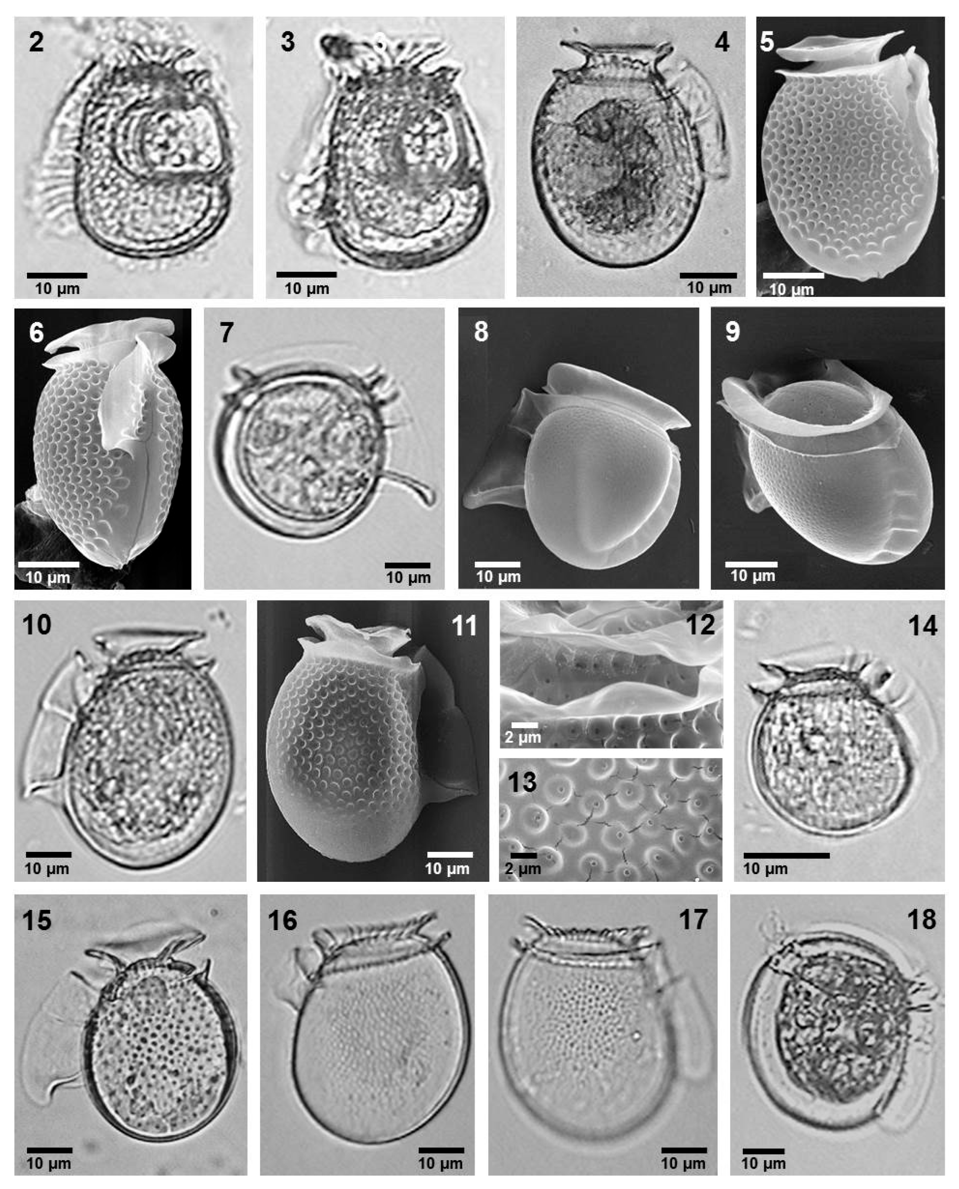
  • Citharistes regius Stein (Figure 2, images 2 and 3)
  • References: [39] (p. 73, pl. 22, figs 1–4) and [40] (p. 20).
Small-sized species, with an irregular ovoid body and a large phaeosomal chamber occurring in the hypotheca. The epitheca is low and very reduced, while the hypotheca is wider and has a C shape. Both cingular lists are vertical and have ribs, and the anterior cingular list is small. The left sulcal list is long with three ribs and is ornamented. N = 2.
  • Dimensions: L 35 µm, W 28 µm.
  • Local distribution: Gulf of California, at 30 °C and 35 salinity (localities g and i, Figure 1).
  • Genus: Dinophysis Ehrenberg
  • Section: Acuta Kofoid et Skogsberg
  • Dinophysis acuminata Claparède et Lachman (Figure 2, images 4–6)
  • References: [39] (p. 39, pl. 5, figs 5–8), [40] (p. 36), [41] (p. 433, fig. 4), and [42] (p. 43, fig. 7a–y).
Potentially toxic species, producing Okadaic acid, Dinophysistoxins, and Pectenotoxins [15].
Medium-sized species, with an oval body in the lateral view and a semi-elliptic body in the dorso-ventral view. The epitheca is reduced, and the hypotheca is convex with the posterior margin having small bumps. The cingulum is convex and ornamentated. The cingular lists are large and lack ribs. The sulcal left list with an R2 to the same distance of R1 than R3.
  • The theca has poroids with one pore. N = 18.
  • Dimensions: L 40–55 µm, W 30–40 µm.
  • Local distribution: western coast of the Baja California and central Mexican Pacific (localities n and bk).
  • Dinophysis exigua Kofoid et Skogsberg (Figure 2, images 7–9)
  • References: [11] (figs 2g,i and 3e), [39] (p. 43, pl. 6, figs 13–17), [40] (p. 34), and [43] (p. 239, fig. 30).
Small- to medium-sized species, with a spherical body in the lateral view and a semi-elliptic body in the dorso-ventral view. The epitheca is not prominent. The hypotheca is convex. The cingulum is wide and convex. The cingular lists lack ribs, and the sulcal left list has an R2 closer to R1 than to R3; R3 is very strong and claviform. N = 3.
  • Dimensions: L 40–50 µm, W 37.5–45 µm.
  • Local distribution: Gulf of California and central Mexican Pacific (localities n and t).
  • Dinophysis fortii Pavillard (Figure 2, images 10–13)
  • References: [39] (p. 43, pl. 6, figs 18–19), [42] (p. 54, fig. 13a–k), and [44] (p. 135, fig. 127a–c).
Medium-sized species, with an oval body in the lateral view. The epitheca is reduced. The hypotheca is convex. The cingulum is wide and convex, and the cingular lists have no ribs. The left sulcal list has an R2 to the same distance of R1 than R3. The theca has poroids with one pore. N = 43.
  • Dimensions: L 40–50 µm, W 32–40 µm.
  • Local distribution: western coast of the Baja California, Gulf of California, central Mexican Pacific, and Gulf de Mexico (localities a, p, t, u, ab, ac, ae, ah, an, ao, as, bj, bk, and bn).
  • Dinophysis infundibulus Schiller (Figure 2, image 14)
  • References: [11] (figs 2k and 3i–j) and [44] (p. 112, fig. 104).
Small species, with a body that is irregularly ovoid in the lateral view. The epitheca is low. The hypotheca has a wide and convex posterior margin. The cingulum is convex. The cingular lists have no ribs. The left sulcal list is poorly developed and lack an R3. N = 2.
  • Dimensions: L 38 µm, W 40 µm.
  • Local distribution: Gulf of Mexico (locality al).
  • Remarks: Potentially toxic species; analysis of selected cells showed a low proportion of Pectenotoxins [15].
  • Dinophysis recurva Kofoid et Skogsberg (Figure 2, image 15)
  • Reference: [44] (p. 113, fig. 105).
Small- to medium-sized species, oval body in lateral view, and reduced epitheca. Hypotheca convex. The cingulum is convex. The cingular lists have few ribs. The anterior cingular list has seven ribs. The left sulcal list has no R3. The theca has approximately 22 poroids per pore. N = 2.
  • Dimensions: L 40 µm, W 35 µm.
  • Local distribution: central Mexican Pacific (locality v).
  • Dinophysis similis Kofoid et Skogsberg (Figure 2, images 16 and 17)
  • References: [11] fig. 2j), [39] (p. 42, pl. 6, figs 1–4), [43] (p. 247, figs 31–1, 2), and [44] (p. 122, fig. 115a, b).
Medium-sized species, with an irregular oval body in the lateral view and low epitheca. The hypotheca is wide and convex. The cingulum is convex. The cingular lists have few ribs. The left sulcal list has no R3. The theca has approximately 22 poroids per pore. N = 2.
  • Dimensions: L 50–55 µm, W 50 µm.
  • Local distribution: Gulf of California, at 29 °C and 35 salinity (locality b).
  • Dinophysis sourniae Balech (Figure 2, Image 18)
  • References: [39] (p. 47, pl. 9, figs 14 and 15), [45] (p. 66, pl. 8, figs 113–118, pl. 9, figs 119 and 120).
Figure 2. Images 2 and 3—Citharistes regius, LM; Images 4–6—Dinophysis acuminata, LM and SEM; Images 7–9—Dinophysis exigua, LM and SEM; Images 10–13—Dinophysis fortii, LM and SEM; Image 14—Dinophysis infundibulus, LM; Image 15—Dinophysis recurva, LM; Images 16 and 17—Dinophysis similis, LM; Image 18—Dinophysis sourniae, LM.
Phycology 05 00048 g002
Medium-sized species, with an oval body in the lateral view. The epitheca is slightly high. The hypotheca has a convex margin. The cingulum is slightly convex. The cingular lists lack ribs. The left sulcal list is considerably long and lacks an R3. N = 3.
  • Dimensions: L 49 µm, W 44 µm.
  • Local distribution: central Mexican Pacific (locality e).
  • Section: Hastata Pavillard
All the species cited here were previously described and illustrated for the Mexican Pacific [27], and they will only be illustrated herein.
  • Dinophysis balechii Norris et Berner (Figure 3, image 19)
  • Reference: [27] (p. 1189, figs 3, 23, 34, 47, 62).
  • Dinophysis conjuncta Parra-Toriz, Esqueda-Lara et Hernández-Becerril (Figure 3, image 20)
  • Reference: [27] (p. 1189, figs 10, 27, 72, 83).
  • Dinophysis hastata Stein (Figure 3, images 21 and 22)
  • Reference: [27] (p. 1189, figs 4, 5, 24, 35, 36, 48, 55, 63).
  • Local distribution in this paper: Gulf of Mexico (localities an, ao, ap, as, ay, bb, bs).
  • Dinophysis monacantha Kofoid et Skogsberg (Figure 3, image 23)
  • Reference: [27] (p. 1189, figs 11 and 28).
  • Dinophysis nias Karsten (Figure 3, images 24 and 25)
  • Reference: [27] (p.1198, figs 18, 19, 32, 45, 54, 70).
  • Dinophysis phalacromoides (Jörgensen) Gómez, López-García et Moreira (Figure 3, image 26)
  • Reference: [27] (p. 1195, fig. 12).
  • Dinophysis pusilla Jörgensen (Figure 3, image 27)
  • Reference: [27] (p. 1196, figs 13, 14, 29, 41, 52, 58, 67).
  • Dinophysis schuettii Murray et Whitting (Figure 3, images 28 and 29)
  • Reference: [27] (p. 1197, figs 15, 16, 30, 42, 53, 68).
  • Local distribution in this paper: Gulf of Mexico (localities ah, ak, ay, bc, and bd).
  • Dinophysis swezyae Kofoid et Skogsberg (Figure 3, images 30 and 31)
  • Reference: [27] (p. 1198, figs 20, 21, 33, 46, 61, 71).
  • Dinophysis uracantha Stein (Figure 3, image 32)
  • Reference: [27] p. 1190, figs 6, 37, 38, 49, 56, 64.
  • Local distribution in this paper: Gulf of Mexico (localities af, ao, be, and bf).
  • Dinophysis uracantha var. mediterranea Jörgensen (Figure 3, image 33)
  • Reference: [27] (p. 1191, figs 7, 8, 39, 50, 65).
  • Dinophysis uracanthoides (Jörgensen) Gómez, López-García et Moreira (Figure 3, image 34)
  • Reference: [27] (p. 1191, figs 9, 26, 40, 51, 57, 66).
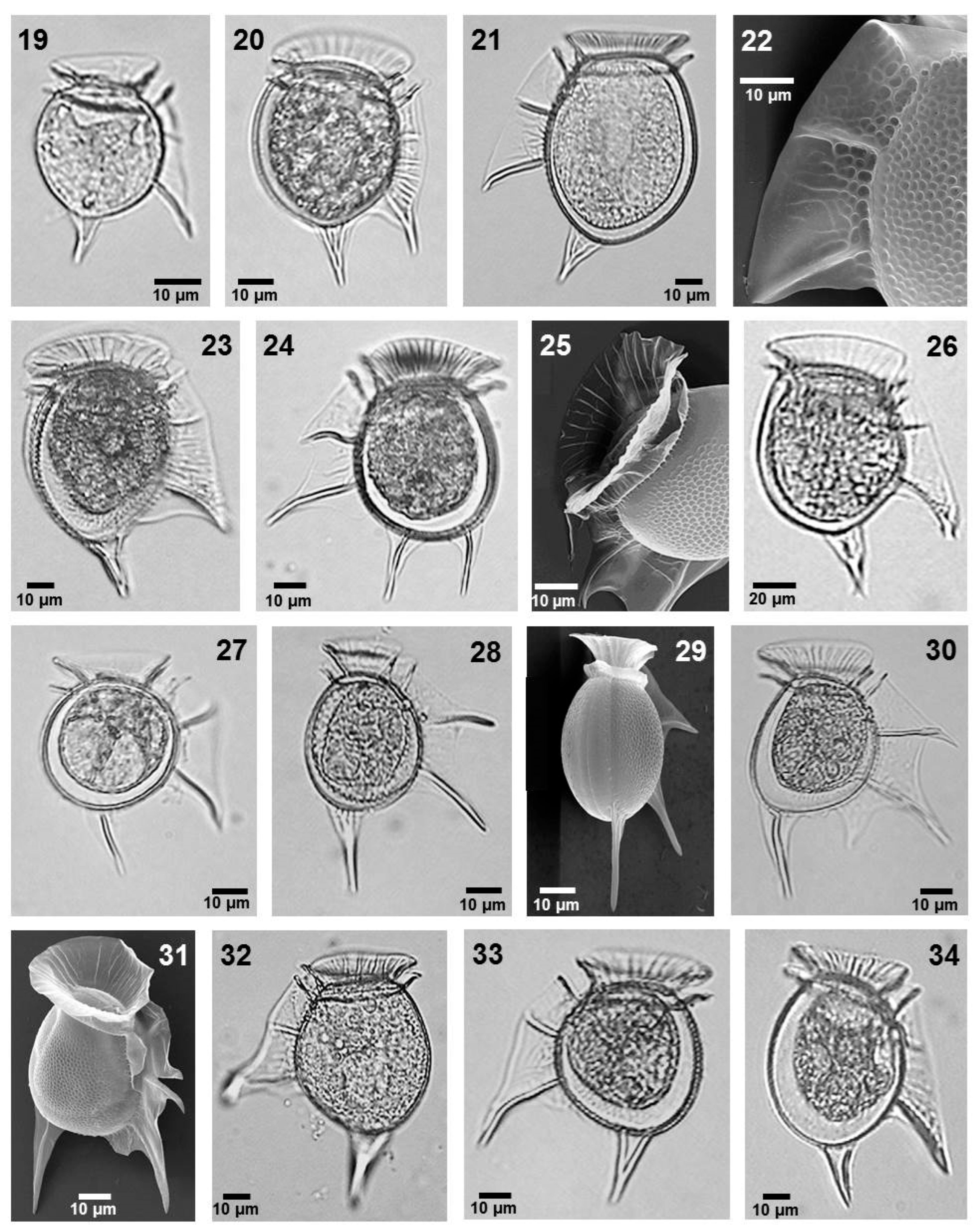
Figure 3. Image 19—Dinophysis balechi, LM; Image 20—Dinophysis conjuncta, LM; Images 21 and 22—Dinophysis hastata, LM and SEM; Image 23—Dinophysis monacantha, LM; Images 24 and 25—Dinophysis nias, LM and SEM; Image 26—Dinophysis phalacromoides, LM; Image 27—Dinophysis pusilla, LM; Images 28 and 29—Dinophysis schuettii, LM and SEM; Images 30 and 31—Dinophysis swezyae, LM and SEM; Image 32—Dinophysis uracantha, LM; Image 33—Dinophysis uracantha var. mediterranea, LM; Image 34—Dinophysis uracanthoides, LM.
Phycology 05 00048 g003
  • Section: Caudata Kofoid et Skogsberg
  • Dinophysis caudata Saville-Kent (Figure 4, images 35–39)
  • References: [39] (p. 45, pl. 8, figs 2 and 3), [40] (p. 37), [42] (p. 56, fig. 14a–d), and [46] (p. 24, figs 30–34).
Solitary or regularly paired cells of large size. Cells are irregularly trapezoidal in the lateral view and semi-elliptical in the dorsal-ventral view. The epitheca is slightly flattened in the center, not prominent. The hypotheca has a long projection directed posteriorly-ventrally, which can have one or two posterior spines. The cinlulum is convex, with ornamentation. The cingular lists are wide and with ribs. The left sulcal list has an R2 closer to R1 than to R3; R3 is long, thin, or slightly clavate. The theca ornamentation is of one pore per poroid. N = 70.
  • Dimensions: L 60–100 µm, W 47–95 µm.
  • Local distribution: western coast of the Baja California, Gulf of California, central Mexican Pacific, and Gulf of Mexico (localities a, b, c, m, n, o, r, s, t, u, ab, ac, ad, af, ah, ai, aj, ak, al, am, ao, ap, aq, ar, as, at, au, aw, ax, ay, az, bb, bc, bd, bk, bn, bo, bq, br, and bs).
  • Remarks: potentially toxic species, producing Okadaic acid and Dinophysistoxins [15].
  • Dinophysis caudata var. diegensis (Kofoid) Wood (Figure 4, images 40 and 41)
  • References: [44] (p. 151, fig. 144), [46] (p. 27, fig. 35), and [47] (p. 202, fig. 50a–g).
Solitary or regularly paired cells of medium size. Cells are irregularly trapezoidal in the lateral view and semi-elliptical in the dorsal-ventral view. The epitheca is slightly flattened in the center. The hypotheca has a short triangular projection in the posterior ventral region directed posteriorly-ventrally. The cingulum is convex. The cingular lists are wide and have ribs. The left sulcal list is long, with an R2 closer to R1 than to R3; R3 is long, thin, or slightly clavate. Theca ornamentation with depressions and pores. N = 2.
  • Dimensions: L 65 µm, W 35–38 µm.
  • Local distribution: central Mexican Pacific (locality n).
  • Dinophysis tripos Gourret (Figure 4, images 42–46)
  • References: [39] (p. 45, pl. 7, fig. 7, pl. 8, fig. 1), [44] (p. 158, fig. 140), and [46] (p. 29, figs 38 and 39).
Solitary or regularly paired cells of large size, with irregularly trapezoidal cells in the lateral view and semi-elliptical in the dorsal-ventral view. The epitheca is slightly flattened in the center. The hypotheca is trapezoidal and has a long projection in the posterior ventral region directed posteriorly-ventrally, which can have one or two posterior spines, and has a short projection in the posterior dorsal region. The cingulum is convex. The cingular lists are wide, showing ribs. The left sulcal list is long, with an R2 closer to R1 than to R3. R3 can be long, thin, or slightly clavate.
  • The theca ornamentation is of one pore per poroid. N = 40.
  • Dimensions: L 100–110 µm, W 50 µm.
  • Local distribution: western coast of the Baja California and Gulf of California (localities a and u).
  • Remarks: potentially toxic species, producing Pectenotoxins and Dinophysistoxins; nevertheless, its toxic potential for human intoxications is low [15].
  • Genus: Metaphalacroma Tai et Skogsberg
  • Metaphalacroma skogsbergi Tai (Figure 4, images 47 and 48)
  • References: [11] (figs 2p and 3l), [39] (p. 38, pl. 5, figs 3 and 4), [41] (p. 458, fig. 11), [45] (p. 43, pl. 3, figs 20–30), and [48] (p. 141, fig. 110a–e).
Figure 4. Images 35–39—Dinophysis caudata, LM and SEM; Images 40 and 41—Dinophysis caudata var. diegensis, LM; Images 42–46—Dinophysis tripos, LM and SEM; Images 47 and 48—Metaphalacroma skogsbergii, LM and SEM; Images 49 and 50—Pseudophalacroma nasutum, LM and SEM.
Phycology 05 00048 g004
Medium-sized species, with an ovoid body in the lateral view. The epitheca is low and the hypotheca is wide. The cingular lists are horizontal and lack ribs. The cingulum has two rows of poroids and a row of pores. The right sulcal list is wider than the left one, both with ornamentations. The theca is ornamented with poroids and pores.
  • The sagittal suture is serrated, except the ventral margin of the epitheca and sulcal lists area. N = 6.
  • Dimensions: L 50–55 μm, W 45–55 μm.
  • Local distribution: Gulf of California (localities a, b, and t).
  • Genus: Pseudophalacroma Jörgensen
  • Pseudophalacroma nasutum (Stein) Jörgensen (Figure 4, images 49 and 50)
  • References: [11] (figs 2r and 3k), [41] (p. 469, fig. 13), [44] (p. 55, fig. 54a–c), and [46] (p. 3, fig. 1).
Medium-sized cells, with a circular shape in the ventral view and an oval shape in the lateral view. The epitheca is prominent, with a notch in the ventral position. The hypotheca is with a convex margin. The cingulum is wide and concave. The cingular lists are horizontal and short, having short ribs. The sulcal lists appear united, with a reduced sulcal left list and no ribs.
  • The theca is ornamented with one pore in every poroid. N = 1.
  • Dimensions: T 45 μm, W 39.5 μm.
  • Local distribution: western coast of Baja California (locality u).
  • Genus: Histioneis Stein
  • Histioneis biremis Stein (Figure 5, images 51–56)
  • References: [11] (fig. 8h), [34] (p. 6, fig. 61), [44] (p. 254, fig. 250), and [49] (p. 44, pl. 9, fig. 89).
Medium-sized species with reduced epitheca, irregular hypotheca, and a dorso-posterior prolongation. The anterior cingular list has a funnel shape and conspicuous ribs, whereas the posterior cingular list has a conspicuous transverse rib. The cingulum is enlarged to contain the phaeosomal chamber, where often the so-called phaeosomes (usually cyanobacteria) are found. The epitheca has a lobular dorso-ventral projection. Both sulcal lists are ornamented and united at the anterior part. The left sulcal list is narrow and irregularly concave with a long and concave R3; R2 is located at the same distance as R1 and R3. The theca has one pore for each six or seven poroids. N = 1.
  • Dimensions: L 31 µm, W 51 µm.
  • Local distribution: Gulf of California (locality a).
  • Histioneis costata Kofoid et Michener (Figure 5, images 57–61)
  • References: [11] (fig. 8j) and [44] (p. 232, fig. 25).
Medium- to large-sized species with a reduced and inclined epitheca and an ovoid hypotheca, funnel-shaped anterior cingular list, with internal ribs, and a narrow phaeosomal chamber. The left sulcal list is long and hyaline, with a continuous marginal rib, from R2 to R3. The right sulcal list is reduced and short, not united to the left one. Theca ornamentation shows one pore for each six or seven poroids. N = 6.
  • Dimensions: L 76–82 µm, W 35 µm.
  • Local distribution: Gulf of California and Gulf of Mexico (localities a and aa).
  • Histioneis crateriforme Stein (Figure 5, images 67–71)
  • References: [11] (fig. 8e) and [39] (p. 63, pl. 15, fig. 8).
Small-sized species with rounded hypotheca and reduced epitheca. The cingulum is concave. The anterior cingular list is wide and low, and with strong ribs and ornamentations at its base. Posterior cingular list is convex, and the phaeosomal chamber is smaller than that of precedent species. Rounded hypotheca. The left sulcal list is narrow, ornamented, and irregularly convex with a thick R3 in the posterior position. R2 is closer to R1 than to R3. The theca has one pore for each four or six poroids. N = 1.
  • Dimensions: L 34 µm, W 34 µm.
  • Local distribution: central Mexican Pacific (locality p).
  • Histioneis depressa Schiller (Figure 5, images 62 and 63)
  • References: [40] (p. 53) and [44] (p. 237, fig. 230).
Figure 5. Images 51–56—Histioneis biremis, LM and SEM; Images 57–61—Histioneis costata, LM and SEM; Images 62 and 63—Histioneis mitchellana, SEM; Image 64—Histioneis depressa, SEM; Images 65 and 66—Histioneis garrettii, LM and SEM; Images 67–71—Histioneis crateriformis, LM and SEM.
Phycology 05 00048 g005
Medium-sized species with low, reduced epitheca and hypotheca with a kidney shape in the slateral view. The anterior cingular list is elongate and funnel-shaped, with ribs at the most anterior portion. The posterior cingular list is well developed and ornamented, in the upper part, above the transverse rib. High phaeosomal chamber (still with phaeosomes). The left sulcal list is long and thin, with incipient wings (additional lists) rising from R3, and the right sulcal list is reduced and short. The theca appears smooth, with randomly distributed, small pores. N = 2
  • Dimensions: L 56 µm, W 30 µm.
  • Local distribution: central Mexican Pacific (locality v).
  • Histioneis garrettii Kofoid (Figure 5, images 65 and 66)
  • References: [39] (p. 64, pl. 15, fig. 3) and [43] (p. 596, pl. 19, fig. 93).
Medium-sized species, with cells that have an ovoid body. The posterior part is slightly acute, and the epitheca is relatively wider than the previous species. The anterior cingular list is wide, low, and has conspicuous ribs. The posterior cingular list has no transverse rib nor defined phaeosomal chamber. The left sulcal list is long, slightly ornamented, and narrow between R1 and R2, becoming wider between R2 and R3. R3 is thick. The theca shows poroids and pores. N = 1.
  • Dimensions: L 88 µm, W 39 µm.
  • Local distribution: central Mexican Pacific, at 28.9 °C and 32 salinity (locality r).
  • Histioneis mitchellana Murray et Whitting (Figure 5, image 64)
  • References: [39] (p. 66, pl. 16, fig. 4) and [40] (p. 53).
Medium- to large-sized species with a low, reduced epitheca and a large hypotheca with a kidney shape. The species is similar to H. depressa, as follows: funnel-shaped anterior cingular list with multiple ribs, and a higher and much more ornamented posterior cingular list. Irregularly shaped quadrangular phaeosomal chamber, and a strong transverse rib. The left sulcal list is irregularly shaped, long, wide, with a strong ornamentation, well-developed wings (additional perpendicular lists) around R3, and a conspicuous window. N = 1.
  • Local distribution: central Mexican Pacific (locality v).
  • Dimensions: L 77 µm, W 57 µm.
  • Genus: Ornithocercus Stein
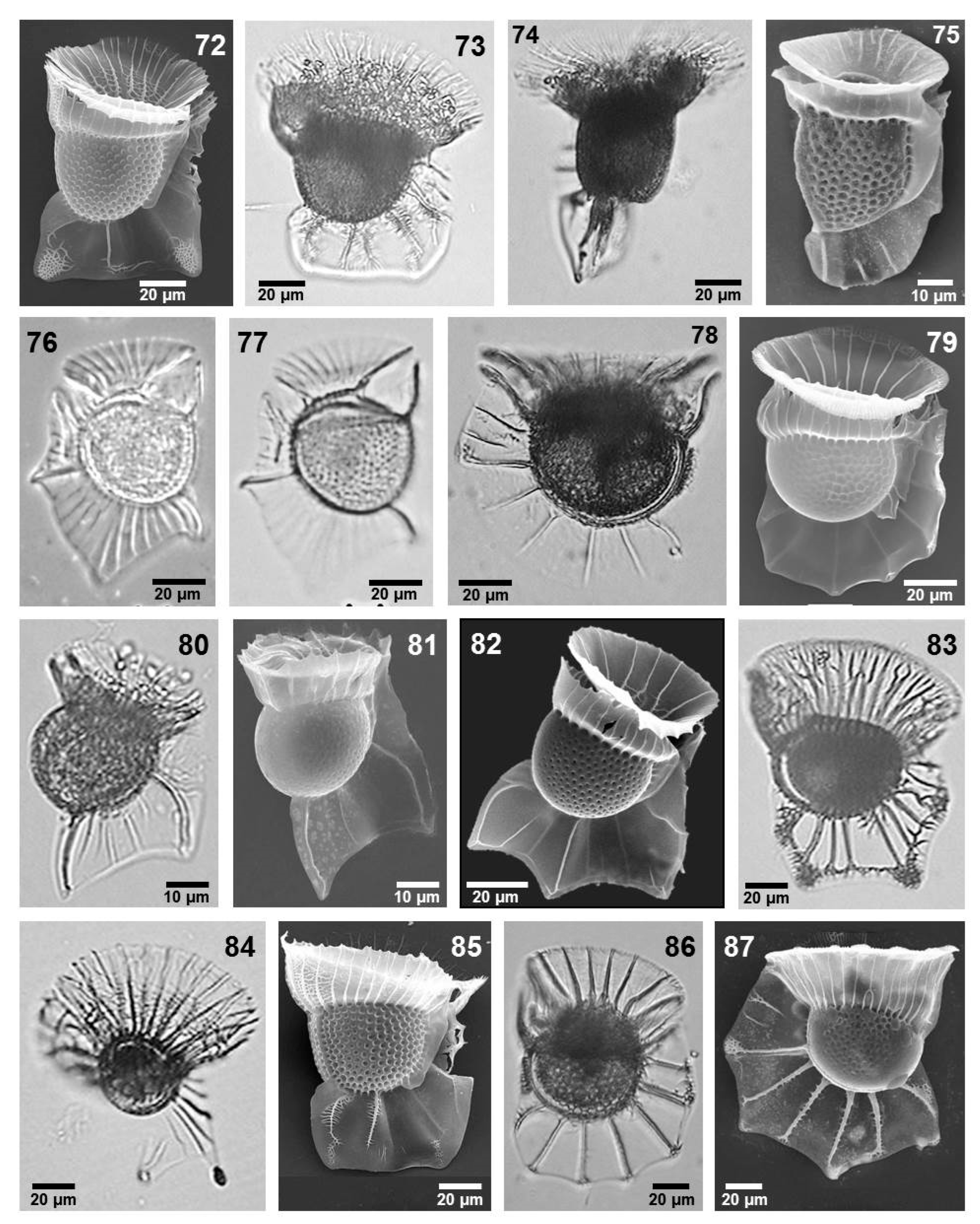
  • Ornithocercus assimilis Jörgensen emend Wilke et Hoppenrath (Figure 6, images 72–74)
  • Synonym: Ornithocercus galea (Pouchet) Abé
  • References: [28] (p. 569, fig. 81), [35] (p. 242, figs 20 and 21), [49] (p. 50, pl. 8, fig. 79), and [50] (p. 165, fig. 158a–c).
Large-sized species, with a round to oval body. The epitheca is flat and broad. The cingular lists have nine to eleven simple complete radial ribs and five to eight incomplete radial ribs on each lateral half, with ornamented fragments at the base, and the posterior cingular list has twelve radial ribs, which may also present as ornamentation. It has an extended and quadrangular left sulcal list with five ribs, three of which are central. Two lobes can be present at the extremes of the list. The right sulcal list is reduced and short. The theca has an ornamentation with seven or eight depressions per pore. N = 8.
  • Dimensions: L 125–154 μm, W 81–115 μm.
  • Local distribution: central Mexican Pacific and Gulf of Mexico (localities b, d, e, f, g, n, and au).
  • Ornithocercus cristatus Matzenauer (Figure 6, image 75)
  • References: [51] (p. 447, fig. 11) and [52] (p. 93, pl. II, figs 38–46).
Medium-sized cells with an irregularly ovoid shape. The epitheca is reduced, and the hypotheca is slightly elongated. The cingular list has a few simple ribs, and the anterior cingular list has incomplete ribs. The left sulcal list is poorly developed, with few incomplete ribs. The ornamentation has poroids and pores. N = 1.
  • Dimensions: L 70 μm, W 47 μm.
  • Local distribution: central Mexican Pacific (locality t).
  • Ornithocercus francescae (Murray et Whitting) Balech (Figure 6, images 76 and 77)
  • References: [53] (p. 333, pl. 32) and [54] (p. 136, pl. XVIII, fig. 259).
Large- to medium-sized species. The body is semispherical in the lateral view. The cingulum is concave. The anterior cingular list has multiple simple ribs, and the posterior one has only a few ribs. The left sulcal list is broad with numerous ribs and three points, where the central lone is the largest. The left sulcal list has many ribs. N = 1.
  • Dimensions: L 92 μm, W 64 μm.
  • Local distribution: central Mexican Pacific (locality t).
  • Ornithocercus heteroporus Kofoid (Figure 6, images 80 and 81)
  • References: [39] (p. 59, pl. 14, fig. 4), [40] (p. 43), [46] (p. 38, fig. 54), and [43] (p. 517, pl. 18, figs 1 and 3).
Medium-sized with an elliptical body, slightly elongated longitudinally. The epitheca is small and flat, and the hypotheca is oval. The cingulum is convex. The cingular lists have five complete ribs on each half. The left sulcal list extends to the posterior margin and has two well-developed ribs. The ornamentation consist of one pore for each poroid. N = 2.
  • Dimensions: L 69–81 μm, W 40–55 μm.
  • Local distribution: central Mexican Pacific (locality s).
  • Ornithocercus magnificus Stein (Figure 6, image 82)
  • References: [35] (p. 244, figs 20 and 21), [39] (p. 61, pl. 14, figs 7 and 8), [40] (p. 44), [43] (p. 529, pl. 16, figs 3 and 79), and [46] (p.35, fig. 48).
Cells of medium-size, with a slightly circular body in the lateral view. The cingular lists bear complete ribs. The anterior cingular list has five to six complete radial ribs. The posterior cingular list bears ten simple, complete ribs. The left sulcal list shows five simple or branched ribs, with three lobes, and the margin between lobes is convex. The R2 is conspicuous. The thecal ornamentation has a pattern of one pore per six poroids. N = 14.
  • Dimensions: L 47–55 μm, W 75–93 μm.
  • Local distribution: central Mexican Pacific and Gulf of Mexico (localities o, r, t, aa, ac, ad, ae, ao, au, and bg).
  • Ornithocercus orbiculatus Kofoid et Michener (Figure 6, images 78 and 79)
  • References: [34] (p. 9, figs 34 and 35), [39] (p. 61, pl. 15, fig. 2), [43] (p. 559, pl. 17, fig. 7), and [50] (p. 164, fig. 157a–c).
Large-sized species, with a circular and wide body. The epitheca is flat, oblique, and relatively extensive. The cingulum is asymmetrical, wide, and concave. The cingular lists are wide, the anterior cingular list has three to five complete, radial ribs and five to eight short ribs between complete ribs. The left sulcal list has a convex margin that is almost smooth, with small or no lobes and weak ribs. There is one pore per poroid. N = 5.
  • Dimensions: L 112–143 μm, W 102–112 μm.
  • Local distribution: central Mexican Pacific and Gulf of Mexico (localities o, r, and ac).
  • Remarks: This species has been considered as a synonym of O. steinii; however, they can be separated according to the number of ribs in the anterior cingular list and the shape of the margin of the left sulcal list.
  • Ornithocercus quadratus Schütt (Figure 6, image 83)
  • References: [35] (p. 244, figs 25 and 26), [39] (p. 60, pl. 14, fig. 10), [40] (p. 45, 46), and [49] (p. 50, pl. 8, fig. 81).
Large-sized species, with a round body, slightly sharp at the posterior margin. The epitheca is flat. The anterior cingular list shows ten complete radial ribs, sometimes bifurcated and numerous incomplete ribs. The posterior cingular list also has ribs. The left sulcal list has ribs (two or three, centered), with ornamentation originating from the rib; its margin is wide and slightly concave, almost rectilinear, with two lobes. The thecal ornamentation shows pores and poroids. N = 1.
  • Dimensions: L 128 μm, W 106 μm.
  • Local distribution: Gulf of California (locality b).
  • Ornithocercus schuetti T. Wilke et Hoppenrath (Figure 6, image 85)
  • References: [28] (p. 569, fig. 80).
Large-sized cells with a round shape in the lateral view. The cingular lists have radial ribs, the anterior cingular list has ten radial ribs on each half, and the posterior cingular list has thirteen complete simple ribs, with ornamentation in the margin dorsal. The left sulcal list has four ribs, which can be branched along their length with a serrate appearance. N = 1.
  • Dimensions: L 85 μm, W 58 μm.
  • Local distribution: Gulf of California (locality e).
  • Ornithocercus splendidus Schütt (Figure 6, image 84)
  • References: [35] (p. 244, figs 31–33), [39] (p. 60, pl. 14, figs 5 and 6), [40] (p. 48), [43] (p. 521, pl. 16, figs 2 and 4, pl. 17, fig. 3), and [44] (p. 198, fig. 190).
Cells of a large size, with a triangular body in the lateral view and irregularly oval in the dorsal or ventral view. The anterior cingular list shows complete radial ribs, with six to eight ribs on each half, ornamented. The left sulcal list extends to the ventral margin, with light radial ornamentation and two large lobes. N = 2.
  • Dimensions: L 141 μm, W 43.6 μm.
  • Local distribution: Gulf of California and Gulf of Mexico (localities i, k, and au).
  • Ornithocercus steinii Schütt (Figure 6, image 86)
  • References: [11] (fig. 7g), [35] (p. 246, figs 27 and 28), [39] (p. 61, pl. 15, fig. 1), [40] (p. 49), [43] (p. 551, pl. 16, figs 1 and 86), and [46] (p. 36, fig. 49).
Large-sized species with a subcircular body. The cingulum is asymmetrical, wide, and slightly concave. The cingular lists are wide, the anterior cingular list has seven to eight complete radial ribs on each half. There are ten short radial ribs between complete ribs or without short ribs. The posterior cingular list also has very ribs. The left sulcal list has four posterior lobes poorly marked, each with a rib and short branches; it has five complete ribs, sometimes branched along their length, with a serrate appearance. The contour between the ribs is clearly concave. The theca ornamentation shows pores and poroids. N = 6.
  • Dimensions: L 137–150 μm, W 100 μm.
  • Local distribution: Gulf of California, central Mexican Pacific and Gulf of Mexico (localities a, b, c, d, e, f, g, h, i, j, k, m, n, q, r, s, t, and ao).
  • Ornithocercus thumii (Schmidt) Kofoid et Skogsberg (Figure 6, image 87)
  • References: [11] (fig. 7h), [35] (p. 246, figs 23 and 24), [39] (p. 61, pl. 14, fig. 9), [40] (p. 50, 51), [43] (p. 540, pl. 18, figs 4, 6, 81), and [44] (p. 200, fig. 191).
Figure 6. Images 72–74—Ornithocercus assimilis, SEM and LM; Image 75—Ornithocercus cristatus, SEM; Images 76 and 77—Ornithocercus francescae, LM; Images 78 and 79—Ornithocercus orbiculatus, LM and SEM; Images 80 and 81—Ornithocercus heteroporus, LM and SEM; Image 82—Ornithocercus magnificus, SEM; Image 83—Ornithocercus quadratus, LM; Image 84—Ornithocercus splendidus, LM; Image 85—Ornithocercus schuettii, SEM; Image 86—Ornithocercus steinii, LM; Image 87—Ornithocercus thumii, SEM.
Phycology 05 00048 g006
The cells have a large size and a subcircular body. The cingulum is asymmetrical, wide, and slightly concave. The cingular lists are wide, and the anterior cingular list has five to seven and eight to ten short ribs on each half. The posterior cingular list has many ribs (10–12). The left sulcal list is extended and has three marked posterior lobes, each with a rib and short branches. The ornamentation consists of pores and poroids. N = 4.
  • Dimensions: L 125–153 μm, W 89–106 μm.
  • Local distribution: Gulf of California, central Mexican Pacific and Gulf of Mexico (localities d, f, g, h, j, k, l, q, r, s, and ae).
  • Family: Oxyphysaceae Sournia
  • Genus: Phalacroma Stein
  • Section: Rotundatum Kofoid et Skogsberg
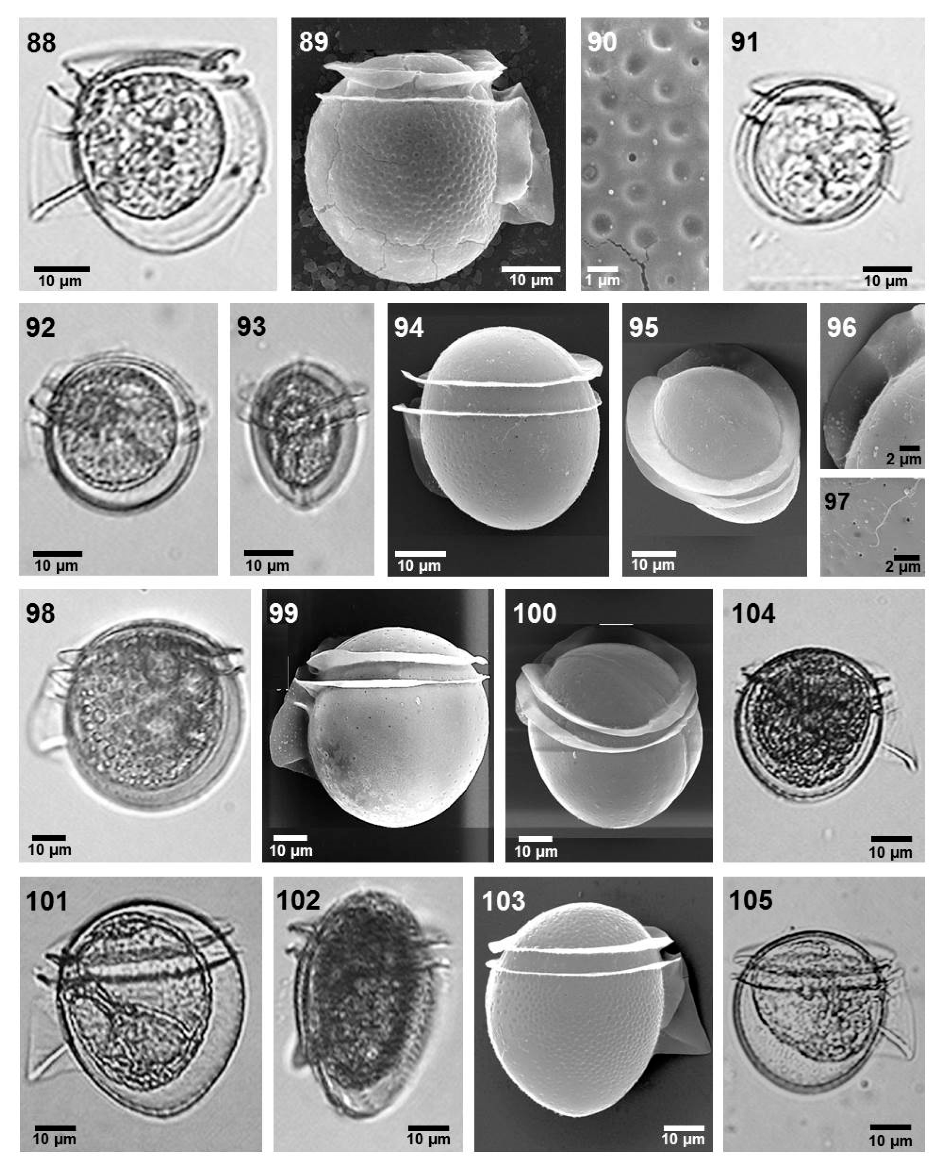
  • Phalacroma laeve (Claparède et Lachman) Díaz-Ramos (Figure 7, images 92–97)
  • References: [39] (p. 52, pl. 12, fig. 4) and [44] (p. 149, fig. 141).
A small-sized species. The body is circular in the lateral view and oval in the dorso-ventral view. The epitheca is prominent, and the hypotheca has a rounded posterior margin. The cingulum is slightly convex. The cingular lists are poorly developed and lack ribs. The left sulcal list is long with a convex margin. R2 is closer to R1 than to R3. R3 is very short. The ornamentation pattern consists of one pore per fourteen poroids. N = 5.
  • Dimensions: L 36 µm, W 38 µm.
  • Local distribution: Gulf of California, central Mexican Pacific and Gulf of Mexico (localities m, t, am, bb, and bo, Figure 1).
  • Phalacroma operculoides Schütt (Figure 7, images 101–103)
  • Reference: [44] (p. 64, fig. 58a,b).
Medium-sized species. The body is spherical in the lateral view and oval in the dorso-central view. The epitheca is prominent, the hypotheca is convex, and the cingulum is convex. The cingular lists are inconspicuous, with no ribs. The sulcal lists have a notch (“reinforcement”) between R1 and R2. R2 is closer to R1 than R3, and R3 is large and clavate. The theca ornamentation shows poroids and pores. N = 9.
  • Dimensions: L 67–75 µm, W 53–57.5 µm.
  • Local distribution: Gulf of Mexico (locality ab, ah, aj, ak, as, and bo).
  • Remarks: See the remarks under Phalacroma ovum Schütt.
  • Phalacroma ornamentatum Esqueda-Lara et Hernández-Becerril (Figure 10, images 147–149)
  • References: [3] (p. 304, figs 2–14, 34) and [55] (p. 37).
Small- to medium-sized species. Cells have an elliptical shape in the lateral view and oval in the dorsal view. The epitheca is prominent. The hypotheca is wide, with its posterior margin rounded. The cingulum has two rows of pores and two to four rows of poroids. The cingular lists have no ribs. The left sulcal list is narrow, with the R2 closer to R1 than to R3. The right sulcal list is ornamented. The thecal ornamentation is characterized by pores and poroids, with an apparent pattern of one pore for each 3–6 poroids. N = 12.
  • Dimensions: L 54–57 µm, W 64–67 µm.
  • Local distribution: central Mexican Pacific and Gulf of Mexico (localities a, aa, ah, ai, aj, aw, ax, ay, bb, bd, dj, and bn).
  • Phalacroma parvulum (Schütt) Jörgensen (Figure 7, images 104 and 105)
  • References: [39] (p. 49, pl. 10, figs 4–6) and [46] (p. 7, fig. 4).
Figure 7. Images 88–90—Phalacroma rotundatum, LM and SEM; Image 91—Phalacroma whittingiae, LM; Images 92–97—Phalacroma laeve, LM and SEM; Images 98–100—Phalacroma rudgei, LM and SEM; Images 101–103—Phalacroma ovum, LM and SEM; Images 104 and 105—Phalacroma parvulum, LM.
Phycology 05 00048 g007
Medium-sized species, and the body is spherical in the lateral view. The epitheca is high, and the hypotheca has convex margins. The cingulum is narrow and convex. The cingular lists are small and have no ribs. The left sulcal list has an R2 closer to R2 than R3. R3 is strong and slightly convex. The ornamentation shows pores and poroids. N = 9.
  • Dimensions: L 50–62 µm, W 50–61 µm.
  • Local distribution: central Mexican Pacific (localities n, o, and r).
  • Phalacroma rotundatum Claparéde et Lachmann (Figure 7, images 88–90)
  • References: [40] (p. 33), [42] (p. 57, fig. 15a–h), [46] (p. 5, fig. 2), and [55] (p. 36).
A vector species of Dinophysistoxins [15], with solitary cells that are small to medium in size, with a rounded body in the lateral view and oval in the dorsal or ventral view. Cingulum slightly convex. The cingular lists are reduced and show no ribs. The left sulcal list has three ribs, with the R2 closer to R1 than to R3, which is thick. The theca is ornamented with one pore for each 16 poroids. N = 11.
  • Dimensions: L 39–46 µm, W 35–45 µm.
  • Local distribution: Gulf of California, central Mexican Pacific and Gulf of Mexico (localities a, b, j, t, ac, af, ag, am, an, ao, and bo).
  • Phalacroma paulsenii Schiller (Figure 8, images 115–117)
  • References: [44] (p. 98, fig. 89a, b) and [52] (p. 88, pl. I, figs 13–19).
Solitary cells of small to medium size, with a rounded body in the lateral view and oval in the dorsal or ventral view. The epitheca is low, and the hypotheca is rounded. Cingular lists are horizontal, rather narrow, and without ribs. The left sulcal list is not wide, inconspicuous, and has three ribs. The R2 is closer to R1 than to R3. The theca is ornamented with poroids and pores. N = 5.
  • Dimensions: L 38–50 µm, W 33–46.5 µm.
  • Local distribution: Gulf of California (at 28–29 °C and 34–35 salinity) (locality a and c).
  • Phalacroma rudgei Murray et Whitting (Figure 7, images 98–100)
  • References: [42] (p. 59, fig. 16a–j), [44] (p. 66, fig. 59a,b), and [56] (p. 21, figs 5 and 6).
Solitary cells that are medium-sized, with an elliptical body in the lateral view and oval in the dorsal or ventral view. The epitheca is domed, the hypotheca is rounded, and the cingulum is not excavated. The cingular lists are semi-horizontal, narrow, and show no ribs. The left sulcal list is rather narrow, with the R2 shorter and closer to R1 than to R3 (Figure 4, images 40 and 41). The theca exhibits pores (Figure 4, image 50). N = 1.
  • Dimensions: L 58 µm, W 52 µm.
  • Local distribution: Gulf of California, at 30 °C and 35 salinity (locality k).
  • Phalacroma whittingiae (Balech) Esqueda-Lara et Hernández-Becerril nov. comb.
  • (Figure 7, image 91)
  • Basyonym: Dinophysis whittingiae Balech
  • Reference: [45] (p. 73, pl. 10, figs 154–167).
Solitary cells, small in size, with a rounded body in the lateral view and oval in the dorsal or ventral view. The epitheca is slightly prominent, the cingulum is not excavated, and the hypotheca is round. The cingular lists have no ribs. The left sulcal list is extremely inconspicuous and narrow and has an R2 closer to R1 than to R3. N = 4.
  • Dimensions: L 30–31 µm, W 30–34 µm.
  • Local distribution: Gulf of California at 30 °C and 34 salinity, Gulf of Mexico (localities a, l, m, and ab).
  • Section: Argus Kofoid et Skogsberg
  • Phalacroma apicatum Kofoid et Skogsberg (Figure 8, images 108 and 109)
  • Reference: [36] (p. 246, figs 34 and 35), [40] (p. 23), and [50] (p. 143, fig. 136).
Solitary cells and large in size. The body is spherical to subspherical and oval in the lateral view. The epitheca is prominent and acute. The cingulum is convex, and the cingular lists have ribs. The sulcal lists have a prominent notch between R1 and R2. The thecal ornamentation consists of large polygons and pores. N = 1.
  • Dimensions: L 100 µm, W 80 µm.
  • Local distribution: Gulf of California and central Mexican Pacific (localities d, e, m, o, r, and s).
  • Phalacroma argus Stein (Figure 8, images 106 and 107)
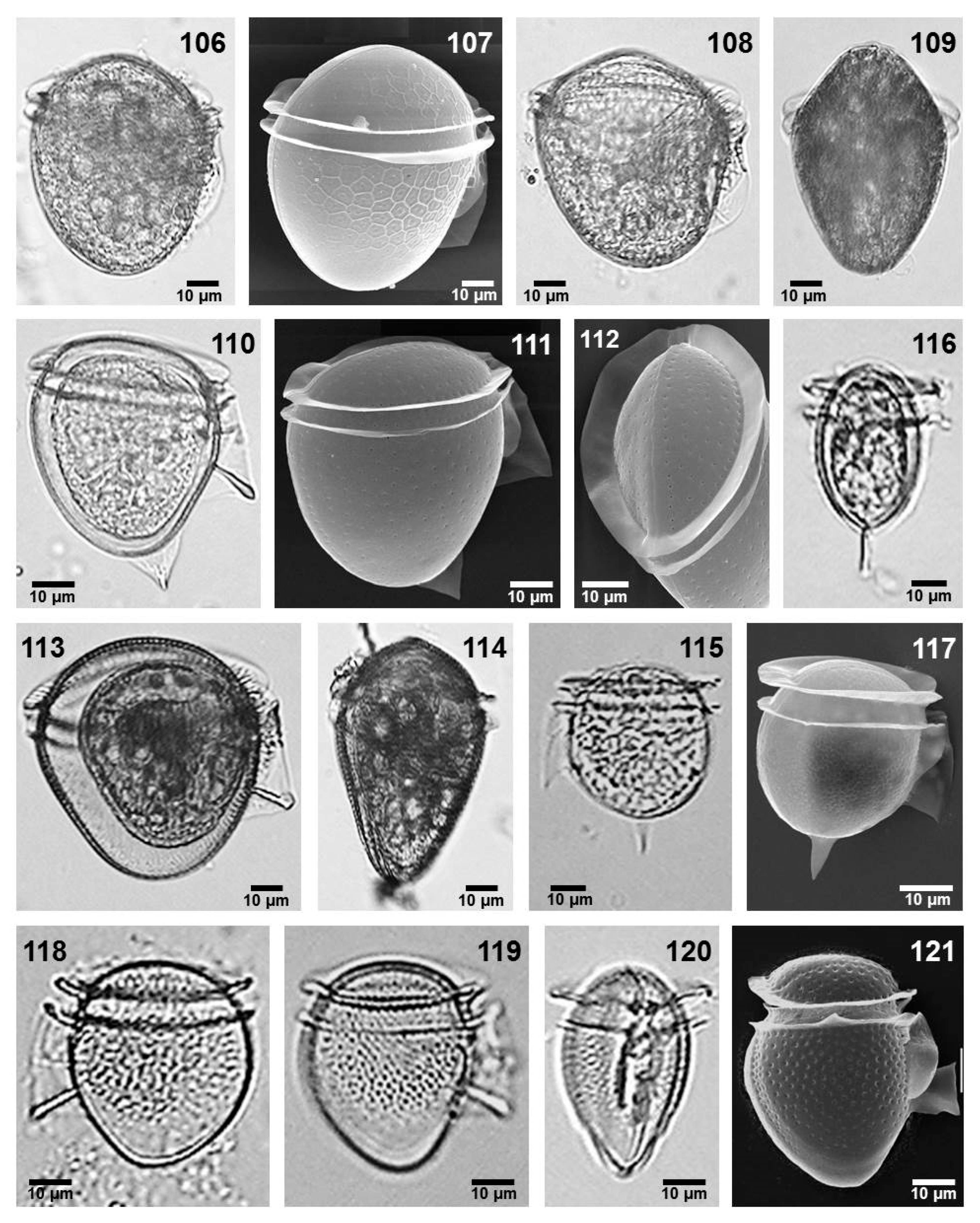
  • Reference: [35] (p. 246, fig. 36), [39] (p. 51, pl. 11, figs 7–10), [40] (p. 24), [42] (p. 71, fig. 23a–b), and [44] (p. 74, fig. 67a).
Figure 8. Images 106 and 107—Phalacroma argus, LM and SEM; Images 108 and 109—Phalacroma apicatum, LM; Images 110–112—Phalacroma doryphorum, LM and SEM; Images 113 and 114—Phalacroma porodictyum, LM; Images 115–117—Phalacroma paulsenii, LM and SEM; Images 118–121—Phalacroma scrobiculatum, LM and SEM.
Phycology 05 00048 g008
Large-sized species, with a round to subrotund body shape in the lateral view and oval in the dorsal or ventral view. The epitheca is prominent, and the hypotheca is round. The cingular lists lack ribs. The left sulcal list has three ribs. The theca is ornamented with large polygons and pores. N = 7.
  • Dimensions: L 80–97.5 µm, W 67.5–80 µm.
  • Local distribution: Gulf of California and central Mexican Pacific (localities d, e, m, o, r, and s).
  • Phalacroma ovum Schütt (Figure 10, images 145 and 146)
  • References: [39] (p. 50, pl. 10, figs 16 and 17) and [43] (p. 118, fig. 11).
  • Synonyms: Dinophysis amygdala Balech, Dinophysis amandula (Balech) Sournia
Medium-sized species, with solitary cells with an oval body. The epitheca is high and rounded or slightly flattened, and the hypotheca is ovoid. The cingulum is convex. The left sulcal list is well developed, with a strong R3. The ornamentation has one pore for seventeen poroids. N = 6.
  • Dimensions: L 55–72.5 µm, W 48–70 µm.
  • Local distribution: Gulf of California and Gulf of Mexico (locality a, m, and af).
Remarks: There has been a long debate about the identity of this name and species. This debate was summarized by Kofoid & Skogsberg [43], who additionally described and illustrated Phalacroma ovum, and, more recently, by Hallegraeff et al. [9]. We consider that there are two species involving Phalacroma operculoides (as it was already described and illustrated above) and P. ovum. Our specimens are quite similar to those illustrated by Kofoid & Skogsberg [43] and Hallegraeff et al. [9], with outstanding characteristics for the external margin of the left sulcal list, between R1 and R2, which is notoriously reinforced (“reinforcement”, following [12]), giving the perception that both sulcal lists are united.
  • Phalacroma scrobiculatum (Balech) Díaz-Ramos et Estrella (Figure 8, images 118–121)
  • References: [39] (p. 46, pl. 9, figs 5 and 6) and [45] (pl. 11, figs 168–173).
This is a medium-sized species, with an elliptical body in the lateral and dorsal or ventral views. The epitheca is prominent, and the hypotheca is acute at the posterior margin. The cingulum is not excavated with two rows of pores. The cingular lists are relatively narrow and show no ribs. The left sulcal list is relatively wide, with an R2 that is curved and closer to R1 than to R3. The theca is covered by poroids and pores. N = 41.
  • Dimensions: L 36–49 µm, W 35–45 µm.
  • Local distribution: Gulf of California, at 30 °C and 35 salinity, and central Mexican Pacific at 28 °C and 35 salinity, and Gulf of Mexico (locality a, b, j, k, l, aa, af, ah, ai, aj, al, ao, ap, aq, ar, as, at, aw, ax, ay, az, bb, bf, bj. bk, bl, and bn).
Remarks: The most particular character of P. scrobiculatum, apart from its shape and size, is the second ratio of the left sulcal list, which is concave and closer to R3 than R1.
  • Section: Cuneus Kofoid et Skogsberg
  • Phalacroma cuneus Schütt (Figure 9, images 122–125)
  • Reference: [35] (p. 247, figs 40 and 41), [39] (p. 51, pl. 11, figs 4–6), [40] (p. 26), [42] (p. 68, fig. 21), and [44] (p. 84, fig. 76).
The cells are solitary and large-sized. The epitheca is wide, and the hypotheca has a rounded posterior margin. The cingulum is convex, and the cingular lists have short ribs. The left sulcal list is narrow and rounded at the posterior margin, with an R3 that is small. The theca has reticules and pores. N = 13.
  • Dimensions: LT 80–100 µm, W 62.5–87.5 µm.
  • Local distribution: Gulf of California and central Mexican Pacific (localities k, m, n, q, r, s, and t).
  • Section: Rapa Kofoid et Skogsberg
  • Phalacroma rapa Stein (Figure 9, images 126–129)
  • References: [39] (p. 44, pl. 8, figs 6–8), [40] (p. 32), [42] (p. 66, fig. 19), and [44] (p. 88, fig. 80).
The cells are solitary and large-sized, with a trapezoid body in the lateral view. The epitheca is not prominent, and the hypotheca is trapezoidal. The cingulum is concave. The anterior cingular list has basal ribs. The left sulcal list is extended, with a thick and large R3. The right sulcal list reaches R3, and it is ornamented with short basal ribs. The theca is strongly ornamented with areolae, and there is one pore in almost every areola. N = 16.
  • Dimensions: L 80–98 µm, W 65–80 µm.
  • Local distribution: central Mexican Pacific and Gulf of Mexico (locality o, r, s, aa, ad, ah, al, bi, bo, bq, bs).
  • Phalacroma capitulatum (Balech) Hernández-Becerril (Figure 9, images 135–137)
  • References: [11] (figs 4h and 5h), [35] (p. 247, figs 49 and 50), and [52] (p. 90, pl. II, figs 25–31).
Solitary cells and small- to medium-sized species. Cells irregularly ovate in the lateral view, with a short posterior process. The epitheca is not prominent, and the cingulum is wide and not excavated. The anterior cingular list has basal ribs. The left sulcal list is rectangular, not extensive, and has a thick R3. The R2 extends the same distance as R1 and R3. The theca exhibits poroids and pores. N = 3.
  • Dimensions: L 40–50 µm, W 38–40 µm.
  • Local distribution: Gulf of California, at 30 °C and 35 salinity, and central Mexican Pacific (localities i, k, m, n, o, p, and r).
  • Phalacroma mitra Schütt (Figure 9, images 130–132)
  • Reference: [35] (p. 250, figs 54 and 55), [39] (p. 45, pl. 8, figs 9–11), [40] (p. 30), and [42] (p. 63, fig. 18a–q).
Potentially toxic species, producing Okadaic acid and Dinophysistoxins [15]. Solitary cells; medium- to large-sized species, with a trapezoid body in the lateral view. The epitheca is not prominent, and the hypotheca has an extensive and slightly convex ventral margin. The cingulum is concave. The anterior cingular list bears strong ribs. The left sulcal list is not prominent, showing a long and thick R3. The theca has large poroids, with one pore for most of the areolae. N = 28.
  • Dimensions: L 40–72 µm, W 38–67 µm.
  • Local distribution: Gulf of California, central Mexican Pacific, and Gulf of Mexico (localities i, k, m, n, o, p, r, t, aa, ae, af, al, bc, bd, bk, and bn).
  • Phalacroma favus Kofoid et Michener (Figure 9, images 133 and 134)
  • References: [40] (p. 28) and [42] (p. 67, fig. 20a–e).
The cells are solitary and large-sized, with å trapezoid body in the lateral view, leading to a posterior process. The epitheca is not prominent, and the hypotheca has a ventral margin that is very extensive and slightly convex until the sulcal list. The cingular lists show strong ribs. The left sulcal list appears rectangular, is not extensive, and has short ribs and a thick R3. The theca has large poroids (areolae), with one pore in almost every areola. N = 4.
  • Dimensions: LT 90–97.5 µm, W 62.5–77.5 µm.
  • Local distribution: central Mexican Pacific and Gulf of Mexico (localities n, p, and az).
Figure 9. Images 122–125—Phalacroma cuneus, LM and SEM; Images 126–129—Phalacroma rapa, LM and SEM; Images 130–132—Phalacroma mitra, LM and SEM; Images 133 and 134—Phalacroma favus, LM and SEM; Images 135–137—Phalacroma capitulata, LM and SEM. Section: Expulsum Kofoid et Skogsberg.
Phycology 05 00048 g009
  • Phalacroma expulsum Kofoid et Michener (Figure 10, images 138–141)
  • References: [35] (p. 250, figs 44 and 48), [40] (p. 27), and [44] (p. 94, fig. 86).
Solitary cells and a medium-sized species. The cells have an oval shape in the ventral view and amphoroid in the dorsal or ventral view and wider at the middle. The epitheca is low, and the hypotheca has rounded margins. The cingulum is concave. The cingular lists have no apparent ribs. The left sulcal list is slightly elongated and has no R3. The ornamentation has a pore in most of the poroids. N = 10.
  • Description: L 61 µm, W 65 µm.
  • Local distribution: central Mexican Pacific and Gulf of Mexico (locality s, v, ac, af, ag, am, ah, and az).
  • Phalacroma stenopterygium Jörgensen (Figure 10, images 142–144)
  • Reference: [45] (p. 11, fig. 10).
Solitary cells and medium-sized species, with an elliptical body in the lateral view and ovoid in the dorsal or ventral view. The epitheca is low, and the hypotheca is wide with a convex margin. The cingulum is convex. The cingular lists are vertical and parallel, and the anterior cingular list has incomplete ribs, which do not reach the edge. The reduced left sulcal list shows no R3. N = 1.
  • Dimensions: LT 50 µm, W 45 µm.
  • Local distribution: Gulf of California, at 28–29 °C and 34.9–35 salinity (localities b and d).
Remarks: This species has been traditionally considered a synonym of P. expulsum (44, 45); however, (1) P. stenopterygium has a narrower epitheca than P. expulsum; (2) P. stenopterygium has a cingulum convex, whereas for P. expulsum the cingulum is concave; and, most importantly, (3) in the ventral or dorsal view, P. stenopterygium shows a convex outline, whereas P. expulsum has an amphoroid outline. This species is a new record for the Mexican Pacific.
  • Section Doryphorum Kofoid et Skogsberg
  • Phalacroma doryphorum Stein (Figure 8, images 110–112)
  • References: [39] (p. 55, pl. 13, figs 11–13; 40, p. 35), [42] (p. 77, fig. 26), and [44] (p. 99, fig. 91).
Solitary cells and medium- to large-sized species, with an ovoid body. The epitheca is slightly flattened, and the hypotheca has a triangular appendage in the posterior margin. The cingulum is concave. It has narrow and horizontal cingular lists without ribs. The left sulcal list shows an R2 closer to R1; R3 is long, thick, and claviform. The theca has poroids and pores. N = 1.
  • Dimensions: LT 65–95 µm, W 55–88 µm.
  • Local distribution: Gulf of California, central Mexican Pacific, and Gulf of Mexico (localities i, k, m, n, q, t, and ah).
  • Phalacroma porodictyum Stein (Figure 8, images 113 and 114)
  • References: [39] (p. 50, pl. 10, figs 18–20), [40] (p. 31), [42] (p. 61, fig. 17a–f), and [46] (p. 6, fig. 9).
Solitary cells and medium- to large-sized species. Cells have an irregular oval shape and are wide in the lateral view. The epitheca is concave, and the hypotheca has a concave ventral margin. The cingular lists are reduced and show ribs. The left sulcal list shows a thick and long R3. The theca ornamentation exhibits poroids and pores. N = 20.
  • Dimensions: LT 68–90 µm, W 62.5–77.5 µm.
  • Local distribution: Gulf of California, central Mexican Pacific, and Gulf of Mexico (localities d, e, f, h, i, l, m, n, o, q, r, s, ah, ak, am, and at).
  • Genus: Oxyphysis Kofoid
  • Oxyphysis oxytoxoides Kofoid (Figure 10, images 151–154)
  • Synonym: Phalacroma oxytoxoides (Kofoid) F. Gómez, P. Lopez-Garcia et D. Moreira
  • Reference: [39] (p. 201, pl. 18, figs 10–12), [41] (p. 474, fig. 14), and [57] (p. 205, pl. 18, figs 1–4).
Figure 10. Images 138–141—Phalacroma expulsum, LM and SEM; Images 142–144—Phalacroma stenopterygium, LM; Images 145 and 146—Phalacroma operculoides, LM; Images 147–149—Phalacroma ornamentatum, LM and SEM; Images 150—Dinofurcula pseudoultima, LM; Images 151–154—Oxyphysis oxytoxoides, LM and SEM.
Phycology 05 00048 g010
Solitary cells or regularly paired and medium-sized. The body is elongate and biconical. The epitheca is prominent, tapering anteriorly. The cingulum is wide, concave, and decorated with poroids and two rows of pores. The hypotheca is acute, ending in a small spine. The cingular lists are narrow with no ribs. The ornamentation shows one small pore for approximately every 22 poroids. N = 2.
  • Dimensions: L 65 µm, W 15 µm.
  • Local distribution: western coast of Baja California, central Mexican Pacific, and Gulf of Mexico (localities o, u, bn, and bo).
  • Genus: Dinofurcula Kofoid et Skogsberg
  • Dinofurcula pseudoultima Hernández-Becerril (Figrue 10, image 150)
  • Reference: [36] (p. 5, figs 1–8).
This species was described elsewhere [37].
  • Local distribution: central Mexican Pacific (locality v). N = 1.
  • Family: Amphisoleniaceae Lindemann
  • Genus: Amphisolenia Stein
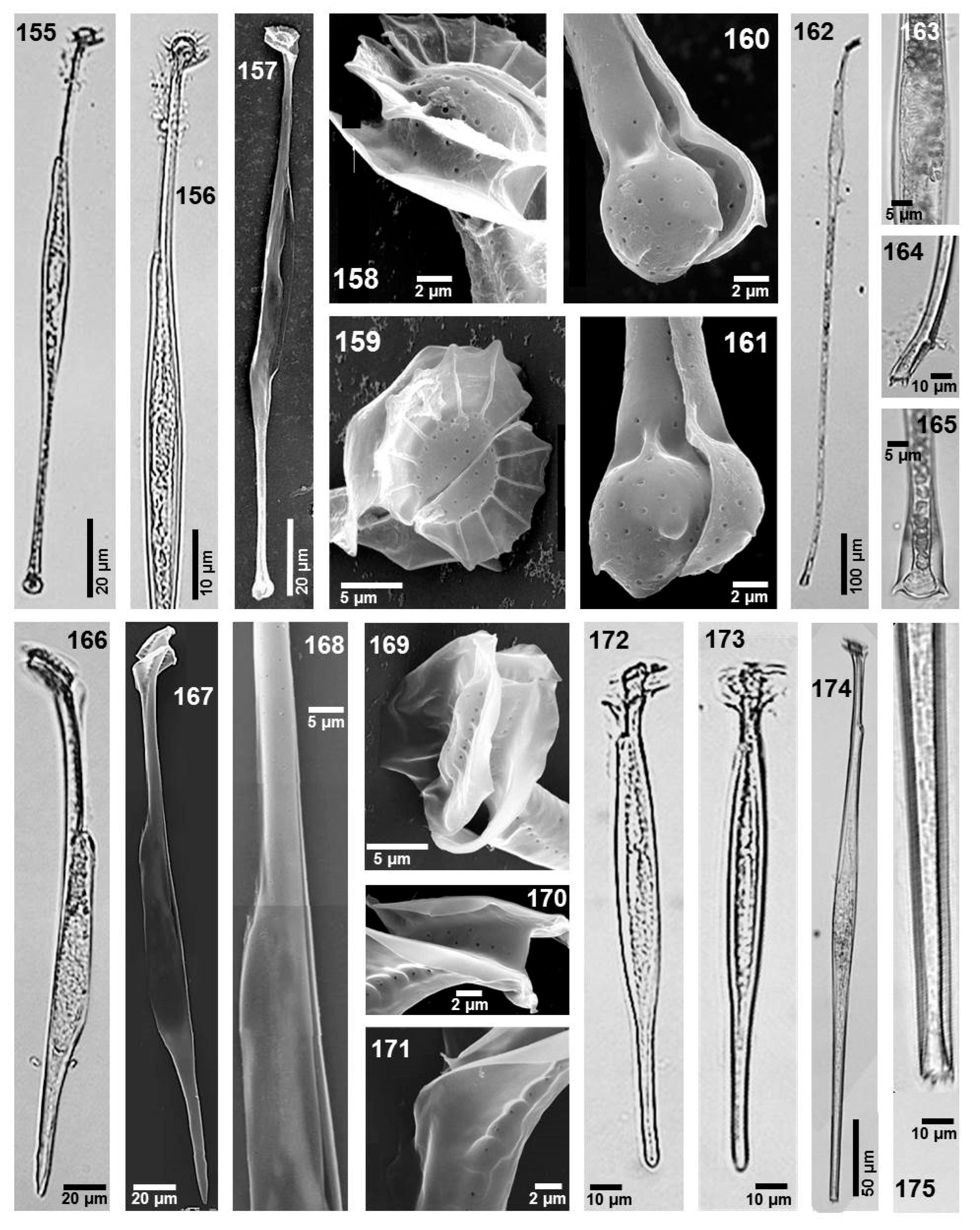
  • Amphisolenia bidentata Schröder (Figure 11, images 162–165)
  • References: [35] (p. 253, figs 67–71), [39] (p. 69, pl. 17, figs 2, 3, 13), [40] (p. 15), [43] (p. 409, figs 54(1–4) and 56(1)), [47] (p. 39, fig. 56), and [50] (p. 28, pl. 2, figs 21, 22, pl. 3, figs 21b and 22b).
Large species, with elongate and slightly sigmoid cells, uniformly wider at the third part of the body (mid-body) and then tapering slightly toward the caudal part. The head is well-developed, with a reduced epitheca. The caudal portion has a foot with two terminal spines. N = 25.
  • Dimensions: L 625–800 µm, W 20–35 µm.
  • Local distribution: Gulf of California, central Mexican Pacific, and Gulf of Mexico (localities m, t, aa, ab, ad, af, am, ap, ar, au, ay, az, bc, bh, bi, bl, bm, bo, and br).
  • Amphisolenia brevicauda Kofoid (Figure 11, images 166–171)
  • Reference: [43] (p. 372, fig. 49), [44] (p. 169, fig. 154), and [58] (p. 31, pl. 12).
Medium-sized species. The cells are elongated with an irregular shape, having a short neck and body wider at the mid-body and an acute ending at the caudal portion. The head is well-developed with a reduced epitheca. The cingular lists are approximately equal, whereas the anterior cingular list has basal ribs. The cingulum is convex, with two rows of pores. The sulcal lists are poorly developed. The theca is smooth, with scattered tiny pores. The sagittal suture is smooth. N = 1.
  • Dimensions: L 250 µm, W 15 µm.
  • Local distribution: Gulf of California, at 29 °C and 35 salinity (locality d, Figure 1).
  • Remarks: This species is a new record for the Mexican Pacific.
  • Amphisolenia deltiana Gul et Saifullah (Figure 12, images 186–190)
  • Reference: [59] (p. 564, fig. 3).
Solitary cells and large-sized. Similar in shape and size to A. bidentata. The body is long, thin, and slightly sigmoid. The epitheca is moderately convex. The mid-body is fusiform. The caudal portion is truncated and rounded, with no foot or spinules. N = 10.
  • Dimensions: L 475 µm, W 18 µm.
  • Local distribution: central Mexican Pacific and Gulf of Mexico (localities t, af, ah, an, ax, az, bc, bl, bn, br).
  • Remarks: This species is a new record for the Mexican Pacific.
  • Amphisolenia globifera Stein (Figure 11, images 155–161)
  • Reference: [39] (p. 70, pl. 18, figs 1 and 40, p. 16), [43] (p. 388, figs 49, 50, pl. 86, figs 1, 2, 4, 8), [47] (p. 41, fig. 61), and [50] (p. 29, pl. 2, figs 26 and 27, pl. 3, figs 26b and 27b).
Medium-sized species, with elongated and almost straight cells, a rounded head, and a spherical posterior structure. The epitheca is prominent and rounded, and the cingulum is concave. The anterior cingular list has well-developed ribs. The mid-body is wider. The posterior spherical process has four-minute spines. The theca is smooth and covered by small pores. The sagittal suture is not serrate. N = 2.
  • Dimensions: L 102 μm, W 12 μm.
  • Local distribution: Gulf of California, at 28 °C and 35 salinity, and Gulf of Mexico (localities a and ao).
  • Amphisolenia inflata Murray et Whitting (Figure 12, images 191–196)
  • References: [35] (p. 253, figs 79–81), [43] (p. 366, figs 47, 49), and [44] (p. 167, 168, fig. 152).
Figure 11. Images 155–161—Amphisolenia globifera, LM and SEM; Images 162–165—Amphisolenia bidentata, LM; Images 166–171—Amphisolenia brevicauda, LM and SEM; Images 172 and 173—Amphisolenia laticincta, LM; Images 174 and 175—Amphisolenia schroederi, LM.
Phycology 05 00048 g011
Medium-sized species, with cells that have a well-defined head, short neck, a very inflated mid-body, at about the third anterior part in the lateral and dorso-ventral views, and a straight caudal part. It has a reduced epitheca, narrow cingulum, and equally developed cingular lists, and the anterior one has ribs. The sulcal lists are reduced and have a conspicuous shoulder. The caudal portion tapers smoothly toward the end. It has a relatively smooth theca, with numerous scattered small pores. N = 2.
  • Dimensions: L 110 µm, W 30 µm.
  • Local distribution: central Mexican Pacific, 29 °C and 32 salinity (locality t).
  • Amphisolenia laticincta Kofoid (Figure 11, images 172 and 173)
  • Reference: [43] (p. 369, fig. 40) and [44] (p. 168, fig. 153a, b).
A species of medium size. The cells are elongated and straight, with a small head, reduced epitheca, and a high and concave cingulum. The neck is straight and short, with sulcal lists that are reduced. Its mid-body is wider than the caudal part. There is no apparent caudal process or spine. N = 2.
  • Dimensions: L 103–110 µm, W 10 µm.
  • Local distribution: Gulf of California, at 28 °C and 35 salinity (localities a and h).
  • Amphisolenia lemmermannii Kofoid (Figure 12, images 176 and 177)
  • References: [34] (p. 11, figs 48 and 49), [39] (p. 70, pl. 17, figs 8 and 12), [43] (p. 419, figs 50(8), 56(2), 57, pl. 9, figs 11 and 12), and [44] (p. 179, fig. 170).
Large-sized species, with very elongated and nearly straight cells. It is also similar to A. bidentata but with the caudal portion having a foot with two short terminal spines. N = 3.
  • Dimensions: L 780–790 µm, W 30 µm.
  • Local distribution: Gulf of California, central Mexican Pacific, and Gulf of Mexico (localities a, t, and ao).
  • Amphisolenia palmata Stein (Figure 12, images 178 and 179)
  • Reference: [34] (pl. 11, figs 44 and 45), [39] (p. 69, pl. 17, figs 4–7), [43] (p. 422, pl. 12, figs 4 and 7), and [47] (p. 40, fig. 57a–c).
Large-sized species, also similar to A. bidentata and A. lemmermanni (elongate and sigmoid), however the caudal portion has a foot with three antapical spines. N= 1.
  • Dimensions: L 780 µm, W 25 µm.
  • Local distribution: Gulf of California (locality a).
  • Amphisolenia rectangulata Kofoid (Figure 12, images 180–185)
  • Reference: [39] (p. 186, pl. 83, figs 3–6), [43] (p. 378, fig. 49, pl. 8, figs 3, 5, 6, 7, 9), and [44] (p. 170, fig. 156a,b).
Large-sized species. It is another species similar to A. bidentata and other related ones. The cingulum is concave, and the cingular lists are equivalent. The anterior cingular has few ribs, and the sulcal lists are reduced. The caudal portion shows a wide foot with four conspicuous spines. The theca is smooth and has pores. The sagittal suture is smooth. N = 3.
  • Dimensions: LT 695 µm, W 15 µm.
  • Local distribution: Gulf of California, 28 °C and 35 salinity (locality a).
  • Amphisolenia schroederi Kofoid (Figure 11, image 174 and 175)
  • Reference: [35] (p. 256, figs 85–90), [39] (p. 187, pl. 83, fig. 9), and [40] (p. 17), [44] (p. 176, fig. 165).
A medium-sized species, with an elongated and straight body, narrow head, and long and straight neck, wider (slightly inflated) mid-body and then tapering gradually toward the caudal part. The caudal portion has a foot ending with small spines. N = 1.
  • Dimensions: L 380 µm, W 18 µm.
  • Local distribution: Gulf of California, at 30 °C and 35 salinity (locality i, ag, ai, au, and bc).
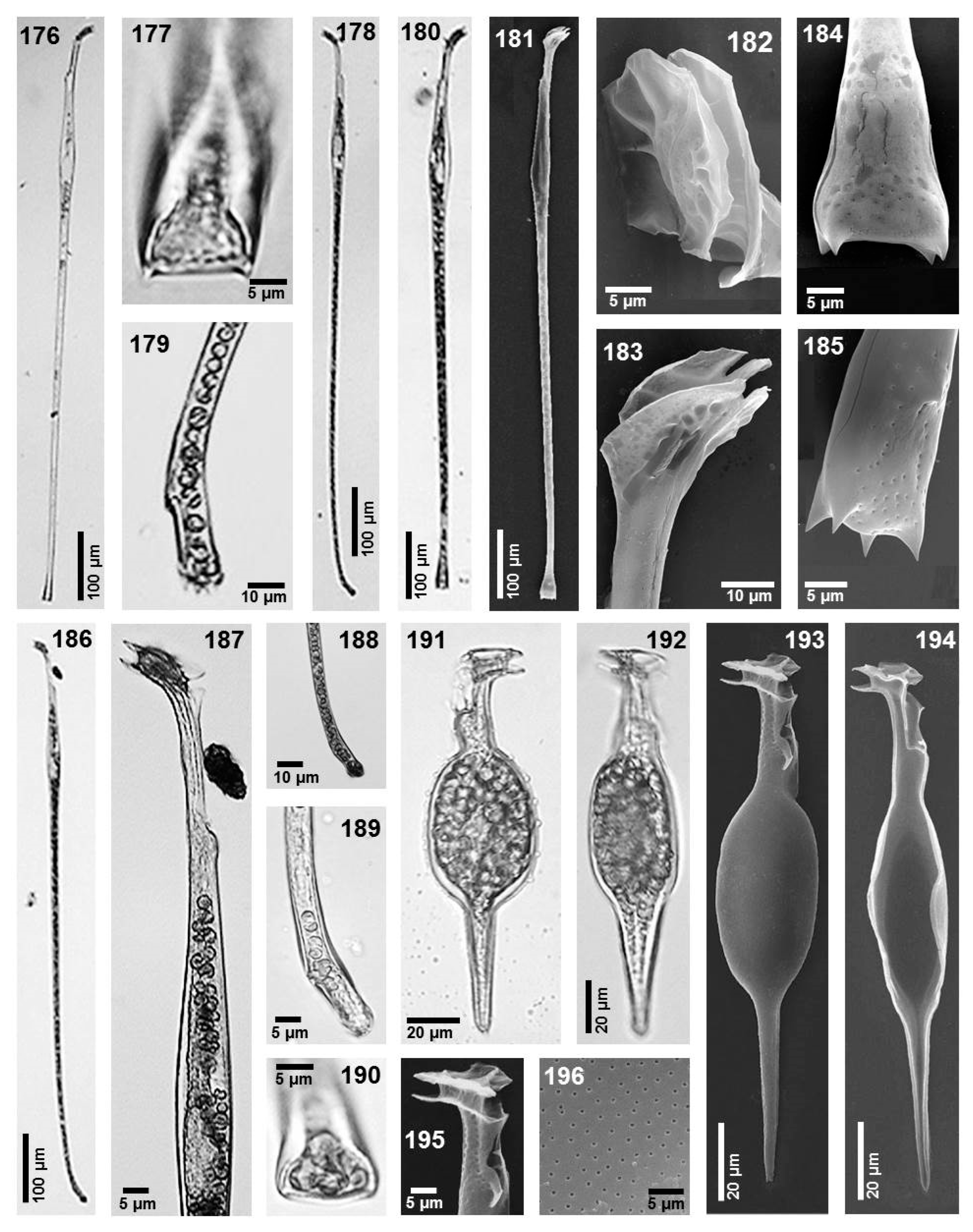
Figure 12. Images 176 and 177. Amphisolenia lemmermannii, LM. Images 178 and 179. Amphisolenia palmata, LM. Images 180–185. Amphisolenia rectangulata, LM and SEM. Images 186–190. Amphisolenia deltiana, LM. Images 191–196. Amphisolenia inflata, LM and SEM.
Phycology 05 00048 g012

4. Discussion

4.1. Taxonomy of Some Dinophysoid Species

Dinophysales have a large number of species and a great diversity of forms. This diversity is such that morphological characteristics with taxonomic value have not been clearly defined for some species or genera [31]. For example, “small cells”, previously described as new species, are difficult to recognize in Dinophysis and Phalacroma, for these cells are now considered as synonyms of other species [18,60]. This is the case of Dinophysis diegensis Kofoid in relation to D. caudata Saville-Kent, as part of a complex life cycle [18,60,61,62,63].
In contrast, species considered at some point as synonyms of others are really validly recognized species, for example, Phalacroma parvulum and P. rotundatum, which are recognized to be distinct species by molecular sequences [31]. Another case is that of Phalacroma stenopterygia, which had been for many years considered a synonym of Phalacroma expulsum [44,45]; however, morphological differences concern not only the size but also the shape (outline) of the cells in the dorsal and ventral views, relative width of the epitheca, and shape of the cingulum (see remarks under the species). We consider it wise to maintain these two species as separate until molecular data are available.
Another species of similar status is Phalacroma rudgei. This species has been traditionally considered a synonym of P. rotundatum [62,64], but P. rudgei has various morphological characteristics that differ from the latter, as follows: a more rounded body, a wider and higher epitheca than P. rotundatum, and a rather shorter R2. They should be considered as two different species, see also [41].
The specimens found here of Phalacroma scrobiculatum were relatively abundant. The lack of previous records may be due to misidentifications, because the species strongly resembles P. ovum and P. parvulum, since they share similarities in shape and size. However, the most particular character of P. scrobiculatum is the second rib (R2) of the left sulcal list, which is curved and located closer to R3 than to R1. All the specimens in this study were similar to those previously studied using light microscopy, but new observations in this paper by SEM show details of the cingular lists and the ornamentation of the cingulum and the theca (Figure 5, images 53–55).
Finally, two apparently confused species (both morphological and nomenclatural) are Phalacroma operculoides and P. ovum [9,43]. This was also discussed in the remarks of P. ovum. They are considered to be separate species [11,43].
We are proposing Phalacroma whittingiae (Balech) Esqueda-Lara et Hernández-Becerril nov. comb. to be part of the genus Phalacroma rather than Dinophysis, where it was originally described.

4.2. Diversity and New Records

In the Mexican Pacific and the Gulf of Mexico, there is a relatively high number of recorded species of dinoflagellates of the Order Dinophysales, but very few have been reliably illustrated or described. Species of the order Dinophysales recorded from the Tropical Pacific were 129 [43] and from the Mexican Pacific in 2006 were 83 [33], in 2008 85 [34], and in 2012 the records increased to 91 [27]. On the other hand, species of this order from the south Atlantic were 83 in 1980 [39]. In this paper, we report five more species; hence, 96 species of Dinophysales are recorded from the Mexican Pacific and 36 species from the Gulf of Mexico; this represents only 34% and 43% of the total Dinophysales described in the literature, respectively, but we consider that the number will increase in the near future, with more systematic studies on the group from the Mexican Pacific and Gulf of Mexico.
In this paper, we illustrate the following three (3) species for first time in the study areas: Dinophysis similis, Histioneis garrettii, and Phalacroma whittingiae. We also studied these species by SEM, which provides important information for future studies on various topics, including biodiversity, taxonomy, and phylogeny.
New records for the Mexican Pacific are Amphisolenia brevicauda, A. deltiana, and Phalacroma stenopterygium, and for the Gulf of Mexico it is P. ornamentatum. Two of them are poorly known species—Amphisolenia brevicauda and Phalacroma stenopterygium, with the latter being considered as a synonym of another (P. expulsum)—whereas the other two are relatively newly described species, Amphisolenia deltiana and Phalacroma ornamentatum, and they extend their geographical range.
These new records are important in terms of biodiversity but also the information obtained by SEM on some morphological details of Amphisolenia globifera, A. rectangulata, Histioneis biremis, and H. reticulata. Both specimens of Amphisolenia are showing only pores in the theca and in A. rectangulata details of the posterior four spines. In the cases of A. brevicauda and A. globifera, we observed a smooth sagittal suture, which differs from the general morphological characteristic of the “serrate” suture in the order [7,10]. Also, A. palmata was illustrated by Abé [42] and A. bidentata depicted by Balech [65], both which have a smooth sagittal suture, although they did not mention it. Although the information given here concerns only three species of Amphisolenia, it is possible that this characteristic may be shared with most species of the genus, supporting the phylogenetic distance within the order [31].
The finding of new species of the order Dinophysales reflects the need to continue studying the group. The scarce number of specimens observed is indicative of the low population densities that make it difficult to obtain more information and certainty of the morphological variation in the taxa. A similar paper is presented by Hallegaeff et al. [9]; these show Dinophysales flora from Australian waters with exceptional illustrations of the electron microscopy; however, they do not show details of the critical morphological characteristics.
We consider that the morphological details provided in this paper are important as complementary knowledge of the species. This knowledge is basic and necessary to understand the possible adaptive and evolutionary patterns of the group.

5. Conclusions

Seventy-five taxa (73 species and two varieties) of thecate dinophysoid dinoflagellates (Dinophysales) from the tropical Mexican Pacific and the southern Gulf of Mexico are reported in this study. Most of the taxa are described, and all of them are illustrated by LM and/or SEM. The taxa belong to the following 10 genera (with the number of species found in brackets): Amphisolenia (10), Citharistes (1), Dinofurcula (1), Dinophysis (22), Histioneis (6), Metaphalacroma (1), Ornithocercus (11), Oxyphysis (1), Phalacroma (21), and Pseudophalacroma (1). The more diverse genera were Dinophysis and Phalacroma. We found the following four new records: three for the Mexican Pacific (Amphisolenia brevicauda, A. deltiana, and Phalacroma stenopterygium) and one for the Gulf of Mexico (Phalacroma ornamentatum); two of them are poorly known species, Amphisolenia brevicauda and Phalacroma stenopterygium, whereas the other two are relatively newly described species, Amphisolenia deltiana and Phalacroma ornamentatum, and they extend their geographical range. Special morphological characteristics, such as the structures of the cingulum and cingular lists, union of the sulcal lists, and theca ornamentation, were observed using SEM; these characteristics should be systematically observed to aid in identifying and characterizing species. Taxonomic discussion on some Phalacroma species included two species traditionally considered synonyms of Phalacroma rotundatum, which are here recognized to be separate species, Phalacroma parvulum and P. rudgei, based on morphological characteristics. There is a similar case with the species Phalacroma operculoides and P. ovum, which should be considered as different species. We additionally believe that Phalacroma stenopterygia is a different species than Phalacroma expulsum, which had been for many years considered a synonym of the latter. Phalacroma whittingiae (Balech) Esqueda-Lara et Hernández-Becerril nov. comb. is a new taxonomic proposal. With this work, we contribute to the knowledge on the diversity and morphology of this important dinoflagellate group in dynamic areas of tropical to subtropical environs.

Author Contributions

Conceptualization, K.E.-L. and D.U.H.-B.; Methodology, K.E.-L. and D.U.H.-B.; Software, K.E.-L. and D.U.H.-B.; Validation, K.E.-L. and D.U.H.-B.; Formal Analysis, K.E.-L., D.U.H.-B., and J.P.G.-G.; Investigation, K.E.-L., D.U.H.-B., and J.P.G.-G.; Resources K.E.-L. and D.U.H.-B.; Data Curation, K.E.-L. and D.U.H.-B.; Writing—Original Draft Preparation, K.E.-L. and D.U.H.-B.; Writing—Review & Editing, K.E.-L. and D.U.H.-B.; Visualization, D.U.H.-B.; Supervision, K.E.-L. and D.U.H.-B.; Project Administration, K.E.-L. and D.U.H.-B.; Funding Acquisition, D.U.H.-B. All authors have read and agreed to the published version of the manuscript.

Funding

The first author had a doctorate fellowship given by CONACYT (now SECIHTT). Approval and support to carry out oceanographic cruises were provided by CIC, UNAM.

Institutional Review Board Statement

The study was conducted in accordance with the Declaration of Helsinki, and the protocol was approved by the Ethics Committee.

Informed Consent Statement

Not applicable.

Data Availability Statement

Informed consent for participation was obtained from all subjects involved in the study. Data Availability Statement: The original contributions presented in this study are included in the article. Further inquiries can be directed to the corresponding author(s).

Acknowledgments

We thank Laura Sánchez-Velasco, Adela Monreal-Gómez, and Maria Luisa Machain-Castillo for providing the facilities to obtain phytoplankton samples in the oceanographic cruises “GOLCA 07”, “DIPAL II”, “TEHUA V”, and “ZOM I”, respectively. Thanks are also due to Yolanda Hornelas Orozco for her assistance in using the SEM equipment.

Conflicts of Interest

The authors declare no conflicts of interest.

References

  1. Gómez, F. Diversity and classification of dinoflagellates. In Dinoflagellates: Classification, Evolution, Physiology and Ecological Significance; Subba Rao, V.D., Ed.; Nova: New York, NY, USA, 2020; pp. 1–38. [Google Scholar]
  2. Guiry, D.R. How many species of algae are there? A reprise. Four kingdoms, 14 phyla, 63 classes and still growing. J. Phycol. 2024, 60, 214–228. [Google Scholar] [CrossRef]
  3. Esqueda-Lara, K.; Hernández-Becerril, D.U. Two new species of the dinoflagellate genus Phalacroma Stein (Dinophyceae) from the tropical Mexican Pacific. Nova Hedwig. 2017, 105, 301–312. [Google Scholar] [CrossRef]
  4. Gómez, F. A checklist and classification of living dinoflagellates (Dinoflagellata, Alveolata). CICIMAR Oceánides 2012, 27, 65–140. [Google Scholar] [CrossRef]
  5. Zonneveld, K.A.F.; Pospelova, V. A determination key for modern dinoflagellate cysts. Palynology 2015, 39, 387–409. [Google Scholar] [CrossRef]
  6. Taylor, F.J.R.; Fukuyo, Y.; Larsen, J.; Hallegraeff, G.M. Taxonomy of harmful dinoflagellates. In Manual on Harmful Marine Microalgae; Hallegraeff, G.M., Anderson, D.M., Cembella, A.D., Eds.; UNESCO-IOC: Paris, France, 2003; pp. 283–318. [Google Scholar]
  7. Fensome, R.A.; Taylor, F.J.R.; Norris, G.; Sarjeant, W.A.S.; Wharton, D.I.; Williams, G.L. A Classification of Living and Fossil Dinoflagellates. Micropaleontol, Spec Publ No. 7; Sheridan Press: Hanover, PA, USA, 1993; p. 147. [Google Scholar]
  8. Sournia, A. Red tide and toxic marine phytoplankton of the world ocean: An inquiry into diversity. In Harmful Marine Algal Blooms; Lassus, P., Arzul, G., Erand, E., Gentienand, E., Marcaillou, C., Eds.; Lavoisier, Intercept Ltd.: Paris, France, 1995; pp. 103–112. [Google Scholar]
  9. Hallegraeff, G.M.; Eriksen, R.S.; Davies, C.H.; Uribe-Palomino, J. Marine planktonic dinophysoid dinoflagellates (order Dinophysales): 60 years of species-level distributions in Australian waters. Austral. Syst. Bot. 2022, 35, 469–500. [Google Scholar] [CrossRef]
  10. Sournia, A. Atlas du Phytoplancton Marin. Volume I. Introduction, Cyanophycées, Dictyochophycées, Dinophycées, Raphidophycées; Éditions du Centre National de la Recherche Scientifique: Paris, France, 1986; p. 219. [Google Scholar]
  11. Zinssmeister, C.; Wilke, T.; Hoppenrath, M. Species diversity of dinophysoid dinoflagellates in the Clarion-Clipperton fracture zone, eastern Pacific. Mar. Biodiv. 2017, 47, 271–287. [Google Scholar] [CrossRef]
  12. Tarangkoon, W.; Hansen, G.; Hansen, P.J. Spatial distribution of symbiont-bearing dinoflagellates in the Indian Ocean in relation to oceanographic regimes. Aquat. Microb. Ecol. 2010, 58, 197–213. [Google Scholar] [CrossRef]
  13. Reguera, B.; García-Portela, M.; Velasco-Senovilla, E.; Rial, P.; Escalera, L.; Díaz, P.A.; Rodríguez, F. Dinophysis, a highly specialized mixoplanktonic protist. Front. Protistol. 2024, 1, 1328026. [Google Scholar] [CrossRef]
  14. Reguera, B.; Velo-Suárez, L.; Raine, R.; Park, M.G. Harmful Dinophysis species: A review. Harmful Algae 2012, 14, 87–106. [Google Scholar] [CrossRef]
  15. Reguera, B.; Riobó, P.; Rodríguez, F.; Díaz, P.A.; Pizarro, G.; Paz, B.; Franco, J.M.; Blanco, J. Dinophysis toxins: Causative organisms, distribution and fate in shellfish. Mar. Drugs 2014, 12, 394–461. [Google Scholar] [CrossRef]
  16. Gaines, G.; Elbrächter, M. Heterotrophic nutrition. In The Biology of Dinoflagellates; Taylor, F.J.R., Ed.; Blackwell Scientific Publications: Oxford, UK, 1987; pp. 224–268. [Google Scholar]
  17. Schnepf, E.; Elbrächter, M. Dinophyte chloroplasts and phylogeny—A review. Grana 1999, 38, 81–97. [Google Scholar] [CrossRef]
  18. Reguera, B.; González-Gil, S. Small cell and intermediate cell formation in species of Dinophysis (Dinophyceae, Dinophysiales). J. Phycol. 2001, 37, 318–333. [Google Scholar] [CrossRef]
  19. Koike, K.; Nishiyama, A.; Sayito, K.; Imai, K.; Koike, K.; Kobiyama, A.; Ogata, T. Mechanisms of gamete fusion in Dinophysis fortii (Dinophyceae, Dinophyta): Light microscopic and ultrastructural observation. J. Phycol. 2006, 42, 1247–1256. [Google Scholar] [CrossRef]
  20. Reguera, B.; González-Gil, S.; Delgado, M. Dinophysis diegensis is a life history stage of Dinophysis caudata (Dinophyceae, Dinophysiales). J. Phycol. 2007, 43, 1083–1093. [Google Scholar] [CrossRef]
  21. Park, M.G.; Kim, S.; Kim, H.S.; Yi, G.M.; Kang, G.; Yih, W. First establishment of the marine dinoflagellate Dinophysis acuminata in culture. Aquat. Microb. Ecol. 2006, 45, 101–106. [Google Scholar] [CrossRef]
  22. Nishitani, G.; Nagai, S.; Sakiyama, S.; Kamiyama, T. Successful cultivation of the toxic dinoflagellate Dinophysis caudata (Dinophyceae). Plankton Benthos Res. 2008, 3, 78–85. [Google Scholar] [CrossRef]
  23. Escalera, L.; Reguera, B.; Moita, T.; Pazos, Y.; Cerejo, M.; Cabanas, J.M.; Ruiz-Villareal, M. Bloom dynamics of Dinophysis acuta in an upwelling system: In situ growth versus transport. Harmful Algae 2010, 9, 312–322. [Google Scholar] [CrossRef]
  24. Okolodkov, Y. Dinophysiales (Dinophyceae) of the National Park Sistema Arrecifal Veracruzano, Gulf of Mexico, with a key for identification. Act. Bot. Mex. 2014, 106, 9–71. [Google Scholar] [CrossRef]
  25. Steidinger, K.A.; Tangen, K. Dinoflagellates. In Identifying Marine phytoplankton; Tomas, C.R., Ed.; Academic Press: San Diego, CA, USA, 1997; pp. 387–584. [Google Scholar]
  26. Parra-Toriz, D.; Esqueda-Lara, K.; Hernández-Becerril, D.U. Morphological observations of the marine planktonic dinoflagellate Phalacroma turbineum Kofoid et Michener (Dinophyta), with discussion on its taxonomy and distribution. Eur. J. Protistol. 2012, 48, 274–282. [Google Scholar] [CrossRef] [PubMed]
  27. Esqueda-Lara, K.; Parra-Toriz, D.; Hernández-Becerril, D.U. Morphology and taxonomy of Dinophysis species of the section Hastata (Dinoflagellata), including the description of Dinophysis conjucta sp. nov., from the Mexican marine waters. J. Mar. Biol. Assoc. UK 2013, 93, 1187–1202. [Google Scholar] [CrossRef]
  28. Wilke, T.; Zinssmeister, C.; Hoppenrath, M. Morphological variability within the marine dinoflagellate Ornithocercus quadratus (Dinophysales, Dinophyceae)—Evidence for three separate morphospecies. Phycology 2018, 57, 555–571. [Google Scholar] [CrossRef]
  29. Handy, S.M.; Bachvaroff, T.R.; Timme, R.E.; Wayne Coasts, W.D.; Kim, S.; Delwiche, C.F. Phylogeny of our dinophysiacean genera (Dinophyceae, Dinophysiales) based on rDNA sequences from single cells and environmental samples. J. Phycol. 2009, 45, 1163–1174. [Google Scholar] [CrossRef]
  30. Jensen, M.H.; Daugbjerg, N. Molecular phylogeny of selected species of the order Dinophysiales (Dinophyceae): Testing the hypothesis of a dinophysiod radiation. J. Phycol. 2009, 45, 1136–1152. [Google Scholar] [CrossRef] [PubMed]
  31. Gómez, F.; López-García, P.; Moreira, D. Molecular phylogeny of dinophysoid Dinoflagellates the systematic position on Oxyphysis oxytoxoides and the Dinophysis Hastata group (Dinophysales, Dinophyceae). J. Phycol. 2011, 47, 303–406. [Google Scholar] [CrossRef]
  32. Hernández-Becerril, D.U.; Meave del Castillo, M.E.; Flores-Granados, C. Dinoflagelados del Orden Dinophysiales en las costas mexicanas. In Planctología Mexicana; Barreiro Güemes, M.T., Meave del Castillo, M.E., Signoret Poillon, M., Figueroa-Torres, M.G., Eds.; Sociedad Mexicana de Planctología; Universidad Autónoma Metropolitana: Mexico City, Mexico, 2003; pp. 19–42. [Google Scholar]
  33. Okolodkov, Y.; Gárate-Lizárraga, I. An annotated checklist of dinoflagellates (Dinophyceae) from the Mexican Pacific. Acta Bot. Mex. 2006, 74, 1–154. [Google Scholar] [CrossRef]
  34. Hernández-Becerril, D.U.; Ceballos-Corona, J.G.A.; Esqueda-Lara, K.; Tovar-Salazar, M.A.; León-Álvarez, D. Marine planktonic dinoflagellates of the order Dinophysiales (Dinophyta) from coasts of the tropical Mexican Pacific, including two new species of the genus. Amphisolenia. J. Mar. Biol. Assoc. UK 2008, 88, 1–15. [Google Scholar] [CrossRef]
  35. Parra-Toriz, D.; Hernández-Becerril, D.U. Planktonic dinoflagellates (Dinophyceae) of the order Dinophysales from the southern Gulf of Mexico. Nova Hedwig. 2023, 116, 231–263. [Google Scholar] [CrossRef]
  36. Hernández-Becerril, D.U.; Meza-García, P.; Villarreal-Martínez, A.M.; García-Anaya, T.M.; Vázquez-Montes, E.D.; Esqueda-Lara, K.; Arana-García, J. Observations on some planktonic dinophysoid dinoflagellates (Dinophysales, Dinophyta) from the Mexican Pacific, including the description of a new species, Dinofurcula pseudoultima sp. nov., and taxonomic and distribution notes about the genera Latifascia and Triposolenia. Protistology 2023, 17, 3–15. [Google Scholar] [CrossRef]
  37. Balech, E. Dinoflagelados. In Fitoplancton Marino; Balech, E., Ferrando, H.J., Eds.; Editorial Universitaria de Buenos Aires: Buenos Aires, Argentina, 1964. [Google Scholar]
  38. Balech, E. On thecal morphology of Dinoflagellates with special emphasis on cingular and sulcal plates. An. Centro Cienc. Mar. Limnol. UNAM 1980, 7, 57–68. [Google Scholar]
  39. Balech, E. Los Dinoflagelados del Atlántico Sudoccidental; Publicaciones Especiales, Instituto Español de Oceanografía: Madrid, Spain, 1988; p. 219. [Google Scholar]
  40. Yang, S.M.; Li, R.X. Atlas of Dinoflagellates in China’s Seas; Ocean Press: Beijing, China, 2014; 213p. [Google Scholar]
  41. Tai, L.-S.; Skogsberg, T. Studies on the Dinophysoidae, marine armored dinoflagellates, of Monterey Bay, california. Arch. Protistenk. 1934, 82, 380–482. [Google Scholar]
  42. Abé, T.H. The armored Dinoflagellata. II. Prorocentridae and Dinophysidae (B)—Dinophysis and its allied genera. Publ. Seto Mar. Biol. Lab. 1967, 15, 37–68. [Google Scholar] [CrossRef][Green Version]
  43. Kofoid, C.A.; Skogsberg, T. The Dinoflagellata: The Dinophysoidae. Mem. Mus. Comp. Zool. 1928, 21, 1–766. [Google Scholar][Green Version]
  44. Schiller, J. Dinoflagellatae (Peridineae). Teil I. Rabenhorst’s Kryptogamen-Flora; Akademie Verlag: Leipzig, Germany, 1933; p. 590. [Google Scholar][Green Version]
  45. Gul, D.; Saifullah, S.M. Taxonomic and ecological studies on three marine genera of Dinophysiales from Arabian Sea shelf of Pakistan. Pak. J. Bot. 2010, 42, 2647–2660. [Google Scholar][Green Version]
  46. Balech, E. Microplancton de la Campaña Productividad III. Rev. Mus. Arg. Cs. Nat. B Rivadavia Hidrobiol. 1971, 3, 1–202. [Google Scholar][Green Version]
  47. Jörgensen, E. Mediterranean Dinophysiaceae. In Report of the Danish Oceanographic Expedition II; Carlsbergfondet: Copenhagen, Denmark, 1923; Volume 2, pp. 1–48. [Google Scholar][Green Version]
  48. Wood, E.J.F. Dinoflagellates in the Australian region. Mar. Freshwat. Res. 1954, 5, 171–351. [Google Scholar] [CrossRef]
  49. Yang, S.M.; Li, R.X.; Dong, S.G. Dinoflagellates in the China’s Seas I (Prorocentrales, Dinophysiales); Ocean Press: Beijing, China, 2014; 156p. [Google Scholar]
  50. Taylor, F.J.R. Dinoflagellates from the International Indian Ocean Expedition: A Report on Material Collected by the R. V. “Anton Bruun” 1963–1964; Bibliotheca Botanica: Adendorf, Germany, 1976; 234p. [Google Scholar]
  51. Esqueda-Lara, K.; Hernández-Becerril, D.U. Dinoflagelados Microplanctónicos Marinos del Pacífico Central Mexicano (Isla Isabel, Nayarit y Costas de Jalisco y Colima); Instituto de Ciencias del Mar y Limnología, Universidad Nacional Autónoma de México: Mexico City, Mexico, 2010; p. 206. [Google Scholar]
  52. Matzenauer, L. Die Dinoflagellaten des Indischen Ozeans. Bot. Arch. 1933, 35, 437–510. [Google Scholar]
  53. Balech, E. Dinoflagelados nuevos o interesantes del Golfo de México y Caribe. Rev. Mus. Arg. Cs. Nat. B Rivadavia Hidrobiol. 1967, 2, 77–126. [Google Scholar]
  54. Murray, G.; Whitting, F.G. New Peridiniaceae from the Atlantic. Trans. Linnean Soc. Lond. 2nd Ser. Bot. 1899, 5, 321–342. [Google Scholar] [CrossRef]
  55. Balech, E. Tintinnoinea y dinoflagellata del Pacífico según material de las expediciones Norpac y Downwind del Instituto Scripps de Oceanografía. Rev. Mus. Argent. Cienc. Nat. (Cienc. Zool.) 1962, 7, 1–253. [Google Scholar]
  56. Hernández-Becerril, D.U.; Barón-Campis, S.A.; Ceballos-Corona, J.G.A.; Alonso-Rodríguez, R.; Rincones-Reyes, K.M.; Becerra-Reynoso, R.T.; Arce-Rocha, G. Catálogo de Fitoplancton del Pacífico Central Mexicano. Cruceros “MareaR” (2009–2019) B/O “El Puma”; Universidad Nacional Autónoma de México: Mexico City, Mexico, 2021; p. 254. [Google Scholar]
  57. Kofoid, C.A. On Oxyphysis oxytoxoides gen. nov. sp. nov. a dinophysoid dinoflagellate convergent toward the peridinioid type. Univ. Calif. Pub. Zool. 1926, 28, 203–216. [Google Scholar]
  58. Rampi, L.; Bernhard, M. Chiave per la Determinazione Delle Peridinee Pelagiche Mediterranee; Comitato Nazionale Energia Nucleare: Rome, Italy, 1980. [Google Scholar]
  59. Gul, D.; Saifullah, S.M. Genus Amphisolenia Stein from north-west Arabian sea shelf of Pakistan. Pak. J. Bot. 2007, 39, 561–576. [Google Scholar]
  60. Dodge, J.D. Taxonomy and systematics of dinoflagellates. In Out of the Past; Norton, T.A., Ed.; British Phycological Society: Belfast, UK, 2003; pp. 69–86. [Google Scholar]
  61. MacKenzie, L. Does Dinophysis (Dinophyceae) have a sexual life cycle? J. Phycol. 1992, 28, 399–406. [Google Scholar] [CrossRef]
  62. Gómez, F. A list of free-living dinoflagellate species in the world’s oceans. Acta Bot. Croat. 2005, 64, 129–212. [Google Scholar]
  63. Hernández-Becerril, D.U. Dinophysis taylorii, sp. nov., y otros Dinophysis de Baja California, México (Dinophyceae). Rev. Biol. Trop. 1992, 40, 101–109. [Google Scholar]
  64. Halim, Y. Dinoflagellates of the south-east Caribbean Sean (east-Venezuela). Int. Revue Ges. Hydrobiol Hydrogr. 1967, 52, 701–755. [Google Scholar] [CrossRef]
  65. Balech, E. Estructura de Amphisolenia bidentata Schröder (Dinoflagellata). Physis Sección A 1977, 37, 25–32. [Google Scholar]
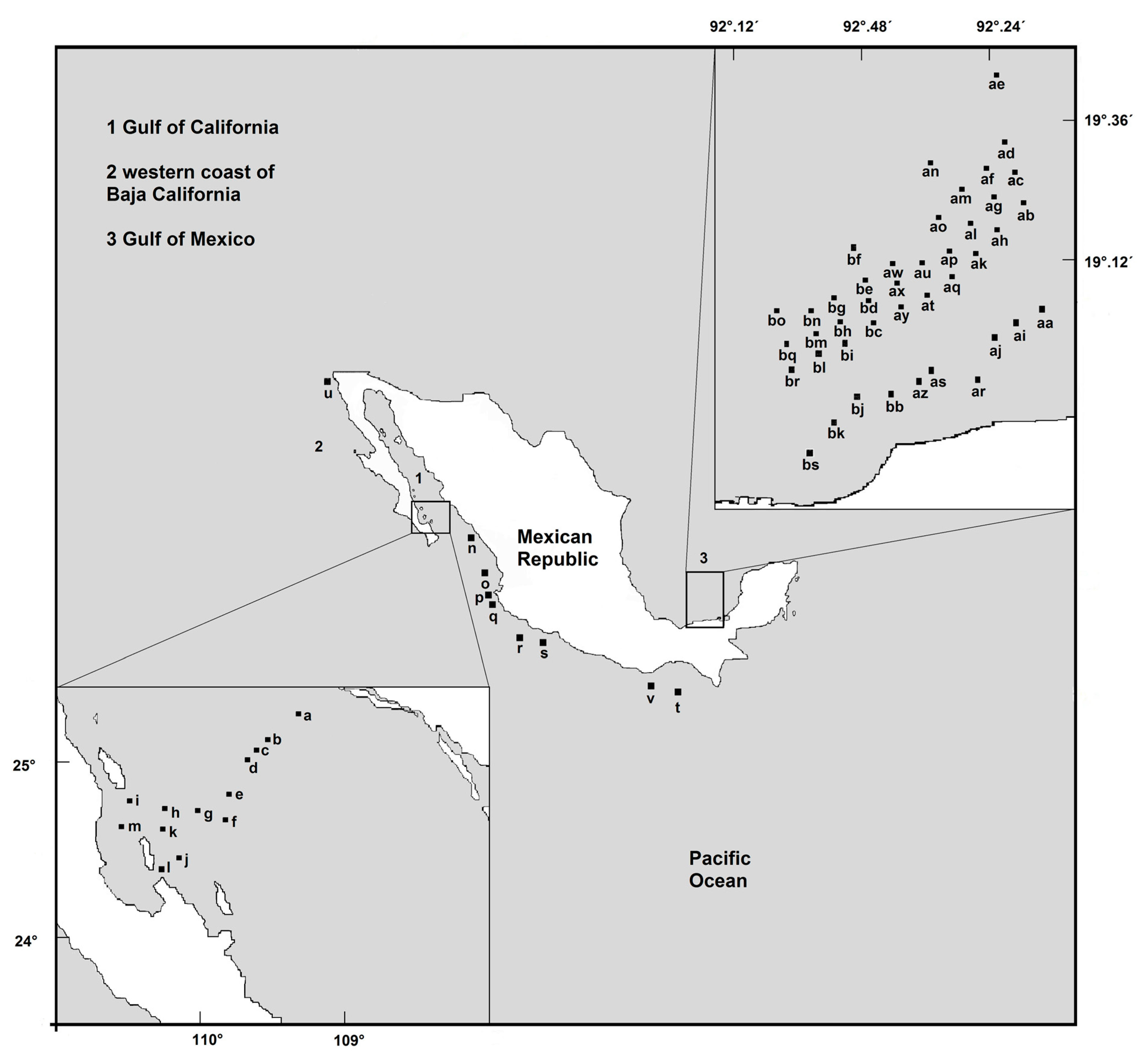
Figure 1. Map of the study areas, including fixed stations in the Gulf of California (a–m), Mexican Pacific (n–t,v), western coast of Baja California (u) and the Gulf of Mexico (aa–bs).
Phycology 05 00048 g001
Table 1. Distribution of Dinophysales in the study area. The small letters are localities in Figure 1.
SpeciesNo. SpecimensStudy AreasLocalities
Citharistes regius2Gulf of Californiag, i, n
Dinophysis acuminata18western coast of Baja Californiau
central Mexican Pacificbk
Dinophysis exigua3Gulf of Californian
central Mexican Pacifict
Dinophysis fortii43western coast of Baja Californiaa
central Mexican Pacificp, t, u
Gulf of Mexicoab, ac, ae, ah, an, ao, as, bj, bk, bn
Dinophysis infundibulus2Gulf of Mexicoal
Dinophysis recurva2central Mexican Pacificv
Dinophysis similis2Gulf of Californiab
Dinophysis sourniae3central Mexican Pacifice
Dinophysis balechii1Gulf of California in [27]see [27]
central Mexican Pacificsee [27]
Dinophysis conjuncta3Gulf of California in [27]see [27]
Gulf of Mexicosee [27]
Dinophysis hastata17Gulf of California in [27]see [27]
central Mexican Pacificsee [27]
Gulf of Mexicoan, ao, ap, as, ay, bb, bs
Dinophysis monacantha1Gulf of California in [27]see [27]
Dinophysis nias4Gulf of California in [27]see [27]
Gulf of Mexicosee [27]
Dinophysis phalacromoides1Gulf of California in [27]see [27]
Dinophysis pusilla3Gulf of California in [27]see [27]
Gulf of Mexicosee [27]
Dinophysis schuettii13Gulf of California in [27]see [27]
Gulf of Mexicoah, ak, ay, bc, bd
Dinophysis swezyae4Gulf of California in [27]see [27]
Gulf of Mexicosee [27]
Dinophysis uracantha3Gulf of California in [27]see [27]
Gulf of Mexicoaf, ao, be, bf
Dinophysis uracantha var. mediterranea4Gulf of California in [27]see [27]
central Mexican Pacificsee [27]
Dinophysis uracanthoides2Gulf of California in [27]see [27]
Gulf of Mexicosee [27]
Dinophysis caudata70western coast of Baja Californiau
Gulf of Californiaa, b, c, m
central Mexican Pacificn, o, r, s, t
Gulf of Mexicoab, ac, ad, af, ah, ai, aj, ak, al, am, ao, ap, aq, ar, as, at, au, aw, ax, ay, az, bb, bc, bd, bk, bn, bo, bq, br, bs
Dinophysis caudata var. diegensis2central Mexican Pacificn
Dinophysis tripos40western coast of Baja Californiaa
Gulf of Californiau
Metaphalacroma skogsbergi6Gulf of Californiaa, b, t
Pseudophalacroma nasutum1western coast of Baja Californiau
Histioneis biremis1Gulf of Californiaa
Histioneis costata6Gulf of Californiaa
Gulf of Mexicoaa
Histioneis crateriforme1central Mexican Pacificp
Histioneis depressa2central Mexican Pacificv
Histioneis garrettii1central Mexican Pacificr
Histioneis mitchellana1central Mexican Pacificv
Ornithocercus assimilis8central Mexican Pacificb, d, e, f, g, n
Gulf of Mexicoau
Ornithocercus cristatus1central Mexican Pacifict
Ornithocercus francescae1central Mexican Pacifict
Ornithocercus heteroporus2central Mexican Pacifics
Ornithocercus magnificus14central Mexican Pacifico, r, t
Gulf of Mexicoaa, ac, ad, ae, ao, au, bg
Ornithocercus orbiculatus5central Mexican Pacifico, r
Gulf of Mexicoac
Ornithocercus quadratus1Gulf of Californiab
Ornithocercus schuetti1Gulf of Californiae
Ornithocercus splendidus2Gulf of Californiai, k
Gulf of Mexicoau
Ornithocercus steinii6Gulf of Californiaa, b, c, d, e, f, g, h, i, j, k, m
central Mexican Pacificn, q, r, s, t
Gulf of Mexicoao
Ornithocercus thumii4Gulf of Californiad, f, g, h, j, k, l
central Mexican Pacificq, r, s
Gulf of Mexicoae
Phalacroma laeve5Gulf of Californiam
central Mexican Pacifict
Gulf of Mexicoam, bb, bo
Phalacroma operculoides9Gulf fo Mexicoab, ah, aj, ak, as, bo
Phalacroma ornamentatum12central Mexican Pacifica
Gulf of Mexicoaa, ah, ai, aj, aw, ax, ay, bb, bd, dj, bn
Phalacroma parvulum9central Mexican Pacificn, o, r
Phalacroma rotundatum11Gulf of Californiaa, b, j
central Mexican Pacifict
Gulf of Mexicoac, af, ag, am, an, ao, bo
Phalacroma paulsenii5Gulf of Californiaa, c
Phalacroma rudgei1Gulf of Californiak
Phalacroma whittingiae4Gulf of Californiaa, l, m
Gulf of Mexicoab
Phalacroma apicatum1Gulf of Californiad, e, m
central Mexican Pacifico, r, s
Phalacroma argus7Gulf of Californiad, e, m
central Mexican Pacifico, r, s
Phalacroma ovum6Gulf of Californiaa, m
Gulf of Mexicoaf
Phalacroma scrobiculatum41Gulf of Californiaa, b, j, k, l
central Mexican Pacificaa, af, ah, ai, aj, al, ao, ap, aq, ar, as at, aw, ax, ay, az, bb, bf, bj, bk, bl, bn
Phalacroma cuneus13Gulf of Californiak
central Mexican Pacificm, n, q, r, s, t
Phalacroma rapa16central Mexican Pacifico, r, s
Gulf of Mexicoaa, ad, ah, al, bj, bo, bq, bs
Phalacroma capitulatum3Gulf of Californiai, k, m, n
central Mexican Pacifico, p, r
Phalacroma mitra28Gulf of Californiai, k, m
central Mexican Pacificn, o, p, r, t
Gulf of Mexicoaa, ae, af, al, bc, bd, bk, bn
Phalacroma favus4central Mexican Pacificn, p
Gulf of Mexicoaz
Phalacroma expulsum10central Mexican Pacifics, v
Gulf of Mexicoac, af, ag, am, ah, az
Phalacroma stenopterygium1Gulf of Californiab, d
Phalacroma doryphorum1Gulf of Californiai, k, m
central Mexican Pacificn, q, t
Gulf of Mexicoah
Phalacroma porodictyum20Gulf of Californiad, e, f, h, j, l, m
central Mexican Pacificn, o, q, r, s
Gulf of Mexicoah, ak, am, at
Oxyphysis oxytoxoides2western coast of Baja Californiau
central Mexican Pacifico
Gulf of Mexicobn, bo
Dinofurcula pseudoultima1central Mexican Pacificv
Amphisolenia bidentata25Gulf of Californiam
central Mexican Pacifict
Gulf of Mexicoaa, ab, ad, af, am, ap, ar, au, ay, az bc, bh, bi, bl, bm, bo, br
Amphisolenia brevicauda1Gulf of Californiad
Amphisolenia deltiana10central Mexican Pacifict
Gulf of Mexicoaf, ah, an, ax, az, bc, bl, bn, br
Amphisolenia globifera2Gulf of Californiaa
Gulf of Mexicoao
Amphisolenia inflata2central Mexican Pacifict
Amphisolenia laticincta2Gulf of Californiaa, h
Amphisolenia lemmermanni3Gulf of Californiaa
central Mexican Pacifict
Gulf of Mexicoao
Amphisolenia palmata1Gulf of Californiaa
Amphisolenia rectangulata3Gulf of Californiaa
Amphisolenia schroederi1Gulf of Californiaj
Gulf of Mexicoag, ai, au, bc
Disclaimer/Publisher’s Note: The statements, opinions and data contained in all publications are solely those of the individual author(s) and contributor(s) and not of MDPI and/or the editor(s). MDPI and/or the editor(s) disclaim responsibility for any injury to people or property resulting from any ideas, methods, instructions or products referred to in the content.