Next Article in Journal
Seaweed Consumption Practices in Coastal Communities of Tawi-Tawi, Philippines
Previous Article in Journal
Hybrid Machine Learning Models for Predicting the Impact of Light Wavelengths on Algal Growth in Freshwater Ecosystems
 
 
Due to scheduled maintenance work on our database systems, there may be short service disruptions on this website between 10:00 and 11:00 CEST on June 14th.
Article

Phylogenetic Proximity vs. Environmental Adaptation: Exploring Photosynthetic Performances in Mediterranean and Andean Isolated Microalgae Under Different Light Intensities

by
Giulio Panicucci
1,
Carolina Chiellini
2,†,
Cristiana Sbrana
2,
Cristina Echeverría
3,
Lorenzo Guglielminetti
1,* and
Thais Huarancca Reyes
1
1
Department of Agriculture, Food and Environment, University of Pisa, 56124 Pisa, Italy
2
Institute of Agricultural Biology and Biotechnology, Italian National Research Council, 56124 Pisa, Italy
3
Department of Biotechnology, Universidad Técnica del Norte, Ibarra 100150, Ecuador
*
Author to whom correspondence should be addressed.
Deceased author.
Phycology 2025, 5(2), 24; https://doi.org/10.3390/phycology5020024
Submission received: 24 April 2025 / Revised: 31 May 2025 / Accepted: 9 June 2025 / Published: 11 June 2025

Abstract

:
The microalgal defense strategies for different white light intensities (70–700 μmol m−2 s−1) were investigated in isolates from unexplored habitats, focusing on photosynthetic performance. Chlorella sorokiniana strain F4 from a Mediterranean inland swamp and two strains related to Pectinodesmus pectinatus (PEC) and Ettlia pseudoalveolaris (ETI) from an Ecuadorian highland lake were exposed to light over 18 h. The results showed that PSII photochemical efficiency was affected with increasing light due to photoinhibition or photodamage. F4 showed a low threshold of saturation light intensity, after which NPQ was compromised and total antioxidant levels were increased, leading to a reduction in its PSII photochemistry performance. F4 exhibited limited capacity for antennae reorganization in response to light stress. ETI and PEC differed in their photophysiological responses, although they came from the same habitat. ETI maintained high Chlb to Chla (i.e., large antennae), exhibited sustained energy dissipation, and preserved a high antioxidant pool (i.e., mycosporine-like amino acids) in all lights. Differently, in PEC, NPQ, antennae rearrangement, and reactive oxygen species scavenger pool were induced in a light-dependent manner. This study revealed the complex relationship between light parameters and microalgal physiology affected by environmental constraint adaptation and phylogenetic diversity.

1. Introduction

Light plays an important role in the survival of photosynthetic organisms such as plants and algae, serving as an energy source and a regulatory factor altering diverse physiological responses. Photosynthetic microorganisms such as microalgae typically use light in a range called photosynthetic active radiation (PAR, 400–700 nm), and convert it into chemical energy under the process named photosynthesis [1]. Depending on the species, optical characteristics such as light periodicity, quality, and quantity can have an impact on microalgal physiology and metabolism. For instance, high PAR intensity may become a stress factor affecting the reactions involved in electron transport and carbon fixation, and ultimately can be detrimental, with the threshold of the damaging light intensity depending on the organisms and their adaptation to specific environments [2]. Indeed, at this level, the electron transport becomes saturated, the efficiency of chlorophyll (Chl) quenching worsens and the excited Chla can donate electrons to oxygen, leading to the production of reactive oxygen species (ROS) [3]. ROS at low concentrations act as signaling molecules; however, when imbalance between ROS production and their removal occurs, they can affect the functionality of chloroplasts and cells through lipid peroxidation, oxidizing proteins and damaging nucleic acids, thus impairing cell development and viability [4,5].
Microalgae have evolved multiple mechanisms in response to high PAR intensity by dissipating light energy into harmless heat (non-photochemical quenching, NPQ), reducing the photosynthetic antenna, or by rapidly repairing damaged proteins in the photosynthetic apparatus [3,6,7]. All these mechanisms decrease the overproduction of ROS and subsequent cell damage. Different bioactive compounds are produced by microalgae in response to ROS, including enzymatic and non-enzymatic antioxidants. Among them, carotenoids (Car) are ubiquitous and essential pigments in photosynthetic organisms that serve not only as components in the light-harvesting process but also prevent photooxidative damage in cells under stressful conditions [8,9]. In addition, mycosporine-like amino acids (MAAs) are small molecules identified in a wide variety of organisms with versatile functions as photoprotectants, antioxidants, and ROS scavengers, whose higher concentrations are induced with increasing light irradiance levels, among other abiotic stresses [10,11]. In microalgae, long-term acclimation to high light levels involves changes in Chl and Car decreasing the incoming excitation of the photosynthetic machinery and promoting ROS quenching. Moreover, these changes can correlate with the re-organization of the photosynthetic machinery and with the modulation of proteins involved in photosynthetic and non-photosynthetic processes, which depend on the species and their adaptive evolution to native locations [12,13,14,15,16,17].
Recent studies have confirmed that the photoinhibition protection in microalgae differed between species [14,18] and even between strains within the same species [19,20], where their mechanisms significantly differed from the well-established ones in model organisms such as Chlamydomonas reinhardtii [3]. Therefore, this diverse response emphasizes the importance of considering diverse environmental contexts and genetic constitutions in understanding photoprotection capabilities. In this context, this study focused on unraveling the differential effects of increasing light intensity on the photosynthetic performance of microalgae, with a specific emphasis on strains isolated from contrasting regions—the Mediterranean lowland and the Andean highlands. By examining key parameters such as photosynthetic pigments, MAAs, total antioxidant compounds and Chla fluorescence, we aimed to shed light on the nuanced strategies employed by these microorganisms in response to varying light conditions, contributing to the broader understanding of their acclimation mechanisms.

2. Materials and Methods

2.1. Microalgal Strains and Growth Conditions

One strain from the collection of the Institute of Agricultural Biology and Biotechnology of the Italian National Research Council in Pisa (strain F4) and two from the collection of Universidad Técnica del Norte (strains PEC and ETI) were used (Table 1). Microalgal cultures were grown in sterile tris-acetate-phosphate (TAP) medium at a controlled temperature (23 ± 1 °C), with 70 μmol m−2 s−1 photosynthetic active radiation (PAR) and a 16 h photoperiod.

2.2. Different Light Intensity Treatments

A volume of 10 mL of microalgal culture was placed into Petri dishes (60 × 15 mm, Greiner Bio-one, Kremsmünster, Austria) under agitation and continuous white light at different intensities (70, 210, 350 and 700 μmol m−2 s−1 PAR) at 23 ± 1 °C. The four light regimes were separately set by adjusting the distance between the lamps and Petri dishes, and the light intensity was measured using a Flame miniature spectrometer (Optical Insight, Orlando, FL, USA). The experiment, conducted at the Department of Agriculture, Food and Environment, University of Pisa, Italy, lasted 18 h, and microalgal cultures were collected at the beginning (T0) and end (T18) of the experimental course.

2.3. Extraction and Determination of Photosynthetic Pigments

The cells were harvested by centrifugation at 3000× g for 10 min. The photosynthetic pigments were extracted from microalgal pellets in acetone 80% (v/v) and analyzed as previously reported [22]. The absorbance (Abs) of extracts was measured at 470.0, 646.8 and 663.2 nm by using a UV-1800 spectrophotometer (Shimadzu, Kyoto, Japan) and the concentration of chlorophyll a (Chla), chlorophyll b (Chlb), and total carotenoids (Car) were calculated according to Lichtenthaler [23], as follows (in µg mL−1):
C h l a = 12.25 A b s 663.2 2.79 A b s 646.8
C h l b = 21.50 A b s 646.8 5.10 A b s 663.2
C a r = 1000 A b s 470 1.82 C h l a 85.02 C h l b 198
Chla + b and Car per mL of culture are expressed in % comparing T18 with T0 as (T18/T0) × 100%, where T0 represents 100%. Results of Chla/Chlb at the end of the experiment were reported. Three biological replicates were considered for these analyses.

2.4. Determination of Mycosporine-Like Amino Acids

Total mycosporine-like amino acids (MAAs) in microalgal samples were determined as described by [24]. Briefly, the absorption spectrum of each microalgal extract from photosynthetic pigments was scanned from 300 to 750 nm using a UV-1800 spectrophotometer (Shimadzu, Kyoto, Japan). The total MAAs were calculated as Chla × (AbsMAAs/AbsChla), where Chla refers to the concentration of Chla (μg mL−1), and AbsMAAs and AbsChla stand for the areas below the whole peak in the range of 319–350 nm and 630–680 nm, respectively. Results were expressed in % comparing T18 with T0, which represented 100%. Three biological replicates were considered for these analyses.

2.5. Extraction and Determination of Total Antioxidant Compounds

Microalgal samples were centrifugated at 3000× g for 10 min and the collected pellets were extracted in 80% ethanol (v/v), as previously described [25]. The ethanolic extracts were recovered and then used for determining the total antioxidant capacity (TAC) by the 2,2-diphenyl-1-picrylhydrazyl (DPPH) assay, as reported [26]. The absorbance was recorded at 515 nm using a UV-1800 spectrophotometer (Shimadzu, Kyoto, Japan). Results were expressed in % comparing T18 with T0, which represented 100%. Three biological replicates were considered for these analyses.

2.6. Chlorophyll Fluorescence Analysis

Chlorophyll fluorescence was measured with a portable pulse-amplitude-modulated fluorometer (Mini-PAM; Heinz Walz GmbH, Effeltrich, Germany) with a saturating light at about 8000 µmol photons m−2 s−1 and actinic light at different intensities. Measurements were performed at T18 on the surface of 1 mL microalgal culture volume (24-well plates, 1.5 cm diameter, Greiner Bio-one, Kremsmünster, Austria), as previously described [25]. Fluorescence parameters were determined at increasing incident light intensities (iPAR) from 0 to 700 μmol photons m−2 s−1. The potential efficiency of photosystem II (PSII) photochemistry (Fv/Fm) was calculated as (Fm − F0)/Fm in cells dark-adapted for 30 min, where Fv, F0, and Fm are the variable fluorescence in the dark, the minimum fluorescence yield in the dark, and the maximum fluorescence yield in the dark after the application of a saturation flash, respectively. The actual photochemical PSII efficiency (ΦPSII) and non-photochemical quenching (NPQ) in the light were calculated, respectively, as ΦPSII = (Fm′ − F)/Fm′ and NPQ = (Fm/Fm′) − 1, where Fm is the maximum fluorescence yield with all PSII reaction centers in the reduced state obtained by superimposing a saturating light flash during exposition to actinic light [27]. All measurements included three biological replicates.

2.7. Statistical Analysis

Statistical evaluation of the data was performed with GraphPad Prism version 10.0.0 (GraphPad Software, Boston, MA, USA). Physiological and biochemical data were tested for significance by a two-way analysis of variance (ANOVA), followed by multiple comparison test and subgroups (Tukey’s test) with a 0.05 probability level (p). Prior to ANOVA, the assumption of normality was assessed using the Shapiro–Wilk test. The two-way ANOVA tested the following effects: strain, light intensity, and the interaction effect between these two factors.

3. Results

The analysis of physiological and biochemical traits revealed a significant strain × treatment interaction (p < 0.05), except where noted otherwise.

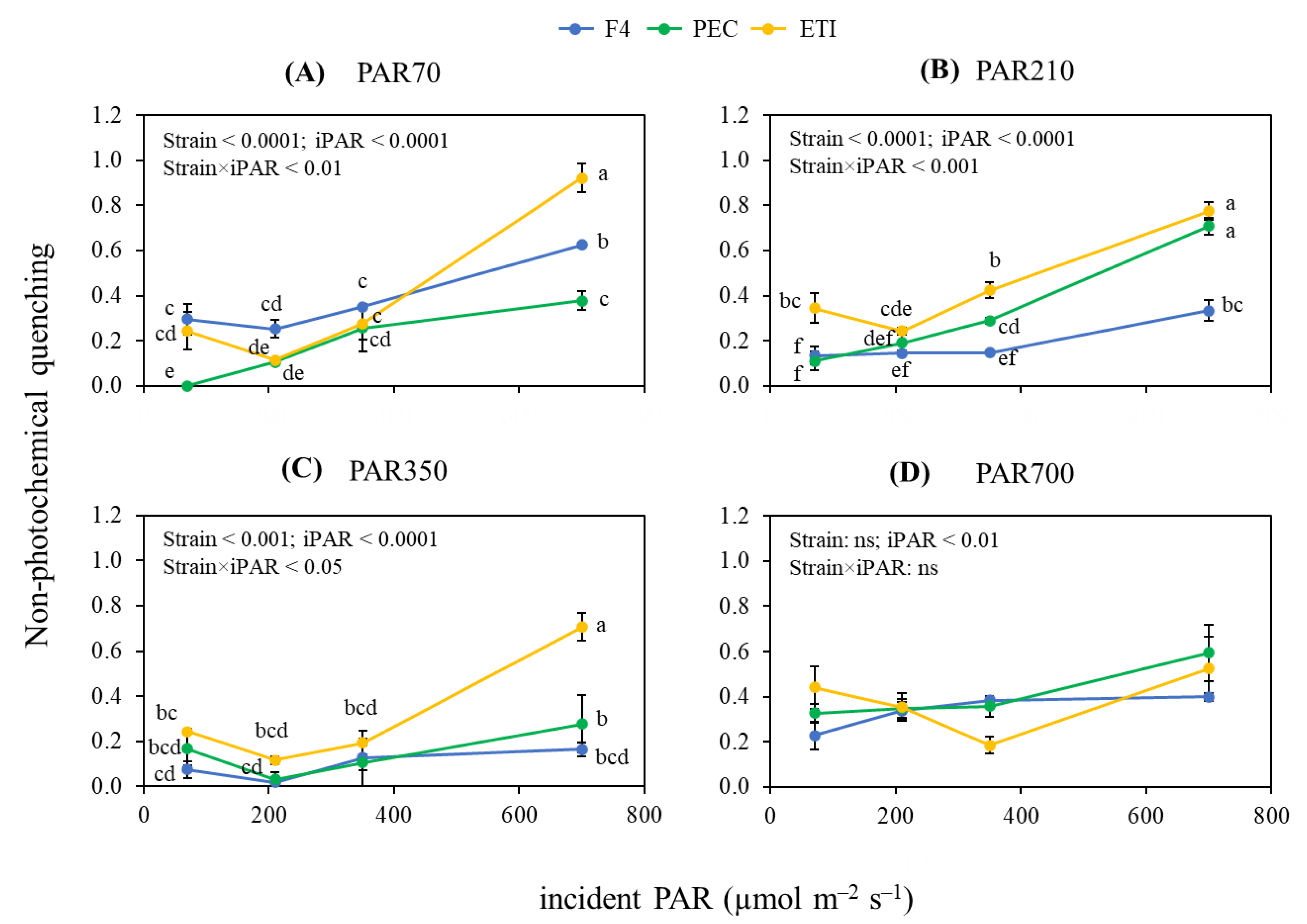
3.1. Chlorophyll Fluorescence

For clarity, data for each light treatment are presented separately. Two-way ANOVA tests showed that the interaction between strain × iPAR did not significantly affect the PSII photochemical efficiency of microalgae, while, as expected, this parameter was significantly affected by iPAR (p < 0.0001; Figure 1A–D). Although the photochemical performance was reduced by an average of 66% when comparing iPAR 0 and 700, the lowest effect was found in microalgae grown at PAR350 (−45%; Figure 1C). Moreover, the strain significantly altered the photochemical efficiency of PSII under light range conditions PAR70—PAR350 (Figure 1A–C), whereas this alteration was not observed at PAR700 (Figure 1D). In detail, no clear trend was observed, although PEC recorded on average the lowest efficiency under intermediate light treatments (i.e., PAR210 and 350; Figure 1B,C).
Concerning the light response curves of NPQ, the two-way ANOVA showed that the individual factors and their interaction significantly affected the dissipation capacity of microalgae grown from PAR70 to PAR350 (Figure 2A–C). In detail, F4 curves became gradually flatter with increasing light growth conditions in comparison to PEC and ETI. Moreover, NPQ patterns in ETI remained consistent for this strain grown from PAR70 to PAR350 (Figure 2A–C), while it was compromised in PEC when cells were grown at PAR350 (Figure 2C). At PAR700, the light response curves of NPQ were not significantly affected by strain × iPAR and strain, while a significant variation was detected due to iPAR (Figure 2D). Although iPAR 700 resulted in the highest dissipation, no differences in NPQ were observed at lower incident lights.

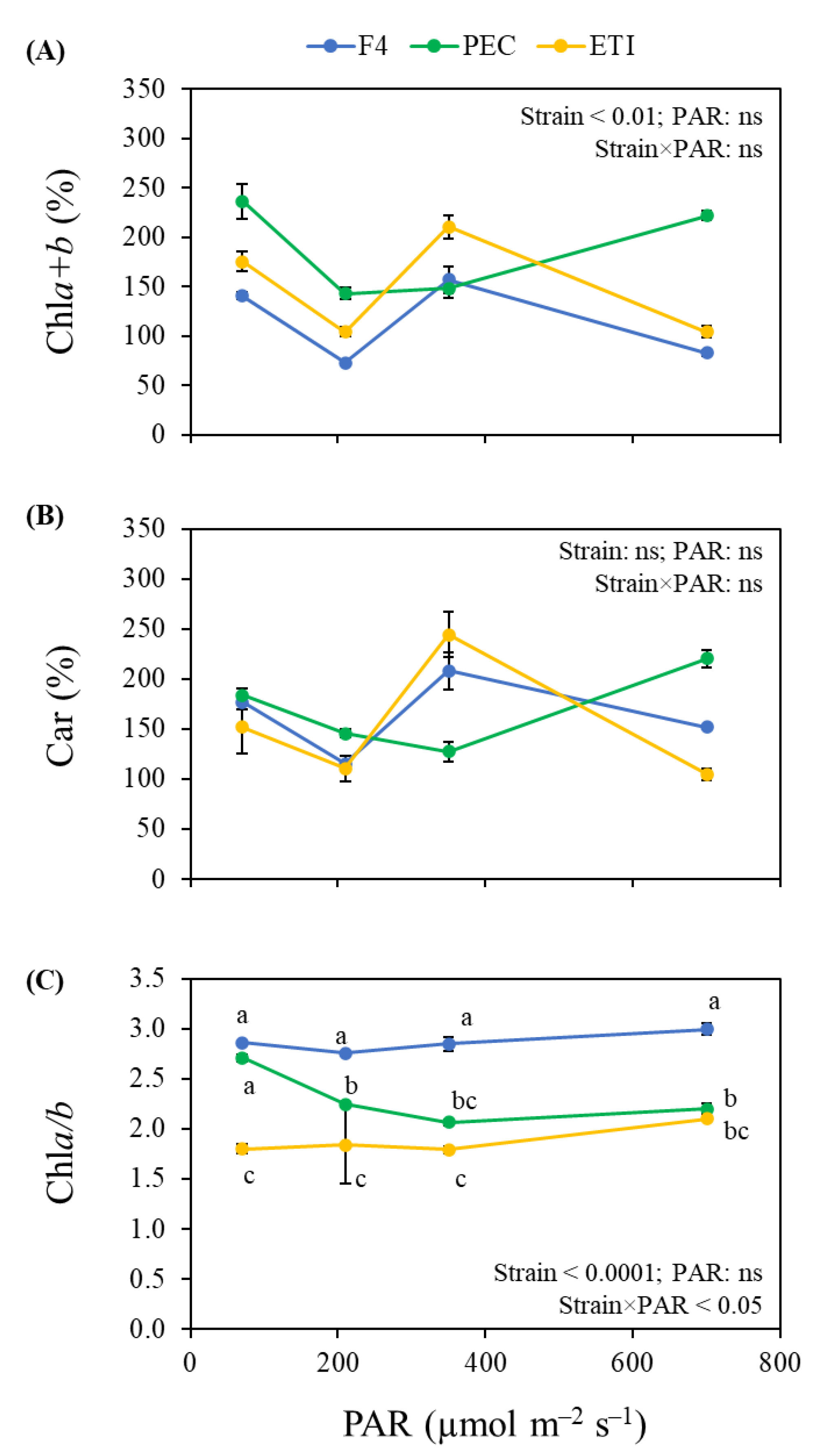
3.2. Photosynthetic Pigments

Two-way ANOVA tests showed that strain × PAR and PAR did not significantly affect Chla + b, while a significant effect was evidenced due to strain (p < 0.01; Figure 3A). Particularly, PEC recorded the highest Chla + b on average, while no differences were detected between ETI and F4 (Figure 3A). Results of Car showed that neither the effect of individual factors nor the interaction of strain with PAR was significant for the percentage of these photosynthetic pigments (Figure 3B). In contrast, the analysis of Chla/b revealed a significant strain × PAR interaction (p < 0.05; Figure 3C). In detail, F4 and ETI generally maintained the highest and lowest Chla/b ratios, respectively, independent of the light intensity, while Chla/b in PEC gradually decreased with increasing light intensity, starting at the same level of F4 at PAR70 and reaching to the lowest level at PAR700, similar to that of ETI (Figure 3C).

3.3. Antioxidant Systems

A significant interaction between microalgal strains and light intensity conditions was observed for MAAs (p < 0.05) and TAC (p < 0.0001) (Figure 4). The individual factors induced significant changes in MAAs (Figure 4A) but did not affect TAC (Figure 4B). From PAR70 to PAR350, all strains showed similar MAAs patterns with a decrease from PAR70 to PAR210 and a subsequent restoration when cells were exposed to PAR350 (Figure 4A). Moreover, ETI exposed to an increasing light intensity up to PAR350 showed a consistently higher level of MAAs than F4 and PEC at the same conditions. At PAR700, PEC strongly induced the production of MAAs whose level was higher than that in F4 and ETI, and did not show significant differences and maintained their levels similar to those found at PAR350 (Figure 4A). Concerning TAC, each strain showed different patterns under increasing light intensity (Figure 4B). In detail, F4 did not show changes in TAC with increasing light intensity from PAR70 to PAR350, whereas at PAR700 TAC was strongly induced, reaching the highest level in comparison with the Ecuadorian strains. Levels of TAC in ETI and PEC at PAR70 were similar but significantly higher than that in F4, whereas at PAR210 and PAR350, ETI showed similar TAC percentages as F4, whose values were significantly lower or higher than those found in PEC depending on the light intensity (Figure 4B). Moreover, increasing the light intensity from PAR350 to PAR700 led to variations in TAC with significant differences between strains.

4. Discussion

Light is an important, critical factor for photosynthesis; however, depending on its intensity, it can be a limiting or inhibiting factor that in turn affects microalgal growth and metabolism. Among the three different species used in this study, ETI and PEC were isolated from a high light environment in the Andes at 2200 m altitude [20], while F4 came from a Mediterranean ecosystem at sea level [21]. Given these features, we expected that ETI and PEC will be more tolerant than F4 in response to increasing light at photosynthetic level, reflecting the differences in their photoacclimation strategies to adapt to their habitats’ origin. However, we found almost no difference in the light curves of the PSII photochemical efficiency between strains under increasing light intensity, showing a clear deficiency in the use of absorbed light when treatment was beyond PAR210. Moreover, PAR350 and PAR700 induced a decline in the Fv/Fm parameter (−50% on average) in all strains, indicating that cells have experienced an excess of energy, thus potentially resulting in photooxidative stress that led to PSII inhibition or damage. Photosynthetic organisms have developed some strategies to prevent and manage oxidative stress, including a decrease in light absorption and ROS scavenging [28]. NPQ is a fast and effective photoprotection mechanism where energy in excess, which cannot be used for photochemistry, is safely dissipated as heat [29]. Among strains, ETI seems to have better energy dissipation capacity as NPQ tended to rise more rapidly as the incident light increased in cells grown up to PAR350. Moreover, the sustained NPQ curves in ETI suggested that this strain may employ persistent energy dissipation. Concordantly, another species (Chlamydomonas priscui) from the Chlamydomonadales like ETI has the capacity to photoprotect their cells by a constitutive NPQ, enabling its survival in extreme environments [30]. As well as concerning PEC, the results showed that although PEC and ETI were isolated from the same environment, the NPQ in PEC seemed to be induced by increasing light and impaired from PAR350. These results indicated that the different energy dissipation capacity in highland strains is probably species dependent. Unlike highland strains, the NPQ in F4 was compromised starting from PAR210, suggesting that this intensity represented a saturating threshold light in F4. The different light effects on NPQ among lowland and highland strains might be related to the selection pressure of their native environments. Indeed, unlike F4, ETI and PEC are adapted to extreme environmental conditions characteristic of the equatorial highland zone, such as high solar radiation and constant photoperiods [31].
Since Car can contribute to NPQ, as they promote the quenching of excess excitation energy, some differences between pigment content in the studied strains were expected. Here, among strains, PEC showed a completely different Car pattern in response to increasing light, while ETI and F4 showed similar patterns, although they displayed different dissipation capacities. This suggested that NPQ in each strain may be differentially dependent from Car. Concordantly, a recent study in different microalgal species showed that exposure to increasing light intensity can activate heat dissipation in the presence or absence of xanthophyll carotenoids [32]. These findings also revealed that these different NPQ mechanisms were more related to the environmental selection pressure than to microalgal phylogeny [32]. However, since only total carotenoids were analyzed in this study, it would be interesting to determine the composition and identify the influence of the xanthophyll cycle in NPQ among strains.
In addition to Car, MAAs and more in general TAC are ROS scavengers involved in minimizing oxidative stress and preventing photodamage. In this study, ETI showed a persistent high content of MAAs in all light conditions, while in PEC they were strongly induced with high light levels. Differently, F4 showed a small increase in MAAs under high light levels. These results suggested that MAAs might be functional metabolites in highland conditions, and their production might be linked to environmental adaptations. Further studies are needed to reveal the ecophysiological role of MAAs in organisms living under stress conditions [10]. Concerning TAC, ETI and PEC showed a declining trend with increasing light intensity, while F4 showed an induction at high light levels. These results suggest that high light-driven ROS production in F4 may be difficult to control by energy dissipation, and thus this strain might induce biochemical regulation pathways. Differently, ETI and PEC were able to balance pro-oxidant production and scavenging capacity well in response to light through heat dissipation and probably via other photoprotective mechanisms (e.g., modulation of thylakoid complexes). Concordantly, Giovagnetti et al. (2012) [33] demonstrated that the biochemical pathway activation in native algae when PAR becomes saturating or over-saturating is a strategy related to the habitats of origin.
The acclimation process to high light requires a long time, during which cells can adapt the number and size of photosynthetic units to optimize energy capture for photochemistry, and thus maintain efficient metabolism and growth [16]. In this line, changes in the Chla/b ratio can be used to estimate the size of the light-harvesting antenna complexes of photosystems, as Chlb is specifically bound to proteins in the antenna [16]. Here, ETI maintained a low Chla/b ratio without changes in all light conditions, suggesting that this strain has a particularly large antenna that is not affected by increasing light intensity. Similarly, the microalga Chlamydomonas sp. UWO241 (belonging to the Chlamydomonadales order as ETI) has the ability to produce chlorophyll with a constitutively increased level of Chlb and efficiently perform photosynthesis in high-latitude environments characterized by continuous cold, high salinity, and low irradiance [34]. Recently, Popson et al. (2024) reported that this Chlamydomonas strain decreases the number of reaction centers instead of adjusting antennae size to regulate the light-harvesting capacity in a short-term high-light-level condition [30]. Altogether, these results have revealed that long- and short-term protection mechanisms against excess light in this species were mainly constitutive, including a particular structure and composition of the photochemical apparatus, continuous energy dissipation not associated with the xanthophyll cycle, high rates of PSI-cyclic electron flow, and high activity of specific antioxidant enzymes [30,35,36]. Concordantly, ETI maintained a low Chla/b ratio, exhibited sustained energy dissipation, and maintained low ROS by possibly keeping a high MAA pool, indicating that this constitutive photoprotection may be a robust strategy to overcome from shade to high-light conditions. Indeed, its native environment is a highland lake of 7 m depth where atmospheric factors can generate waves mixing different layers in the water column [37], meaning that ETI can easily move up and downwards and thus might be well adapted to large changes in light intensity. However, further studies are needed to explore in more detail the structural and functional features in the photochemical apparatus, which will provide insights into the mechanisms of ETI in the energy balancing regulation.
The Chlorella sorokiniana F4 also kept its Chla/b ratio constant in all light conditions, but the values were higher (~3) in comparison with those of ETI (~1.8). High Chla/b ratio is commonly found in different microalgal species, including those from the Chlorella genus [2,17,18,19,38,39]. Moreover, most of these species, like plants, increase their ratio under high light conditions, thus highlighting that the reduction in antenna size is a suitable strategy to avoid photoinhibition [18,38,39,40,41]. Although F4 showed high phylogenetic proximity with some of these species (i.e., algae from the Trebouxiophyceae class), this strain surprisingly did not exhibit changes in the Chla/b ratio with increasing light intensity. This suggested that the organization and assembly of the photosystems in F4 might be influenced by the native environmental adaptive pressure rather than phylogenic characteristics. For instance, native species from the Trebouxiophyceae class like F4, adapted to extreme high-light conditions, were able to modify the antenna size upon light stress [14], while those adapted to low–moderate light could not [41]. It is worth noting that F4 comes from a lowland shallow swamp of 2 m depth with calm water [42], suggesting that this strain might slowly move in the column water and probably not experience so many photic changes, with a total irradiance remaining low if compared with the harsh environment of the highland strains. Altogether, the features of F4 indicated that this strain was less tolerant to high light than highland strains.
Different from ETI and F4, PEC was the only strain that modified the Chla/b ratio in response to light intensity, reducing its values from ~3 to ~1.8 with increasing light. It is worth noting that although the low ratio in PEC was mainly due to the increased level of Chlb, like in ETI, the regulation of chlorophyll composition differed between both strains. In fact, the increased level of Chlb in PEC was activated by moderate–high light intensity, while in ETI it seemed to be light-independent. We hypothesize that PEC adjusts the antennae sizes in a light-dependent manner to regulate the light-harvesting capacity, thus highlighting its plasticity in response to light. Moreover, increasing the antennae sizes seemed to be a robust photoacclimative strategy related to the adaptive evolution to a highland environment, while its regulation is rather linked to phylogenetic diversity. Concordantly, a comparative study of microalgal productivity related to photosynthesis showed that species from the Chlamydomonadales and Sphaeropleales orders, to which ETI and PEC belong, respectively, have different regulation mechanisms of photosynthetic complexes’ stoichiometry and thylakoid proteins [17]. Further studies are needed to understand the light-driven mechanisms behind the rearrangement of the antenna organization in PEC.
In conclusion, we found Chlorella sorokiniana F4 to be the most sensitive to increasing light intensity, as reflected by its lower threshold of oversaturation light intensity compared to other strains. Indeed, high light in F4 compromised its capacity for energy dissipation and increased ROS scavengers (i.e., TAC), reducing PSII photochemistry performance. These features were probably related to its adaptation to lowland environment characterized by constant low–moderate light, resulting in a low ability to reorganize its antennae in comparison with other native strains from the same lineage. Although the Ettlia pseudoalveolaris ETI and the Pectinodesmus pectinatus PEC were more tolerant to increasing light, both strains showed particular photophysiological responses, probably linked to their distinct photoacclimation strategies to highland environmental conditions (e.g., high solar irradiance). In fact, ETI maintained high Chlb to Chla, exhibited sustained energy dissipation, and preserved a high antioxidant pool (i.e., MAAs) in all light conditions, indicating that this strain has a constitutive photoprotective strategy. On the other hand, PEC showed the induction of NPQ, rearrangement of the antennae, and the ROS scavenger pool in a light-dependent manner, highlighting its physiological plasticity in response to light. Altogether, these results demonstrated that the photophysiological responses in native microalgae are much more intricated than in model organisms, as the adaptive evolution to specific environment is affected not only by the native ecosystem constraints but also by the phylogenetic diversity. These findings also pave the way for practical application in bioenergy production, algal biomass cultivation, and photobioreactor optimization, where light-resilient strains can contribute to improving efficiency and sustainability.

Author Contributions

G.P.: investigation, formal analysis, writing—original draft preparation. C.C.: investigation, methodology, resources. C.S.: resources, writing—reviewing and editing. C.E.: resources, writing—reviewing and editing. L.G.: conceptualization, supervision, writing—reviewing and editing. T.H.R.: conceptualization, methodology, investigation, visualization, writing—original draft preparation. All authors have read and agreed to the published version of the manuscript.

Funding

This work was supported by a PhD fellowship from the University of Pisa (to T.H.R). The CNR-MLIP collection was granted by the European Commission—NextGenerationEU, Project SUSMIRRI.IT “Strengthening the MIRRI Italian Research Infrastructure for Sustainable Bioscience and Bioeconomy”, code n. IR0000005.

Institutional Review Board Statement

Not applicable.

Informed Consent Statement

Not applicable.

Data Availability Statement

Data are contained within the article.

Acknowledgments

T.H.R. and G.P. would like to thank Giulia Volpi for her assistance in the collection of data. This article is dedicated to the memory of Carolina Chiellini, who suddenly and very untimely passed away and who will be remembered for her contributions to algal biology.

Conflicts of Interest

The authors declare no conflicts of interest. The funders had no role in the design of the study; in the collection, analyses, or interpretation of data; in the writing of the manuscript; or in the decision to publish the results.

References

  1. Masojídek, J.; Torzillo, G.; Koblížek, M. Photosynthesis in microalgae. In Handbook of Microalgal Culture: Applied Phycology and Biotechnology; Richmond, A., Hu, Q., Eds.; John Wiley & Sons, Ltd.: Oxford, UK, 2013; pp. 21–36. [Google Scholar]
  2. Levin, G.; Kulikovsky, S.; Liveanu, V.; Eichenbaum, B.; Meir, A.; Isaacson, T.; Tadmor, Y.; Adir, N.; Schuster, G. The desert green algae Chlorella ohadii thrives at excessively high light intensities by exceptionally enhancing the mechanisms that protect photosynthesis from photoinhibition. Plant J. 2021, 106, 1260–1277. [Google Scholar] [CrossRef] [PubMed]
  3. Erickson, E.; Wakao, S.; Niyogi, K.K. Light stress and photoprotection in Chlamydomonas reinhardtii. Plant J. 2015, 82, 449–465. [Google Scholar] [CrossRef]
  4. Mullineaux, P.M.; Exposito-Rodriguez, M.; Laissue, P.P.; Smirnoff, N. ROS-dependent signalling pathways in plants and algae exposed to high light: Comparisons with other eukaryotes. Free Radic. Biol. Med. 2018, 122, 52–64. [Google Scholar] [CrossRef]
  5. Rezayian, M.; Niknam, V.; Ebrahimzadeh, H. Oxidative damage and antioxidative system in algae. Toxicol. Rep. 2019, 6, 1309–1313. [Google Scholar] [CrossRef] [PubMed]
  6. Finazzi, G.; Minagawa, J. High light acclimation in green microalgae. In Non-Photochemical Quenching and Energy Dissipation in Plants, Algae and Cyanobacteria; Demmig-Adams, B., Garab, G., Adams, W., III, Govindjee, Eds.; Springer: Dordrecht, The Netherlands, 2014; pp. 445–469. ISBN 978-94-017-9031-4. [Google Scholar]
  7. Müller, P.; Li, X.P.; Niyogi, K.K. Non-photochemical quenching. A response to excess light energy. Plant Physiol. 2001, 125, 1558–1566. [Google Scholar] [CrossRef] [PubMed]
  8. Niyogi, K.K.; Truong, T.B. Evolution of flexible non-photochemical quenching mechanisms that regulate light harvesting in oxygenic photosynthesis. Curr. Opin. Plant Biol. 2013, 16, 307–314. [Google Scholar] [CrossRef]
  9. Solovchenko, A.; Neverov, K. Carotenogenic response in photosynthetic organisms: A colorful story. Photosynth. Res. 2017, 133, 31–47. [Google Scholar] [CrossRef]
  10. Nascimento, L.B.d.S.; Tattini, M. Beyond photoprotection: The multifarious roles of flavonoids in plant terrestrialization. Int. J. Mol. Sci. 2022, 23, 5284. [Google Scholar] [CrossRef]
  11. Geraldes, V.; Pinto, E. Mycosporine-like amino acids (Maas): Biology, chemistry and identification features. Pharmaceuticals 2021, 14, 63. [Google Scholar] [CrossRef]
  12. Virtanen, O.; Khorobrykh, S.; Tyystjärvi, E. Acclimation of Chlamydomonas reinhardtii to extremely strong light. Photosynth. Res. 2021, 147, 91–106. [Google Scholar] [CrossRef]
  13. Smith, B.M.; Morrissey, P.J.; Guenther, J.E.; Nemson, J.A.; Harrison, M.A.; Allen, J.F.; Melis, A. Response of the photosynthetic apparatus in Dunaliella salina (green algae) to irradiance stress. Plant Physiol. 1990, 93, 1433–1440. [Google Scholar] [CrossRef] [PubMed]
  14. Levin, G.; Yasmin, M.; Liveanu, V.; Burstein, C.; Hanna, R.; Kleifeld, O.; Simanowitz, M.C.; Meir, A.; Tadmor, Y.; Hirschberg, J.; et al. A desert Chlorella sp. that thrives at extreme high-light intensities using a unique photoinhibition protection mechanism. Plant J. 2023, 115, 510–528. [Google Scholar] [CrossRef]
  15. Belgio, E.; Trsková, E.; Kotabová, E.; Ewe, D.; Prášil, O.; Kaňa, R. High light acclimation of Chromera velia points to photoprotective NPQ. Photosynth. Res. 2018, 135, 263–274. [Google Scholar] [CrossRef] [PubMed]
  16. Bonente, G.; Pippa, S.; Castellano, S.; Bassi, R.; Ballottari, M. Acclimation of Chlamydomonas reinhardtii to different growth irradiances. J. Biol. Chem. 2012, 287, 5833–5847. [Google Scholar] [CrossRef]
  17. Baldisserotto, C.; Sabia, A.; Giovanardi, M.; Ferroni, L.; Maglie, M.; Pancaldi, S. Chlorophyta microalgae as dietary protein supplement: A comparative analysis of productivity related to photosynthesis. J. Appl. Phycol. 2022, 34, 1323–1340. [Google Scholar] [CrossRef]
  18. Cecchin, M.; Simicevic, J.; Chaput, L.; Hernandez Gil, M.; Girolomoni, L.; Cazzaniga, S.; Remacle, C.; Hoeng, J.; Ivanov, N.V.; Titz, B.; et al. Acclimation strategies of the green alga Chlorella vulgaris to different light regimes revealed by physiological and comparative proteomic analyses. J. Exp. Bot. 2023, 74, 4540–4558. [Google Scholar] [CrossRef] [PubMed]
  19. Suwannachuen, N.; Leetanasaksakul, K.; Roytrakul, S.; Phaonakrop, N.; Thaisakun, S.; Roongsattham, P.; Jantasuriyarat, C.; Sanevas, N.; Sirikhachornkit, A. Palmelloid formation and cell aggregation are essential mechanisms for high light tolerance in a natural strain of Chlamydomonas reinhardtii. Int. J. Mol. Sci. 2023, 24, 8374. [Google Scholar] [CrossRef]
  20. Huarancca Reyes, T.; Chiellini, C.; Barozzi, E.; Sandoval, C.; Echeverría, C.; Guglielminetti, L. Exploring the physiological multiplicity of native microalgae from the Ecuadorian highland, Italian lowland and indoor locations in response to UV-B. Int. J. Mol. Sci. 2023, 24, 1346. [Google Scholar] [CrossRef]
  21. Chiellini, C.; Mariotti, L.; Huarancca Reyes, T.; de Arruda, E.J.; Fonseca, G.G.; Guglielminetti, L. Remediation capacity of different microalgae in effluents derived from the cigarette butt cleaning process. Plants 2022, 11, 1770. [Google Scholar] [CrossRef]
  22. Huarancca Reyes, T.; Pompeiano, A.; Ranieri, A.; Volterrani, M.; Guglielminetti, L.; Scartazza, A. Photosynthetic performance of five cool-season turfgrasses under UV-B exposure. Plant Physiol. Biochem. 2020, 151, 181–187. [Google Scholar] [CrossRef]
  23. Lichtenthaler, H.K. Chlorophylls and carotenoids: Pigments of photosynthetic biomembranes. In Methods in Enzymology; Elsevier: Amsterdam, The Netherlands, 1987; Volume 148, pp. 350–382. [Google Scholar] [CrossRef]
  24. Huang, J.J.; Bunjamin, G.; Teo, E.S.; Ng, D.B.; Lee, Y.K. An enclosed rotating floating photobioreactor (RFP) powered by flowing water for mass cultivation of photosynthetic microalgae. Biotechnol. Biofuels 2016, 9, 218. [Google Scholar] [CrossRef] [PubMed]
  25. Huarancca Reyes, T.; Mariotti, L.; Chiellini, C.; Guglielminetti, L.; Fonseca, G.G. UV-B irradiation effect on microalgae performance in the remediation of effluent derived from the cigarette butt cleaning process. Plants 2022, 11, 2356. [Google Scholar] [CrossRef]
  26. Moles, T.M.; Guglielminetti, L.; Huarancca Reyes, T. Differential effects of sodium chloride on germination and post-germination stages of two tomato genotypes. Sci. Hortic. 2019, 257, 108730. [Google Scholar] [CrossRef]
  27. Pompeiano, A.; Huarancca Reyes, T.; Moles, T.M.; Guglielminetti, L.; Scartazza, A. Photosynthetic and growth responses of Arundo donax L. plantlets under different oxygen deficiency stresses and reoxygenation. Front. Plant Sci. 2019, 10, 408. [Google Scholar] [CrossRef]
  28. Li, Z.; Wakao, S.; Fischer, B.B.; Niyogi, K.K. Sensing and responding to excess light. Annu. Rev. Plant Biol. 2009, 60, 239–260. [Google Scholar] [CrossRef] [PubMed]
  29. Bassi, R.; Dall’Osto, L. Dissipation of light energy absorbed in excess: The molecular mechanisms. Annu. Rev. Plant Biol. 2021, 72, 47–76. [Google Scholar] [CrossRef] [PubMed]
  30. Popson, D.; D’Silva, S.; Wheeless, K.; Morgan-Kiss, R. Permanent stress adaptation and unexpected high light tolerance in the shade-adapted Chlamydomonas priscui. Plants 2024, 13, 2254. [Google Scholar] [CrossRef]
  31. Benito, X.; Luethje, M.; Schneider, T.; Fritz, S.C.; Baker, P.A.; Pedersen, E.J.; Gaüzère, P.; de Novaes Nascimento, M.; Bush, M.; Ruhi, A. Ecological resilience in tropical Andean lakes: A paleolimnological perspective. Limnol. Oceanogr. 2022, 67, S23–S37. [Google Scholar] [CrossRef]
  32. Quaas, T.; Berteotti, S.; Ballottari, M.; Flieger, K.; Bassi, R.; Wilhelm, C.; Goss, R. Non-photochemical quenching and xanthophyll cycle activities in six green algal species suggest mechanistic differences in the process of excess energy dissipation. J. Plant Physiol. 2015, 172, 92–103. [Google Scholar] [CrossRef]
  33. Giovagnetti, V.; Cataldo, M.L.; Conversano, F.; Brunet, C. Growth and photophysiological responses of two picoplanktonic Minutocellus species, strains RCC967 and RCC703 (Bacillariophyceae). Eur. J. Phycol. 2012, 47, 408–420. [Google Scholar] [CrossRef]
  34. Cvetkovska, M.; Orgnero, S.; Hüner, N.P.A.; Smith, D.R. The enigmatic loss of light-independent chlorophyll biosynthesis from an Antarctic green alga in a light-limited environment. New Phytol. 2019, 222, 651–656. [Google Scholar] [CrossRef] [PubMed]
  35. Stahl-Rommel, S.; Kalra, I.; D’Silva, S.; Hahn, M.M.; Popson, D.; Cvetkovska, M.; Morgan-Kiss, R.M. Cyclic electron flow (CEF) and ascorbate pathway activity provide constitutive photoprotection for the photopsychrophile, Chlamydomonas sp. UWO 241 (renamed Chlamydomonas priscuii). Photosynth. Res. 2022, 151, 235–250. [Google Scholar] [CrossRef] [PubMed]
  36. Szyszka, B.; Ivanov, A.G.; Hüner, N.P.A. Psychrophily is associated with differential energy partitioning, photosystem stoichiometry and polypeptide phosphorylation in Chlamydomonas raudensis. Biochim. Biophys. Acta Bioenerg. 2007, 1767, 789–800. [Google Scholar] [CrossRef] [PubMed]
  37. Jácome, G.; Valarezo, C.; Yoo, C. Assessment of water quality monitoring for the optimal sensor placement in lake Yahuarcocha using pattern recognition techniques and geographical information systems. Environ. Monit. Assess. 2018, 190, 259. [Google Scholar] [CrossRef]
  38. Li, Y.; Gu, W.; Huang, A.; Xie, X.; Wu, S.; Wang, G. Transcriptome analysis reveals regulation of gene expression during photoacclimation to high irradiance levels in Dunaliella salina (Chlorophyceae). Phycol. Res. 2019, 67, 291–302. [Google Scholar] [CrossRef]
  39. Cazzaniga, S.; Dall’Osto, L.; Szaub, J.; Scibilia, L.; Ballottari, M.; Purton, S.; Bassi, R. Domestication of the green alga Chlorella sorokiniana: Reduction of antenna size improves light-use efficiency in a photobioreactor. Biotechnol. Biofuels 2014, 7, 157. [Google Scholar] [CrossRef]
  40. Ballottari, M.; Dall’Osto, L.; Morosinotto, T.; Bassi, R. Contrasting behavior of higher plant photosystem I and II antenna systems during acclimation. J. Biol. Chem. 2007, 282, 8947–8958. [Google Scholar] [CrossRef]
  41. La Rocca, N.; Sciuto, K.; Meneghesso, A.; Moro, I.; Rascio, N.; Morosinotto, T. Photosynthesis in extreme environments: Responses to different light regimes in the Antarctic alga Koliella antarctica. Physiol. Plant. 2015, 153, 654–667. [Google Scholar] [CrossRef]
  42. Bartolini, A. La Riserva Naturale del Padule di Fucecchio. Dieci anni di Gestione (1996–2006); Centro di Ricerca, Documentazione e Promozione del Padule di Fucecchio: Larciano, Italy, 2007. [Google Scholar]
Figure 1. Light response curves of (AD) photochemical efficiency of photosystem (PSII) under increasing incident photosynthetically active radiation (iPAR) from 0 to 700 μmol photons m−2 s−1. The measurements were performed in each microalgal strain (F4, PEC, and ETI) after being exposed to (A) PAR70, (B) 210, (C) 350, and (D) 700 for 18 h. Data are expressed as means of 3 different replicates ± standard error (SE). The p-values for the main factors and the Strain × iPAR interaction term of the ANOVA are shown. ns: Not significant.
Phycology 05 00024 g001
Figure 2. Light response curves of (AD) non-photochemical quenching (NPQ) under increasing incident photosynthetically active radiation (iPAR). The measurements were performed in each microalgal strain (F4, PEC, and ETI) after being exposed to (A) PAR70, (B) 210, (C) 350, and (D) 700 for 18 h. Data are expressed as means of 3 different replicates ± standard error (SE). The p-values for the main factors and the strain × iPAR interaction term of the ANOVA are shown. Means followed by the same letter are not significantly different based on Tukey’s test (p < 0.05). ns: Not significant.
Phycology 05 00024 g002
Figure 3. Effect of light condition on the photosynthetic pigments. (A) Total chlorophyll (Chla + b), (B) carotenoids (Car), and (C) the ratio of Chla to Chlb (Chla/b) were determined in each microalgal strain (F4, PEC and ETI) at the beginning (T0) and end (T18) of the experiment. Microalgal strains were exposed to different light intensities (70, 210, 350 and 700 μmol m−2 s−1 photosynthetically active radiation (PAR)). Chla + b and Car are expressed in % and were obtained by the comparison of values obtained at T18 with respect to that at T0. Chla/Chlb at the end of the experiment are reported. Data are expressed as means of 3 different replicates ± standard error (SE). The p-values for the main factors and the Strain × PAR interaction term of the ANOVA are shown. Means followed by the same letter are not significantly different based on Tukey’s test (p < 0.05). ns: Not significant.
Phycology 05 00024 g003
Figure 4. Effect of light condition on secondary metabolites. (A) Mycosporine-like amino acids (MAAs) and (B) total non-enzymatic antioxidant compounds (TAC) were determined in each microalgal strain (F4, PEC and ETI) at the beginning (T0) and end (T18) of the experiment. Microalgal strains were exposed to different light intensities (70, 210, 350, and 700 μmol m−2 s−1 photosynthetically active radiation (PAR)). MAAs and TAC are expressed in % and were obtained by the comparison of values obtained at T18 with respect to that at T0. Data are expressed as means of 3 different replicates ± standard error (SE). The p-values for the main factors and the strain × PAR interaction term of the ANOVA are shown. Means followed by the same letter are not significantly different based on Tukey’s test (p < 0.05). ns: Not significant.
Phycology 05 00024 g004
Table 1. List of microalgal strains.
StrainIsolation SourceGeographic LocationTaxonomic AffiliationReference
F4
(MLIP M004)
Mediterranean
Marshland (Italy)
43°48′31″ N
10°48′18″ E
Chlorella
sorokiniana
[21]
PECAndean
Lake (Ecuador)
00°22′10″ N
78°06′09″ W
Pectinodesmus pectinatus[20]
ETIAndean
Lake (Ecuador)
00°22′10″ N
78°06′09″ W
Ettlia
pseudoalveolaris
[20]
Disclaimer/Publisher’s Note: The statements, opinions and data contained in all publications are solely those of the individual author(s) and contributor(s) and not of MDPI and/or the editor(s). MDPI and/or the editor(s) disclaim responsibility for any injury to people or property resulting from any ideas, methods, instructions or products referred to in the content.

