Article

Sexual Propagation in the Green Seaweed Codium tomentosum—An Emerging Species for Aquaculture

by
Maria Francisca Sá
1,2,
Teresa Cunha Pacheco
1,2,
Isabel Sousa-Pinto
1,2 and
Gonçalo Silva Marinho
1,2,*
1
Department of Biology, Faculty of Sciences, University of Porto, Rua do Campo Alegre s/n, 4169-007 Porto, Portugal
2
CIIMAR/CIMAR-LA, Interdisciplinary Centre of Marine and Environmental Research, University of Porto, Terminal de Cruzeiros do Porto de Leixões, Av. General Norton de Matos s/n, 4450-208 Matosinhos, Portugal
*
Author to whom correspondence should be addressed.
Phycology 2024, 4(4), 533-547; https://doi.org/10.3390/phycology4040029
Submission received: 6 August 2024 / Revised: 13 September 2024 / Accepted: 29 September 2024 / Published: 3 October 2024

Abstract

:
Codium tomentosum holds a variety of bioactive compounds, high nutritional value and health benefits, which makes it a valuable natural resource for the food, cosmetic and pharmaceutical industries. Currently, C. tomentosum is farmed at a small-scale targeting niche markets, and further expansion of production is limited by a lack of optimised propagation and cultivation methods. This study aims to identify the conditions required to control key production parameters including gametogenesis, gamete release and suitable culture conditions for the early stages of development of C. tomentosum. Wild specimens of C. tomentosum were collected on the Aguçadoura shore, north of Portugal. Gametogenesis was successfully induced in infertile specimens cultured under a short-day photoperiod (8 h:16 h; L:D). Gamete release was optimised through a combination of hydric shock and ultrasounds, with the highest gamete yield obtained after a 2 h 30 min desiccation period, followed by re-hydration and a series of three ultrasounds. Germlings, precursors of the adult C. tomentosum, grew faster when cultured under a lower light intensity (20 μmol m−2 s−1) compared to higher intensities (40 and 60 μmol m−2 s−1) in every light spectrum; additionally, the growth of germlings exposed to the lowest light intensity was significantly higher under white, red and green light spectra compared to blue light. The results on key production parameters constitute an important contribution to the establishment of nursery protocols based on sexual reproduction for aquaculture of the species.

1. Introduction

One of the EU Blue Bioeconomy’s promising new sectors is the European algae industry including seaweed [1]. Aquaculture is considered to have the greatest potential to supply the increasing market demand for seaweed biomass, as wild stocks are already under pressure due to climate change and disturbance from human activity [2]. Nevertheless, to reach its full potential and ensure long-term sustainability, the industry needs to diversify the commercialised species to boost its resilience and capacity to cope with major challenges ahead, such as genetic deprivation, decline in resistance to disease and climate change, which could lead to mass crop loss events [3]. In this context, it is necessary to develop propagation and cultivation techniques for novel species, particularly those underexploited but with recognised commercial potential [4].
The green seaweed Codium tomentosum Stackhouse, 1797, order Bryopsidales, has recently drawn more interest due to its high nutritional value, health benefits and potential for a broad range of industries, namely cosmetic, pharmaceutic, biomedicine and food industries [4,5]. Codium species are an important source of bioactive compounds, phytochemicals, stigmasterol α-tocopherol and many others [6,7,8]. Additionally, high levels of essential fatty acids have also been described for C. tomentosum [9].
Propagation in Codium species can be performed through sexual reproduction by the settlement of wild zygotes onto culture lines [10,11]. Asexual reproduction is carried out by artificial seed production via regeneration of isolated utricles and medullary filaments attached to twine in a nursery [10,12,13] or by cuttings of spongy thalli (approx. 2 cm fragments) in free-floating culture [4].
Codium species present a diplontic life cycle, and sexual reproduction occurs by the fusion of haploid biflagellate gametes. Male and female gametes exhibit a round to ovoid shape, green to olive green colour, measuring 3 to 14 µm and 17 to 30 µm in diameter, respectively. Zygotes germinate into siphonated diploid filaments, germlings, which are the precursors of the spongy thallus, the upright macroscopic Codium, constituted by many utricles [13,14,15]. Ovoid, oblong or fusiform gametangia, one to four per utricle, arise laterally from the utricles in C. tomentosum [16].
C. tomentosum has recently been domesticated, and it is cultivated in tumble culture through vegetative propagation (cuttings) in integrated multi-trophic aquaculture (IMTA) [4,17]. This is still small-scale production, and further expansion requires optimisation of culture methods. The establishment of nursery protocols based on sexual reproduction and/or artificial seeding has the potential to facilitate the establishment of a large biobank, allowing for the mass production of propagules/seedlings and reducing dependence from natural stocks, time and labour intensity. These would also potentiate cultivation in seeded substrates, allowing for expansion of production to the sea. Despite the great interest in developing sexual propagation for this species, life cycle control under laboratory conditions is challenging [18]. Important knowledge gaps include gametogenesis induction, gamete release optimisation and light requirements to sustain germling growth.
In the laboratory, gamete release from fertile wild-collected specimens of Codium fragile was achieved by resorting to osmotic shock [19,20,21]. However, studies are still required to evaluate the effectiveness of such methods to obtain high gamete yields in C. tomentosum.
To our knowledge, gametogenesis in C. tomentosum has not been previously investigated under laboratory-controlled conditions. Seaweeds present species-specific patterns of growth and reproduction, which are affected by the interaction of physical and chemical parameters, among the most important: temperature, salinity, light, water motion and nutrients [17,22,23,24]. Light plays a major role in inducting reproduction (reviewed by Santelices [25]), with the photoperiod acting as the main instigator for gametogenesis in long-lived algae [26,27]. Both short-day and long-day conditions can induce gametogenesis, depending on the species, although short-day responses are more common [28]. C. tomentosum from the northern Portuguese coast experiences gametogenesis during late spring and reaches sexual maturity in summer [29,30], which matches a period of increased temperature and daylength. The same seasonal pattern was observed in populations of C. fragile from the Northeastern USA, and it was proposed that a minimum temperature of 12 to 15 °C was required for the development of gametangia, with gametogenesis being inhibited at lower temperatures [20,21].
The establishment of culture conditions required for the development of zygotes and germlings is a key step for the development of sexual propagation protocols for aquaculture. Hwang et al. [11], studied the effects of light intensity and temperature on the development of zygotes of C. fragile, reporting the maximum growth of germlings at a low light intensity (20 μmol m−2 s−1) and 15 °C, as opposed to isolated utricles (vegetative propagation) which performed better under a higher light intensity and a higher temperature. Irradiance and water movement have been reported as key factors controlling the development of filamentous and spongy thalli of C. fragile under laboratory culture conditions [12,13,31]. A minimum light intensity of 60 μmol m−2 s−1 together with water movement is required to induce the development of the spongy thalli in C. fragile from the precursor filamentous thalli [12,13]. Additionally, Marques et al. [17] reported a higher growth rate in spongy thalli of C. tomentosum cultured under a long-day photoperiod (16 h:8 h; light:dark; L:D) and when exposed to red light, relatively to those exposed to white and blue lights. However, further studies focusing on optimal culture conditions for the early development of C. tomentosum and the differentiation of spongy thallus (the conditions required to induce the development of spongy thallus from the precursors filamentous thallus and germlings) are needed.
The present study aims to determine the conditions required to control key production parameters such as induce gametogenesis, improve gamete release and optimise light conditions to enhance growth in the early stages, zygotes and germlings, of C. tomentosum, which provides valuable knowledge for establishing nursery protocols based on sexual reproduction for aquaculture.

2. Materials and Methods

2.1. Induction of Gametogenesis

The effects of the long-day photoperiod (16 h:8 h; L:D) and the short-day photoperiod (8 h:16 h; L:D) on the gametogenesis of infertile specimens of C. tomentosum were studied under laboratory conditions. Young specimens were randomly collected during low tide from a natural population on the Aguçadoura rocky shore (Póvoa de Varzim, Portugal, 41°25′47.6″ N, 8°47′05.3″ W) in April 2021. For a detailed description of the sampling area, see Cabral [32]. In the laboratory, thalli were rinsed with sterilised seawater to remove any debris. Following this, the specimens were examined to confirm the absence of reproductive structures (small sections of apical thalli were homogenised in sterile seawater, and the resulting suspension of utricles was observed under an inverted microscope, Nikon Eclipse TE200, Tokyo, Japan). In each photoperiod, 3 flasks (n = 3) containing 4 thalli with an initial approximate total length of 7 cm (initial density of approx. 30 g L−1) were kept growing under 16 °C and 100 μmol m−2 s−1 light intensity, with air filtered through a 0.2 μm filter (600 L h−1; model Air-Flow 4; SuperFish, Klundert, Netherlands). Provasoli’s Enriched Seawater (PES) medium prepared according to Harrison et al. [33] and diluted 10 times in sterile seawater was used and changed weekly. After a 3-week culture, a scale-up from a 1 L flask to a 2 L flask was carried out to not limit the growth of the specimens. Every week, thalli were observed in the stereomicroscope Leica EZ4 (Leica Microsystems, Wetzlar, Germany) to examine for possible development of gametangia. The specimens were weighed weekly throughout the 8-week culture period, and the relative growth rate (RGR) and the productivity (P) were calculated as follows:
RGR (% week−1) = [(ln Wf − ln Wi) × t−1] × 100%,
where Wf is the final fresh weight (g), Wi is the initial fresh weight (g), and t is the culture time (weeks) [17].
P (g L−1 week−1) = (FWf − FWi) × V−1 × t−1,
where FWf is the final fresh weight (g), FWi is the initial fresh weight (g), V is the culture flask volume (L), and t is the time in the culture (weeks) [34].
After the culture period, the apical thalli of the individuals grown in both the long-day and the short-day photoperiods were homogenised in sterile seawater to obtain a suspension of utricles. The suspension was then transferred to graduated Petri dishes, and if present, the gametangia density was determined under the inverted microscope connected to a computer with Nikon’s microscope imaging software (NIS). Images of gametangia were captured, and their lengths and diameters were measured using Image J Software 1.53e (U. S. National Institutes of Health, Bethesda, MD, USA), a processing program that allows for measurement and analysis.
Thalli that bearded gametangia after the culture period were used for the gamete release process. Gametes were released based on Churchill et al. [21], resorting to hydric shock, desiccation of thalli for 2 h followed by submersion in sterile seawater. The resulting gamete suspension was transferred to graduated Petri dishes and observed under the inverted microscope to check gamete motility and image recording. Gamete suspensions were cultured at 20 μmol m−2 s−1 light intensity and 16 °C, to assess their viability based on zygote development and germination to form the siphonous filament (germling). The numbers of female gametes and germlings were determined after 1 and 7 days in culture, respectively. The germination (G) rate was calculated as follows [35]:
G (%) = (mean number of germlings/mean number of female gametes) × 100

2.2. Optimisation of Gamete Release

To optimise gamete release, two linked assays were conducted. In the first assay, the fertile thalli were subjected to increasing desiccation periods—0 h, 2 h 30 min and 5 h, followed by three rounds of ultrasounds (total 9 conditions; n = 3) to test their combined effect on the number of female gametes released. Reproductive C. tomentosum was collected from the Aguçadoura shore in November 2021. Specimens (n = 5 per replicate) were dried with laboratory paper and left to desiccate at room temperature according to each desiccation period to be tested. Following this, fertile thalli were submerged in seawater and subjected to a series of three ultrasounds (three minutes each), using an ultrasonic water bath (HF45KHz, model USC300T, VWR International, Radnor, PA, USA). Ten minutes after each ultrasound, three aliquots with 5 mL of suspension were transferred into three Petri dishes, and the number of female gametes was counted under the inverted microscope. Between ultrasounds, the suspension was replaced with new autoclaved seawater to avoid repeated counts of the same gametes.
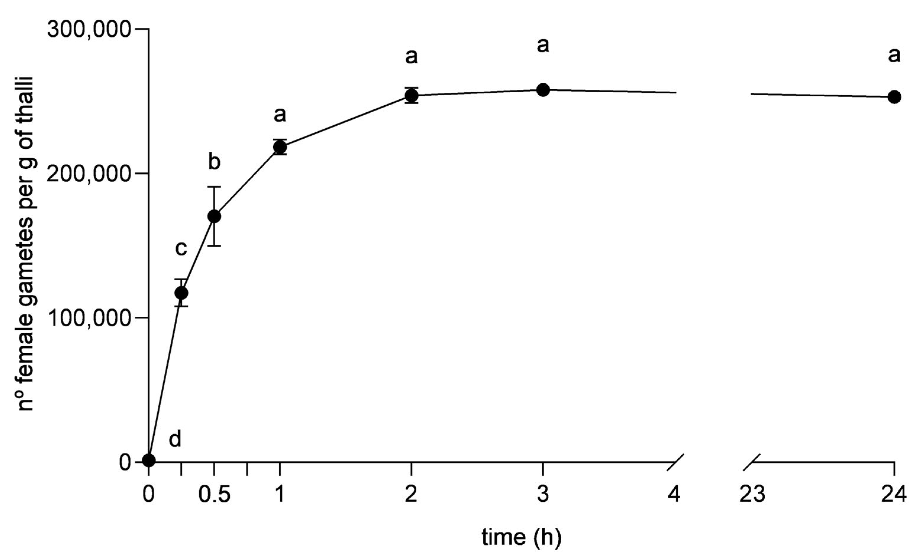
The second trial was performed to draw a 24 h curve of gametes release and determine gamete yield over time. The desiccation and ultrasound of reproductive C. tomentosum were carried out as previously described (for the best-performing treatment), and the numbers of female gametes released after 0 min, 15 min, 30 min, 1 h, 2 h, 3 h and 24 h were determined under the inverted microscope. The motility of the female gametes was also monitored in each count.

2.3. Light Intensity and Spectrum

Different light intensities and spectra were tested to assess their effects on the length (µm) of C. tomentosum germlings for 28 days. Gametes of C. tomentosum were obtained based on the studies described by Churchill et al. [21], adding three minutes of ultrasound to potentiate gamete release from fertile thalli. Afterwards, the gamete suspension was filtered through a 100 μm mesh and a 30 μm mesh to reduce contamination from larger fragments and other organisms before use.
White light was provided by cool-white fluorescent lamps (BIOLUX, OSRAM L58W/840), and blue, green and red lights were obtained using blue, green and red Lee filters, allowing the blue, green and red parts of the light spectra to be transmitted, respectively. The light spectrum was measured using the lighting Passport Spectrometer (AsenseTek, Taiwan; Figure 1). To each light spectrum, three different irradiances were applied (20, 40 and 60 μmol m−2 s−1) and quantified using a US-SQS/L spherical micro quantum sensor connected to a Universal Light Meter ULM-500 (Heinz Walz GmbH, Bad Waldsee, Germany). In total, 12 light conditions were tested.
Gamete suspension was added to graduated Petri dishes and distributed in each light condition (n = 3). Cultures were incubated at 16 °C, a 12 h:12 h (L:D) photoperiod, with 10 times diluted PES changed every two weeks. Cultures were monitored weekly under the inverted microscope to follow germling growth and visually assess the possible differentiation of spongy thallus (i.e., the formation of an entangled mesh of medullary filaments forming the centre of the thallus, which were closely adjoined and swollen into utricles in the surrounding cortex). Images were captured, and the germling length (µm) was determined using Image J software.

2.4. Statistical Analysis

Results were reported as mean ± standard error. The data were tested for normality using the Shapiro−Wilk test and homogeneity of variance using the Brown−Forsythe test. A one-way ANOVA was applied to test the effect of the waiting period on the number of gametes released. A two-way ANOVA was applied to test the effects of photoperiod and time on the RGR and productivity of C. tomentosum thalli, the effects of drying time and number of ultrasounds on the number of gametes released and at last the effects of light intensity and spectrum on the germling length. If a significant difference between sample means or interaction of factors was revealed by ANOVA, a pairwise comparison among levels of factors was performed using Tukey’s test. Means were considered significantly different when p < 0.05.

3. Results

3.1. Induction of Gametogenesis

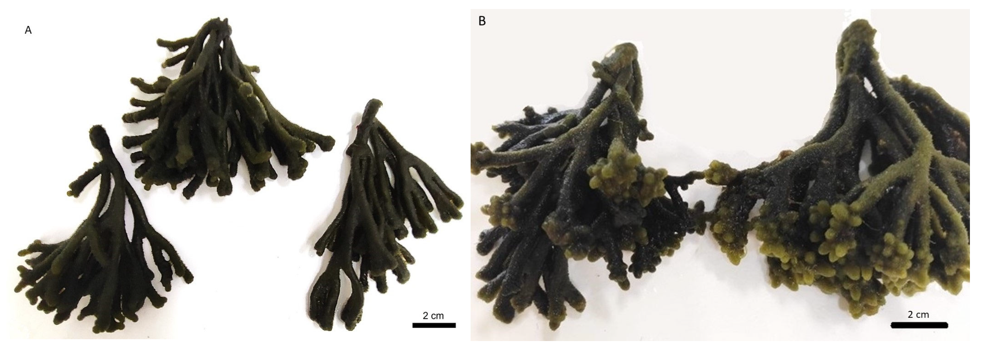
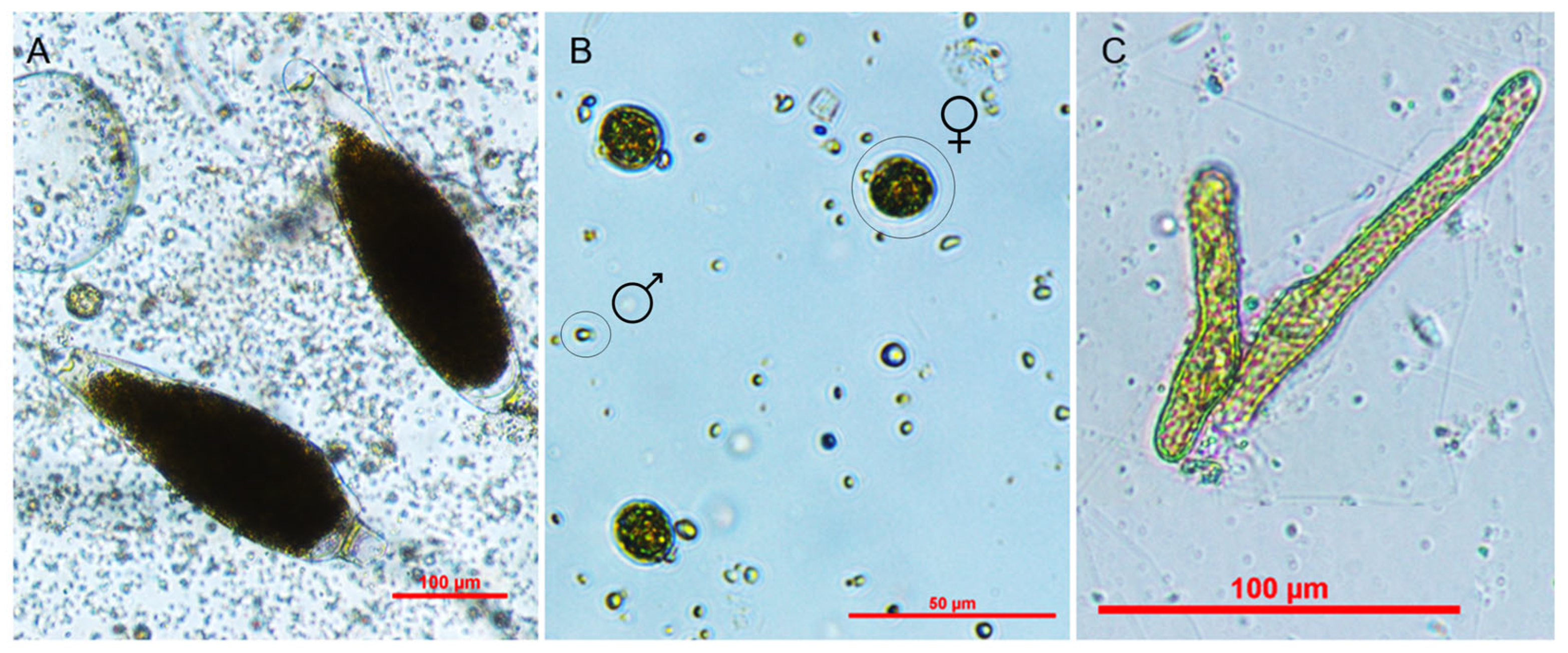
The development of reproductive structures in infertile specimens collected from the wild was observed only in the short-day photoperiod after the 8-week culture under laboratory-controlled conditions (16 °C; 100 μmol m−2 s−1). Thalli grown under the short-day photoperiod appeared dark green, and their average length of 9.1 ± 0.7 cm was measured, with no apparent development of new shoots (Figure 2A). Fertile thalli bearded gametangia with an average density of 3328 gametangia per gram of reproductive tissue (Figure 3A). After gamete release, motile female and male gametes were observed (Figure 3B). The fertility of the thalli and viability of the gametes were confirmed by germination (35.02% ± 6.46% germination rate) and germling growth (Figure 3C). On the other hand, the development of reproductive structures was not observed in the specimens cultured in the long-day photoperiod. Thalli cultured under the long-day photoperiod showed a light green appearance, with a measured average length of 9.0 ± 0.5 cm, and new shoots were observed in the apical ends (Figure 2B).
Growth of C. tomentosum was observed throughout the 8-week culture period (Figure 4). The results from the two-way ANOVA showed that both the culture time and the photoperiod affected the RGR (p < 0.05), with no interaction between the two factors (F(7,32) = 0.596; p = 0.755). In general, the specimens exposed to the long-day photoperiod presented higher growth than those exposed to the short-day photoperiod, with average RGRs of 4.93 ± 0.21% week−1 and 2.84 ± 0.13% week−1, respectively, during the 8-week trial (p < 0.05).
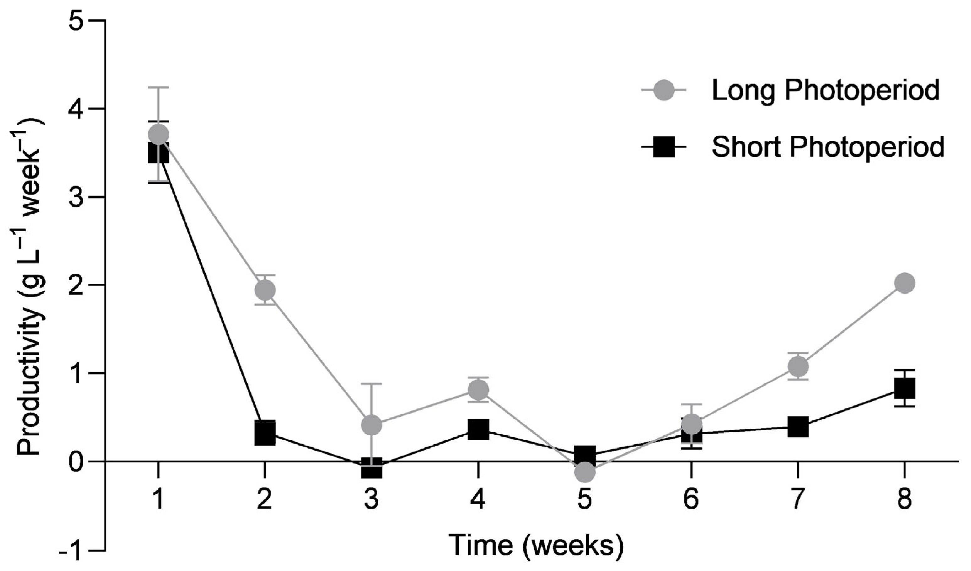
The productivity of C. tomentosum was also assessed throughout the culture period (Figure 5). The results from the two-way ANOVA showed an interaction between the culture time and the photoperiod that significantly affected productivity (F(7,32) = 3.165; p = 0.012). The highest productivities in both the long-day photoperiod (3.71 ± 0.53 g L⁻¹ week⁻¹) and the short-day photoperiod (3.51 ± 0.35 g L⁻¹ week⁻¹) were recorded in week 1 (p < 0.001), with no significative differences between the two photoperiods (p = 0.998). From week 1 until week 3, there was a decrease in productivity in both photoperiods (p < 0.001). In week 3, a scale-up in the culture flask capacity was carried out to reduce growth limitation by low volume. After a recovery period, productivity increased significantly until the end of the experiment in the long-day photoperiod (p < 0.001), reaching 2.02 ± 0.10 g L⁻¹ week⁻¹. On the other hand, the short-day photoperiod did not show a significant recovery in productivity (p = 0.386) until the end of the experiment (0.83 ± 0.21 g L⁻¹ week⁻¹).

3.2. Optimisation of Gamete Release

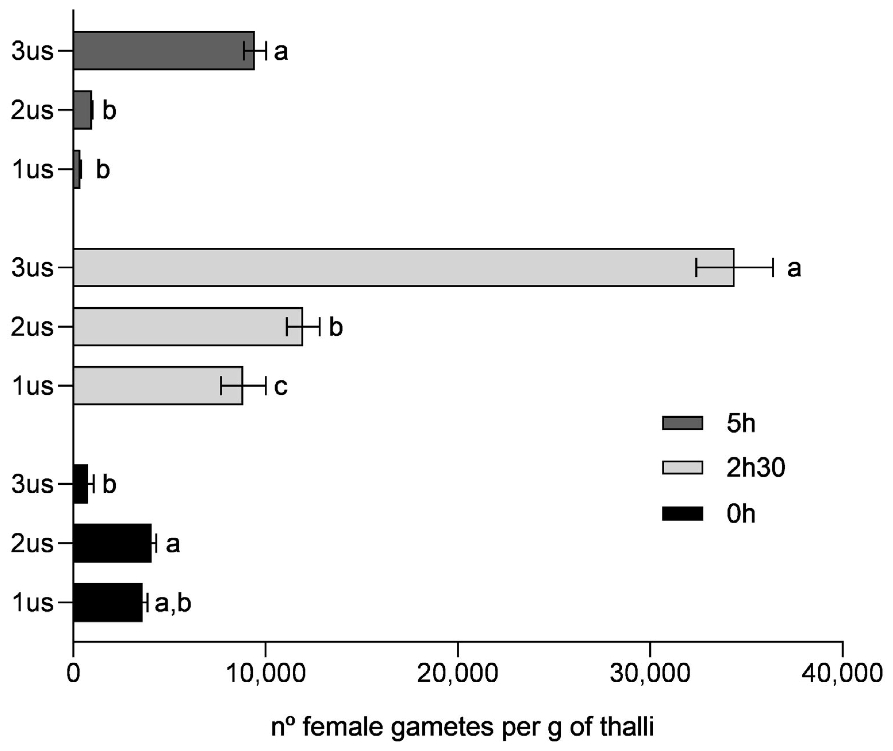
In the first trial, the number of female gametes released through each experimental condition was counted to identify which method induced the highest gamete yield. The combination of 2 h 30 min desiccation time, followed by a series of three ultrasounds, reached a maximum of 34,390 ± 2000 female gametes released per gram of fertile thalli, which outperformed all the other treatments (Figure 6)). The results from the two-way ANOVA revealed an interaction between factors, the desiccation period and the ultrasound, which significantly affected gamete yield (F(4,18) = 84.00; p < 0.001). Generally, the ultrasound worked better when performed on desiccated seaweed (2 h 30 min and 5 h). The third ultrasound, following the 2 h 30 min and the 5 h desiccations, yielded 34,390 ± 2000 and 9452 ± 576 gametes per gram of thalli, respectively, which was significantly higher (p < 0.001) than the yield obtained for the thalli not exposed to desiccation (757 ± 308 gametes per gram of thalli). Additionally, for the thalli desiccated for 2 h 30 min, the number of gametes released was increasingly higher with each ultrasound, ranging from 8848 ± 1166 up to 34,390 ± 2 000 gametes per gram of thalli (p < 0.05). On the other hand, in the thalli not exposed to desiccation (0 h), a different pattern was observed. The first two ultrasounds reached a similar gamete yield (3611 ± 253–4077 ± 232 gametes per gram of thalli; p = 9.92), whereas the yield obtained in the final ultrasound was significantly reduced (757 ± 308 gametes per gram of thalli; p < 0.05).
In the follow-up trial, female gamete release was monitored for 24 h after applying a 2 h 30 min desiccation period and three ultrasounds—the best-performing treatment from the previous trial. A clear pattern of gradual female gamete release was observed over the 24 h trial (Figure 7). The one-way ANOVA showed a significative increase in the number of female gametes released within the first hour (p < 0.05), from 1459 ± 145 up to 218,284 ± 5126 female gametes per gram of thalli. On the other hand, there was no significant change in the number of female gametes released after the first hour ranging from 218,284 ± 5126 to 257,989 ± 4032 female gametes per gram of thalli (p ≥ 0.05) until the end of the trial (24 h).
Most female gametes released within the first 15–30 min were motile. After 1–3 h, the proportion of motile gametes decreased, and after 24 h, few gametes still moved. For most, signs of disintegration became apparent.

3.3. Light Intensity and Spectrum

The two-way ANOVA revealed an interaction between light intensity and spectra, which significantly affected the germling length of C. tomentosum after 28 days in the culture (F(6,204) = 10.78; p < 0.001; Figure 8). Germlings cultured at a light intensity of 20 µmol m−2 s−1 in white, red, green and blue lights grew larger compared to those exposed to 40 and 60 µmol m−2 s−1 for the same light spectrum (p < 0.001). Additionally, germlings cultured at a light intensity of 20 µmol m−2 s−1 grew larger when exposed to white, red and green spectra (1338.5 ± 64.2 µm, 1296.0 ± 58.8 µm and 1468.4 ± 64.6 µm, respectively) compared to those exposed to the blue spectrum (841.4 ± 40.8 µm; p < 0.001).

4. Discussion

4.1. Induction of Gametogenesis

It is well known that the photoperiod and other abiotic factors play a key role in controlling reproduction and gametogenesis in most seaweed species [28,36]. In the present study, gametogenesis was successfully induced in specimens of C. tomentosum cultured under a short-day photoperiod for 8 weeks, which was confirmed by the presence of gametangia and zygote germination. The fertile thalli yielded an average density of 3328 gametangia per gram of reproductive biomass, which was similar to the highest gametangia yield obtained from the naturally fertile specimens collected in November 2020 and August 2021 [29]. This fact suggests that gametogenesis induced in the laboratory produces equivalent amounts of reproductive structures to those observed in nature. Both long-day and short-day photoperiods were reported to induce gametogenesis in different seaweed species [28]. For instance, a long-day photoperiod (16 h:8 h; L:D) induced gametogenesis in Laminaria digitata [37], whereas a short-day photoperiod induced gametogenesis in Gigartina acicularis, Fucus distichus and Himantothallus grandifolius [28]. On the other hand, Ulva lactuca has demonstrated limited gametogenesis under a short-day photoperiod (9 h:15 h; L:D) [38].
Throughout the trial, higher growth was observed in the specimens of C. tomentosum grown under the long-day photoperiod (16 h:8 h; L:D) compared to those grown in the short-day photoperiod (8 h:16 h; L:D). Marques et al. [17] also reported higher growth rates for C. tomentosum cultivated under a long-day photoperiod than under a short-day photoperiod. At the end of the trial, thalli grown in the long-day photoperiod displayed a light green appearance and the development of new shoots growing from the old thalli was observed, while thalli grown in the short-day photoperiod looked darker with no visible development of new vegetative shoots. During photosynthesis, seaweeds absorb light that is converted into electrochemical energy. The more the available light, the higher the photosynthetic rate and consequently the higher the energy obtained [39,40] until the maximum saturating light, which for C. tomentosum is approximately 200 μmol m−2 s−1 [17]. This could explain the higher growth and development of vegetative shoots observed in the thalli exposed to more light. On the other hand, thalli exposed to the short-day photoperiod presented lower growth rates and developed reproductive structures. These results may be explained by the trade-off in energy allocation between reproduction and growth (reviewed by Liu et al. [36]). For instance, Guillemin et al. [41] observed that growth of Gracilaria chilensis was significantly higher in infertile thalli compared to reproductive thalli and Chu et al. [42] observed the same pattern for Sargassum thunbergia.
The fact that in controlled culture conditions gametogenesis only occurred under a short-day photoperiod is contrary to what occurs in wild populations from Aguçadoura where this species becomes reproductive in summer following a period of increasing day length [29,30]. However, it should also be pointed out that the photoperiod also influences other correlated parameters such as total incident light. According to Lüning et al. [43], seasonal development of reproductive structures can be directly triggered by environmental factors (type 1), or these factors may control a circadian clock which then regulates reproduction (type 2). Temperature, irradiance and nutrients are considered “ultimate factors” controlling type 1 reproduction, which is expected to prevail in short-lived species (e.g., Dictyota dichotoma), whereas type 2 reproduction is controlled largely by photoperiod (e.g., Porphyra spp. [44]). If C. tomentosum follows type 1, factors such as light and temperature would directly trigger the development of reproductive structures (i.e., gametangia). In type 2, these factors regulate an internal biological clock that controls reproduction cycles, with a certain degree of independence from the immediate environment. In this context, it is paramount to distinguish between genuine and non-genuine photoperiod effects (reviewed by Liu et al. [36]). In the present study, it was not possible to distinguish whether reproduction was induced by the lower incident light or the short photoperiod; nevertheless, the results strongly suggest that the photoperiod and/or its correlated parameters play a key role in this process. Further research using a multifactorial approach under controlled culture conditions should be carried out to elucidate the process controlling gametogenesis of C. tomentosum.

4.2. Optimisation of Gamete Release

Optimising and standardising the process of gamete release helps to maximise gamete yield, reduce gamete deterioration and improve nursery protocols for aquaculture. In C. tomentosum, gametangia are borne laterally from the utricles [16] and bear anisogamous flagellated gametes, previously described for C. fragile [45] and also observed by our team for C. tomentosum. The description of the mechanism that causes the rupture of gametangia apical end triggering gamete release is incomplete for Codium species [14,18,45,46]. For the gametangia of green seaweed, Enteromorpha intestinalis was observed an altered membrane structure and loosening of wall microfibrils in the future exit pore area for gamete expulsion [47]. For Derbesia tenuissima, another green seaweed, the gametangia release mechanism was caused by an increase in turgor pressure, which in this case was caused by light, breaking the frail pore area and forcing an expulsion of gametes [48]. In the present study, C. tomentosum gamete release responded positively to a combination of hydric shock and ultrasound. The highest number of gametes released was achieved after the 2 h 30 min desiccation period followed by submersion in seawater, regardless of the number of ultrasounds applied. This result is in line with previous studies [49,50] where desiccation was also used to induce gamete release in C. fragile, probably explained by an increase in turgor pressure and consequent rupture of the membrane from the apical end of the gametangia. Despite this result, there seems to be a window of time for the induction of gamete release, as the 5 h desiccation period followed by ultrasound resulted in a lower gamete yield. Additionally, the absence of desiccation prior to the ultrasound treatment also led to a small gamete yield presumably explained by the absence of turgor pressure variation.
Gamete yield was also favoured by the use of ultrasound following desiccation, as the highest number of gametes was achieved after three consecutive ultrasounds. Optimal ultrasonic conditions including frequency, intensity and duration facilitate cell membrane disruption [51,52]. The combined effect of variation in turgor pressure caused by desiccation and the use of ultrasound likely potentiated the disruption of the gametangia apical end, promoting gamete discharge.
Gametes of Codium species are discharged slowly and immersed in a slimy substance through an apical opening following the disruption of the apical end of the gametangia, as reported for C. fragile, C. elongatum and C. bursa [18,53,54,55]. The curve from female gamete release showed that C. tomentosum released gametes gradually during approximately 1–2 h after the last ultrasound, with no significant changes in their numbers after that. This result suggests that a 1 h-to-2 h waiting period should be applied to ensure a maximum gamete yield. Besides gamete yield, their activity was also considered. In the initial waiting times, 15 min and 30 min, almost all female gametes observed were motile. Prince [55] reported that gametes of C. fragile become motile right after extrusion, and Miravalles et al. [14] stated that female gametes are motile during few minutes after release. Gametes which are not fecundated lose activity after some time and eventually become non-viable. In this study, the proportion of motile female gametes 1 h to 3 h after release was reduced. After 24 h, very few gametes moved, and a significant number of them disintegrated. Thus, considering both gamete yield and viability, a 1 h-to-3 h waiting period is recommended.

4.3. Light Intensity and Spectrum

The control of light irradiance and spectra during nursery production is fundamental to improve growth and reduce energy costs [34]. To our knowledge, this is the first study that investigates the effects of the light intensity and the spectrum on the early stages of development of C. tomentosum. In this experiment, the preference for the less intense light condition of 20 μmol m−2 s−1 for germling growth was evident in every light spectrum—white, blue, red and green. A study by Leukart and Lüning [56] regarding spectral light requirements for long-term germling growth of different seaweeds showed that growth of germlings belonging to the green seaweed Ulva peseudocurvata requires at least 3 μmol m−2 s−1 in green light spectra and only 1 μmol m−2 s−1 in blue and red lights. In the same investigation, it was reported that for sublittoral species light saturation occurred between 10 and 20 μmol m−2 s−1 and photoinhibition at 50–100 μmol m−2 s−1 [56], and it is important to note that Codium spp. are described as lower-shore seaweeds [57]. Hwang et al. [11] studied the growth of zygotes of C. fragile under different irradiances (at 15 °C and 16 h:8 h; L:D). After 13 days in the culture, the zygotes reached the highest length under 20 μmol m−2 s−1 and 10 μmol m−2 s−1 irradiance (261.3 μm and 162.7 μm, respectively), whereas the zygotes exposed to 40, 60 and 100 μmol m−2 s−1 were bleached. Likewise, the results here presented show that a light intensity of 20 μmol m−2 s−1 was enough to sustain germling growth in C. tomentosum, whereas higher light intensities, 40 μmol m−2 s−1 and 60 μmol m−2 s−1, led to reduced growth, possibly due to photoinhibition. Excess light energy leads to the formation of harmful reactive oxygen species in oxygen-enriched environments, causing slow growth and reduced productivity [34,58]. Following the trial, the cultures of filamentous germlings were monitored for several months and kept growing continuously, but the formation of the spongy thallus, the macroscopic upright thallus, was not observed. A minimum light intensity of 60 μmol m−2 s−1 together with water movement was required to induce the development of the spongy thallus in C. fragile from the precursor filamentous thalli [12,13]. Additionally, it is known that the spongy thallus of C. tomentosum needs an irradiance of around 120–230 μmol m−2 s−1 to sustain growth [17]. Thus, these results indicate that the microscopic and macroscopic stages of the life cycle of C. tomentosum have distinct light demands, and consequently, an increased irradiance (together with other factors, e.g., water movement and phytohormones) is likely necessary to induce morphogenesis.
The spectral range of light plays an important role in photosynthesis affecting seaweed growth and morphology [59,60]. In this study, germling growth was higher under white, red and green lights compared to under blue light for the intensities of 20 and 40 μmol m−2 s−1. Light harvest pigments have a characteristic absorption spectrum, and the sum of all the pigments in the seaweed gives thallus absorption spectra [15]. Ramus [61] observed that C. fragile thallus absorbed 97% to 99% of the incident light in the visible wavelengths of 400 to 700 nm, regardless of pigment concentration, that is, they are “optically black” [15], which may explain the growth performance under white, red and green lights here reported. The growth patterns of germlings cultured under red and blue light were similar to that observed for the macroscopic thallus by Marques et al. [17]. The authors noticed that relative growth rates were higher under red light, intermediate under white light, and lower under blue light. This could suggest that the micro- and macroscopic stages of C. tomentosum may have the same light absorption spectra. Since C. tomentosum has a high concentration of chlorophyll a and Siph and Siph-do pigments, which account for 54% of the total carotenoid content, have a peak of absorption in the blue-green region [17,62], the results obtained for the blue spectrum were surprising. On the other hand, as this species absorbs preferentially in the blue-green region, it cannot be excluded the possibility that even low light intensities of 20–40 μmol m−2 s−1 may be enough to cause photoinhibition, leading to reduced growth of germlings.

5. Conclusions

In conclusion, gametogenesis was induced in infertile specimens of C. tomentosum cultured under a short-day photoperiod in the laboratory. A high gamete yield was obtained through a combination of desiccation followed by a series of three ultrasounds. Germlings showed a better growth performance when cultured under a lower intensity of 20 μmol m−2 s−1 compared to those under 40 and 60 μmol m−2 s−1 in all tested light spectra. The results indicated that there was a difference in light intensity requirement between the micro- and macroscopic stages of C. tomentosum, which must be further elucidated and considered when developing nursery protocols. The results on reproduction and early stages of C. tomentosum provide important knowledge for future establishment of nursery protocols based on sexual reproduction.

Author Contributions

Conceptualisation, M.F.S., I.S.-P. and G.S.M.; methodology, M.F.S., T.C.P. and G.S.M.; investigation, M.F.S., T.C.P. and G.S.M.; formal analysis, M.F.S., T.C.P. and G.S.M.; writing—original draft preparation, M.F.S. and T.C.P.; writing—review and editing, G.S.M. and I.S.-P.; supervision, G.S.M. and I.S.-P.; project administration, G.S.M. and I.S.-P.; funding acquisition, I.S.-P. All authors have read and agreed to the published version of the manuscript.

Funding

This work was integrated in the research and innovation AquaVitae (AV) project funded by the European Union’s Horizon 2020 Research and Innovation Programme under Grant Agreement No. 818173.

Institutional Review Board Statement

Not applicable.

Informed Consent Statement

Not applicable.

Data Availability Statement

The datasets generated and/or analysed in this study are available from the corresponding author upon reasonable request.

Conflicts of Interest

The authors declare no conflicts of interest.

References

  1. Araújo, R.; Vázquez Calderón, F.; Sánchez López, J.; Azevedo, I.C.; Bruhn, A.; Fluch, S.; Garcia Tasende, M.; Ghaderiardakani, F.; Ilmjärv, T.; Laurans, M.; et al. Current Status of the Algae Production Industry in Europe: An Emerging Sector of the Blue Bioeconomy. Front. Mar. Sci. 2021, 7, 626389. [Google Scholar] [CrossRef]
  2. Barbier, M.; Charrier, B.; Araujo, R.; Holdt, S.L.; Jacquemin, B.; Rebours, C. Pegasus—Phycommorph European Guidelines for Sustainable Aquaculture of Seaweeds. In Proceedings of the COST Action FA1406; Barbier, M., Charrier, B., Eds.; Roscoff, France, 2019. Available online: https://www.phycomorph.org/pegasus-phycomorph-european-guidelines-for-a-sustainable-aquaculture-of-seaweeds (accessed on 6 August 2024).
  3. Sugumaran, R.; Padam, B.S.; Yong, W.T.L.; Saallah, S.; Ahmed, K.; Yusof, N.A. A Retrospective Review of Global Commercial Seaweed Production—Current Challenges, Biosecurity and Mitigation Measures and Prospects. Int. J. Environ. Res. Public Health 2022, 19, 7087. [Google Scholar] [CrossRef] [PubMed]
  4. Moreira, A.; Cruz, S.; Marques, R.; Cartaxana, P. The Underexplored Potential of Green Macroalgae in Aquaculture. Rev. Aquac. 2022, 14, 5–26. [Google Scholar] [CrossRef]
  5. Trowbridge, C.D.; Farnham, W.F.; White, L.F. Thriving Populations of the Native Macroalga Codium tomentosum on Guernsey Rocky Shores. J. Mar. Biol. Assoc. U. K. 2004, 84, 873–877. [Google Scholar] [CrossRef]
  6. Fernández, P.V.; Arata, P.X.; Ciancia, M. Polysaccharides from Codium Species: Chemical Structure and Biological Activity. Their Role as Components of the Cell Wall; Elsevier: Amsterdam, The Netherlands, 2014; Volume 71, ISBN 9780124080621. [Google Scholar]
  7. Wang, L.; Wang, X.; Wu, H.; Liu, R. Overview on Biological Activities and Molecular Characteristics of Sulfated Polysaccharides from Marine Green Algae in Recent Years. Mar. Drugs 2014, 12, 4984–5020. [Google Scholar] [CrossRef] [PubMed]
  8. Santos, S.A.O.; Vilela, C.; Freire, C.S.R.; Abreu, M.H.; Rocha, S.M.; Silvestre, A.J.D. Chlorophyta and Rhodophyta Macroalgae: A Source of Health Promoting Phytochemicals. Food Chem. 2015, 183, 122–128. [Google Scholar] [CrossRef]
  9. da Costa, E.; Melo, T.; Moreira, A.S.P.; Alves, E.; Domingues, P.; Calado, R.; Abreu, M.H.; Domingues, M.R. Decoding Bioactive Polar Lipid Profile of the Macroalgae Codium tomentosum from a Sustainable IMTA System Using a Lipidomic Approach. Algal Res. 2015, 12, 388–397. [Google Scholar] [CrossRef]
  10. Hwang, E.K.; Park, C.S. Seaweed Cultivation and Utilization of Korea. Algae 2020, 35, 107–121. [Google Scholar] [CrossRef]
  11. Hwang, E.K.; Baek, J.M.; Park, C.S. Artificial Seed Production Using the Reproduction Methods in Codium fragile (Chlorophyta). Korean J. Fish. Aquat. Sci. 2005, 38, 164–171. [Google Scholar]
  12. Hwang, E.K.; Baek, J.M.; Park, C.S. Cultivation of the Green Alga, Codium fragile (Suringar) Hariot, by Artificial Seed Production in Korea. J. Appl. Phycol. 2008, 20, 469–475. [Google Scholar] [CrossRef]
  13. Nanba, N.; Kado, R.; Ogawa, H.; Nakagawa, T.; Sugiura, Y. Effects of Irradiance and Water Flow on Formation and Growth of Spongy and Filamentous Thalli of Codium fragile. Aquat. Bot. 2005, 81, 315–325. [Google Scholar] [CrossRef]
  14. Miravalles, A.B.; Leonardi, P.I.; Cáceres, E.J. Female Gametogenesis and Female Gamete Germination in the Anisogamous Green Alga Codium fragile subsp. novae-zelandiae (Bryopsidophyceae, Chlorophyta). Phycol. Res. 2012, 60, 77–85. [Google Scholar] [CrossRef]
  15. Hurd, C.L.; Harrison, P.J.; Bischof, K.; Lobban, C.S. Seaweed Ecology and Physiology; Cambridge University Press: Cambridge, UK, 2014; Volume 53, ISBN 9788578110796. [Google Scholar]
  16. Silva, P.C. The Dichotomous Species of Codium in Britain. J. Mar. Biol. Assoc. U. K. 1955, 34, 565–577. [Google Scholar] [CrossRef]
  17. Marques, R.; Cruz, S.; Calado, R.; Lillebø, A.; Abreu, H.; Pereira, R.; Pitarma, B.; da Silva, J.M.; Cartaxana, P. Effects of Photoperiod and Light Spectra on Growth and Pigment Composition of the Green Macroalga Codium tomentosum. J. Appl. Phycol. 2020, 33, 471–480. [Google Scholar] [CrossRef]
  18. Borden, C.A.; Stein, J.R. Reproduction and Early Development in Codium fragile (Suringar) Hariot: Chlorophyceae. Phycologia 1969, 8, 91–99. [Google Scholar] [CrossRef]
  19. Ramus, J. Differentiation of the Green Alga Codium fragile. Am. J. Bot. 1972, 59, 478. [Google Scholar] [CrossRef]
  20. Hanisak, M.D. Growth Patterns of Codium fragile ssp. tomentosoides in Response to Temperature, Irradiance, Salinity, and Nitrogen Source. Mar. Biol. 1979, 50, 319–332. [Google Scholar] [CrossRef]
  21. Churchill, A.C.; Moeller, H.W. Seasonal Patterns of Reproduction in New York Populations of Codium fragile (Sur.) Hariot subsp. tomentosoides (Van Goor) Silva. J. Phycol. 1972, 8, 147–152. [Google Scholar] [CrossRef]
  22. Bast, F. An Illustrated Review on Cultivation and Life History of Agronomically Important Seaplants. In Seaweed: Mineral Composition, Nutritional and Antioxidant Benefits and Agricultural Uses; Nova Publishers: New York, NY, USA, 2014; pp. 39–70. ISBN 9781631175756. [Google Scholar]
  23. Kerrison, P.D.; Stanley, M.S.; Edwards, M.D.; Black, K.D.; Hughes, A.D. The Cultivation of European Kelp for Bioenergy: Site and Species Selection. Biomass Bioenergy 2015, 80, 229–242. [Google Scholar] [CrossRef]
  24. McHugh, D.J. A Guide to the Seaweed Industry; FAO Fisheries Technical Paper 441; FAO: Rome, Italy, 2003; p. 105. [Google Scholar]
  25. Santelices, B. Patterns of Reproduction; Dispersal and Recruitment in Seaweeds. Eanography Mar. Biol. 1990, 28, 177–276. [Google Scholar]
  26. Dring, M.J. Photoperiodism and Phycology. Prog. Phycol. Res. 1984, 3, 159–192. [Google Scholar]
  27. Dring, M.J. Photocontrol of Development in Algae. Annu. Rev. Plant Physiol. Plant Mol. Biol. 1988, 39, 174. [Google Scholar] [CrossRef]
  28. Brawley, S.H.; Johnson, L.E. Garnet Ogenesis, Gametes and Zygotes: An Ecological Perspective on Sexual Reproduction in the Algae. Br. Phycol. J. 1992, 27, 233–252. [Google Scholar] [CrossRef]
  29. Sá, M.F. Reproductive Status and Early Stages of Development in the Green Macroalgae Codium tomentosum. Master’s Thesis, University of Porto, Porto, Portugal, 2021. Available online: https://hdl.handle.net/10216/138709 (accessed on 1 June 2024).
  30. Pacheco, T. Propagation of Macroalga Codium tomentosum for Aquaculture Production. Master’s Thesis, University of Porto, Porto, Portugal, 2022. Available online: https://hdl.handle.net/10216/146493 (accessed on 1 June 2024).
  31. Park, C.S.; Soth, C.H. Effects of Light and Temperature on Morphogenesis of Codium fragile (Suringar) Hariot in Laboratory Culture. Korean J. Phycol. 1992, 7, 213–223. [Google Scholar]
  32. Cabral, J.P. Characterization and Multivariate Analysis of Patella intermedia, Patella ulyssiponensis and Patella vulgata from Povoa de Varzim (Northwest Portugal). Iberus 2003, 21, 1–17. [Google Scholar]
  33. Harrison, P.J.; Berges, J.A. Marine Culture Media. In Algal Culturing Techniques; Academic Press: Cambridge, MA, USA, 2005; pp. 21–33. [Google Scholar] [CrossRef]
  34. Marques, R.; Moreira, A.; Cruz, S.; Calado, R.; Cartaxana, P. Controlling Light to Optimize Growth and Added Value of the Green Macroalga Codium tomentosum. Front. Mar. Sci. 2022, 9, 906332. [Google Scholar] [CrossRef]
  35. Huo, Y.; Kim, J.K.; Yarish, C.; Augyte, S.; He, P. Responses of the Germination and Growth of Ulva prolifera Parthenogametes, the Causative Species of Green Tides, to Gradients of Temperature and Light. Aquat. Bot. 2021, 170, 103343. [Google Scholar] [CrossRef]
  36. Liu, X.; Bogaert, K.; Engelen, A.H.; Leliaert, F.; Roleda, M.Y.; De Clerck, O. Seaweed Reproductive Biology: Environmental and Genetic Controls. Bot. Mar. 2017, 60, 89–108. [Google Scholar] [CrossRef]
  37. Martins, N.; Tanttu, H.; Pearson, G.A.; Bartsch, I. Interactions of Daylength, Temperature and Nutrients Affect Thresholds for Life Stage Transitions in the Kelp Laminaria digitata (Phaeophyceae ). Bot. Mar. 2017, 60, 109–121. [Google Scholar] [CrossRef]
  38. Balar, N.; Jaiswar, S.; Mantri, V.A. Phycological Society Effects of Extrinsic Abiotic Factors on Induction of Gametogenesis and Efficacy of a Device for the Segregation of Non-Fused Gametes and Zygotes in the Green Alga Ulva lactuca. Appl. Phycol. 2021, 2, 1–9. [Google Scholar] [CrossRef]
  39. Khoeyi, Z.A.; Seyfabadi, J.; Ramezanpour, Z. Effect of Light Intensity and Photoperiod on Biomass and Fatty Acid Composition of the Microalgae, Chlorella vulgaris. Aquac. Int. 2012, 20, 41–49. [Google Scholar] [CrossRef]
  40. Streckaite, S.; Llansola-Portoles, M.J.; Pascal, A.A.; Ilioaia, C.; Gall, A.; Seki, S.; Fujii, R.; Robert, B. Pigment Structure in the Light-Harvesting Protein of the Siphonous Green Alga Codium fragile. Biochim. Biophys. Acta-Bioenerg. 2021, 1862, 148384. [Google Scholar] [CrossRef] [PubMed]
  41. Guillemin, M.L.; Valenzuela, P.; Gaitán-Espitia, J.D.; Destombe, C. Evidence of Reproductive Cost in the Triphasic Life History of the Red Alga Gracilaria chilensis (Gracilariales, Rhodophyta). J. Appl. Phycol. 2014, 26, 569–575. [Google Scholar] [CrossRef]
  42. Chu, S.; Zhang, Q.; Liu, S.; Zhang, S.; Tang, Y.; Lu, Z.; Yu, Y. Trade-off between Vegetative Regeneration and Sexual Reproduction of Sargassum thunbergii. Hydrobiologia 2011, 678, 127–135. [Google Scholar] [CrossRef]
  43. Lüning, K.; Dieck, I.T. Environmental Triggers in Algal Seasonality. Bot. Mar. 1989, 32, 389–398. [Google Scholar] [CrossRef]
  44. Tala, F.; Chow, F. Phenology and Photosynthetic Performance of Porphyra spp. (Bangiophyceae, Rhodophyta): Seasonal and Latitudinal Variation in Chile. Aquat. Bot. 2014, 113, 107–116. [Google Scholar] [CrossRef]
  45. Miravalles, A.B.; Leonardi, P.I.; Cáceres, E.J. Male Gametogenesis Ultrastructure of Codium fragile subsp. Novae-Zelandiae (Bryopsidophyceae, Chlorophyta). Phycologia 2011, 50, 370–378. [Google Scholar] [CrossRef]
  46. Prince, J.S.; Trowbridge, C.D. Reproduction in the Green Macroalga Codium (Chlorophyta): Characterization of Gametes. Bot. Mar. 2004, 47, 461–470. [Google Scholar] [CrossRef]
  47. McArthur, D.M.; Moss, B.L. Gametogenesis and Gamete Structure of Enteromorpha intestinalis (L.) Link. Br. Phycol. J. 1979, 14, 43–57. [Google Scholar] [CrossRef]
  48. Wheeler, A.E.; Page, J.Z. The Ultrastructure of Derbesia tenuissima (de Notaris) Crouan. I. Organization of the Gametophyte Protoplast, Gametangium, and Gametangial PORE1, 2, 3. J. Phycol. 1974, 10, 336–352. [Google Scholar] [CrossRef]
  49. Moeller, H. Ecology and Life History of Codium fragile subsp. tomentosoides; Rutgers: Newark, NJ, USA, 1969; p. 258. [Google Scholar]
  50. Hanisak, M.D. Nitrogen Limitation of Codium fragile ssp. tomentosoides as Determined by Tissue Analysis. Mar. Biol. 1979, 50, 333–337. [Google Scholar] [CrossRef]
  51. Liu, Y.; Liu, X.; Cui, Y.; Yuan, W. Ultrasound for Microalgal Cell Disruption and Product Extraction: A Review. Ultrason. Sonochem. 2022, 87, 106054. [Google Scholar] [CrossRef] [PubMed]
  52. Fernandes, F.A.N.; Gallão, M.I.; Rodrigues, S. Effect of Osmotic Dehydration and Ultrasound Pre-Treatment on Cell Structure: Melon Dehydration. LWT 2008, 41, 604–610. [Google Scholar] [CrossRef]
  53. Oltmanns, F. Morphologie Und Biologie Der Algen: Chrysophyceae—Chlorophyceae, 2nd ed.; Gustav Fischer: Jena, Germany, 1922. [Google Scholar] [CrossRef]
  54. Berthold, G. Zur Kenntnis Der Siphoneen Und Bangiaceen. Mitt. Zool. Sta. Neapel. 1881, 2, 72–82. [Google Scholar]
  55. Prince, J.S. Sexual Reproduction in Codium fragile subsp. tomentosoides (Chlorophyceae) from the Northeast Coast of North America. J. Phycol. 1988, 24, 112–114. [Google Scholar] [CrossRef]
  56. Leukart, P.; Lüning, K. Minimum Spectral Light Requirements and Maximum Light Levels for Long-Term Germling Growth of Several Red Algae from Different Water Depths and a Green Alga. Eur. J. Phycol. 1994, 29, 103–112. [Google Scholar] [CrossRef]
  57. Melo, R.; Sousa-pinto, I.; Antunes, S.C.; Costa, I. Temporal and Spatial Variation of Seaweed Biomass and Assemblages in Northwest Portugal. J. Sea Res. 2021, 174, 102079. [Google Scholar] [CrossRef]
  58. Seki, S.; Yamano, Y.; Oka, N.; Kamei, Y.; Fujii, R. Discovery of a Novel Siphonaxanthin Biosynthetic Precursor in Codium fragile That Accumulates Only by Exposure to Blue-Green Light. FEBS Lett. 2022, 596, 1544–1555. [Google Scholar] [CrossRef]
  59. Figueroa, F.L.; Aguilera, J.; Niell, F.X. Red and Blue Light Regulation of Growth and Photosynthetic Metabolism in Porphyra umbilicalis (Bangiales, Rhodophyta). Eur. J. Phycol. 1995, 30, 11–18. [Google Scholar] [CrossRef]
  60. Luimstra, V.M.; Schuurmans, J.M.; Verschoor, A.M.; Hellingwerf, K.J.; Huisman, J.; Matthijs, H.C.P. Blue Light Reduces Photosynthetic Efficiency of Cyanobacteria through an Imbalance between Photosystems I and II. Photosynth. Res. 2018, 138, 177–189. [Google Scholar] [CrossRef]
  61. Ramus, J. Seaweed Anatomy and Photosynthetic Performance: The Ecological Significance of Light Guides, Heterogeneous Absorption Ans Multiple Scatter. J. Phycol. 1978, 14, 352–362. [Google Scholar] [CrossRef]
  62. Uragami, C.; Galzerano, D.; Gall, A.; Shigematsu, Y.; Meisterhans, M.; Oka, N.; Iha, M.; Fujii, R.; Robert, B.; Hashimoto, H. Light-Dependent Conformational Change of Neoxanthin in a Siphonous Green Alga, Codium intricatum, Revealed by Raman Spectroscopy. Photosynth. Res. 2014, 121, 69–77. [Google Scholar] [CrossRef] [PubMed]
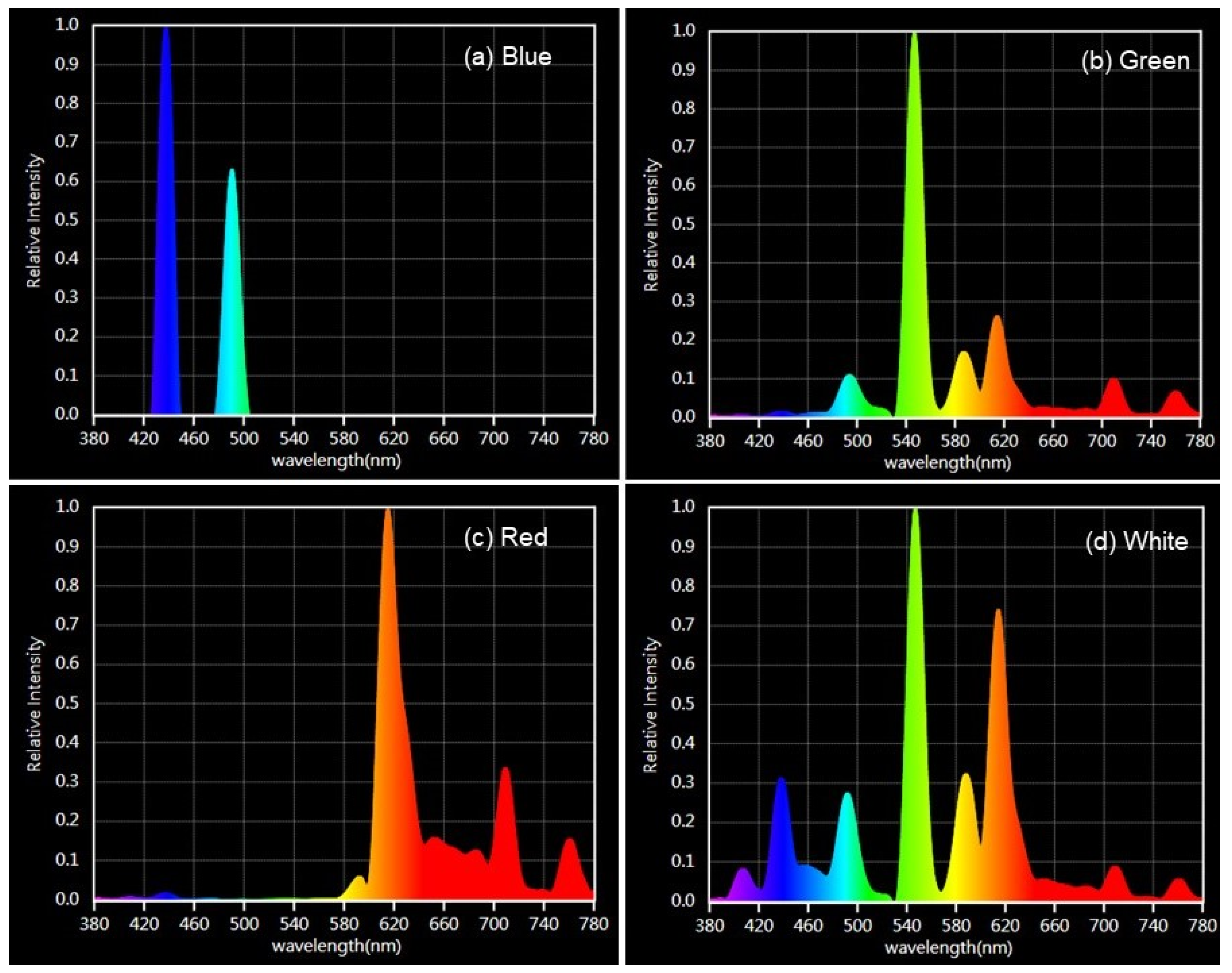
Figure 1. Emission spectra of the four light conditions.
Phycology 04 00029 g001
Figure 2. Thalli of C. tomentosum after the 8-week culture in the short-day photoperiod (8 h:16 h; L:D) (A) and the long-day photoperiod (16 h:8 h; L:D) (B) under controlled conditions (100 µmol m−2 s−1 and 16 °C). Scale bar = 2 cm.
Phycology 04 00029 g002
Figure 3. Reproductive structures and early development stages of C. tomentosum: (A) gametangia developed after 8 weeks in the culture under the short-day photoperiod (8 h:16 h; L:D; 16 °C and 100 µmol m−2 s−1); (B) female and male gametes right after release; and (C) germlings seven days after gamete release. Scale bar = 100 µm (A,C); scale bar = 50 µm (B).
Phycology 04 00029 g003
Figure 4. Relative growth rates (RGRs) of C. tomentosum (mean ± SEM; n = 3) along the 8-week culture period under the long-day photoperiod (16 h:8 h; L:D) and the short-day photoperiod (8 h:16 h; L:D) at 100 µmol m−2 s−1 intensity and 16 °C.
Phycology 04 00029 g004
Figure 5. Productivity of C. tomentosum (mean ± SEM; n = 3) along the 8-week culture period under the long-day photoperiod (16 h:8 h; L:D) and the short-day photoperiod (8 h:16 h; L:D) at 100 µmol m−2 s−1 intensity and 16 °C.
Phycology 04 00029 g005
Figure 6. Effects of the desiccation period (h) and the number of ultrasounds (US) on the yield of released female gametes (number of gametes per g of fertile thalli; mean ± SEM; n = 3). Different letters denote significant differences between ultrasounds within each desiccation period (p < 0.05).
Phycology 04 00029 g006
Figure 7. Yield curve of female gametes (number of female gametes per gram of fertile thalli) released over 24 h (mean ± SEM; n = 3). Different letters denote significant differences between means (p < 0.05).
Phycology 04 00029 g007
Figure 8. Lengths of germlings exposed to blue, green, red and white spectra at 20, 40 and 60 µmol m−2 s−1 light intensities over 28 days in culture at 16 °C and a 12 h:12 h (L:D) photoperiod (mean ± SEM).
Phycology 04 00029 g008
Disclaimer/Publisher’s Note: The statements, opinions and data contained in all publications are solely those of the individual author(s) and contributor(s) and not of MDPI and/or the editor(s). MDPI and/or the editor(s) disclaim responsibility for any injury to people or property resulting from any ideas, methods, instructions or products referred to in the content.