Article

Mycosporine-like Amino Acids from Red Alga Dulse (Devaleraea inkyuleei): Monthly Variation and Improvement in Extraction

1
Chair of Marine Chemical Resource Development, Graduate School of Fisheries Sciences, Hokkaido University, Hakodate 041-8611, Hokkaido, Japan
2
Food Research Institute, Aomori Prefectural Industrial Technology Research Center, Agricultural Products Institute, 91, Yanagisawa, Inuotose, Rokunohe-machi, Kamikita-gun, Aomori 033-0071, Aomori-ken, Japan
3
JSPS Postdoctoral Fellowship for Research in Japan, Graduate School of Fisheries Sciences, Hokkaido University, Hakodate 041-8611, Hokkaido, Japan
4
Faculty of Science, University of Maroua, Maroua P.O. Box 814, Cameroon
5
Laboratory of Marine Chemical Resource Development, Faculty of Fisheries Sciences, Hokkaido University, Hakodate 041-8611, Hokkaido, Japan
*
Author to whom correspondence should be addressed.
Phycology 2023, 3(3), 394-404; https://doi.org/10.3390/phycology3030026
Submission received: 28 July 2023 / Revised: 30 August 2023 / Accepted: 5 September 2023 / Published: 7 September 2023

Abstract

:
Mycosporine-like amino acids (MAAs) are natural UV-absorbing compounds found in microalgae and macroalgae. The content of MAAs in algae varies with the seasons and environmental factors. Red alga dulse in Usujiri (Hokkaido, Japan) is an underutilized resource. Therefore, we investigated the amount of MAAs in Usujiri dulse in 2022 to clarify the suitable months for MAA extraction. In addition, we also evaluated the extraction method focusing on the extraction volume. MAAs were prepared via the 20 volumes of 25% ethanol extraction method and detected via HPLC. The results showed that the amount of MAAs on 25 March 2022 showed the highest value (40.4 μmol/g DW) among the samples from 24 January to 13 May. The tendency of suitable samples for MAA preparation corresponded to the term from mid-February to early April, which was the same as the previous three years. Although the surveys from 2019–2021 were performed by using the successive water–methanol method, it was found that the improved method also reflected the monthly variation in MAAs. The extraction of MAAs was performed via 20 or 40 volumes of 25% ethanol at 4 °C for 24 h. The amount of MAAs with 40 volumes of 25% ethanol extraction increased 1.3-fold compared to that with 20 volumes of 25% ethanol extraction. These data are useful information for valuable compound extraction from Usujiri dulse.

1. Introduction

Ultraviolet radiation (UVR) is a part of the solar electromagnetic spectrum encompassing wavelengths ranging from 200 to 400 nm. UVR consists of ultraviolet A (UVA) (315–400 nm), ultraviolet B (UVB) (280–315 nm), and ultraviolet C (UVC) (200–280 nm). UVR reaching the Earth’s surface is only a small portion of the entire UVR, which is composed of wavelengths above 290 nm (mainly UVA and up to 10% of UVB) [1,2,3]. Excessive exposure to UVA and UVB rays causes sunburn (erythema) [4], persistent pigment darkening (PPD) [5], and collagen network alterations in skin tissue [6]. This is caused by oxidative stress due to the high production of reactive oxygen species (ROS), which leads to DNA damage and premature skin aging (wrinkling) [7]. Therefore, organisms under UVR must develop defense strategies to minimize UV-induced damage [8].
Recently, there has been a major shift towards the use of natural UV protection ingredients, clean labeling, and non-synthetic chemicals. This shift is primarily driven by an increased awareness of the toxicity and chemical cocktails in cosmetic products [9,10,11]. Traditional sunscreens, such as oxybenzone and octinoxate, have been banned in several regions of the world because of harmful effects on the marine environment. The search for new, natural UV protection ingredients on sunscreen is now unavoidable because of our changing minds about the awareness of marine environmental protection [12]. Marine organisms in intertidal zones are exposed to sunlight; therefore, they have evolved photoprotection mechanisms to protect themselves [13,14,15]. Marine organisms, such as seaweed, cyanobacteria, phytoplankton, and marine animals, protect themselves by involving the accumulation of MAAs as UVR absorbers [16,17,18]. Depending on their molecular structures, the UV absorption maxima of MAAs range from 310 to 360 nm [19]. MAAs exhibit high water solubility, high stability, low toxicity, and antioxidant activity [20,21]. These diverse functions of MAAs make them highly promising natural products in the cosmetic and biotechnology industries.
Although most MAAs have maximum absorption wavelengths of around 320–330 nm, several MAAs with wavelengths of around 350–360 nm have been reported [1,22,23]. Usujirene is one MAA that has a long absorption wavelength of 357 nm. Protection from broad-spectrum UV is possible via a combination of MAAs with an absorption of around 320–330 nm and usujirene. However, usujirene has been detected in limited species, such as cyanobacteria (Phormidium angustissimum), red alga Gracilaria vermiculophylla, and dulse [24,25]. Porphyra-334 is a major MAA in Bangiophyceae. It has been speculated that usujirene is synthesized from porphyra-334 via palythenic acid [22]. Dulse is rich in usujirene compared to other red algae. The amount of usujirene differs according to the season and habitat place [26,27]. Dulse from the Atlantic (Palmaria palmata) showed different MAA compositions in the two places [28,29]. The amount of usujirene in Japanese dulse (Devaleraea inkyuleei) changed monthly between 8.2 mol% and 32 mol% [30,31,32]. We previously studied Japanese dulse as Palmaria palmata in Japan, as the scientific name was changed in 2022 [33]. Therefore, investigations into the environmental conditions of dulse provide clues to the production of large amounts of usujirene.
Red alga dulse, which is grown on Kombu rope in Hakodate, is regarded as an underused marine resource in the area. Therefore, attempts have been made to utilize dulse as a novel local food, and to evaluate its functions, such as its antioxidant activity [34,35]. It has also been studied as a source of peptides for the inhibition of angiotensin I-converting enzyme activity [36] and of xylooligosaccharides for prebiotics [37,38]. Our previous studies showed that the content and composition of MAAs from dulse varied from month to month [30,31,32]. We thought that the MAA variability may be related to the marine environment. In addition, we previously developed an extraction method to improve the MAA yield via the evaluation of the solvent and extraction time [32]. In this study, we investigated the monthly variation in MAA content in dulse collected in 2022 from Usujiri, Hokkaido, Japan, using the 25% ethanol extraction method. The extraction volumes were varied, as reported (e.g., 1:10–25 g/mL solid–liquid ratio) [26,27,39,40]. There are fewer studies on the extraction volumes. The effects of the extraction solvent volume and extraction time on the MAA content and composition have also been evaluated to improve the MAA yield.
In this study, we investigated the monthly variation in MAAs in Usujiri, Hokkaido, Japan, from January to May 2022 to clarify suitable dulse sampling periods using the 25% ethanol extraction method. In addition, we attempted to improve the 25% ethanol extraction efficiency by evaluating the extraction volumes.

2. Materials and Methods

2.1. Algal Sample Preparation

All dulse samples (Devaleraea inkyuleei in Japan, formerly Palmaria palmata [33]) were collected at a 1–2 m depth in Usujiri, Hakodate, Japan, from January to May 2022. Thalli were washed with tap water to remove sea salt and impurities. The samples were lyophilized and ground into a fine powder via a Wonder Blender WB-1 (Osaka Chemical Co., Osaka, Japan).

2.2. Extraction of Crude MAAs from Dulse

The extraction of MAAs from dulse was performed according to our previous study [32]. Namely, a powder sample was suspended in 20 volumes (w/v) of 25% ethanol and extracted at 4 °C for 24 h. The extracts were collected via centrifugation at 4 °C, 27,200× g, for 10 min. The supernatant was evaporated and dissolved in water, the solution was centrifuged, and the supernatant was lyophilized. Solid samples were designated as crude MAAs and used for experimental purposes.

2.3. HPLC Analysis of MAAs

After the extraction process, the crude MAA samples were dissolved in water containing 0.1% trifluoroacetic acid (TFA) and subjected to sequential filtration via Millex GV (pore size: 0.22 μm) (Merck Millipore, Billerica, MA, USA) and Millex LG (pore size: 0.20 μm) (Merck Millipore). The filtrated MAA samples were isolated via reversed-phase HPLC with a Mightysil RP-18GP column (5 µm, 10 × 250 mm) (Kanto Kagaku, Tokyo, Japan), with the column oven set to 40 °C and a detection wavelength of 330 nm, and using an isocratic elution of pure water containing 0.1% TFA for 7 min, and a linear gradient of acetonitrile (0–70%) containing 0.1% TFA for 13 min, at a flow rate of 4.73 mL/min. The MAA content was expressed in terms of the molecule per gram of dry dulse powder (μmol/g DW (dry weight)). The lambda max of each MAA was identified using a spectrophotometer (UV-1800, Shimadzu, Kyoto, Japan) after separation via HPLC.
A standard curve for each MAA in the HPLC was constructed from the peak area and the extinction coefficient of each MAA using the Lambert–Beer law. The peaks of usujirene and palythene were eluted at the same retention time. Therefore, we expressed these MAAs as usujirene + palythene and employed a high extinction coefficient from palythene (usujirene: 45,070 M−1 cm−1; palythene: 47,521 M−1 cm−1) to avoid the risk of overestimation.

2.4. Protein, Phycoerythrin (PE), and Sugar Contents

The amount of crude protein was determined gravimetrically. One gram of fine powder was dissolved in 20 mL water and extracted at 4 °C for 12 h. After centrifugation at 12,000× g for 5 min, the supernatant was dialyzed against water at 4 °C and lyophilized. For each sample, phycoerythrin (PE) was prepared from the fine powders. A 10 mg amount of powder was dissolved in 1 mL distilled water and extracted at 4 °C for 12 h. After centrifugation at 12,000× g for 5 min, the spectra of the supernatant were measured via the UV–visible ray absorption spectrum using a spectrophotometer. The amount of PE was determined via the following equation [41]: PE (mg/mL) = [(A564 − A592) − (A455 − A592) × 0.2] × 0.12. The main sugar content (glucose and xylose) was determined via colorimetric methods. Powder was hydrolyzed using TFA, and the glucose and xylose contents were determined using a glucose assay kit (Fujifilm Wako Shibayagi, Gunma, Japan) and a D-xylose assay kit (Megazyme, Wicklow, Ireland), respectively.

2.5. Abiotic Data from Hakodate

Monthly means of the daily maximum ultraviolet index (UVI) were obtained from the Japan Meteorological Agency (JMA: https://www.data.jma.go.jp/gmd/env/uvhp/info_uv.html, accessed on 5 May 2023). The erythemal UV intensity (mW/m2) was calculated by multiplying the UVI by a factor of 25. The data on the near-surface chlorophyll concentration (mg/m3) were obtained from NASA’s Ocean Color WEB (https://oceancolor.gsfc.nasa.gov, accessed on 5 May 2023).

2.6. Compositional Comparison of Usujiri Dulse from 2019 to 2022

Data on the Usujiri dulse’s MAAs between 2019 and 2021 were used from our previous studies [30,31,32]. Sampling of dulse was performed in the same Usujiri area at a depth of 1–2 m, as previously mentioned. Data on the erythemal UVI and near-surface chlorophyll concentrations from 2019 to 2021 were obtained from the JMA and NASA’s Ocean Color WEB.

2.7. Effect of Extraction Volume on MAA Extraction

To improve the amount of extracted MAAs, the extraction volume was evaluated. The dulse powder from April 2021 was suspended in 20 or 40 volumes (w/v) of 25% ethanol and extracted at 4 °C for 24 h. The supernatant was collected via centrifugation at 4 °C, 27,200× g, for 10 min, and the MAA composition was evaluated via HPLC.

2.8. Effect of Extraction Time on MAA Extraction

To evaluate whether all the MAAs were extracted in a single extraction from the dulse, the extraction of MAAs from the extraction residue was examined. The dulse powder from April was suspended in 20 volumes (w/v) of 25% ethanol and extracted at 4 °C for 24 h. The supernatant was collected via centrifugation at 4 °C, 27,200× g, for 10 min. The MAAs in residue were again suspended in the same extraction solvent at 4 °C for 24 h, and the supernatant was collected via centrifugation under the same conditions. This process was repeated four times. The composition of MAAs was evaluated via HPLC.

2.9. Statistical Analysis

Data are expressed as means ± standard errors. All values are the means of triplicate analysis. The Student’s t-test was applied for pairwise comparisons. Statistical analyses were carried out using Tukey–Kramer’s multiple comparisons test. All statistical analyses were performed using Statcel 3 software (Version No. 3, OMS Publisher, Tokorozawa, Japan).

3. Results and Discussion

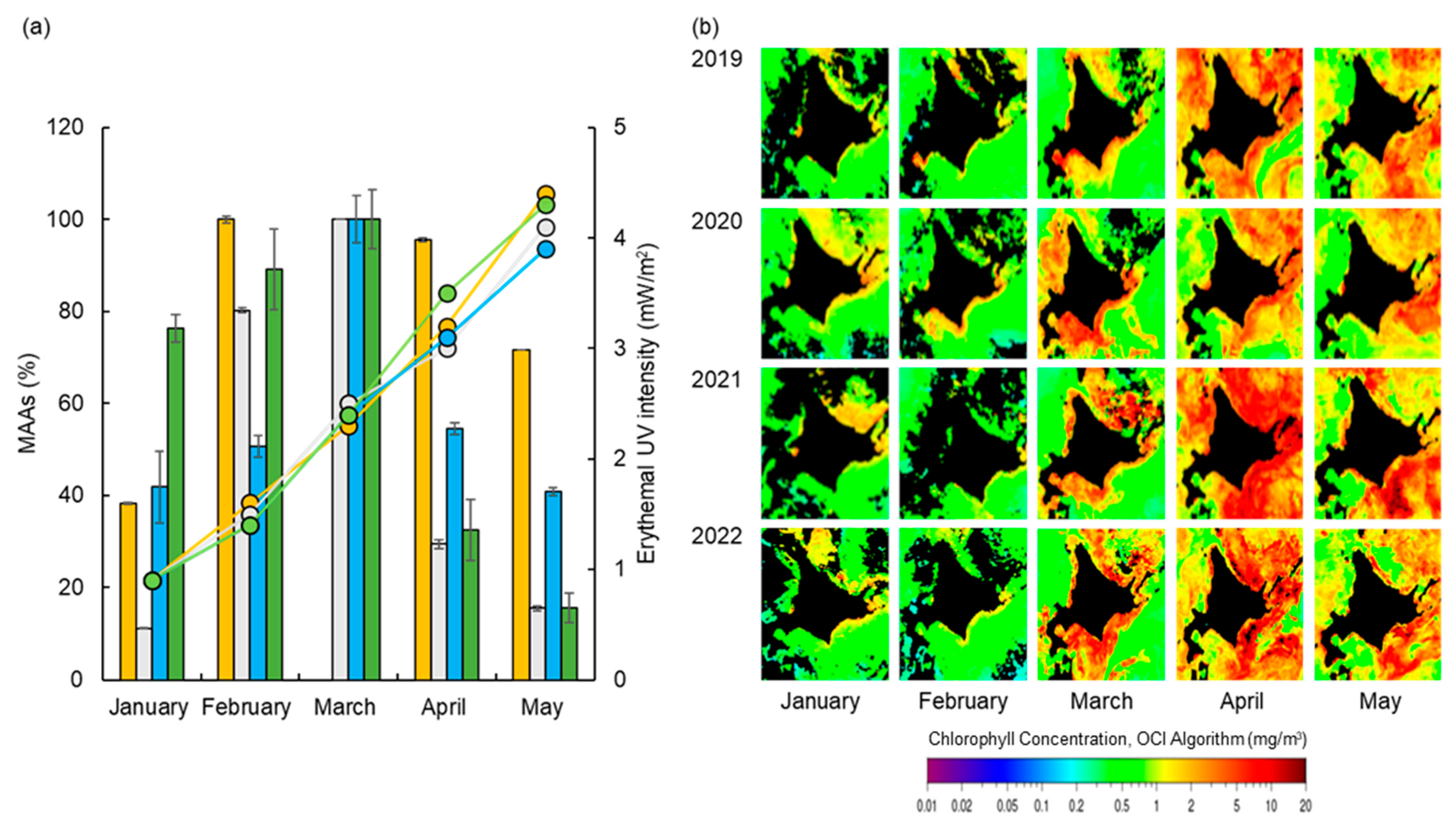
3.1. Monthly Variation in MAAs from Usujiri Dulse in 2022

We previously extracted MAAs from each month via the water–methanol extraction method. A previous study showed that the amount of MAA extraction was increased via the 25% ethanol extraction method [32]. In this study, we used the extraction method to evaluate the monthly variation in MAAs from Usujiri dulse. An analysis of the HPLC chromatogram revealed six peaks: shinorine, palythine, asterina-330, porphyra-334, usujirene, and palythene, as for previous dulse MAAs [32]. The monthly variation in the 25% ethanol extraction is shown in Table 1. The total MAA content gradually increased from January (30.8 µmol/g DW) to March (40.4 µmol/g DW) and decreased sharply in April (13.1 µmol/g DW). The MAA content was lowest in May (6.29 µmol/g DW).
The maximum MAA content in 2022 (40.4 µmol/g DW) was approximately 5.7 times higher than that in 2021, which was extracted via the water–methanol extraction method (7.03 µmol/g DW) [32]. The high MAA content was due to the improved extraction method. This method reflected the monthly variation in the increase in MAAs in March and the decrease in MAAs in April. Comparing the 2022 data with data from the previous three years, the trend in the palythine content in 2022 was similar to that in 2020. Namely, the palythine content was unchanged up to March and decreased sharply in April. The trends in the porphyra-334 and usujirene + palythene contents were similar over four years, reaching the maximum in March and decreasing significantly in April.

3.2. Monthly Variation in MAA Contents

The molar percentages (mol%) of MAAs in 2022 were compared (Figure 1). The molar percentages of shinorine (a maximum of 3.6 mol% on 24 January and a minimum of 2.6 mol% on 24 March) and asterina-330 (a maximum of 1.7 mol% on 13 May and a minimum of 1.4 mol% on 24 March) were stable at low values. Palythine was stable at 29 mol% on 24 February and 35 mol% on 13 May. The rest of the major MAAs showed the same variations. Namely, usujirene + palythene was stable from 21 mol% on 24 January to 22 mol% on 24 March and reached approximately 29–30 mol% on 12 April and 13 May. Porphyra-334 was stable at 43–47 mol% up to 24 March and dropped 32–35 mol% on 12 April and 13 May.
In the previous three years, the mol% of polphyra-334 decreased in April [28,29,30]. Because of the small decrease in polphyra-334 and the stable mol% of palythine, polphyra-334 was the major MAA up to April. Palythine became the major MAA in May. Although the change in the mol% of the MAAs slightly differed over the years, we observed that palythine replaced porphyra-334 as the major MAA in late April or early May.
Studies on dulse MAAs were performed in western Ireland [42], New Brunswick (Canada) [28], Spitsbergen (Norway) [27], and Brittany (France) [25,26]. Monthly variations in MAAs were surveyed in Ireland and France. An analysis of MAAs from French P. palmata revealed the presence of eight different MAAs, with seven of them identified as shinorine, palythine, asterina-330, porphyra-334, palythinol, usujirene, and palythene [25]. The proportion of porphyra-334 in relation to the total MAA area increased from 33% to 50% between March and August of 2018 [26]. In the case of Irish dulse (P. palmata), porphyra-334 constituted 45–55% of the total MAA content in September 2014, the month with the lowest content of the survey. It rose to 75–80% in April 2015, the month with the highest content of the survey [42]. Japanese dulse and Irish dulse showed similarities in terms of the seasonal variations in the contents of porphyra-334. Despite seasonal changes, Irish dulse and Japanese dulse maintained high levels of porphyra-334 content. The results of the monthly variation in the dulse MAAs in Japan were consistent with those of other studies [25,42]. We deposited data for several years in the same Usujiri region. There were no long-term observation data. Therefore, we convincingly showed that the amount of MAAs varies depending on environmental factors.

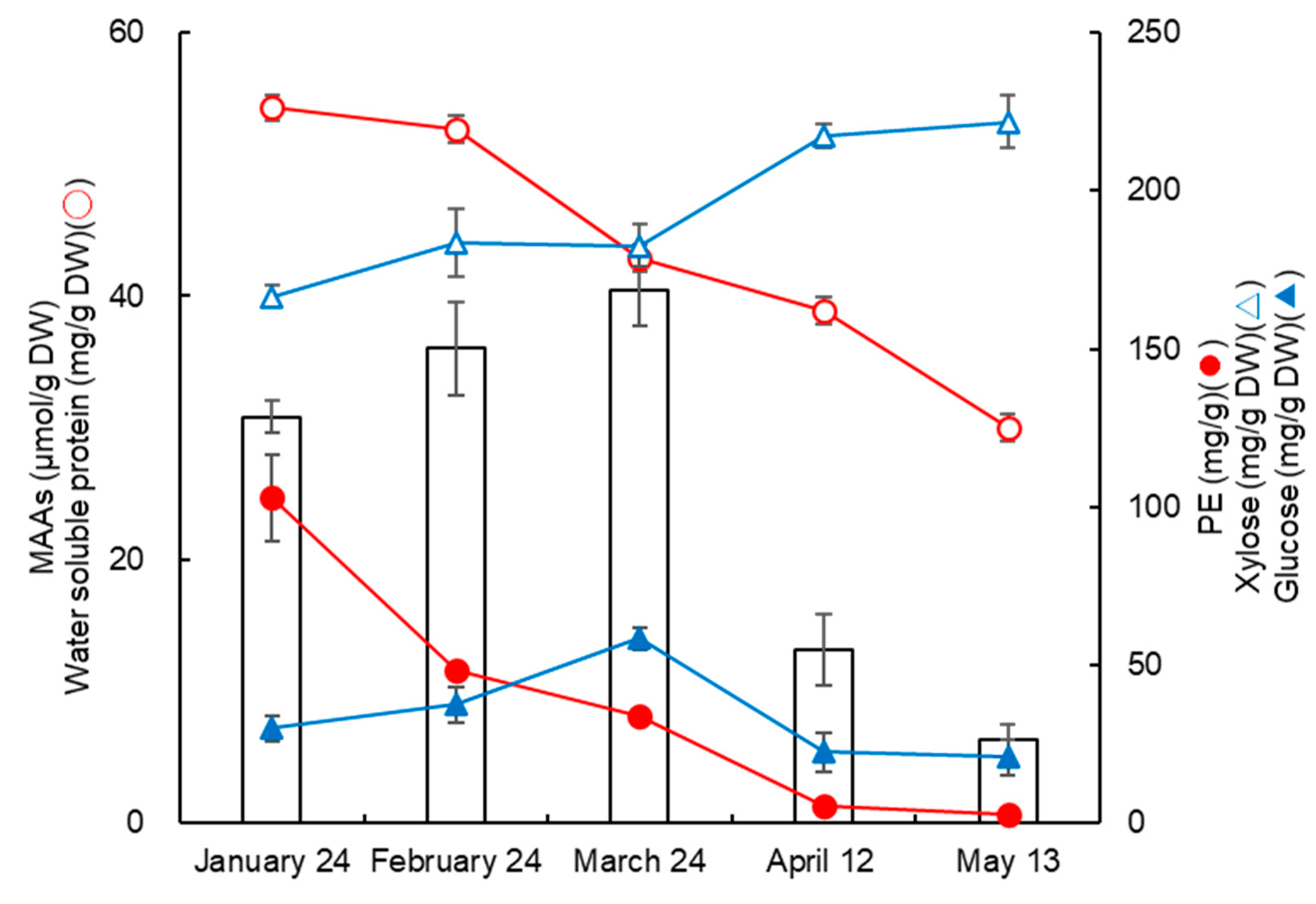
3.3. Changes in MAAs, Proteins, Saccharides, and Erythemal UV Intensity

Our data showed that the MAA content of the dulse changed monthly. Therefore, we examined the relationship between MAAs and the composition of dulse (proteins and saccharides) (Figure 2). The data represented the amount of micromolecules in 1 g dry-weight dulse powder. Proteins and saccharides are the major components of dulse. We designated water-soluble protein as the protein, and the main component of the soluble protein was PE. The water-soluble protein and PE showed the same trend. Xylose and glucose are considered the main constituted saccharides. The acid hydrolysate of the cell wall components of xylan and the storage polysaccharide of floridean starch were xylose and glucose, respectively. The amount of protein decreased from January to May, while that of xylose gradually increased from January to May.
In addition, erythema UV intensity increased constantly from January to May (Figure 3). The amount of MAAs in the dulse increased in response to the erythemal UV intensity until March. However, it decreased in April. We employed the erythemal UV intensity and chlorophyll concentration (data from JMA and NASA’s Ocean Color WEB) (Figure 3). The concentration of chlorophyll in the Usujiri area was low from January to February, but the concentration increased from March to April. In Funka Bay, the nutrient circulation from the sediment occurred via changes in the water temperature from winter to spring [43]. The massive proliferation of phytoplankton was induced by inorganic nitrogen [43,44]. We previously suggested that a thickened xylan cell wall protected dulse from UV radiation [43]. Floridean starch is a photosynthesis product and increases in sunlight, which might promote the starch synthesis until the loss of nitrogen compounds in the Usujiri area. Not only proteins but also MAAs are nitrogen compounds in red algae [45,46]. Therefore, we concluded that the decrease in MAAs could be associated with phytoplankton proliferation around the Usujiri area [30,31,32].

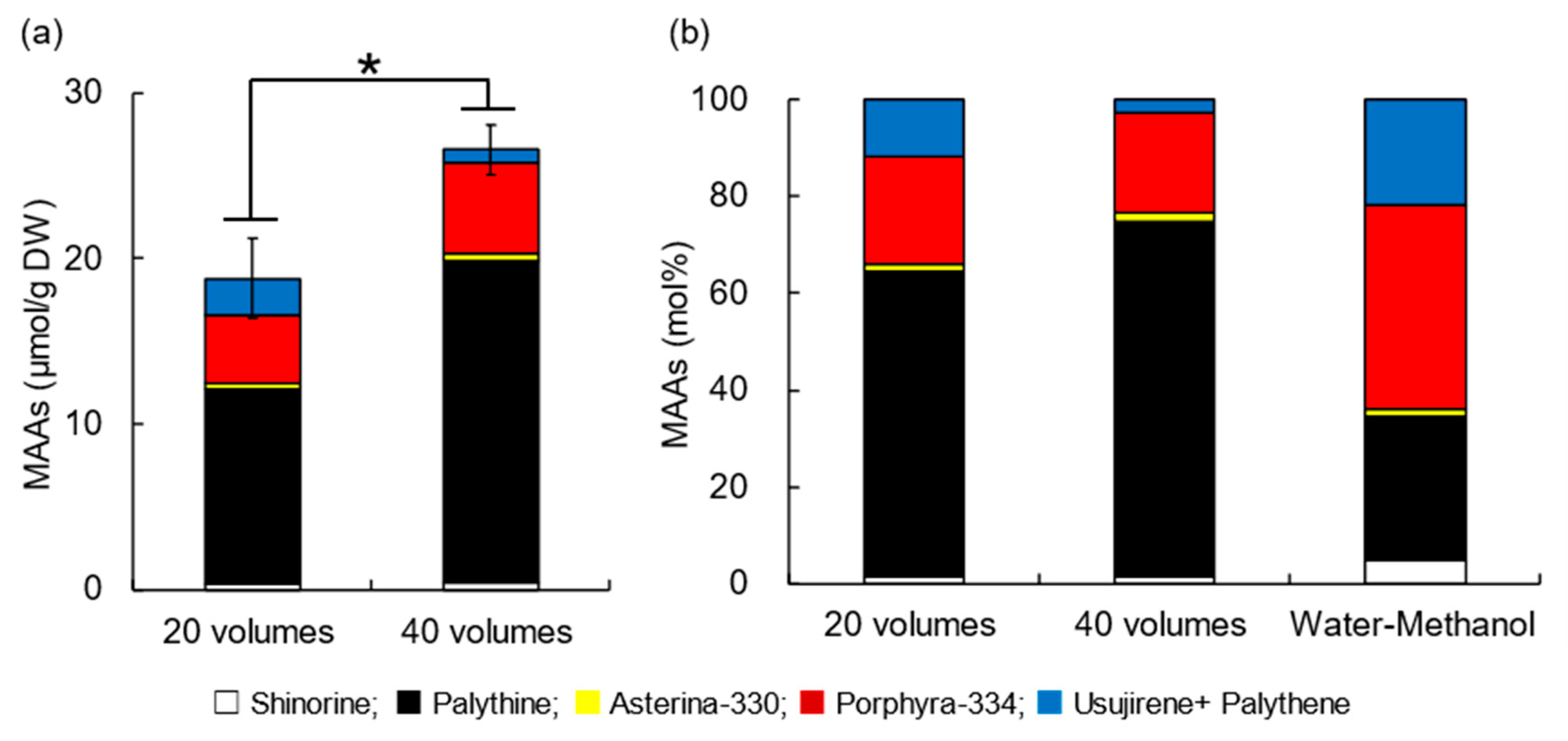
3.4. Effect of Extraction Volume on MAA Extraction from Dulse

In a previous study, we improved the MAA extraction method from dulse using 25, 50, and 99% ethanol, resulting in the development of the extraction method via 25% ethanol for 24 h at 4 °C [32]. In this study, we evaluated the effect of the solvent volumes (1/20 and 1/40, w/v) for MAAs from the dulse powder sample (April 2021) using 25% ethanol for 24 h at 4 °C (Figure 4a). The amount of MAAs obtained via 40 volumes of 25% ethanol increased (28.8 µmol/g DW) 1.5-fold higher than that by 20 volumes (18.8 µmol/g DW). The MAA contents were as follows: shinorine: 0.6 µmol/g DW; palythine: 16.5 µmol/g DW; asterina-330: 1.4 µmol/g DW; porphyra-334: 6.9 µmol/g DW; usujirene + palythene: 3.5 µmol/g DW. The increase in the MAA content differed for each MAA. Among them, the amount of palythine mostly increased (4.7 µmol/g DW). The mol% of the MAA composition is shown in Figure 4b. The tendency of the mol% was similar in the three samples. The composition of palythine in the 20-volume extract was 62.8 mol%, while that in the 40-volume extract was slightly decreased at 57.1 mol%. The difference happened via the increase in each MAA (e.g., porphyra-334 from 22.0 mol% to 23.8 mol%; usujirene + palythine from 11.8 mol% to 12.1 mol%; asterina-330 from 1.7 mol% to 4.8 mol%; and shinorine from 1.6 mol% to 2.2 mol%). In this experiment, the 40-volume extraction with 25% ethanol was suitable for the MAA extraction from dulse.
The MAA composition varied by using different solvent volumes for extraction. The amount of palythine increased compared to the 20-volume and water–methanol extraction. It is thought that usujirene is hydrolyzed to palythine [30]. Although the synthetic and hydrolysis pathways of MAAs are still not clear, the high amount of palythine might be attributed to hydrolyzed MAAs.

3.5. Effect of Extraction Time on MAA Extraction from Dulse

We previously improved our MAA extraction methods from dulse [32]. Other studies have used different extraction volumes; however, fewer studies on the evaluation of the extraction time have been reported. Therefore, we investigated the effect of the extraction times on the MAAs from residue (Figure 5). MAAs were extracted (26.6 µmol/g DW) first by 40 volumes of 25% ethanol extraction. The MAAs were repeatedly extracted from the residue via the same method, obtaining 4.6 µmol/g DW in the second extraction. MAAs were extracted from 2.6 µmol/g DW from the third to the fifth extraction. As a result, the total MAA content obtained by the fifth extraction with the 40-volume method was reached (33.8 µmol/g DW). These data showed that 92% of the MAAs were obtained via two-time extraction. Similarly, five-time extraction was performed via 20 volumes of 25% ethanol extraction, and the total content of MAAs obtained was 26.4 µmol/g DW. The amount of MAAs from the two methods was significantly higher in the five-times extraction via 40 volumes. The results indicated that two or more extractions via 40 volumes yielded the most extracted MAAs from the dulse.
Sun et al. also reported variations in the MAAs obtained based on the extraction degree in four species of red algae [40]. However, red algae have different types of polysaccharides (κ-, λ-, and ι-carrageenan, agar, and agaropectin). Thus, the more that the amount of water in the extraction solvent increases, the more polysaccharides are extracted. Polysaccharides increase in viscosity, resulting in the loss of the recovery of MAAs. MAAs cannot be recovered sufficiently in the high organic solvent because of the low solubility of MAAs in organic solvent compared to water. Therefore, it is necessary to evaluate an extraction method using each seaweed. The absolute amount of the MAA content in red algae remains unclear. The development of extraction methods will lead to the amounts and selectivity of MAAs from seaweeds.

4. Conclusions

We studied the content and composition of dulse ingredients from Usujiri and their monthly variation. The trends in the content and composition of MAAs in dulse were the same as those in the previous three years, regardless of the different extraction methods. The highest amount of MAAs was obtained from the March dulse sample. The extraction conditions affected the yield of MAAs. We improved the MAA extraction method to 40 volumes of 25% ethanol at 4 °C for 24 h, resulting in a 1.3-fold increase in the MAA yield. However, there are still remaining factors to improve the MAA yield. We will continue to refine the extraction conditions of MAAs from Usujiri dulse. This study will be useful in determining the optimal extraction conditions of MAAs from seaweeds. A preparation method for obtaining a large amount of the desired MAAs is expected in the future.

Author Contributions

Y.K. and H.K. conceived of and designed the research; R.Y. and K.T. performed the experiments; R.Y. and Y.M. analyzed the data; Y.K., R.Y., M.A.M.M. and H.K. contributed to the writing and editing of the manuscript. All authors have read and agreed to the published version of the manuscript.

Funding

This research received no external funding.

Institutional Review Board Statement

Not applicable.

Informed Consent Statement

Not applicable.

Data Availability Statement

Data sharing is not applicable to this article.

Acknowledgments

We gratefully acknowledge the sampling assistance for Devaleraea inkyuleei at Usujiri by Hiroyuki Munehara and Atsuya Miyajima.

Conflicts of Interest

The authors declare no conflict of interest.

References

  1. Chrapusta, E.; Kaminski, A.; Duchnik, K.; Bober, B.; Adamski, M.; Bialczyk, J. Mycosporine-Like Amino Acids: Potential health and beauty ingredients. Mar. Drugs. 2017, 15, 326. [Google Scholar] [CrossRef]
  2. Afaq, F.; Mukhtar, H. Botanical antioxidants in the prevention of photo carcinogenesis and photoaging. Exp. Dermatol. 2006, 15, 678–684. [Google Scholar] [CrossRef]
  3. Rastogi, R.P.; Sinha, R.P. Biotechnological and industrial significance of cyanobacterial secondary metabolites. Biotech. Advan. 2009, 27, 521–539. [Google Scholar] [CrossRef] [PubMed]
  4. Ryu, J.; Park, S.-J.; Kim, I.-H.; Choi Hee, Y.; Nam, T.-J. Protective effect of porphyra-334 on UVA-induced photoaging in human skin fibroblasts. Int. J. Mol. Med. 2014, 34, 796–803. [Google Scholar] [CrossRef]
  5. Antoniou, C.; Kosmadaki, M.G.; Stratigos, A.J.; Katsambas, A.D. Sunscreens—What’s important to know. J. Eur. Acad. Dermatol. Venereol. 2008, 22, 1110–1119. [Google Scholar] [CrossRef]
  6. Orfanoudaki, M.; Hartmann, A.; Alilou, M.; Gelbrich, T.; Planchenault, P.; Derbré, S.; Schinkovitz, A.; Richomme, P.; Hensel, A.; Ganzera, M. Absolute configuration of mycosporine-like amino acids, their wound healing properties and in vitro anti-aging effects. Mar. Drugs. 2020, 18, 35. [Google Scholar] [CrossRef] [PubMed]
  7. Lawrence, K.P.; Gacesa, R.; Long, P.F.; Young, A.R. Molecular photoprotection of human keratinocytes in vitro by the naturally occurring mycosporine-like amino acid palythine. Br. J. Dermatol. 2018, 178, 1353–1363. [Google Scholar] [CrossRef]
  8. Tang, S.; Chen, Y.; Song, G.; Liu, X.; Shi, Y.; Xie, Q.; Chen, D. A Cocktail of Industrial Chemicals in Lipstick and Nail Polish: Profiles and Health Implications. Environ. Sci. Technol. Lett. 2021, 8, 760–765. [Google Scholar] [CrossRef]
  9. Pangestuti, R.; Siahaan, E.A.; Kim, S.K. Photoprotective substances derived from marine algae. Mar. Drugs 2018, 16, 11. [Google Scholar] [CrossRef]
  10. Danovaro, R.; Bongiorni, L.; Corinaldesi, C.; Giovannelli, D.; Damiani, E.; Astolfi, P.; Greci, L.; Pusceddu, A. Sunscreens cause coral bleaching by promoting viral infections. Environ. Health Perspect. 2008, 116, 441–447. [Google Scholar] [CrossRef]
  11. Downs, C.A.; Kramarsky-Winter, E.; Segal, R.; Fauth, J.; Knutson, S.; Bronstein, O.; Ciner, F.R.; Jeger, R.; Lichtenfeld, Y.; Woodley, C.M.; et al. Toxicopathological Effects of the Sunscreen UV Filter, Oxybenzone (Benzophenone-3), on Coral Planulae and Cultured Primary Cells and Its Environmental Contamination in Hawaii and the U.S. Virgin Islands. Arch. Environ. Contam. 2016, 70, 265–288. [Google Scholar] [CrossRef]
  12. Hermund, D.B.; Torsteinsen, H.; Vega, J.; Figueroa, F.L.; Jacobsen, C. Screening for New Cosmeceuticals from Brown Algae Fucus vesiculosus with Antioxidant and Photo-Protecting Properties. Mar. Drugs. 2022, 20, 687. [Google Scholar] [CrossRef]
  13. Núñez-Pons, L.; Avila, C.; Romano, G.; Verde, C.; Giordano, D. UV-protective compounds in marine organisms from the Southern Ocean. Mar. Drugs. 2018, 16, 336. [Google Scholar] [CrossRef]
  14. Álvarez-Gómez, F.; Korbee, N.; Casas-Arrojo, V.; Abdala-Díaz, R.T.; Figueroa, F.L. UV photoprotection, cytotoxicity and immunology capacity of red algae extracts. Molecules 2019, 24, 341. [Google Scholar] [CrossRef]
  15. Bonomi-Barufi, J.; Figueroa, F.L.; Korbee, N.; Momoli, M.M.; Martins, A.P.; Colepicolo, P.; van Sluys, M.A.; Oliveira, M.C. How macroalgae can deal with radiation variability and photoacclimation capacity: The example of Gracilaria tenuistipitata (Rhodophyta) in laboratory. Algal Res. 2020, 50, 102007. [Google Scholar] [CrossRef]
  16. Karentz, D.; Mceuen, E.S.; Land, M.C.; Dunlap, W.C. Survey of mycosporine-like amino acid compounds in Antarctic marine organisms: Potential protection from ultraviolet exposure. Mar. Biol. 1991, 108, 157–166. [Google Scholar] [CrossRef]
  17. Jain, S.; Prajapat, G.; Abrar, M.; Ledwani, L.; Singh, A.; Agrawal, A. Cyanobacteria as efficient producers of mycosporine-like amino acids. J. Basic Microbiol. 2017, 57, 715–727. [Google Scholar] [CrossRef]
  18. Sinha, R.P.; Singh, S.P.; Häder, D.P. Database on mycosporines and mycosporine-like amino acids (MAAs) in fungi, cyanobacteria, macroalgae, phytoplankton and animals. J. Photochem. Photobiol. B Biol. 2007, 89, 29–35. [Google Scholar] [CrossRef]
  19. Geraldes, V.; Pinto, E. Mycosporine-like amino acids (MAAs): Biology, chemistry and identification features. Pharmaceuticals 2021, 14, 63. [Google Scholar] [CrossRef]
  20. Conde, F.; Churio, M.; Previtali, C. The photoprotector mechanism of mycosporine-like amino acids. Excited-state properties and photostability of porphyra-334 in aqueous solution. J. Photochem. Photobiol. B Biol. 2020, 56, 139–144. [Google Scholar] [CrossRef]
  21. Torres, P.; Santos, J.P.; Chow, F.; Pena Ferreira, M.J.; dos Santos, D.Y.A.C. Comparative analysis of in vitro antioxidant capacities of mycosporine-like amino acids (MAAs). Algal Res. 2018, 34, 57–67. [Google Scholar] [CrossRef]
  22. Carreto, J.I.; Carignan, M.O. Mycosporine-like amino acids: Relevant secondary metabolites. chemical and ecological aspects. Mar. Drugs. 2011, 9, 387. [Google Scholar] [CrossRef] [PubMed]
  23. Rastogi, R.P.; Richa Sinha, R.P.; Singh, S.P.; Häder, D.P. Photoprotective compounds from marine organisms. J. Ind. Microbiol. Biotechnol. 2010, 37, 537–558. [Google Scholar] [CrossRef] [PubMed]
  24. Browne, N.; Otero, P.; Murray, P.; Saha, S.K. Rapid Screening for Mycosporine-like Amino Acids (MAAs) of Irish Marine Cyanobacteria and Their Antioxidant Potential. Sustainability 2023, 15, 3792. [Google Scholar] [CrossRef]
  25. Lalegerie, F.; Lajili, S.; Bedoux, G.; Taupin, L.; Stiger-Pouvreau, V.; Connan, S. Photo-protective compounds in red macroalgae from Brittany: Considerable diversity in mycosporine-like amino acids (MAAs). Mar. Environ. Res. 2019, 147, 37–48. [Google Scholar] [CrossRef]
  26. Lalegerie, F.; Stiger-Pouvreau, V.; Connan, S. Temporal variation in pigment and mycosporine-like amino acid composition of the red macroalga Palmaria palmata from Brittany (France): Hypothesis on the MAA biosynthesis pathway under high irradiance. J. Appl. Phycol. 2020, 32, 2641–2656. [Google Scholar] [CrossRef]
  27. Karsten, U.; Wiencke, C. Factors controlling the formation of UV-absorbing mycosporine-like amino acids in the marine red alga Palmaria palmata from Spitsbergen (Norway). J. Plant Physiol. 1999, 155, 407–415. [Google Scholar] [CrossRef]
  28. Yuan, Y.V.; Westcott, N.D.; Hu, C.; Kitts, D.D. Mycosporine-like amino acid composition of the edible red alga, Palmaria palmata (dulse) harvested from the west and east coasts of Grand Manan Island, New Brunswick. Food Chem. 2009, 112, 321–328. [Google Scholar] [CrossRef]
  29. Athukorala, Y.; Trang, S.; Kwok, C.; Yuan, Y.V. Antiproliferative and antioxidant activities and mycosporine-Like amino acid profiles of wild-Harvested and cultivated edible Canadian marine red macroalgae. Molecules 2016, 21, 119. [Google Scholar] [CrossRef]
  30. Nishida, Y.; Kumagai, Y.; Michiba, S.; Yasui, H.; Kishimura, H. Efficient extraction and antioxidant capacity of mycosporine-like amino acids from red alga dulse Palmaria palmata in Japan. Mar. Drugs. 2020, 18, 502. [Google Scholar] [CrossRef]
  31. Nishida, Y.; Miyabe, Y.; Kishimura, H.; Kumagai, Y. Monthly Variation and Ultraviolet Stability of Mycosporine-like Amino Acids from Red Alga Dulse Palmaria palmata in Japan. Phycology 2021, 1, 119–128. [Google Scholar] [CrossRef]
  32. Yamamoto, R.; Mune Mune, M.A.; Miyabe, Y.; Kishimura, H.; Kumagai, Y. Monthly Variation in Mycosporine-like Amino Acids from Red Alga Dulse (Devaleraea inkyuleei, Formerly Palmaria palmata in Japan). Phycology 2023, 3, 127–137. [Google Scholar] [CrossRef]
  33. Skriptsova, V.S.; Suzuki, M.; Semenchenko, A.A. Morphological variation in Northwest Pacific Devaleraea mollis and description of D. inkyuleei sp. Nov. (Palmariaceae, Rhodophyta). Phycologia 2022, 61, 606–615. [Google Scholar] [CrossRef]
  34. Nishida, Y.; Saburi, W.; Miyabe, Y.; Kishimura, H.; Kumagai, Y. Characterization of Antioxidant Activity of Heated Mycosporine-like Amino Acids from Red Alga Dulse Palmaria palmata in Japan. Mar. Drugs. 2022, 20, 184. [Google Scholar] [CrossRef] [PubMed]
  35. Sato, N.; Furuta, T.; Takeda, T.; Miyabe, Y.; Ura, K.; Takagi, Y.; Yasui, H.; Kumagai, Y.; Kishimura, H. Antioxidant activity of proteins extracted from red alga dulse harvested in Japan. J. Food Biochem. 2019, 43, e12709. [Google Scholar] [CrossRef] [PubMed]
  36. Furuta, T.; Miyabe, Y.; Yasui, H.; Kinoshita, Y.; Kishimura, H. Angiotensin I converting enzyme inhibitory peptides derived from phycobiliproteins of dulse Palmaria palmata. Mar. Drugs. 2016, 14, 32. [Google Scholar] [CrossRef]
  37. Yamamoto, Y.; Kishimura, H.; Kinoshita, Y.; Saburi, W.; Kumagai, Y.; Yasui, H.; Ojima, T. Enzymatic production of xylooligosaccharides from red alga dulse (Palmaria sp.) wasted in Japan. Process Biochem. 2019, 82, 117–122. [Google Scholar] [CrossRef]
  38. Kobayashi, M.; Kumagai, Y.; Yamamoto, Y.; Yasui, H.; Kishimura, H. Identification of a key enzyme for the hydrolysis of β-(1-3)-xylosyl linkage in red alga dulse xylooligosaccharide from Bifidobacterium adolescentis. Mar. Drugs. 2020, 18, 174. [Google Scholar] [CrossRef]
  39. Tartarotti, B.; Sommaruga, R. The effect of different methanol concentrations and temperatures on the extraction of mycosporine-like amino acids (MAAs) in algae and zooplankton. Arch. Hydrobiol. 2002, 154, 691–703. [Google Scholar] [CrossRef]
  40. Sun, Y.; Han, X.; Hu, Z.; Cheng, T.; Tang, Q.; Wang, H.; Deng, X.; Han, X. Extraction, isolation and characterization of mycosporine-like amino acids from four species of red macroalgae. Mar. Drugs. 2021, 19, 615. [Google Scholar] [CrossRef]
  41. Beer, S. Determining phycoerythrin and phycocyanin concentrations in aqueous crude extracts of red algae. Mar. Freshw. Res. 1985, 36, 785. [Google Scholar] [CrossRef]
  42. Guihéneuf, F.; Gietl, A.; Stengel, D.B. Temporal and spatial variability of mycosporine-like amino acids and pigments in three edible red seaweeds from western Ireland. J. Appl. Phycol. 2018, 30, 2573–2586. [Google Scholar] [CrossRef]
  43. Kudo, I.; Yoshimura, T.; Lee, C.; Yanada, M.; Maita, Y. Nutrient Regeneration at Bottom after a Massive Spring Bloom in a Subarctic Coastal Environment, Funka Bay, Japan. J. Oceanogr. 2007, 63, 791–801. [Google Scholar] [CrossRef]
  44. Isada, T.; Hirawake, T.; Nakada, S.; Kobayashi, T.; Sasaki, K.; Tanaka, Y.; Watanabe, S.; Suzuki, K.; Saitoh, S.I. Influence of hydrography on the spatiotemporal variability of phytoplankton assemblages and primary productivity in Funka Bay and the Tsugaru Strait. Estuar. Coast. Shelf Sci. 2017, 188, 199–211. [Google Scholar] [CrossRef]
  45. Ekman, P.; Yu, S.; Pedersen, M. Effects of altered salinity, darkness and algal nutrient status on floridoside and starch content, α-galactosidase activity and agar yield of cultivated Gracilaria sordida. Br. Phycol. J. 1991, 26, 123–131. [Google Scholar] [CrossRef]
  46. Kudo, I.; Matsunaga, K. Environmental factors affecting the occurrence and production of the spring phytoplankton bloom in funka bay, Japan. J. Oceanogr. 1999, 55, 505–513. [Google Scholar] [CrossRef]
Figure 1. Molar percentages of MAAs in 2022. The contents of six MAAs (shinorine, palythine, asterina-330, porphyra-334, and usujirene + palythene) in dulse collected between January and May 2022 from Usujiri, Hakodate, Japan. The mol% data are converted from µmol/g DW in Table 1. The values of 100 mol% of MAAs represent 30.8, 36.0, 40.4, 13.1, and 6.29 µmol/g DW of the samples from 24 January, 24 February, 24 March, 12 April, and 13 May, respectively.
Phycology 03 00026 g001
Figure 2. Monthly variations in MAAs, proteins, and saccharides in dulse collected in 2022. The amount of MAAs was expressed as the total MAA content. Water-soluble protein was determined via gravimetric methods. The amount of PE from dulse powder was determined using the equation of Reference 37. Xylose and glucose were determined via colorimetric methods. The data show mean values (n = 3).
Phycology 03 00026 g002
Figure 3. Monthly mean MAA contents, daily maximum erythemal UV intensities, and chlorophyll concentrations from 2019 to 2022. (a) MAA content and erythemal UV intensity. Bars of orange, gray, blue, and green represent MAA content (%) in 2019, 2020, 2021, and 2022, respectively. The data show the mean values ± SEs (n = 3). Lines of orange, gray, blue, and green represent erythemal UV intensities (mW/m2) in 2019, 2020, 2021, and 2022, respectively. Erythemal UV intensity was obtained from the Japan Meteorological Agency (https://www.data.jma.go.jp/gmd/env/uvhp/info_uv.html, accessed on 5 May 2023). (b) Chlorophyll concentrations. The sampling place was Usujiri, at the entrance to Funka Bay. These data were obtained from JMA and NASA’s Ocean Color WEB (https://oceancolor.gsfc.nasa.gov, accessed on 5 May 2023). All data were recorded in 2019–2022.
Phycology 03 00026 g003
Figure 4. Effect of solvent volumes on MAA yields. Dulse collected on 12 April 2022 was used. (a) The amount of MAAs by different extraction volumes. Two volumes (20 and 40 volumes) of 25% ethanol were evaluated. The contents of MAAs are expressed as µmol/g DW. Error bars show the average of the total MAAs (n = 3). The asterisk indicates a significant difference in mean value (Student’s t-test, p < 0.05). (b) Molar percentages of MAAs obtained via different extraction methods. The data of (a) are converted to mol%. The successive water–methanol extraction data were obtained from dulse collected on March 25, 2021 [32]. The data show the mean values (n = 3). The values of 100 mol% in 25% ethanol in 20-volume and 40-volume and successive water–methanol extraction were 22.7, 28.8, and 3.8 µmol/g DW, respectively.
Phycology 03 00026 g004
Figure 5. Effect of extraction times on MAA yield. Dulse collected on 24 March 2022 was used. MAAs were extracted via 20 or 40 volumes of 25% ethanol at 4 °C for 24 h. The extractions were continued five times in each method. The contents of MAAs are expressed as µmol/g DW. The total of 20 and 40 volumes of MAAs are 26.4 and 33.8 µmol/g DW, respectively. Error bars show the average of total MAAs (n = 3). The asterisk indicates a significant difference in mean value (Student’s t-test, p < 0.05).
Phycology 03 00026 g005
Table 1. MAA content in Usujiri dulse.
MAAsCollection Date (2022)
(µmol/g DW)24 January 24 February 24 March 12 April 13 May
Shinorine1.14 ± 0.07 a1.16 ± 0.09 a1.04 ± 0.40 a0.41 ± 0.17 b0.23 ± 0.13 b
Palythine9.71 ± 0.16 a10.3 ± 0.9 a12.0 ± 4.2 a4.01 ± 1.09 b2.17 ± 0.58 b
Asterina-3300.47 ± 0.03 b0.60 ± 0.05 a0.58 ± 0.21 ab0.22 ± 0.02 c0.11 ± 0.01 c
Porphyra-33413.2 ± 0.6 b17.0 ± 1.5 ab17.8 ± 7.3 a4.59 ± 0.52 c1.98 ± 0.23 c
Usujirene +
Palythene
6.32 ± 0.27 ab6.94 ± 1.14 a8.97 ± 4.05 a3.91 ± 0.41 bc1.80 ± 0.21 c
Total30.8 ± 1.2 b36.0 ± 3.5 ab40.4 ± 2.6 a13.1 ± 2.7 c6.29 ± 1.27 c
Dulse was collected on 24 January, 24 February, 24 March, 12 April, and 13 May 2022. The contents of MAAs are expressed as µmol/g DW. The data show mean values ± SEs (n = 3). Different letters in each MAA indicate significant differences in mean value (Tukey–Kramer’s multiple comparisons test, a, b, c, p < 0.05).
Disclaimer/Publisher’s Note: The statements, opinions and data contained in all publications are solely those of the individual author(s) and contributor(s) and not of MDPI and/or the editor(s). MDPI and/or the editor(s) disclaim responsibility for any injury to people or property resulting from any ideas, methods, instructions or products referred to in the content.

