Review

Using the Carnivorous Sponge Lycopodina hypogea as a Nonclassical Model for Understanding Apoptosis-Mediated Shape Homeostasis at the Organism Level

1
ISEM, Univ Montpellier, CNRS, IRD, 34095 Montpellier, France
2
IMBE, CNRS, Aix Marseille Université, IRD, Avignon Université, Station Marine d’Endoume, Rue de la Batterie des Lions, 13007 Marseille, France
*
Authors to whom correspondence should be addressed.
n° ISEM 2023-006.
Foundations 2023, 3(2), 220-230; https://doi.org/10.3390/foundations3020018
Submission received: 13 January 2023 / Revised: 10 March 2023 / Accepted: 29 March 2023 / Published: 18 April 2023

Abstract

:
The dynamic equilibrium between death and regeneration is well established at the cell level. Conversely, no study has investigated the homeostatic control of shape at the whole organism level through processes involving apoptosis. To address this fundamental biological question, we took advantage of the morphological and functional properties of the carnivorous sponge Lycopodina hypogea. During its feeding cycle, this sponge undergoes spectacular shape changes. Starved animals display many elongated filaments to capture prey. After capture, prey are digested in the absence of any centralized digestive structure. Strikingly, the elongated filaments actively regress and reform to maintain a constant, homeostatically controlled number and size of filaments in resting sponges. This unusual mode of nutrition provides a unique opportunity to better understand the processes involved in cell renewal and regeneration in adult tissues. Throughout these processes, cell proliferation and apoptosis are interconnected key actors. Therefore, L. hypogea is an ideal organism to study how molecular and cellular processes are mechanistically coupled to ensure global shape homeostasis.

1. Introduction

Originally, homeostasis was defined as the process through which an organism tends to maintain constant its biological and physiological features, for instance, those of its internal medium [1,2]. Although this concept generally focuses on the stability of the internal environment (i.e., physiological homeostasis), it can also be generalized and applied to the whole organism and its relationship to its local environment through its shape. In this more general context, apoptosis is a powerful tool to generate, renew, and maintain shapes during embryonic development and throughout adult life [3]. Thus, homeostasis can be extended to all organization levels of living matter. To obtain an integrated view of organism homeostasis in relation to its environment, different organization levels must often be considered (e.g., the molecular level, in relation to the internal medium, and the structural level) in a temporal framework. Accordingly, the changes that ensure the homeostasis of functional structures are slow and prolonged, whereas the homeostasis of the internal environment is maintained through rapid processes. The study of global homeostasis (i.e., the whole-organism homeostasis in relation to its environment) requires biological models that can respond to energy variations in the environment in a measurable manner. This is the case of the carnivorous sponge Lycopodina hypogea (Vacelet & Boury-Esnault, 1996) [4]. In this sponge, and apparently in all carnivorous sponges (Cladorhizidae family), the response to nutritional stimuli results in a dramatic change in the structures involved in prey capture that requires the complete reorganization of the underlying molecular networks (see below). The digestive system of these sponges, which has been rarely studied due to their deep-sea habitat, has evolved from a “static” state (filter feeders) to a dynamic state (carnivores) to allow adaptation to their environment. Therefore, these sponges can be defined as structures that maintain their homeostasis through an ordered succession of events in which the apparent persistence of higher levels of organization is supported by the change in subordinated systems. In L. hypogea, the recovery of macroscopic structures at the end of a feeding cycle is closely correlated with the return to the initial state of the whole transcriptome and of the molecular networks directly involved in cell turnover [5]. Moreover, L. hypogea can easily stay several months without eating macroprey. Therefore, this sponge alternates between states of dynamic equilibrium without any consequence for its global shape (in the absence of nutrients) and drastic changes in shape with a return to the initial state (in the presence of nutrients). L. hypogea can adapt its global homeostasis to the presence/absence of food in its environment.
It is well documented how individual cells influence the development of multicellular organisms. Conversely, it is less well understood how the molecular and cellular features of these cells collectively lead to the emergence of robust homeostatic structures [6]. The process of cell renewal is a critical mechanism for ensuring homeostatic morphological and functional structures in adults. This process requires the differentiation of adult stem cells into new differentiated cells and the elimination of old cells by apoptosis. This dynamic equilibrium has been well documented at the cellular level [4,5,6,7], but no study has investigated how the shape of a functional structure or of a whole organism is homeostatically controlled.

2. Using L. hypogea as a Model to Study Homeostasis at the Organism/Structure Level

To better understand the processes involved in the homeostasis of functional structures during physiological cell turnover, we took advantage of the intriguing biological properties of the carnivorous sponge L. hypogea (class Demospongiae) [4]. This sponge was collected in its environment: a submarine cave in the Marseille region (France) that reproduces deep-water conditions (i.e., the classical habitat of carnivorous sponges). Once collected, L. hypogea samples could be easily maintained in aquarium conditions. Sponges were fed Artemia nauplii shrimps every 1–2 months, but they can survive more than one year without any food. This is probably an adaptation to the low nutrient levels in their natural environment, as suggested by the absence of filtration feeding systems in this sponge. During its feeding cycle, L. hypogea undergoes dramatic shape changes. Starved animals are attached to the rock by a 1–2 cm long peduncle (see Figure 1a, 0 h). This peduncle is relayed to an ovoid body covered with many elongated filaments with hook-like spicules that are crucial for capturing prey. After prey capture, filaments rapidly regress until they almost disappear. At the end of the digestion process, filaments slowly reform. The number and length of filaments is maintained between feeding cycles (Figure 1). We previously showed that this succession of morphological changes involves cell apoptosis, phagocytosis, and cell proliferation [5].
As the L. hypogea genome has yet not been sequenced, we performed a large transcriptomic study that revealed the complex molecular networks implicated in tissue homeostasis [5]. Specifically, we found that the intrinsic caspase-dependent apoptosis pathway is present in the sponge with a quite complex network (Figure 2a). We identified a few apoptotic regulators (i.e., SMAC/Diablo and BH-3-only proteins). Conversely, the extrinsic apoptosis pathway was more limited (without TNFR and with proteins that directly or indirectly interact with FAS only). These observations are in agreement with data obtained in some unicellular eukaryotes [12,13] and in another sponge, Amphimedon queenslandica [14]. This suggests that the intrinsic apoptosis pathway is more ancestral than the extrinsic one. We identified the caspase-independent AIF but not ENDOG. The autophagic machinery also displayed a complexity comparable to that observed in humans (Figure 2b). Similarly, cell proliferation actors and the cell cycle regulation were comparable to what is described in mammals but with the absence of cyclin-dependent kinase inhibitors (CDKN) (Figure 2c).
In L. hypogea, all these networks can be monitored with the cell biology methods used in more classical models: BrdU and EdU labeling for cell proliferation, he TUNEL assay for cell apoptosis, and anti-ATG8 antibodies for autophagy. This allows their spatiotemporal visualization in vivo. Moreover, in adult L. hypogea, apoptosis can be efficiently inhibited with the caspase inhibitor Z-VAD-FMK [5]. In addition, cell proliferation, apoptosis, and autophagy can be induced in vivo upon feeding in controlled conditions.
Therefore, L. hypogea is an ideal model to study how cellular processes are mechanistically coupled to maintain a consistent shape at the whole-organism level. This model also allows studying mechanisms that have been necessary for the acquisition and maintenance of multicellularity in the sister group of all other metazoans [15].

3. Cell Turnover and Adult Tissue Homeostasis in L. hypogea

Many adult metazoans remain in a dynamic equilibrium throughout their life, giving the impression of the stability of adult structures. This counterintuitive concept is very old and can be attributed to Heraclitus of Ephesus: “No man ever steps in the same river twice, for it’s not the same river and he’s not the same man”. A living organism is stable and invariant only in appearance: it is actually the manifestation of a perpetual flow of events. Without any interruption, most differentiated cells in living systems “commit suicide” and new stem cells differentiate to replace them. This concept is valid not only for cells but for all levels of life, including at the molecular level because all biomolecules are continuously renewed at varying rates. This dynamic concept of the organism is one of the most important principles of modern biology. Cell death and cell proliferation have been extensively studied separately, but very little is known about the identity of the molecular actors and networks underlying their coupling that maintains morphological homeostasis (i.e., the capacity to return to the initial shape) in adult organs and organisms. In this context, it is crucial to better understand how the regulation of cell turnover integrates the apoptosis of differentiated cells and the division and differentiation of stem cells to maintain and/or form functional structures again (here, the prey capture system of L. hypogea). How do these two seemingly opposite processes regulate each other in time and space? Do apoptotic cells and dividing/differentiating cells cross-talk to maintain an invariant number of cells between two feeding cycles? If this dialogue actually exists, what is the biochemical nature of the involved signals?
Addressing these questions implies identifying the molecules that connect the molecular networks controlling cell proliferation, differentiation, and programmed death at the cell, tissue, and whole-organism levels. To tackle this challenge, the carnivorous sponge L. hypogea is an attractive model organism.
This sponge ensures its homeostasis via trans-differentiation processes that involve the proliferation and differentiation of adult stem cells (archeocytes) with a continuous cell turnover mediated by apoptosis [5,16]. In addition to cell proliferation/differentiation and apoptosis, cell migration is implicated. Briefly, after prey capture, the filaments are destroyed and resorbed through a complex mechanism that concomitantly involves apoptosis at the top of the filaments and the active centripetal migration of phagocytic cells from all parts of the sponge toward the prey. At this stage, archeocyte proliferation is already observed. During the digestion process, the filaments reform from newly differentiated cells, including sclerocytes (i.e., cells secreting the skeleton of the new filaments), that adopt a centrifugal migration (Figure 3). In the resting adult sponge, the two processes of creation and destruction (and all their subprocesses) are active and in a dynamic equilibrium to efficiently maintain the filament number and length. Therefore, in L. hypogea, homeostasis is achieved at the molecular and cellular levels (restricted homeostasis) and at the structural level to maintain length and number of functional structures (adaptive homeostasis), as demonstrated by the quantitative analysis of filament structures during the feeding cycle (Figure 1).
The regulation of homeostasis by cell proliferation and apoptosis is also observed in noncarnivorous sponges, such as Halisarca dujardinii (class Demospongiae) and Leucosolenia variabilis (class Calcarea) [18]. In these sponges, choanocytes play an important role. Choanocytes have been described as stem cells [19], but this cell type has not been identified in L. hypogea. Moreover, inflammation has never been detected at any step of the feeding cycle in L. hypogea. This suggests that necrosis and pyroptosis (other types of cell death) are not implicated in the regulation of tissue homeostasis. However, this needs to be confirmed. Similarly, ferroptosis has never been described in L. hypogea or in sponges in general. These different types of cell death have been implicated in the regulation of cell homeostasis in various models other than sponges [20,21,22].

4. Caspase-Dependent Apoptosis Controls Homeostatic Cell Turnover and Tissue Regeneration in L. hypogea

Our transcriptomic study of the different steps of the L. hypogea feeding cycle allowed us to identify changes in the expression of the genes encoding stem cell markers and the proteins implicated in prey digestion [23], cell cycle control, phagocytosis, autophagy, and apoptosis [5]. For instance, among the four caspases (two of which may be initiator caspases because they present a long pro-domain with CARD and DED domains), one effector caspase is transcriptionally activated by feeding. Several actors of the caspase-dependent intrinsic pathway of apoptosis also are upregulated by feeding: a member of the BCL2 family, APAF-1, and OMI/HTRA2. Conversely, survivin is downregulated upon feeding (Figure 2a). In parallel, different genes encoding proteins implicated in autophagy are downregulated by food ingestion (ATG2, ATG9, ATG14, and VPS15), while mTOR (autophagy inhibitor) is activated (Figure 2b). The expression of some cell cycle regulators is also regulated by feeding (some cyclin-dependent kinases and cyclins, E2F, Wee-1, and IKBE) (Figure 2c). We then extensively monitored cell proliferation, digestive cell differentiation, and apoptosis during the feeding cycle in a large cell-biology study [5]. Surprisingly, apoptosis inhibition by treating L. hypogea with Z-VAD-FMK (a pan-caspase inhibitor) did not prevent the complete shortening of filaments but inhibited their regeneration (Figure 1). This unexpected result might be partly explained by caspase-independent apoptosis induction being correlated with the increase in AIF expression (a caspase-independent cell death effector). Concomitantly, the massive cell migration observed in regressing filaments strongly suggests that many cells migrate and differentiate into phagocytic-like cells to ingest food. Apoptotic cells are essentially observed at the filament extremity [5,16]. Unexpectedly, our results also revealed increased cell proliferation in regressing filaments [5]. Our data suggest that during filament regeneration, cell proliferation and migration concomitantly occur to form new filaments. However, they are not enough because the inhibition of apoptosis using the pan-caspase inhibitor Z-VAD-FMK after filament regression is sufficient to prevent filament reformation [5]. Studies in eumetazoans have shown that apoptosis plays an important role in regeneration. This caspase-dependent regenerative cell death acts by allowing the release of long- or short-distance regeneration signals and by eliminating cells that suppress regeneration [24,25,26]. In eumetazoans, the stem cell control model is an alternative model for the regulation of cell turnover and regeneration. In this scenario, stem cells control the cell turnover rate by producing apoptotic signals that activate molecular cell death pathways in the surrounding differentiated cells (mainly sclerocytes in sponges) (reviewed in [27]). Our data preferentially support the cell death control model, suggesting that the rate of caspase-dependent apoptosis plays a pivotal role in the cell turnover level. In this case, it could be hypothesized that differentiated cells undergoing apoptosis induce stem cell proliferation, similar to what is observed during compensatory proliferation in Drosophila melanogaster [13,14,15,16]. In this scenario, the inhibition of cell death should decrease the rate of stem cell division, as observed in L. hypogea when caspases are inhibited by Z-VAD-FMK followed by TUNEL and EdU labeling [5]. This type of study in L. hypogea allowed us to show that this apoptosis function and the underlying signaling pathways are already functional in sponges. This finding is also supported by the complexity of the signaling pathways involved in apoptosis: the “Janus face” feature. Moreover, we identified a p53 homolog (Lh-p53) that might have a coordinating role in the proliferation/apoptosis processes observed during the destruction/reconstruction of the prey capture structures. Finally, the presence of a c-MYC homolog (Lh-myc) could also link and coordinate the cell cycle entry of quiescent cells and apoptosis [28,29]. With Lh-p53, Lh-myc might increase the robustness of the molecular regulations that ensure organism homeostasis in direct relation to its energetic environment. All these findings show that the apoptosis signaling pathways in sponges are similar in complexity to those in eumetazoans, although sponges have only 5 cell types compared with vertebrates, which have approximately 100 cell types organized in various tissues and organs. This high molecular complexity and the remarkable cell simplicity may help us to improve our understanding of the cell death pathways and their impact on tissue homeostasis.

5. Homeostasis of Functional Structures

In adult tissues, cell renewal involves the permanent removal of old differentiated cells and their replacement by new cells derived from the division of adult stem cells, as observed during the continuous renewal of the intestinal epithelium in vertebrates. Together with many other processes (e.g., cell cycle quality control), this is crucial for tissue homeostasis and the stability of morphogenetic processes throughout adult life [30] and during embryo development [17]. This explains why the term “homeostasis” is used in different contexts, such as adaptive homeostasis in the case of an organism in its close environment and developmental homeostasis during embryogenesis. This fundamental difference highlights the relevance of defining the context and illustrates the importance of in vivo studies focusing on differentiated cell apoptosis and stem cell division/differentiation in adult model organisms. The contribution of cell proliferation and apoptosis to shape modeling has been much studied in development. During prenatal life, apoptosis is implicated in sculpting shapes that mostly remain unchanged after birth. Our model allows studying a transient and functional change of shape in the adult. Therefore, L. hypogea is an ideal model because a high level of proliferation/apoptosis coexists throughout the feeding cycle, followed by an extremely low global metabolic level associated with somatic cell stasis between successive feeding cycles. In these intercycle periods, the low metabolic level seems to be mainly mobilized for germ cell maturation (oocytes [31] and spermatophores [32]). Then, spermatophores migrate and accumulate in the filaments (Figure 4) to be released into the environment after long starvation periods or just after digestion. Therefore, filaments have a double function: prey capture/feeding and reproduction. This physiological adaptation, directly linked to the general homeostasis, allows L. hypogea to resist starvation for several months while ensuring efficient reproduction, regardless of the level of energy available in the surrounding environment. Moreover, the homeostasis of such a bifunctional structure (feeding and reproduction) contributes to ensuring sponge reproduction throughout the whole year, as is generally the case for most invertebrates living in deep water [33]. Indeed, the physical environment and temperature are very stable in deep water (the cave in which L. hypogea lives is at a temperature of 13 °C all year long with low water movements) [34]. Importantly, L. hypogea can be used to better understand the different homeostasis levels that ensure the crucial functions of feeding and reproduction (i.e., restricted, general, and adaptive homeostasis) by simply starting a new feeding cycle (addition of Artemia nauplii on the filaments). This perfectly controlled stimulus very efficiently triggers the apoptosis of differentiated cells (mainly sclerocytes) and activates the division/migration/differentiation of totipotent stem cells (archaeocytes that differentiate into phagocytic-like cells and new sclerocytes). Our findings show that cell death and proliferation are much more interconnected than currently described because both mechanisms are active during filament regression and regrowth. These features and the available tools to visualize and manipulate caspase-dependent apoptosis of differentiated cells (i.e., the TUNEL assay and Z-VAD-FMK) and cell regeneration (e.g., BrdU and EdU labeling) form the bases to carry out hypothesis-driven experiments on the regulatory mechanisms implicated in adult tissue turnover and regeneration.

6. Concluding Remarks

Finally, according to Heraclitus’ vision of nature, a better understanding of the impact of cell death signaling pathways on the homeostasis of tissues, organs, and organisms requires widening our knowledge of the molecular actors that regulate and coordinate cell death and regeneration. In this context, SOX genes play a pivotal role, and their robust induction after a nutritional stimulus in L. hypogea strongly suggests their involvement in the maintenance of archeocyte totipotency. The in situ hybridization analysis of these actors (i.e., Lh-caspases, Lh-p53, Lh-myc, and Lh-sox) in Z-VAD-FMK-treated and untreated L. hypogea sponges during a feeding cycle should provide insights into the spatiotemporal regulations required to ensure homeostasis at different organization levels (from the molecular to the whole-organism level with its environment). The concept of the organism as an expression of event-driven fluxes leads to the concept of dynamic morphology (perfectly illustrated by L. hypogea), the aim of which is to derive organic shapes from a set of homeostatic functions subject to quantitative rules. This will unify the fields of metabolism, growth, and morphogenesis.

Author Contributions

Conceptualization, N.G. and S.B.; methodology, E.L.G. and L.P.; validation, N.G., S.B., E.L.G., L.P. and J.V.; formal analysis, N.G.; investigation, N.G. and S.B.; resources, J.V.; data curation, N.G.; and E.L.G. writing—original draft preparation, N.G., S.B., E.L.G., L.P. and J.V.; writing—review and editing, N.G., E.L.G. and S.B.; supervision, N.G. and S.B.; project administration, N.G.; All authors have read and agreed to the published version of the manuscript.

Funding

This research received no external funding.

Institutional Review Board Statement

Not applicable.

Informed Consent Statement

Not applicable.

Conflicts of Interest

The authors declare no conflict of interest.

References

  1. Bernard, C. Introduction à L’étude de la Médecine Expérimentale; J.B. Baillière et Fils, Librairie de l’Académie Impériale de Médecine: Paris, France, 1865. [Google Scholar]
  2. Cannon, W.B. The Wisdom of the Body; W. W. Norton&Co., Inc.: New York, NY, USA, 1932. [Google Scholar]
  3. Jacobson, M.D.; Weil, M.; Raff, M.C. Programmed Cell Death in Animal Development. Cell 1997, 88, 347–354. [Google Scholar] [CrossRef] [PubMed]
  4. Vacelet, J.; Boury-Esnault, N. Carnivorous Sponges. Nature 1995, 373, 333–335. [Google Scholar] [CrossRef]
  5. Le Goff, E.; Martinand-Mari, C.; Belkhir, K.; Vacelet, J.; Nidelet, S.; Godefroy, N.; Baghdiguian, S. Molecular Complexity and Gene Expression Controlling Cell Turnover during a Digestive Cycle of Carnivorous Sponge Lycopodina Hypogea. Cell Tissue Res. 2022, 388, 399–416. [Google Scholar] [CrossRef] [PubMed]
  6. Memar, N.; Schiemann, S.; Hennig, C.; Findeis, D.; Conradt, B.; Schnabel, R. Twenty Million Years of Evolution: The Embryogenesis of Four Caenorhabditis Species Are Indistinguishable despite Extensive Genome Divergence. Dev. Biol. 2019, 447, 182–199. [Google Scholar] [CrossRef]
  7. Kaczanowski, S.; Sajid, M.; Reece, S.E. Evolution of Apoptosis-like Programmed Cell Death in Unicellular Protozoan Parasites. Parasit Vectors 2011, 4, 44. [Google Scholar] [CrossRef]
  8. Kaczanowski, S. Symbiotic Origin of Apoptosis. In Symbiosis: Cellular, Molecular, Medical and Evolutionary Aspects; Kloc, M., Ed.; Results and Problems in Cell Differentiation; Springer International Publishing: Cham, Switzerland, 2020; pp. 253–280. ISBN 978-3-030-51849-3. [Google Scholar]
  9. Srivastava, M.; Simakov, O.; Chapman, J.; Fahey, B.; Gauthier, M.E.A.; Mitros, T.; Richards, G.S.; Conaco, C.; Dacre, M.; Hellsten, U.; et al. The Amphimedon Queenslandica Genome and the Evolution of Animal Complexity. Nature 2010, 466, 720–726. [Google Scholar] [CrossRef]
  10. Simion, P.; Philippe, H.; Baurain, D.; Jager, M.; Richter, D.J.; Di Franco, A.; Roure, B.; Satoh, N.; Quéinnec, É.; Ereskovsky, A.; et al. A Large and Consistent Phylogenomic Dataset Supports Sponges as the Sister Group to All Other Animals. Curr. Biol. 2017, 27, 958–967. [Google Scholar] [CrossRef]
  11. Martinand-Mari, C.; Vacelet, J.; Nickel, M.; Wörheide, G.; Mangeat, P.; Baghdiguian, S. Cell Death and Renewal during Prey Capture and Digestion in the Carnivorous Sponge Asbestopluma Hypogea (Porifera: Poecilosclerida). J. Exp. Biol. 2012, 215, 3937–3943. [Google Scholar] [CrossRef]
  12. Melnikov, N.P.; Bolshakov, F.V.; Frolova, V.S.; Skorentseva, K.V.; Ereskovsky, A.V.; Saidova, A.A.; Lavrov, A.I. Tissue Homeostasis in Sponges: Quantitative Analysis of Cell Proliferation and Apoptosis. J. Exp. Zool. B Mol. Dev. Evol. 2022, 338, 360–381. [Google Scholar] [CrossRef]
  13. Funayama, N. The Stem Cell System in Demosponges: Suggested Involvement of Two Types of Cells: Archeocytes (Active Stem Cells) and Choanocytes (Food-Entrapping Flagellated Cells). Dev. Genes Evol. 2013, 223, 23–38. [Google Scholar] [CrossRef]
  14. Chautan, M.; Chazal, G.; Cecconi, F.; Gruss, P.; Golstein, P. Interdigital Cell Death Can Occur through a Necrotic and Caspase-Independent Pathway. Curr. Biol. 1999, 9, 967–970. [Google Scholar] [CrossRef] [PubMed]
  15. LaRock, D.L.; Johnson, A.F.; Wilde, S.; Sands, J.S.; Monteiro, M.P.; LaRock, C.N. Group A Streptococcus Induces GSDMA-Dependent Pyroptosis in Keratinocytes. Nature 2022, 605, 527–531. [Google Scholar] [CrossRef] [PubMed]
  16. Armenta, D.A.; Laqtom, N.N.; Alchemy, G.; Dong, W.; Morrow, D.; Poltorack, C.D.; Nathanson, D.A.; Abu-Remaileh, M.; Dixon, S.J. Ferroptosis Inhibition by Lysosome-Dependent Catabolism of Extracellular Protein. Cell Chem. Biol. 2022, 29, 1588-1600.e7. [Google Scholar] [CrossRef] [PubMed]
  17. Godefroy, N.; Le Goff, E.; Martinand-Mari, C.; Belkhir, K.; Vacelet, J.; Baghdiguian, S. Sponge Digestive System Diversity and Evolution: Filter Feeding to Carnivory. Cell Tissue Res. 2019, 377, 341–351. [Google Scholar] [CrossRef]
  18. Duffy, D.J. Instructive Reconstruction: A New Role for Apoptosis in Pattern Formation: Instructive Apoptotic Patterning Establishes de Novo Tissue Generation via the Apoptosis Linked Production of Morphogenic Signals. Bioessays 2012, 34, 561–564. [Google Scholar] [CrossRef]
  19. Vriz, S.; Reiter, S.; Galliot, B. Cell Death. In Current Topics in Developmental Biology; Elsevier: Amsterdam, The Netherlands, 2014; Volume 108, pp. 121–151. ISBN 978-0-12-391498-9. [Google Scholar]
  20. Huh, J.R.; Guo, M.; Hay, B.A. Compensatory Proliferation Induced by Cell Death in the Drosophila Wing Disc Requires Activity of the Apical Cell Death Caspase Dronc in a Nonapoptotic Role. Curr. Biol. 2004, 14, 1262–1266. [Google Scholar] [CrossRef]
  21. Pellettieri, J.; Sánchez Alvarado, A. Cell Turnover and Adult Tissue Homeostasis: From Humans to Planarians. Annu. Rev. Genet. 2007, 41, 83–105. [Google Scholar] [CrossRef]
  22. Askew, D.S.; Ashmun, R.A.; Simmons, B.C.; Cleveland, J.L. Constitutive C-Myc Expression in an IL-3-Dependent Myeloid Cell Line Suppresses Cell Cycle Arrest and Accelerates Apoptosis. Oncogene 1991, 6, 1915–1922. [Google Scholar]
  23. Evan, G.I.; Wyllie, A.H.; Gilbert, C.S.; Littlewood, T.D.; Land, H.; Brooks, M.; Waters, C.M.; Penn, L.Z.; Hancock, D.C. Induction of Apoptosis in Fibroblasts by C-Myc Protein. Cell 1992, 69, 119–128. [Google Scholar] [CrossRef]
  24. Bao, L.; Shi, B.; Shi, Y.-B. Intestinal Homeostasis: A Communication between Life and Death. Cell Biosci. 2020, 10, 66. [Google Scholar] [CrossRef]
  25. Riesgo, A.; Taylor, C.; Leys, S.P. Reproduction in a Carnivorous Sponge: The Significance of the Absence of an Aquiferous System to the Sponge Body Plan. Evol. Dev. 2007, 9, 618–631. [Google Scholar] [CrossRef] [PubMed]
  26. Vacelet, J.; Boury-Esnault, N.; Le Goff, E.; Ereskovsky, A. Spermatogenesis in the Carnivorous Sponge Lycopodina Hypogea (Porifera, Demospongiae). Zoomorphology 2022, 141, 1–17. [Google Scholar] [CrossRef]
  27. Orton, J.H. Sea-Temperature, Breeding and Distribution in Marine Animals. J. Mar. Biol. Assoc. UK 1920, 12, 339–366. [Google Scholar] [CrossRef]
  28. Vacelet, J. Deep-Sea Sponge in a Mediterranean Cave. In Deap-Sea and Extreme Shallow Water Habitats: Affinities and Adaptations; Uiblein, F., Ott, J., Stachowitsch, M., Eds.; Austrian Academy of Sciences: Vienna, Austria, 1996; pp. 299–312. [Google Scholar]
  29. Boruczkowski, T.; Boruczkowska, H.; Drożdż, W.; Miszczak, M.; Leszczyński, W. Use of ImageJ Software for Assessment of Mechanical Damage to Starch Granules. Processes 2022, 10, 630. [Google Scholar] [CrossRef]
  30. Martin, T.N.; Fipke, G.M.; Winck, J.E.M.; Marchese, J.A. ImageJ Software as an Alternative Method for Estimating Leaf Area in Oats. Acta Agronómica 2020, 69, 162–169. [Google Scholar] [CrossRef]
  31. Miura, K.; Sladoje, N. Bioimage Data Analysis Workflows—Advanced Components and Methods; Springer Nature: Berlin/Heidelberg, Germany, 2022; ISBN 978-3-030-76394-7. [Google Scholar]
  32. Young, K.; Morrison, H. Quantifying Microglia Morphology from Photomicrographs of Immunohistochemistry Prepared Tissue Using ImageJ. J. Vis. Exp. 2018, 136, 57648. [Google Scholar] [CrossRef]
  33. Yasmin, N.Z.; Tohir, R.S.; Prajitno, P.; Soejoko, D.S. Quantification of Pericardial and Epicardial Fat Using ImageJ. J. Phys. Conf. Ser. 2021, 2019, 012078. [Google Scholar] [CrossRef]
  34. Le Goff, E.; Martinand-Mari, C.; Martin, M.; Feuillard, J.; Boublik, Y.; Godefroy, N.; Mangeat, P.; Baghdiguian, S.; Cavalli, G. Enhancer of Zeste Acts as a Major Developmental Regulator of Ciona Intestinalis Embryogenesis. Biol. Open 2015, 4, 1109–1121. [Google Scholar] [CrossRef]
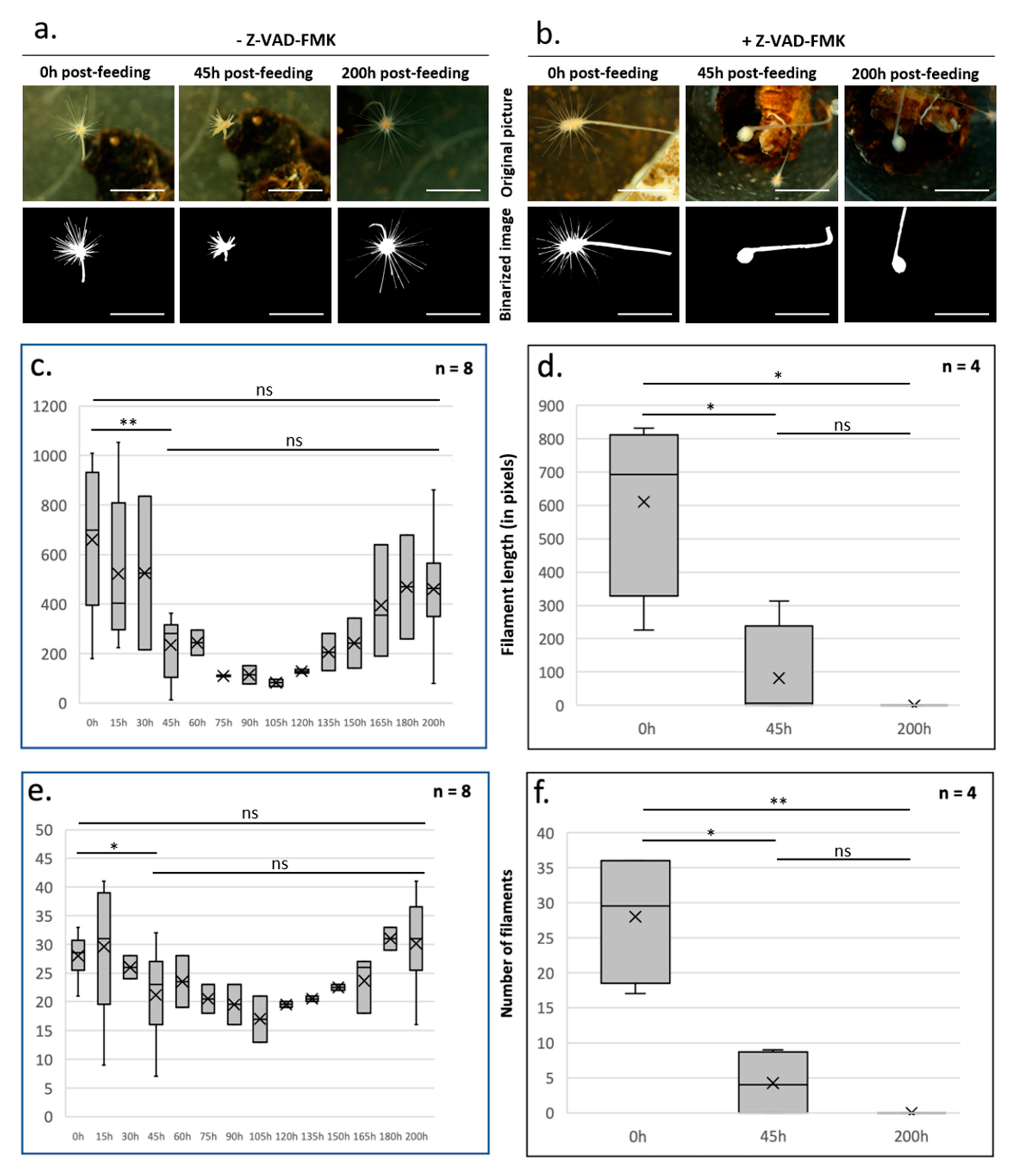
Figure 1. Quantitative approach to investigate caspase-dependent morphofunctional homeostasis during the feeding cycle of Lycopodina hypogea. After 2 months of starvation, a feeding cycle was initiated by putting Artemia nauplii shrimp on the sponge filaments involved in prey capture, in the absence (a,c,e) or presence of Z-VAD-FMK (pan-caspase inhibitor, Euromedex, 1050A) (b,d,f). Sponges were filmed for 72 h, and images were acquired at the indicated times with a camera (SONY E3CMOS) coupled to a stereoscopic microscope (Nikon SMZ-2B) (upper panels). Scale bar: 5 mm. The original images were binarized with ImageJ (lower panels) [7,8,9] for the quantitative analysis, expressed in pixels, of the length (c,d) and number (e,f) of prey capture filaments. Specifically, images were preprocessed to retain only the green channel (Split channel) [10] in which the sponge was better visualized (brightness, contrast, and grain). The step before the morphological analysis (detecting and delimiting the sponge edge and biomass and detaching it from the rest of the image, such as support, water particles, and prey) was image binarization, performed by adjusting the threshold (Adjust threshold) [8,11]. This resulted in an image where the pixels of interest (sponge) had a value of 1 and the background of the image had a value of 0. To permanently remove the background noise, a mask was applied (Clear outside). The last step was to segment and quantify the object with a particle analysis (Analyze particles) [7,8]. The result was the number of pixels that constituted the object. As there is no scale on the image, the image resolution was previously standardized. Knowing the pixel size of each image, the sponge biomass and the filament length were quantified using the pixels as the unit (Measure). In (cf), the box and whisker plots represent the 10th and 90th percentiles, with the first, second (median), and third quartiles. X represents the mean. Error bars indicate the standard error of the mean (n = 8 sponges without Z-VAD-FMK and n = 4 sponges with Z-VAD-FMK); ns, not significant (p value > 0.01); * p value < 0.01; ** p value < 0.001 (t test, R software). The first analysis with the t test confirmed the homogeneity of samples/data in each condition. Then, the analysis compared the two conditions.
Foundations 03 00018 g001
Figure 2. Schematic representation of the main mechanisms involved in tissue homeostasis: apoptosis (a), autophagy (b), and cell cycle regulation (c). Dashed outline and grey color transcripts of proteins not found in any step of the L. hypogea feeding cycle in the transcriptomic study; colored names, transcripts of proteins detected in the transcriptomic study; bold, red, and underlined names, transcripts of proteins expression that is regulated during feeding. Red arrows indicate a decrease or increase of the gene expression level.
Foundations 03 00018 g002
Figure 3. Regulation of apoptosis, autophagy, and proliferation during a feeding cycle in L. hypogea. In unfed animals, proliferation is observed in all parts of the sponge. Apoptosis is mainly present in the peduncle. Autophagy occurs in the peduncle and at the base of filaments (a). During the regression phase after feeding, autophagy is inhibited in regressing filaments, and the peduncle undergoes high levels of apoptosis. Proliferation is still present (b). The regeneration phase is associated with a moderate level of cell proliferation in filaments and a high level of proliferation in the peduncle. No autophagy is observed. Apoptosis is present everywhere but with higher rates at the tip of the filaments and in the peduncle. (c). Examples of immunofluorescent labeling obtained at the end of the filaments are presented together with phase images showing the silica filaments. The sponges were fixed for 30 min with 4% paraformaldehyde in filtered seawater and then permeabilized for 20 min at room temperature with 0.2% Triton X-100 in TS buffer (150 mM NaCl, 25 mM Tris, and pH 7.5). Fixed and permeabilized sponges were labeled with DAPI (Euromedex, 1050A) and TUNEL (Merck, In Situ Cell Death Rhodamine Detection Kit, 12,156,792,910) as previously described [17]. The blue labeling corresponds to a DAPI labeling of nuclei. Red labeling corresponds to TUNEL-positive nuclei. Scale bar: 3 mm (stereoscopic microscope Nikon SMZ-2B images) and 150 μm (confocal microscope Leica TCS-SPE images).
Foundations 03 00018 g003
Figure 4. Accumulation of spermatophores in the prey capture filaments after two months of starvation (a) and during filament regression after feeding (b) in L. hypogea. Fluorescence stereomicroscope (Olympus MC5) images after DAPI staining (Euromedex, 1050A) (a,b). Confocal microscopy (Leica TCS-SPE) image of mature spermatophores on the filament (starved sponge) after DAPI staining of spermatophore nuclei (blue). The sponges were fixed for 30 min with 4% paraformaldehyde in filtered seawater and then permeabilized for 20 min at room temperature with 0.2% Triton X-100 in TS buffer (150 mM NaCl, 25 mM Tris, and pH 7.5). Fixed and permeabilized sponges were labeled with DAPI as previously described [17]. (c) (Montpellier RIO Imaging platform, France). Scale bar: 3 mm (a,b), 35 µm (c).
Foundations 03 00018 g004
Disclaimer/Publisher’s Note: The statements, opinions and data contained in all publications are solely those of the individual author(s) and contributor(s) and not of MDPI and/or the editor(s). MDPI and/or the editor(s) disclaim responsibility for any injury to people or property resulting from any ideas, methods, instructions or products referred to in the content.