Review

Enterococcus and COVID-19: The Emergence of a Perfect Storm?

by
Dan Alexandru Toc
1,*,
Razvan Marian Mihaila
2,
Alexandru Botan
1,*,
Carina Nicoleta Bobohalma
1,
Giulia Andreea Risteiu
1,
Bogdan Nicolae Simut-Cacuci
1,
Bianca Steorobelea
1,
Stefan Troanca
1 and
Lia Monica Junie
1
1
Department of Microbiology, Iuliu Hatieganu University of Medicine and Pharmacy, 400012 Cluj-Napoca, Romania
2
Centre Hospitalier Régional d’Orléans, 14 Av. de l’Hôpital, 45100 Orléans, France
*
Authors to whom correspondence should be addressed.
Int. J. Transl. Med. 2022, 2(2), 220-229; https://doi.org/10.3390/ijtm2020020
Submission received: 30 April 2022 / Revised: 1 June 2022 / Accepted: 6 June 2022 / Published: 7 June 2022
(This article belongs to the Special Issue Translational Medicine Approach against the COVID-19 Pandemic)

Abstract

:
(1) Background: Based on the uncontrolled use of antibiotics and the lack of worldwide-accepted healthcare policies, the COVID-19 pandemic has provided the best premises for the emergence of life-threatening infections. Based on changes described in the intestinal microbiome, showing an increased number of Enterococcus bacteria and increased intestinal permeability due to viral infection, infections with Enterococcus have taken the spotlight in the healthcare setting; (2) Methods: We conducted a brief review in order to analyze the relationship between the two pathogens: the SARS-CoV-2 virus and the Enterococcus bacterial genus. We searched in PubMed, the Cochrane Library electronic database and MedNar and included twenty-one studies based on relevance; (3) Results: The existing studies show a statistically significant difference in the composition of the intestinal microbiome, favoring Enterococcus genus, when compared to a control group. Changes also seem to persist over a period of time, suggesting possible implications for long COVID. Regarding bloodstream infections, Enterococcus is statistically significantly isolated more often when compared to the pre-COVID-19 era, and to a control group of non-COVID-19 patients. (4) Conclusions: The intimate synergy between COVID-19 and Enterococcus has the potential to pose a real threat to human healthcare, and more extensive research is needed to explore the relationship between these two pathogens.

1. Introduction

The COVID-19 pandemic has taken the world on a race against the clock for providing the best diagnostic tools, proper treatment and the implementation of appropriate prevention measures. The implications of this pandemic from a socioeconomic and scientific point of view cannot be easily comprehended, since there is a continuous analysis of its aftermath. Since many aspects are still lacking proper understanding, research in this field continues to provide much-needed information in order to bridge the existing gaps [1,2,3].
The Enterococcus genus provides some of the most dangerous bacterial species involved in human infections, such as Enterococcus faecium (vancomycin-resistant), or the latest, Enterococcus faecium (linezolid-resistant) [4,5,6]. In the past five years, many efforts were executed worldwide in order to develop new drugs able to cure this type of infection or to develop new strategies meant to keep the trend of increased antimicrobial resistance under control. Beside the most notorious species of the genus, such as Enterococcus faecalis and Enterococcus faecium, of notable importance are Enterococcus gallinarum and Enterococcus caseliflavus, which are intrinsic resistant to glycopeptides, due to the vanC gene, but also Enterococcus raffinosus, which has proven to be able to harbor the dangerous vanA gene. New insights in non-faecalis non-faecium enterococcus have shown that almost every species of this genus has the potential to pose a real threat for human health, and continuous research on this niche is necessary [7,8,9,10,11].
An unexpected aftermath of the COVID-19 pandemic is the high number of cases of Enterococcus diagnosed in patients with COVID-19. Many hypotheses have arisen for these findings, but since the physiopathology of the infection with SARS-CoV-2 remains incompletely described, no definitive explanation is available [12,13]. In this paper we aim to untangle the complex interrelation between viral infection with SARS-CoV-2 and the bacterial genus Enterococcus by providing a brief review of the existing medical literature.

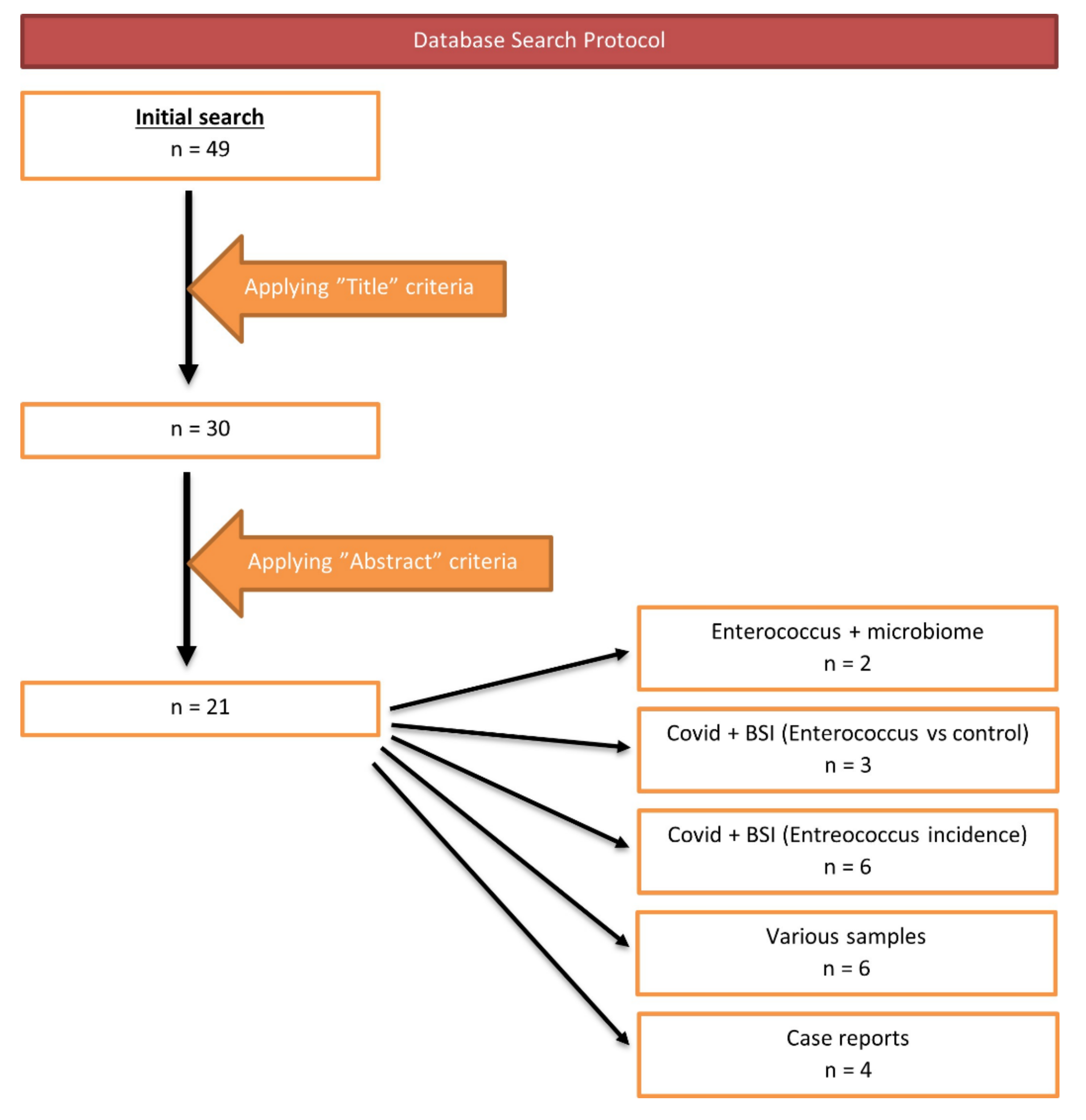
2. Materials and Methods

In order to evaluate the relationship between Enterococcus and COVID-19, we performed a brief review of the existing literature. We searched relevant articles on PubMed, the Cochrane Library electronic database and MedNar, up to the 1st of September 2021.
We considered the following terms included in the studies’ titles or abstracts: “enterococcus” combined with the operator “AND” along with “COVID-19”, “SARS-CoV-2”, “COVID-19 infection” or “SARS-CoV-2 infection”. The search was performed by two individual researchers and the results were confronted afterwards. We excluded duplicates and studies written in languages other than English, French or Spanish.
Figure 1 presents the flow diagram of the search we performed based on the criteria describes above, as well as the number of articles included, and their distribution based on selected criteria.

3. Results

Based on the protocol described above, we included twenty-one studies in our review. We organized them as follows: Enterococcus and bloodstream infections (BSIs), Enterococcus from various samples from COVID-19 patients, Enterococcus and gut microbiota (GM) in COVID-19 patients and Enterococcus and COVID-19 case reports.
Table 1 presents the articles included in our article that involved the study of microbiome in patients with COVID-19. Both studies, published in 2021, used stool samples to describe the changes in the intestinal microbiome that were present in patients with COVID-19.
Table 2 and Table 3 contain the articles that describe BSIs involving Enterococcus in patients with COVID-19. Based on the study design, the control group is usually composed of patients without COVID-19. The diagnostic tools that were used in these studies were different, showing a heterogenous availability of proficient diagnostic tools. Enterococcus faecalis seems to be the specie of the genus Enterococcus most commonly isolated from patients with COVID-19. Studies that described the incidence of Enterococcus in BSIs in patients with COVID-19 showed a variable outcome. Vancomycin-resistant Enterococcus (VRE) remains a constant threat, being described in most included articles.
Enterococcus was also described from other, different samples. Table 4 presents the studies included in our review that evaluate E. faecium and E. faecalis from all the samples.
Table 5 presents the case reports involving the isolation of Enterococcus from different samples from patients with COVID-19. E. faecalis was the species involved in most case reports. The sites of the infections described were representative for most of the common sites of infections for Enterococcus: endocarditis, pneumonia, etc.

4. Discussion

The COVID-19 pandemic has launched the best premises for the development of highly resistant bacterial strains due to unregulated antimicrobial use and the lack of proper worldwide-accepted protocols. Enterococcus genus represents one of the most common findings in human infections. It is no surprise that during the pandemic, a high number of this type of infection was anticipated. However, it is unclear why the number of Enterococcus is so high in COVID-19 patients. The relationship between Enterococcus and the SARS-CoV-2 virus is also unclear.
We wanted to tackle this issue by analyzing the existing articles in a brief review format. Based on the included articles, we tackled the issue of Enterococcus infections in the COVID-19 era in a step-by-step approach. We initially focused on the BSIs (bloodstream infections) involving Enterococcus from an incidence and outcome point of view without ignoring other types of samples. We then compared the findings from microbiome studies. Lastly, we analyzed the existing case reports involving these two pathogens.

4.1. Enterococcus and Bloodstream Infections

Most of the articles in this brief review described Enterococci solely in the context of BSI. In order to better understand the relationship between COVID-19 and BSIs with Enterococcus, we further classified them into articles that compared their findings with a control group (such as patients with influenza, COVID-19-negative patients or a different time period) and articles that simply described the incidence of Enterococcus infections among COVID-19 patients that developed a BSI [16,17,18,19,20,21,22,23,24].
In the first group, one study compared the number of Enterococcus cases within a COVID-19-positive group with the number of cases within an influenza-positive group [16]; another one used two control groups, one consisting of influenza patients and one consisting of just COVID-19-negatives [17]; a third one made a comparison between the incidence of Enterococcus BSI during similar periods, 1 year apart, before and after the start of the pandemic (2019 and 2020) [18]; and the fourth study compared the proportions of BSIs caused by Enterococcus during three different years [23].
No Enterococcus isolates were found from either group of influenza patients from the two studies that analyzed them, so no statistical analysis comparing them to COVID-19 groups could be made. The incidence of Enterococcus BSI among COVID-19-positive patients varied between 0.47% and 2.6% [16,17].
All the BSI studies that used control groups other than influenza found a solid association between Enterococcus BSI and COVID-19. De Voe at al. describe that Enterococcus spp. BSI occurred in 2.6% of COVID-19 patients (8/314 cases—6 E. faecalis and 2 E. faecium), compared to the 0.33% of the COVID-19-negative group (48/14,332 cases), the adjusted odds ratio being 3.75 (95% CI: 1.49–9.41) [17]. Cuntrò et al. compared the incidence of E. faecalis and E. faecium BSIs in an ICU before the pandemic (Feb 22nd to May 21st, 2019) with their incidence during the pandemic (Feb 22nd to May 21st, 2020). What they determined was that there was a substantial increase in the number of E. faecalis cases—28 in 2019 vs. 74 in 2020 (p < 0.001, Fisher’s test), but no significant increase for E. faecium—27 in 2019 vs. 32 in 2020 (p = 0.41, Fisher’s test) [18]. Bonazzetti et al. compared the proportions of BSIs caused by Enterococcus between 2020, 2019 and 2018. The rate was significantly higher in 2020 (71.7% vs. 33.3% and 20%; p = 0.016) [23].
The rest of the studies included in this brief review, involving BSI with Enterococcus, mainly provide an incidence of this infection that varies between 17.2% and 37.5% [19,20,22,24]. Giacobbe et al. and Posteraro et al. went one step further, and described the incidence of the most common species, E. faecium and E. faecalis [19,22]. In those studies, the incidence of E. faecium varied between 3.5% and 8.88%, while E. faecalis remained somehow constant at around 17%. Therefore, we can notice that there could be a tendency for a greater occurrence of E. faecalis cases in COVID-19 patients, rather than E. faecium, which would be consistent with the other earlier findings described by Bonazzetti et al. [23].
Vancomycin-resistant Enterococci were also present within these studies. Most of them report the frequency between 2.38% and 18.1% of all Enterococci [19,20,21]. However, Signorini et al. reported a much higher incidence of VRE − 33.3% [24].
Palanisamy et al. described that 81.8% of Enterococci was MDR: resistance to erythromycin was reported in 90.9% of isolates, to ampicillin and ciprofloxacin in 81.8%, to tetracycline in 54.5% and to teicoplanin in 18.1%. In addition, another relevant observation from this study was the high mortality [20].
As presented so far, Enterococcus seems to be isolated from BSI at much higher rate than before the pandemic, and mortality in cases that involve COVID-19 and Enterococcus BSI is high. Additionally, antimicrobial resistance appears to be an additional threat in this situation. Further analysis is required in order to provide an adequate conclusion.

4.2. Enterococcus from Various Samples from COVID-19 Patients

Six of the studies included in this review analyzed secondary infections in patients diagnosed with COVID-19. It was observed that a wide range of bacterial infections occurred in these patients, notably: Streptococcus pneumoniae, Staphylococcus aureus, Staphylococcus epidermidis, Pseudomonas aeruginosa, Escherichia coli, Klebsiella pneumoniae, Enterococcus faecium, Enterococcus faecalis, Acinetobacter baumannii or Haemophilus influenzae.
Two of the articles emphasized finding Enterococcus not only in samples recovered from patients, but in environmental samples too. In one of them, after detecting five cases of COVID-19 with VRE infections, some tests from environmental sites were made. Eleven cases of VRE were determined from those samples. E. faecium was the bacteria isolated, and further analysis described two clusters of closely related strains. Interestingly, one of these clusters corresponded to COVID-19-positive patients. The other study revealed that in the USA, E. faecalis and E. faecium have emerged as further common nosocomial infections after MRSA, being responsible for 7.4% and 3.7% of all HAIs. The researchers detected isolates of VRE in environmental samples, highlighting what an important role contaminated surfaces have in VRE transmission to COVID-19 patients [25,26].
Serano et al. outlined the descending order of the specimen types depending on the number of positive cultures in secondary infection: blood, endotracheal aspirate, urine, sputum wound swab or bronchoalveolar lavage. Regarding E. faecalis and E. faecium, these were in the first half of frequency scale from positive cultures [33].
Furthermore, Saeed N et al. have shown that these two bacteria occurred in 44 cases of 261 patients with secondary co-infections. The pathological products that were used to find the microorganisms were blood culture, sputum culture, stool culture, endotracheal aspirate and bronchoalveolar lavage culture [29].
Enterococcus has been isolated from a wide variety of pathological products from patients with COVID-19 and, thus, implies an eminent threat for human health. It also raises awareness of the importance of microbiologic diagnosis in order to provide long-term proper healthcare.

4.3. Enterococcus and Gut Microbiota of COVID-19 Patients

Two of the articles focused on the gut microbiota (GM) changes that occurred in patients diagnosed with COVID-19 from Italy and China. Gaibani et al. analyzed stool samples from patients with COVID-19 from three different hospitals in Bologna (Italy), where the results from the next-generation sequencing method were compared with the publicly available sequences from matching sex and age in healthy Italians and critically ill non-COVID-19 patients [14]. Zhou et al. analyzed samples from Wuhan Union Hospital, where the GM of two groups of moderate COVID-19 patients were compared, one of the groups presenting fever (≥37.3 °C) and the other one without fever. Compared to the data prior to the pandemic, an increased number of secondary infections was reported, which led to monitoring the GM of COVID-19 patients. It was decided that it has an important effect on mediating the inflammatory response, favoring the production of pro-inflammatory cytokines, such as IL-6, thus causing fever in virus-infected patients [15].
In both articles, following examination of samples from COVID-19 patients, a disruption to the microbiota homeostasis was observed. Particular to this infection was the reduced diversity of the microbiota (alpha diversity) in comparison to the healthy controls (p value = 0.0008, Wilcoxon test) [14)], with reduced health-associated microorganisms from the Ruminococcaceae, Bacteroidaceae and Lachnospiraceae families, responsible for producing short-chain fatty acids (SCFA), important in human immunological and metabolic homeostasis, featured by an increasing growth of potential pathogens, especially Enterococcus, in addition to Staphylococcus, Lactobacillus and Serratia (p value ≤ 0.02) [14]. There seems to be a causal relationship between Enterococcus spp. and bloodstream infections (BSIs) developed in COVID-19 patients. Two of the dominant species responsible were E. faecalis (1.8%) and E. faecium (8.4%) [14].
An interesting observation was made when analyzing the GM profiles of COVID-19 patients. Those who entered the ICU and developed BSI presented a loss of alpha diversity (p value ≤ 0.004) accompanied by an abundance of Enterococcus (p value ≤ 0.001), compared to the opposite profiles, who did not enter the ICU and did not develop BSI [14]. A similar depletion in alpha diversity was observed in patients presenting fever, in contrast to those without fever. Moreover, fever could be associated with a decreased relative abundance of Bacteroidetes, as well as a significant enrichment of Enterococcus faecalis, Saccharomyces cerevisiae and Haemophilus parainfuenzae, whereas the GM of non-fever patients indicated the growth of Anaerostipes, a butyrate-producing bacteria which suppresses the inflammatory cytokine production. The latter has been observed to be lacking in those with fever [15]. Furthermore, the GM of critically ill patients who tested negative for COVID-19 showed the presence of Enterobacteriaceae (Klebsiella spp.) (p value ≤ 0.001), which mainly distinguished the samples from patients who tested positive for the infection [14].
Due to the ability of SARS-CoV-2 to enter the cells of the digestive tract, therefore leading to enteric manifestations through virus-induced immune-mediated damage, the GM of the infected patients developed an accumulation of opportunistic pathogens or pathobionts, potentially antibiotic-resistant [14]. Consequently, the crossing of the bacteria to the circulation through the damaged intestinal epithelium was facilitated, which is very clinically relevant. Finally, studies have found that GM plays a major role in the pathogenesis of inflammation and the evolution of infection with SARS-CoV-2, controlling the immune responses.

4.4. Enterococcus and COVID-19—Case Reports

There were reported cases of COVID-19 patients presenting a secondary bacterial infection in four of the articles, which is not atypical in the context of infection with SARS-CoV-2.
In one case, a patient presented with the following symptoms: fever, dyspnea and cough. He was later diagnosed with nosocomial pneumonia caused by Enterococcus faecalis and tested positive for COVID-19. It is yet unknown if the pulmonary affection was determined by the bacterial co-infection or by the virus alone [31].
One article outlined a higher incidence of hospital-acquired infected endocarditis (HAIE) between March and April 2020, compared to the correspondent months 5 years prior to the pandemic (p = 0.033). Four cases of HAIE were reported in this period, the pathogenesis of two of them being Enterococcus faecalis, and for the others, Staphylococcus aureus and Candida albicans. The diagnosis was made after analyzing blood or valve culture. All four patients with HAIE underwent central venous and urinary catheterization when admitted to the hospital, which appeared to be the source of infection [32]. Co-infections with bacteria and fungi are a common complication in COVID-19 cases [35]. Another article discussed the case of a patient diagnosed with both COVID-19 and infected endocarditis (IE), who presented nonproductive cough, shortness of breath and fatigue. After a blood culture analysis, the etiology of IE was Enterococcus faecalis. After a persistent bacteremia following antibiotic therapy with vancomycin and gentamicin and an insertion of a new catheter, the patient suffered an aortic valve surgery, which identified a vegetation >10 mm of Enterococcus faecalis. The treatment changed to ampicillin and ceftriaxone [34].
The last case-report article presents a patient who underwent a simultaneous heart-kidney transplant (SHKT) previous to the first cases of SARS-CoV-2 in the respective area. In the weeks following the intervention, he was admitted to the hospital for respiratory failure, open non-healing wounds and multiple secondary infections due to opportunistic bacteria. A perinephric collection showed vancomycin-resistant Enterococcus (VRE) and his blood samples grew out MRSA bacteremia. He was also tested positive for SARS-CoV-2, in spite of being asymptomatic. The patient was immunosuppressed following the procedure, which led to complications [33].

5. Conclusions

In the COVID-19 era, a new threat seems to be emerging: the Enterococcus genus. From BSI, to changes in the microbiome, a new pathogen-to-pathogen relationship between SARS-CoV-2 and Enterococcus seems to take the spotlight. Although the exact mechanism remains unknown, the viral infection seems to cause changes in the bacterial microbiome, favoring Enterococcus and increasing intestinal permeability, which provides the perfect circumstances for Enterococcus bacteria to develop invasive infections. With a constant number of VRE being reported in these articles, the threat of life-threatening infections seems to be higher than ever [36]. Moreover, special attention should be considered regarding long COVID and changes in the microbiome, which also seem to be linked. With COVID-19’s aftermath being constantly evaluated, we might be facing the emergence of a perfect storm, and we should be prepared to tackle it in the best possible way.

Author Contributions

Conceptualization, D.A.T. and L.M.J.; methodology, D.A.T. and R.M.M.; software, R.M.M. and A.B.; validation, D.A.T., R.M.M., A.B. and L.M.J.; formal analysis, D.A.T., C.N.B., G.A.R., B.N.S.-C., B.S. and S.T.; investigation, C.N.B., G.A.R., B.N.S.-C., B.S. and S.T.; resources, C.N.B., G.A.R., B.N.S.-C., B.S. and S.T.; data curation, D.A.T. and A.B.; writing—original draft preparation, C.N.B., G.A.R., B.N.S.-C., B.S. and S.T.; writing—review and editing, D.A.T., A.B., R.M.M. and L.M.J.; visualization, A.B.; supervision, L.M.J. All authors have read and agreed to the published version of the manuscript.

Funding

This research received no external funding.

Institutional Review Board Statement

Not applicable.

Informed Consent Statement

Not applicable.

Data Availability Statement

Not applicable.

Conflicts of Interest

The authors declare no conflict of interest.

References

  1. Joshi, A.; Kaur, M.; Kaur, R.; Grover, A.; Nash, D.; El-Mohandes, A. Predictors of COVID-19 Vaccine Acceptance, Intention, and Hesitancy: A Scoping Review. Front. Public Health 2021, 9, 698111. [Google Scholar] [CrossRef]
  2. Vandenberg, O.; Martiny, D.; Rochas, O.; van Belkum, A.; Kozlakidis, Z. Considerations for diagnostic COVID-19 tests. Nat. Rev. Microbiol. 2021, 19, 171–183. [Google Scholar] [CrossRef] [PubMed]
  3. Van Oosterhout, C.; Hall, N.; Ly, H.; Tyler, K.M. COVID-19 Evolution during the Pandemic–Implications of New SARS-CoV-2 Variants on Disease Control and Public Health Policies; Bellwether Publishing, Ltd.: Columbia, MD, USA, 2021; Volume 12, pp. 507–508. [Google Scholar]
  4. Bender, J.K.; Cattoir, V.; Hegstad, K.; Sadowy, E.; Coque, T.M.; Westh, H.; Hammerum, A.M.; Schaffer, L.; Burns, K.; Murchan, S.; et al. Update on prevalence and mechanisms of resistance to linezolid, tigecycline and daptomycin in Enterococci in Europe: Towards a common nomenclature. Drug Resist. Updates 2018, 40, 25–39. [Google Scholar] [CrossRef] [PubMed]
  5. Egan, S.A.; Shore, A.C.; O’Connell, B.; Brennan, G.I.; Coleman, D.C. Linezolid resistance in Enterococcus faecium and Enterococcus faecalis from hospitalized patients in Ireland: High prevalence of the MDR genes optrA and poxtA in isolates with diverse genetic backgrounds. J. Antimicrob. Chemother. 2020, 75, 1704–1711. [Google Scholar] [CrossRef]
  6. Van Hal, S.J.; Willems, R.J.L.; Gouliouris, T.; Ballard, S.A.; Coque, T.M.; Hammerum, A.M.; Hegstad, K.; Westh, H.T.; Howden, B.P.; Malhotra-Kumar, S.; et al. The global dissemination of hospital clones of Enterococcus faecium. Genome Med. 2021, 13, 52. [Google Scholar] [CrossRef] [PubMed]
  7. Kim, S.G.; Becattini, S.; Moody, T.U.; Shliaha, P.V.; Littmann, E.R.; Seok, R.; Gjonbalaj, M.; Eaton, V.; Fontana, E.; Amoretti, L.; et al. Microbiota-derived lantibiotic restores resistance against vancomycin-resistant Enterococcus. Nature 2019, 572, 665–669. [Google Scholar] [CrossRef]
  8. Toc, D.A.; Pandrea, S.L.; Botan, A.; Mihaila, R.M.; Costache, C.A.; Colosi, I.A.; Junie, L.M. Enterococcus raffinosus, Enterococcus durans and Enterococcus avium Isolated from a Tertiary Care Hospital in Romania—Retrospective Study and Brief Review. Biology 2022, 11, 598. Available online: https://www.mdpi.com/2079-7737/11/4/598 (accessed on 21 April 2022). [CrossRef] [PubMed]
  9. Morrisette, T.; Lev, K.L.; Kebriaei, R.; Abdul-Mutakabbir, J.C.; Stamper, K.C.; Morales, S.; Lehman, S.M.; Canfield, G.S.; Duerkop, B.A.; Arias, C.A.; et al. Bacteriophage-Antibiotic Combinations for Enterococcus faecium with Varying Bacteriophage and Daptomycin Susceptibilities. Antimicrob. Agents Chemother. 2020, 64, e00993-20. [Google Scholar] [CrossRef] [PubMed]
  10. Monticelli, J.; Knezevich, A.; Luzzati, R.; di Bella, S. Clinical management of non-faecium non-faecalis vancomycin-resistant Enterococci infection. Focus on Enterococcus gallinarum and Enterococcus casseliflavus/flavescens. J. Infect. Chemother. 2018, 24, 237–246. [Google Scholar] [CrossRef] [Green Version]
  11. Campanini-Salinas, J.; Andrades-Lagos, J.; Rocha, G.G.; Choquesillo-Lazarte, D.; Dragnic, S.B.; Faúndez, M.; Alarcon, P.; Silva, F.; Vidal, R.; Salas-Huenuleo, E.; et al. A new kind of quinonic-antibiotic useful against multidrug-resistant S. aureus and E. faecium Infections. Molecules 2018, 23, 1776. [Google Scholar] [CrossRef] [PubMed] [Green Version]
  12. Aktas, B.; Aslim, B. Gut-lung axis and dysbiosis in COVID-19. Turk. J. Biol. 2020, 44, 265–272. [Google Scholar] [CrossRef]
  13. Oliva, A.; Miele, M.C.; di Timoteo, F.; de Angelis, M.; Mauro, V.; Aronica, R.; Al Ismail, D.; Ceccarelli, G.; Pinacchio, C.; d’Ettorre, G.; et al. Persistent Systemic Microbial Translocation and Intestinal Damage During Coronavirus Disease-19. Front. Immunol. 2021, 12, 2810. [Google Scholar] [CrossRef] [PubMed]
  14. Gaibani, P.; D’Amico, F.; Bartoletti, M.; Lombardo, D.; Rampelli, S.; Fornaro, G.; Coladonato, S.; Siniscalchi, A.; Re, M.C.; Viale, P.; et al. The Gut Microbiota of Critically Ill Patients With COVID-19. Front. Cell. Infect. Microbiol. 2021, 11, 670424. [Google Scholar] [CrossRef] [PubMed]
  15. Zhou, Y.; Shi, X.; Fu, W.; Xiang, F.; He, X.; Yang, B.; Wang, X.; Ma, W.-L. Gut microbiota dysbiosis correlates with abnormal immune response in moderate COVID-19 patients with fever. J. Inflamm. Res. 2021, 14, 2619–2631. [Google Scholar] [CrossRef]
  16. Hughes, S.; Troise, O.; Donaldson, H.; Mughal, N.; Moore, L.S.P. Bacterial and fungal coinfection among hospitalized patients with COVID-19: A retrospective cohort study in a UK secondary-care setting. Clin. Microbiol. Infect. 2020, 26, 1395–1399. [Google Scholar] [CrossRef] [PubMed]
  17. DeVoe, C.; Segal, M.R.; Wang, L.; Stanley, K.; Madera, S.; Fan, J.; Schouest, J.; Graham-Ojo, R.; Nichols, A.; Prasad, P.A.; et al. Increased rates of secondary bacterial infections, including Enterococcus bacteremia, in patients hospitalized with coronavirus disease 2019 (COVID-19). Infect Control Hosp Epidemiol. 2021, 1–8. [Google Scholar] [CrossRef]
  18. Cuntrò, M.; Manisco, A.; Guarneri, D.; Zuglian, G.; Vailati, F.; Passera, M.; Cavallini, M.; Raglio, A.; Farina, C. Blood stream infections during the first wave of COVID-19. A short microbiological retrospective picture at Papa Giovanni XXIII Hospital, Bergamo, Italy. New Microbiol. 2021, 44, 51–58. [Google Scholar]
  19. Giacobbe, D.R.; Battaglini, D.; Ball, L.; Brunetti, I.; Bruzzone, B.; Codda, G.; Crea, F.; de Maria, A.; Dentone, C.; di Biagio, A.; et al. Bloodstream infections in critically ill patients with COVID-19. Eur. J. Clin. Investig. 2020, 50, e13319. [Google Scholar] [CrossRef]
  20. Palanisamy, N.; Vihari, N.; Meena, D.S.; Kumar, D.; Midha, N.; Tak, V.; Sharma, A.; Bohra, G.K.; Kothari, N.; Dutt, N.; et al. Clinical profile of bloodstream infections in COVID-19 patients: A retrospective cohort study. BMC Infect. Dis. 2021, 21, 933. [Google Scholar] [CrossRef]
  21. Abelenda-Alonso, G.; Rombauts, A.; Gudiol, C.; Oriol, I.; Simonetti, A.; Coloma, A.; Rodríguez-Molinero, A.; Izquierdo, E.; Díaz-Brito, V.; Sanmartí, M.; et al. Immunomodulatory therapy, risk factors and outcomes of hospital-acquired bloodstream infection in patients with severe COVID-19 pneumonia: A Spanish case–control matched multicentre study (BACTCOVID). Clin. Microbiol. Infect. 2021, 27, 1685–1692. [Google Scholar] [CrossRef]
  22. Posteraro, B.; de Angelis, G.; Menchinelli, G.; D’inzeo, T.; Fiori, B.; de Maio, F.; Cortazzo, V.; Sanguinetti, M.; Spanu, T. Risk factors for mortality in adult COVID-19 patients who develop bloodstream infections mostly caused by antimicrobial-resistant organisms: Analysis at a large teaching hospital in italy. J. Clin. Med. 2021, 10, 1752. [Google Scholar] [CrossRef]
  23. Bonazzetti, C.; Morena, V.; Giacomelli, A.; Oreni, L.; Casalini, G.; Galimberti, L.R.; Bolis, M.; Rimoldi, M.; Ballone, E.; Colombo, R.; et al. Unexpectedly High Frequency of Enterococcal Bloodstream Infections in Coronavirus Disease 2019 Patients Admitted to an Italian ICU: An Observational Study. Crit. Care Med. 2020, 49, E31–E40. [Google Scholar] [CrossRef]
  24. Signorini, L.; Moioli, G.; Calza, S.; van Hauwermeiren, E.; Lorenzotti, S.; del Fabro, G.; Renisi, G.; Lanza, P.; Saccani, B.; Zambolin, G.; et al. Epidemiological and Clinical Characterization of Superinfections in Critically Ill Coronavirus Disease 2019 Patients. Crit. Care Explor. 2021, 3, e0430. [Google Scholar] [CrossRef]
  25. Kampmeier, S.; Tönnies, H.; Correa-Martinez, C.L.; Mellmann, A.; Schwierzeck, V.A. Nosocomial cluster of vancomycin resistant Enterococci among COVID-19 patients in an intensive care unit. Antimicrob. Resist. Infect. Control 2020, 9, 154. [Google Scholar] [CrossRef]
  26. O’Toole, R.F. The interface between COVID-19 and bacterial healthcare-associated infections. Clin. Microbiol. Infect. 2021, 27, 1772–1776. [Google Scholar] [CrossRef]
  27. Senok, A.; Alfaresi, M.; Khansaheb, H.; Nassar, R.; Hachim, M.; al Suwaidi, H.; Almansoori, M.; Alqaydi, F.; Afaneh, Z.; Mohamed, A.; et al. Coinfections in patients hospitalized with COVID-19: A descriptive study from the United Arab Emirates. Infect. Drug Resist. 2021, 14, 2289–2296. [Google Scholar] [CrossRef] [PubMed]
  28. Cultrera, R.; Barozzi, A.; Libanore, M.; Marangoni, E.; Pora, R.; Quarta, B.; Spadaro, S.; Ragazzi, R.; Marra, A.; Sagala, D.; et al. Co-infections in critically ill patients with or without COVID-19: A comparison of clinical microbial culture findings. Int. J. Environ. Res. Public Health 2021, 18, 4358. [Google Scholar] [CrossRef] [PubMed]
  29. Saeed, N.K.; Al-Khawaja, S.; Alsalman, J.; Almusawi, S.; Albalooshi, N.A.; Al-Biltagi, M. Bacterial co-infection in patients with SARS-CoV-2 in the Kingdom of Bahrain. World J. Virol. 2021, 10, 168–181. [Google Scholar] [CrossRef] [PubMed]
  30. Calderaro, A.; Buttrini, M.; Montecchini, S.; Piccolo, G.; Martinelli, M.; Dell’Anna, M.L.; Maio, A.D.; Arcangeletti, M.C.; Maccari, C.; de Conto, F.; et al. Detection of SARS-CoV-2 and other infectious agents in lower respiratory tract samples belonging to patients admitted to intensive care units of a tertiary-care hospital, located in an epidemic area, during the italian lockdown. Microorganisms 2021, 9, 185. [Google Scholar] [CrossRef]
  31. Amaral, L.T.W.; Beraldo, G.L.; Brito, V.M.; Rosa, M.E.E.; Matos, M.J.R.; de Fonseca, E.K.U.N.; Yokoo, P.; Silva, M.M.A.; Teles, G.B.D.S.; Shoji, H.; et al. Lung cavitation in COVID-19: Co-infection complication or rare evolution? Einstein (Sao Paulo) 2020, 18, eAI5822. [Google Scholar] [CrossRef] [PubMed]
  32. Ramos-Martínez, A.; Fernández-Cruz, A.; Domínguez, F.; Forteza, A.; Cobo, M.; Sánchez-Romero, I.; Asensio, A. Hospital-acquired infective endocarditis during COVID-19 pandemic. Infect. Prev. Pract. 2020, 2, 100080. [Google Scholar] [CrossRef] [PubMed]
  33. Serrano, O.K.; Kutzler, H.L.; Rochon, C.; Radojevic, J.A.; Lawlor, M.T.; Hammond, J.A.; Gluck, J.; Feingold, A.D.; Jaiswal, A. Incidental COVID-19 in a heart-kidney transplant recipient with malnutrition and recurrent infections: Implications for the SARS-CoV-2 immune response. Transpl. Infect. Dis. 2020, 22, e13367. [Google Scholar] [CrossRef] [PubMed]
  34. Sanders, D.J.; Sutter, J.S.; Tatooles, A.; Suboc, T.M.; Rao, A.K. Endocarditis Complicated by Severe Aortic Insufficiency in a Patient with COVID-19: Diagnostic and Management Implications. Case Rep. Cardiol. 2020, 2020, 8844255. [Google Scholar] [CrossRef] [PubMed]
  35. Toc, D.A.; Costache, C.; Botan, A.; Mihaila, R.M.; Colosi, I.A.; Buksa, S.B.; Chiorescu, R.M. Mixed etiology COVID-19 associated pulmonary aspergillosis (Capa)—a case report and brief review of the literature. J. Fungi 2021, 7, 877. [Google Scholar] [CrossRef] [PubMed]
  36. Toc, D.A.; Butiuc-Keul, A.L.; Iordache, D.; Botan, A.; Mihaila, R.M.; Costache, C.A.; Colosi, I.A.; Chiorean, C.; Neagoe, D.S.; Gheorghiu, L.; et al. Descriptive Analysis of Circulating Antimicrobial Resistance Genes in Vancomycin-Resistant Enterococcus (VRE) during the COVID-19 Pandemic. Biomedicines 2022, 10, 1122. [Google Scholar] [CrossRef]
Figure 1. Flow diagram of the database search protocol.
Ijtm 02 00020 g001
Table 1. Intestinal microbiome.
No.AuthorYearCountrySampleDiagnostic TechniqueCOVID-19 PatientsAdditional Information
1Gaibani, P. et al. [14]2021ItalyStool sampleIllumina MiSeq69The GM of COVID-19 patients showed the enrichment of known or potential opportunistic pathogens, such as Enterococcus, Staphylococcus, Serratia and Collinsella (p value ≤ 0.02)
2Zhou, Y. et al. [15]2021Republic of ChinaStool sampleMagPure Stool DNA KF kit B127Saccharomyces and Enterococcus were significantly enriched in patients with fever
Table 2. BSI (COVID vs. control).
No.AuthorYearCountryDiagnostic TechniqueCOVID-19 PatientsNon-COVID-19 PatientsEnterococcus Spp. of COVID-19 +Enterococcus Spp. of COVID-19 −E. FaeciumE. Faecalis
1Hughes, S. et al. [16]2020UKMALDI-TOF8362161
(0.47%)
---
2DeVoe, C. et al. [17]2021USAMiSeq31414,3328
(2.6%)
48
(0.3%)
2
(0.6%)
6
(1.9%)
3Cuntrò, M. et al. [18]2021ItalyVITEK21911-106
(5.54%)
56
(2.93%)
32
(1.67%)
74
(3.78%)
Table 3. BSI (Enterococcus incidence).
No.AuthorYearCountryDiagnostic TechniqueCOVID-19 PatientsEnterococcus Spp. of COVID-19 +E. FaeciumE. FaecalisVREOther Resistances
1Giacobbe, D.R. et al. [19]2020ItalyVITEK-27812/45 BSI
(26.6%)
4
(8.8%)
8
(17.7%)
1 VRE
(E. faecium)
4/4 E. faecium were ampicillin-resistant (100%)
2Palanisamy, N. et al. [20]2021IndiaVITEK-275011/64 BSI
(17.2%)
--2 VRE81.8% of Enterococci were MDRO.
Ampicillin (81.8%),
Ciprofloxacin (81.8%),
Tetracycline (54.5%),
Erythromycin (90.9%),
Teicoplanin (18.1%)
3Abelenda-Alonso, G. et al. [21]2021Spain-10042/169 isolates
(24.85%)
10
(5.91%)
32 (18.93%)1 VRE-
4Posteraro, B. et al. [22]2021ItalyMALDI-TOF29315/58 BSI
(20.7%)
2
(3.44%)
10
(17.24%)
--
5Bonazzetti, C. et al. [23]2020ItalyVITEK MS MALDI-TOF8953/93 BSI
(55.8%)
26
(27.95%)
26
(27.95%)
5 VRE
(E. faecium)
-
6Signorini, L. et al. [24]2021Italy-926/57 BSI
(10.5%)
--3 VRE-
Table 4. Various samples.
No.AuthorYearCountrySampleDiagnostic TechniqueCOVID-19 PatientsE. FaeciumE. Faecalis
1Kampmeier, S. et al. [25]2020GermanyBlood culture samples and pleural drainageMALDI-TOF-MS33-
2O’Toole, R.F. et al. [26]2021SpainUrine-724-
ItalyBlood culture-78-14
3Senok, A. et al. [27]2021United Arab EmiratesBlood and central-line cultures, endotracheal
aspirates and urine
BioFire FilmArray29,8021018
4Cultrera, R. et al. [28]2021ItalyBlood, urine, or respiratory specimens obtained with bronchoalveolar lavage (BAL) or bronchial aspirate (BASP)/BSIMALDI-TOF by VITEK MS, VITEK 2281014
5Saeed, N.K. et al. [29]2021Kingdom of
Bahrain
Blood culture, sputum culture, stool culture, endotracheal aspirate or bronchoalveolar
lavage culture
MALDI-TOF MS; BD Phoenix2612420
6Calderaro, A. et al. [30]2021ItalyLower respiratory tractMALDI-TOF using a VITEK MS instrument90311
Table 5. Case reports.
No.AuthorYearCountrySampleDiagnostic TechnizqueCOVID-19 PatientsE. FaeciumE. Faecalis
1Amaral, L. et al. [31]2020BrazilNosocomial pneumonia-1-1
2Ramos-Martínez, A. et al. [32]2020SpainBlood or valve culture-2-2
3Serrano, O.K. et al. [33]2020USAPerinephric collection-11 vancomycin-resistant Enterococcus
4Sanders, D.J. et al. [34]2020USAAortic valve culture-1-1
Publisher’s Note: MDPI stays neutral with regard to jurisdictional claims in published maps and institutional affiliations.

