
Biological Impacts of Microplastic Exposure in Zebrafish (Danio rerio): A Systematic Review Across Developmental, Physiological, and Neurobehavioral Endpoints

 ,
,  ,
,  ,
,  , , , ,
Abstract
1. Introduction
- i.
- How does exposure of zebrafish to MPs affect their developmental, physiological, and neurobehavioral endpoints in relation to the underlying toxicity mechanisms, dose-response relationships, and severity of biological impacts?
- ii.
- What are the key methodological constraints, exposure variables (such as polymer type, particle size, concentration, and degree of weathering), and inconsistencies in experimental studies that limit the interpretation of biological impacts and risk assessment in zebrafish models?
2. Materials and Methods
2.1. Search Criteria
2.2. Selection Process (Inclusion and Exclusion Criteria)
2.3. SYRCLE Risk of Bias Assessment
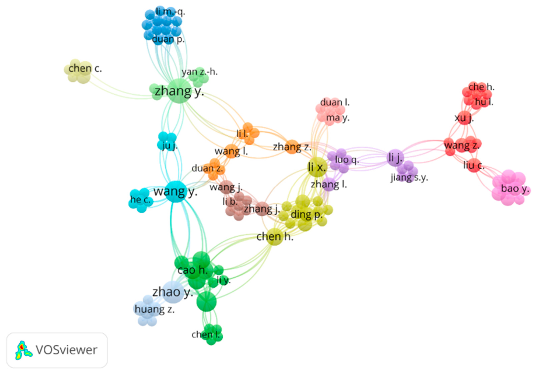
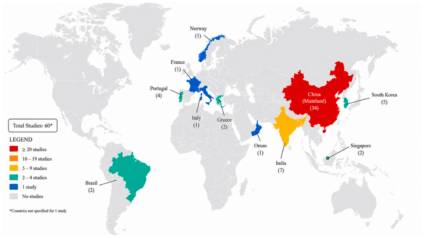
2.4. Bibliometric, Co-Occurrence, Author Coupling Analyses and Geographic Distribution of Published Studies
3. Developmental Anomalies and Consequences of Microplastic Exposure in Zebrafish

| Life Stage | Plastic Characterization (Type/Shape/Size/Concentrations) | Endpoints | Exposure Time | Effects | Ref. |
|---|---|---|---|---|---|
| Adult | PP/Spherical/Virgin: 33.2 ± 14.4 μm/UV: 20.8 ± 10.5 μm/50 mg/L for PP and UV-PP | Survival rate, hatching rate, time to hatch | 14 days | Excretion began immediately after MP removal, GI residual after 1 day: PP = 7.9%, UV-PP = 12.3%, elimination half-life: PP = 0.78 days, UV-PP = 0.38 days, 99.9% excreted after 5 days. | [42] |
| PE & PES/Fragmented beads/average size of 180 ± 210 µm/1 mg/L both PE & PES | Growth and development: body length and survival rate | 96 h (4 days) acute exposure | No significant developmental toxicity. | [43] | |
| PS/5 µm/2 mg/L | Hatching rate (72 hpf), body length (7 dpf), heart rate, and malformation rate (pericardial edema, yolk sac edema, spinal deformity) | 14 days | No recorded mortality for PS MP exposure. | [58] | |
| PE & PES/Fragments/PE mean size: 180 ± 210 µm PES mean length: 350 ± 220 µm/0.2 mg/L, 1 mg/L both PE & PES | Cardiac and developmental: heart rate, pericardial edema, cardiac looping, blood flow abnormalities, survival and hatching | 30 days | No mortality observed, no major difference between 0.2 and 1 mg/L (threshold effect). | [45] | |
| PS/Spherical beads/5 µm diameter/20 µg/L | Growth indices: body weight, body length, condition factor (K = W/L3 × 100) | 21 days | No mortality occurred; final body length and body weight did not change. | [44] | |
| PGA/~1 μm in diameter/1 mg/L & 100 mg/L | Neurochemistry: brain 5-HT system | 28 days | ↓ brain 5-HT both 1 mg/L & 100 mg/L. Serotonin-pathway gene expression: ↑ tph1b while ↓ tph1a. Brain inflammatory gene expression: ↑ (il-1β, tnf-α, il-10) after PGA exposure. | [47] | |
| Embryos | PS/Spherical beads/1, 5 µm/10, 100, 1000 µg/L | Survival rate, hatching rate, time to hatch | 96 h (4 days post-fertilization) | PS (10 µg/L) No significant effects on survival or hatching. Minor but non-significant increase in pericardial edema (5%). PS (100 µg/L) ↓ Hatching rate (~10% reduction); slight increase in tail curvature (8–10%). PS (1000 µg/L) Significant developmental delay; ↓ hatching (−25%), ↓ heart rate (−13% vs. control), ↑ deformities (22% embryos malformed). | [37] |
| Virgin & Photo-aged PS/1 μm diameter/0, 0.1, 1, 10, 100 μg/L for virgin and photo-aged PS MPs | Embryo development and mortality (daily observation); neurotoxicity-focused endpoints: locomotor behavior, neurotransmitter changes, neuronal development and gene expression related to neurotransmission/adipocytokine signaling. | 24 h | Motor neuron development (Tg(hb9-GFP), 120 hpf): A-PS exposure reduced motor neurons in brain and spinal cord relative to control; GFP fluorescence intensity decreased from 238 ± 2.58 AU (control) to 234 ± 2.88 (0.1 μg/L), 232 ± 4.97 (1 μg/L), 230 ± 3.95 (10 μg/L), and 229 ± 2.53 AU (100 μg/L) (p < 0.05). Neurodevelopment- and nervous system-related gene expression (120 hpf): A-PS significantly altered expression of nervous system function/development genes (nestin, gfap, manf, shha, alpha–tubulin, mbp). Relative to control, A-PS significantly inhibited gabra1 and manf, and significantly elevated nestin, gfap, alpha–tubulin, and mbp. | [48] | |
| Virgin & Artificially weathered PP & PS/PS: ~15–36 µm PP: ~50–148 µm; after weathering: (≤230 µm)/Environmentally relevant (particle based): 2000, 20,000, 200,000 MP·L−1; high concentrations (mass based): 12.5, 25, 50, 100 mg·L−1 | Mortality, hatching rate | 96 h (outcomes recorded at 24/48/72/96 h; heart rate at 72 h; length at 96 h) | 2000 MP·L−1: ↓ heart rate, ↓ body length. 20,000 MP·L−1: sublethal growth effects. 200,000 MP·L−1: no linear increase in toxicity. 12.5–100 mg·L−1: no significant embryotoxicity. | [38] | |
| PS/spherical/0.1 μm diameter/0, 0.1, 1, 10, 50, 100 mg/L | Development: cumulative mortality, hatching, malformation (48–96 hpf); morphometrics: yolk sac area, pericardial edema, body length, eye size, heart rate. | 96 h | PS attenuated AgNP toxicity rather than acting as a strong developmental toxicant at the selected dose. | [49] | |
| Embryos → Larvae | Fluorescent plastic microspheres/spherical/1–5 μm/2 mg/L (~1.09 × 10⁸ particles/L) | Developmental and biodistribution: mortality, growth, distribution of MPs in tissues | 2 hpf to 14 dpf | Increased PCNA-positive cells in retina → increased cell proliferation, downregulation of neurogenesis genes (sox2, neuroD, olig2), altered DNMT expression (epigenetic modulation), presence of plastic particles in retina. | [50] |
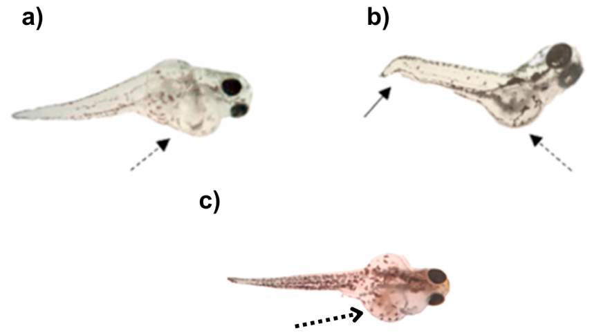
| Not disclosed/MPs: ~1 µm diameter/MPs suspended at 0.006%, 0.0045%, 0.003%, 0.0015% solids | Mortality rate, tail and vascular morphology (larval stage), angiogenesis, growth metric (caudal body length), caudal vein morphometrics, and heart morphology | Exposed for 3 days (1 dpf → 4 dpf) | Developmental toxicity included: pathological changes of caudal vein plexus (angiogenesis abnormalities), caudal tissue impairment and reduced growth/body length, peripheral microcirculation dysfunction (caudal region). Mortality (1 dpf → +1day exposure): mortality from 29.2% (MP1) to 95.8% (MP4), with intermediate values 33.3% (MP2) and 58.3% (MP3). Heart morphology: described as largely transformed (malformed) relative to control zebrafish. | [60] | |
| PS/Fragmented/2 µm/10 mg/L | Developmental toxicity: blood disorder, heartbeat, hatch/death rates, malformations, morphometrics; neurodevelopmental marker: atoh1a expression in cerebellar area (fluorescent reporter) | 24–72 hpf | Developmental toxicity: single PS 157 μm showed no observed effect. Neurodevelopmental toxicity: μ-PS did not change cerebellar fluorescence. | [61] | |
| PGA, PLA, PBS, PHA, PBAT/No data/1 mg/L and 100 mg/L for each polymer | Survival: assessed repeatedly from 3–96 hpf; hatching assessed at 48/72/96 hpf. | 3, 6, 10, 24, and 96 hpf | Early morphology (3–95 hpf): no significant morphological changes at 6, 10, and 24 hpf across groups. Survival (96 hpf): survival rate significantly decreased in mg/L PHA and 1 mg/L PBAT, and in 100 mg/L PGA, PLA, PBS, and PHA groups. Hatching: at 48 hpf, hatching rates were significantly increased in high-concentration MP groups; at 72 and 96 hpf, hatching showed a decreased trend. Larval morphometrics (96 hpf): no significant malformations at 96 hpf, but body length and head area were markedly reduced in all exposure groups except 1 mg/L PGA; eye area decreased except in 1 mg/L PGA and 1 mg/L PBAT. Retinal histology (5 dpf): IPL thickness was significantly reduced in 1 mg/L PGA and 100 mg/L PBAT; ONL thickness significantly decreased in 1 mg/L PBS and 1 mg/L PBAT; RGL thickness significantly decreased in all treatment groups. Eye/retina gene expression (5 dpf): 100 mg/L PGA, PLA, and PBAT significantly decreased pax6a, pax6b, rx1, gnat2, grk1b, and opn1mw1; 100 mg/L PBS increased pax6b, gnat2, grk1b, and opn1mw1 but reduced rx1; 100 mg/L PHA increased pax6a, gnat2, and grk1b but reduced rx1. | [62] | |
| PVC/mean size ~250 µm/100, 200, 300, 400 ppm | Phenotype endpoints: survival, hatching, edema, tail malformation. | 30 days | Survival: no mortality reported through 120 h post-fertilization (hpf) across all exposure scenarios (MP-only, phenanthrene-only, co-exposure). Hatching: hatching rate was reported as not affected, including under the study’s “extremely high” exposure conditions. Morphology/teratogenic endpoints (monitored at 48, 72, 96, 120 hpf): edema rate and tail malformation rate were recorded as developmental indicators.
| [54] | |
| PET/irregular fragments/Average 30–100 µm/1 mg/L & 10 mg/L | Growth (length/weight) | 0 to 96- or 120-h post-fertilization (hpf) | Significant growth alterations. | [55] | |
| Virgin & weathered PS & PE/spherical/virgin 10 µm and 30 µm; weathered PS (lab prepared, ~1–7 µm range) and weathered PE (lab prepared, ~1–10 µm range)/virgin MPs targeted ~105–106 particles/L, Weathered MPs targeted ~104 particles/L | Survival/mortality, teratogenic outcomes (spinal/tail defects, edema) | 10 days | Virgin PS: 10–20% (PS 30 µm ~10%; PS 10 µm ~20%; not significant vs. control), weathered MPs: ~80–82% (weathered PS ~80%; weathered PE ~82%; highly significant). Malformations: weathered groups: spinal malformations ~13–14%, bent tails ~21%, significantly higher than control, virgin PS groups: low/non-significant malformations. | [39] | |
| PS/microsphere/5 μm/1 mg/L | Mortality/survival and body length | 2 h post-fertilization (hpf) and continued to 7 days post-fertilization (dpf). | Embryo hatching: no significant effect on hatching. Heart rate: no significant effect on heart rate. Mortality: increased embryo and larval cumulative mortality during the 7-day exposure Growth (body length): no significant effect on growth. | [45] | |
| Larvae | PS & PVC/spherical/PS (spherical ~7.0 µm mean size) PVC (spherical ~3.8 µm mean size)/both MPs 20 mg L−1 | Survival and development: daily mortality, morphological abnormalities | 10 days | PS MPs alone: higher mortality than PVC (10–20%), strong locomotion suppression. PVC MPs alone: moderate mortality, moderate suppression. | [57] |
| PS/virgin & photo-aged/spherical beads/1 µm/0, 1, 10, and 100 µg/L both virgin & photo-aged | Growth and development: body length and survival rate | 96 h post-fertilization (hpf) | More strongly inhibited larval growth, and ↓ survival rate in photo-aged PS. | [63] | |
| PS/microbeads/5 ± 3 μm & 50 ± 3 μm/10 mg/L at 5 μm (MPs-5) or 50 μm (MPs-50) | Growth indices: body weight, body length, condition factor (K = W/L3 × 100) | Up to 96 hpf | Developmental endpoints: PS-MPs 10 mg/L (5 μm): malformation 31.3%; mortality 11.1%; hatching 72 hpf 72.2%, 96 hpf 87.5%; PS-MPs 10 mg/L (50 μm): malformation 27.8%; mortality 8.33%; hatching 72 hpf 68.1%, 96 hpf 88.9%. | [56] | |
| Virgin & UV-aged PA/irregular fragments/~5 µm (mean particle diameter)/0, 10, 100, and 1000 µg/L | Growth and development: body length and survival rate | 2 days post-fertilization (dpf) → 10 dpf | 10 (µg/L): Slight growth inhibition (photo-aged > pristine). 100 (µg/L): reduced body length; intestinal structural changes. 1000 (µg/L): significant growth inhibition; severe intestinal damage; impaired lipid adsorption; photo-aging significantly enhanced PA toxicity. | [48] | |
| Virgin & photo-aged PS/virgin: 10 μm Photo-aged: 6.5 μm/0.1–100 μg/L for both V-PS and P-PS. | Neurotransmission: neurotransmitters (5-HT, GABA, DA, ACh); enzymes (AChE, ChAT, ChE) | 1 hfp to 120 hpf | V-PS significantly increased neurotransmitter levels and cholinergic enzyme activity; response trends with concentration and IBR weighting toward neurotransmitter disruption; only V-PS has the effect on the neurotransmitter level of the zebrafish. | [47] | |
| PS/~25 μm in diameter/25, 250 μg/L | Neurobehavior: light–dark locomotor response (movement distance, max acceleration, average velocity); neurodevelopment genes: gap43, α1-tubulin | Early dpf stages | No significant change in neurobehavioral or neurodevelopment-related gene expression (p > 0.05). | [64] |
4. Physiological and Oxidative Response of Zebrafish Exposed to Microplastics

| Life Stage | Plastic Characterization (Type/Shape/Size/Concentrations) | Endpoints | Exposure Time | Effects | Ref. |
|---|---|---|---|---|---|
| Adult | PE/Spherical/Average diameter of 40 ± 10 µm/100 µg/L | Oxidative stress biomarkers: SOD, CAT, GPx, MDA, T-AOC. | 21 d | 100 µg/L: Slight oxidative stress (↑ SOD, CAT activities); minor intestinal villi damage. | [80] |
| PS/Spherical beads/1 μm & 3 μm/0.01, 0.1, 1.0, 10.0 mg/L | Cardiac physiology (heart rate at 72 h); Redox homeostasis/oxidative stress biomarkers. | 96 h (4 days post-fertilization) | ↑ in heart rate both concentrations with more strongly at 10 mg/L. ↓ ROS content at 10 mg/L; ↑ Lipid hydroperoxides but more significant in 10 mg/L. Antioxidant enzymes: GPX: Unaffected; GR: ↑ significantly only in 1 mg/L; SOD: ↑ significantly only in 10 mg/L. | [37] | |
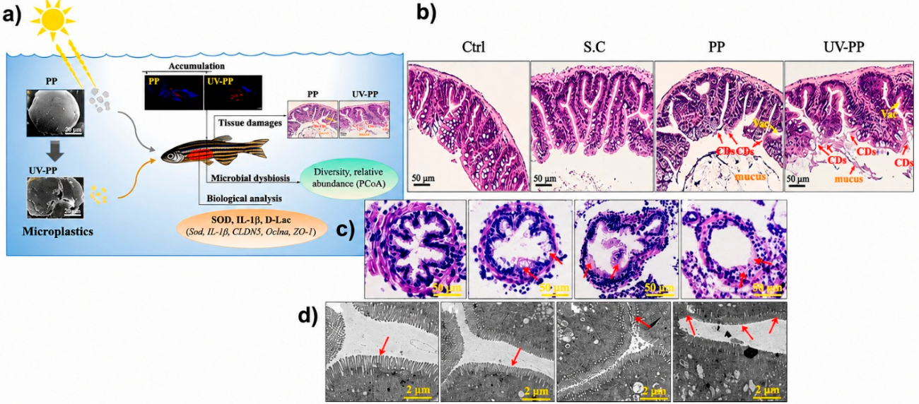
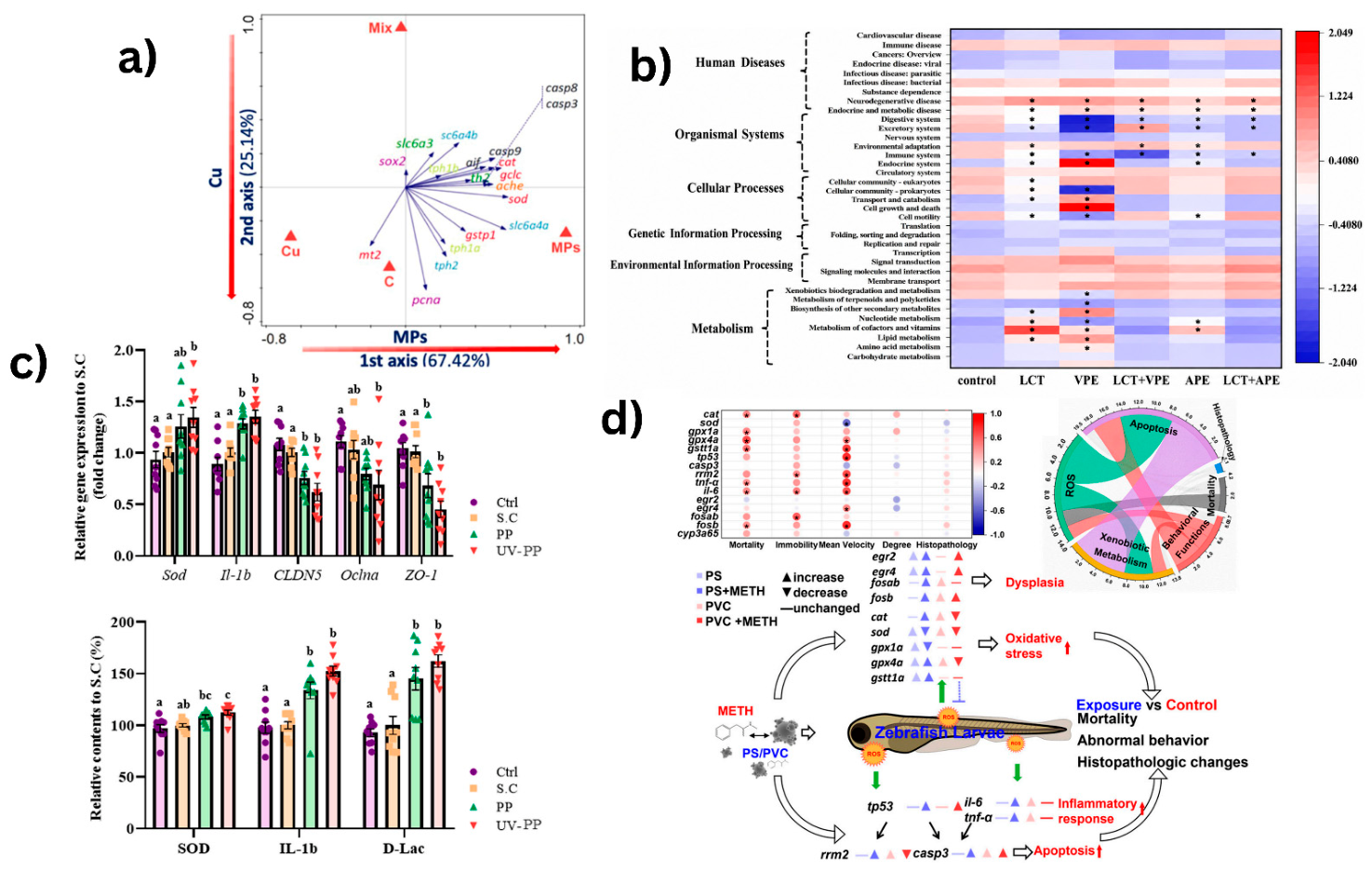
| Virgin & UV PP/Virgin: 33.2 ± 14.4 μm UV: 20.8 ± 10.5 μm/50 mg/L both V & UV | Histopathology: Intestinal villi length, mucus secretion, goblet cell number, vacuolization, ciliary defects. Gene expression: Sod1, IL-1β, CLDN5, Oclna, ZO-1. Enzyme biomarkers: SOD, IL-1β (inflammation), and D-lactate (intestinal permeability). Gut microbiome: Alpha diversity (Chao1, Shannon), Beta diversity (PCoA, UniFrac), OTU abundance, taxonomic shifts at phylum & genus levels, KEGG pathway analysis using PICRUSt & MinPath. | 14 d | Tissue distribution and accumulation: Both V & UV weathered PP particles were detected in the GI tract, liver, and gills. Peak GI burden (day 3): PP = 383.4 ± 50.3 particles; UV-PP = 2053.7 ± 371.4 particles. Excretion kinetics (GI): Elimination half-life PP = 0.78 d; UV-PP = 0.38 d; after 5 days of depuration, both MPs reached 99.9% excretion. Intestinal histopathology: Exposure to PP and UV-PP produced mucosal damage characterized by structural damage, vacuolization, ciliary defects, mucus secretion, and reduced goblet cells. Goblet cells: Decreased by 34% (PP) and 51% (UV-PP). Oxidative stress/inflammation markers (gut): SOD mRNA upregulated significantly in UV-PP; il-1β mRNA upregulated significantly in both PP and UV-PP vs. S.C; SOD and IL-1β enzyme-level changes were consistent with gene expression patterns. Tight junction/barrier markers: cldn5 and zo-1 transcription downregulated in PP and UV-PP; D-lactate (D-Lac) increased. Gut microbiota diversity and composition: Shannon index increased significantly in the PP group; PCoA indicated microbiome shifts after PP and UV-PP exposure. Differential taxa (reported comparisons): Rhizobium, Gemmobacter, and Cloacibacterium were significantly higher in PP than UV-PP; Luteolibacter and Rodobacter were significantly higher in UV-PP; Porphyromonadaceae and Aeromonas tended to decrease in MP-exposed groups. | [41] | |
| Virgin MPs (propriety polymer)/Spherical/1–5 μm/2 mg/L (~1.09 × 10⁸ particles/L) | Biochemical biomarkers: ROS, LPO, SOD, CAT, GPx, GST, GR, GSH/GSSG, LDH, AChE, and MT. Molecular responses: Lipid metabolism genes (fabp, apoa1, etc.), and intestinal barrier genes (zo-1, claudin). | 2 hpf to 14 dpf | Significant inhibition of acetylcholinesterase (AChE) activity compared with control, indicating neurotoxicity. AChE inhibition showed correlation with mortality increase and reduced growth. Other biomarkers such as ROS, LPO, SOD, CAT, GST, GR, GSH/GSSG, LDH, significantly affect the overall physiological capacity of the zebrafish. | [65] | |
| PE & PES/Fragmented beads/Average size of 180 ± 210 µm/1 mg/L both PE & PES | Molecular responses: Lipid metabolism genes (fabp, apoa1, etc.) and intestinal barrier genes (zo-1, claudin). Oxidative stress: ROS production, antioxidant enzymes (SOD, CAT), lipid peroxidation (MDA). | 96 h (4 days) acute exposure | PE exposure: Significantly altered lipid metabolism in the intestine and liver, and perturbation of lipid metabolism, fatty acid metabolism, vitamin metabolism, TCA cycle, and amino acid metabolism. PES exposure: Upregulated metabolites included phosphocholine and 2-lysophosphatidylcholine; and downregulation metabolites included triglyceride, 13-HDoHE, n-triacontanol, and phosphatidylserine. | [43] | |
| Propriety polymer (composition undisclosed/Spherical/1–5 µm/2 mg/L | Molecular responses: Oxidative stress-related genes, detoxification-related genes. Metabolomics: Whole-body metabolomics, LC-MS/MS (QTOF) untargeted metabolomics. | 30 d | MPs (2 mg/L): ↑ Metallothionein (MT), ↑ tph1a (serotonin synthesis gene), ↓ LDH (metabolic alteration). Gut microbiota: Increased; Fusobacteria, and Protobacteria; Decreased: Firmicute; Clear dysbiosis in both PE and PES groups. | [64] | |
| PS/Microbeads/200 µm, 40 µm, 10 µm/100 µg/L | Biochemical biomarkers: ALT, AST (hepatic injury), SOD, CAT, GSH (oxidative stress), integrated biomarker response (IBR). | 30 d | MP exposure resulted in liver pathological changes, wherein liver damage included the presence of ballooning of the hepatocytes (vacuolization), nuclear abnormalities (including the formation of pyknotic or peripherally located nuclei), and the presence. CAT activities decreased with the decrease of MPs size, and the smallest MP group showed a significant decrease in CAT under single exposure. In the MP single-exposure groups, SOD activity increased with decreasing plastic size. GSH content significantly rose under the single MP treatments, but the magnitude of GSH increase showed a decline with a decrease in MPs size. | [73] | |
| PS/5 µm/2 mg/L | Oxidative stress biomarkers: Superoxide dismutase (SOD), catalase (CAT), glutathione peroxidase (GPx), malondialdehyde (MDA). Histopathology: Liver tissue, hematoxylin & eosin (H&E) staining, hepatocyte vacuolization, cellular degeneration, histopathological scoring. Gene expression (qRT-PCR): Metallothionein (mt2), antioxidant-related genes, apoptosis-related genes. | 14 d | Gills (Histopathology): There were no histopathological changes in the gills of the fish that were exposed to not contaminated. Intestine (Histopathology): Intestinal changes are minor, including cracking of villi, caused by MPs alone. Liver (histopathology): There was no obvious impact of MPs alone on histopathology. | [44] | |
| PE & PES/Fragments/PE mean size: 180 ± 210 µm PES mean length: 350 ± 220 µm/0.2 mg/L, 1 mg/L both PE & PES | Whole-body untargeted LC-MS/MS metabolomics to quantify MP-associated shifts in molecular metabolites and enriched metabolic pathways. Gut microbiome physiology and gut microbiota profiling after MP exposure. | 30 d | PE-specific metabolomic changes: 0.2 mg/L and 1 mg/L overlapped with minimal separation, while both separated from controls. Both concentrations had no significant different and does not change metabolomics of zebrafish. Gut microbiome (16S metagenomics): Community composition: Dominant phyla included Proteobacteria, Fusobacteria, Firmicutes, Actinobacteria, Bacteroidetes, and Verrucomicrobia; Fusobacteria increased (0.3–11.7%) in microplastics-exposed groups vs. control, while Proteobacteria decreased (0.4–9.0%) across exposures vs. control. Potential pathogens were reported only in exposed groups (examples: Mycobacterium in PES2; Aeromonas in PE1/PE2). Diversity: Alpha diversity indices (observed OTU, Shannon, Faith FD, Simpson) were reported as not significantly altered across exposure groups. PE: Physical epithelial damage, oxidative stress, inflammation, disrupted membrane and energy metabolism. PES: Fibers interfered more strongly with gut microbiota interactions, lipid digestion, and endocrine-related metabolism. | [45] | |
| PVC & PP/Irregular fragments/5–50 µm/PVC MPs (100 µg/L), and PP MPs (100 µg/L) | Intestinal histopathology (H&E); intestinal inflammation biomarkers/gene expressions: TNF-α levels and IL-1β gene expression. Oxidative stress biomarkers/antioxidant-related expression; intestinal metabolomics; and intestinal microbiota | 21 d | Intestinal histopathology (H&E): Control intestines showed intact villi shape and regularly arranged epithelial cells. PVC-only and PP-only groups showed slight shedding of intestinal villi. Intestinal inflammation biomarkers/gene expression: TNF-α levels significantly increased in all treatment groups except the cadmium-only group; this includes PVC-only and PP-only. IL-1β gene expression significantly increased in all treatment groups; this includes PVC-only and PP-only. Oxidative stress biomarkers/antioxidant-related expression: No remarkable shifts in SOD activity or MDA levels were reported after exposure to PVC alone or PP alone. PP (but not PVC) significantly increased GPx expression (detoxification-related). Intestinal metabolomics: PVC-only: 32 significant differential metabolites downregulated and 24 upregulated vs. control. PP-only: 85 significant differential metabolites downregulated and 10 upregulated vs. control. KEGG pathway enrichment: both PVC-only and PP-only showed disturbances in purine metabolism and arginine/proline metabolism. PVC-only additionally disrupted primary bile acid biosynthesis and arachidonic acid metabolism. PP-only additionally affected arginine biosynthesis and glutamate metabolism. Intestinal microbiota: At the phylum level, Firmicutes relative abundance increased in PVC-only and PP-only groups. Proteobacteria relative abundance increased in all treatment groups, including PVC-only and PP-only. | [65] | |
| PS/Spherical beads/2 μm diameter/0.44 mg/L (~108 items/L) | Oxidative biomarkers: SOD, CAT, GSH, and MDA | 7 d | Ocular oxidative stress biomarkers (eyes; ELISA; normalized to protein): MP exposure showed no significant main effect on SOD, CAT, GSH, or MDA in the model analysis (p > 0.1). GSH specifically: The MP-only group had significantly higher ocular GSH than the control group. MDA: No significant difference was detected in ocular MDA among groups. | [66] | |
| PS/Spherical beads/2–4 µm diameter/440 µg/L | Oxidative stress biomarkers: SOD, CAT activities, MDA content, and protein normalization. Histopathology: Villus number, villus height and width, and intestinal wall thickness; and gut microbiota analysis. | 21 d | PS beads were observed in the gut at day 1 and day 21 (control showed only autofluorescence). PS distributed along anterior–middle–posterior gut and tended to accumulate in the mid-posterior gut and/or be excreted with fecal pellets. Oxidative stress/anti-oxidant response: CAT activity was significantly increased in the PS group compared with the control group (and AMI group). SOD activity was not reported as significantly increased in the PS group (significance was reported for the PS + AMI group vs. control/PS/AMI). MDA was not reported as significantly increased in the PS group (the significant change reported was a decrease in the AMI group vs. control/PS). Morphometrics: Villus width was significantly increased in the PS-MP group; intestinal wall thickness was significantly decreased in the PS-MP group. The number of villi showed decreasing tendency in the PS-MP group. Gut microbiota: PS-MPs showed an increasing tendency in Proteobacteria, which includes Exiguobacterium, Candidatus paracaedibacter, and Staphylococcus. | [69] | |
| PS/Spherical beads/5 µm diameter/20 µg/L | Hepatic biochemical parameters: Energy metabolism: glucose, pyruvate, Lipid metabolism: TG, T-CHO, LDL-C, NEFA, oxidative stress: SOD, CAT, GSH, MDA Glycolipid metabolism genes: Glycolysis/gluconeogenesis: Gk, Hk1, Pepckc, β-oxidation: Aco, Cpt1, Ppar-α, Lipid synthesis: Acc, Fas, Ppar-γ, Lipid transport: Apo, Fabp6. Oxidative stress genes: Mn-sod, Cu/Zn-sod, Cat, Nrf2, Keap1, Gpx, Bcl2; Inflammatory genes: IL-1β, IL-6, IL-8, TNF-α, IFN, C3, IL-10. | 21 d | Hepatic metabolism: PS: ↓ pyruvate, TG, T-CHO. PS specific significantly changes: Hepatic pyruvate, TG, and T-CHO with significant decreased; PS had no significant effect on NEFA. Hepatic glycolipid metabolism genes: Based from the results, PS had no significant effect on glycolipid metabolism genes. Oxidative biomarkers: CAT activity had a significant increase in PS exposure vs. control.; the GSH content was reduced after 21 days of exposure; oxidative stress-related mRNA such as Mn-sod, Nrf2, Keap1, Gpx, and Bcl2 mRNA levels were significantly inhibited in PS-MPs. | [46] | |
| Virgin & Photo-aged PLA/~100 µm/5 mg·L−1 | Female reproductive endpoints: Gonadosomatic index (GSI), ovary histology (H&E), sex hormones: Testosterone (T), estradiol (E2), Emphasis on E2/T; ovary metabolomics. | 5 w | DPLA > UPLA toxicity, ovarian structural damage, disrupted steroid hormones, altered metabolomic pathways, offspring: ↑ mortality, ↓ hatching, ↓ body length. | [48] | |
| PE/Spherical/146.2 ± 8.9 µm/5 µg/L and 50 µg/L | Oxidative stress & antioxidant biomarkers: Catalase (CAT), glutathione-S-transferase (GST), lipid peroxidation (MDA); ion regulation: Na+/K+-ATPase activity (gills). | 10 d & 20 d | 5 µg/L: 10 days: ↓ CAT and GST (liver); 20 days: ↑ GST activity (adaptive response), ↑ lipid peroxidation (brain). 50 µg/L: Stronger oxidative stress than 5 µg/L, significant ↑ MDA (brain), marked ↑ Na+/K+-ATPase activity in gills. | [70] | |
| PS/Spherical beads/1 µm/30 mg/L | MP distribution (gills vs. gut), gut histopathology, oxidative stress enzyme, liver metabolomics, gut microbiota (16S rRNA), sex hormones, and reproductive output. | Chronic (multi-week). | Females accumulated more PS, gut microbiota dysbiosis stronger in females, altered hepatic lipid & energy metabolism, reduced egg production, clear sex-specific toxicity. | [71] | |
| PS/Fragmented/5 µm/20 & 200 mg/L | Cd accumulation (liver, gut, gill), oxidative stress enzymes, histopathology, inflammatory gene expression. | Chronic (multi-week). | MPs enhanced Cd bioaccumulation, there was an increased trend in oxidative damage and inflammation synergistically increased, MPs acted as toxic vector. | [72] | |
| PS/Fragmented/5 µm/LMPs: 0.1, 1, 10, 50, 100 mg/L; SMPs: 0.1, 1, 10, 50, 100 mg/L | Antioxidant enzymes: SOD, CAT, GPx, lipid peroxidation (MDA). | 96 h post-exposure | SOD activity: At day 4, PS-MP exposure significantly increased hepatic SOD. CAT activity: At day 4, CAT activity was significantly higher and remained significantly higher at day 8. GPx: At day 4, GPx activity was significantly higher and remained significantly higher at day 8. Lipid peroxidation (MDA): PS-MPs had no significant effect on hepatic MDA at day 4 but evidently reduced at day 8. | [49] | |
| PS/Not disclosed/0, 50, 500 µg/L | Gene expression: qPCR normalization to β-actin, ovarian histology: (oocyte stage scoring; H&E), oxidative stress markers: MDA (lipid peroxidation), SOD, CAT. | 60 d | 50 and 500 µg/L: Decreased ovarian SOD/CAT activity (oxidative stress), increased NO, increased apoptosis; dose response noted (500 µg/L highest TUNEL-positive), all dosage groups: MDA boosted (membrane damage). | [74] | |
| PS/Spherical/2 µm/0.1 mg/L and 1.0 mg/L | Endpoints measured: Thyroid axis & maternal transfer: co-exposure increased maternal transfer of T3 and T4, and reduced thyroid hormones in the “F2 generation.” | 63 d | Results: “At both concentrations” (0.1 and 1.0 mg/L), µ-PS exacerbated acetochlor-induced reductions in thyroid hormones and promoted maternal transfer. | [75] | |
| PE/Not disclosed/60 mg/L | Genotoxicity/cytotoxicity (blood cell biomarkers): Increased nuclear abnormalities, changes in erythrocyte and nuclear size/shape (mutagenic + cytotoxic signals), morphometric RBC nuclear/shape endpoints are shown in later figures (e.g., elongation/circularity). | 10 d | Results: “At both concentrations” (0.1 and 1.0 mg/L), µ-PS exacerbated acetochlor-induced reductions in thyroid hormones and promoted maternal transfer. Genotoxicity (erythrocytes; comet assay): The DNA damage index was ~64% higher than control, and tail intensity increased by >60% relative to unexposed fish. Hydrogen peroxide (H2O2): PE-MP exposure was associated with higher H2O2 in brain (vs control) and higher H2O2 in liver (PE-MPs alone). In gills, PE-MP exposure showed reduced H2O2 versus control (this reduction was reported for PE-MPs alone). | [79] | |
| PSE/Spherical/100 µm/40.1 µg/L | Endocrine profile: Serum LH, FSH, and β-estradiol (E2) quantified by ELISA; kit ranges provided; absorbance read at 450 nm for E2; metabolomic/oxidative stress markers: Measured via glucometer; molecular biomarkers (qPCR). | 21 d | Ovary testosterone: PS-MP increased ovarian testosterone by 75% vs. control; brain testosterone: PS-MP increased brain testosterone by 39.3% vs. control after 21 days. ovarian histology and oocyte maturation arrest (PCOS-like morphology): PS-MP ovaries: More developing immature (stage I/II) follicles and significantly fewer mature follicles (interpreted as chronic anovulation-like). Oxidative stress: MDA was significantly higher in PS-MP than control (direction/significance stated), with PS-MP MDA reported as 0.015 μM/mg ovarian tissue (LET: 0.104 μM/mg). | [76] | |
| PGA/~1 μm in diameter/1 mg/L & 100 mg/L | Gut barrier/intestinal permeability: Regulation downstream of Wnt/β-catenin; supported by qPCR, ELISA, tissue section analysis; gut microbiome: 16S rRNA sequencing; liver injury + histopathology and molecular assays (qPCR/ELISA) connecting gut disruption to systemic effects. | 28 d | 1 mg/L PGA MPs (28 d): Evidence of gut barrier disruption, microbiota dysbiosis, and behavioral/neurochemical disturbance (anxiety-like and impaired cognition/visual preference; altered 5-HT system). Wnt/β-catenin pathway genes (intestine): Low PGA reduced wnt-4a, high PGA increased wnt-4a and wnt-10b expression, while both doses reduced gsk3β and dkk1 expression, and high PGA increased β-catenin expression. Taxonomic changes (phylum-level trends): Reduced Proteobacteria, and increased Fusobacteria and Bacteroidetes after PGA exposure. | [47] | |
| PE/Spherical/25 μm 100 μg/L | Endocrine biomarkers (adults; sex hormones + VTG): Analytes: Estradiol (E2), testosterone (T), 11-keto testosterone (11-KT), vitellogenin (VTG); Genes: 17 HPGL-axis related genes listed in the paper (e.g., gnrh2/3, gnrhr2/3, lhβ, fshβ, fshr, lhr, star, cyp11a, cyp19a/b, erα/β, ar, vtg1, etc.); qPCR approach: SYBR Green real-time PCR; three biological replicates per treatment (as stated for this assay) | 35 d | Pathological changes described including the loss of contact between oocyte membrane and follicular cell layer, yolk cell breakdown, and cell lysis; gnrhr2/gnrhr3: significantly reduced in all groups except MP group; additional gene-level guidance is mentioned in the text (e.g., cyp11a, lhr, erα, vtg downregulation; lhβ and erβ upregulation with MA significant for erβ; and MA-specific trends for cyp19a/cyp19b/star). | [78] | |
| PE/Microsphere/10–300 μm/0.1, 2, and 300 mg/L | Primary endpoints: Uptake, accumulation, and elimination of MPs (counts of fluorescent microspheres) in daphnia and zebrafish; fish tissues assessed for MPs: gill and digestive gland; feces collected for removal quantification in pathway experiment. | 72 h | MP10 (0.1 mg/L): only a few particles at 1 h and only individual particles detectable at 12 h; interpreted by authors as inadvertent ingestion. (2 mg/L): accumulation peaked at 6 h with 163.18 particles/fish; then declined with time. MPs in gut (67.39% at 6 h; 53.85% at 12 h), while Exp2–Exp4 showed most MPs in feces; after 12 h in Exp2–Exp4, fecal proportions ranged ~87.86% to 98.22%. | [79] | |
| PE/Not disclosed/PE-MPs: VPE or APE at 1 mg/L (non-lethal; LC50 of MPs alone > 100 mg/L) | Oxidative stress: Enzymes: SOD, CAT; histopathology; immune gene expression: Genes: TLR-2, TLR-4, MyD88, NF-κB, NF-κB1, c-Rel, TNF-α, TNF-β, IL-1β; immune protein: NF-κB; gut microbiome. | 35 d | Virgin polyethylene microplastics (VPE) and aged polyethylene microplastics (APE) caused significant changes in the intestinal microbial community. Both VPE and APE increased the relative abundance of Proteobacteria and reduced Fusobacteria. Oxidative stress responses: Lipid peroxidation (MDA) was increased in the APE group (~3.99%). Antioxidant enzyme activities indicated compensatory responses: Superoxide dismutase (SOD) activity was increased in VPE-treated fish. Catalase (CAT) activity indicated adaptive responses related to oxidative stress. Immune-related gene expression. Microplastic exposure increased intestinal immune signaling pathway markers: TLR-2, c-Rel APE treatment caused more immune activation than VPE. Intestinal barrier and inflammation. Indications of intestinal stress and inflammation related to microbiota imbalance and oxidative status. | [81] | |
| Virgin & Artificially weathered PP & PS/PS: ~15–36 µm PP: ~50–148 µm; After weathering: (≤230 µm)/Environmentally relevant (particle based): 2000, 20,000, 200,000 MP·L−1; High concentrations (mass based): 12.5, 25, 50, 100 mg·L−1 | Malformations (edema, scoliosis, hemorrhage), heart rate, body length, swimming bladder, microplastic–chorion interaction. | 96 h (outcomes recorded at 24/48/72/96 h; heart rate at 72 h; length at 96 h) | 2000 MP·L−1: ↓ heart rate, ↓ body length. 20,000 MP·L−1: Sublethal growth effects. 200,000 MP·L−1: No linear increase in toxicity. 12.5–100 mg·L−1: No significant embryotoxicity. | [38] | |
| Weathered PE/Fragmented/32 µm/1 µg/L | Innate immune metrics: lysozyme, antimicrobial, antiprotease activity; hematology: differential counts + RBC indices (MCV/MCH/MCHC discussion indicates anemia typing); stress physiology: Plasma cortisol elevated (stress response). | 40 d | Results 1 μg/L: Significant modulation of lysozyme, antimicrobial, antiprotease activity, plus altered blood differential counts; male fish more susceptible than females after chronic exposure; hematological interpretation suggests macrocytic-type anemia signatures; (MCV/MCH changes; MCHC decreased in both sexes after 40 d, with weaker change in females); cortisol increased, consistent with chronic stress from MP accumulation | [82] | |
| Embryos | PE/Spherical/10–150 µm/100 mg/L, 500 mg/L, and 1000 mg/L | Cellular & physiological: Microplastic accumulation (fluorescence microscopy), excretion patterns, reactive oxygen species (ROS)—H2DCFDA staining, cell death—acridine orange staining. Molecular (qRT-PCR): Antioxidant genes: sod2, cat, hmox1, nfe2l2a, keap1a, DNA damage/apoptosis genes: puma, mdm2, tp53. | Up to 120 hpf (5 days); excretion observed up to 11 d | 100 mg/L: Accumulation: Detected at ≥96 hpf (eye, gut, liver); ROS: ↑ ROS (moderate); cell death: ↑ apoptosis; gene expression: Early antioxidant gene upregulation. 500 mg/L: Accumulation: Increased accumulation; ROS: ↑↑ ROS; cell death: ↑↑ apoptosis; gene expression: downregulation of antioxidant genes. 1000 mg/L: Accumulation: Highest accumulation; ROS: ↑↑↑ ROS; cell death: ↑↑↑ apoptosis; gene expression: Strong oxidative stress & DNA damage response. | [83] |
| Virgin & Chlorinated PS/Both Pristine and Chlorinated MPs ~5 µm/0.25 mg/L, 1.0 mg/L, and 4.0 mg/L | Intestinal histopathology, gut microbiota, oxidative stress biomarkers in gut (SOD, CAT, MDA), immune signaling (gene transcription), and nf-κb protein. | 96–120 hpf | Gut microbiota: Chao1 index: increased significantly in VPE-only and APE-only groups vs. control; VPE-only higher than APE-only. Shannon index: Increased significantly in VPE-only and APE-only groups vs. control; VPE-only showed the largest increase (~2.88× control). After 7 days: SOD and CAT activities in “other treatment groups” (i.e., non-PTH-alone) increased significantly (range given 17.32–143.84%). After 14 days: CAT activity remained elevated in each treatment group vs. control (increase ~55.00–113.18%); MDA in the APE-only group was significantly increased by 3.99% vs. control. After 21 days: SOD activity in the PE-MPs treatment group returned to baseline levels; MDA content decreased in all treatment groups by ~17.03–57.70% vs. control. VPE-only (gene expression): TLR-2, MyD88, c-Rel, TNF-β, and IL-1β increased by 1.13–1.79× vs. control. APE-only (gene expression): TLR-2 and c-Rel upregulated (~2.10× and ~1.95×), while NF-κB1 and IL-1β downregulated (~0.61× and ~0.73×) vs. control. nf-κb protein content in gut: VPE-only and APE-only did not show a significant change vs. control (the significant change described is for penthiopyrad alone and for combined groups). | [84] | |
| PE/Microsphere/8.0 µm/50 µg/L and 500 µg/L | qRT-PCR gene expression: IGF-related: igf1, igf2a, igf2b, igfra, igfrb; GH-related: ghrh, gh1, ghra, ghrb. | 72 h | Low MPs (50 µg/L) and low PFOS (0.02 µg/L) activated gene expression rapidly (short time window). High MPs (500 µg/L) and high PFOS (0.1 µg/L) activate genes rapidly and sustain elevated expression longer. | [85] | |
| PS/Spheres/0.1 μm diameter/0, 0.1, 1, 10, 50, 100 mg/L | Oxidative stress: ROS (DCFH-DA), antioxidant/related markers (later sections); apoptosis: Acridine orange staining; transcriptomics + metabolomics; DEG/GO/KEGG (cell cycle, retinol, ferroptosis, p53). | 96 h | Oxidative stress: ROS production was measured in larvae following embryo-larval exposure, with a PS-MP-alone group added (“Larvae exposed to 1 mg/L PS”). Apoptosis: Acridine orange (AO) staining-based apoptosis analysis included a PS-MPs-alone group (“Larvae exposed to 1 mg/L PS”). | [49] | |
| Embryos → Larvae | PS/Spherical/1 µm in diameter/0.1, 1, and 10 mg/L | Oxidative biomarkers: SOD, CAT, GPx, MDA, ROS | 21 d | 0.1 mg/L: Slight oxidative stress; mild increase in SOD and CAT activities; no behavioral alteration. 10 mg/L: High oxidative stress (↑MDA levels 2× control); reduced swimming velocity; histopathological changes in liver | [86] |
| PE/Mean diameter 58.9 ± 4.52 µm/0.0, 12.5, 50 and 100 mg·L−1 | Juveniles: Gastrointestinal retention and depuration of PE microplastics. Adults: Histology: Organs analyzed: Intestine, gills, liver. Adult: Genotoxicity and cytotoxicity: Micronucleus test, nuclear abnormalities, Comet assay (alkaline). Adults: Biochemical biomarkers: Acetylcholinesterase (AChE), glutathione-S-transferase (GST), lactate dehydrogenase (LDH). | Embryo: 96 h, Juvenile: 72 h, Adult: 96 h | Juveniles’ depuration: after exposure, the gastrointestinal tract eliminated the microplastics gradually; after 15 days in clean water (post-exposure), the intestinal lumen agglomerate disappeared (recovery test). Adult tissue distribution/histology: PE microplastics agglomerated with fecal content in the intestinal lumen and were detectable there, but were not observed in the intestinal wall/villi, nor in gill or liver. Genotoxicity/cytotoxicity: Micronucleus test showed no chromosome breaks/malsegregation and no nuclear abnormalities across exposure levels (p > 0.05). Comet assay: no increase in DNA break indices across exposure levels (p > 0.05), except the positive control (H2O2 0.1%, p < 0.05). Neurotoxicity-related biomarker (AChE): AChE activity in adult head showed significant differences at 50 and 100 mg/L versus control (*** p < 0.001) indicating highly significant, while tail AChE showed no difference (p > 0.05). LDH (tail): LDH activity was not modified versus control (p > 0.05). GST: body GST activity significantly decreased at 50 and 100 mg/L (* p < 0.05) which indicates statistically significant, while gill GST activity increased with a concentration–effect relationship (*** p < 0.001) with high significant. | [87] | |
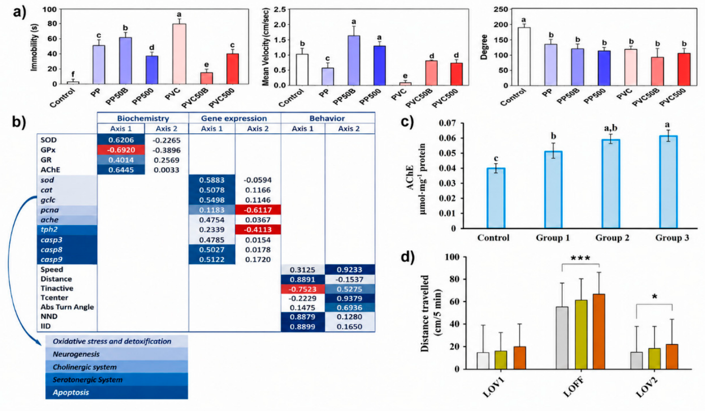
| Fluorescent plastic microspheres/Spherical/1–5 μm/2 mg/L (~1.09 × 10⁸ particles/L). | Gene expression: Neurogenesis/proliferation: sox2, pcna, ngn1, neuroD, olig2; motor neuron development: islet1, islet2a, islet2b; epigenetic regulation; dnmt1, dnmt3–dnmt8; related to antioxidant activity (sod1, cat), apoptosis (casp3, casp8, casp9), neurogenesis (pcna, sox2), and neurotransmitter systems (cholinergic, serotonergic, dopaminergic) Cellular and histopathology endpoints: Immunohistochemistry (PCNA, ISL1&2), stereological analysis of retina and brain, histopathology (retina, brain). | 2 hpf to 14 dpf. | Virgin MPs (proprietary polymer): Inhibited GPx activity; upregulated sod1, casp8, casp9, casp3, th, and slc6a3 genes; increased AChE activity. Induced behavioral changes in mean speed and distance moved. Apoptosis: MPs alone significantly increased the expression of apoptosis-related genes (casp3, casp8, casp9). | [50] | |
| PS/Spherical/20 µm/2 mg/L | Thyroid axis parameters: T3, T4, TSH. Metabolomics: metabolites such as BHA, arachidonic acid and glycerophospholipid pathways. | 7 d | Uptake/distribution: After 7 days, the 20 μm fluorescent polystyrene MPs were mainly found to be accumulated in the GI tract of the larvae (none were found in the controls). Thyroid axis endpoint: MPs did not significantly change T3. No significant difference in the results in metabolomic analysis. | [88] | |
| PS/Spherical/5 µm and 10 µm/1, 10, and 100 mg/L | Molecular: Oxidative stress biomarkers, antioxidant enzyme activities, lipid peroxidation, expression of heart-related genes. Histopathological: Heart tissue structure, cardiomyocyte alterations, apoptotic markers. | 96 hpf | Oxidative damage and genotoxicity in heart tissue after dietary PS-MP exposure (21 days): Lipid peroxidation was higher in PS-MPs treated fish (reported as +528.5% vs. control), and DNA damage was higher (reported as ~100× higher vs. control). Autophagy markers: LC3 II/I ratio increased (reported as 2.2-fold higher) and SQSTM1/p62 decreased (reported as 2.8-fold lower) in PS-MPs treated fish vs. control. Apoptosis markers: Bax/Bcl-2 ratio increased (reported as 5.1-fold higher) and caspase-3 and caspase-9 increased (reported as 2.5-fold higher) in PS-MP-treated fish vs. control. Heart metabolomics (PS-MPs exposed vs. control): The metabolic profile of heart tissue was altered, with most metabolites reduced; pyruvic acid (+38%) and acetylcarnitine (+14%) increased, while TCA intermediates (e.g., succinic acid −75%, α-ketoglutaric acid −56%) and multiple amino acids were reduced. | [67] | |
| PS/Microbeads/5 ± 3 μm & 50 ± 3 μm/10 mg/L at 5 μm (MPs-5) or 50 μm (MPs-50) | Oxidative stress and oxidative damage genes: ROS, MDA, DNA damage markers, CAT; apoptosis-related gene expression such as p53, Bax, and Bcl-2; Dioxin-like marker genes (CYP1A1 and CYP1B1). | Up to 96 hpf | Oxidative stress and oxidative damage: increased ROS with associated increases in lipid peroxidation (MDA) and oxidative DNA damage marker (8-OHdG), and activation/induction of antioxidant enzymes like SOD and CAT. Apoptosis-related genes: p53 and Bax were upregulated and Bcl-2 was downregulated vs. controls and caspase 3, 8, 9 were upregulated in PS-MP-exposed treatments. At 5 µm PS-MPs: CYP1A1 and CYP1B1 significantly upregulated vs. control (CK). At 50 µm PS-MPs: No significant difference vs. control (CK) for these CYP responses. | [56] | |
| PP/Mixed fragments/11.9–44.6 µm/0 mg/L, 1 mg/L, 10 mg/L, 100 mg/L | Gut MP load; oxidative stress; ROS; SOD, CAT; neurotoxicity; acetylcholinesterase (AChE); Histopathology; Liver; Brain. | 28 d | 0 (control): No effect 1 mg/L: Mild ROS elevation, early antioxidant imbalance. 10 mg/L: Significant oxidative stress, liver histological damage, increased AChE activity. 100 mg/L: Severe oxidative stress, marked hepatic and neural injury, apoptosis of blood cells, high MP bioaccumulation in gut. | [89] | |
| PE/0–10 mm/ 0 mg/L, 10 mg/L, 100 mg/L, 1000 mg/L | Gut microbiota: qPCR at phylum level (all 4 groups) + 16S sequencing. Biochemical indicators (physiology): TG, GLU, TCHO, TBA, LDL, HDL, pyruvic acid, NEFA. Gene expression (glycolipid & phospholipid metabolism): RT-qPCR in 0/10/100/1000 mg/L groups. Metabolomics: Nontargeted LC-MS metabolomics only for Control vs. 1000 mg/L (6 parallels; 400 larvae/sample). | 7 d | 0 mg/L (control): Nontargeted LC-MS metabolomics only for control vs. 1000 mg/L (6 parallels; 400 larvae/sample). 10 mg/L: Minimal to lower significant. 100 mg/L: Microbiome (qPCR, phylum level): Firmicutes and Bacteroidetes significantly lower vs. control; Actinobacteria, β-Proteobacteria, γ-Proteobacteria significantly reduced vs. control (also true at 1000). 1000 mg/L: Microbiome (16S sequencing vs. control): alpha diversity shifts; OTUs decrease; clear separation in beta diversity (PCA), Proteobacteria/Chloroflexi/Fusobacteria ↑; Firmicutes/Bacteroidetes/Actinobacteria and others ↓, Genus-level dysbiosis (Aeromonas/Shewanella etc ↑; many beneficial taxa ↓), biochemical indicators: TG, TCHO, NEFA, TBA, GLU significantly increased; pyruvic acid significantly decreased; LDL/HDL show a decreasing trend, Glycolysis/glucose genes: PK, HK1, GK decreased significantly at 1000 mg/L; PEPCKc decreased at 100 and 1000. | [90] | |
| PS/Spherical/1 µm diameter/100 µg/L and 1000 µg/L | Gene expression (il1b, cat, sod). | 4 hpf → 96–120 hpf. | 100 µg/L: No effect on the gene expression. 1000 µg/L: Significant ↑ inflammatory gene (il1b), ↑ oxidative stress marker (cat). | [91] | |
| Not disclosed/~1 µm diameter/MPs suspended at 0.006%, 0.0045%, 0.003%, 0.0015% solids | Microcirculation/RBC velocity using high-speed CCD and micro-PIV analysis. Heart morphology. | Exposed for 3 days (1 dpf → 4 dpf) | The caudal artery (CA), systole–diastole cycle duration was elongated in MP3-ZF without changing RBC velocity. The duration and magnitude of RBC velocity at the dorsal artery (DA) were not significantly altered. | [51] | |
| PS/Not disclosed/1, 10, 100 µg/L | Endpoints measured: Cardiac development: heart rate (HR) at multiple hpf; cardiac morphology; SV–BA distance; histopathology. Thrombosis: thrombus in caudal vein in 100 µg/L MC-LR + PS-MPs/NPs group. Angiogenesis: Inhibited angiogenesis (DLAV/ISV loss) in Tg(kdrl:EGFP) model; amelioration by ASTA, oxidative stress + inflammation: ROS staining; MDA/SOD/GSH; IL-6/IL-8 mRNA by qRT-PCR at 168 hpf. Gene expression pathways: Cardiovascular development genes & calcium signaling pathway genes altered (heatmaps). | 7 d | Results: At 96 & 168 hpf: MC-LR + PS-MPs → lower heart rate vs. control and MC-LR alone groups. 100 µg/L MC-LR + PS-MPs: thrombus observed; vascular loss; oxidative stress/inflammation increased; ASTA provides partial rescue. | [92] | |
| PGA, PLA, PBS, PHA, PBAT/No data/1 mg/L and 100 mg/L for each polymer | Thyroid axis/molecular endpoints: Hormones (ELISA): T3, T4, TSH in 120 hpf larvae (tested using 100 mg/L groups); Gene expression (qRT-PCR): HPT axis genes + eye/retina development genes (samples taken at 72 hpf; again, focusing on 100 mg/L groups for gene work). | 3, 6, 10, 24, and 96 hpf | 1 mg/L: Survival: Significantly decreased for PHA (1 mg/L) and PBAT (1 mg/L) at 96 hpf; body length/head area: Reduced in most low-dose groups except PGA 1 mg/L (which showed no significant reduction). Eye area: decreased in most low-dose groups except PGA 1 mg/L and PBAT 1 mg/L (no significant decrease reported for those two). Retina thickness (5 dpf): IPL decreased at PGA 1 mg/L (reported significant reduction); ONL decreased at PBS 1 mg/L and PBAT 1 mg/L; RGL decreased in all 1 mg/L groups (and also all 100 mg/L groups). Visually-mediated behavior (light–dark): In light, locomotor parameters were reduced in all treatment groups (interpreted as inhibited motor ability during light period. 100 mg/L: Early embryonic development: PBS, PHA, PBAT (100 mg/L) caused developmental delay at 3 hpf (PBS notably with embryonic cell mound defects). Retina & eye development gene expression: Eye/retina genes (e.g., pax6a, pax6b, rx1, gnat2, grk1b, opn1mw1) were abnormally altered; notably, PGA/PLA/PBAT 100 mg/L significantly decreased multiple eye/retina development genes. RGL thickness decreased in all groups, supporting retinal injury across polymers/doses. Thigmotaxis (3 dpf): Thigmotaxis increased significantly in PGA, PLA, PBS, PBAT at 100 mg/L, described as anxiety-like behavior. Visually-mediated behavior (5 dpf): PBS 100 mg/L is explicitly flagged as disrupting visually-mediated behavior (impaired light-to-dark response pattern). Thyroid hormones (ELISA, 100 mg/L only): TSH: No significant change overall. T3 and T4: Significantly decreased in PHA 100 mg/L and PBAT 100 mg/L; not significantly changed for PGA/PLA/PBS 100 mg/L. HPT axis gene disruption (100 mg/L groups): crh downregulated (notably PGA/PBS/PHA), tshβ upregulated in all groups. dio1 and ttr generally down; dio2 up with PLA 100 mg/L. TH synthesis genes: tg down in PBAT; nis down in PLA and PBAT; tpo up in PBAT. TH receptors: thrab increased in PGA/PBS/PHA/PBAT; thrb decreased in PLA but increased in PBS/PHA/PBAT. | [53] | |
| PS/Microsphere/5 μm/1 mg/L | Oxidative stress biomarkers: CAT, SOD, GPX activities; MDA level; ROS measurement (DCFH-DA assay described); TG level (lipid-related index) at 7 dpf. | 2 h post-fertilization (hpf) and continued to 7 days post-fertilization (dpf). | Oxidative stress biomarker: CAT down by 1.55× (NPs) and 1.22× (MPs), SOD down by 1.76× (NPs) and 1.62× (MPs), GPX down by 1.96× (NPs) and 1.58× (MPs), ROS up by 1.20× (NPs) and 1.41× (MPs). | [57] | |
| PET & PE/Irregular fragments/Average 30–100 µm/1 mg/L & 10 mg/L | Enzymatic biomarkers (EROD, AChE), reproductive output (fecundity/fertility), and offspring (F1) fitness. | 0 to 96- or 120-h post-fertilization (hpf) | Biomarkers: EROD and AChE activities were altered only in marine medaka, not zebrafish. | [55] | |
| Larvae | PS/0.2, 1.0, and 10 µm/20 µg/mL for all particle sizes (0.2, 1.0, 10 µm) | Mortality; vascular development; CYP1A activity (EROD Assay). Oxidative stress (ROS); cell death (acridine orange staining). | Larvae: Chorion removed at 24 hpf, Exposure from 24–96 hpf (72 h post-initiation). | CYP1A activity (EROD, heart region): PS-MPs alone did not induce CYP1A activity. ROS in heart region: PSMPs alone were not significantly different from control for ROS. Cell death (acridine orange signal, heart region): PSMPs alone did not affect cell death. | [93] |
| PS & PVC/Spherical/PS (Spherical ~7.0 µm mean size) PVC (Spherical ~3.8 µm mean size)/Both MPS 20 mg L−1 | Histopathology: Intestinal MP deposition, inflammation, pleural effusion, cellular damage (H&E staining). Molecular and biochemical: Oxidative stress genes (sod, cat, gpx1a, gpx4a, gstt1a), inflammation genes (tnf-α, il-6), apoptosis/p53 pathway genes (tp53, casp3, rrm2), development & metabolism genes (egr2, egr4, fosab, fosb, cyp3a65). | 10 d | MPs visibly accumulated in intestinal tract, Inflammatory infiltration and pleural effusion observed, PS caused slightly more damage than PVC. Gene expression: MPs alone significantly upregulated oxidative stress genes, PS induced stronger oxidative stress than PVC. PS MPs were more toxic than PVC MPs to zebrafish larvae, Joint toxicity depended strongly on MP polymer type; Oxidative stress was the central mechanism linking mortality, behavior, histopathology, and gene expression. | [58] | |
| PP/Irregular fragments/8–10 µm/0 mg/L (control), 1 mg/L, 10 mg/L, 100 mg/L | Cytotoxicity and tissue damage: Liver histopathology, intestinal histopathology, cellular vacuolization, inflammatory infiltration. Oxidative stress biomarkers: Reactive oxygen species (ROS), superoxide dismutase (SOD), catalase (CAT), malondialdehyde (MDA). Apoptosis and molecular markers: Caspase-3 activity, bax, bcl-2 gene transcription, p53 transcription. Neurotoxicity and metabolic endpoints: Acetylcholinesterase (AChE) & ATP content. | 96 h (acute exposure) | Control: Normal liver and intestinal morphology, baseline ROS, enzyme activity, and gene transcription. 1 mg/L: Slight increase in ROS, no significant histological damage, Minor antioxidant response. 10 mg/L: Significant ROS elevation, decreased SOD and CAT, increased MDA, mild liver vacuolization, increased bax and caspase-3 expression, reduced ATP levels. 100 mg/L: Severe oxidative stress, strong lipid peroxidation, marked liver and intestinal tissue damage; high apoptosis activation, suppressed AChE activity, significant metabolic disruption. | [94] | |
| PS/5 µm/1 mg/L | Toxicity endpoints (larval): Oxidative stress gene expression such as GSH/GSSG. | 7 d | Oxidative-stress–linked metabolite: Glutathione (GSH) increased in the MP-only group (fold change 1.17). GSH/GSSG: No significant difference in the results. | [95] | |
| PS/~25 μm in diameter/250 μg/L | Apoptosis markers: bax/bcl2, caspase3; AO staining brain. | Early dpf stages | Apoptosis-related genes (whole larvae): PS-MPs upregulated the bax/bcl2 ratio and caspase3 expression at 1 dpf, but not at 3 dpf and 5 dpf. Apoptosis staining (brain region): At 5 dpf, there were no apoptotic bodies in the larval brain regions exposed to PS-MPs. | [62] | |
| Virgin & UV-aged PA/Irregular fragments/~5 µm (mean particle diameter)/0, 10, 100, and 1000 µg/L | Intestinal health: Intestinal morphology (H&E staining), Goblet cell number, Tight junction–related gene expression. Lipid absorption: Oil Red O staining and Triglyceride content. Molecular responses: Lipid metabolism genes (fabp, apoa1, etc.), and Intestinal barrier genes (zo-1, claudin). | 2 d post-fertilization (dpf) → 10 dpf | Mild changes in the intestinal structure were noticed, such as slight desquamation of enterocytes and vacuolation in the intestinal mucosa. Oxidative stress responses were triggered, as evidenced by enhanced production of reactive oxygen species and changes in the activity of antioxidant enzymes. There was impairment in lipid metabolism, as evidenced by decreased expression of lipid metabolites (triglycerides, diglycerides, monoglycerides, cholesterol esters, phospholipids). Inhibition of lipoprotein lipase activity: Decreased expression of genes involved in fat digestion and absorption. Decreased triglyceride and cholesterol concentrations, suggesting impaired triglyceride and cholesterol absorption. | [48] | |
| Virgin & Photo-aged PS/Virgin: 10 μm Photo-aged: 6.5 μm/0.1–100 μg/L for both V-PS and P-PS. | Oxidative stress biomarkers: SOD, CAT, GST, MDA; Molecular: expression of neurotransmission genes (ache, drd3, 5ht2c, gat1, etc.) and oxidative stress genes (cat1, sod1, gpx1a, gstrl, etc.). | 1 hfp to 120 hpf | Oxidative stress: Antioxidant enzymes + MDA significantly altered at 10–100 μg/L P-PS. As well as the SOD, CAT, and GST are significantly altered. | [47] | |
| PS/Virgin & Photo-aged/Spherical beads/1 µm/0, 1, 10, and 100 µg/L both Virgin & Photo-aged | Molecular responses: Lipid metabolism genes (fabp, apoa1, etc.), and Intestinal barrier genes (zo-1, claudin). | 96 h post-fertilization (hpf) | Photo-aged PS MPs: caused intestinal epithelial damage, Reduced goblet cell number, disrupted intestinal barrier genes, severely impaired lipid absorption. | [59] |
5. Neurobehavior and Its Corresponding Effects on Zebrafish Exposed to Microplastics
| Life Stage | Plastic Characterization (Type/Shape/Size/Concentrations) | Endpoints | Exposure Time | Effects | Ref. |
|---|---|---|---|---|---|
| Adult | PS/Irregular fragments/~400 µm/0.5 mg/L | Behavioral parameters: Latency to first MP capture, capture frequency, spitting frequency, swallowing ratio (%), feeding duration, time in feeding zone, swimming activity, speed, total distance moved. | 10 m per trial | Latency to first capture: Most zebrafish started MP capture within ~10 s of adding MPs. Shy zebrafish showed longer latency than bold zebrafish (not significant). Capture frequency: Bold zebrafish captured MPs significantly more often than shy zebrafish on exposure days (p < 0.05). Spitting behavior: Zebrafish often spat MPs out after capture. About 30–47% of total capture events happened in the first minute. Spitting frequency was positively correlated with capture frequency. Swallowing ratio: About 40–60% of captured MPs were swallowed. Bold fish had higher effective exposure because of higher capture frequency. Swimming activity during feeding: Zebrafish showed immediate increase in activity after MP addition. Bold zebrafish showed significantly higher levels of feeding activity (activity %, total distance, average speed) than shy zebrafish on days 2–3 (p < 0.05). Bold zebrafish spent significantly more time in feeding areas (upper water layer). Ingestion/egestion behavior: Bold zebrafish ingested more MPs than shy zebrafish when exposed to MPs only (significant on day 2). Very few MPs were left in intestines after 3 days, suggesting efficient egestion. | [96] |
| Propriety polymer (composition undisclosed/Spherical/1–5 µm/2 mg/L | Behavior: Locomotor activity (Open Field Test), anxiety (Light-Dark Test), and social. | 30 d | Locomotor activity (open field test): Mean speed: Markedly reduced in MP group (584 ± 33.5 cm/min) compared to control (696 ± 27.1 cm/min) (p < 0.01). Total distance moved: Significantly reduced in MP group (2863.4 ± 1.8 cm) compared to control (3445.8 ± 234.4 cm) (p = 0.0395). Absolute turn angle: Not significantly different between MP group and control. Time inactive: Significantly increased in MP group (10.4 ± 0.3%) compared to control (3.0 ± 0.3%) (p < 0.0001). Time in center zone: no significant difference between MP group and control. Anxiety-like behavior (light–dark test): MPs did not significantly alter anxiety-like behavior of the zebrafish. Social/shoaling behavior: MPs altered social behavior significantly, leading to tighter shoaling patterns. | [64] | |
| PS/Spherical beads/2 μm diameter/0.44 mg/L (~108 items/L) | Locomotion behavior: Average swimming velocity (ASV), duration of high mobility (DHM), frequency of high mobility (FHM), duration of thigmotaxis (DTH). | 7 d | Results: No locomotion alteration. No change in thigmotaxis or high mobility metrics. No change in post-stimulation escape response. While there is a significant increase in shoaling duration. While it enhances the shoaling behavior, it did not induce hyperactivity or hypoactivity or alter startle performance. | [66] | |
| PE/Not disclosed/60 mg/L | Behavioral endpoints: School cohesion, school depth, distance from predator (anti-predator response deficit inferred). | 10 d | Results: Direct exposure to PE MPs at 60 mg/L for 10 days did not significantly alter: Latency to reach the top, time spent in the top zone, anxiety index in open field test, swimming speed, or total distance traveled. Additionally, no significant increase in shoal cohesion when predator was present, impaired defensive aggregation response, and reduced prey–predatory distance compared to control. | [78] | |
| PGA/~1 μm in diameter/1 mg/L & 100 mg/L | Behavior: anxiety-like behavior + cognitive/visual preference behaviors (novel tank and color preference experiments). | 28 d | After zebrafish exposed to 1 mg/L and 100 mg/L of PGA microplastics for 28 days, some clear changes showed up in their behavior. First, in the novel tank test, the fish started sticking to the bottom more as the concentration went up. Their movement became really limited to the bottom zone. Significant decreased in: Time spent in the top zone; Distance traveled in the top zone; The ratio of time spent top versus bottom; Number of times they entered the top zone. Interestingly, their average swimming speed did not change much. For cognitive effects, researchers used a color preference test with a four-arm maze. Normally, the fish prefer colors in this order: Blue, then no color, then green, red, and yellow last. After exposure: The high concentration group spent more time in the blue and no-color areas. Their color preference shifted compared to the control group. The low concentration group did not show much change in color preference. | [47] | |
| PS/Spheres/0.1 μm diameter/0, 0.1, 1, 10, 50, 100 mg/L | Locomotion: Moving distance, swimming speed. | 96 h | Swimming pace through the trial did not change reliably when it compares to the control. The pattern of behavior seen in PS-MP-treated subjects matched levels observed in animals getting no treatment. At 1 mg/L, polystyrene MPs it showed no obvious change in fish behavior after 96 h. Movement patterns stayed the same regardless of exposure. | [48] | |
| Embryos → Larvae | PS/Spherical/1 µm in diameter/0.1, 1, and 10 mg/L | Swimming velocity, mobility ratio, avoidance response. | 21 d | Swimming velocity: No significant effect in MP exposure. Mobility ratio: No significant effect. Avoidance response: No significant differences relative to the control for MPs alone. | [86] |
| PS/Spherical/1 µm diameter/100 µg/L and 1000 µg/L | Swimming distance, swimming speed, and Dark-light avoidance behavior | 4 hpf → 96–120 hpf | Swimming speed dropped by 3.5% when fish were exposed to 1000 μg/L in dark water after 120 hpf. That level reduced movement more than any other. Swimming speed dropped slightly—1.5 plus or minus 0.3 mm per second compared to 1.5 plus or minus 0.3 in healthy animals; that difference turned out statistically significant, p equals 0.03. At 1000 micrograms per liter, overall movement shrunk, cutting the total distance by roughly 3.2 percent. A shift appeared with 15.3 ± 2.8 cm measured against 15.8 ± 2.7 cm baseline values—difference found significant at p = 0.021. Exposure to 100 μg/L showed no clear impact during dark swimming trials. Light and dark cycles switched every few days in a test lasting around five days at 120 hpf. Light period: Swimming length and speed showed no clear changes across the board—whether at 100 or 1000 μg/L. Dark period: At 100 μg/L, fish swam shorter distances—by 4.6% fewer beats. Swimming slowed down by 4.9% at 100 μg/L. A shift appeared—movement slowed in one condition compared to the other. Speed dropped by about 1.41 to 1.48 mm per second. That difference was highly significant, showing up clearly beyond doubt. The p value hit a near-zero mark after testing. At 1000 μg/L, animals swam 2.8 percent slower. Speed rose slightly in treatment versus control (1.44 ± 0.24 mm/s vs. 1.48 ± 0.23 mm/s; p = 0.0065). | [99] | |
| PGA, PLA, PBS, PHA, PBAT/No data/1 mg/L and 100 mg/L for each polymer | Behavioral: Thigmotaxis at 3 dpf (edge preference); light–dark test at 5 dpf: max speed, total distance, movement count in light vs. dark. | 3, 6, 10, 24, and 96 hpf | Thigmotaxis behavior in 3-day-old larvae: Larvae exposed to strong concentrations—like 100 mg/L—of PGA, PLA, PBS, and PBAT showed a clearer tendency toward edge avoidance. This shift toward thigmotaxis became more pronounced, suggesting heightened anxiety behavior. Even at 100 mg/L, PHA did not cause much thigmotactic response. At just 1 mg/L, there was barely any change in thigmotaxis worth noting. Larvae at five days old responded to changes in light and dark. During shifts in illumination, their actions were observed. Light at 80 lux levels: Every single one of the five natural breakdown microplastics—made from PGA, PLA, PBS, PHA, PBAT—dissolved more easily when tested at both light and heavy doses. Their ability to break down happened clearly across every sample exposure. Dark conditions (0.1×): At 100 mg/L PBS and 1 mg/L PBAT caused slower movement, lower overall travel, and fewer shifts in position. At 100 mg/L PHA cut down movement speed and overall motion total. Not every option lowered dark-phase movement—some changes did not affect it significantly. Light–dark responsiveness: Beyond 100 mg/L PBS plus 1 mg/L PBAT, movement patterns stayed largely unchanged across light-to-dark shifts. These conditions weakened the body’s reliance on visual cues when responding to sudden changes. Light-driven reactivity simply fell short under such exposure. Other groups still showed clear differences in movement between light and dark periods. | [62] | |
| Larvae | PS & PVC/Spherical/PS (Spherical ~7.0 µm mean size) PVC (Spherical ~3.8 µm mean size)/Both MPS 20 mg L−1 | Immobility duration, mean swimming velocity, turn angle (locomotor behavior) | 10 d | Locomotor activity: PS & PVC MPs significantly suppressed swimming activity compared to control. Immobility duration: Immobility duration significantly increased in larvae and immobility in PVC group showed U-shaped trajectory, indicating high immobility observed under PVC-MP exposure. Mean velocity and turn angle: Both PS & PVC-MPs reduced the mean swimming velocity of zebrafish. The turn angle was altered in a concentration-dependent manner. | [57] |
| Virgin & Photo-aged PS/Virgin: 10 μm Photo-aged: 6.5 μm/0.1–100 μg/L for both V-PS and P-PS. | Behavior: Locomotor activity (reduced locomotion). | 1 hfp to 120 hpf | Locomotor activity: After 120 h of development, young zebrafish moved less when exposed to 100 μg/L of virgin PS-MPs than those in clean water. Their ability to swim declined noticeably under that condition. Swimming changes showed up when larvae were exposed to photo-aged PS-MPs at 1–100 μg/L levels. Speed shifted in response to those doses. P-PS given at 10 and 100 μg/L caused fish to swim slower, with lower average speeds than in the control and V-PS groups (p < 0.05). Light–dark response: Most changes happened while the lights were off. The P-PS group showed clearer shifts than others. Correlation with neurochemical biomarkers measures of behavior—like how fast zebrafish swam—tended to drop as brain chemicals such as 5-HT, GABA, and ACh went down. What also followed a similar pattern was the activity of enzymes like AChE and ChE, which moved in step with those neurotransmitters but in opposite directions. | [47] |
6. Summary and Future Perspectives
- Future studies should prioritize standardized reporting of microplastic physicochemical properties, including polymer identity, particle size distribution, morphology, aging status, surface characteristics, and concentration metrics. The reviewed studies used diverse exposure designs and reporting approaches, making direct comparisons difficult and limiting quantitative synthesis. Establishing harmonized experimental protocols would improve reproducibility and facilitate cross-study comparisons.
- Although advanced analytical approaches, including imaging technologies, omics analyses, and adverse outcome pathway (AOP) frameworks, may contribute to future mechanistic investigations, relatively few of the studies included in this review employed these approaches. In addition, these advanced methodologies are presented only as potential research opportunities rather than evidence-based conclusions arising from the present systematic review.
- Artificial intelligence and machine learning technologies are emerging as promising tools in MP ecotoxicology research. AI image analysis is seen as having the potential to enhance MP detection, identification, and quantification in environmental samples and biological tissues. Machine learning can also be applied to analyze large datasets from multi-omics approaches to identify predictive toxicity pathways and biomarker networks. In zebrafish behavioral assays, AI video tracking systems are being increasingly applied to quantify locomotor activity, anxiety-like behavior, and social behavior with greater accuracy and without subjective bias.
Supplementary Materials
Author Contributions
Funding
Institutional Review Board Statement
Data Availability Statement
Acknowledgments
Conflicts of Interest
Abbreviations
| 5-HT | 5-hydroxytryptamine |
| Ach | Acetylcholine |
| AChE | Acetylcholinesterase |
| AgNP | Silver nanoparticles |
| BDNF | Brain-derived neurotrophic factor |
| Bcl2 | B-cell lymphoma 2 |
| CAT | Catalase |
| ChAT | Choline acetyltransferase |
| ChE | Cholinesterase |
| CYP1A | Cytochrome P450 1A |
| DA | Acetylcholinesterase |
| DNMT | DNA methyltransferase |
| Dpf | Days post-fertilization |
| EROD | Ethoxy resorufin-O-demethylase |
| GABA | Gamma-aminobutyric acid |
| GFP | Green fluorescent protein |
| GI | Gastrointestinal |
| GPx | Glutathione peroxidase |
| GR | Glutathione reductase |
| GSH | Reduced glutathione |
| GST | Glutathione S-transferase |
| HMOX1 | Heme oxygenase 1 |
| Hpf | Hours post-fertilization |
| keap1a | Kelch-like ECH-associated protein 1a |
| LDH | Lactate dehydrogenase |
| LDL | Low-density lipoprotein |
| MDA | Malondialdehyde |
| Mn-sod | Manganese superoxide dismutase |
| MPs | Microplastics |
| Mt | Million metrics tons |
| NE-FA | Non-esterified fatty acids |
| nfe212a | Nuclear factor erythroid 2-related factor 2a |
| OECD | Organization for Economic Co-operation and Development |
| PA | Polyamide |
| PBAT | Polybutylene adipate terephthalate |
| PBS | Polybutylene succinate |
| PCNA | Proliferating cell nuclear antigen |
| PCoA | Principal Coordinate Analysis |
| PE | Polyethylene |
| PES | Polyester |
| PET | Polyethylene terephthalate |
| PGA | Polyglycolic acid |
| PHA | Polyhydroxyalkanoate |
| PLA | Polylactic acid |
| PP | Polypropylene |
| PPM | Parts per million |
| PRISMA | Preferred Reporting Items for Systematic Reviews and Meta-Analyses |
| PS | Polystyrene |
| PVC | Polyvinyl chloride |
| ROS | Reactive oxygen species |
| SOD | Superoxide dismutase |
| T-CHO | Total cholesterol |
| TEM | Transmission electron microscopy |
| TG | Triglycerides |
| tp53 | Tumor protein p53 |
| UV | Ultraviolet |
| W | Weeks |
References
- Borrelle, S.B.; Ringma, J.; Law, K.L.; Monnahan, C.C.; Lebreton, L.; McGivern, A.; Murphy, E.; Jambeck, J.; Leonard, G.H.; Hilleary, M.A.; et al. Predicted growth in plastic waste exceeds efforts to mitigate plastic pollution. Science 2020, 369, 1515–1518. [Google Scholar] [CrossRef] [Scilit]
- Phengsaart, T.; Srichonphaisarn, P.; Villacorte-Tabelin, M.; Silwamba, M.; Janjaroen, D.; Tabelin, C.B.; Alonzo, D.; Ta, A.T.; Juntarasakul, O. Microplastic Removal by Flotation: Systematic Review, Meta-Analysis, and Research Trends. Water 2025, 17, 3394. [Google Scholar] [CrossRef] [Scilit]
- Dokl, M.; Copot, A.; Krajnc, D.; Van Fan, Y.; Vujanović, A.; Aviso, K.B.; Tan, R.R.; Kravanja, Z.; Čuček, L. Global projections of plastic use, end-of-life fate and potential changes in consumption, reduction, recycling and replacement with bioplastics to 2050. Sustain. Prod. Consum. 2024, 51, 498–518. [Google Scholar] [CrossRef] [Scilit]
- Lebreton, L.; Andrady, A. Future scenarios of global plastic waste generation and disposal. Palgrave Commun. 2019, 5, 6. [Google Scholar] [CrossRef] [Scilit]
- European Food Safety Authority (EFSA). Presence of microplastics and nanoplastics in food, with particular focus on seafood. EFSA J. 2021, 19, e06713. [Google Scholar] [CrossRef] [Scilit]
- Juntarasakul, O.; Julapong, P.; Srichonphaisarn, P.; Meekoch, T.; Janjaroen, D.; Tabelin, C.B.; Phengsaart, T. Weave structures of polyester fabric affect the tensile strength and microplastic fiber emission during the laundry process. Sci. Rep. 2025, 15, 2272. [Google Scholar] [CrossRef] [Scilit]
- Soonthornwiphat, N.; Srichonphaisarn, P.; Villacorte-Tabelin, M.; Julapong, P.; Tabelin, C.B.; Janjaroen, D.; Phengsaart, T. Ultraviolet Irradiation Affects Microplastic Properties and Removal from Water Using Agglomeration–Micro-Flotation. Water 2026, 18, 1588. [Google Scholar] [CrossRef] [Scilit]
- Hsu, W.-T.; Domenech, T.; McDowall, W. How circular are plastics in the EU?: MFA of plastics in the EU and pathways to circularity. Clean. Environ. Syst. 2021, 2, 100004. [Google Scholar] [CrossRef] [Scilit]
- de Sá, L.C.; Oliveira, M.; Ribeiro, F.; Rocha, T.L.; Futter, M.N. Studies of the effects of microplastics on aquatic organisms: What do we know and where should we focus our efforts in the future? Sci. Total Environ. 2018, 645, 1029–1039. [Google Scholar] [CrossRef] [Scilit]
- Kramm, J.; Völker, C.; Wagner, M. Superficial or Substantial: Why Care about Microplastics in the Anthropocene? Environ. Sci. Technol. 2018, 52, 3336–3337. [Google Scholar] [CrossRef] [Scilit]
- Amobonye, A.; Bhagwat, P.; Raveendran, S.; Singh, S.; Pillai, S. Environmental Impacts of Microplastics and Nanoplastics: A Current Overview. Front. Microbiol. 2021, 12, 768297. [Google Scholar] [CrossRef] [Scilit]
- Atamanalp, M.; Köktürk, M.; Uçar, A.; Duyar, H.A.; Özdemir, S.; Parlak, V.; Esenbuğa, N.; Alak, G. Microplastics in Tissues (Brain, Gill, Muscle and Gastrointestinal) of Mullus barbatus and Alosa immaculata. Arch. Environ. Contam. Toxicol. 2021, 81, 460–469. [Google Scholar] [CrossRef] [Scilit]
- Barboza, L.G.A.; Lopes, C.; Oliveira, P.; Bessa, F.; Otero, V.; Henriques, B.; Raimundo, J.; Caetano, M.; Vale, C.; Guilhermino, L. Microplastics in wild fish from North East Atlantic Ocean and its potential for causing neurotoxic effects, lipid oxidative damage, and human health risks associated with ingestion exposure. Sci. Total Environ. 2019, 717, 134625. [Google Scholar] [CrossRef] [Scilit]
- Habib, R.Z.; Thiemann, T. Microplastic in commercial fish in the Mediterranean Sea, the Red Sea and the Arabian Gulf. Part 1: The Mediterranean Sea. J. Water Resour. Prot. 2021, 13, 563–587. [Google Scholar] [CrossRef]
- Bhagat, J.; Zang, L.; Nishimura, N.; Shimada, Y. Zebrafish: An emerging model to study microplastic and nanoplastic toxicity. Sci. Total Environ. 2020, 728, 138707. [Google Scholar] [CrossRef] [Scilit]
- Stock, F.; Narayana, V.K.B.; Scherer, C.; Löder, M.G.J.; Brennholt, N.; Laforsch, C.; Reifferscheid, G. Pitfalls and limitations in microplastic analyses. In Plastics in the Aquatic Environment—Part I. The Handbook of Environmental Chemistry; Springer: Cham, Switzerland, 2020; pp. 13–42. [Google Scholar] [CrossRef] [Scilit]
- Khan, F.R.; Alhewairini, S.S. Zebrafish (Danio rerio) as a Model Organism. In Current Trends in Cancer Management; Streba, L., Gheonea, D.I., Schenker, M., Eds.; IntechOpen: London, UK, 2019. [Google Scholar]
- Savuca, A.; Nicoara, M.N.; Ciobica, A.; Gorgan, D.L.; Ureche, D.; Balmus, I.M. Current Aspects on the Plastic Nano- and Microparticles Toxicity in Zebrafish—Focus on the Correlation between Oxidative Stress Responses and Neurodevelopment. Animals 2023, 13, 1810. [Google Scholar] [CrossRef] [Scilit]
- Lu, Y.; Zhang, Y.; Deng, Y.; Jiang, W.; Zhao, Y.; Geng, J.; Ding, L.; Ren, H. Uptake and accumulation of polystyrene microplastics in zebrafish (Danio rerio) and toxic effects in liver. Environ. Sci. Technol. 2016, 50, 4054–4060. [Google Scholar] [CrossRef] [Scilit]
- Lei, L.; Wu, S.; Lu, S.; Liu, M.; Song, Y.; Fu, Z.; Shi, H.; Raley-Susman, K.M.; He, D. Microplastic particles cause intestinal damage and other adverse effects in zebrafish Danio rerio and nematode Caenorhabditis elegans. Sci. Total Environ. 2018, 619–620, 1–8. [Google Scholar] [CrossRef] [Scilit]
- Smith, M.; Love, D.C.; Rochman, C.M.; Neff, R.A. Microplastics in seafood and the implications for human health. Curr. Environ. Health Rep. 2018, 5, 375–386. [Google Scholar] [CrossRef] [Scilit]
- Bhuyan, M.S. Effects of microplastics on fish and in human health. Front. Environ. Sci. 2022, 10, 827289. [Google Scholar] [CrossRef] [Scilit]
- Prata, J.C.; da Costa, J.P.; Lopes, I.; Duarte, A.C.; Rocha-Santos, T. Environmental exposure to microplastics: An overview on possible human health effects. Sci. Total Environ. 2020, 702, 134455. [Google Scholar] [CrossRef] [Scilit]
- Campos, A.R.A.; Luza, K.M.B.; Subebe, M.J.B.; Tabelin, C.B.; Phengsaart, T.; Arima, T.; Seno, R.; Butalid, R.; Escabarte, A.B.; Mazahery, A.R.F.; et al. Polytetrafluoroethylene (PTFE) microplastics affect angiogenesis and central nervous system (CNS) development of duck embryo. Emerg. Contam. 2024, 11, 100433. [Google Scholar] [CrossRef] [Scilit]
- Cassar, S.; Adatto, I.; Freeman, J.L.; Gamse, J.T.; Iturria, I.; Lawrence, C.; Muriana, A.; Peterson, R.T.; Van Cruchten, S.; Zon, L.I. Use of zebrafish in drug discovery toxicology. Chem. Res. Toxicol. 2020, 33, 95–118. [Google Scholar] [CrossRef] [Scilit]
- Hill, A.J.; Teraoka, H.; Heideman, W.; Peterson, R.E. Zebrafish as a model vertebrate for investigating chemical toxicity. Toxicol. Sci. 2005, 86, 6–19. [Google Scholar] [CrossRef] [Scilit]
- Limonta, G.; Mancia, A.; Benkhalqui, A.; Bertolucci, C.; Abelli, L.; Fossi, M.C.; Panti, C. Microplastics induce transcriptional changes, immune response and behavioral alterations in adult zebrafish. Sci. Rep. 2019, 9, 15775. [Google Scholar] [CrossRef] [Scilit]
- Howe, K.; Clark, M.D.; Torroja, C.F.; Torrance, J.; Berthelot, C.; Muffato, M.; Collins, J.E.; Humphray, S.; McLaren, K.; Matthews, L.; et al. The zebrafish reference genome sequence and its relationship to the human genome. Nature 2013, 496, 498–503. [Google Scholar] [CrossRef] [Scilit]
- Stewart, A.M.; Braubach, O.; Spitsbergen, J.; Gerlai, R.; Kalueff, A.V. Zebrafish models for translational neuroscience research: From tank to bedside. Trends Neurosci. 2014, 37, 264–278. [Google Scholar] [CrossRef] [Scilit]
- Gerlai, R. Evolutionary conservation, translational relevance and cognitive function: The future of zebrafish in behavioral neuroscience. Neurosci. Biobehav. Rev. 2020, 116, 426–435. [Google Scholar] [CrossRef] [Scilit]
- Zhao, Y.; Jin, R.; Chen, Y.; Zhang, J.; Tao, S.; Liu, S.; Shen, M. Constructed wetlands as neglected fixed source of microplastics and antibiotic resistance genes in natural water bodies? Sci. Total Environ. 2023, 902, 166474. [Google Scholar] [CrossRef] [Scilit]
- Luan, J.; Liu, J.-L.; Xu, Y.-X.; Zhang, Z.; Chen, X. Effects of microplastic exposure on the early developmental period and circadian rhythm of zebrafish (Danio rerio): A comparative study of polylactic acid and polyglycolic acid. Ecotoxicol. Environ. Saf. 2023, 258, 114994. [Google Scholar] [CrossRef] [Scilit]
- Huang, W.; Mo, J.; Li, J.; Wu, K. Exploring developmental toxicity of microplastics and nanoplastics (MNPS): Insights from investigations using zebrafish embryos. Sci. Total Environ. 2024, 933, 173012. [Google Scholar] [CrossRef] [Scilit]
- Zhang, Y. Cell toxicity mechanism and biomarker. Clin. Transl. Med. 2018, 7, 34. [Google Scholar] [CrossRef] [Scilit]
- Auten, R.L.; Davis, J.M. Oxygen toxicity and reactive oxygen species: The devil is in the details. Pediatr. Res. 2009, 66, 121–127. [Google Scholar] [CrossRef] [Scilit]
- Pedersen, A.F.; Meyer, D.N.; Petriv, A.M.V.; Soto, A.L.; Nelissen, K. Nanoplastics impact the zebrafish (Danio rerio) transcriptome: Associated developmental and neurobehavioral consequences. Environ. Pollut. 2020, 266, 115090. [Google Scholar] [CrossRef] [Scilit]
- La Pietra, A.; Fasciolo, G.; Lucariello, D.; Motta, C.M.; Venditti, P.; Ferrandino, I. Polystyrene microplastics effects on zebrafish embryological development: Comparison of two different sizes. Environ. Toxicol. Pharmacol. 2024, 106, 104371. [Google Scholar] [CrossRef] [Scilit]
- Prata, J.C.; Venâncio, C.; Girão, A.V.; Da Costa, J.P.; Lopes, I.; Duarte, A.C.; Rocha-Santos, T. Effects of virgin and weathered polystyrene and polypropylene microplastics on Raphidocelis subcapitata and embryos of Danio rerio under environmental concentrations. Sci. Total Environ. 2021, 816, 151642. [Google Scholar] [CrossRef] [Scilit]
- Mansuri, A.; Trivedi, C.; Kumar, A. Impact of virgin and weathered microplastics on zebrafish: Bioaccumulation, developmental toxicity and molecular pathway disruptions. Sci. Total Environ. 2024, 957, 177510. [Google Scholar] [CrossRef] [Scilit]
- Page, M.J.; McKenzie, J.E.; Bossuyt, P.M.; Boutron, I.; Hoffmann, T.C.; Mulrow, C.D.; Shamseer, L.; Tetzlaff, J.M.; Akl, E.A.; Brennan, S.E.; et al. The PRISMA 2020 statement: An updated guideline for reporting systematic reviews. BMJ 2021, 372, n71. [Google Scholar] [CrossRef] [Scilit]
- Van Eck, N.J.; Waltman, L. Software survey: VOSviewer, a computer program for bibliometric mapping. Scientometrics 2009, 84, 523–538. [Google Scholar] [CrossRef] [Scilit]
- Choi, J.S.; Yoon, H.; Heo, Y.; Kim, T.H.; Park, J. Comparison of gut toxicity and microbiome effects in zebrafish exposed to polypropylene microplastics: Interesting effects of UV-weathering on microbiome. J. Hazard. Mater. 2024, 470, 134209. [Google Scholar] [CrossRef] [Scilit]
- Medriano, C.A.; Bae, S. Acute exposure to microplastics induces metabolic disturbances and gut dysbiosis in adult zebrafish (Danio rerio). Ecotoxicol. Environ. Saf. 2022, 245, 114125. [Google Scholar] [CrossRef] [Scilit]
- Luo, T.; Weng, Y.; Huang, Z.; Zhao, Y.; Jin, Y. Combined hepatotoxicity of imidacloprid and microplastics in adult zebrafish: Endpoints at gene transcription. Comp. Biochem. Physiol. Part C Toxicol. Pharmacol. 2021, 246, 109043. [Google Scholar] [CrossRef] [Scilit]
- Yang, H.; Ju, J.; Wang, Y.; Zhu, Z.; Lu, W.; Zhang, Y. Micro-and nano-plastics induce kidney damage and suppression of innate immune function in zebrafish (Danio rerio) larvae. Sci. Total Environ. 2024, 931, 172952. [Google Scholar] [CrossRef] [Scilit]
- Medriano, C.A.; Kim, S.; Kim, L.H.; Bae, S. Chronic exposure of adult zebrafish to polyethylene and polyester-based microplastics: Metabolomic and gut microbiome alterations reflecting dysbiosis and resilience. J. Hazard. Mater. 2024, 484, 136691. [Google Scholar] [CrossRef] [Scilit]
- Ding, P.; Xiang, C.; Li, X.; Chen, H.; Shi, X.; Li, X.; Huang, C.; Yu, Y.; Qi, J.; Li, A.J.; et al. Photoaged microplastics induce neurotoxicity via oxidative stress and abnormal neurotransmission in zebrafish larvae (Danio rerio). Sci. Total Environ. 2023, 881, 163480. [Google Scholar] [CrossRef] [Scilit]
- Zhang, X.; Xia, M.; Zhao, J.; Cao, Z.; Zou, W.; Zhou, Q. Photoaging enhanced the adverse effects of polyamide microplastics on the growth, intestinal health, and lipid absorption in developing zebrafish. Environ. Int. 2021, 158, 106922. [Google Scholar] [CrossRef] [Scilit]
- Xiao, Y.; Hu, L.; Duan, J.; Che, H.; Wang, W.; Yuan, Y.; Xu, J.; Chen, D.; Zhao, S. Polystyrene microplastics enhance microcystin-LR-induced cardiovascular toxicity and oxidative stress in zebrafish embryos. Environ. Pollut. 2024, 352, 124022. [Google Scholar] [CrossRef] [Scilit]
- Santos, D.; Luzio, A.; Bellas, J.; Monteiro, S.M. Microplastics- and copper-induced changes in neurogenesis and DNA methyltransferases in the early life stages of zebrafish. Chem.-Biol. Interact. 2022, 363, 110021. [Google Scholar] [CrossRef] [Scilit]
- Koelmans, A.A.; Besseling, E.; Shim, W.J. Nanoplastics in the aquatic environment: Critical review. In Marine Anthropogenic Litter; Bergmann, M., Gutow, L., Klages, M., Eds.; Springer: Berlin/Heidelberg, Germany, 2015; pp. 325–340. [Google Scholar] [CrossRef] [Scilit]
- Lambert, S.; Wagner, M. Formation of microscopic particles during the degradation of different polymers. Chemosphere 2016, 161, 510–517. [Google Scholar] [CrossRef] [Scilit]
- Rochman, C.M.; Hoh, E.; Hentschel, B.T.; Kaye, S. Long-term field measurement of sorption of organic contaminants to five types of plastic pellets: Implications for plastic marine debris. Environ. Sci. Technol. 2013, 47, 1646–1654. [Google Scholar] [CrossRef] [Scilit]
- Li, J.; Liu, X.; Fu, J.; Gong, Z.; Jiang, S.Y.; Chen, J.P. Metabolic profile changes of zebrafish larvae in the single- and co-exposures of microplastics and phenanthrene. Sci. Total Environ. 2024, 953, 175994. [Google Scholar] [CrossRef] [Scilit]
- Cormier, B.; Cachot, J.; Blanc, M.; Cabar, M.; Clérandeau, C.; Dubocq, F.; Bihanic, F.L.; Morin, B.; Zapata, S.; Bégout, M.; et al. Environmental microplastics disrupt swimming activity in acute exposure in Danio rerio larvae and reduce growth and reproduction success in chronic exposure in D. rerio and Oryzias melastigma. Environ. Pollut. 2022, 308, 119721. [Google Scholar] [CrossRef] [Scilit]
- Zhang, J.; Bai, Y.; Meng, H.; Zhu, Y.; Yue, H.; Li, B.; Wang, J.; Wang, J.; Zhu, L.; Du, Z. Combined toxic effects of polystyrene microplastics and 3,6-dibromocarbazole on zebrafish (Danio rerio) embryos. Sci. Total Environ. 2024, 913, 169787. [Google Scholar] [CrossRef] [Scilit]
- De Souza Freire, I.; Fascineli, M.L.; Piau, T.B.; Pereira, B.F.; Grisolia, C.K. Multilevel Toxicity Evaluations of Polyethylene Microplastics in Zebrafish (Danio rerio). Int. J. Environ. Res. Public Health 2023, 20, 3617. [Google Scholar] [CrossRef] [Scilit]
- Marshoudi, M.A.; Reasi Ha, A.; Al-Habsi, A.; Barry, M.J. Additive effects of microplastics on accumulation and toxicity of cadmium in male zebrafish. Chemosphere 2023, 334, 138969. [Google Scholar] [CrossRef] [Scilit]
- Araújo, A.P.C.; da Luz, T.M.; Rocha, T.L.; Ahmed, M.A.I.; e Silva, D.M.; Rahman, M.M.; Malafaia, G. Toxicity evaluation of the combination of emerging pollutants with polyethylene microplastics in zebrafish: Perspective study of genotoxicity, mutagenicity, and redox unbalance. J. Hazard. Mater. 2022, 432, 128691. [Google Scholar] [CrossRef] [Scilit]
- Park, S.H.; Kim, K. Microplastics induced developmental toxicity with microcirculation dysfunction in zebrafish embryos. Chemosphere 2021, 286, 131868. [Google Scholar] [CrossRef] [Scilit]
- Wang, J.; Wu, J.; Cheng, H.; Wang, Y.; Fang, Y.; Wang, L.; Duan, Z. Polystyrene microplastics inhibit the neurodevelopmental toxicity of mercury in zebrafish (Danio rerio) larvae with size-dependent effects. Environ. Pollut. 2022, 314, 120216. [Google Scholar] [CrossRef] [Scilit]
- Wen, L.; Man, X.; Luan, J.; Zhang, S.; Zhao, C.; Bao, Y.; Liu, C.; Feng, X. Early-life exposure to five biodegradable plastics impairs eye development and visually-mediated behavior through disturbing hypothalamus-pituitary-thyroid (HPT) axis in zebrafish larvae. Comp. Biochem. Physiol. Part C Toxicol. Pharmacol. 2024, 284, 109981. [Google Scholar] [CrossRef] [Scilit]
- Li, X.; Zheng, T.; Zhang, J.; Chen, H.; Xiang, C.; Sun, Y.; Dang, Y.; Ding, P.; Hu, G.; Yu, Y. Photoaged polystyrene microplastics result in neurotoxicity associated with neurotransmission and neurodevelopment in zebrafish larvae (Danio rerio). Environ. Res. 2024, 250, 118524. [Google Scholar] [CrossRef] [Scilit]
- Cao, Q.; Wei, D.; Ma, X.; Liu, R.; Qi, Y.; Yuan, C.; Huang, D. Polystyrene microplastics mitigate lead-induced neurotoxicity by reducing heavy metal uptake in zebrafish larvae. Sci. Total Environ. 2024, 919, 170790. [Google Scholar] [CrossRef] [Scilit]
- Santos, D.; Luzio, A.; Félix, L.; Cabecinha, E.; Bellas, J.; Monteiro, S.M. Microplastics and copper induce apoptosis, alter neurocircuits, and cause behavioral changes in zebrafish (Danio rerio) brain. Ecotoxicol. Environ. Saf. 2022, 242, 113926. [Google Scholar] [CrossRef] [Scilit]
- Harikrishnan, T.; Paramasivam, P.; Sankar, A.; Sakthivel, M.; Sanniyasi, E.; Raman, T.; Thangavelu, M.; Singaram, G.; Muthusamy, G. Weathered polyethylene microplastics induced immunomodulation in zebrafish. Environ. Toxicol. Pharmacol. 2024, 109, 104478. [Google Scholar] [CrossRef] [Scilit]
- Zhao, Y.; Chen, H.; Liang, H.; Liang, H.; Zhao, T.; Ren, B.; Li, Y.; Liang, H.; Liang, H.; Liu, Y.; et al. Combined toxic effects of polyethylene microplastics and lambda-cyhalothrin on gut of zebrafish (Danio rerio). Ecotoxicol. Environ. Saf. 2024, 276, 116296. [Google Scholar] [CrossRef] [Scilit]
- Xu, J.; Yang, W.; Wang, D.; Wang, Z.; Liu, C.; Li, J. Methamphetamine shows different joint toxicity for different types of microplastics on zebrafish larvae by mediating oxidative stress. Toxics 2023, 12, 9. [Google Scholar] [CrossRef] [Scilit]
- Luan, J.; Wen, L.; Bao, Y.; Bai, H.; Zhao, C.; Zhang, S.; Man, X.; Yin, T.; Feng, X. Systemic toxicity of biodegradable polyglycolic acid microplastics on the gut-liver-brain axis in zebrafish. Sci. Total Environ. 2024, 954, 176898. [Google Scholar] [CrossRef] [Scilit]
- Rangasamy, B.; Malafaia, G.; Maheswaran, R. Evaluation of antioxidant response and Na+-K+-ATPase activity in zebrafish exposed to polyethylene microplastics: Shedding light on a physiological adaptation. J. Hazard. Mater. 2021, 426, 127789. [Google Scholar] [CrossRef] [Scilit]
- Wu, D.; Carter, L.; Kay, P.; Holden, J.; Yin, Y.; Guo, H. Female zebrafish are more affected than males under polystyrene microplastics exposure. J. Hazard. Mater. 2024, 482, 136616. [Google Scholar] [CrossRef] [Scilit]
- Shi, Y.; Chen, C.; Han, Z.; Chen, K.; Wu, X.; Qiu, X. Combined exposure to microplastics and amitriptyline caused intestinal damage, oxidative stress and gut microbiota dysbiosis in zebrafish (Danio rerio). Aquat. Toxicol. 2023, 260, 106589. [Google Scholar] [CrossRef] [Scilit]
- Yu, Z.; Yan, C.; Qiu, D.; Zhang, X.; Wen, C.; Dong, S. Accumulation and ecotoxicological effects induced by combined exposure of different sized polyethylene microplastics and oxytetracycline in zebrafish. Environ. Pollut. 2022, 319, 120977. [Google Scholar] [CrossRef] [Scilit]
- Dimitriadi, A.; Papaefthimiou, C.; Genizegkini, E.; Sampsonidis, I.; Kalogiannis, S.; Feidantsis, K.; Bobori, D.C.; Kastrinaki, G.; Koumoundouros, G.; Lambropoulou, D.A.; et al. Adverse effects polystyrene microplastics exert on zebrafish heart—Molecular to individual level. J. Hazard. Mater. 2021, 416, 125969. [Google Scholar] [CrossRef] [Scilit]
- Adhikari, M.; Biswas, C.; Mazumdar, P.; Sarkar, S.; Pramanick, K. Evaluating the potential of daily intake of polystyrene microplastics via drinking water in inducing PCOS and its ovarian fibrosis progression using female zebrafish. NanoImpact 2024, 34, 100507. [Google Scholar] [CrossRef] [Scilit]
- Xue, M.; Jia, M.; Qin, Y.; Francis, F.; Gu, X. Toxicity of parental co-exposure of microplastic and bisphenol compounds on adult zebrafish: Multi-omics investigations on offspring. Sci. Total Environ. 2024, 955, 176897. [Google Scholar] [CrossRef] [Scilit]
- Jiang, X.; Yan, S.; Sun, H.; Kong, X.Z.; Li, S.; Shi, H.; Zhu, X.; Gu, X. Preparation of fluorescent polyurethane microspheres and their applications as reusable sensor for 4-nitrophenol detection and as microplastics model for visualizing polyurethane in cells and zebrafish. J. Colloid Interface Sci. 2024, 673, 550–563. [Google Scholar] [CrossRef] [Scilit]
- Zhang, L.; Luo, Y.; Zhang, Z.; Pan, Y.; Li, X.; Zhuang, Z.; Li, J.; Luo, Q.; Chen, X. Enhanced reproductive toxicity of photodegraded polylactic acid microplastics in zebrafish. Sci. Total Environ. 2023, 912, 168742. [Google Scholar] [CrossRef] [Scilit]
- Lu, K.; Qiao, R.; An, H.; Zhang, Y. Influence of microplastics on the accumulation and chronic toxic effects of cadmium in zebrafish (Danio rerio). Chemosphere 2018, 202, 514–520. [Google Scholar] [CrossRef] [Scilit]
- Zhao, Y.; Ma, C.; Wei, W.; Wang, Y.; Cao, H.; Cui, N.; Liu, Y.; Liang, H. Effects of single and combined exposure of virgin or aged polyethylene microplastics and penthiopyrad on zebrafish (Danio rerio). Sci. Total Environ. 2024, 921, 171160. [Google Scholar] [CrossRef] [Scilit]
- Kim, J.; Haque, M.N.; Lee, S.; Lee, D.; Rhee, J. Exposure to environmentally relevant concentrations of polystyrene microplastics increases hexavalent chromium toxicity in aquatic animals. Toxics 2022, 10, 563. [Google Scholar] [CrossRef] [Scilit]
- Santos, D.; Luzio, A.; Félix, L.; Bellas, J.; Monteiro, S.M. Oxidative stress, apoptosis and serotonergic system changes in zebrafish (Danio rerio) gills after long-term exposure to microplastics and copper. Comp. Biochem. Physiol. Part C Toxicol. Pharmacol. 2022, 258, 109363. [Google Scholar] [CrossRef] [Scilit]
- Mohan, M.; Gaonkar, A.A.; Nanjappa, D.P.; Vittal, R.; Chakraborty, A.; Chakraborty, G. Screening for microplastics in drinking water and its toxicity profiling in zebrafish. Chemosphere 2023, 341, 139882. [Google Scholar] [CrossRef] [Scilit]
- Li, H.; Yang, Z.; Liu, Y.; Sun, P.; Wu, B.; Chen, L. Combined effects of polyvinyl chloride or polypropylene microplastics with cadmium on the intestine of zebrafish at environmentally relevant concentrations. Sci. Total Environ. 2024, 954, 176289. [Google Scholar] [CrossRef] [Scilit]
- Shu, Q.; Xie, S.; Junaid, M.; Zheng, R.; Tang, H.; Zou, J.; Zhou, A. MPs and PFOS single and combined exposure significantly alter genetic expressions of growth hormone and insulin growth factor-related biomarkers during zebrafish embryonic development. Sci. Total Environ. 2024, 949, 174925. [Google Scholar] [CrossRef] [Scilit]
- Yan, Z.; Zhao, H.; Zhu, P.; Wang, Y.; Hou, J.; Lu, G.; He, C. Polystyrene microplastics alter the trophic transfer and biotoxicity of fluoxetine in an aquatic food chain. J. Hazard. Mater. 2024, 470, 134179. [Google Scholar] [CrossRef] [Scilit]
- Zhang, Y.; Chen, C.; Chen, K. Combined exposure to microplastics and amitriptyline induced abnormal behavioral responses and oxidative stress in the eyes of zebrafish (Danio rerio). Comp. Biochem. Physiol. Part C Toxicol. Pharmacol. 2023, 273, 109717. [Google Scholar] [CrossRef] [Scilit]
- Zhao, H.; Xu, J.; Yan, Z.; Ren, H.; Zhang, Y. Microplastics enhance the developmental toxicity of synthetic phenolic antioxidants by disturbing the thyroid function and metabolism in developing zebrafish. Environ. Int. 2020, 140, 105750. [Google Scholar] [CrossRef] [Scilit]
- Priyadharshini, S.; Jeyavani, J.; Al-Ghanim, K.A.; Govindarajan, M.; Karthikeyan, S.; Vaseeharan, B. Eco-toxicity assessment of polypropylene microplastics in juvenile zebrafish (Danio rerio). J. Contam. Hydrol. 2024, 266, 104415. [Google Scholar] [CrossRef] [Scilit]
- Zhao, Y.; Qin, Z.; Huang, Z.; Bao, Z.; Luo, T.; Jin, Y. Effects of polyethylene microplastics on the microbiome and metabolism in larval zebrafish. Environ. Pollut. 2021, 282, 117039. [Google Scholar] [CrossRef] [Scilit]
- Qiang, L.; Cheng, J. Exposure to microplastics decreases swimming competence in larval zebrafish (Danio rerio). Ecotoxicol. Environ. Saf. 2019, 176, 226–233. [Google Scholar] [CrossRef] [Scilit]
- Li, C.; Yuan, S.; Zhou, Y.; Li, X.; Duan, L.; Huang, L.; Zhou, X.; Ma, Y.; Pang, S. Microplastics reduce the bioaccumulation and oxidative stress damage of triazole fungicides in fish. Sci. Total Environ. 2021, 806, 151475. [Google Scholar] [CrossRef] [Scilit]
- Sim, Y.; Cho, H.; Lee, J.; Lee, W.S.; Kim, H.; Jeong, J. Combined effects of microplastics and benz[a]anthracene on cardiotoxicity in zebrafish (Danio rerio) larvae: Size matters. Chemosphere 2023, 330, 138723. [Google Scholar] [CrossRef] [Scilit]
- Bobori, D.C.; Feidantsis, K.; Dimitriadi, A.; Datsi, N.; Ripis, P.; Kalogiannis, S.; Sampsonidis, I.; Kastrinaki, G.; Ainali, N.M.; Lambropoulou, D.A.; et al. Dose-Dependent cytotoxicity of polypropylene microplastics (PP-MPs) in two freshwater fishes. Int. J. Mol. Sci. 2022, 23, 13878. [Google Scholar] [CrossRef] [Scilit]
- Zhu, J.; Zhang, Y.; Xu, Y.; Wang, L.; Wu, Q.; Zhang, Z.; Li, L. Effects of microplastics on the accumulation and neurotoxicity of methylmercury in zebrafish larvae. Mar. Environ. Res. 2022, 176, 105615. [Google Scholar] [CrossRef] [Scilit]
- Chen, Y.; Li, W.; Xiang, L.; Mi, X.; Duan, M.; Wu, C. Fish personality affects their exposure to microplastics. Ecotoxicol. Environ. Saf. 2022, 233, 113301. [Google Scholar] [CrossRef] [Scilit]
- Gong, N.; Wang, Z.; Wang, X.; Shao, K. Uptake, removal and trophic transfer of fluorescent polyethylene microplastics by freshwater model organisms: The impact of particle size and food availability. Aquat. Toxicol. 2024, 277, 107165. [Google Scholar] [CrossRef] [Scilit]
- Wang, X.; Ding, N.; Liu, H. Effect of microplastics on sodium hypochlorite disinfection and changes in its toxicity on zebrafish. Chemosphere 2024, 363, 142594. [Google Scholar] [CrossRef] [Scilit]
- Gupta, P.; Mahapatra, A.; Suman, A.; Ray, S.S.; Malafaia, G.; Singh, R.K. Polystyrene microplastics disrupt female reproductive health and fertility via sirt1 modulation in zebrafish (Danio rerio). J. Hazard. Mater. 2023, 460, 132359. [Google Scholar] [CrossRef] [Scilit]


Disclaimer/Publisher’s Note: The statements, opinions and data contained in all publications are solely those of the individual author(s) and contributor(s) and not of MDPI and/or the editor(s). MDPI and/or the editor(s) disclaim responsibility for any injury to people or property resulting from any ideas, methods, instructions or products referred to in the content. |
© 2026 by the authors. Licensee MDPI, Basel, Switzerland. This article is an open access article distributed under the terms and conditions of the Creative Commons Attribution (CC BY) license.
Share and Cite
Yahsin, A.S.; Tabelin, C.B.; Phengsaart, T.; Andalan, J.R.; Mondejar, A.J.S.; Subebe, M.J.B.; Orbecido, A.H.; Tse, W.K.F.; Ogino, Y.; Villacorte-Tabelin, M. Biological Impacts of Microplastic Exposure in Zebrafish (Danio rerio): A Systematic Review Across Developmental, Physiological, and Neurobehavioral Endpoints. Microplastics 2026, 5, 173. https://doi.org/10.3390/microplastics5030173
Yahsin AS, Tabelin CB, Phengsaart T, Andalan JR, Mondejar AJS, Subebe MJB, Orbecido AH, Tse WKF, Ogino Y, Villacorte-Tabelin M. Biological Impacts of Microplastic Exposure in Zebrafish (Danio rerio): A Systematic Review Across Developmental, Physiological, and Neurobehavioral Endpoints. Microplastics. 2026; 5(3):173. https://doi.org/10.3390/microplastics5030173
Chicago/Turabian StyleYahsin, Assiddik Sapii, Carlito Baltazar Tabelin, Theerayut Phengsaart, Janna R. Andalan, Alissa Jane S. Mondejar, Merrah Joy Blaya Subebe, Aileen H. Orbecido, William Ka Fai Tse, Yukiko Ogino, and Mylah Villacorte-Tabelin. 2026. "Biological Impacts of Microplastic Exposure in Zebrafish (Danio rerio): A Systematic Review Across Developmental, Physiological, and Neurobehavioral Endpoints" Microplastics 5, no. 3: 173. https://doi.org/10.3390/microplastics5030173
APA StyleYahsin, A. S., Tabelin, C. B., Phengsaart, T., Andalan, J. R., Mondejar, A. J. S., Subebe, M. J. B., Orbecido, A. H., Tse, W. K. F., Ogino, Y., & Villacorte-Tabelin, M. (2026). Biological Impacts of Microplastic Exposure in Zebrafish (Danio rerio): A Systematic Review Across Developmental, Physiological, and Neurobehavioral Endpoints. Microplastics, 5(3), 173. https://doi.org/10.3390/microplastics5030173