Article

Microplastic Volatile Organic Compounds Found within Chrysaora chesapeakei in the Patuxent River, Maryland

Department of Biology and Chemistry, Morgan State University, Baltimore, MD 21251, USA
*
Author to whom correspondence should be addressed.
Microplastics 2024, 3(2), 250-263; https://doi.org/10.3390/microplastics3020015
Submission received: 15 March 2024 / Revised: 15 April 2024 / Accepted: 1 May 2024 / Published: 7 May 2024
(This article belongs to the Special Issue Microplastics in Aquatic Enviroments)

Abstract

:
Microplastics are tangible particles of less than 0.2 inches in diameter that are ubiquitously distributed in the biosphere and accumulate in water bodies. During the east-coast hot summers (23–29 °C) of 2021 and 2022, June through September, we captured copious amounts of the jellyfish Chrysaora chesapeakei, a predominant species found in the Patuxent River of the Chesapeake Bay in Maryland on the United States East Coast. We determined that their gelatinous bodies trapped many microplastics through fluorescent microscopy studies using Rhodamine B staining and Raman Spectroscopy. The chemical nature of the microplastics was detected using gas chromatography–mass spectroscopy headspace (SPME-GC-MS) and solvent extraction (GC-MS) methods through a professional commercial materials evaluation laboratory. Numerous plastic-affiliated volatile organic compounds (VOCs) from diverse chemical origins and their functional groups (alkanes, alkenes, acids, aldehydes, ketones, ethers, esters, and alcohols) along with other non-microplastic volatile organic compounds were observed. Our findings corroborate data in the available scientific literature, distinguishing our finding’s suitability.

1. Introduction

Microplastics are made of hydrocarbons, and they include polyamide (PA), polystyrene (PS), polyethylene (PE), polyvinyl chloride (PVC), and polyethylene terephthalate (PET). Upon degradation due to environmental weathering (temperature, ultraviolet light, chemical reaction) or microbial decomposition, they generate volatile organic compounds (VOCs) that are distributed within different environments. When the environmental weathering of plastic debris releases volatile organic compounds (VOCs), it further threatens ecosystems and harms biota and human health [1]. VOCs have a low water solubility, semi-volatility, and high lipid solubility, enabling them to pass through biological membranes to accumulate in fatty tissues [2].
In our biosphere, microplastics (MPs) increase exponentially and deposit at 575–1008 various-sized microplastic particles/m2/day [3]. The distribution of microplastics in the ocean is always under dynamic equilibrium, and their movement can be vertical or horizontal due to factors associated with tides, salinity, temperature, and winds [3]. As plastic debris becomes exposed to environmental weathering, it degrades due to photo-oxidative, thermal, and hydrolytic processes, often resulting in the fragmentation of the polymeric material into microparticles made of volatile organic compounds [2,4].
One hundred fifty million tons of plastic materials are estimated to be dispersed in the world’s oceans and accumulate in subtropical latitudes of the ocean basins [5,6]. MPs are found worldwide in sediments, floating material in oceans, estuaries, and coastal areas, as well as in freshwaters and soils [7,8,9,10]. Primary MPs directly enter oceans through freshwater waterways carrying domestic wastewater [11,12].
Due to their hydrophobic surface, MPs are vectors for transporting contaminants to organisms but also release harmful substances such as additives or residual monomers, enhancing the toxicity effects in marine organisms [13,14,15]. In particular, the marine environments present the final sink for the irresponsible disposal of plastic waste [16]. These facts are critical to human health since MPs ingested by fish and shellfish easily contaminate the human food chain [17,18].
This study focuses on microplastics, and their residual volatile organic compounds found within the Chrysaora chesapeakei (C. chesapeakei) of Chesapeake Bay, Maryland. Significantly, C. chesapeakei is not limited to the Chesapeake Bay but lives in several different types of water, including the open ocean, brackish water, bays, and estuaries. Even though named after the Chesapeake Bay, the same species also dwells in many bays and estuaries along the U.S. East Coast and the Gulf of Mexico, further affecting those food webs with microplastics embedded in its fatty tissue [19]. This study is crucial because the food chain of the C. chesapeakei can directly impact biota.
Research has shown that microplastics are abundant in Chesapeake Bay, readily ingested by several zooplankton taxa, and have associated negative impacts on biological processes. Zooplankton in the Chesapeake Bay is a crucial food for jellyfish like the C. chesapeakei [20].
Also, benthic invertebrates—the small animals, such as clams, worms, and crustaceans, that live on or in the bottom substrate of a water body—are known to ingest microplastics involuntarily, which reduces their somatic growth, delays metamorphosis, and lowers their reproductive output [21,22,23,24,25]. They are also known to be effective marine planktonic predators that initiate trophic cascades [26,27,28,29]. These organisms ingest plastic debris [30,31,32] and chemicals that may enter aquatic ecosystems [33].

C. chesapeakei

Similarly, C. chesapeakei, of the cnidaria phylum, is an invertebrate that dwells in various water depths from the bottom to the top of the Patuxent River, Chesapeake Bay, during the summer months of June through early September when the water temperature ranges from 22 to 25 °C. The stages of its growth are polymorphic and sessile in the polyp stage but mobile in the medusa stage. Stages of their development can range from the bottom of the estuary floor to a higher water depth level, depending on where their eggs can latch onto and develop. For example, they develop on oyster and clam shells as their eggs float and land on the solid shell with a strong foundation to grow. They can grow on any ocean debris as well. They can live for up to six months in the wild with incessant feeding dependent upon a sequence of chemically mediated behaviors, triggering (a) a discharge of cnidocytes by compounds usually associated with cell membranes, mucin, and chitin of the prey, such as N-acetylated sugars; (b) a retraction of tentacles triggered by endogenous compounds to move captured prey to the mouth; and (c) an ingestion of the prey, promoted by a reversed ciliary beating on the mouth and pharynx [34,35,36,37]. Their different growth development stages in the water allow them to encounter microplastics at different water column depths and their different growth stages.

2. Microplastic Movements and C. chesapeakei

Microplastics move with water currents due to their lightweight and specific chemical and physical properties, giving jellyfish involuntarily or voluntarily abilities to capture microplastics within their gelatinous bodies [38,39]. C. chesapeakei fares better than many other sea creatures in the eutrophic polluted waters of the Patuxent River as they do not need much oxygen to flourish within dead zones and have a minimal threat of predators. Moreover, marine pollutants induce feeding behaviors in the C. chesapeakei of the Cnidaria (phylum) [40,41,42,43]. Since microplastics cannot decompose in the Patuxent River’s water temperatures of 6.5 °C to 26 °C, they can become trapped by jellyfish through vortex pressures [44,45,46,47].

3. Material and Methods

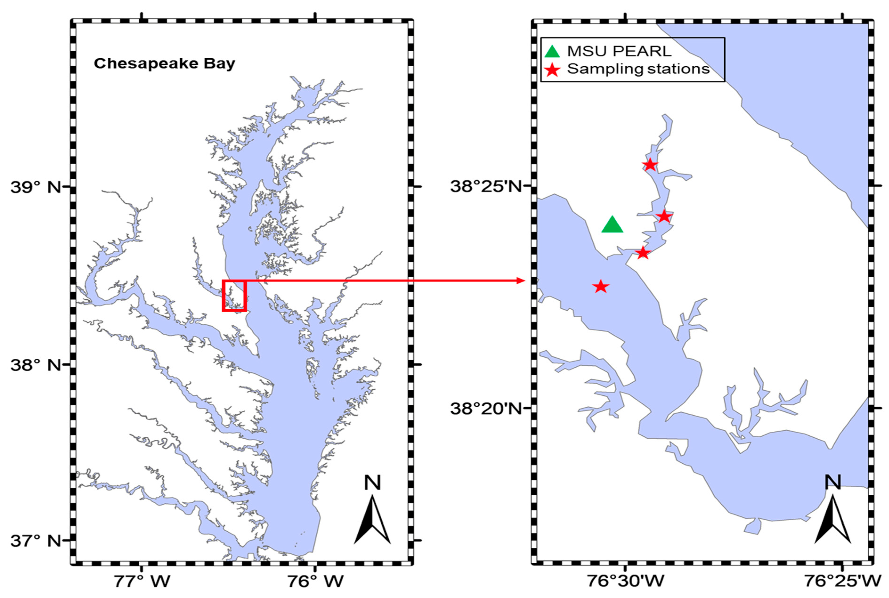
3.1. Sample Collections

The Patuxent River empties into the west side of Chesapeake Bay, 89.3 miles above the Virginia Capes. Commercial traffic consists chiefly of shellfish and shells and petroleum products. The river has natural depths of 25 to 30 feet in the approach, 30 to over 100 feet for 16 miles upstream. During the hot summers (23–29 °C) of 2021 and 2022, approximately forty C. chesapeakei of various sizes were collected using a twenty-foot handle metal strainer, and all river water was drained from the jellies. C. chesapeakei collection sizes and experimental methods were humane.
The coordinates of the collection were as follows (Figure 1):
  • 38°23.104 N, 076°30.025 W;
  • 38°23.470 N, 076°29.584 W;
  • 38°26.170 N, 076°29.386 W;
  • 38°25.481 N, 076°29.372 W.
Figure 1. The sampling area (Patuxent River) and its location relative to Chesapeake Bay, MD. The green triangle represents the location of the Pearl of Morgan State University. The red stars represent the stations where samples of C. chesapeakei were collected.
Microplastics 03 00015 g001

3.2. Contamination Prevention

All the various jellyfish caught, totaling 1478 mL (50 oz), were placed in two separate sterile 100 oz. glass jars; approximately 25 oz. (about 739.34 mL), a small amount of 100% glycerol (0.01), was added to the samples, and they were then frozen at –20 degree Celsius until aliquots were needed for examination. Small- and medium-sized C. chesapeakeis were humanely captured with the least amount required at the coordinates mentioned in this study. Their medusas ranged from 1/2 inches to 2 inches per diameter.

4. Procedures

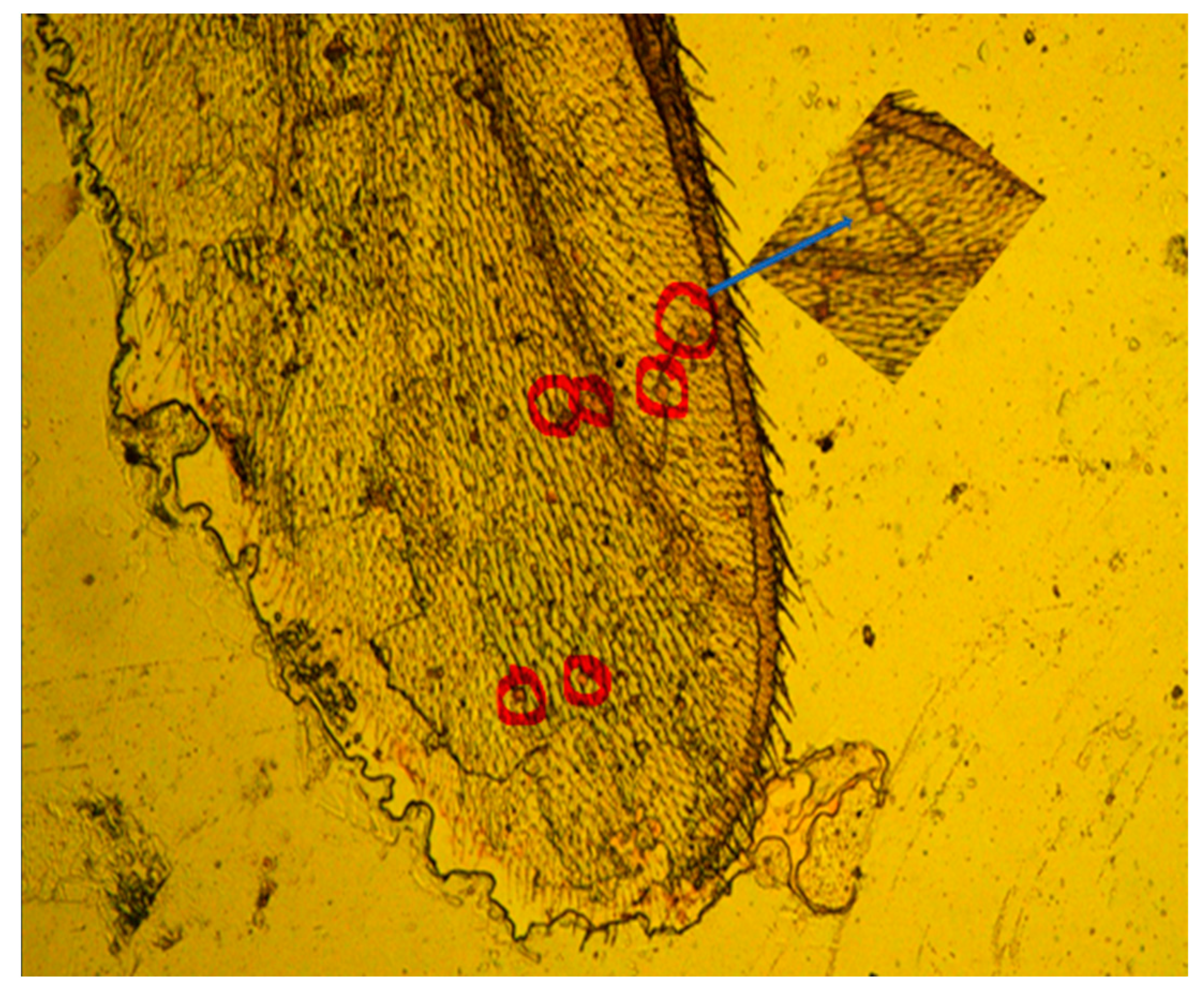
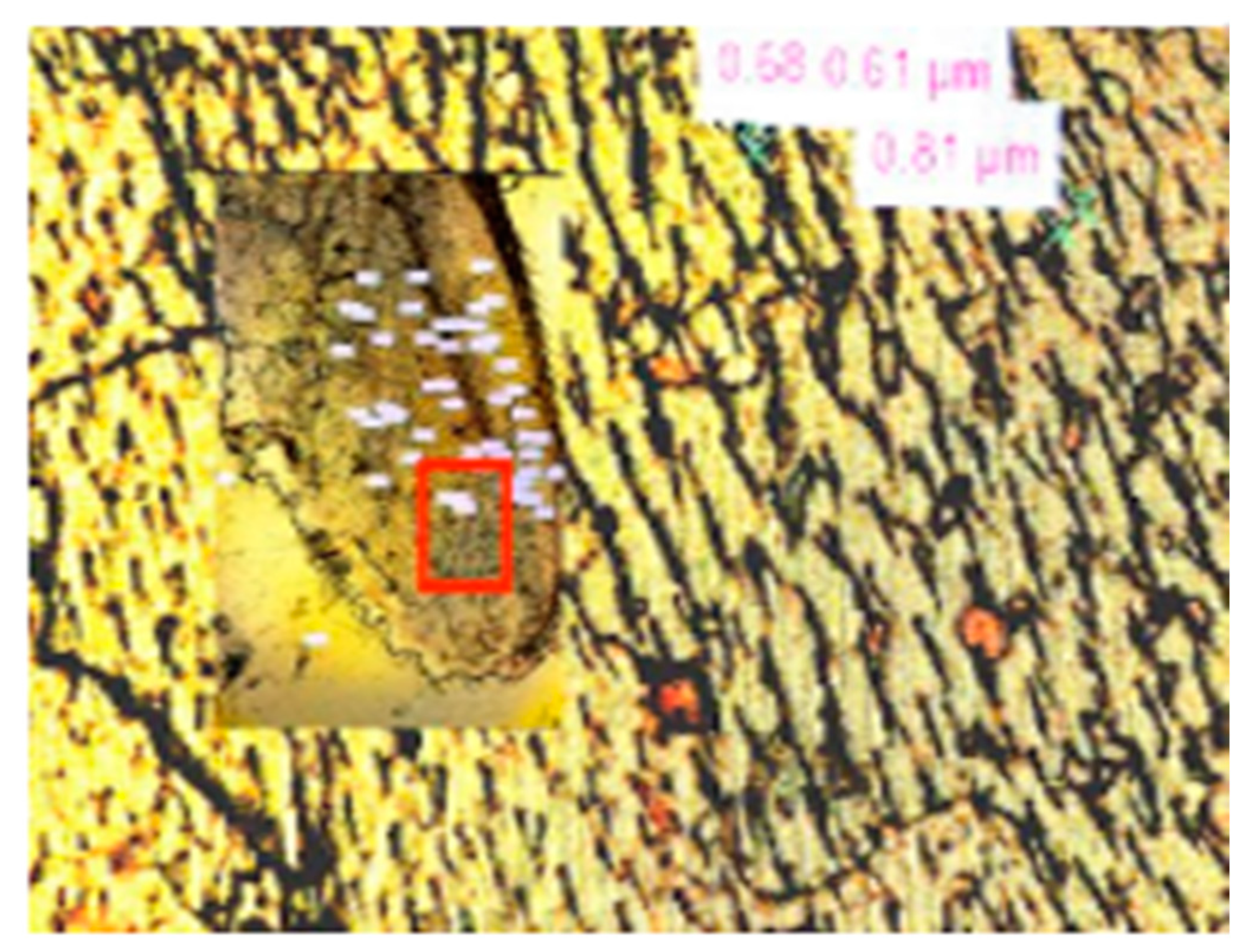
4.1. Identification of Native Microplastic in Jellyfish (Before GC-MS)

Rhodamine B Staining

Microplastics are transparent and invisible under an optical microscope. To track the distribution of MPs on the jellyfish membrane, they were stained with RhB. The purpose of using the RhB stain is that it is a polar solvent-soluble stain that will not interfere with microplastics and adheres to microplastics to provide a pinkish stain that is fluorescent.
Before using the slides, they were washed with distilled water to avoid airborne microplastics. We developed a procedure for microplastic staining and the jellyfish used in this study [45]. In brief, a portion of the jellyfish tissue from the field was placed on the distilled water-washed glass slides, and then a drop of 0.01% RhB in an ethanol stain was added. Clean glass sterile coverslips were applied. After an hour of incubation, the slides were visualized under an LED microscope.
The jellyfish captured weighed approximately 0.89 oz/per inch since jellyfish are an aqueous species. An amount of 75 mL of various sizes of jellyfish (0.01 percent RhB and ethanol) was added and then observed on 100 microscopic slides separately aliquoted with a glass dropper in 0.10 mL per microscopic slide. Each 0.10 mL liquid aliquoted to the microscopic slides was dried (for at least one hour) and then separately observed under the optical and LED microscopes. The RhB staining technique enables the inexpensive identification of various microplastics under an optical microscope, as noted by Tong et al., 2021 [48]. We observed jellyfish tissue centrifuged and digested with 30% hydrogen peroxide and tissue non-digested with no centrifugation. All of them visually contained microplastics with RhB staining.

4.2. Microplastic Identification Imaging

On a conservative average, there were ten visual pieces of MP per 0.10 mL of jellyfish per separate microscopic slide, which amounted to 1000 microplastics per 10 mL of the Chrysaora chesapeakei jellyfish.
A notable sample of a tentacle from the jellyfish inundated with MPs and the RHB staining identifying the MPs is imaged below. There was no digestion, as it would have destroyed the physical structure of the tentacle. The images collected reflected the fluorescence system at an excitation wavelength range of approximately 450–490 and 515–565 nm (Figure 2 and Figure 3).
The photo below shows an abundance of at least 60 microplastics sized 0.32 µm to 33.0 µm trapped and held within one very small C. chesapeakei’s (caught directly from the field) tentacle. The C. chesapeakei either as prey or involuntarily trapped the MPs as it came into bodily contact with the enormous amount of microplastics present in the Patuxent River of the Chesapeake Bay water column.
Our initial research demonstrated that various microplastics were present in C. chesapeakei, as shown in Figure 4 (60 pieces of microplastics in one partial C. chesapeakei tentacle); thus, we sought to then investigate further what were the chemical molecules of these microplastics and if they are volatile organic compounds. Moreover, we sought to review how it may impact the food web hierarchy of the C. chesapeakei since it is in the Chesapeake Bay, Patuxent River, where a significant number of fish and shellfish that eat the C. chesapeakei are frequently consumed by human beings.

4.3. Gas Chromatography Studies for Volatile Organic Compounds

From the confirmed results of microplastics within the jellyfish, we sought to develop an analytical procedure for determining the volatile organic compounds released and present from the degradation process of plastic debris found in the jellyfish. The method to capture volatile organic compounds captured in the C. chesapeakei from the Patuxent River, Chesapeake Bay, was based on gas chromatography/mass spectrometry (GC–MS), which utilized the headspace-Solid-Phase Micro-Extraction technique and also the methanol solvent extraction method. We determined that since chromatography/mass spectrometry (GC–MS) is commonly used to determine VOCs from direct contact with microplastics, we sought to investigate whether this method could detect VOCs from within the mucous body of the Chrysaora chesapeakei. Our results confirmed the presence of volatile organic compounds in the samples of the C. chesapeakei investigated. To the best of our knowledge, no other studies of the VOCs emitted from MPs from within Chrysaora chesapeakei have been conducted using the GC-MS method and/or have been reported to date in the literature.

4.4. GC-MS Techniques

All data were collected using a Thermo Fisher Scientific Trace 1310 Gas Chromatograph (GC) instrument equipped with a single-quadrupole mass spectrometer (MS, ISQ7000) along with a Flame Ionization Detector (FID) and Thermal Conductivity Detector (TCD) and performed at the Anderson commercial material evaluation laboratory in Columbia, Maryland.

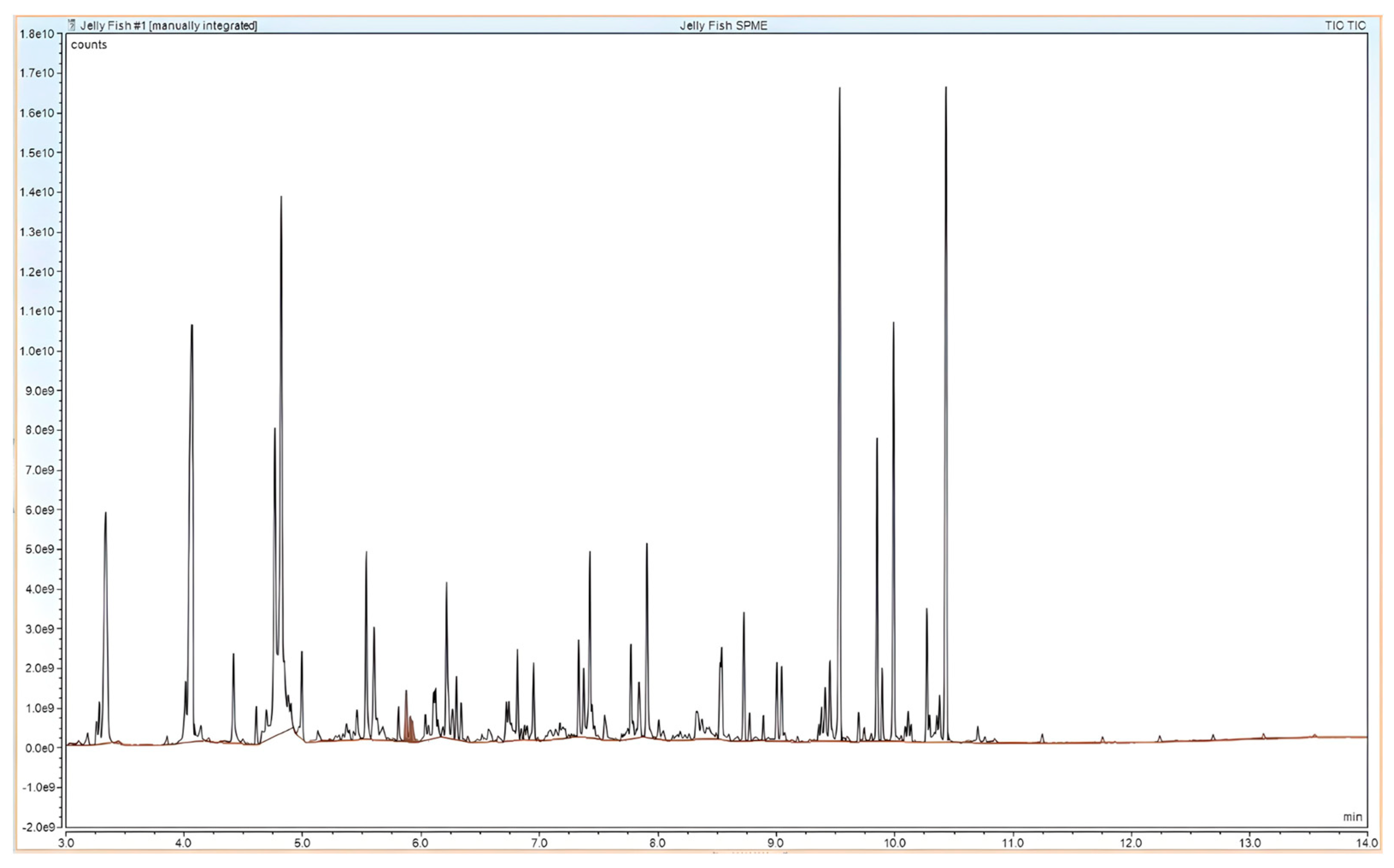
4.4.1. Headspace-SPME-GC-MS Method

An RTX-200 capillary column (29.45 m length × 0.25 mm I.D × 0.25 µm film thickness) was used for analysis. The contents of the jellyfish sample were analyzed by the headspace-Solid-Phase Micro-Extraction technique. The sample was taken in a 15 mL SPME vial. The vial was immersed in a water bath and heated to 80 °C and held at 80 °C for 30 min. Later, the SPME fiber was exposed to the sample (held over the sample without any contact with the sample) for 20 min to adsorb all the volatile organic compounds from the sample. Finally, the fiber was injected in the GC inlet port (inlet port temperature—260 °C) to desorb the volatile organic compounds into the GC column. The GC oven (column temperature) was kept at 40 °C for 1 min after the injection of the sample. The temperature was then increased to 280 °C at a rate of 20 °C/min and maintained at this temperature for 1 min. During the analysis, helium (1.4 mL/min) was used as the carrier gas. Qualitative analysis was performed using the National Institute of Standards and Technology (NIST) 17 MS library. Based on the relative area percent for each compound, a semi-quantitative analysis was performed (area of a peak is proportional to the amount of compound reaching the detector).

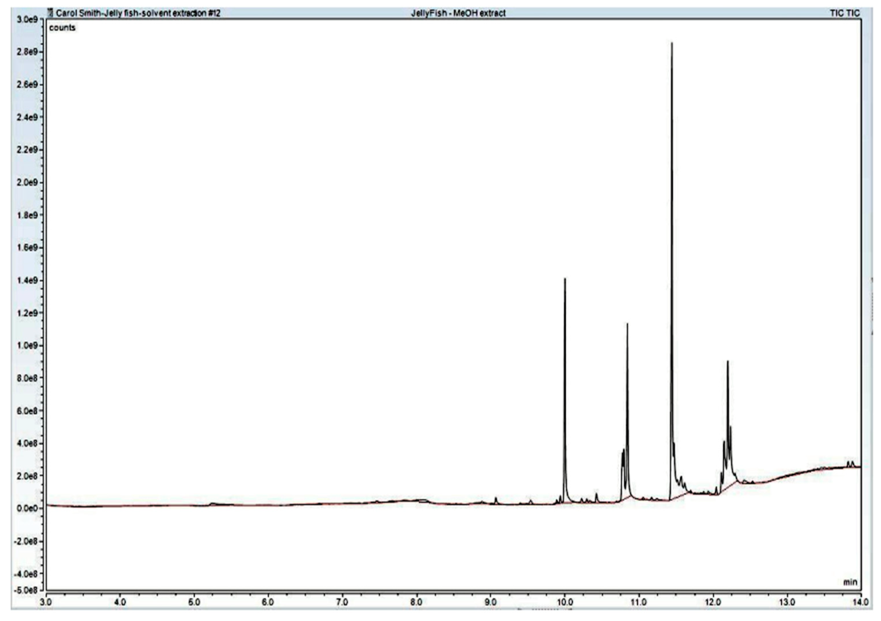
4.4.2. Solvent Extraction Method
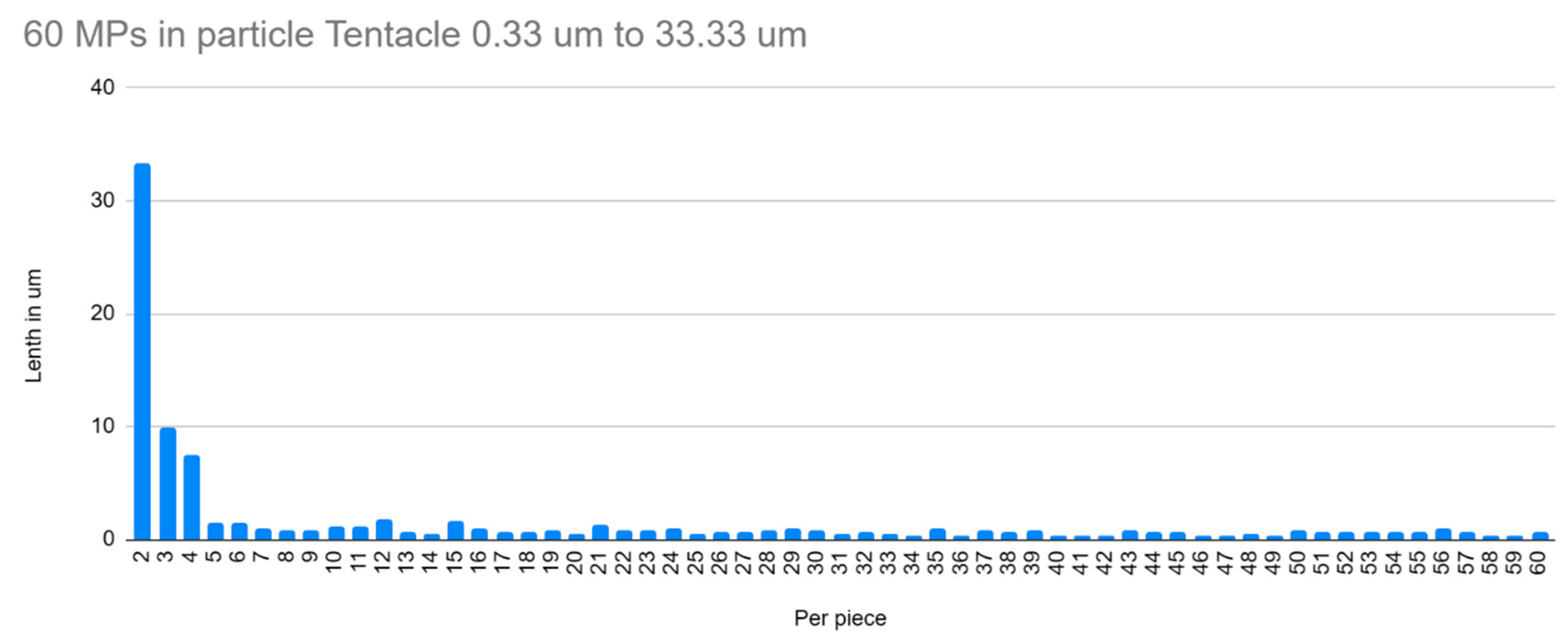
An RTX-200 capillary column (29.45 m length × 0.25 mm I.D × 0.25 µm film thickness) was used for analysis. The organic chemicals used from the jellyfish were methanol in the solvent extraction method. One microliter (µL) of the extracted solution was then injected into the GC with a 20:1 split. The GC oven was kept at 40 °C for 1 min during the injection of the sample. The temperature was then increased to 280 °C at a rate of 20 °C/min and maintained at this temperature for 2 min. During the analysis, helium (1.4 mL/min) was used as the carrier gas. Qualitative analysis was performed using the NIST 17 MS library. Based on the relative area percent for each compound, a semi-quantitative analysis was performed (area of a peak is proportional to the amount of compound reaching the detector).

5. Results

Table 1 lists the microplastic volatile organic compounds (VOC) found with GC-MS defined in the published papers and discovered in the Chrysora chesapeaki specimens while also utilizing GC-MS techniques. Our results have broken the result down into functional groups to include aromatic compounds, esters, ethers, ketones, aldehydes, alcohols, alkanes, alkenes, and siloxanes (Table 1).

6. Discussion

Marine microplastic litter of the Patuxent River, Chesapeake Bay, is a primary concern since it accumulates high hydrophobic organic and inorganic pollutants in a short time, making them an ideal attraction for the gelatinous body of the C. chesapeakei [60,61,62,63]. Consequently, the short accumulation period gives ample time for the C. chesapeakei, during its short life cycle of no longer than six months in the wild, to accumulate microplastics as it floats through the water column [63,64,65].
Earlier science publications guided this research to define specific volatile organic molecules derived from microplastics utilizing extraction with the direct injection of the jellyfish fluid into the GC-MS headspace (SPME) as well as the GC-MS MeOH techniques (Figure 3 and Figure 4), verifying that microplastic volatile organic compounds can become trapped in the C. chesapeakei jellyfish [49,50,51,52,53,54,55,56,57,58,59]. The total ion currency (TIC) graphs in Figure 5 and Figure 6 show that the VOC detection derived from the headspace GC-MS SPME direct injection technique was much more sensitive in capturing the presence of all types of VOCs. Compared to the MeOH application, which mainly detected alkenes and acids. SPME GC-MS detected many ketones, acids, aldehydes, alcohols, esters, alkanes, and alkenes. The TIC graphs provide the spectra of both. In addition, Table 1 and Table 2 also show functional group differences detected between these two GC-MS applications.
The GC-MS (SPME) method enabled precise recoveries for various microplastic VOCs. Between the detectors, the SPME-GC-MS method detected a higher amount of volatile organic compounds from microplastic within the jellyfish as listed from Table 1. Our GC-MS results of volatile organic microplastic compounds from within the C. chesapeakei are identical to MP VOCs reported in previous publications (Table 1). The total molecular MP VOC profile in Table 1 is broken into groups listed here: Ketone-Heptanone; Aldehydes-Octadecanal 9-Octadecenal, (Z)-13-Methyltetradecanal trans-2-Nonenal Nonanal, Benzeneacetaldehyde, Hexadecanal, Vanillin Phenolic, and Lilac aldehyde D Octanal; Alcohols-(R)-(-)-(Z)-14-Methyl-8-hexadecen-1-ol, 12-Methyl-E, E-2,13-octadecadien-1-ol,Ethanol, 2-(9-octadecenyloxy)-, (Z), 1-Undecanol, 2-Decen-1-ol,1,3,5-Pentanetriol, 3-methyl trans-2-Ethyl-2-hexen-1-ol, 6 trans-2-Dodecen-1-ol; Alkanes Decane, 2,3,5,8-tetramethyl, and 9-Oxabicyclo [6.1.0] nonane; Peroxide-2,5-Dimethylhexane-2,5-dihydroperoxide; Alkenes -5-Ethyl-1-nonene Cyclohexene and 1,5,5-trimethyl-6-acetylmethyl; Siloxanes-Cyclononasiloxane, octadecamethyl, Cyclohexasiloxane, dodecamethyl, Cyclotrisiloxane, hexamethyl Ester 3-Trifluoroacetoxypentadecane, 9,12,15-Octadecatrienoic acid, 2-(acetyloxy)-1-[(acetyloxy)methyl] ethyl ester, and (Z, Z,Z); Aromatic-1,1-Biphenyl and 3,4-diethyl; Acids-Acetic acid Tetradecanoic acid, 9-Hexadecenoic acid, Z-8-Methyl-9-tetradecenoic acid, n-Hexadecanoic acid, Dodecanoic acid, 3-hydroxy n-Hexadecanoic acid, Oleic Acid, and Octadecanoic acid; Ethers-Furan, and 2-pentyl Cyclic.

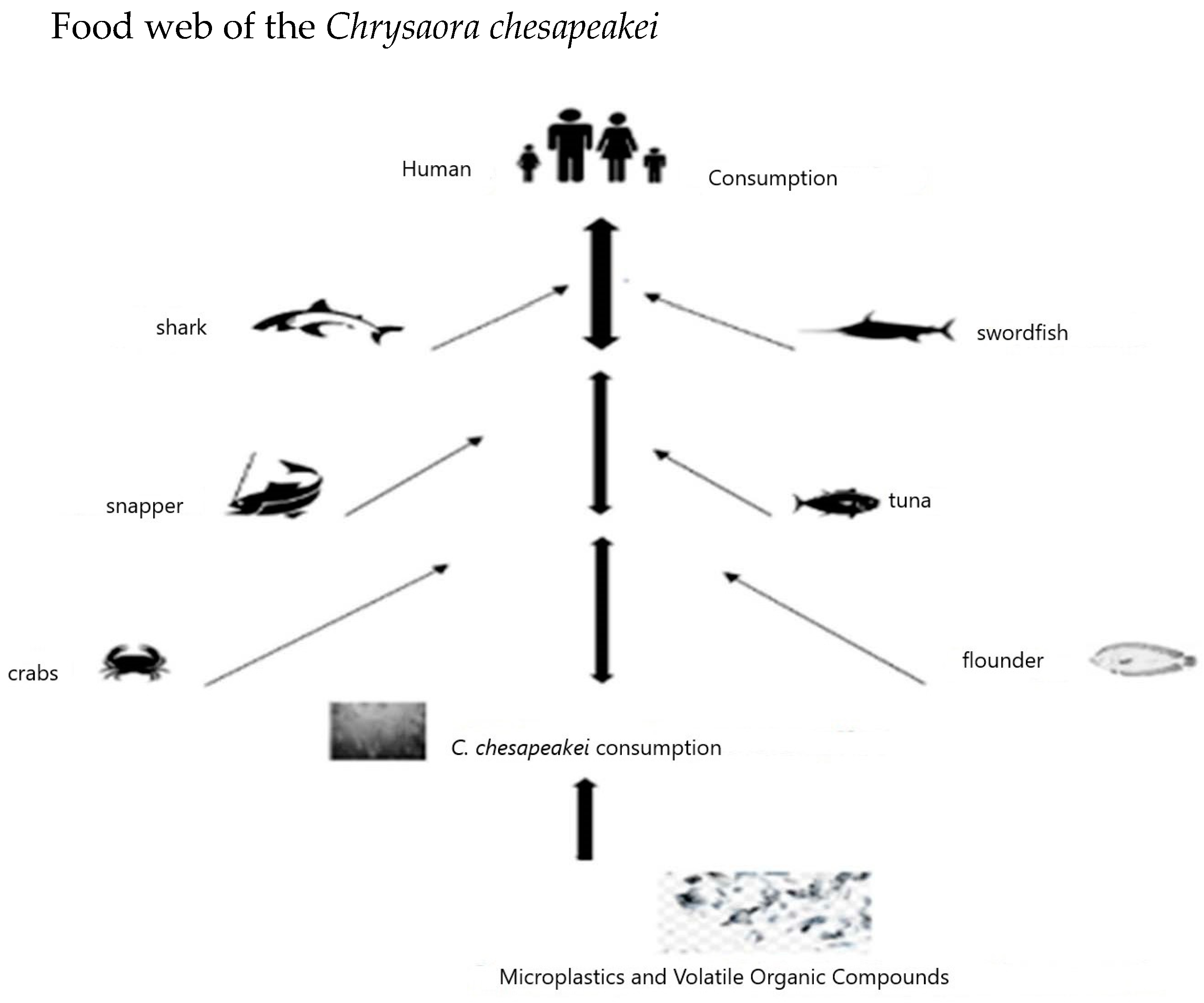
6.1. How Does the Chrysaora chesapeakei Microplastic Content Reach the Human Population from the Chesapeake Bay?

When the C. chesapeakei comes into contact voluntarily or involuntarily with abundant microplastics in the water, they become embedded in the jellyfish. The C. chesapeakei is consumed directly by crabs, swordfish, sharks, snappers, tuna, sunfish, and flounders (Figure 7). Humans then often directly consume these common fish and shellfish. Microplastics are well known to negatively impact humans and other aquatic animals biochemically.
The Patuxent River’s significant presence of microplastics in Chesapeake Bay, MD, increases C. chesapeakei’s vulnerability and exposure to floating microplastics and their VOCs. Notably, the jellyfish’s gelatinous bodies capture and accumulate microplastics from their surrounding environment. The VOCs released from C. chesapeakei jellyfish are known to be from many types of microplastic synthetic materials made of organic polymers whose chemical structure allows the production of a wide variety of resins such as polystyrene (PS), polyethylene terephthalate (PET), polyurethane (PU), polyvinyl chloride (PVC), polyethylene (PE), and polypropylene (PP). The release of VOCs due to the degradation of polymers is well known; for instance, the degradation of polyolefins generates VOCs belonging to the families of lactones, esters, ketones, and carboxylic acids, with the consequent reduction in molecular weight [66,67,68]. The MPs found in the C. chesapeakei were of reduced weight and size and produced molecules found in plastic VOCs.
Our objective was reached, which distinguished the chemical composition of microplastics. Exact functional groups concerning MP were also distinctly observed from the VOCs from the Chrysora chesapeakei. The compounds detected most were aldehydes, followed by alcohols and esters. The least volatile molecules detected were ethers and peroxides.
The limitation of this study is that similar VOCs are derived from many different plastics, thus reducing the ability to specify where each MP originated from in the rivers’ watery environment. Our analyses, while powerful, were demanding and expensive for accurate VOC analysis.
The bigger picture is that the microplastics trapped and present in the gelatinous tissue of the jellyfish demonstrate how harmful microplastics made of volatile organic compounds enter the aquatic food chain since C. chesapeakei are readily eaten by swordfish, flounder, hermit crabs, and sharks, which are then eaten commonly by humans. Through the fundamental laws of bioaccumulation, MPs within the C. chesapeakei are a direct vector to transfer VOCs throughout the food chain, resulting in its presence in foods consumed by humans. Recent studies have determined that interactions between microplastics and organic pollutants in aquatic environments can increase the toxicity of microplastics by a factor of 10. Further, because of the MP’s high durability and lack of biodegradability, it will disperse to those fish that eat the Chrysaora chesapeakei through the fundamental law of bioaccumulation as bioaccumulation refers to the increase in a pollutant, like microplastics, in an organism over time [69].

6.2. C. chesapeakei Tentacle from the Field

The capture of MP in the C. chesapeakei tentacle directly from the field in Figure 2 and Figure 3 demonstrates how the abundance of MPs becomes trapped in the smallest of spaces on the C. chesapeakei. JF, being 95% aqueous, is evaluated as a liquid; thus, our 100 slides with 0.10 mL per slide amount to approximately one thousand pieces of microplastic conservatively in 10 mL (0.33 oz (0.64 cu in)) of jellyfish. One small to medium medusas of jellyfish is usually 0.5 cu to 2 cu in inches, and the tentacles can be up to 12 inches long. An enormous amount of MP is present and trapped in one exceedingly small partial tentacle.

6.3. Volatile Compound Adverse Effects

The volatile organic compounds (VOCs) derived from MPs are significant contaminants in water matrices. Such VOCs are directly connected to physiological adverse effects on the human body, such as cancer, genetic mutations, eye irritation, nasopharyngeal mucosa, dizziness and headache, and short-term memory loss. MPs are considered a vector for transporting contaminants to organisms but can also release harmful substances such as additives or residual monomers, enhancing the toxicity effects in marine organisms [13,14,15].
In our jellyfish aquatic samples, the VOC molecules reported are in the range for the trace determination of VOCs. Research publications with reference to sampling, sampling preparation, method development, and analysis of diverse VOCs have defined and guided the chemical profile for the jellyfish’s microplastic VOCs. Direct aqueous injection through SPME GC-MS in our research recovered an immense amount of MP VOCs present compared to the MeOH GC-MS. It was successfully applied to the aqueous analysis of the jellyfish, which is made of 95 percent water. Further research on C.chesapeakei could help us understand microplastics by observing the effects of specific volatile organic compounds on the jellyfish [69].

7. Conclusions

C. chesapeakei’s hydrostatic skeleton and its gelatinous bodies capture floating microplastics to accumulate in their bodies. Chemically, plastics are polymers of hydrocarbons and volatile compounds that can cause damage to biota and human cells upon releasing toxic chemicals. Microplastics trapped in the gelatinous tissue of the jellyfish demonstrate how harmful microplastics made of volatile organic compounds can enter the aquatic food chain since C. chesapeakei is readily eaten by swordfish, flounder, hermit crabs, and sharks, which are then eaten commonly by humans. The degradation of microplastics through depolymerization can release VOCs in acidic environments and at low temperatures [70,71,72]. Recent literature has verified the toxic effects of MPs on human cell lines; moreover, people with tiny plastic particles lodged in blood vessels are more likely to experience heart attack, stroke, or death [73,74,75,76]. Further research on the effect of microplastic VOCs would assist in determining how microplastics continue impacting biota, human cells, and tissues.

Author Contributions

C.A.S.; Investigation, conceptualization, methodology, writing—original draft preparation, data curation, visualization, tables, figures, methodology, data writing, chemical analysis: S.P., S.M., and C.F.; investigation, Anderson Materials Evaluation Inc; GC-MS datawriting, writing—review and editing, methodology, validation: Anderson Materials Evaluation Inc. All authors have read and agreed to the published version of the manuscript.

Funding

This work was supported in part by the National Institute on Minority Health and Health Disparities of the National Institutes of Health under Award Number U54MD013376 and the National Science Foundation (award number 2022887) to Fan (PI) and (Co-PIs) Pramanik and Mandal in enhancing the research and education infrastructure of the Bioenvironmental Science Ph.D. program at Morgan State University: Microplastics in estuarine ecosystem Title III grant (College of Computer, Mathematical and Natural Sciences, MSU).

Institutional Review Board Statement

The animal collection protocol was approved by the Mary-land Department of Natural Resources for Morgan State University Pearl 10545 Mackall Road Rd St.Leonard, MD 20685. Permit No SCP-2024-37 expires 12/31/2024 for studies involving invertebrate.

Informed Consent Statement

All authors consent to this manuscript and the content is solely the responsibility of the authors.

Data Availability Statement

The data presented in this publication are available on request.

Acknowledgments

Authors are highly indebted to Anderson Materials Evaluation Lab, Inc., 9051 Red Branch Rd. Ste C, Columbia, MD, 21045, and Classical Music and Art, Patuxent Environmental & Aquatic Research Laboratory, Saint Leonard, MD, and the RCMI Core facilities of Morgan State University, Baltimore, MD Award (NIH 5U54MD013376 and 5UL1GM118973).

Conflicts of Interest

The authors declare no conflicts of interest.

References

  1. Lomonaco, T.; Manco, E.; Corti, A.; La Nasa, J.; Ghimenti, S.; Biagini, D.; Di Francesco, F.; Modugno, F.; Ceccarini, A.; Fuoco, R.; et al. Release of harmful volatile organic compounds (VOCs) from photo-degraded plastic debris: A neglected source of environmental pollution. J. Hazard. Mater. 2020, 394, 122596. [Google Scholar] [CrossRef]
  2. Chary, N.S.; Fernandez-Alba, A.R. Determination of volatile organic compounds in drinking and environmental waters. TrAC Trends Anal. Chem. 2012, 32, 60–75. [Google Scholar] [CrossRef]
  3. Wright, S.; Ulke, J.; Font, A.; Chan, K.; Kelly, F. Atmospheric microplastic deposition in an urban environment and an evaluation of transport. Environ. Int. 2019, 136, 105411. [Google Scholar] [CrossRef]
  4. Gewert, B.; Plassmann, M.M.; MacLeod, M. Pathways for degradation of plastic polymers floating in the marine environment. Environ. Sci. Process. Impacts 2015, 17, 1513–1521. [Google Scholar] [CrossRef]
  5. Eriksen, M.; Lebreton, L.C.; Carson, H.S.; Thiel, M.; Moore, C.J.; Borerro, J.C.; Galgani, F.; Ryan, P.G.; Reisser, J. Plastic Pollution in the World’s Oceans: More than 5 Trillion Plastic Pieces Weighing over 250,000 Tons Afloat at Sea. PLoS ONE 2014, 9, e111913. [Google Scholar] [CrossRef]
  6. Lebreton, L.-M.; Greer, S.; Borrero, J. Numerical modelling of floating debris in the world’s oceans. Mar. Pollut. Bull. 2012, 64, 653–661. [Google Scholar] [CrossRef]
  7. Barnes, D.K.A.; Galgani, F.; Thompson, R.C.; Barlaz, M. Accumulation and fragmentation of plastic debris in global environments. Philos. Trans. R. Soc. Lond. B Biol. Sci. 2009, 364, 1985–1998. [Google Scholar] [CrossRef]
  8. Frère, L.; Paul-Pont, I.; Rinnert, E.; Petton, S.; Jaffré, J.; Bihannic, I.; Soudant, P.; Lambert, C.; Huvet, A. Influence of environmental and anthropogenic factors on the composition, concentration and spatial distribution of microplastics: A case study of the Bay of Brest (Brittany, France). Environ. Pollut. 2017, 225, 211–222. [Google Scholar] [CrossRef]
  9. Ceccarini, A.; Corti, A.; Erba, F.; Modugno, F.; La Nasa, J.; Bianchi, S.; Castelvetro, V. The Hidden Microplastics: New Insights and Figures from the Thorough Separation and Characterization of Microplastics and of Their Degradation Byproducts in Coastal Sediments. Environ. Sci. Technol. 2018, 52, 5634–5643. [Google Scholar] [CrossRef]
  10. Horton, A.A.; Walton, A.; Spurgeon, D.J.; Lahive, E.; Svendsen, C. Microplastics in freshwater and terrestrial environments: Evaluating the current understanding to identify the knowledge gaps and future research priorities. Sci. Total. Environ. 2017, 586, 127–141. [Google Scholar] [CrossRef]
  11. Castelvetro, V.; Corti, A.; Bianchi, S.; Ceccarini, A.; Manariti, A.; Vinciguerra, V. Quantification of poly(ethylene terephthalate) micro- and nanoparticle contaminants in marine sediments and other environmental matrices. J. Hazard. Mater. 2019, 385, 121517. [Google Scholar] [CrossRef]
  12. Fendall, L.S.; Sewell, M.A. Contributing to marine pollution by washing your face: Microplastics in facial cleansers. Mar. Pollut. Bull. 2009, 58, 1225–1228. [Google Scholar] [CrossRef]
  13. Teuten, E.L.; Saquing, J.M.; Knappe, D.R.; Barlaz, M.A.; Jonsson, S.; Björn, A.; Rowland, S.J.; Thompson, R.C.; Galloway, T.S.; Yamashita, R.; et al. Transport and release of chemicals from plastics to the environment and to wildlife. Philos. Trans. R. Soc. B Biol. Sci. 2009, 364, 2027–2045. [Google Scholar] [CrossRef] [PubMed]
  14. Hermabessiere, L.; Dehaut, A.; Paul-Pont, I.; Lacroix, C.; Jezequel, R.; Soudant, P.; Duflos, G. Occurrence and effects of plastic additives on marine environments and organisms: A review. Chemosphere 2017, 182, 781–793. [Google Scholar] [CrossRef] [PubMed]
  15. Zhang, Z.; Ran, C.; Ding, Q.-W.; Liu, H.-L.; Xie, M.-X.; Yang, Y.-L.; Xie, Y.-D.; Gao, C.-C.; Zhang, H.-L.; Zhou, Z.-G. Ability of prebiotic polysaccharides to activate a HIF1α-antimicrobial peptide axis determines liver injury risk in zebrafish. Commun. Biol. 2019, 2, 1–17. [Google Scholar] [CrossRef] [PubMed]
  16. Geyer, R.; Jambeck, J.R.; Law, K.L. Production, use, and fate of all plastics ever made. Sci. Adv. 2017, 3, e1700782. [Google Scholar] [CrossRef] [PubMed]
  17. Smith, M.; Love, D.C.; Rochman, C.M.; Neff, R.A. Microplastics in Seafood and the Implications for Human Health. Curr. Environ. Health Rep. 2018, 5, 375–386. [Google Scholar] [CrossRef] [PubMed]
  18. Toussaint, B.; Raffael, B.; Angers-Loustau, A.; Gilliland, D.; Kestens, V.; Petrillo, M.; Rio-Echevarria, I.M.; Van den Eede, G. Review of micro- and nanoplastic contamination in the food chain. Food Addit. Contam. Part A 2019, 36, 639–673. [Google Scholar] [CrossRef] [PubMed]
  19. Bay Nettle (Chrysaora chesapeakei)—Jungle Dragon. (n.d.). Available online: https://www.jungledragon.com/specie/26324/bay_nettle.html (accessed on 10 March 2024).
  20. Botterell, Z.L.R.; Beaumont, N.; Dorrington, T.; Steinke, M.; Thompson, R.C.; Lindeque, P.K. Bioavailability and effects of microplastics on marine zooplankton: A review. Environ. Pollut. 2019, 245, 98–110. [Google Scholar] [CrossRef] [PubMed]
  21. Leung, J.; Chan, K.Y.K. Microplastics reduced posterior segment regeneration rate of the polychaete Perinereis aibuhitensis. Mar. Pollut. Bull. 2018, 129, 782–786. [Google Scholar] [CrossRef]
  22. Murphy, F.; Quinn, B. The effects of microplastic on freshwater Hydra attenuata feeding, morphology & reproduction. Environ. Pollut. 2018, 234, 487–494. [Google Scholar]
  23. Lo, H.K.A.; Chan, K.Y.K. Negative effects of microplastic exposure on growth and development of Crepidula onyx. Environ. Pollut. 2018, 233, 588–595. [Google Scholar] [CrossRef] [PubMed]
  24. Ziajahromi, S.; Kumar, A.; Neale, P.A.; Leusch, F.D. Environmentally relevant concentrations of polyethylene microplastics negatively impact the survival, growth and emergence of sediment-dwelling invertebrates. Environ. Pollut. 2018, 236, 425–431. [Google Scholar] [CrossRef] [PubMed]
  25. Cole, M.; Lindeque, P.; Fileman, E.; Halsband, C.; Galloway, T.S. The Impact of Polystyrene Microplastics on Feeding, Function and Fecundity in the Marine Copepod Calanus helgolandicus. Environ. Sci. Technol. 2015, 49, 1130–1137. [Google Scholar] [CrossRef] [PubMed]
  26. Sussarellu, R.; Suquet, M.; Thomas, Y.; Lambert, C.; Fabioux, C.; Pernet, M.E.J.; Le Goïc, N.; Quillien, V.; Mingant, C.; Epelboin, Y.; et al. Oyster reproduction is affected by exposure to polystyrene microplastics. Proc. Natl. Acad. Sci. USA 2016, 113, 2430–2435. [Google Scholar] [CrossRef] [PubMed]
  27. Dinasquet, J.; Titelman, J.; Møller, L.F.; Setälä, O.; Granhag, L.; Andersen, T.; Båmstedt, U.; Haraldsson, M.; Hosia, A.; Katajisto, T.; et al. Cascading effects of the ctenophore Mnemiopsis leidyi on the planktonic food web in a nutrient-limited estuarine system. Mar. Ecol. Prog. Ser. 2012, 460, 49–61. [Google Scholar] [CrossRef]
  28. McNamara, M.; Lonsdale, D.; Cerrato, R. Role of eutrophication in structuring planktonic communities in the presence of the ctenophore Mnemiopsis leidyi. Mar. Ecol. Prog. Ser. 2014, 510, 151–165. [Google Scholar] [CrossRef]
  29. Wright, R.M.; Le Quéré, C.; Buitenhuis, E.; Pitois, S.; Gibbons, M.J. Role of jellyfish in the plankton ecosystem revealed using a global ocean biogeochemical model. Biogeosciences 2021, 18, 1291–1320. [Google Scholar] [CrossRef]
  30. GESAMP (2016) Sources, Fate, and Effects of Microplastics in the Marine Environment: Part Two of a Global Assessment Joint Group of Experts on the Scientific Aspects of Marine Environmental Protection. Rep. Stud. No. 93. Sources, Fate and Effects of Microplastics in the Marine Environment: A Global Assessment|UNEP—UN Environment Programme. Available online: http://www.gesamp.org/site/assets/files/1275/sources-fate-and-effects-of-microplastics-in-the-marine-environment-part-2-of-a-global-assessment-en.pdf (accessed on 10 March 2024).
  31. Tanaka, K.; Takada, H.; Yamashita, R.; Mizukawa, K.; Fukuwaka, M.-A.; Watanuki, Y. Accumulation of plastic-derived chemicals in tissues of seabirds ingesting marine plastics. Mar. Pollut. Bull. 2013, 69, 219–222. [Google Scholar] [CrossRef] [PubMed]
  32. Setälä, O.; Fleming-Lehtinen, V.; Lehtiniemi, M. Ingestion and transfer of microplastics in the planktonic food web. Environ. Pollut. 2014, 185, 77–83. [Google Scholar] [CrossRef]
  33. Mato, Y.; Isobe, T.; Takada, H.; Kanehiro, H.; Ohtake, C.; Kaminuma, T. Plastic Resin Pellets as a Transport Medium for Toxic Chemicals in the Marine Environment. Environ. Sci. Technol. 2000, 35, 318–324. [Google Scholar] [CrossRef]
  34. García-Gómez, J.C.; Garrigós, M.; Garrigós, J. Plastic as a Vector of Dispersion for Marine Species With Invasive Potential. A Review. Front. Ecol. Evol. 2021, 9. [Google Scholar] [CrossRef]
  35. Americus, B.; Lotan, T.; Bartholomew, J.L.; Atkinson, S.D. A comparison of the structure and function of nematocysts in free-living and parasitic cnidarians (Myxozoa). Int. J. Parasitol. 2020, 50, 763–769. [Google Scholar] [CrossRef] [PubMed]
  36. Thorington, G.U.; Hessinger, D.A. Efferent Mechanisms of Discharging Cnidae: II. A Nematocyst Release Response in the Sea Anemone Tentacle. Biol. Bull. 1998, 195, 145–155. [Google Scholar] [CrossRef] [PubMed]
  37. Lindstedt, K.J. Biphasic Feeding Response in a Sea Anemone: Control by Asparagine and Glutathione. Science 1971, 173, 333–334. [Google Scholar] [CrossRef]
  38. Wei, X.-F.; Bohlén, M.; Lindblad, C.; Hedenqvist, M.; Hakonen, A. Microplastics generated from a biodegradable plastic in freshwater and seawater. Water Res. 2021, 198, 117123. [Google Scholar] [CrossRef]
  39. NOAA. What Are Jellyfish Bodies Made from? What Are Jellyfish Made of? NOAA: Washington, DC, USA, 2023.
  40. Purcell, J.E.; Decker, M.B. Effects of climate on relative predation by scyphomedusae and ctenophores on copepods in Chesapeake Bay during 1987–2000. Limnol. Oceanogr. 2005, 50, 376–387. [Google Scholar] [CrossRef]
  41. Morabito, R.; Marino, A.; La Spada, G. Nematocytes’ activation in Pelagia noctiluca (Cnidaria, Scyphozoa) oral arms. J. Comp. Physiol. A 2012, 198, 419–426. [Google Scholar] [CrossRef] [PubMed]
  42. Morabito, R.; Marino, A.; Dossena, S.; La Spada, G. Nematocyst discharge in Pelagia noctiluca (Cnidaria, Scyphozoa) oral arms can be affected by lidocaine, ethanol, ammonia and acetic acid. Toxicon 2014, 83, 52–58. [Google Scholar] [CrossRef] [PubMed]
  43. Ormond, R.F.; Caldwell, S. The effect of oil pollution on the reproduction and feeding behavior of the sea anemone Actinia equina. Mar. Pollut. Bull. 1982, 13, 118–122. [Google Scholar] [CrossRef]
  44. Available online: https://www.seatemperature.org/north-america/united-states/solomons (accessed on 10 March 2024).
  45. Smith, C.; Denaro, F.; Fan, C.; Pramanik, S. The Effective Use of the Inexpensive LED Microscope with Rhodamine Blue Staining to Identify Microplastics. Microsc. Today 2023, 31, 36–37. [Google Scholar] [CrossRef]
  46. Sauls-Smith, C.A.; Pramanik, S.; Drichko, N.; Fan, C. The Occurrence of Microplastics in Chrysaora Chesapeakei in the Patuxent River, Maryland; Science Symposium Morgan State University: Baltimore, MD, USA, 2022. [Google Scholar]
  47. Gemmell, B.J.; Du Clos, K.T.; Colin, S.P.; Sutherland, K.R.; Costello, J.H. The most efficient metazoan swimmer creates a ‘virtual wall’ to enhance performance. Proc. R. Soc. B Biol. Sci. 2021, 288, 20202494. [Google Scholar] [CrossRef] [PubMed]
  48. Tong, H.; Jiang, Q.; Zhong, X.; Hu, X. Rhodamine B dye staining for visualizing microplastics in laboratory-based studies. Environ. Sci. Pollut. Res. 2020, 28, 4209–4215. [Google Scholar] [CrossRef]
  49. Cabanes, A.; Valdés, F.; Fullana, A. A review on VOCs from recycled plastics. Sustain. Mater. Technol. 2020, 25, e00179. [Google Scholar] [CrossRef]
  50. Han, X.; Wu, Y.; Chen, G.; Wang, X.; Yuan, W.; Lv, Z. Optimization of a headspace solid-phase microextraction-gas chromatography–mass spectrometry procedure for odor compounds from polyolefin resin used in plastic food packaging. Packag. Technol. Sci. 2020, 33, 515–529. [Google Scholar] [CrossRef]
  51. Fabris, S.; Freire, M.T.d.A.; Wagner, R.; Reyes, F.G. A method to determine volatile contaminants in polyethylene terephthalate (PET) packages by HDC-GC-FID and its application to post-consumer materials. Food Sci. Technol. 2010, 30, 1046–1055. [Google Scholar] [CrossRef]
  52. Strangl, M.; Fell, T.; Schlummer, M.; Maeurer, A.; Buettner, A. Characterization of odorous contaminants in post-consumer plastic packaging waste using multidimensional gas chromatographic separation coupled with olfactometric resolution. J. Sep. Sci. 2017, 40, 1500–1507. [Google Scholar] [CrossRef] [PubMed]
  53. Camacho, W.; Karlsson, S. Quality-determination of recycled plastic packaging waste by identification of contaminants by GC–MS after microwave assisted extraction (MAE). Polym. Degrad. Stab. 2000, 71, 123–134. [Google Scholar] [CrossRef]
  54. Chen, Z.-F.; Lin, Q.-B.; Song, X.-C.; Chen, S.; Zhong, H.-N.; Nerin, C. Discrimination of Virgin and Recycled Polyethylene Based on Volatile Organic Compounds Using a Headspace GC-MS Coupled with Chemometrics Approach. Food Packag. Shelf Life 2020, 26, 100553. [Google Scholar] [CrossRef]
  55. He, Z.; Li, G.; Chen, J.; Huang, Y.; An, T.; Zhang, C. Pollution characteristics and health risk assessment of volatile organic compounds emitted from different plastic solid waste recycling workshops. Environ. Int. 2015, 77, 85–94. [Google Scholar] [CrossRef]
  56. Curran, K.; Strlič, M. Polymers and volatiles: Using VOC analysis for the conservation of plastic and rubber objects. Stud. Conserv. 2014, 60, 1–14. [Google Scholar] [CrossRef]
  57. Huang, Z.; Lin, H.; Peng, X.; Wang, Z.; Wang, R.; Li, Z.; Fang, P. Aging behaviors of silicone rubber composite materials under outdoor environment. J. Wuhan Univ. Technol. Sci. Ed. 2016, 31, 1289–1293. [Google Scholar] [CrossRef]
  58. Nerìn, C.; Albiñana, J.; Philo, M.R.; Castle, L.; Raffael, B.; Simoneau, C. Evaluation of some screening methods for the analysis of contaminants in recycled polyethylene terephthalate flakes. Food Addit. Contam. 2003, 20, 668–677. [Google Scholar] [CrossRef]
  59. Mihreteab, M.; Stubblefield, B.A.; Gilbert, E.S. Microbial bioconversion of thermally depolymerized polypropylene by Yarrowia lipolytica for fatty acid production. Appl. Microbiol. Biotechnol. 2019, 103, 7729–7740. [Google Scholar] [CrossRef]
  60. Oseji, O.F.; Fan, C.; Chigbu, P. Composition and Dynamics of Phytoplankton in the Coastal Bays of Maryland, USA, Revealed by Microscopic Counts and Diagnostic Pigments Analyses. Water 2019, 11, 368. [Google Scholar] [CrossRef]
  61. Hirose, E.; Sakai, D.; Iida, A.; Obayashi, Y.; Nishikawa, J. Exumbrellar Surface of Jellyfish: A Comparative Fine Structure Study with Remarks on Surface Reflectance. Zool. Sci. 2021, 38, 170–178. [Google Scholar] [CrossRef]
  62. Britannica, T. Editors of Encyclopedia (2009, April 1). epithelium. Encyclopedia Britannica. Available online: https://www.britannica.com/science/epithelium (accessed on 10 March 2024).
  63. Hydrophobic Interactions. (2020, August 15). Available online: https://chem.libretexts.org/@go/page/1506 (accessed on 10 March 2024).
  64. Gall, S.; Thompson, R. The impact of debris on marine life. Mar. Pollut. Bull. 2015, 92, 170–179. [Google Scholar] [CrossRef]
  65. Wu, Y.; Wang, S.; Wu, L.; Yang, Y.; Yu, X.; Liu, Q.; Liu, X.; Li, Y.; Wang, X. Vertical distribution and river-sea transport of microplastics with tidal fluctuation in a subtropical estuary, China. Sci. Total. Environ. 2022, 822, 153603. [Google Scholar] [CrossRef]
  66. Rabek, J.F. Polymer Photodegradation: Mechanisms and Experimental Methods; Springer: Dordrecht, The Netherlands, 1995. [Google Scholar]
  67. Ray, S.; Cooney, R.P. Thermal Degradation of Polymer and Polymer Composites. In Handbook of Environmental Degradation of Materials; Elsevier: Amsterdam, The Netherlands, 2018. [Google Scholar]
  68. Gardette, M.; Perthue, A.; Gardette, J.; Janecska, T.; Földes, E.; Pukánszky, B.; Therias, S. Photo- and thermal-oxidation of polyethylene: Comparison of mechanisms and influence of unsaturation content. Polym. Degrad. Stabil. 2013, 98, 2383–2390. [Google Scholar] [CrossRef]
  69. McMullen, K.; Vargas, F.H.; Calle, P.; Alavarado-Cadena, O.; Pakhomov, E.A.; Alava, J.J. Modelling microplastic bioaccumulation and biomagnification potential in the Galápagos penguin ecosystem using Ecopath and Ecosim (EwE) with Ecotracer. PLoS ONE 2024, 19, e0296788. [Google Scholar] [CrossRef]
  70. Kang, P.; Wu, P.; Jin, Y.; Shi, S.; Gao, D.; Chen, G.; Li, Q. Formation and Emissions of Volatile Organic Compounds from Homo-PP and Co-PP Resins during Manufacturing Process and Accelerated Photoaging Degradation. Molecules 2020, 25, 2761. [Google Scholar] [CrossRef]
  71. Romera-Castillo, C.; Lucas, A.; Mallenco-Fornies, R.; Briones-Rizo, M.; Calvo, E.; Pelejero, C. Abiotic plastic leaching contributes to ocean acidification. Sci. Total Environ. 2023, 854, 158683. [Google Scholar] [CrossRef] [PubMed]
  72. Available online: https://www.nationalgeographic.com/science/article/plastic-breaks-down-in-ocean-after-all-and-fast (accessed on 10 March 2024).
  73. Yee, M.S.-L.; Hii, L.-W.; Looi, C.K.; Lim, W.-M.; Wong, S.-F.; Kok, Y.-Y.; Tan, B.-K.; Wong, C.-Y.; Leong, C.-O. Impact of Microplastics and Nanoplastics on Human Health. Nanomaterials 2021, 11, 496. [Google Scholar] [CrossRef] [PubMed]
  74. Kozlov, M. Landmark study links microplastics to serious health problems. Nature 2024. [Google Scholar] [CrossRef] [PubMed]
  75. Marfella, R.; Prattichizzo, F.; Sardu, C.; Fulgenzi, G.; Graciotti, L.; Spadoni, T.; D’onofrio, N.; Scisciola, L.; La Grotta, R.; Frigé, C.; et al. Microplastics and Nanoplastics in Atheromas and Cardiovascular Events. N. Engl. J. Med. 2024, 390, 900–910. [Google Scholar] [CrossRef]
  76. Braun, A.; Seitz, H. Uptake and Cellular Effects of Polymethylmethacrylate on Human Cell Lines. Microplastics 2024, 3, 205–216. [Google Scholar] [CrossRef]
Figure 2. Microplastics found in small tentacles. Microplastics are (stained dark pinkish red using RhB staining technique) at 40× magnification found by a regular microscope.
Microplastics 03 00015 g002
Figure 3. Mapped sizes found in a piece of tentacle from C. chesapeakei from the field in July 2021. The MP size and location within fluorescence mapping using Nikon Eclipse 90i upright microscope equipped with BF, FL, and DIC optics, a motorized focus, and a Nikon 100W mercury power supply; a Lumenera 3 digital camera; Image-Pro Plus image analysis software; and a Dell OptiPlex 5040 workstation.
Microplastics 03 00015 g003
Figure 4. A total of 60 pieces of microplastics found in the small tentacle from Figure 2 and Figure 3 are shown above with MPs sized 0.32 µm to 33.32 µm contained within the C. chesapeakei tentacle.
Microplastics 03 00015 g004
Figure 5. TIC of VOCs from jellyfish samples with He GCMS-headspace (SPME).
Microplastics 03 00015 g005
Figure 6. TIC of VOCs from jellyfish sample with MeOH solvent.
Microplastics 03 00015 g006
Figure 7. C. chesapeakei encounters microplastics in the food web, and they are eaten by fish and crabs of the Chesapeake Bay, which humans frequently eat.
Microplastics 03 00015 g007
Table 1. GC-MS identification of MP volatile organic compounds from C. chesapeakei with publication verification of the VOC as a microplastic volatile compound with method distinguished as either SPME or MeOH.
Volatile Organic Compounds from within the C. chesapeakeiPublications Verifying the MP VOC Distinction
Ketone (SPME)
Heptanone
Cabanes et al., 2020 [48]
Aldehyde (SPME)
Octadecanal Cababes et al., 2020 [48], Han et al., 2020 [49]
9-Octadecenal, (Z)-Cababes et al., 2020, Han et al., 2020
13-Methyltetradecanal Cababes et al., 2020, Han et al., 2020
trans-2-NonenalCababes et al., 2020, Han et al., 2020
NonanalCabanes et al., 2020, Han et al., 2020,Fabris et al. 2008 [50]
Benzeneacetaldehyde(Cababes et al., 2020, Han et al., 2020)
HexadecanalCababes et al., 2020
Vanillin PhenolicCababes et al., 2020, Han et al., 2020, Stragel et al. 2017 [51]
Lilac aldehyde D
OctanalCabanes et al., 2020, Han 2020, Fabris et al., 2008
Alcohols (SPME)
(R)-(-)-(Z)-14-Methyl-8-hexadecen-1-olCabanes et al., 2020, Camacho and Karlsson 2000 [52]
12-Methyl-E, E-2,13-octadecadien-1-olCamacho and Karlsson 2000 [53]
Ethanol, 2-(9-octadecenyloxy)-, (Z)Cabanes et al., 2020, Han et al., 2020
1-UndecanolHan et al., 2020
2-Decen-1-olCabanes et al., 2020
1,3,5-Pentanetriol, 3-methyl
trans-2-Ethyl-2-hexen-1-olCabanes et al., 2020, Han et al., 2020
trans-2-Dodecen-1-olCabanes et al., 2020, Camacho and Karlsson 2000
Alkanes (SPME)
Decane, 2,3,5,8-tetramethyl Chen et al. 2020 [54], Han et al., 2020
9-Oxabicyclo [6.1.0] nonaneCabanes et al., 2020, Han et al., 2020
Peroxide (SPME)
2,5-Dimethylhexane-2,5-dihydroperoxide Han et al., 2020
Alkene (SPME)
5-Ethyl-1-noneneCabanes et al., 2020
Cyclohexene, 1,5,5-trimethyl-6-acetylmethyl He et al., 2015 [55]
Siloxanes (SPME)
Cyclononasiloxane, octadecamethylCurran and Strlic 2015 [56], Huang et al. 2016 [57]
Cyclohexasiloxane, dodecamethylHuang, Zhen, Lin, Peng et al. 2016
Cyclotrisiloxane, hexamethylHuang, Zhen, Lin, Peng et al. 2016
Ester (SPME and MeOH)
3-TrifluoroacetoxypentadecaneNerin et al., 2001 [58]
9,12,15-Octadecatrienoic acid, 2-(acetyloxy)-1-[(acetyloxy)methyl] ethyl ester, (Z, Z,Z)Camacho and Karlsson 2000
Aromatic (SPME)
1,1-Biphenyl, 3,4-diethyl Camacho and Karlsson 2000
Acids (MeOH)
Acetic acidCabanes et al., 2020
Tetradecanoic acidCabanes et al., 2020
9-Hexadecenoic acidCabanes et al., 2020, Camacho and Karlsson 2000
Z-8-Methyl-9-tetradecenoic acidCabanes et al., 2020
n-Hexadecanoic acidCabanes et al., 2020, Camacho and Karlsson 2000
Dodecanoic acid, 3-hydroxyCabanes et al., 2020, Camacho and Karlsson 2000
n-Hexadecanoic acidCabanes et al., 2020, Camacho and Karlsson 2000
Oleic AcidMihreteab et al. 2019 [59]
Octadecanoic acid MeOHCabanes et al., 2020, Fabris et al., 2008, Camacho Karlsson 2000
Ether (SPME)
Furan, 2-pentyl CyclicCabanes et al., 2020
Table 2. Relative area percentage of MP VOCs using SPME and MeOH.
Chemical GroupsSPMEMeOH
Ketone0.25
Aldehydes25.81
Alcohols6.63
Alkanes7.57
Siloxanes4.8
Esters0.3
Alkene0.63.71
Aromatic0.08
Acid3.0630.71
Ether0.11
Other50.7965.58
Disclaimer/Publisher’s Note: The statements, opinions and data contained in all publications are solely those of the individual author(s) and contributor(s) and not of MDPI and/or the editor(s). MDPI and/or the editor(s) disclaim responsibility for any injury to people or property resulting from any ideas, methods, instructions or products referred to in the content.

