


Abstract
Fetal therapy has evolved into a rapidly advancing field with the potential to alter the natural history of many severe congenital and genetic disorders before irreversible injury occurs. Progress in prenatal imaging, molecular diagnostics, and fetal intervention techniques now enables the earlier identification of disease and, in select settings, targeted prenatal treatment. This review synthesizes the current landscape of fetal therapies, spanning established surgical interventions for structural anomalies and emerging biologic and molecular approaches, including enzyme replacement therapy, stem cell-based strategies, gene therapy, and gene editing. The intrauterine environment provides a distinct therapeutic context, with developmental plasticity, immune immaturity, enhanced tissue accessibility, and relatively permissive central nervous system exposure that together define a time-sensitive window for intervention. Preclinical studies and early clinical experience across both structural anomalies and genetic disorders, including lysosomal storage disorders, osteogenesis imperfecta, and spinal muscular atrophy, support the premise that prenatal treatment can preserve organ development and improve pediatric outcomes. However, translation remains constrained by procedural risks, uncertainty regarding long-term safety and durability, ethical and regulatory complexities, and challenges with equitable access, alongside the need for robust comparative evidence versus early postnatal therapy. As the field advances, multidisciplinary collaboration, rigorous trial design with meaningful developmental endpoints, and ethically grounded implementation frameworks will be essential to guide responsible clinical adoption and maximize benefit for children and families.
1. Introduction
The field of fetal medicine has undergone a remarkable transformation, evolving from a primarily diagnostic discipline into one that offers genuine therapeutic potential [1]. Advances in prenatal imaging, maternal fetal medicine, and genomic sequencing now enable the early and precise identification of a broad range of fetal structural anomalies and genetic conditions, many of which can cause irreversible damage before birth [2,3,4,5,6]. These developments have paved the way for fetal treatment strategies, encompassing both surgical and medical interventions, with the goal of altering the natural course of disease during fetal development to maximize therapeutic outcomes [7,8].
Fetal surgical interventions were introduced to correct structural anomalies, aiming to improve both survival and long-term functional outcomes [9]. For example, the prenatal repair of myelomeningocele (MMC) has been shown to significantly improve motor outcomes and reduce the need for postnatal cerebrospinal fluid shunting, as demonstrated by the landmark Management of Myelomeningocele Study (MOMS) [10]. However, open fetal surgery poses risks such as preterm birth and uterine dehiscence [10,11]. Emerging fetoscopic approaches aim to reduce maternal morbidity, with early results showing promise [12,13,14]. Other examples include fetoscopic endoluminal tracheal occlusion (FETO) to promote lung growth in congenital diaphragmatic hernia (CDH), fetal cardiac intervention to treat critical aortic or pulmonary stenosis, the debulking of sacrococcygeal teratoma (SCT), and shunting in lower urinary tract obstruction (LUTO) [15,16,17,18,19,20,21,22].
Prenatal medical interventions are uniquely justified by the distinct biological characteristics of the developing fetus and the intrauterine environment. During gestation, a critical therapeutic window exists. The fetal immune system’s immaturity allows for the safe administration of otherwise immunogenic therapies—such as gene editing or stem cell transplantation—without eliciting a significant immune response [23,24,25]. Moreover, the increased permeability of the fetal blood–brain barrier (BBB) facilitates a more effective systemic delivery of therapeutic agents to the central nervous system (CNS), enhancing therapeutic reach [26,27]. The abundance and proliferative nature of progenitor cells further support the efficacy of regenerative strategies [28]. Additionally, the small size of the fetus permits lower, more efficient dosing, a critical advantage in the context of therapeutic manufacturing and cost constraints [23].
Technical advances in fetal medicine have enabled targeted medical therapies for a range of disorders, such as inborn errors of metabolism (IEM). To address this, fetal enzyme replacement therapy (ERT) is emerging as a promising approach to prevent disease progression and preserve organ function, with early reports showing encouraging outcomes [29]. Fetal stem cell therapies are being explored for various genetic disorders, but success has mainly been limited to Severe Combined Immunodeficiency (SCID), where only T-cell lineage engraftment [split chimerism] is achieved, with no clear prenatal advantage [30,31]. Prenatal and postnatal mesenchymal stem cell transplantation (MSCT) for osteogenesis imperfecta (OI) have shown low-level engraftment, reduced fractures, and improved growth [32,33]. These promising outcomes are now being evaluated in the ongoing BOOSTB4 trial (NCT03706482) [34].
Fetal gene therapy is an emerging strategy to correct genetic diseases before birth and prevent irreversible damage [35]. Early preclinical studies demonstrated successful in utero gene addition using adeno-associated viruses (AAVs) and lentiviral vectors in murine and primate models, achieving the long-term expression of therapeutic proteins like hFIX, hFX, and β-globin [36,37]. More recent work has focused on fetal gene editing, such as CRISPR–Cas9 delivered via AAV for hereditary tyrosinemia and the nanoparticle-based delivery of peptide-nucleic acids for β-thalassaemia [24,38,39]. These approaches show potential for correcting genetic diseases before irreversible damage occurs. However, regulatory discussions, safety concerns—especially the potential for vectors to cross the placental barrier into the maternal circulation—and the risk of off-target effects and/or severe immune-mediated reactions remain key challenges to advancing clinical translation [35].
Here, we aim to provide a comprehensive overview of this rapidly evolving field. The literature discussed in this review was identified through structured searches of PubMed, Embase, and Web of Science, including studies published up to March 2026. Search terms included combinations of “fetal therapy”, “prenatal therapy”, “in utero treatment”, “fetal surgery”, “gene therapy”, “gene editing”, “stem cell transplantation”, and “enzyme replacement therapy”. Emphasis was placed on studies published between 2010 and 2026, alongside selected earlier landmark studies of particular relevance. Both preclinical and clinical studies were considered to reflect the translational nature of fetal therapy; however, distinctions between experimental, clinical, and mechanistic evidence are made throughout the manuscript. Priority was given to primary research articles and key clinical studies, while review articles were used selectively to provide context and minimize potential source bias. Figure 1 provides an overview of the major fetal organ systems targeted by these interventions, illustrating the range of congenital conditions and therapeutic strategies currently under investigation or in clinical use.
Figure 1.
Targeted fetal organ systems for fetal surgical and medical therapies. Schematic representation of a developing fetus highlighting key organ systems targeted by current and emerging fetal interventions. Surgical and interventional approaches address structural congenital anomalies involving the central nervous system, heart, lungs, and lower urinary tract, while medical therapies—including enzyme replacement, cell-based therapies, gene therapy, antisense oligonucleotides, and gene editing—target organs affected by inherited metabolic and genetic disorders.
2. Current State of Fetal Therapy
2.1. Surgical Therapies for Congenital Abnormalities
Fetal surgical therapies aim to correct structural abnormalities to mitigate disease progression before birth and improve postnatal outcomes. There are three main approaches: open fetal surgery, minimally invasive fetoscopic procedures, and image-guided percutaneous interventions. Each modality carries distinct risks and benefits, including considerations related to gestational age, maternal morbidity, and procedural complexity. Advances in imaging, instrumentation, and perioperative care have significantly expanded the range of conditions amenable to prenatal surgical intervention.
2.1.1. Myelomeningocoele (MMC)
MMC, the most common form of spina bifida, remains the leading congenital anomaly of the CNS compatible with life, despite widespread folic acid supplementation and fortification efforts. Neurological damage is typically present at birth and irreversible, with the severity of motor dysfunction closely linked to the spinal lesion level. Nearly all cases are associated with Arnold–Chiari II malformation, involving hindbrain herniation, often leading to hydrocephalus that requires lifelong cerebrospinal fluid (CSF) shunting [40].
The first human prenatal MMC repair via open hysterotomy was performed in 1997, and over 200 cases had been completed by 2003 [41]. Early data showed prenatal MMC repair improved hindbrain herniation but increased maternal and fetal risks. The MOMS trial compared prenatal and postnatal repair, safety, and efficacy and found that performing surgery before 26 weeks of gestation significantly lowered the need for shunt placement, with only 40% of infants in the fetal group requiring shunts compared to 82% in the postnatal group (relative risk, 0.48; 97.7% CI, 0.36 to 0.64; p < 0.001) [10,41,42]. Fetal surgery was also associated with notable improvements in cognitive and motor function at 30 months (p = 0.007), as well as a reduction in hindbrain herniation by 12 months and a greater likelihood of ambulation by 30 months [10,43]. A five-year follow-up of 30 fetal surgery cases before the MOMS trial also showed a 47–55% ventriculoperitoneal [VP] shunt rate. About 90% had average or high-average IQ, though scores were lower in those with shunts [44]. A separate 10-year follow-up study of 42 fetal surgery cases demonstrated improved long-term functional outcomes, with only 14% of children being wheelchair-dependent [45].
Although open fetal surgery for MMC improves neurological outcomes, it carries inherent risks, including uterine dehiscence and potential rupture during the current and future pregnancies [10,11,46]. To reduce these risks, surgeons have developed minimally invasive fetoscopic repair—either laparotomy-assisted (hybrid) or entirely percutaneous—based on extensive animal research, aiming to preserve neurological benefits while improving maternal and fetal safety [11]. A study comparing 300 patients undergoing fetoscopic repair with 78 from the MOMS trial and 100 from a later open surgery cohort found similar spinal lesion levels, gestational age at delivery, neonatal motor function, and perinatal death rates across groups. However, fetoscopic patients had surgery earlier and about one-third delivered vaginally, unlike open surgery where all were delivered by cesarean section. Importantly, uterine scar dehiscence or thinning occurred in 34–49% of open surgery cases but was absent in the fetoscopic group. Hydrocephalus treatment rates at 12 months were also comparable. These results suggest that fetoscopic repair offers similar early outcomes to open surgery while significantly reducing maternal risks by preventing uterine scar complications and allowing vaginal delivery [47]. However, a recent meta-analysis showed that laparotomy-based fetoscopic approaches have lower rates of preterm prelabor rupture of membranes (PPROM) and higher gestational age at delivery compared to percutaneous techniques, highlighting that each approach carries distinct risks and requires individualized surgical planning [48].
2.1.2. Congenital Diaphragmatic Hernia (CDH)
CDH affects approximately 1 in 4000 births, with 85% of cases occurring on the left side. It results from the herniation of abdominal organs into the chest cavity, which compresses normal lung and vascular development, leading to a high risk of neonatal death due to pulmonary hypoplasia and pulmonary hypertension [49,50]. The prenatal assessment of CDH severity is based on the presence of other anomalies, liver herniation, and lung size, typically measured using the observed-to-expected lung-to-head ratio (O/E LHR) to classify CDH as mild, moderate, or severe, with corresponding survival estimates of 85%, 55%, and 20% [51,52]. Fetal lung growth can be stimulated by tracheal occlusion, which traps fluid in the lungs and promotes airway expansion. Building on this concept, the minimally invasive FETO technique was developed, involving the insertion of a balloon into the fetal trachea around 27–28 weeks’ gestation and removal at about 34 weeks [53]. Studies show that, in fetuses with severe CDH, FETO improves survival (49% vs. 24% in historical controls) and reduces early respiratory complications, although it increases the risk of preterm birth [15,16,54]. Two randomized trials from the TOTAL study assessed the effectiveness of FETO in improving outcomes for fetuses with isolated left-sided CDH. In the trial for severe CDH (NCT01240057), FETO performed at 27–29 weeks significantly increased survival to NICU discharge (40% vs. 15%; RR, 2.67; p = 0.009), with the benefit sustained at 6 months, although it was associated with higher rates of PPROM [55]. In contrast, the trial for moderate CDH (NCT00763737) found no significant survival benefit for FETO performed at 30–32 weeks compared to expectant care, while also reporting increased risks of prematurity-related complications [56]. These findings suggest that FETO may offer a survival advantage in severe, but not moderate, cases of CDH, with a trade-off of increased obstetric risks. In a recent multi-institutional retrospective study across over 90 centers participating in the CDH Study Group registry [2015–2023], 106 out of 4524 infants with CDH (2.3%) underwent FETO. The overall survival rate was 72.1%, decreasing to 57.1% among preterm infants with suboptimal fetal lung growth, while survival reached 90.0% among term infants who demonstrated an increase in lung growth [57].
2.1.3. Lower Urinary Tract Obstruction (LUTO)
Fetal LUTO affects approximately 2–3 in 10,000 fetuses and can lead to significant perinatal morbidity and mortality due to progressive bladder distension, hydronephrosis, kidney hypodysplasia, and oligohydramnios, which impairs lung development and causes pulmonary hypoplasia and fetal demise in up to 80% of cases when diagnosed in the second trimester [58]. The most common etiologies include posterior urethral valves (PUVs) and urethral atresia, but prenatal diagnosis is complicated by overlapping ultrasound findings and a high rate of false positives due to non-obstructive causes like vesicoureteral reflux [59]. Vesicoamniotic shunting (VAS) is the most commonly used fetal intervention, aimed at decompressing the bladder, restoring amniotic fluid volume, and reducing the risk of pulmonary hypoplasia. However, VAS has a complication rate of approximately 40%, including shunt dislodgement, fetal demise, and preterm labor [21]. The cystoscopic laser ablation (CLA) of PUVs offers a targeted alternative by removing the obstruction directly under fetal cystoscopy. While CLA improved perinatal survival [OR 20.51, 95% CI 3.87–108.69] compared with no treatment, there was no difference in perinatal survival between CLA and VAS placement [OR 1.49, 95% CI 0.13–16.97] [19]. Similarly, a retrospective study of 48 procedures [23 CLA, 25 VAS] performed on 33 patients between 2008 and 2018 found no difference between CLA and VAS regarding long-term kidney function or urological outcomes [60].
2.1.4. Congenital Heart Disease (CHD)
CHD has an incidence of approximately 6 per 1000 live births for moderate and severe forms [61]. Fetal cardiac intervention is primarily indicated for specific diagnoses such as severe aortic stenosis, pulmonary atresia with intact ventricular septum, critical pulmonary stenosis, hypoplastic left heart syndrome (HLHS) with intact or restrictive atrial septum, and the transposition of the great arteries with restricted atrial septum [17,62]. Fetal aortic valvuloplasty in severe AS aims to prevent progression to HLHS by promoting left ventricular growth and preserving biventricular circulation, with technical success rates improving to over 90% in experienced centers with fetal loss rates below 10% [63]. Similarly, fetal pulmonary valvuloplasty in pulmonary atresia with intact ventricular septum or critical pulmonary stenosis aims to reduce right ventricular afterload, encouraging right heart growth and increasing the chances of biventricular circulation. While fetal pulmonary valvuloplasty is a technically challenging procedure with significant risks, including fetal loss, successful interventions are associated with improved right ventricular growth and better postnatal outcomes [64,65,66]. Atrial septoplasty and stenting for HLHS with intact atrial septum reduce left atrial hypertension and improve pulmonary venous flow, addressing a key factor linked to postnatal morbidity and mortality [67]. Although technically successful in approximately 65–87% of cases, these interventions carry risks including fetal demise, and the long-term survival benefits remain unclear [63,67,68]. Overall, fetal cardiac interventions appear to be feasible and safe with limited maternal risk when performed in in specialized centers, yet outcomes vary significantly depending on lesion severity, fetal condition, and timing of intervention [62]. Further study, with long term follow-up, with prospective controlled studies, is required to improve survival and the quality of life for affected neonates.
2.2. Medical Therapies in Fetal Treatment
In addition to surgical approaches, a range of fetal medical therapies are increasingly employed to manage conditions diagnosed prenatally. These interventions may be delivered transplacentally via maternal administration or directly to the fetus and are aimed at stabilizing or modifying disease progression in utero. Commonly utilized approaches include the maternal administration of antiarrhythmic agents for fetal tachyarrhythmias, and targeted pharmacological therapies such as thyroid hormone replacement for fetal hypothyroidism.
Fetal arrhythmias such as supraventricular tachycardia (SVT) are a well-recognized indication for fetal therapy. First-line management typically involves the transplacental administration of antiarrhythmic agents, which can achieve therapeutic fetal drug levels via the maternal circulation and are effective in many cases. However, in the presence of hydrops fetalis, transplacental drug transfer may be reduced, limiting treatment efficacy.
In refractory or severe cases, direct fetal therapy may be required to achieve adequate therapeutic levels. This includes the intravascular (umbilical vein) or intraperitoneal administration of antiarrhythmic agents. These approaches have been successfully used to restore sinus rhythm and resolve hydrops in selected cases, but are associated with increased procedural risks and are therefore generally reserved for cases where transplacental therapy has failed or when the rapid control of arrhythmia is required [69,70,71].
Some of the current surgical and medical approaches used in fetal therapy are summarized in Table 1. These interventions represent the core of contemporary clinical fetal therapy, requiring multidisciplinary expertise and specialized centers, and have transitioned from research settings to standard care in many jurisdictions.
Table 1.
Summary of currently available fetal therapies.
3. Emerging Fetal Surgical Therapies
Advances in fetal medicine are rapidly pushing the boundaries of surgical intervention, moving beyond established techniques into innovative and minimally invasive therapies currently under preclinical evaluation or clinical trial.
3.1. Neural Tube Defects: Regenerative Approaches for Myelomeningocele and Occipital Encephalocele
One promising area in fetal surgery is the regenerative fetoscopic repair of MMC. In a rat model of in utero spina bifida repair, cryopreserved human umbilical cord (HUC) patches demonstrated reduced inflammation and apoptosis, along with more organized regenerative cellular growth, compared to acellular dermal matrix patches. The HUC patch exhibited distinct cell polarity, with a preferential growth of meningeal and keratinocyte cells on respective surfaces, suggesting it may serve as a superior meningeal patch to reduce spinal cord tethering and improve functional outcomes after fetal repair [72]. Similarly, in a twin ovine spina bifida model, the HUC patch used for in utero repair showed a superior preservation of spinal cord function and significantly reduced inflammation compared to acellular dermal matrix and primary skin closure [73]. Building on these successful preclinical results, a prospective human study evaluated 57 of 92 patients who underwent prenatal MMC repair via a laparotomy-assisted, 3-miniport fetoscopic technique using cryopreserved HUC for dural closure at a mean gestational age of 24.8 weeks. The study reported effective watertight closure without CSF leaks at birth, low wound complication rates, and, at 30 months’ follow-up, over half of the patients were independent ambulators; only 35.3% required CSF diversion for hydrocephalus, and 84.3% had complete reversal of hindbrain herniation [74]. Currently, a single-arm Phase III clinical trial evaluating safety and outcomes, sponsored by The University of Texas Health Science Centre at Houston, is recruiting at one site to test the efficacy of a laparotomy-assisted fetoscopic surgical approach using cryopreserved HUC [NEOX Cord 1K®] as a meningeal and skin patch for spina bifida repair [NCT06042140] [34].
In a prenatal lamb model of MMC repair using a fibrin patch seeded with GFP-tagged umbilical cord-derived mesenchymal stromal cells [UC-MSCs], no GFP DNA was detected in the organs at birth, indicating limited or no systemic cell migration [75]. In two ovine models, fetal repair with an allogenic UC-MSC patch led to improved motor and sphincter function, reduced fibrosis, and a greater preservation of spinal cord neurons compared to acellular patches, suggesting enhanced neurological recovery and tissue repair following prenatal MMC surgery [76,77].
Placental mesenchymal stromal cells (PMSCs) have been investigated as a regenerative adjunct to in utero MMC repair. In a novel ovine model, long-term safety evaluation demonstrated that PMSC transplantation during fetal repair was technically feasible and well-tolerated, with no evidence of ectopic tissue formation, tumorigenicity, or adverse fetal or maternal effects [78]. Treated lambs showed improved tissue coverage and spinal cord preservation compared with surgical repair alone, supporting the potential of cell-based therapies to augment fetal surgery. These findings establish the long-term safety profile of PMSCs and provide the basis for ongoing clinical translation, including the CuRe Trial (Cellular Therapy for In Utero Repair of Myelomeningocele; ClinicalTrials.gov ID NCT04652908) [34].
An emerging frontier in fetal surgical therapy is the prenatal treatment of occipital encephalocele (OE). In this condition, intrauterine repair may theoretically limit the progression of sac herniation and reduce the secondary distortion of the developing brain. Early clinical evidence supports the technical feasibility of this approach: in a retrospective cohort study of 22 patients, intrauterine repair performed between 19 and 27 weeks’ gestation in selected fetuses with microcephaly was associated with the reversal of microcephaly in all treated cases and significantly improved neurocognitive outcomes compared with postnatal management (median Bayley scores 98.7 vs. 27.8), although some patients required ventriculoperitoneal shunting [79]. A subsequent analysis of this cohort further identified the regression of microcephaly as a factor associated with improved neuropsychomotor development, with the normalization of head circumference growth during the first year of life in the fetal surgery group [80]. Complementing these data, a recent case report describing open fetal repair at 26 weeks for an isolated OE demonstrated procedural feasibility and favorable early outcomes, including normocephaly at birth without hydrocephalus, infection, or overt neurological deficit [81]. However, despite these encouraging findings, the available evidence remains limited to small, non-randomized cohorts and isolated reports. Unlike myelomeningocele, there is currently no robust high-level evidence that fetal intervention consistently alters disease trajectory or reliably reverses established microcephaly. Larger, controlled studies with long-term follow-up are required before this approach can be widely adopted.
3.2. Abdominal Wall Defects: Prenatal Intervention for Gastroschisis
Gastroschisis is another condition that may be amenable to prenatal treatment. It is a congenital anterior abdominal wall defect, typically located to the right of the umbilicus, in which the bowel herniates directly into the amniotic cavity without a protective sac, resulting in inflammatory injury. Gastroschisis occurs in around 2–5 per 10,000 live births and is readily diagnosed using fetal ultrasound scanning, and antenatal bowel dilatation has been associated with adverse long-term outcomes, including short bowel syndrome and intestinal failure [82]. Encouraging preclinical data have led to the exploration of the fetoscopic repair of the defect and/or enlargement of the constricting abdominal wall ring, causing bowel ischemia. This is currently under evaluation in a clinical trial [83]. Intervention-related risks—particularly, the preterm rupture of membranes and extreme prematurity—may offset potential benefits, and clinical outcomes including survival, feeding tolerance, and the length of hospital stay are being assessed. Currently, the fetal treatment of gastroschisis remains limited to ethically approved research protocols, including the Fetal Repair of Complex Gastroschisis: A Safety and Feasibility Trial [ClinicalTrials.gov ID NCT05704257] [34].
3.3. Vascular Malformations: Prenatal Treatment of Vein of Galen Malformation
The prenatal treatment of the Vein of Galen malformation, the most common congenital cerebrovascular anomaly, which is associated with a high risk of mortality and neurodevelopmental impairment, has been explored. A recent single-center study demonstrated the technical feasibility of ultrasound-guided in utero embolization in selected fetuses, with reduction in cardiac output and normal neurodevelopment in surviving embolized infants. However, the intervention was associated with high rates of unscheduled and preterm delivery, and any potential benefit must be balanced against procedure-related risks [84].
3.4. Technological Advances in Fetal Surgery
Minimally invasive fetoscopic techniques are emerging but have a steep learning curve and debate exists over surgical approaches. Effective training using realistic, low-cost models is needed to ensure surgical competency before clinical use. Several high-fidelity in vivo models, including rabbits, sheep, and rhesus monkeys have been proposed for fetoscopic surgery training. However, logistical challenges, variability, and ethical concerns have resulted in the development of synthetic models as a reliable, ethical, and cost-effective alternative [85,86,87,88].
Robot-assisted surgical platforms may offer significant advantages over traditional laparoscopic approaches, which rely on 2D vision and rigid instruments. Robotic systems provide 3D vision, articulated instruments, tremor filtering, and improved ergonomics, enhancing dexterity, hand–eye coordination, and surgical precision [89]. This enables even novices to perform surgical tasks more successfully than with classical fetoscopic approaches, supporting the increased adoption of robotic assistance in complex fetal surgeries [90]. Synthetic models have been helpful in testing and refining these robotic fetal surgery approaches before clinical application. A study was conducted to evaluate the feasibility of a robot-assisted fetoscopic approach (FROST) using a high-fidelity simulator designed to replicate the fetal surgical environment. Both novice and expert surgeons achieved competency after 15–21 simulated procedures, based on Global Evaluative Assessment of Robotic Skills (GEARS) scores and successful surgical closures. The simulator demonstrated strong face and content validity, and using it for training led to skill retention and consistent performance, suggesting that a robot-assisted approach may shorten the learning curve and enhance technical outcomes in fetal MMC surgery [91].
These emerging surgical therapies, summarized in Table 2, supported by both preclinical and early clinical evidence, underscore a shift towards minimally invasive fetal interventions that may improve outcomes while reducing maternal morbidity. Continued research, trials and refinement with long-term follow-up on outcomes are critical to validate these approaches before routine clinical adoption.
Table 2.
Summary of emerging surgical therapies in fetal treatment.
4. Emerging Fetal Medical Therapies
The emerging prenatal molecular, cellular, and genetic therapies discussed in this section are summarized in Table 3. New technologies applied in utero are revolutionizing treatment by enabling early intervention during critical developmental windows, aiming to prevent irreversible damage before birth.
Table 3.
Summary of emerging medical therapies in fetal treatment.
The development of fetal therapies relies heavily on robust preclinical models to evaluate safety, efficacy, and feasibility prior to clinical translation [8,23,35]. Both in vitro and in vivo systems play essential roles in this process [23,28,35].
In vitro models, including cell cultures and organoid systems, provide controlled environments to study molecular mechanisms, gene delivery efficiency, and cellular responses to therapeutic interventions [23,28]. However, these models are limited in their ability to replicate the complex physiological environment of the fetus and maternal–fetal interface [28,35].
In vivo models, particularly in small and large animals such as mice and sheep, are widely used to assess procedural feasibility, pharmacokinetics, and long-term outcomes [26,27,37,83]. Large animal models are especially valuable due to their anatomical and physiological similarities to humans, allowing for the refinement of surgical techniques and delivery methods [72,73,86].
Despite their importance, preclinical models face several challenges, including species-specific differences in development, immune responses, and placental structure [28,41]. Ethical considerations, cost, and technical complexity further limit their widespread use [35,83]. Bridging the gap between preclinical findings and clinical application remains a critical challenge in the advancement of fetal therapies [8,23,35].
These limitations underscore the need for the continued refinement of translational models and careful interpretation of preclinical findings. Nevertheless, substantial progress in this area has enabled the development of a range of emerging prenatal therapeutic strategies, which are discussed below.
4.1. Enzyme Replacement Therapies
ERT is the standard postnatal treatment for lysosomal storage diseases [LSDs], a group of rare genetic conditions caused by lysosomal enzyme deficiencies that lead to toxic substrate accumulation and progressive multi-organ damage [123]. However, postnatal ERT has critical limitations: it does not reverse damage done in utero, poorly penetrates the BBB, and often triggers immune responses. In utero ERT addresses these challenges by delivering enzyme therapy during fetal development, when the BBB is still permeable, immune tolerance can be induced, and early organ injury may be prevented [92,123]. Following the positive outcomes in a preclinical study in a mouse model of mucopolysaccharidosis [MPS] type VII [MPS VII] demonstrated that in utero ERT significantly improved survival, reduced disease burden, and induced immune tolerance compared to postnatal treatment [92]. These promising results led to the initiation of a Phase 1 clinical trial [PrEnAtal Enzyme Replacement Therapy for Lysosomal Storage Disorders [PEARL]; NCT04532047], which was initiated to evaluate the safety and feasibility of in utero ERT for eight LSDs with antenatal onset and available recombinant enzymes. The trial aims to enroll ten pregnant women carrying affected fetuses confirmed by molecular diagnosis via chorionic villus sampling or amniocentesis. ERT will be administered every 2–4 weeks by umbilical vein injection between 18- and 35-weeks’ gestation. After birth, infants will receive standard care [34].
In the first reported case, a fetus with infantile-onset Pompe disease received six in utero infusions of alfa glucosidase, followed by standard postnatal therapy and immune tolerance induction. Unlike two previously affected siblings—who developed severe cardiomyopathy and died in infancy—the treated infant had normal cardiac structure and function, was meeting developmental milestones, and had normal biomarker levels at 13 months of age, with persistently low anti-enzyme antibody titers. These early results demonstrate the potential of in utero ERT to modify the natural history of severe lysosomal storage disorders [29].
4.2. Cell-Based Therapies
Fetal stem cell transplantation is a pioneering strategy in prenatal medicine aiming to correct or alleviate disease processes and improve long-term outcomes. Initial efforts have concentrated on hematopoietic stem cells, with growing interest in mesenchymal stem cells for treating disorders of connective tissue and bone.
Initial research focused on in utero HSCT [IUHSCT], particularly for immunodeficiency disorders such as SCID. The first successful case, reported in the late 1980s, involved a fetus with Bare Lymphocyte Syndrome who received an infusion of fetal liver hematopoietic and thymic cells via the umbilical vein at 30 weeks’ gestation. The infant showed donor T cell engraftment, but B cell development was absent, requiring multiple postnatal stem cell infusions [124]. Subsequent IUHSCT procedures for SCID fetuses have demonstrated encouraging outcomes, with most treated fetuses born alive without graft-versus-host disease, and thus may be an option for cases with a significant infection risk [30,31,93]. However, IUHSCT trials in hemoglobinopathies and lysosomal storage diseases have shown limited efficacy, with ongoing transfusion dependence or no alteration of postnatal disease course observed [94,95,96,97,98]. A Phase 1 trial [NCT02986698] combining fetal blood transfusion and maternal bone marrow-derived HSC transplantation delivered between 18- and 26-weeks’ gestation alongside serial intrauterine blood transfusions for alpha-thalassemia showed the resolution of hydrops in two cases but postnatal transfusion dependence persisted. The trial was terminated early as most families chose pregnancy termination over participation [34].
The scope of fetal therapy has since expanded to include in utero MSCT [IUMSCT] for osteogenesis imperfecta (OI), a connective tissue disorder characterized by bone fragility, recurrent fractures, and growth restriction. Postnatal MSC infusions in infants with severe OI have been shown to improve growth velocity significantly [36]. Building on these findings, IUMSCT was performed in two fetuses with severe OI [types III and IV], and showed improved growth velocity and reduced fracturing, although one required a booster dose of MSC at 13 months In both cases, no immune reactions or adverse effects were observed, and donor-derived osteoblast activity was confirmed in follow-up biopsies [32,33]. These promising early results led to the development of the BOOSTB4 trial [NCT03706482], a multicenter Phase I/II clinical study designed to evaluate the safety and efficacy of postnatal and prenatal plus postnatal MSC transplantation in fetuses with severe OI. For the prenatal plus postnatal cohort, the trial involves a single in utero administration of first-trimester human fetal liver-derived MSCs via ultrasound-guided umbilical vein injection, followed by three postnatal booster infusions [100]. To date, more than 70 same-donor MSC doses have been administered with no reported safety concerns [125]. Short-term follow-up is complete, and long-term follow-up is ongoing until participants reach 10 years of age [100].
4.3. Protein Replacement Therapies
Recombinant protein and fluid replacement into the amniotic fluid is being studied as a treatment for X-linked hypohidrotic ectodermal dysplasia [XLHED], a rare genetic disorder caused by EDA pathogenic variants that disrupt sweat gland development and can cause life-threatening hyperthermia. The prenatal intramniotic administration of Fc-EDA [a fusion protein made up of the constant domain of IgG1 and the receptor-binding portion of echtodysplasin A] has been shown in animal studies. Human sweat glands develop between gestational weeks 20 and 30, and three fetuses treated with Fc-EDA at weeks 26 and 31 exhibited restored sweat gland function, normal sweating, and improved tooth and meibomian gland development, with no XLHED complications over 14 to 22 months of follow-up [101]. A trial in a larger group of male fetuses evaluating IA ER004—a first-in-class protein targeting the EDA1 receptor—showed that EDA1 replacement led to sweat gland development and pilocarpine-responsive sweating in all treated individuals, who also developed more permanent teeth compared to their untreated relatives. In the two oldest boys who received repeated in utero Fc-EDA treatment, normal sweating has been sustained for six years [102,103].
4.4. Gene Therapy (Viral Vector-Mediated Approaches)
Fetal gene therapy holds promise for treating genetic disorders before irreversible damage occurs. Proposed advantages include immune tolerance, broader stem cell targeting, improved access to developing organs, and lower vector doses due to small fetal size [35]. The successful application of these approaches is critically dependent on the choice of delivery platform, which influences transduction efficiency, tissue targeting, durability of expression, and overall safety in the fetal environment. Viral vectors, including AAV, lentiviral, and adenoviral systems, remain the most widely studied due to their high efficiency and capacity for in vivo gene transfer, although their use may be limited by immunogenicity, insertional mutagenesis risk, and payload constraints [35,126]. In parallel, non-viral systems such as lipid nanoparticles and other engineered biomaterials are emerging as promising alternatives, offering improved safety profiles, lower immunogenicity, and greater scalability, albeit often with reduced delivery efficiency and more transient expression [127,128]. Importantly, delivery strategies in the prenatal setting must also consider unique biological factors, including gestational age-dependent tissue accessibility, transplacental transfer, and the relative immaturity of the fetal immune system, all of which can influence biodistribution, persistence, and therapeutic outcomes. In addition, the route of administration plays a key role in determining targeting specificity and clinical applicability.
Hemophilia, IEMs including LSDs, Spinal Muscular Atrophy [SMA], and other genetic disorders have emerged as key targets for in utero gene therapy in preclinical models. Viral vectors like retroviruses and lentiviruses are commonly used due to their ability to integrate into the host genome. Lentiviral vectors can be used for safe, efficient, and stable gene transfer in quiescent cells, making them attractive candidates for in utero gene therapy. They have shown superiority over retroviral vectors in fetal gene transfer studies, including in rhesus monkeys, and have been demonstrated to transduce a variety of fetal tissues in mice [126]. Adenoviral and AAV vectors have been developed to be administered in vivo with high transduction efficiency and tissue specificity. AAVs are often favored for their lower immunogenicity and ability to target diverse cell types, although their payloads often remain episomal and leading to loss of expression in rapidly dividing cells [35].
The success of recently approved gene therapies for adult hemophilia patients has prompted several preclinical studies investigating in utero gene therapy approaches. AAV5 and AAV8 vectors encoding human coagulation factors IX or X were administered to non-human primate fetuses via intravenous, intracardiac, intraperitoneal, or intrahepatic injection, depending on fetal vein accessibility. Findings highlighted a dose-dependent transgenic protein expression across both serotypes and transgenes. Three low-expressing animals [<1%] received a postnatal AAV5 vector challenge at 2 × 10¹¹ vg/kg, which led to transient elevations in circulating factor levels ranging from 2.4% to 13.2%. PCR-based integration analysis showed that 43.9–73.7% of vector genomes were integrated by 10 months, increasing to 87.5% by 42 months. No preferential integration into specific chromosomes or coding regions was observed, and, importantly, no integrations occurred at genomic loci previously implicated in AAV-induced oncogenesis [1].
IEMs are key targets for fetal gene therapy due to early treatment benefits, immune tolerance, and the potential for efficient gene delivery, with preclinical studies conducted for several disorders. One example is pyruvate kinase deficiency [PKD], an autosomal recessive condition caused by pathogenic variants in the PKLR gene, leading to impaired glycolysis in red blood cells resulting in chronic nonspherocytic hemolytic anemia. Symptoms range from mild to severe anemia, often correlating with reduced enzyme activity below 25% of normal. Currently, no definitive cure exists, with treatments like splenectomy or allogenic HSCT required in severe cases. In a study on PKD mice, HSCs were genetically corrected ex vivo using retroviral vectors carrying human RPK cDNA, and then transplanted into lethally irradiated female PKD mice. This led to stable and significant improvement in anemia, including normalized red blood cell counts, reduced reticulocytosis, and restored hematological parameters up to 90 days post-transplant. These therapeutic effects were accompanied by increased erythrocyte pyruvate kinase activity and ATP levels, reduced splenomegaly, and decreased liver iron overload. Flow cytometry analysis revealed that RPK expression in erythroid precursors improved late erythroid precursor differentiation, which is crucial for erythrocyte maturation and survival. Cells from primary corrected recipients were transplanted into secondary and then tertiary recipients, preserving their rescuing capacity at each stage; tertiary recipients exhibited a full and sustained restoration of erythroid function, demonstrating the long-term stability and efficacy of the gene correction without adverse effects. Then, 14.5-day-old fetuses from pregnant PKD mice were transplanted with 5 × 10⁵ genetically corrected hematopoietic progenitors per fetus. Red blood cell, hemoglobin, and hematocrit levels gradually improved by 90 days postnatal, indicating a partial correction of anemia. Bone marrow analysis showed improved, though not fully normalized, erythroid maturation. Although engraftment efficiency was very low with fetal transplantation, similar therapeutic effects were observed in secondary recipients who received tenfold more corrected cells from in utero treated mice [104].
Crigler–Najjar syndrome type I is a rare and severe autosomal recessive IEM caused by a complete absence of the hepatic enzyme UDP-glucuronosyltransferase 1A1 [UGT1A1], essential for bilirubin conjugation and excretion, resulting in persistent postnatal jaundice and a high risk of bilirubin encephalopathy [kernicterus]. Liver transplantation [LT] remains the only curative option. In a rat model of Crigler–Najjar type I, the in utero injection of lentiviral vectors carrying UGT1A1 resulted in a sustained reduction of around 45% in serum bilirubin compared with controls. The organ distribution of the integrated vector was variable, with liver and pancreas consistently positive, though only two pancreatic samples exceeded 1% integration. According to very low mRNA levels detected in treated animal livers, UGT1A1 expression is estimated to be less than 0.1% of that found in normal human liver [105]. A subsequent study also demonstrated that in utero lentiviral gene transfer effectively corrected hyperbilirubinemia, with sustained effects lasting over a year in rats, whereas treatment on the day of birth resulted in correction lasting only up to 18 weeks. However, all treated animals developed antibodies to UGT1A1, indicating that fetal gene therapy does not necessarily induce immune tolerance to the therapeutic protein [106].
Hypophosphatasia [HPP] is caused by the deficient activity of tissue-nonspecific alkaline phosphatase [TNSALP], an enzyme primarily expressed in bone, liver, and kidney. This deficiency leads to the accumulation of inorganic pyrophosphate and pyridoxal 5′-phosphate, resulting in impaired bone mineralization and a wide range of skeletal, dental, muscular, and renal complications. Perinatal HPP, the most severe form, often leads to respiratory failure and early mortality without treatment. Asfotase alfa [STRENSIQ®] is the approved ERT for pediatric-onset HPP in the US and EU, and for all ages in Japan. Efzimfotase alfa [ALXN1850], a second-generation ERT, has shown favorable safety and biomarker reduction in adults [NCT04980248] and is currently being evaluated in Phase 3 trials in children and adults [NCT06079281, NCT06079359, NCT06079372] [129]. Intraperitoneal AAV gene delivery targeted lethal HPP in fetal mice using AAV9 vectors encoding a bone-targeted TNSALP transgene at embryonic day 15 significantly extended survival for at least 56 days without seizures, while untreated mice exhibited seizures and died within 11 days. Plasma and tissue alkaline phosphatase activities in treated mice were substantially elevated throughout the study without bone deformities. Histological examination showed robust alkaline phosphatase expression primarily on the surface of endosteal bone and within hypertrophic chondrocytes of the growth plate cartilage, as well as in muscles, correlating with marked improvement in bone mineralization. Vector biodistribution analysis revealed that the AAV9 vector was widely distributed to all organs examined except the gonads, with significantly higher AAV sequence levels detected in brain and bone compared to neonatal intraperitoneal injection [107].
LSDs are a heterogeneous group of autosomal recessive IEMs, including Tay–Sachs, MPS VII and I, Gaucher disease, and Pompe disease, where existing postnatal treatments such as ERT and HSCT often have limited success. In utero gene therapy offers the potential to halt disease progression before irreversible damage occurs [123]. To date, studies have largely been confined to murine or canine models.
Tay–Sachs disease, caused by a deficiency of hexosaminidase A [HexA], results in the pathological accumulation of GM2 gangliosides predominantly within the CNS. Clinically, the disease manifests in infantile, juvenile, and adult-onset forms, with the infantile variant characterized by rapid neurodegeneration. Current treatment strategies remain primarily supportive. Neural progenitor cell lines, transduced using ectotropic retroviral vectors encoding the human HexA α-subunit, resulted in a 6- to 15-fold increase in HexA enzyme activity compared to parental cells. When these genetically modified cells were transplanted into fetal [embryonic day 14.5] and newborn mice, they engrafted widely throughout the brain. Fetal-transplanted brains showed a 37–43% increase in HexA enzymatic activity compared to controls, while newborn-transplanted brains exhibited a 14–76% increase. Histochemical analyses confirmed enhanced lysosomal HexA activity localized to donor-derived cells, demonstrating that transgene expression was sufficient to correct the metabolic defect in the CNS [108].
MPS VII is a progressive LSD caused by pathogenic variants in the GUSB gene, leading to β-glucuronidase enzyme deficiency and the accumulation of glycosaminoglycans. The disease presents variably, often with non-immune hydrops fetalis during pregnancy, or skeletal deformities, hepatosplenomegaly, and cognitive impairment early in life. Current treatments include HSCT and ERT with vestronidase alpha [130]. Several studies in mice suggest that fetal treatment may ameliorate disease severity. Murine fetal liver cells [FLCs] from MPS VII-affected mice were successfully transduced with retroviral vectors at various gestational ages and transplanted in utero on embryonic day 13.5. The transduced FLCs survived and engrafted, and vector sequences were detected in multiple tissues, demonstrating effective gene transfer and expression in developing fetuses [109]. In a follow-up study, the in utero transplantation of the transduced FLCs showed low-level long-term engraftment [0.01–0.1% chimerism], with donor cells mainly detected in the spleen and liver but not the brain. The enrichment of donor FLCs for hematopoietic stem and progenitor cells increased enzyme expression in recipient tissues but high doses reduced fetal survival. Improved survival was observed when fewer than 1 × 10⁵ cells were transplanted. The transplantation of transgenic FLCs overexpressing human β-glucuronidase into MPS VII fetuses temporarily improved clinical symptoms and enzyme activity in some mice, but these improvements were lost by 7 months of age and overall diminished over time, indicating that the further optimization of dose and transplantation strategies is needed for sustained correction [110]. Another study used the intracerebroventricular injection of AAV1 and AAV2 vectors encoding human β-glucuronidase at embryonic day 15.5 [E15.5], with resultant widespread, robust β-glucuronidase expression throughout the brain and spinal cord, sustained for at least 1 year, effectively preventing neuropathology in these MPS VII mice. Survival was significantly improved in treated MPS VII mice and was comparable to that of normal mice, despite persistent skeletal abnormalities. Small amounts of β-glucuronidase activity were also detected in the spleen, liver, lung, and gonads of treated mice, but no vector DNA was found outside the CNS, indicating localized and effective gene delivery [111]. In a different study, the adenoviral delivery of human β-glucuronidase into the cerebral ventricles of MPS VII mouse embryos on gestational day 12 resulted in the stable, widespread brain expression of β-glucuronidase, with enzyme activity maintained at 3–10% of normal levels for up to four months postnatally. Treated mice showed significant reductions in glycosaminoglycan accumulation and lysosomal storage in neurons and glia across multiple brain regions, indicating effective enzymatic function [112].
MPS type I [MPS I], caused by pathogenic variants in the IDUA gene, leads to α-L-iduronidase deficiency and the subsequent accumulation of dermatan and heparan sulfate. The clinical spectrum ranges from the severe Hurler phenotype, presenting in infancy with progressive neurodegeneration, skeletal abnormalities, organomegaly, and mortality by 10 years of age if untreated, to the milder Scheie form. Although postnatal ERT and HSCT can improve symptoms, their effectiveness in addressing CNS involvement is limited due to poor BBB penetration and the advanced stage of disease at the time of treatment initiation. In a canine model of MPS I, the in utero transplantation of HSCs transduced with a retroviral vector encoding α-L-iduronidase achieved engraftment and induced immune tolerance, but failed to maintain therapeutic enzyme levels or improve disease outcomes [113]. A new retroviral vector [MND-MFG-α-ID] carrying human α-L-iduronidase cDNA showed strong, lasting expression in vitro and successfully transduced multiple tissues following in utero delivery in a canine MPS I model. However, enzyme activity was transient and detected only in the liver and kidney of one stillborn pup, with no sustained expression in surviving animals [114].
Gaucher disease is caused by biallelic pathogenic variants in the GBA1 gene, which impair the function of glucocerebrosidase and lead to the accumulation of glucocerebroside in various tissues. Gaucher disease type 2 [GD2], the acute neuronopathic form, is the most severe, presenting in infancy with rapid neurodegeneration, visceral involvement, and features such as strabismus, dysphagia, and fatal apnea. Current postnatal therapies like ERT cannot cross the blood–brain barrier and offer limited benefit. A fetal intracerebroventricular injection of AAV9-GUSB-GFP on embryonic day 16 in mice resulted in widespread bilateral neuronal transduction from the olfactory bulbs to the brainstem by postnatal day 30. Using the same approach, AAV9-GBA was delivered in utero to Gba knockout mice, restoring GCase expression specifically within neurons of the thalamus and gigantocellular nucleus, reducing microglial and astrocyte activation, and preventing neuronal loss. Long-term, treated knockouts survived past 70 days without paralysis but showed only a partial correction of disease phenotype, with residual motor deficit, biochemical abnormalities, microglial activation, and ventriculomegaly. To assess efficacy at later stages, newborn mice received either intracerebroventricular or intravenous AAV9-GBA. All treated mice survived to 180 days regardless of delivery route, with intravenous therapy showing superior visceral outcomes. Finally, ultrasound-guided in utero AAV9 delivery to the right lateral ventricles of mid-gestation macaques confirmed extensive bilateral neuronal transduction across multiple brain regions, supporting the potential of fetal CNS gene therapy [27].
Pompe disease is caused by lysosomal acid α-glucosidase deficiency due to GAA gene pathogenic variants, leading to glycogen accumulation and progressive muscle damage. Presentation can vary from fatal infantile forms with cardiomyopathy to late-onset forms with skeletal and respiratory muscle weakness. ERT with recombinant acid α-glucosidase improves survival, as well as respiratory and motor function, although patient responses vary. In a knockout mouse model for Pompe disease, the in utero delivery of recombinant AAV vectors encoding human GAA cDNA to the fetal liver or peritoneum achieved high-level diaphragm transduction, up to a 10-fold increase in GAA activity, restored contractile function, and prevented lysosomal glycogen accumulation [123]. Another study administered high doses of recombinant AAV1 vectors encoding human GAA, α-1-antitrypsin, or mini-dystrophin to rhesus monkey fetuses via ultrasound-guided intraperitoneal injection in the late first trimester. The treatment resulted in strong, stable protein expression in the diaphragm and peritoneum with high vector copy numbers, no adverse effects, and minimal off-target liver transduction, demonstrating safe and effective fetal gene delivery to diaphragm muscle [116].
4.5. Nucleic Acid-Based Therapies (ASOs)
Antisense oligonucleotides [ASOs] are short, modified nucleic acids that bind specific RNA sequences to alter splicing or promote RNA degradation, thereby modulating gene expression. Usher syndrome, usually caused by pathogenic variants in the Ush1c gene encoding the harmonin protein essential for inner ear function, is the leading cause of combined deafness and blindness. In Ush1c mutant mice, ASOs targeting the c.216G>A variant delivered via transuterine microinjection into the amniotic cavity partially improved hearing and balance, while direct injection into the embryonic inner ear robustly corrected splicing, restored harmonin expression, prevented hair cell loss, and significantly improved auditory and vestibular function into adulthood [119,120].
Angelman syndrome [AS] is characterized by intellectual disability, sleep disturbances, seizures, and ataxia, and is caused by pathogenic variants in chromosome 15q11–13, particularly affecting the imprinted UBE3A gene. In neurons, the paternal UBE3A allele is normally silenced by the nuclear long non-coding RNA UBE3A antisense transcript [UBE3A-AS], so the disruption of the maternal allele leads to impaired neuronal function. ASOs targeting UBE3A-AS can degrade this transcript and reactivate the paternal UBE3A allele, offering a potential genetic rescue. Several Phase I and II clinical trials are ongoing for ASO treatment in pediatric AS patients, including GTX-102 (NCT04239534) and rugonersen (NCT04428281) following studies in transgenic mice demonstrating the importance of maternal Ube3a expression during prenatal development for improving behavioral outcomes [34]. The embryonic delivery of ASOs via intracranial injection is being explored to reactivate paternal Ube3a expression in a mouse model. Embryos carrying a paternal Ube3a-YFP reporter allele received in utero intracerebroventricular or intra-amniotic injections of targeting UBE3A-AS on embryonic day 13.5–14.5. These embryonic injections resulted in dose-dependent survival and widespread ASO distribution in key brain regions, including the cortex and hippocampus. ASO treatment induced robust UBE3A-YFP expression, a significant downregulation of the UBE3A-AS, and durable protein increases lasting 7–9 weeks post-injection. Behavioral testing demonstrated a partial rescue of cognitive and motor deficits in mice. Additionally, intra-amniotic ASO delivery was shown to reach the embryonic brain through multiple routes and achieve even higher UBE3A-YFP rescue than ICV injection without liver toxicity. These findings highlight prenatal ASO administration as a promising early therapeutic approach for AS [121].
Spinal muscular atrophy [SMA] is an early-onset motor neuron disease caused by loss-of-function variants in the SMN1 gene, leading to reduced SMN protein levels. The SMN2 gene partially compensates, with its copy number inversely correlating with disease severity; most patients with severe type I SMA have two SMN2 copies and experience rapid muscle weakness and early death without treatment. The three approved postnatal therapies, intravenous AAV9-based gene therapy [onasemnogene abeparvovec], intrathecal ASO [nusinersen], and oral small molecules [risdiplam], increase SMN levels and are more effective when given soon after birth. However, many severely affected patients still develop significant symptoms despite early treatment, likely due to prenatal motor neuron pathology, highlighting the need for even earlier intervention. Supporting this concept, preclinical studies in severe SMA mouse models demonstrated that the restoration of SMN expression during embryonic development is necessary to prevent early motor axon degeneration. Fetal treatment in a severe SMA mouse model (SMAΔ7 mice) with small-molecule SMN2 splicing modifiers markedly improved motor axon development, myelination, electrophysiological function, and long-term motor outcomes, whereas postnatal treatment alone failed to prevent early axon loss despite improving survival. These findings indicate a critical prenatal window during which SMN is required to support normal motor unit development and explain why postnatal therapy may be insufficient to fully reverse established prenatal pathology [117]. Extending these observations to humans, a case report described fetal exposure to risdiplam by maternal administration from 32 weeks until delivery followed neonatal daily oral risdiplam from 8 days after birth. Transplacental drug exposure was demonstrated, with increased SMN levels and reduced neurofilament biomarkers, and at 30 months there were no clinical features of SMA, with motor and electrophysiological studies being age-appropriate. Maternal risdiplam is increasingly being used to treat fetuses with severe SMA (2 SMN2 copies) with good outcomes [1]. Further preclinical work in mouse and sheep has been done to evaluate the feasibility of the prenatal intra-amniotic injection of a nusinersen-like ASO. Single fetal treatments on embryonic days 13 or 16 resulted in high survival rates and increased SMN protein expression in CNS and peripheral tissues. Functional motor assessments showed marked improvement in IA treated mice relative to untreated or postnatally treated groups. In a second model, SMAΔ7 mice, which display prenatal motor neuron pathology, IA administration of a modified ASO on embryonic day 14.5 led to ASO distribution in CNS and peripheral tissues without toxicity, with improved survival, motor function, motor neuron counts, and motor axon myelination compared to postnatal dosing. Combining IA with postnatal SC ASO dosing further enhanced functional outcomes and prolonged therapeutic benefits. To assess clinical translatability, ASOs were administered to fetal lambs via intracranial and IA injections. IC injections achieved therapeutic CNS concentrations with minimal maternal exposure and no significant adverse effects. IA injections at midgestation resulted in therapeutic ASO levels in the spinal cord and brain at higher doses, with widespread distribution in multiple peripheral organs and acceptable safety profiles in both fetuses and mothers. These findings demonstrate that the prenatal IA delivery of ASOs can effectively target CNS and systemic tissues in severe SMA animal models, supporting its potential for fetal SMA treatment [122].
4.6. Gene Editing
Gene editing has revolutionized gene therapy by enabling precise, targeted modifications to the genome, thereby overcoming many limitations inherent in traditional gene addition techniques. This approach utilizes targeted DNA double-strand breaks that are repaired through either the error-prone nonhomologous end joining pathway, facilitating gene disruption, or the homology-directed repair pathway, allowing for accurate gene correction or insertion. The primary nuclease platforms employed include zinc finger nucleases, transcription activator-like effector nucleases, meganucleases, and the highly versatile CRISPR/Cas system, with CRISPR/Cas distinguished by its ease of use and capacity for multiplexed editing.
Recent advances have extended gene editing applications to investigate the potential for therapeutic intervention during fetal development. A recent study of hereditary tyrosinaemia type 1 [HT1] murine models used CRISPR-Cas9 and base editor 3 to target Pcsk9 in wild-type mice and Hpd in Fah−/− mice. Pcsk9 editing resulted in reduced plasma PCSK9 and cholesterol levels without liver toxicity, with stable editing observed over three months. Moreover, prenatal treatment showed significantly lower immune responses compared to postnatal delivery. In the HT1 model, Hpd editing rescued the lethal phenotype, with improved liver function and survival in treated mice following the withdrawal of nitisinone [24].
In a cystic fibrosis (CF) F508del mouse model, in utero gene editing using peptide nucleic acid [PNA] and donor DNA-loaded nanoparticles (NPs) successfully edited the l CFTR variant in the developing fetal lung. A single IV administration of NPs restored sustained postnatal CFTR function in multiple tissues affected by cystic fibrosis, including respiratory and gastrointestinal systems, without detectable off-target effects.
A study of β-thalassemia investigating in utero gene editing using NPs showed that intravenous delivery primarily targeted the murine fetal liver, while intra-amniotic injection enabled delivery to the fetal lung and gut. NPs injected in the vitelline vein successfully corrected the variant responsible in fetal mice, resulting in sustained postnatal benefits such as increased hemoglobin levels, improved red blood cell morphology, reduced reticulocyte counts, and decreased splenomegaly. Gene editing was confirmed in HSCs and progenitor cells within both fetal and adult bone marrow, with minimal off-target effects. Additionally, treated mice exhibited significantly improved long-term survival compared to untreated controls [38].
These findings highlight the potential of systemic in utero gene editing as a promising strategy to treat CF and other monogenic diseases before birth [39].
4.7. Base Editing
Base editing is a genome editing approach that targets precise point variations by chemically modifying single-stranded DNA without inducing double-stranded break or relying on homology-directed repair. It employs a catalytically inactive Cas protein fused to a deaminase enzyme, enabling targeted base conversions; cytosine base editors [CBEs] convert C-G to T-A pairs, while adenine base editors convert A-T to G-C pairs. The in utero AAV9 delivery of an adenine base editor targeting the Idua G→A [W392X] variant was assessed in the MPS-IH mouse model, resulting in durable biochemical improvements, including increased IDUA enzyme activity and reduced glycosaminoglycan accumulation. These mice also showed improved cardiac function, normalized skeletal features, and enhanced muscle strength compared to untreated controls. Importantly, no off-target or maternal editing was detected, and treated mice demonstrated significantly improved survival rates. These findings suggest that in utero base editing may offer a promising strategy for the early treatment of MPS-IH and other disorders [28].
5. Innovations Driving Fetal Therapy Evolution and Future Directions
Medical imaging advances, including high-resolution ultrasound, fetal MRI, and 3D/4D visualization, have been central to the evolution of fetal therapy, enabling earlier diagnosis, more precise interventions, and the emergence of novel treatment modalities [131]. Additionally, minimally invasive fetoscopic techniques, guided by real-time imaging, have been developed to potentially reduce maternal morbidity and improve fetal outcomes. Robotic-assisted and steerable fetoscopes, flexible instruments, and enhanced endoscopic visualization systems are expanding the scope of procedures that can be safely performed in utero [88,89,132].
On the diagnostic front, innovations such as non-invasive prenatal diagnosis [NIPD] for monogenic conditions based on the analysis of cell free DNA in maternal plasma and rapid fetal sequencing are transforming prenatal screening and diagnosis, enabling the early detection of genetic conditions, and informing eligibility for targeted therapies [133,134]. Research is also exploring the utility of fetal transcriptomics and cell-free fetal RNA as early biomarkers for disease processes that may be amenable to prenatal intervention [135,136].
The rapid evolution of drug delivery technologies is reshaping the landscape of fetal therapy, enabling safer, more precise, and minimally invasive treatment options for intrauterine conditions. Among the most transformative advancements are lipid-based nanocarriers, particularly lipid nanoparticles [LNPs], which have demonstrated significant potential in selectively targeting maternal fetal interfaces such as the placenta. These carriers, characterized by high biocompatibility and the ability to encapsulate mRNA, siRNA, and other nucleic acids, have shown promise in delivering therapeutic payloads without breaching the fetal compartment. Ionizable LNPs were engineered to deliver mRNA specifically to the placenta as a therapeutic strategy for addressing placental insufficiency. A library of ionizable lipids was designed to formulate LNPs capable of targeting placental trophoblasts, endothelial cells, and immune cells. The lead LNP formulation, when loaded with VEGF-A mRNA, successfully induced placental vasodilation, indicating its potential to improve placental blood flow and support maternal and fetal health through mRNA-based protein replacement therapy during pregnancy [127].
Beyond lipid-based systems, polymeric nanocarriers and hydrogels offer additional promise for fetal therapy. These systems allow for the sustained, localized release of therapeutics and can be tailored to respond to the biochemical cues of the intrauterine environment. For example, hydrogel-based nanomotors activated by near-infrared light have been developed for deep tissue penetration and intracellular drug release, facilitating synergy between chemotherapy, phototherapy, and immunomodulation. In fetal applications, such technologies could be harnessed to deliver immunotherapies or anti-inflammatory agents directly to fetal tissues or membranes without the systemic toxicity typically associated with maternal drug administration [137,138].
Another significant innovation lies in the development of molecularly imprinted polymers [MIPs] and DNA origami nanostructures. These platforms are designed to provide high specificity and controlled release based on the recognition of template molecules. MIPs mimic natural binding interactions and are particularly well-suited for applications requiring long-term or repeated drug release, as might be required in the management of chronic or progressive fetal disorders. Extracellular vesicles [EVs], especially those derived from immune or placental cells, have been investigated as natural carriers due to their low immunogenicity and innate targeting capabilities. These systems offer the potential for the non-invasive delivery of bioactive molecules across placental barriers, opening avenues for targeted fetal gene or enzyme therapy [138].
Artificial intelligence [AI] and machine learning [ML] are increasingly integrated into the design and optimization of these delivery systems. By applying AI-driven predictive modeling, researchers can accelerate the development of nanocarriers with improved biodistribution, reduced immunogenicity, and enhanced targeting precision. In the context of fetal therapy, where the safety margins are exceptionally narrow, such computational tools are invaluable in minimizing off-target effects and enhancing therapeutic index. For example, AI has been employed to optimize the composition of lipid nanoparticles for selective organ targeting [SORT], enabling the precise delivery of gene therapies to maternal organs or the placenta without affecting the fetus [128].
The future of fetal therapy lies in personalized, programmable, and minimally invasive platforms that integrate advanced biomaterials, responsive nanotechnology, and intelligent design. While challenges remain—such as ensuring long-term biocompatibility, placental-specific targeting, and scalable manufacturing—ongoing advancements in drug delivery science are steadily overcoming these barriers. With the further integration of AI-guided design, synthetic biology, genomic medicine, and nanomedicine, fetal therapy is poised to transition from generalized interventions to precision-targeted, individualized treatments that begin before birth [138].
6. Risks to Fetus and Mother
Fetal therapy, encompassing medical, surgical, and gene-based interventions, carries inherent risks to both the fetus and the mother. Among surgical options, open fetal surgery is the most invasive, requiring a maternal laparotomy and hysterotomy under general anesthesia to access the fetus. This approach carries significant maternal risks during the index pregnancy, including pulmonary edema, placental abruption, chorioamnionitis, uterine scar dehiscence, and the need for blood transfusion at delivery [139]. The uterine scar remains a vulnerable site, with partial or complete dehiscence at delivery reported in 7–11% of cases [10,46]. Consequently, elective cesarean delivery is recommended for both the index and future pregnancies, with a minimum two-year interval advised to allow for optimal uterine healing. While fertility is not impaired and miscarriage rates in subsequent pregnancies are similar to the general population, the ongoing risk of rupture may contribute to maternal anxiety, necessitating psychological support [139].
In contrast, fetoscopic procedures are minimally invasive, involving small [<5 mm] abdominal and uterine incisions, usually performed under local anesthesia. These carry lower maternal complication rates than open surgery. Common risks include intraperitoneal amniotic fluid leakage [1–7%], placental abruption [1–3%], and chorioamnionitis [2–3%], with histologic chorioamnionitis and funisitis reported in 13% and 8% of cases, respectively [140,141]. A systematic review estimates the overall maternal complication rate at 5%, with serious complications such as pulmonary edema and placental abruption occurring in only 1% [142]. More extensive or technically complex fetoscopic procedures are associated with a modest increase in risk; however, uterine scar complications have not been observed following fetoscopic interventions. Unlike open surgery, cesarean delivery is not required for the current or future pregnancies. Additionally, fetoscopy does not increase miscarriage rates or impair fertility, with the observed 8% miscarriage rate comparable to that of the general population [139,143].
Medical fetal therapies, including pharmacologic interventions like enzyme replacement, immunomodulation, small molecules, ASOs, and gene-based treatments, carry distinct risk profiles. The systemic delivery of therapeutic agents may cause off-target effects in both the fetus and the mother. Drug transfer across the placenta raises concerns about unintended fetal toxicity, particularly during early gestation when vital organs are developing [35].
Vector-based gene therapies present additional risks, such as immune reactions in the mother or fetus, insertional mutagenesis, and off-target genetic changes. The use of integrating vectors, such as lentiviral vectors, carries the risk of insertional mutagenesis, as these vectors can activate proto-oncogenes or disrupt tumor-suppressor genes, potentially leading to cancer. Although adenoviruses and AAVs are generally considered to pose a lower risk of insertional mutagenesis, preclinical studies have reported the development of carcinoma in fetal mice following injection not only with lentiviral vectors but also with non-integrating vectors like AAV [144,145].
Prenatal gene therapy raises concerns about unintended germline modification due to potential vector integration into germ cells. While animal studies show little to no germline transmission with lentiviral and adenoviral vectors, low-level transmission was reported with retroviral vectors in fetal sheep [146,147,148]. In humans, AAV5 sequences were found in semen for up to 12 months after postnatal gene therapy for hemophilia A, though not in spermatozoa [149]. Additionally, AAV9 vector DNA and RNA were detected in the reproductive tissues of two infants treated for SMA, indicating that germline risks, while low, are not limited to fetal gene therapy [150].
Maternal immune responses to the therapeutic vector or transgenic protein may reduce the efficiency of gene transfer to the fetus and could pose risks to maternal health. There is also concern about unintended maternal gene integration or editing, although this risk appears low due to the small doses used in fetal therapy. Animal studies have shown no evidence of maternal gene modification following in utero delivery [24,28]. However, further research is needed to confirm these findings in humans and to better understand how gene therapy agents behave in the maternal body during pregnancy. Since the intervention is intended to benefit the fetus, it is essential to carefully evaluate maternal safety, including immune, genetic, and obstetric risks, during the development and clinical use of these therapies.
The long-term impacts of these therapies on fetal development and maternal health remain poorly understood, highlighting the need for thorough preclinical evaluation, extended post-treatment monitoring and the development of clinical guidelines [151,152].
7. Ethical and Regulatory Considerations
Fetal therapy raises important ethical challenges that need careful consideration. Since the fetus cannot give consent, parents must make decisions weighing uncertain risks and benefits to both the fetus and mother, especially when the severity of the condition is unclear. While these issues are common to many fetal interventions, fetal therapy may involve unique concerns, such as risks that could affect future generations or the possibility of the possibility of partial correction, leading to the survival of a severely disabled child [153]. Clear medical guidelines and ethical support are necessary to help parents make informed and unbiased choices.
A key ethical consideration is determining which conditions are appropriate candidates for fetal therapy. Decisions should be guided by reliable prenatal diagnosis, a clear understanding of disease severity, evidence that prenatal intervention provides improved outcomes compared with early postnatal treatment, and a consideration of cost-effectiveness and broader health system impact [154]. The relationship between genotype and phenotype presents an additional challenge, as variability in clinical severity can complicate prognostication. When outcomes cannot be predicted with confidence, parents must factor this uncertainty into decision-making, underscoring the need for careful counseling and transparent communication [155].
Access and equity are also key issues. Fetal therapy can be costly and complex, potentially limiting availability and increasing health disparities. Efforts should focus on making effective treatments widely accessible, with an emphasis on reaching underserved populations.
In addition, differences in national and regional termination-of-pregnancy laws may influence parental decision-making and access to fetal therapies, adding further ethical and legal complexity to counseling pathways and clinical implementation [156].
Public understanding is critical for the acceptance of fetal therapies, including surgical and medical interventions. Clear communication about the goals, risks, uncertainties, and benefits for both the fetus and the mother is essential. Robust and transparent regulatory frameworks are necessary to ensure safety and maintain public trust. These regulations must carefully balance maternal and fetal welfare while addressing the specific medical and ethical complexities of fetal treatment.
In this context, the clinical translation of fetal therapies is inherently dependent on rigorous regulatory oversight that reflects the dual-patient nature of these interventions, encompassing both the mother and the fetus. Regulatory processes require a comprehensive maternal–fetal risk assessment, careful evaluation of potential benefits, and demonstration of an acceptable safety profile prior to clinical application. These considerations are particularly important for emerging biologic and gene-based interventions, which require a careful evaluation of long-term safety, the durability of therapeutic effect, and the potential for unintended biological consequences.
An essential component of regulatory oversight in fetal therapy is the requirement for long-term follow-up. As these interventions are performed during critical stages of development, extended monitoring is necessary to identify delayed adverse outcomes and to evaluate sustained efficacy. This is particularly relevant for advanced therapeutic approaches, where long-term clinical, developmental, and functional outcomes must be assessed to ensure both safety and effectiveness [154].
Ethically, fetal therapy requires balancing the mother’s rights with the fetus’s well-being. Families should receive thorough, unbiased counseling, including the option not to undergo treatment and the option for the termination of pregnancy. Treatments should only be offered when benefits clearly outweigh risks, and in those clinical situations or conditions where there is documented evidence of the better outcome of prenatal versus early postnatal treatment.
Ethical governance is closely aligned with regulatory oversight. Institutional review processes play a central role in evaluating the balance of risks and benefits, ensuring that proposed interventions offer a reasonable prospect of direct fetal benefit while minimizing maternal risk. Clinical implementation also requires robust and transparent informed consent processes that address uncertainties, alternative management strategies, and the potential need for ongoing care after birth [154]. International collaborations are crucial when developing new therapies for rare conditions. Regulatory heterogeneity across jurisdictions further complicates the development and implementation of fetal therapies. Variability in clinical trial approval processes, definitions of acceptable risk, and legal frameworks governing prenatal interventions may influence study design, feasibility, and patient access. Greater international alignment will be important to support multicenter collaboration and equitable access to emerging therapies, particularly for rare conditions.
Collaboration among healthcare providers, ethicists, counsellors, and legal experts is essential. As fetal therapy advances, prioritizing ethical standards, patient-centered care, and equitable access will be vital for responsible clinical use. Notably, a “points to consider” framework for prenatal therapies has recently been developed to provide guidance relevant from the point of research through to clinical implementation [155]. In addition, the International Fetal Transplantation and Immunology Society [IFeTIS] has issued recommendations for the development and implementation of fetal gene therapy, while the North American Fetal Therapy Network has published guidelines for innovative practices that emphasize ethical principles, research barriers, informed consent, and professional responsibilities [157,158].
8. Stakeholder Perspectives
It is important that innovations in fetal therapies are aligned with stakeholder views on acceptability and consider perceived benefits and concerns which can inform processes for supporting parents. A small number of studies have considered the views and experiences of parents offered fetal surgery for spina bifida [159,160,161,162]. These studies indicate a high acceptability of fetal surgery, driven by the hope for improved mobility and independence and feelings of parental responsibility. Most parents are also clear that they do not regret the decision to have surgery [157,159]. One study of healthcare professionals indicated a recognition of the potential for fetal surgery to improve clinical outcomes for spina bifida, while retaining caution due to a lack of information around the long-term effects on the child and mother [162]. Studies exploring hypothetical views and attitudes of parents and people with genetic conditions towards fetal therapies is expanding, with research conducted for conditions such as sickle cell, OI, hemophilia, lysosomal storage disorder, SMA and X-linked hypohidrotic ectodermal dysplasia. Across these studies, fetal therapies are largely viewed positively, driven by the hope that early intervention will improve long-term clinical outcomes and quality of life [161,162,163,164,165,166]. Areas of concern raised by parents and patients center on procedural safety, uncertainty around the efficacy and long-term safety of a new therapy, and the challenges in making complex decisions given the emotional distress that accompanies the prenatal diagnosis of serious conditions and the inherent time pressures of pregnancy [161,162,163,164,165,166]. The concerns for decision making highlight that parents will need to receive comprehensive support, including access to specialist counseling, peer support, and clear and transparent information from professionals. Possible practical barriers for fetal therapies, including financial constraints and the potential for unequal access, have also been noted [161,163]. There is also some evidence that views may differ depending on the type of fetal therapy. For example, a qualitative study by Vasquez-Loarte et al. involving parents and patients affected by hemophilia found that gene therapy was perceived as more acceptable than gene editing, largely due to ethical concerns regarding heritable genetic modifications [163]. Ultimately, more research with a much broader range of stakeholders is needed, as considerations will be unique to both the condition and the therapy. It is also crucial that research is conducted to evaluate parent and professional experiences of offering fetal therapies in clinical trials to inform best practice.
9. Conclusions
Fetal therapy has made remarkable strides, progressing from open surgical interventions to include advanced minimally invasive techniques and targeted medical treatments. These approaches have already improved outcomes in various structural anomalies and are now being investigated for a wider spectrum of inherited genetic and metabolic disorders. Established treatments demonstrate that intervening during fetal development can prevent irreversible damage and significantly improve postnatal function.
Emerging therapies, such as enzyme replacement, stem cell-based strategies, RNA therapy and gene therapy/editing remain largely experimental but show transformative potential. Early preclinical and clinical studies suggest these approaches may correct or mitigate the effects of severe monogenic and metabolic diseases before birth, offering the possibility not just of treatment but, in some cases, of cure.
Future research will likely focus on improving the safety, precision, and efficacy of both surgical and medical techniques. Advances in imaging, prenatal diagnostics, and molecular delivery systems are expected to refine targeting and minimize risks. There is also growing interest in combining therapeutic modalities to further enhance treatment outcomes.
Despite the promise, fetal therapy carries inherent risks for both mother and fetus and presents important ethical challenges. These include considerations around informed consent, long-term follow-up, and the implications of modifying the fetal genome. Continued progress will depend on rigorous clinical research, thoughtful regulatory guidance, international collaboration and sustained ethical oversight to ensure that fetal therapies are delivered safely, equitably, and with long-term accountability.
Funding
This review received no external funding.
Institutional Review Board Statement
Not applicable.
Informed Consent Statement
Not applicable.
Data Availability Statement
No new data were created or analyzed in this study. Data sharing is not applicable to this article.
Acknowledgments
L.S.C., P.G., P.D.C., S.L., G.B., M.H. and B.S.Y. are partially funded by the NIHR Great Ormond Street Hospital Biomedical Research Centre. The views expressed are those of the authors and not necessarily those of the NHS, the NIHR, or the UK Department of Health. All research at Great Ormond Street Hospital NHS Foundation Trust and UCL Great Ormond Street Institute of Child Health is made possible by the NIHR Great Ormond Street Hospital Biomedical Research Centre.
Conflicts of Interest
The authors declare no conflicts of interest.
Abbreviations
The following abbreviations are used in this manuscript:
| AAV | Adeno-associated virus |
| AS | Aortic stenosis |
| ASO | Antisense oligonucleotide |
| BBB | Blood–brain barrier |
| CDH | Congenital diaphragmatic hernia |
| CF | Cystic fibrosis |
| CFTR | Cystic fibrosis transmembrane conductance regulator |
| CHD | Congenital heart disease |
| CLA | Cystoscopic laser ablation |
| CNS | Central nervous system |
| CSF | Cerebrospinal fluid |
| CRISPR | Clustered regularly interspaced short palindromic repeats |
| ERT | Enzyme replacement therapy |
| FCI | Fetal cardiac intervention |
| FETO | Fetoscopic endoluminal tracheal occlusion |
| GFP | Green fluorescent protein |
| HPP | Hypophosphatasia |
| HLHS | Hypoplastic left heart syndrome |
| HSC | Hematopoietic stem cell |
| HSCT | Hematopoietic stem cell transplantation |
| HT1 | Hereditary tyrosinemia type 1 |
| HUC | Human umbilical cord |
| IA | Intra-amniotic |
| IEM | Inborn error of metabolism |
| ICV | Intracerebroventricular |
| IUHSCT | In utero hematopoietic stem cell transplantation |
| IUMSCT | In utero mesenchymal stem cell transplantation |
| LSD | Lysosomal storage disease |
| LT | Liver transplantation |
| LUTO | Lower urinary tract obstruction |
| MMC | Myelomeningocele |
| MOMS | Management of Myelomeningocele Study |
| MPS | Mucopolysaccharidosis |
| MSC | Mesenchymal stromal cell |
| OI | Osteogenesis imperfecta |
| O/E LHR | Observed-to-expected lung-to-head ratio |
| PKD | Pyruvate kinase deficiency |
| PMSC | Placental mesenchymal stromal cell |
| PPROM | Preterm prelabor rupture of membranes |
| PUV | Posterior urethral valve |
| SC | Subcutaneous |
| SCID | Severe combined immunodeficiency |
| SMA | Spinal muscular atrophy |
| SVT | Supraventricular tachycardia |
| TNSALP | Tissue-nonspecific alkaline phosphatase |
| UC-MSC | Umbilical cord-derived mesenchymal stromal cell |
| UGT1A1 | UDP-glucuronosyltransferase 1A1 |
| VAS | Vesicoamniotic shunt |
| VG | Vector genome |
| XLHED | X-linked hypohidrotic ectodermal dysplasia |
References
- Moon-Grady, A.J.; Baschat, A.; Cass, D.; Choolani, M.; Copel, J.A.; Crombleholme, T.M.; Deprest, J.; Emery, S.P.; Evans, M.I.; Luks, F.I.; et al. Fetal Treatment 2017: The Evolution of Fetal Therapy Centers—A Joint Opinion from the International Fetal Medicine and Surgical Society (IFMSS) and the North American Fetal Therapy Network (NAFTNet). Fetal Diagn. Ther. 2017, 42, 241–248. [Google Scholar] [CrossRef] [Scilit]
- Vora, N.L.; Norton, M.E. Prenatal exome and genome sequencing for fetal structural abnormalities. Am. J. Obstet. Gynecol. 2023, 228, 140–149. [Google Scholar] [CrossRef] [Scilit]
- Rayburn, W.F.; Jolley, J.A.; Simpson, L.L. Advances in ultrasound imaging for congenital malformations during early gestation. Birth Defects Res. A Clin. Mol. Teratol. 2015, 103, 260–268. [Google Scholar] [CrossRef] [Scilit] [PubMed]
- Vianey-Saban, C.; Acquaviva, C.; Cheillan, D.; Collardeau-Frachon, S.; Guibaud, L.; Pagan, C.; Pettazzoni, M.; Piraud, M.; Lamazière, A.; Froissart, R. Antenatal manifestations of inborn errors of metabolism: Biological diagnosis. J. Inherit. Metab. Dis. 2016, 39, 611–624. [Google Scholar] [CrossRef] [Scilit] [PubMed]
- Guibaud, L.; Collardeau-Frachon, S.; Lacalm, A.; Massoud, M.; Rossi, M.; Cordier, M.P.; Vianey-Saban, C. Antenatal manifestations of inborn errors of metabolism: Prenatal imaging findings. J. Inherit. Metab. Dis. 2017, 40, 103–112. [Google Scholar] [CrossRef] [Scilit]
- Allen, S.K.; Chandler, N.J.; Kinning, E.; Harrison, V.; Brothwell, S.L.C.; Vijay, S.; Castleman, J.; Cilliers, D. Diagnosis of inborn errors of metabolism through prenatal exome sequencing with targeted analysis for fetal structural anomalies. Prenat. Diagn. 2024, 44, 432–442. [Google Scholar] [CrossRef] [Scilit] [PubMed]
- Maselli, K.M.; Badillo, A. Advances in fetal surgery. Ann. Transl. Med. 2016, 4, 394. [Google Scholar] [CrossRef] [Scilit] [PubMed]
- Phithakwatchara, N.; Nawapun, K.; Panchalee, T.; Viboonchart, S.; Mongkolchat, N.; Wataganara, T. Current Strategy of Fetal Therapy I: Principles of In-utero Treatment, Pharmacologic Intervention, Stem Cell Transplantation and Gene Therapy. J. Fetal Med. 2017, 4, 131–138. [Google Scholar] [CrossRef] [Scilit]
- De Bie, F.R.; Tate, T.; Antiel, R.M. Maternal-fetal surgery as part of pediatric palliative care. Semin. Fetal Neonatal Med. 2023, 28, 101440. [Google Scholar] [CrossRef] [Scilit]
- Adzick, N.S.; Thom, E.A.; Spong, C.Y.; Brock, J.W.; Burrows, P.K.; Johnson, M.P.; Howell, L.J.; Farrell, J.A.; Dabrowiak, M.E.; Sutton, L.N.; et al. A Randomized Trial of Prenatal versus Postnatal Repair of Myelomeningocele. N. Engl. J. Med. 2011, 364, 993–1004. [Google Scholar] [CrossRef] [Scilit] [PubMed]
- Goodnight, W.H.; Bahtiyar, O.; Bennett, K.A.; Emery, S.P.; Lillegard, J.; Fisher, A.; Goldstein, R.; Jatres, J.; Lim, F.-Y.; McCullough, L. Subsequent pregnancy outcomes after open maternal-fetal surgery for myelomeningocele. Am. J. Obstet. Gynecol. 2019, 220, 494.e1–494.e7. [Google Scholar] [CrossRef] [Scilit] [PubMed]
- Belfort, M.A.; Whitehead, W.E.; Shamshirsaz, A.A.; Bateni, Z.H.; Olutoye, O.O.; Olutoye, O.A.; Mann, D.G.; Espinoza, J.; Williams, E.; Lee, T.C. Fetoscopic open neural tube defect repair: Development and refinement of a two-port, carbon dioxide insufflation technique. Obstet. Gynecol. 2017, 129, 734–743. [Google Scholar] [CrossRef] [Scilit] [PubMed]
- Kohn, J.R.; Rao, V.; Sellner, A.A.; Sharhan, D.; Espinoza, J.; Shamshirsaz, A.A.; Whitehead, W.E.; Belfort, M.A.; Cortes, M.S. Management of labor and delivery after fetoscopic repair of an open neural tube defect. Obstet. Gynecol. 2018, 131, 1062–1068. [Google Scholar] [CrossRef] [Scilit]
- Belfort, M.; Whitehead, W.; Shamshirsaz, A.; Espinoza, J.; Nassr, A.; Lee, T.; Olutoye, O.; Keswani, S.; Sanz Cortes, M. Comparison of two fetoscopic open neural tube defect repair techniques: Single-vs three-layer closure. Ultrasound Obstet. Gynecol. 2020, 56, 532–540. [Google Scholar] [CrossRef] [Scilit]
- Jani, J.; Nicolaides, K.H.; Gratacós, E.; Valencia, C.; Doné, E.; Martinez, J.M.; Gucciardo, L.; Cruz, R.; Deprest, J.A. Severe diaphragmatic hernia treated by fetal endoscopic tracheal occlusion. Ultrasound Obstet. Gynecol. Off. J. Int. Soc. Ultrasound Obstet. Gynecol. 2009, 34, 304–310. [Google Scholar] [CrossRef] [Scilit] [PubMed]
- Al-Maary, J.; Eastwood, M.P.; Russo, F.M.; Deprest, J.A.; Keijzer, R. Fetal tracheal occlusion for severe pulmonary hypoplasia in isolated congenital diaphragmatic hernia: A systematic review and meta-analysis of survival. Ann. Surg. 2016, 264, 929–933. [Google Scholar] [CrossRef] [Scilit] [PubMed]
- Provinciatto, H.; Simioni, C.; Peixoto, A.B.; Micheletti, T.; da Silva, M.B.; Pilla, C.B.; Araujo Júnior, E. Fetal Cardiac Intervention: A Comprehensive Review of Current Evidence. World J. Pediatr. Congenit. Heart Surg. 2025, 16, 98–106. [Google Scholar] [CrossRef] [Scilit]
- Guseh, S.; Tworetzky, W. Transforming congenital heart disease management: Advances in fetal cardiac interventions. Prenat. Diagn. 2024, 44, 733–738. [Google Scholar] [CrossRef] [Scilit] [PubMed]
- Morris, R.; Ruano, R.; Kilby, M. Effectiveness of fetal cystoscopy as a diagnostic and therapeutic intervention for lower urinary tract obstruction: A systematic review. Ultrasound Obstet. Gynecol. 2011, 37, 629–637. [Google Scholar] [CrossRef] [Scilit] [PubMed]
- Morris, R.K.; Malin, G.L.; Quinlan-Jones, E.; Middleton, L.J.; Hemming, K.; Burke, D.; Daniels, J.P.; Khan, K.S.; Deeks, J.; Kilby, M.D. Percutaneous vesicoamniotic shunting versus conservative management for fetal lower urinary tract obstruction (PLUTO): A randomised trial. Lancet 2013, 382, 1496–1506. [Google Scholar] [CrossRef] [Scilit]
- Morris, R.K.; Middleton, L.J.; Malin, G.L.; Quinlan-Jones, E.; Daniels, J.; Khan, K.S.; Deeks, J.; Kilby, M.D. Outcome in fetal lower urinary tract obstruction: A prospective registry study. Ultrasound Obstet. Gynecol. 2015, 46, 424–431. [Google Scholar] [CrossRef] [Scilit]
- Abiad, M.; Zargarzadeh, N.; Javinani, A.; Krispin, E.; Shamshirsaz, A.A. Fetal Teratomas: Advances in Diagnosis and Management. J. Clin. Med. 2024, 13, 6245. [Google Scholar] [CrossRef] [Scilit] [PubMed]
- Hartman, H.A.; Rossidis, A.C.; Peranteau, W.H. In utero gene therapy and genome editing. Curr. Stem Cell Rep. 2018, 4, 52–60. [Google Scholar] [CrossRef] [Scilit]
- Rossidis, A.C.; Stratigis, J.D.; Chadwick, A.C.; Hartman, H.A.; Ahn, N.J.; Li, H.; Singh, K.; Coons, B.E.; Li, L.; Lv, W. In utero CRISPR-mediated therapeutic editing of metabolic genes. Nat. Med. 2018, 24, 1513–1518. [Google Scholar] [CrossRef] [Scilit] [PubMed]
- Mingozzi, F.; Maus, M.V.; Hui, D.J.; Sabatino, D.E.; Murphy, S.L.; Rasko, J.E.; Ragni, M.V.; Manno, C.S.; Sommer, J.; Jiang, H. CD8+ T-cell responses to adeno-associated virus capsid in humans. Nat. Med. 2007, 13, 419–422. [Google Scholar] [CrossRef] [Scilit]
- Rahim, A.A.; Wong, A.M.; Hoefer, K.; Buckley, S.M.; Mattar, C.N.; Cheng, S.H.; Chan, J.K.; Cooper, J.D.; Waddington, S.N. Intravenous administration of AAV2/9 to the fetal and neonatal mouse leads to differential targeting of CNS cell types and extensive transduction of the nervous system. FASEB J. 2011, 25, 3505–3518. [Google Scholar] [CrossRef] [Scilit]
- Massaro, G.; Mattar, C.N.; Wong, A.M.; Sirka, E.; Buckley, S.M.; Herbert, B.R.; Karlsson, S.; Perocheau, D.P.; Burke, D.; Heales, S. Fetal gene therapy for neurodegenerative disease of infants. Nat. Med. 2018, 24, 1317–1323. [Google Scholar] [CrossRef] [Scilit] [PubMed]
- Bose, S.K.; Menon, P.; Peranteau, W.H. InUtero Gene Therapy: Progress and Challenges. Trends Mol. Med. 2021, 27, 728–730. [Google Scholar] [CrossRef] [Scilit] [PubMed]
- Cohen, J.L.; Chakraborty, P.; Fung-Kee-Fung, K.; Schwab, M.E.; Bali, D.; Young, S.P.; Gelb, M.H.; Khaledi, H.; DiBattista, A.; Smallshaw, S.; et al. In Utero Enzyme-Replacement Therapy for Infantile-Onset Pompe’s Disease. N. Engl. J. Med. 2022, 387, 2150–2158. [Google Scholar] [CrossRef] [Scilit] [PubMed]
- Buckley, R.H.; Schiff, S.E.; Schiff, R.I.; Markert, M.L.; Williams, L.W.; Roberts, J.L.; Myers, L.A.; Ward, F.E. Hematopoietic stem-cell transplantation for the treatment of severe combined immunodeficiency. N. Engl. J. Med. 1999, 340, 508–516. [Google Scholar] [CrossRef] [Scilit] [PubMed]
- Flake, A.W.; Roncarolo, M.G.; Puck, J.M.; Almeida-Porada, G.; Evans, M.I.; Johnson, M.P.; Abella, E.M.; Harrison, D.D.; Zanjani, E.D. Treatment of X-linked severe combined immunodeficiency by in utero transplantation of paternal bone marrow. N. Engl. J. Med. 1996, 335, 1806–1810. [Google Scholar] [CrossRef] [Scilit] [PubMed]
- Götherström, C.; Westgren, M.; Shaw, S.S.; Åström, E.; Biswas, A.; Byers, P.H.; Mattar, C.N.; Graham, G.E.; Taslimi, J.; Ewald, U. Pre-and postnatal transplantation of fetal mesenchymal stem cells in osteogenesis imperfecta: A two-center experience. Stem Cells Transl. Med. 2014, 3, 255–264. [Google Scholar] [CrossRef] [Scilit] [PubMed]
- Le Blanc, K.; Götherström, C.; Ringdén, O.; Hassan, M.; McMahon, R.; Horwitz, E.; Anneren, G.; Axelsson, O.; Nunn, J.; Ewald, U.; et al. Fetal Mesenchymal Stem-Cell Engraftment in Bone after In Utero Transplantation in a Patient with Severe Osteogenesis Imperfecta. Transplantation 2005, 79, 1607–1614. [Google Scholar] [CrossRef] [Scilit] [PubMed]
- ClinicalTrials.gov. Available online: https://www.clinicaltrials.gov (accessed on 30 January 2026).
- Waddington, S.N.; Peranteau, W.H.; Rahim, A.A.; Boyle, A.K.; Kurian, M.A.; Gissen, P.; Chan, J.K.Y.; David, A.L. Fetal gene therapy. J. Inherit. Metab. Dis. 2024, 47, 192–210. [Google Scholar] [CrossRef] [Scilit] [PubMed]
- Chan, J.K.; Gil-Farina, I.; Johana, N.; Rosales, C.; Tan, Y.W.; Ceiler, J.; Mcintosh, J.; Ogden, B.; Waddington, S.N.; Schmidt, M. Therapeutic expression of human clotting factors IX and X following adeno-associated viral vector–mediated intrauterine gene transfer in early-gestation fetal macaques. FASEB J. 2018, 33, 3954. [Google Scholar] [CrossRef] [Scilit] [PubMed]
- Shangaris, P.; Loukogeorgakis, S.P.; Subramaniam, S.; Flouri, C.; Jackson, L.H.; Wang, W.; Blundell, M.P.; Liu, S.; Eaton, S.; Bakhamis, N. In utero gene therapy (IUGT) using GLOBE lentiviral vector phenotypically corrects the heterozygous humanised mouse model and its progress can be monitored using MRI techniques. Sci. Rep. 2019, 9, 11592. [Google Scholar] [CrossRef] [Scilit]
- Ricciardi, A.S.; Bahal, R.; Farrelly, J.S.; Quijano, E.; Bianchi, A.H.; Luks, V.L.; Putman, R.; López-Giráldez, F.; Coşkun, S.; Song, E. In utero nanoparticle delivery for site-specific genome editing. Nat. Commun. 2018, 9, 2481. [Google Scholar] [CrossRef] [Scilit]
- Ricciardi, A.S.; Barone, C.; Putman, R.; Quijano, E.; Gupta, A.; Nguyen, R.; Mandl, H.; Piotrowski-Daspit, A.S.; Lopez-Giraldez, F.; Luks, V.; et al. Systemic in utero gene editing as a treatment for cystic fibrosis. Proc. Natl. Acad. Sci. USA 2025, 122, e2418731122. [Google Scholar] [CrossRef] [Scilit] [PubMed]
- Mitchell, L.E.; Adzick, N.S.; Melchionne, J.; Pasquariello, P.S.; Sutton, L.N.; Whitehead, A.S. Spina bifida. Lancet 2004, 364, 1885–1895. [Google Scholar] [CrossRef] [Scilit]
- Tulipan, N.; Bruner, J.P.; Hernanz-Schulman, M.; Lowe, L.H.; Walsh, W.F.; Nickolaus, D.; Oakes, W.J. Effect of intrauterine myelomeningocele repair on central nervous system structure and function. Pediatr. Neurosurg. 1999, 31, 183–188. [Google Scholar] [CrossRef] [Scilit]
- Sutton, L.N.; Adzick, N.S.; Bilaniuk, L.T.; Johnson, M.P.; Crombleholme, T.M.; Flake, A.W. Improvement in hindbrain herniation demonstrated by serial fetal magnetic resonance imaging following fetal surgery for myelomeningocele. Jama 1999, 282, 1826–1831. [Google Scholar] [CrossRef] [Scilit]
- Farmer, D.L.; Thom, E.A.; Brock, J.W., III; Burrows, P.K.; Johnson, M.P.; Howell, L.J.; Farrell, J.A.; Gupta, N.; Adzick, N.S. The Management of Myelomeningocele Study: Full cohort 30-month pediatric outcomes. Am. J. Obstet. Gynecol. 2018, 218, e256–e2566-256.e213. [Google Scholar] [CrossRef] [Scilit]
- Danzer, E.; Gerdes, M.; Bebbington, M.W.; Zarnow, D.M.; Adzick, N.S.; Johnson, M.P. Preschool neurodevelopmental outcome of children following fetal myelomeningocele closure. Am. J. Obstet. Gynecol. 2010, 202, 450.e1–450.e9. [Google Scholar] [CrossRef] [Scilit]
- Danzer, E.; Thomas, N.H.; Thomas, A.; Friedman, K.B.; Gerdes, M.; Koh, J.; Adzick, N.S.; Johnson, M.P. Long-term neurofunctional outcome, executive functioning, and behavioral adaptive skills following fetal myelomeningocele surgery. Am. J. Obstet. Gynecol. 2016, 214, 269.e1–269.e8. [Google Scholar] [CrossRef] [Scilit]
- Johnson, M.P.; Bennett, K.A.; Rand, L.; Burrows, P.K.; Thom, E.A.; Howell, L.J.; Farrell, J.A.; Dabrowiak, M.E.; Brock, J.W., III; Farmer, D.L. The Management of Myelomeningocele Study: Obstetrical outcomes and risk factors for obstetrical complications following prenatal surgery. Am. J. Obstet. Gynecol. 2016, 215, 778.e1–778.e9. [Google Scholar] [CrossRef] [Scilit] [PubMed]
- Sanz Cortes, M.; Chmait, R.H.; Lapa, D.A.; Belfort, M.A.; Carreras, E.; Miller, J.L.; Brawura Biskupski Samaha, R.; Sepulveda Gonzalez, G.; Gielchinsky, Y.; Yamamoto, M.; et al. Experience of 300 cases of prenatal fetoscopic open spina bifida repair: Report of the International Fetoscopic Neural Tube Defect Repair Consortium. Am. J. Obstet. Gynecol. 2021, 225, 678.e1–678.e11. [Google Scholar] [CrossRef] [Scilit] [PubMed]
- Kabagambe, S.K.; Jensen, G.W.; Chen, Y.J.; Vanover, M.A.; Farmer, D.L. Fetal Surgery for Myelomeningocele: A Systematic Review and Meta-Analysis of Outcomes in Fetoscopic versus Open Repair. Fetal Diagn. Ther. 2017, 43, 161–174. [Google Scholar] [CrossRef] [Scilit] [PubMed]
- Harting, M.T.; Lally, K.P. The congenital diaphragmatic hernia study group registry update. In Proceedings of the Seminars in Fetal and Neonatal Medicine; Elsevier: Amsterdam, The Netherlands, 2014; pp. 370–375. [Google Scholar]
- Ameis, D.; Khoshgoo, N.; Keijzer, R. Abnormal lung development in congenital diaphragmatic hernia. In Proceedings of the Seminars in Pediatric Surgery; Elsevier: Amsterdam, The Netherlands, 2017; pp. 123–128. [Google Scholar]
- Jani, J.; Nicolaides, K.; Keller, R.; Benachi, A.; Peralta, C.; Favre, R.; Moreno, O.; Tibboel, D.; Lipitz, S.; Eggink, A. Observed to expected lung area to head circumference ratio in the prediction of survival in fetuses with isolated diaphragmatic hernia. Ultrasound Obstet. Gynecol. 2007, 30, 67–71. [Google Scholar] [CrossRef] [Scilit]
- DeKoninck, P.; Gratacos, E.; Van Mieghem, T.; Richter, J.; Lewi, P.; Ancel, A.M.; Allegaert, K.; Nicolaides, K.; Deprest, J. Results of fetal endoscopic tracheal occlusion for congenital diaphragmatic hernia and the set up of the randomized controlled TOTAL trial. Early Hum. Dev. 2011, 87, 619–624. [Google Scholar] [CrossRef] [Scilit] [PubMed]
- Deprest, J.; Gratacos, E.; Nicolaides, K. Fetoscopic tracheal occlusion (FETO) for severe congenital diaphragmatic hernia: Evolution of a technique and preliminary results. Ultrasound Obstet. Gynecol. Off. J. Int. Soc. Ultrasound Obstet. Gynecol. 2004, 24, 121–126. [Google Scholar] [CrossRef] [Scilit] [PubMed]
- Júnior, E.A.; Tonni, G.; Martins, W.P.; Ruano, R. Procedure-related complications and survival following fetoscopic endotracheal occlusion (FETO) for severe congenital diaphragmatic hernia: Systematic review and meta-analysis in the FETO era. Eur. J. Pediatr. Surg. 2017, 27, 297–305. [Google Scholar]
- Deprest, J.A.; Benachi, A.; Gratacos, E.; Nicolaides, K.H.; Berg, C.; Persico, N.; Belfort, M.; Gardener, G.J.; Ville, Y.; Johnson, A.; et al. Randomized Trial of Fetal Surgery for Moderate Left Diaphragmatic Hernia. N. Engl. J. Med. 2021, 385, 119–129. [Google Scholar] [CrossRef] [Scilit]
- Deprest, J.A.; Nicolaides, K.H.; Benachi, A.; Gratacos, E.; Ryan, G.; Persico, N.; Sago, H.; Johnson, A.; Wielgoś, M.; Berg, C.; et al. Randomized Trial of Fetal Surgery for Severe Left Diaphragmatic Hernia. N. Engl. J. Med. 2021, 385, 107–118. [Google Scholar] [CrossRef] [Scilit]
- Holden, K.I.; Ebanks, A.H.; Bergh, E.P.; Johnson, A.; Tsao, K.; Lally, K.P.; Harting, M.; Group, O.b.o.t.C.S. Fetoscopic endoluminal tracheal occlusion in congenital diaphragmatic hernia: Unraveling the impact of prematurity and suboptimal fetal lung growth. Fetal Diagn. Ther. 2025, 52, 644–649. [Google Scholar] [CrossRef] [Scilit]
- Capone, V.; Persico, N.; Berrettini, A.; Decramer, S.; De Marco, E.A.; De Palma, D.; Familiari, A.; Feitz, W.; Herthelius, M.; Kazlauskas, V.; et al. Definition, diagnosis and management of fetal lower urinary tract obstruction: Consensus of the ERKNet CAKUT-Obstructive Uropathy Work Group. Nat. Rev. Urol. 2022, 19, 295–303. [Google Scholar] [CrossRef] [Scilit]
- Ruano, R.; Sananes, N.; Sangi-Haghpeykar, H.; Hernandez-Ruano, S.; Moog, R.; Becmeur, F.; Zaloszyc, A.; Giron, A.; Morin, B.; Favre, R. Fetal intervention for severe lower urinary tract obstruction: A multicenter case–control study comparing fetal cystoscopy with vesicoamniotic shunting. Ultrasound Obstet. Gynecol. 2015, 45, 452–458. [Google Scholar] [CrossRef] [Scilit] [PubMed]
- Vinit, N.; Gueneuc, A.; Bessières, B.; Dreux, S.; Heidet, L.; Salomon, R.; Lapillonne, A.; De Bernardis, G.; Salomon, L.J.; Stirnemann, J.J.; et al. Fetal Cystoscopy and Vesicoamniotic Shunting in Lower Urinary Tract Obstruction: Long-Term Outcome and Current Technical Limitations. Fetal Diagn. Ther. 2020, 47, 74–83. [Google Scholar] [CrossRef] [Scilit] [PubMed]
- Hoffman, J.I.; Kaplan, S. The incidence of congenital heart disease. J. Am. Coll. Cardiol. 2002, 39, 1890–1900. [Google Scholar] [CrossRef] [Scilit] [PubMed]
- Moon-Grady, A.J.; Morris, S.A.; Belfort, M.; Chmait, R.; Dangel, J.; Devlieger, R.; Emery, S.; Frommelt, M.; Galindo, A.; Gelehrter, S.; et al. International Fetal Cardiac Intervention Registry: A Worldwide Collaborative Description and Preliminary Outcomes. J. Am. Coll. Cardiol. 2015, 66, 388–399. [Google Scholar] [CrossRef] [Scilit] [PubMed]
- Friedman, K.G.; Sleeper, L.A.; Freud, L.R.; Marshall, A.C.; Godfrey, M.E.; Drogosz, M.; Lafranchi, T.; Benson, C.B.; Wilkins-Haug, L.E.; Tworetzky, W. Improved technical success, postnatal outcome and refined predictors of outcome for fetal aortic valvuloplasty. Ultrasound Obstet. Gynecol. 2018, 52, 212–220. [Google Scholar] [CrossRef] [Scilit] [PubMed]
- Tulzer, A.; Arzt, W.; Gitter, R.; Prandstetter, C.; Grohmann, E.; Mair, R.; Tulzer, G. Immediate effects and outcome of in-utero pulmonary valvuloplasty in fetuses with pulmonary atresia with intact ventricular septum or critical pulmonary stenosis. Ultrasound Obstet. Gynecol. 2018, 52, 230–237. [Google Scholar] [CrossRef] [Scilit]
- Luo, G.; Zhang, B.; Wang, D.; Pan, S.; Sun, Y.; Wang, S. Fetal pulmonary valvuloplasty for pulmonary atresia with intact ventricular septum: A single-center clinical experience. Chin. Med. J. 2023, 136, 740–742. [Google Scholar] [CrossRef] [Scilit] [PubMed]
- Hogan, W.J.; Grinenco, S.; Armstrong, A.; Devlieger, R.; Dangel, J.; Ferrer, Q.; Frommelt, M.; Galindo, A.; Gardiner, H.; Gelehrter, S.; et al. Fetal Cardiac Intervention for Pulmonary Atresia with Intact Ventricular Septum: International Fetal Cardiac Intervention Registry. Fetal Diagn. Ther. 2020, 47, 731–739. [Google Scholar] [CrossRef] [Scilit] [PubMed]
- Jantzen, D.W.; Moon-Grady, A.J.; Morris, S.A.; Armstrong, A.K.; Berg, C.; Dangel, J.; Fifer, C.G.; Frommelt, M.; Gembruch, U.; Herberg, U.; et al. Hypoplastic Left Heart Syndrome with Intact or Restrictive Atrial Septum: A Report From the International Fetal Cardiac Intervention Registry. Circulation 2017, 136, 1346–1349. [Google Scholar] [CrossRef] [Scilit]
- Mustafa, H.J.; Aghajani, F.; Jawwad, M.; Shah, N.; Abuhamad, A.; Khalil, A. Fetal cardiac intervention in hypoplastic left heart syndrome with intact or restrictive atrial septum, systematic review, and meta-analysis. Prenat. Diagn. 2024, 44, 747–757. [Google Scholar] [CrossRef] [Scilit] [PubMed]
- Mangione, R.; Guyon, F.; Vergnaud, A.; Jimenez, M.; Saura, R.; Horovitz, J. Successful treatment of refractory supraventricular tachycardia by repeat intravascular injection of amiodarone in a fetus with hydrops. Eur. J. Obstet. Gynecol. Reprod. Biol. 1999, 86, 105–107. [Google Scholar] [CrossRef] [Scilit]
- Adenin, I.; Kapnosa Hasani, R.D. Multiple direct fetal amiodarone administration for supraventricular tachycardia with hydrops fetalis: A case report. Eur. Heart J.-Case Rep. 2023, 7, ytad128. [Google Scholar] [CrossRef] [Scilit]
- Strasburger, J.F.; Eckstein, G.; Butler, M.; Noffke, P.; Wacker-Gussmann, A. Fetal Arrhythmia Diagnosis and Pharmacologic Management. J. Clin. Pharmacol. 2022, 62, S53–S66. [Google Scholar] [CrossRef] [Scilit] [PubMed]
- Mann, L.K.; Won, J.H.; Trenton, N.J.; Garnett, J.; Snowise, S.; Fletcher, S.A.; Tseng, S.C.G.; Diehl, M.R.; Papanna, R. Cryopreserved human umbilical cord versus acellular dermal matrix patches for in utero fetal spina bifida repair in a pregnant rat model. J. Neurosurg. Spine 2020, 32, 321–331. [Google Scholar] [CrossRef] [Scilit] [PubMed]
- Mann, L.K.; Won, J.H.; Patel, R.; Bergh, E.P.; Garnett, J.; Bhattacharjee, M.B.; Narayana, P.A.; Jain, R.; Fletcher, S.A.; Lai, D.; et al. Allografts for Skin Closure during In Utero Spina Bifida Repair in a Sheep Model. J. Clin. Med. 2021, 10, 4928. [Google Scholar] [CrossRef] [Scilit] [PubMed]
- Kwasnicki, A.; Stevenson, C.B.; Forde, B.; Habli, M.; McKinney, D.; Goetz, E.; Lim, F.Y.; Peiro, J.L. Cryopreserved decellularized human umbilical cord matrix allograft as duraplasty for fetoscopic prenatal spina bifida repair. J. Neurosurg. Pediatr. 2025, 35, 149–157. [Google Scholar] [CrossRef] [Scilit]
- Athiel, Y.; Nasone, J.; Arakelian, L.; Faivre, L.; Dugas, A.; Jouannic, J.M.; Larghero, J.; Guilbaud, L. Biodistribution of allogenic umbilical cord-derived mesenchymal stromal cells after fetal repair of myelomeningocele in an ovine model. Stem Cell Res. Ther. 2022, 13, 300. [Google Scholar] [CrossRef] [Scilit] [PubMed]
- Athiel, Y.; Jouannic, J.M.; Mauffré, V.; Dehan, C.; Adam, C.; Blot, S.; Lallemant, P.; De Saint Denis, T.; Larghero, J.; Nasone, J.; et al. Allogenic umbilical cord-derived mesenchymal stromal cells improve motor function in prenatal surgical repair of myelomeningocele: An ovine model study. BJOG Int. J. Obstet. Gynaecol. 2024, 131, 759–767. [Google Scholar] [CrossRef] [Scilit] [PubMed]
- Athiel, Y.; Cariot, L.; Jouannic, J.M.; Maillet, C.; Mauffré, V.; Adam, C.; Huet, H.; Larghero, J.; Nasone, J.; Guilbaud, L. Safety and efficacy of human umbilical cord-derived mesenchymal stromal cells in fetal ovine myelomeningocele repair. Stem Cell Res. Ther. 2024, 15, 444. [Google Scholar] [CrossRef] [Scilit] [PubMed]
- Stokes, S.C.; Theodorou, C.M.; Jackson, J.E.; Pivetti, C.; Kumar, P.; Yamashiro, K.J.; Paxton, Z.J.; Reynaga, L.; Hyllen, A.; Wang, A.; et al. Long-term safety evaluation of placental mesenchymal stromal cells for in utero repair of myelomeningocele in a novel ovine model. J. Pediatr. Surg. 2022, 57, 18–25. [Google Scholar] [CrossRef] [Scilit]
- Cavalheiro, S.; Costa, M.D.S.D.; Nicácio, J.M.; Dastoli, P.A.; Suriano, I.C.; Barbosa, M.M.; Milani, H.J.; Sarmento, S.G.; de Faria, T.C.; Moron, A.F. Fetal surgery for occipital encephalocele. J. Neurosurg. Pediatr. 2020, 26, 605–612. [Google Scholar] [CrossRef] [Scilit]
- Nicácio, J.M.; Cavalheiro, S.; Costa, M.D.S.D.; Dastoli, P.A.; Suriano, I.C.; Barbosa, M.M.; Sarmento, S.G.; de Faria, T.C.; Moron, A.F. Regression of microcephaly as a protective factor of neuropsychomotor development in fetal surgery for occipital encephalocele. Child’s Nerv. Syst. 2024, 40, 4225–4236. [Google Scholar] [CrossRef] [Scilit]
- García Méndez, A.; Helue Mena, A.; Aguilar, F.A.; Segura, J.A.; Guerrero, M.Á. Fetal surgery for occipital encephalocele: A case report. Surg. Neurol. Int. 2024, 15, 433. [Google Scholar] [CrossRef] [Scilit]
- Ghionzoli, M.; James, C.P.; David, A.L.; Shah, D.; Tan, A.W.; Iskaros, J.; Drake, D.P.; Curry, J.I.; Kiely, E.M.; Cross, K.; et al. Gastroschisis with intestinal atresia--predictive value of antenatal diagnosis and outcome of postnatal treatment. J. Pediatr. Surg. 2012, 47, 322–328. [Google Scholar] [CrossRef] [Scilit] [PubMed]
- Arai, T.; Tianthong, W.; Russo, F.M.; Basurto, D.; Joyeux, L.; De Coppi, P.; Deprest, J. Experimental Induction of Complex Gastroschisis in the Fetal Lamb: Systematic Review. Prenat. Diagn. 2024, 44, 1372–1380. [Google Scholar] [CrossRef] [Scilit]
- Orbach, D.B.; Shamshirsaz, A.A.; Wilkins-Haug, L.; Guseh, S.; Krispin, E.; Tworetzky, W.; O’Leary, B.; Larson, A.M.; Rangwala, S.D.; Oushy, S.; et al. In Utero Embolization for Fetal Vein of Galen Malformation. JAMA 2025, 334, 878–885. [Google Scholar] [CrossRef] [Scilit]
- Joyeux, L.; Javaux, A.; Eastwood, M.P.; De Bie, F.R.; Van den Bergh, G.; Degliuomini, R.S.; Vergote, S.; Micheletti, T.; Callewaert, G.; Ourselin, S.; et al. Validation of a high-fidelity training model for fetoscopic spina bifida surgery. Sci. Rep. 2021, 11, 6109. [Google Scholar] [CrossRef] [Scilit]
- Joyeux, L.; De Bie, F.; Danzer, E.; Van Mieghem, T.; Flake, A.W.; Deprest, J. Safety and efficacy of fetal surgery techniques to close a spina bifida defect in the fetal lamb model: A systematic review. Prenat. Diagn. 2018, 38, 231–242. [Google Scholar] [CrossRef] [Scilit] [PubMed]
- Ahmad, M.A.; Watananirun, K.; De Bie, F.; Page, A.-S.; De Coppi, P.; Vergote, S.; Vercauteren, T.; Vander Poorten, E.; Joyeux, L.; Deprest, J. High-fidelity, low-cost synthetic training model for fetoscopic spina bifida repair. Am. J. Obstet. Gynecol. MFM 2024, 6, 101278. [Google Scholar] [CrossRef] [Scilit] [PubMed]
- Windrim, R.; Ryan, G.; Lebouthillier, F.; Campisi, P.; Kelly, E.N.; Baud, D.; Yoo, S.J.; Deprest, J. Development and use of a high-fidelity simulator for fetal endotracheal balloon occlusion (FETO) insertion and removal. Prenat. Diagn. 2014, 34, 180–184. [Google Scholar] [CrossRef] [Scilit]
- Ishikawa, N.; Watanabe, G.; Hirano, Y.; Inaki, N.; Kawachi, K.; Oda, M. Robotic dexterity: Evaluation of three-dimensional monitoring system and non-dominant hand maneuverability in robotic surgery. J. Robot. Surg. 2007, 1, 231–233. [Google Scholar] [CrossRef] [Scilit] [PubMed]
- Park, Y.S.; Oo, A.M.; Son, S.-Y.; Shin, D.J.; Jung, D.H.; Ahn, S.-H.; Park, D.J.; Kim, H.-H. Is a robotic system really better than the three-dimensional laparoscopic system in terms of suturing performance?: Comparison among operators with different levels of experience. Surg. Endosc. 2016, 30, 1485–1490. [Google Scholar] [CrossRef] [Scilit]
- Kunpalin, Y.; Kik, C.C.; Lebouthillier, F.; Abbasi, N.; Ryan, G.; Spoor, J.; Looi, T.; Kulkarni, A.V.; Van Mieghem, T. Fetoscopic Robotic Open Spina Bifida Treatment (FROST): A Preclinical Feasibility and Learning Curve Study. BJOG Int. J. Obstet. Gynaecol. 2025, 132, 1259–1268. [Google Scholar] [CrossRef] [Scilit]
- Nguyen, Q.H.; Witt, R.G.; Wang, B.; Eikani, C.; Shea, J.; Smith, L.K.; Boyle, G.; Cadaoas, J.; Sper, R.; MacKenzie, J.D.; et al. Tolerance induction and microglial engraftment after fetal therapy without conditioning in mice with Mucopolysaccharidosis type VII. Sci. Transl. Med. 2020, 12, eaay8980. [Google Scholar] [CrossRef] [Scilit]
- Magnani, A.; Jouannic, J.M.; Rosain, J.; Gabrion, A.; Touzot, F.; Roudaut, C.; Kracker, S.; Mahlaoui, N.; Toubert, A.; Clave, E.; et al. Successful in utero stem cell transplantation in X-linked severe combined immunodeficiency. Blood Adv. 2019, 3, 237–241. [Google Scholar] [CrossRef] [Scilit]
- Westgren, M.; Ringden, O.; Eik-Nes, S.; Ek, S.; Anvret, M.; Brubakk, A.-M.; Giambona, A.; Kiserud, T.; Kjaeldgaard, A.; Maggio, A. Lack of evidence of permanent engraftment after in utero fetal stem cell transplantation in congenital hemoglobinopathies1. Transplantation 1996, 61, 1176–1179. [Google Scholar] [CrossRef] [Scilit]
- Hayward, A.; Ambruso, D.; Battaglia, F.; Donlon, T.; Eddelman, K.; Giller, R.; Hobbins, J.; Hsia, Y.E.; Quinones, R.; Shpall, E.; et al. Microchimerism and tolerance following intrauterine transplantation and transfusion for alpha-thalassemia-1. Fetal Diagn. Ther. 1998, 13, 8–14. [Google Scholar] [CrossRef] [Scilit] [PubMed]
- Cowan, M.J.; Golbus, M. In utero hematopoietic stem cell transplants for inherited diseases. Am. J. Pediatr. Hematol. Oncol. 1994, 16, 35–42. [Google Scholar]
- Touraine, J.L. Treatment of human fetuses and induction of immunological tolerance in humans by in utero transplantation of stem cells into fetal recipients. Acta Haematol. 1996, 96, 115–119. [Google Scholar] [CrossRef] [Scilit] [PubMed]
- Bambach, B.; Moser, H.; Blakemore, K.; Corson, V.; Griffin, C.; Noga, S.; Perlman, E.; Zuckerman, R.; Wenger, D.; Jones, R. Engraftment following in utero bone marrow transplantation for globoid cell leukodystrophy. Bone Marrow Transplant. 1997, 19, 399–402. [Google Scholar] [CrossRef] [Scilit]
- MacKenzie, T.C.; Frascoli, M.; Sper, R.; Lianoglou, B.R.; Gonzalez Velez, J.; Dvorak, C.C.; Kharbanda, S.; Vichinsky, E. In Utero Stem Cell Transplantation in Patients with Alpha Thalassemia Major: Interim Results of a Phase 1 Clinical Trial. Blood 2020, 136, 1. [Google Scholar] [CrossRef] [Scilit]
- Sagar, R.L.; Åström, E.; Chitty, L.S.; Crowe, B.; David, A.L.; DeVile, C.; Forsmark, A.; Franzen, V.; Hermeren, G.; Hill, M. An exploratory open-label multicentre phase I/II trial evaluating the safety and efficacy of postnatal or prenatal and postnatal administration of allogeneic expanded fetal mesenchymal stem cells for the treatment of severe osteogenesis imperfecta in infants and fetuses: The BOOSTB4 trial protocol. BMJ Open 2024, 14, e079767. [Google Scholar] [PubMed]
- Schneider, H.; Faschingbauer, F.; Schuepbach-Mallepell, S.; Körber, I.; Wohlfart, S.; Dick, A.; Wahlbuhl, M.; Kowalczyk-Quintas, C.; Vigolo, M.; Kirby, N.; et al. Prenatal Correction of X-Linked Hypohidrotic Ectodermal Dysplasia. N. Engl. J. Med. 2018, 378, 1604–1610. [Google Scholar] [CrossRef] [Scilit]
- Schneider, H.; Hadj-Rabia, S.; Faschingbauer, F.; Bodemer, C.; Grange, D.K.; Norton, M.E.; Cavalli, R.; Tadini, G.; Stepan, H.; Clarke, A.; et al. Protocol for the Phase 2 EDELIFE Trial Investigating the Efficacy and Safety of Intra-Amniotic ER004 Administration to Male Subjects with X-Linked Hypohidrotic Ectodermal Dysplasia. Genes 2023, 14, 153. [Google Scholar] [CrossRef] [Scilit] [PubMed]
- Schneider, H.; Schweikl, C.; Faschingbauer, F.; Hadj-Rabia, S.; Schneider, P. A Causal Treatment for X-Linked Hypohidrotic Ectodermal Dysplasia: Long-Term Results of Short-Term Perinatal Ectodysplasin A1 Replacement. Int. J. Mol. Sci. 2023, 24, 7155. [Google Scholar] [CrossRef] [Scilit]
- Meza, N.W.; Alonso-Ferrero, M.E.; Navarro, S.; Quintana-Bustamante, O.; Valeri, A.; Garcia-Gomez, M.; Bueren, J.A.; Bautista, J.M.; Segovia, J.C. Rescue of pyruvate kinase deficiency in mice by gene therapy using the human isoenzyme. Mol. Ther. 2009, 17, 2000–2009. [Google Scholar] [CrossRef] [Scilit]
- Seppen, J.; Van Der Rijt, R.; Looije, N.; Van Til, N.P.; Lamers, W.H.; Oude Elferink, R.P. Long-term correction of bilirubin UDPglucuronyltransferase deficiency in rats by in utero lentiviral gene transfer. Mol. Ther. 2003, 8, 593–599. [Google Scholar] [CrossRef] [Scilit]
- Seppen, J.; Van Til, N.; Van der Rijt, R.; Hiralall, J.; Kunne, C.; Elferink, R. Immune response to lentiviral bilirubin UDP-glucuronosyltransferase gene transfer in fetal and neonatal rats. Gene Ther. 2006, 13, 672–677. [Google Scholar] [CrossRef] [Scilit] [PubMed]
- Sugano, H.; Matsumoto, T.; Miyake, K.; Watanabe, A.; Iijima, O.; Migita, M.; Narisawa, S.; Millán, J.L.; Fukunaga, Y.; Shimada, T. Successful gene therapy in utero for lethal murine hypophosphatasia. Hum. Gene Ther. 2012, 23, 399–406. [Google Scholar] [CrossRef] [Scilit] [PubMed]
- Lacorazza, H.D.; Flax, J.D.; Snyder, E.Y.; Jendoubi, M. Expression of human β–hexosaminidase α–subunit gene (the gene defect of Tay–Sachs disease) in mouse brains upon engraftment of transduced progenitor cells. Nat. Med. 1996, 2, 424–429. [Google Scholar] [CrossRef] [Scilit]
- Casal, M.; Wolfe, J. Amphotropic and ecotropic retroviral vector viruses transduce midgestational murine fetal liver cells in a dual-chambered cocultivation system. Gene Ther. 1997, 4, 39–44. [Google Scholar] [CrossRef] [Scilit] [PubMed]
- Casal, M.L.; Wolfe, J.H. In utero transplantation of fetal liver cells in the mucopolysaccharidosis type VII mouse results in low-level chimerism, but overexpression of β-glucuronidase can delay onset of clinical signs. Blood J. Am. Soc. Hematol. 2001, 97, 1625–1634. [Google Scholar] [CrossRef] [Scilit]
- Karolewski, B.A.; Wolfe, J.H. Genetic correction of the fetal brain increases the lifespan of mice with the severe multisystemic disease mucopolysaccharidosis type VII. Mol. Ther. 2006, 14, 14–24. [Google Scholar] [CrossRef] [Scilit]
- Shen, J.S.; Meng, X.L.; Maeda, H.; Ohashi, T.; Eto, Y. Widespread gene transduction to the central nervous system by adenovirus in utero: Implication for prenatal gene therapy to brain involvement of lysosomal storage disease. J. Gene Med. 2004, 6, 1206–1215. [Google Scholar] [CrossRef] [Scilit]
- Lutzko, C.; Omori, F.; Abrams-Ogg, A.C.; Shull, R.; Li, L.; Lau, K.; Ruedy, C.; Nanji, S.; Gartley, C.; Dobson, H. Gene therapy for canine alpha-L-iduronidase deficiency: In utero adoptive transfer of genetically corrected hematopoietic progenitors results in engraftment but not amelioration of disease. Hum. Gene Ther. 1999, 10, 1521–1532. [Google Scholar] [CrossRef] [Scilit] [PubMed]
- Meertens, L.; Zhao, Y.; Rosic-Kablar, S.; Li, L.; Chan, K.; Dobson, H.; Gartley, C.; Lutzko, C.; Hopwood, J.; Kohn, D. In utero injection of α-L-iduronidase-carrying retrovirus in canine mucopolysaccharidosis type I: Infection of multiple tissues and neonatal gene expression. Hum. Gene Ther. 2002, 13, 1809–1820. [Google Scholar] [CrossRef] [Scilit] [PubMed]
- Rucker, M.; Fraites, T.J., Jr.; Porvasnik, S.L.; Lewis, M.A.; Zolotukhin, I.; Cloutier, D.A.; Byrne, B.J. Rescue of enzyme deficiency in embryonic diaphragm in a mouse model of metabolic myopathy: Pompe disease. Co. Biol. 2004, 131, 3007–3019. [Google Scholar] [CrossRef] [Scilit] [PubMed]
- Conlon, T.J.; Mah, C.S.; Pacak, C.A.; Rucker Henninger, M.B.; Erger, K.E.; Jorgensen, M.L.; Lee, C.C.I.; Tarantal, A.F.; Byrne, B.J. Transfer of therapeutic genes into fetal rhesus monkeys using recombinant adeno-associated type I viral vectors. Hum. Gene Ther. Clin. Dev. 2016, 27, 152–159. [Google Scholar] [CrossRef] [Scilit] [PubMed]
- Kong, L.; Valdivia, D.O.; Simon, C.M.; Hassinan, C.W.; Delestrée, N.; Ramos, D.M.; Park, J.H.; Pilato, C.M.; Xu, X.; Crowder, M.; et al. Impaired prenatal motor axon development necessitates early therapeutic intervention in severe SMA. Sci. Transl. Med. 2021, 13, eabb6871. [Google Scholar] [CrossRef] [Scilit] [PubMed]
- Finkel, R.S.; Hughes, S.H.; Parker, J.; Civitello, M.; Lavado, A.; Mefford, H.C.; Mueller, L.; Kletzl, H. Risdiplam for Prenatal Therapy of Spinal Muscular Atrophy. N. Engl. J. Med. 2025, 392, 1138–1140. [Google Scholar] [CrossRef] [Scilit]
- Wang, L.; Kempton, J.B.; Jiang, H.; Jodelka, F.M.; Brigande, A.M.; Dumont, R.A.; Rigo, F.; Lentz, J.J.; Hastings, M.L.; Brigande, J.V. Fetal antisense oligonucleotide therapy for congenital deafness and vestibular dysfunction. Nucleic Acids Res. 2020, 48, 5065–5080. [Google Scholar] [CrossRef] [Scilit]
- Depreux, F.F.; Wang, L.; Jiang, H.; Jodelka, F.M.; Rosencrans, R.F.; Rigo, F.; Lentz, J.J.; Brigande, J.V.; Hastings, M.L. Antisense oligonucleotides delivered to the amniotic cavity in utero modulate gene expression in the postnatal mouse. Nucleic Acids Res. 2016, 44, 9519–9529. [Google Scholar] [CrossRef] [Scilit]
- Clarke, M.T.; Remesal, L.; Lentz, L.; Tan, D.J.; Young, D.; Thapa, S.; Namuduri, S.R.; Borges, B.; Kirn, G.; Valencia, J.; et al. Prenatal delivery of a therapeutic antisense oligonucleotide achieves broad biodistribution in the brain and ameliorates Angelman syndrome phenotype in mice. Mol. Ther. 2024, 32, 935–951. [Google Scholar] [CrossRef] [Scilit]
- Borges, B.; Brown, S.M.; Chen, W.J.; Clarke, M.T.; Herzeg, A.; Park, J.H.; Ross, J.; Kong, L.; Denton, M.; Smith, A.K.; et al. Intra-amniotic antisense oligonucleotide treatment improves phenotypes in preclinical models of spinal muscular atrophy. Sci. Transl. Med. 2025, 17, eadv4656. [Google Scholar] [CrossRef] [Scilit]
- Herzeg, A.; Borges, B.; Lianoglou, B.R.; Gonzalez-Velez, J.; Canepa, E.; Munar, D.; Young, S.P.; Bali, D.; Gelb, M.H.; Chakraborty, P.; et al. Intrauterine enzyme replacement therapies for lysosomal storage disorders: Current developments and promising future prospects. Prenat. Diagn. 2023, 43, 1638–1649. [Google Scholar] [CrossRef] [Scilit] [PubMed]
- Touraine, J.; Raudrant, D.; Royo, C.; Rebaud, A.; Roncarolo, M.G.; Souillet, G.; Philippe, N.; Touraine, F.; Betuel, H. In-utero transplantation of stem cells in bare lymphocyte syndrome. Lancet 1989, 333, 1382. [Google Scholar] [CrossRef] [Scilit]
- European Commission. Periodic Reporting for Period 5—BOOSTB4 (Boost Brittle Bones Before Birth). Available online: https://cordis.europa.eu/project/id/681045 (accessed on 30 January 2026).
- MacKenzie, T.C.; Kobinger, G.P.; Kootstra, N.A.; Radu, A.; Sena-Esteves, M.; Bouchard, S.; Wilson, J.M.; Verma, I.M.; Flake, A.W. Efficient transduction of liver and muscle after in utero injection of lentiviral vectors with different pseudotypes. Mol. Ther. 2002, 6, 349–358. [Google Scholar] [CrossRef] [Scilit] [PubMed]
- Swingle, K.L.; Safford, H.C.; Geisler, H.C.; Hamilton, A.G.; Thatte, A.S.; Billingsley, M.M.; Joseph, R.A.; Mrksich, K.; Padilla, M.S.; Ghalsasi, A.A.; et al. Ionizable Lipid Nanoparticles for In Vivo mRNA Delivery to the Placenta during Pregnancy. J. Am. Chem. Soc. 2023, 145, 4691–4706. [Google Scholar] [CrossRef] [Scilit] [PubMed]
- Cheng, Q.; Wei, T.; Farbiak, L.; Johnson, L.T.; Dilliard, S.A.; Siegwart, D.J. Selective organ targeting (SORT) nanoparticles for tissue-specific mRNA delivery and CRISPR–Cas gene editing. Nat. Nanotechnol. 2020, 15, 313–320. [Google Scholar] [CrossRef] [Scilit] [PubMed]
- Dahir, K.M.; Shannon, A.; Dunn, D.; Voegtli, W.; Dong, Q.; Hasan, J.; Pradhan, R.; Pelto, R.; Pan, W.J. Safety, pharmacokinetics, and pharmacodynamics of efzimfotase alfa, a second-generation enzyme replacement therapy: Phase 1, dose-escalation study in adults with hypophosphatasia. J. Bone Miner. Res. 2024, 39, 1412–1423. [Google Scholar] [CrossRef] [Scilit] [PubMed]
- Grant, C.L.; López-Valdez, J.; Marsden, D.; Ezgü, F. Mucopolysaccharidosis type VII (Sly syndrome)—What do we know? Mol. Genet. Metab. 2024, 141, 108145. [Google Scholar] [CrossRef] [Scilit] [PubMed]
- Dawood, Y.; Buijtendijk, M.F.J.; Shah, H.; Smit, J.A.; Jacobs, K.; Hagoort, J.; Oostra, R.J.; Bourne, T.; van den Hoff, M.J.B.; de Bakker, B.S. Imaging fetal anatomy. Semin. Cell Dev. Biol. 2022, 131, 78–92. [Google Scholar] [CrossRef] [Scilit] [PubMed]
- Ahmad, M.A.; Ourak, M.; Wenmakers, D.; Valenzuela, I.; Basurto, D.; Ourselin, S.; Vercauteren, T.; Deprest, J.; Poorten, E.V. Development and validation of a flexible fetoscope for fetoscopic laser coagulation. Int. J. Comput. Assist. Radiol. Surg. 2023, 18, 1603–1611. [Google Scholar] [CrossRef] [Scilit] [PubMed]
- Shaw, J.; Scotchman, E.; Chandler, N.; Chitty, L.S. PREIMPLANTATION GENETIC TESTING: Non-invasive prenatal testing for aneuploidy, copy-number variants and single-gene disorders. Reproduction 2020, 160, A1–A11. [Google Scholar] [CrossRef] [Scilit]
- Mellis, R.; Oprych, K.; Scotchman, E.; Hill, M.; Chitty, L.S. Diagnostic yield of exome sequencing for prenatal diagnosis of fetal structural anomalies: A systematic review and meta-analysis. Prenat. Diagn. 2022, 42, 662–685. [Google Scholar] [CrossRef] [Scilit]
- Kiani, A.K.; Paolacci, S.; Scanzano, P.; Michelini, S.; Capodicasa, N.; D’Agruma, L.; Notarangelo, A.; Tonini, G.; Piccinelli, D.; Farshid, K.R.; et al. Prenatal genetic diagnosis: Fetal therapy as a possible solution to a positive test. Acta Biomed. 2020, 91, e2020021. [Google Scholar] [CrossRef] [Scilit]
- Ozen, M.; Aghaeepour, N.; Marić, I.; Wong, R.J.; Stevenson, D.K.; Jantzie, L.L. Omics approaches: Interactions at the maternal-fetal interface and origins of child health and disease. Pediatr. Res. 2023, 93, 366–375. [Google Scholar] [CrossRef] [Scilit] [PubMed]
- Wang, Y.; Chen, W.; Wang, Z.; Zhu, Y.; Zhao, H.; Wu, K.; Wu, J.; Zhang, W.; Zhang, Q.; Guo, H. NIR-II Light Powered Asymmetric Hydrogel Nanomotors for Enhanced Immunochemotherapy. Angew. Chem. 2023, 135, e202212866. [Google Scholar] [CrossRef] [Scilit]
- Chen, Q.; Yang, Z.; Liu, H.; Man, J.; Oladejo, A.O.; Ibrahim, S.; Wang, S.; Hao, B. Novel Drug Delivery Systems: An Important Direction for Drug Innovation Research and Development. Pharmaceutics 2024, 16, 674. [Google Scholar] [CrossRef] [Scilit] [PubMed]
- Al-Refai, A.; Ryan, G.; Van Mieghem, T. Maternal risks of fetal therapy. Curr. Opin. Obstet. Gynecol. 2017, 29, 80–84. [Google Scholar] [CrossRef] [Scilit]
- Senat, M.V.; Deprest, J.; Boulvain, M.; Paupe, A.; Winer, N.; Ville, Y. Endoscopic laser surgery versus serial amnioreduction for severe twin-to-twin transfusion syndrome. N. Engl. J. Med. 2004, 351, 136–144. [Google Scholar] [CrossRef] [Scilit] [PubMed]
- Rustico, M.A.; Lanna, M.M.; Faiola, S.; Schena, V.; Dell’avanzo, M.; Mantegazza, V.; Parazzini, C.; Lista, G.; Scelsa, B.; Consonni, D.; et al. Fetal and maternal complications after selective fetoscopic laser surgery for twin-to-twin transfusion syndrome: A single-center experience. Fetal Diagn. Ther. 2012, 31, 170–178. [Google Scholar] [CrossRef] [Scilit]
- Merz, W.; Tchatcheva, K.; Gembruch, U.; Kohl, T. Maternal complications of fetoscopic laser photocoagulation (FLP) for treatment of twin-twin transfusion syndrome (TTTS). J. Perinat. Med. 2010, 38, 439–443. [Google Scholar] [CrossRef] [Scilit] [PubMed]
- Gregoir, C.; Engels, A.; Gomez, O.; DeKoninck, P.; Lewi, L.; Gratacos, E.; Deprest, J. Fertility, pregnancy and gynecological outcomes after fetoscopic surgery for congenital diaphragmatic hernia. Hum. Reprod. 2016, 31, 2024–2030. [Google Scholar] [CrossRef] [Scilit] [PubMed]
- Chandler, R.J.; LaFave, M.C.; Varshney, G.K.; Trivedi, N.S.; Carrillo-Carrasco, N.; Senac, J.S.; Wu, W.; Hoffmann, V.; Elkahloun, A.G.; Burgess, S.M.; et al. Vector design influences hepatic genotoxicity after adeno-associated virus gene therapy. J. Clin. Investig. 2015, 125, 870–880. [Google Scholar] [CrossRef] [Scilit] [PubMed]
- Chandler, R.J.; Sands, M.S.; Venditti, C.P. Recombinant Adeno-Associated Viral Integration and Genotoxicity: Insights from Animal Models. Human. Gene Ther. 2017, 28, 314–322. [Google Scholar] [CrossRef] [Scilit]
- Waddington, S.N.; Mitrophanous, K.A.; Ellard, F.M.; Buckley, S.M.K.; Nivsarkar, M.; Lawrence, L.; Cook, H.T.; Al-Allaf, F.; Bigger, B.; Kingsman, S.M.; et al. Long-term transgene expression by administration of a lentivirus-based vector to the fetal circulation of immuno-competent mice. Gene Ther. 2003, 10, 1234–1240. [Google Scholar] [CrossRef] [Scilit]
- Lee, C.C.I.; Jimenez, D.F.; Kohn, D.B.; Tarantal, A.F. Fetal Gene Transfer Using Lentiviral Vectors and the Potential for Germ Cell Transduction in Rhesus Monkeys (Macaca mulatta). Human. Gene Ther. 2005, 16, 417–425. [Google Scholar] [CrossRef] [Scilit] [PubMed]
- Park, P.J.; Colletti, E.; Ozturk, F.; Wood, J.A.; Tellez, J.; Almeida-Porada, G.; Porada, C.D. Factors Determining the Risk of Inadvertent Retroviral Transduction of Male Germ Cells After In Utero Gene Transfer in Sheep. Hum. Gene Ther. 2009, 20, 201–215. [Google Scholar] [CrossRef] [Scilit]
- Rangarajan, S.; Walsh, L.; Lester, W.; Perry, D.; Madan, B.; Laffan, M.; Yu, H.; Vettermann, C.; Pierce, G.F.; Wong, W.Y. AAV5–factor VIII gene transfer in severe hemophilia A. N. Engl. J. Med. 2017, 377, 2519–2530. [Google Scholar] [CrossRef] [Scilit] [PubMed]
- Thomsen, G.; Burghes, A.H.; Hsieh, C.; Do, J.; Chu, B.T.; Perry, S.; Barkho, B.; Kaufmann, P.; Sproule, D.M.; Feltner, D.E. Biodistribution of onasemnogene abeparvovec DNA, mRNA and SMN protein in human tissue. Nat. Med. 2021, 27, 1701–1711. [Google Scholar] [CrossRef] [Scilit] [PubMed]
- Sagar, R.L.; Walther-Jallow, L.; Götherström, C.; Westgren, M.; David, A.L. Maternal and fetal safety outcomes after in utero stem cell injection: A systematic review. Prenat. Diagn. 2023, 43, 1622–1637. [Google Scholar] [CrossRef] [Scilit] [PubMed]
- Spencer, R.N.; Hecher, K.; Norman, G.; Marsal, K.; Deprest, J.; Flake, A.; Figueras, F.; Lees, C.; Thornton, S.; Beach, K.; et al. Development of standard definitions and grading for Maternal and Fetal Adverse Event Terminology. Prenat. Diagn. 2022, 42, 15–26. [Google Scholar] [CrossRef] [Scilit]
- Horn, R.; Hall, A.; Lucassen, A. Ethical considerations in prenatal genomic testing. Best. Pract. Res. Clin. Obstet. Gynaecol. 2024, 97, 102548. [Google Scholar] [CrossRef] [Scilit] [PubMed]
- Benachi, A.; Vivanti, A.J. Ethical issues in fetal therapies of life-threatening malformations. Eur. J. Pediatr. 2024, 184, 110. [Google Scholar] [CrossRef] [Scilit]
- Meslin, E.M.; Kant, C.; Mazzuri, S.; Knoppers, B.M. Prenatal therapies: A Points to Consider framework for responsible innovation. BMC Med. Genom. 2025, 18, 191. [Google Scholar] [CrossRef] [Scilit] [PubMed]
- Luks, F.I.; Johnson, A.; Polzin, W.J. Innovation in maternal-fetal therapy: A position statement from the North American Fetal Therapy Network. Obstet. Gynecol. 2015, 125, 649–652. [Google Scholar] [CrossRef] [Scilit] [PubMed]
- Crombag, N.; Sacco, A.; Stocks, B.; De Vloo, P.; van der Merwe, J.; Gallagher, K.; David, A.; Marlow, N.; Deprest, J. ‘We did everything we could’—A qualitative study exploring the acceptability of maternal-fetal surgery for spina bifida to parents. Prenat. Diagn. 2021, 41, 910–921. [Google Scholar] [CrossRef] [Scilit]
- Antiel, R.M.; Janvier, A.; Feudtner, C.; Blaine, K.; Fry, J.; Howell, L.J.; Houtrow, A.J. The experience of parents with children with myelomeningocele who underwent prenatal surgery. J. Pediatr. Rehabil. Med. 2018, 11, 217–225. [Google Scholar] [CrossRef] [Scilit] [PubMed]
- Fry, J.T.; Frader, J.E. “We want to do everything”: How parents represent their experiences with maternal-fetal surgery online. J. Perinatol. 2018, 38, 226–232. [Google Scholar] [CrossRef] [Scilit] [PubMed]
- Sacco, A.; Ahmed, S.; Deprest, J.; David, A.L. A study to assess knowledge and acceptability of foetal surgery for spina bifida amongst healthcare professionals in the UK. J. Obstet. Gynaecol. 2020, 40, 448–454. [Google Scholar] [CrossRef] [Scilit]
- Hill, M.; Lewis, C.; Riddington, M.; Crowe, B.; DeVile, C.; David, A.L.; Semler, O.; Westgren, M.; Götherström, C.; Chitty, L.S. Stakeholder views and attitudes towards prenatal and postnatal transplantation of fetal mesenchymal stem cells to treat Osteogenesis Imperfecta. Eur. J. Hum. Genet. 2019, 27, 1244–1253. [Google Scholar] [CrossRef] [Scilit]
- Antiel, R.M.; Halpern, S.D.; Stevens, E.M.; Vrecenak, J.D.; Patterson, C.A.; Tchume-Johnson, T.; Smith-Whitley, K.; Peranteau, W.H.; Flake, A.W.; Barakat, L.P. Acceptability of In Utero Hematopoietic Cell Transplantation for Sickle Cell Disease. Med. Decis. Mak. 2017, 37, 914–921. [Google Scholar] [CrossRef] [Scilit]
- Vasquez-Loarte, T.C.; Lucas, T.L.; Harris-Wai, J.; Bowen, D.J. Beliefs and Values About Gene Therapy and In-Utero Gene Editing in Patients with Hemophilia and Their Relatives. Patient 2020, 13, 633–642. [Google Scholar] [CrossRef] [Scilit]
- Schneider, H.; Schneider, M.; Lia, M.; Grange, D.K.; Hadj-Rabia, S.; Clarke, A.; Fete, M.; Jaulent, A.; Guiraud, M.; Odibo, A.; et al. Attitudes of female carriers of X-linked hypohidrotic ectodermal dysplasia towards prenatal treatment and their decisions during a pregnancy with a male fetus. Orphanet J. Rare Dis. 2025, 20, 182. [Google Scholar] [CrossRef] [Scilit]
- Schwab, M.E.; Brown, J.E.H.; Lianoglou, B.; Jin, C.; Conroy, P.C.; Gallagher, R.C.; Harmatz, P.; MacKenzie, T.C. Fetal therapies and trials for lysosomal storage diseases: A survey of attitudes of parents and patients. Orphanet J. Rare Dis. 2022, 17, 25. [Google Scholar] [CrossRef] [Scilit] [PubMed]
- Pham, Q.D.M.; Thomson, S.M.; Schaible, B.N.; Mills, K.D.; Atala, A.; Porada, C.D.; Almeida-Porada, G. Acceptability of prenatal diagnosis and prenatal treatment of haemophilia using cell and gene therapies within US haemophilia community. Haemophilia 2023, 29, 1024–1031. [Google Scholar] [CrossRef] [Scilit] [PubMed]
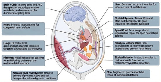
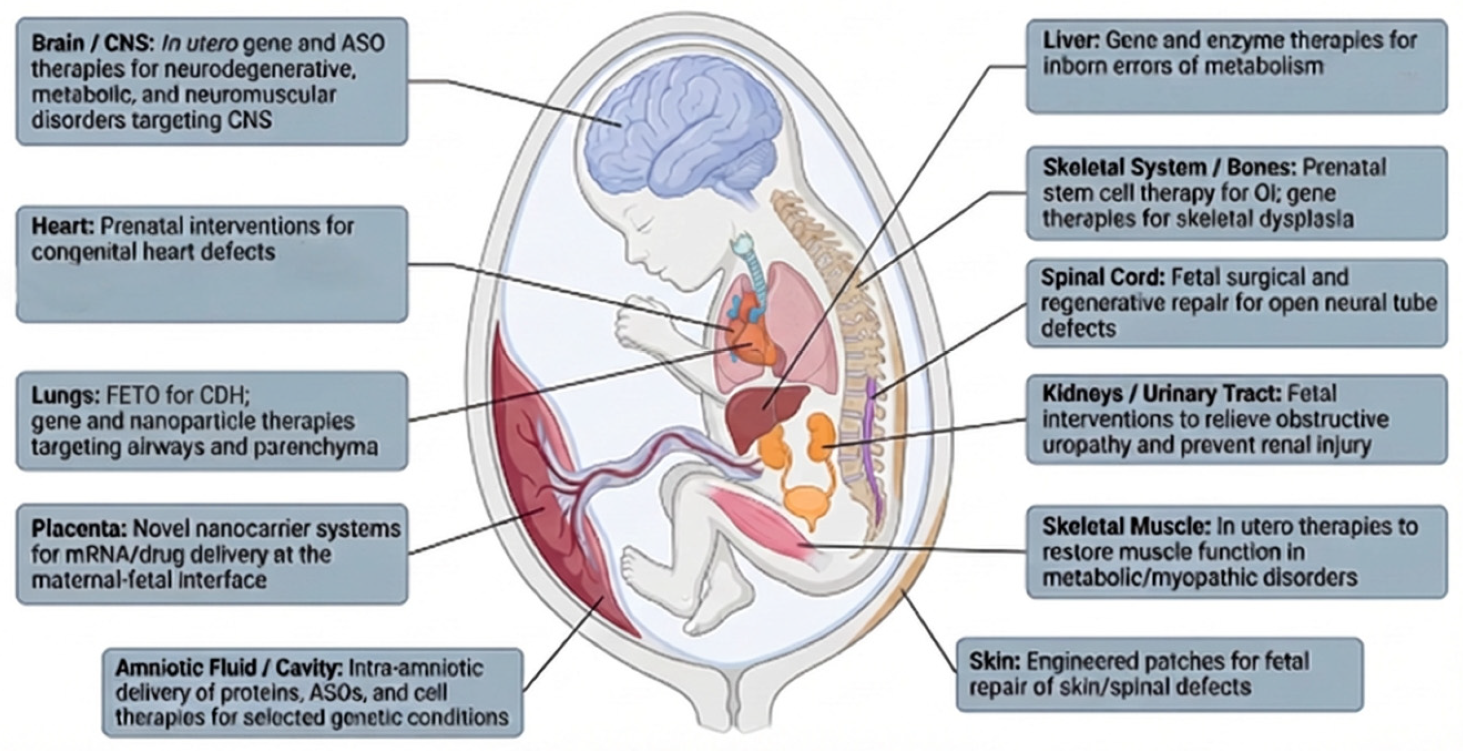

Disclaimer/Publisher’s Note: The statements, opinions and data contained in all publications are solely those of the individual author(s) and contributor(s) and not of MDPI and/or the editor(s). MDPI and/or the editor(s) disclaim responsibility for any injury to people or property resulting from any ideas, methods, instructions or products referred to in the content. |
© 2026 by the authors. Licensee MDPI, Basel, Switzerland. This article is an open access article distributed under the terms and conditions of the Creative Commons Attribution (CC BY) license.