Article

Immunotherapy Potential of Animal-Sourced Probiotic Bacteria

by
Isaac Oluseun Adejumo
Department of Animal Science, University of Ibadan, Ibadan 200005, Nigeria
Biologics 2025, 5(3), 17; https://doi.org/10.3390/biologics5030017
Submission received: 6 May 2025 / Revised: 16 June 2025 / Accepted: 23 June 2025 / Published: 27 June 2025

Abstract

Background/Objectives: Research efforts and substantial funding have been dedicated to finding cost-effective and sustainable alternatives to antibiotics. Probiotics have been proposed as promising substitutes for antibiotics in human nutrition and livestock production; however, their functional mechanisms remain incompletely understood, limiting their sustainable applications as food supplements, feed additives and for therapeutic and cosmetic purposes. Methods: In this study, the probiotic potential of two bacterial genomes, Ligilactobacillus saerimneri and Ligilactobacillus salivarius, were explored. Their protein-coding hypothetical proteins were analyzed for their potential to induce interleukin-5 (IL-5) and interleukin-13 (IL-13). Results: The IL-5- and IL-13-inducing peptides were identified as immunogens against bacterial and tumor peptides. Conclusions: These findings provide insights into the probiotic bacteria’s immune functionality pathways, sustainability and potential as therapeutic feed additives, food supplements and candidates for vaccine development.

1. Introduction

The demand for probiotics in animal and human nutrition is rising, owing to their health-promoting benefits and the concerns against the consistent use of antibiotics, including as growth promoters in livestock production, particularly chickens. The importance of chicken in the livestock industry and fundamental biology cannot be over-stated. For their protection from diseases, molecular analysis of their immunity plays a significant role. Bioinformatic tools have been used in the past to identify various cytokine genes in the chicken genome, leading to the discovery of a T helper 2 (Th2) cytokine cluster comprising interleukin-3 (IL-3), interleukin-4 (IL-4), interleukin-5 (IL-5), interleukin-13 (IL-13) and Granulocyte Macrophage Colony-Stimulating-Factor (GMCSF) genes [1]. The use of bioinformatic tools in studies involving human health is also common.
Vaccination plays a vital role in ameliorating disease outbreak in poultry industry and to improve vaccine and immunity efficacy, chicken cytokines associated with antibody production need to be identified and studied. IL-5 has been suggested to play a key role in chicken antibody production, with possible unique functions [2].
Research efforts and funding have been invested in the search for cost-effective and sustainable alternatives to antibiotics [3,4,5,6]. Probiotics have been suggested as potent alternatives to antibiotics in human nutrition and livestock production; however, their functional mechanisms have not fully been understood, restraining their sustainable applications as food supplements and feed additives, as well as for therapeutic and cosmetic purposes. On-going studies seek to unravel the mystery behind the functional mechanisms of probiotics [7,8,9], but such studies have currently yielded little, requiring more efforts to unravel the mystery. A full understanding of the functional mechanisms of probiotics promises to benefit not only the livestock industry but it also promises to benefit a wide range of fields including food science and technology, nutritional science, medicine, vaccine development, the cosmetic industry and a lot more.
In order to contribute to existing literature and close the existing knowledge gap regarding understanding the mechanisms underlying the health-promoting functions and sustainability issues of probiotics, the present study was conducted to investigate the probiotic potential of Ligilactobacillus salivarius and Ligilactobacillus saerimmeri genomes and evaluate peptides from their protein-coding hypothetical proteins for immunogenicity and interleukin-5 (IL-5) and interleukin-13 (IL-13) induction.

2. Materials and Methods

2.1. Bacterial Isolatation and Sequence Retrieval

Two complete bacterial genomes, Ligilactobacillus saerimneri M-11 and Ligilactobacillus salivarius strain ZLp4b, were utilized in this study. The nucleotide sequence of L. saerimneri M-11 (CP144759.1), which was isolated from the cecum of a 20-day-old chicken, and L. salivarius (CP062071.1), which was isolated from the feces of swine, were retrieved from the National Center for Biotechnology Information (NCBI) at https://www.ncbi.nlm.nih.gov/nuccore/CP144759.1/ (accessed on 2 January 2025) and https://www.ncbi.nlm.nih.gov/nuccore/CP062071.1/ (accessed on 1 January 2025), respectively. The sequencing technology employed for L. saerimneri was Nanopore and Illumina, with the assembly method being Unicycler v.0.5.0 and a genome coverage of 741.18x. The genome size is 1.6 Mb, with a GC content of 42.5%, comprising 1561 genes, of which 1453 are protein-coding. The sequencing method for L. salivarius was PacBio Sequel, with a genome coverage of 100.0× and an assembly method of FALCON v.0.3. It has a genome size of 2.3 Mb and a GC content of 33%. iProbiotics (http://bioinfor.imu.edu.cn/iprobiotics/public/, accessed on 23 January 2025) was used to determine the probiotic characteristics of the genomes [10].

2.2. Bacterial Toxin Investigation

Considering the significant role that bacterial toxins play in causing diseases, as well as in producing various symptoms and lesions during infection, I investigated selected proteins from both bacterial genomes for their potential as bacterial toxins. This assessment aimed to determine their safety for animal and human consumption, particularly as therapeutic feed additives and food supplements. I utilized BTXpred (https://webs.iiitd.edu.in/raghava/btxpred, accessed on 22 January 2025), a tool capable of identifying bacterial toxins and their functions from primary amino acid sequences using Support Vector Machines, HMM, and PSI-BLAST [11].

2.3. Selection of Protein-Coding Hypothetical Proteins

One hundred protein-coding hypothetical proteins were selected from L. salivarius (out of 327) and L. saerimneri (out of 135) as representative samples since the entire population cannot be used. The idea was to select that much to enhance accuracy. The nucleotide and amino acid sequences of the selected proteins were retrieved from the NCBI.

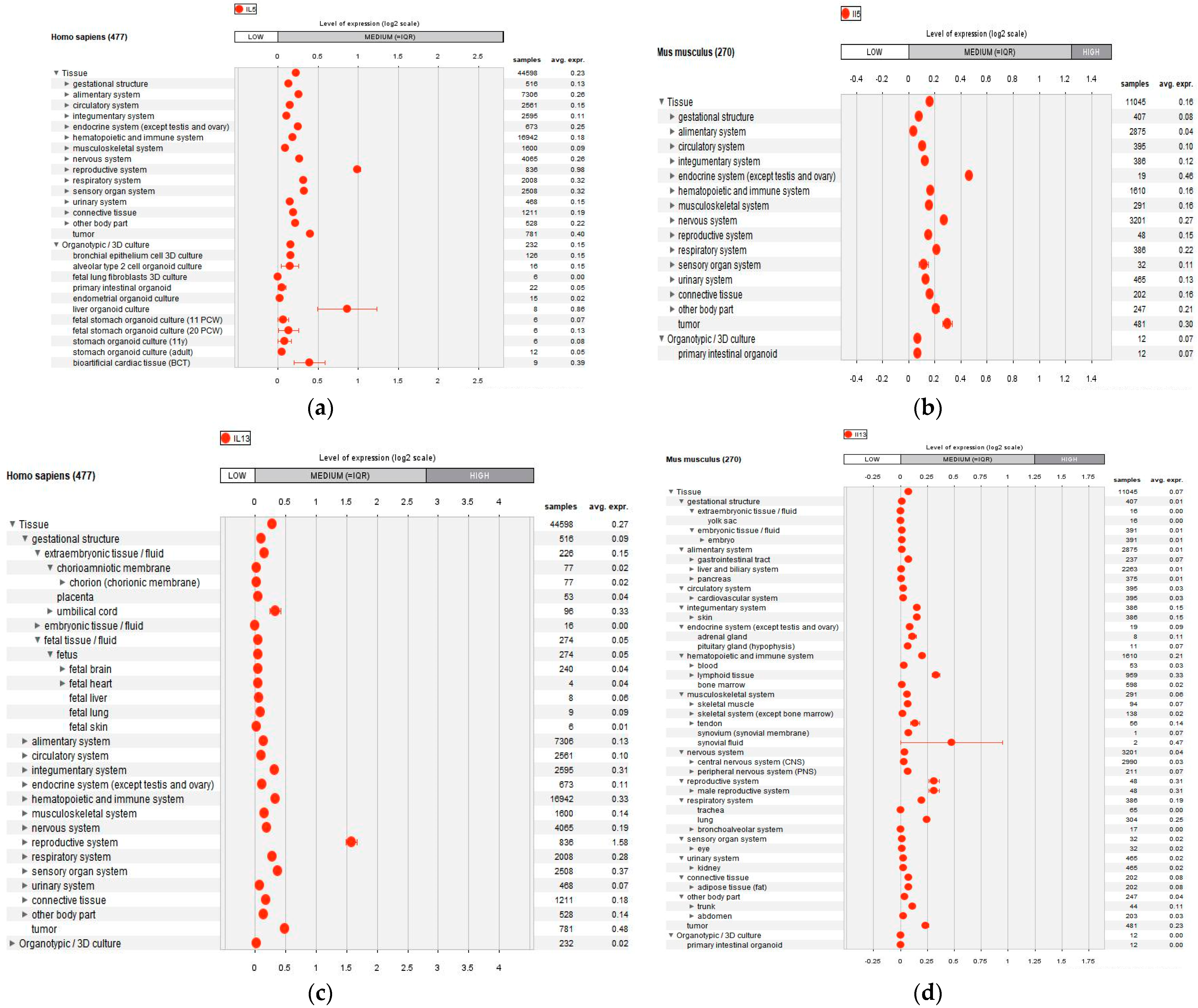
2.4. IL-5 and IL-13 Anaysis

Previously identified IFN-γ-inducing peptides [12] from each protein of both bacteria were assessed for their potential to induce IL-5 and IL-13. A total of 20 peptides from each protein group were selected, resulting in 400 peptides (20 peptides from 20 proteins) for L. salivarius and 280 peptides (20 peptides from 14 proteins) for L. saerimneri. The selection was proportionally representative based on the availability of hypothetical proteins. The selected proteins are listed in Table 1. Their IL-5- and IL-13-inducing potentials were evaluated using IL5Pred (https://webs.iiitd.edu.in/raghava/il5pred/, accessed on 14 January 2025) and IL-13Pred (https://webs.iiitd.edu.in/raghava/il13pred/, accessed on 17 January 2025), respectively [13,14,15].

2.5. Statistical Analysis and Gene Expression

The IL-5-inducing and IL-13-inducing peptides were statistically analyzed using SAS v. 9.4 ((SAS Institute Inc., Cary, NC, USA) in a completely randomized design. Tukey’s and Duncan’s Multiple Range Tests were employed for post hoc analysis to generate diffograms and mean separations. A significance level of α = 0.05 was established.
From each organism, five protein groups with the highest mean values from both bacteria were selected. Additionally, eight IL-5-inducing and IL-13-inducing peptides with the highest mean scores were chosen from each group and tested for immunogenicity. This testing utilized bacteria and tumor peptides as target organisms, employing VaxiJen v3.0 (https://www.ddg-pharmfac.net/vaxijen3/home/, accessed on 8 April 2025) [16].
From the antigenic peptides, the ones with the highest scores (one from each group) were selected based on their respective groups. Subsequently, their physicochemical properties were evaluated using ExPASy ProtParam, available at https://web.expasy.org/protparam/ (accessed on 18 March 2025) [17]. Gene expression analysis was conducted to gain a better understanding of IL-5 and IL-13 across various anatomical regions and conditions, as well as to identify positively correlated genes in both human and mouse models, using GENEVESTIGATOR. The default threshold values recommended by the respective tools were used.

3. Results

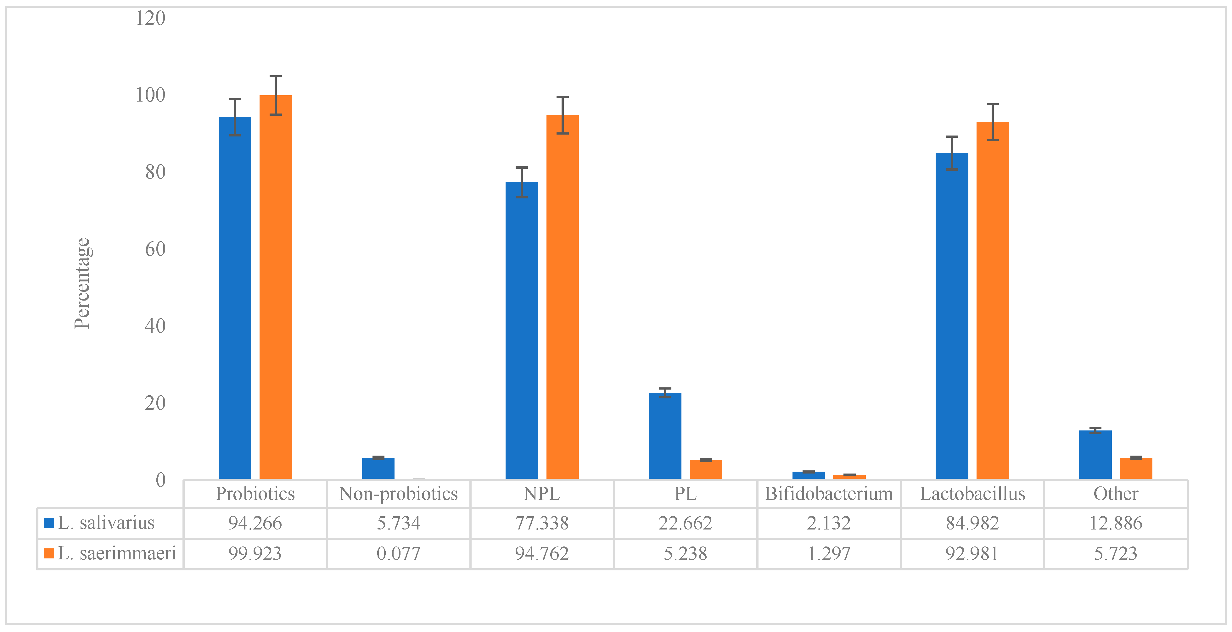
3.1. Probiotic Characteristics of the Genomes

The probiotic characteristics of the investigated genomes are shown in Figure 1. L. salivarius contains about 94.3% probiotic and 5.7% non-probiotic, while L. saerimmeri is almost completely probiotic (99.9%). The non-probiotic lactobacillus (NPL) in L. saerimmeri (94.8%) is more than the NPL in L. salivarius. L. saerimmeri is more lactobacillus (93%) than L. salivarius (85%). However, overall, both genomes are probiotic, with over a 90% score, and they are lactobacillus, containing about 85 and 93 percent for L. salivarius and L. saerimmeri, respectively.

3.2. Prediction of Bacterial Toxins

None of the evaluated proteins from either L. salivarius or L. saerimneri were found to contain bacterial toxin. The analysis returned no hit for all the investigated proteins from both genomes.

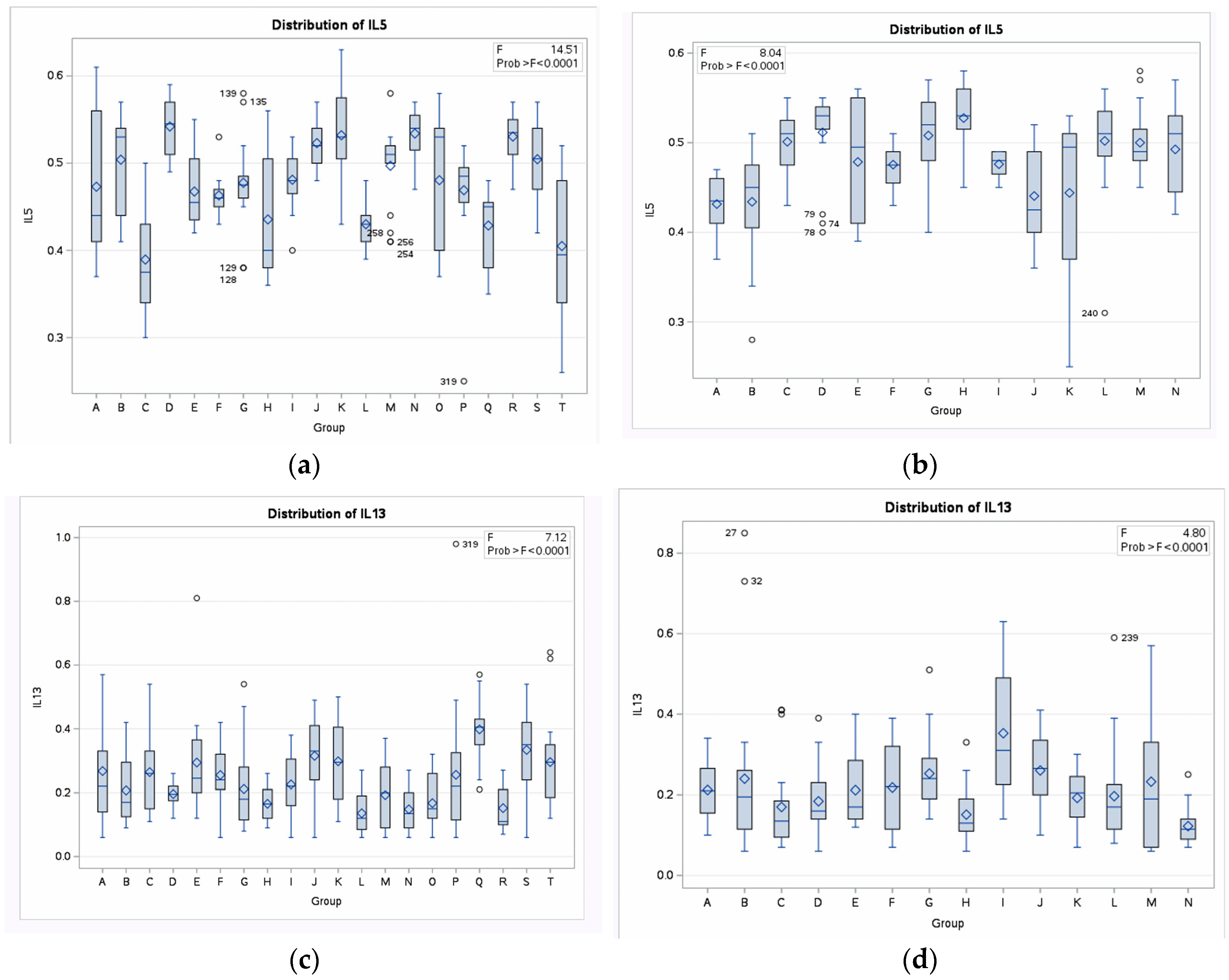
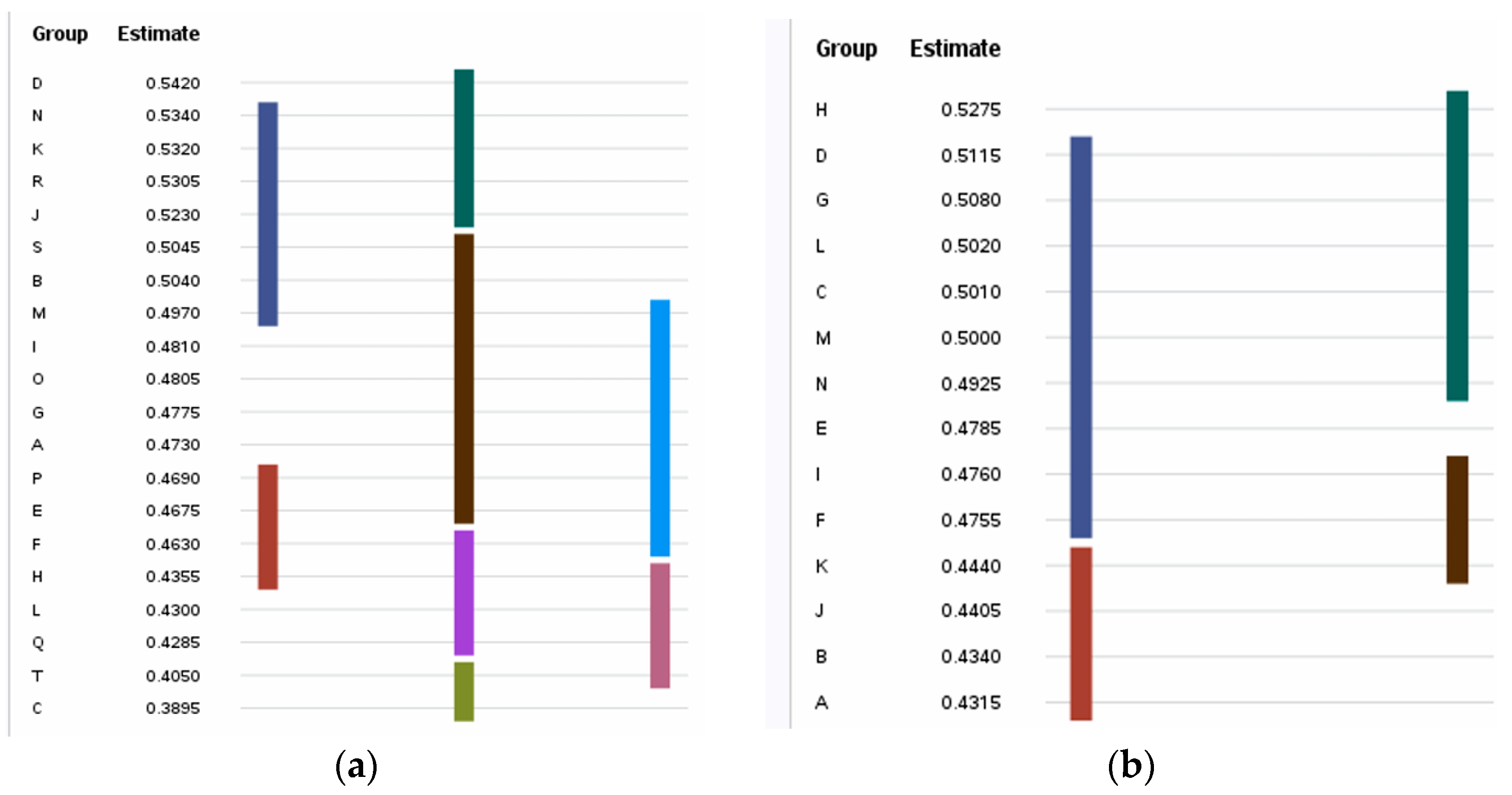
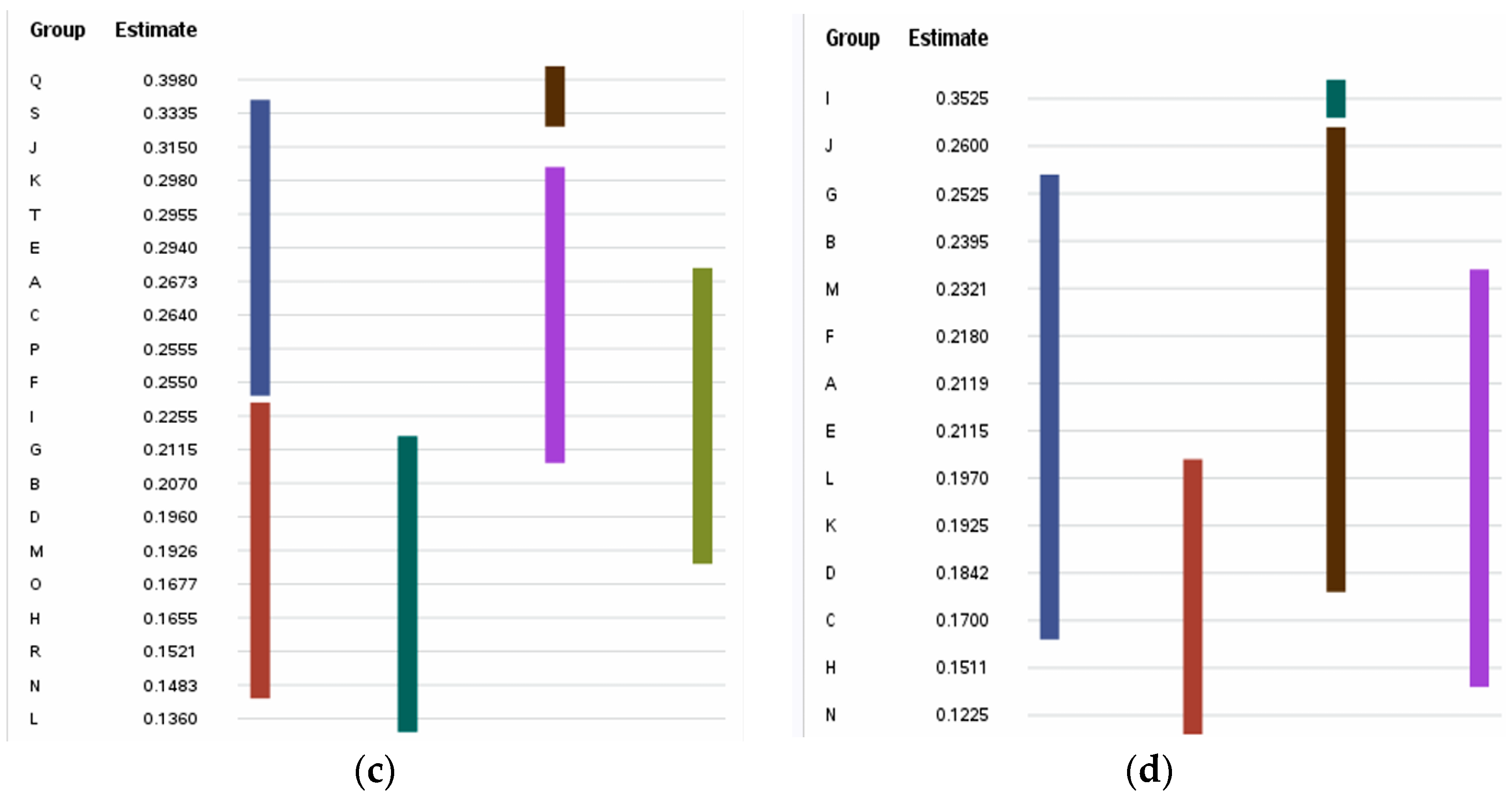
3.3. IL-5- and IL-13-Inducing Capacity

The statistical analysis result of the IL-5- and IL-13-inducing capacities of the selected peptides obtained from protein-coding hypothetical proteins of L. salivarius and L. saerimneri, their group distribution, Duncan grouping for their means and their diffograms showing group comparison are presented in Table 2, Figure 2, Figure 3 and Figure 4 For L. salivarius, group D (0.542) has the highest score, which is statistically similar to J (0.523), K (0.532), N (0.534) and R (0.531). B (0.504) is significantly (p < 0.05) higher than the scores obtained for C, F, H, L, Q and T but statistically similar to A, E, G, I, J, K, M, N, O, R and S. The significantly (p < 0.05) lowest score is obtained by group C (0.390), which is significantly (p < 0.05) lower than the scores obtained for A, B, D, E, F, G, H, I, J, K, L, M, N, O, P, Q, R and S but statistically similar to T (0.405). T is statistically similar to Q, L, H and C.
For IL-13-inducing capacity, the obtained score is highest for Q (0.398), which is statistically similar to S (0.334). Q is closely followed by J (0.315), which is significantly (p < 0.05) higher than the scores obtained for B, D, H, I, L, M, N, O and R, but statistically similar to A (0.267), C (0.264), E (0.294), F (0.255), K (0.298), P (0.256), S (0.334) and T (0.296). B, C, D, F, G, H, I, L, M, N, O and R are statistically similar. L (0.136) obtains the least score, which is statistically similar to B, D, G, H, M, N, O and R but significantly (p < 0.05) lower when compared with A, C, E, F, I, J, K, P, Q, S and T.
For L. saerimneri, the IL-5-inducing capacity score is highest for H (0.578), which is statistically similar to C (0.501), D (0.512), G (0.508), L (0.502), M (0.500) and N (0.493). E (0.479) is statistically similar to C, D, F, G, I, L, M and N. A (0.432) and J (0.441) obtained the lowest score, which is significantly (p < 0.05) lower than the scores obtained for other groups except A, B and K, to which it is statistically similar. F, I and K are statistically similar.
Group I (0.353) obtained the highest IL-13-inducing capacity score, which is closely followed by J (0.260) which is consequently statistically similar to A, B, D, E, F, G, K, L and M. N (0.123) obtained the least significantly (p < 0.05) score, which is statistically similar to C (0.170), D (0.184), H (0.151), K (0.193) and L (0.197).

3.4. Immunogenic IL-5- and IL-13-Inducing Peptides

Figure 5a–d is a group of charts that show IL-5- and IL-13-inducing immunogenic peptides derived from hypothetical proteins of L. salivarius (a and b) and L. saerimneri (c and d) using bacteria and tumor peptide as target organisms; a and c are the IL-5-inducing capacity while b and d are the IL-13-inducing capacity for L. salivarius and L. saeriimmeri, respectively. For IL-5 (L. salivarius), all the investigated peptides are immunogens against tumor peptides. However, when the target organisms are bacteria, 75% of R, 62.5% of J, 50% of D and 25% of N are immunogens. For L. saerimneri, all the peptides are immunogens against tumor peptides, while 62.5% of C, 50% of H and 37.5% of G and I are immunogens against bacteria.
For IL-13-inducing peptides all L. salivarius peptides are immunogens against tumor peptide, against bacteria, 100% of E, 87.5% of Q, S, T and 50% of J are immunogens. It was found that 100% of G, I and M and 75% of B and J of L. saerimneri peptides are immunogens against tumor peptides, while 100% of M, 75% of I, 62.5% of B and 37.5% of G are immunogens against bacteria.

3.5. Physicochemical Properties

Table 3 shows IL-5- and IL-13-inducing peptides selected for physicochemical property evaluation, based on their immunogenicity and group means. For IL-5, there are four groups for each of L. salivarius (D, J, N and R) and L. saerimneri (C, G, H and L), while for IL-13, there are five groups for L. salivarius (E, J, Q, S and T) and four for L. saerimneri (B, G, I and M).
The physicochemical properties of selected immunogenic IL-5- and IL-13-inducing peptides from the hypothetical proteins of L. salivarius and L. saerimneri are presented in Table 4. Molecular weight (MW) ranges between 2510.61 and 2125.46. The theoretical isoelectric point (pI) ranges between 12.01 and 4.00. The estimated half-life for IL-5-inducing peptides from L. salivarius varies. For D, it is 1.1 h (mammalian reticulocytes, in vitro), 3 min (yeast, in vivo) and 2 min (Escherichia coli, in vivo); for J, it is 7.2 h (mammalian reticulocytes, in vitro), >20 h (yeast, in vivo) and >10 h (Escherichia coli, in vivo). The estimated half-life for N is100 h (mammalian reticulocytes, in vitro), >20 h (yeast, in vivo) and >10 h (Escherichia coli, in vivo) and for R it is 5.5 h (mammalian reticulocytes, in vitro), 3 min (yeast, in vivo) and 2 min (Escherichia coli, in vivo). For L. saerimneri peptides, the estimated half-life for C is 30 h (mammalian reticulocytes, in vitro), >20 h (yeast, in vivo) and >10 h (Escherichia coli, in vivo); it is 30 h (mammalian reticulocytes, in vitro), >20 h (yeast, in vivo) and >10 h (Escherichia coli, in vivo) for G; 1 h (mammalian reticulocytes, in vitro), 30 min (yeast, in vivo) and >10 h (Escherichia coli, in vivo) for H and 1.1 h (mammalian reticulocytes, in vitro), 3 min (yeast, in vivo) and >10 h (Escherichia coli, in vivo) for L. For IL-13-inducing L. salivarius peptides, it is 0.8 h (mammalian reticulocytes, in vitro), 10 min (yeast, in vivo) and 10 h (Escherichia coli, in vivo) for E; 4.4 h (mammalian reticulocytes, in vitro), >20 h (yeast, in vivo) and >10 h (Escherichia coli, in vivo) for J; 1 h (mammalian reticulocytes, in vitro), 2 min (yeast, in vivo) and 2 min (Escherichia coli, in vivo) for Q; 1.9 h (mammalian reticulocytes, in vitro), >20 h (yeast, in vivo) and >10 h (Escherichia coli, in vivo) for S and for T it is 30 h (mammalian reticulocytes, in vitro), >20 h (yeast, in vivo) and >10 h (Escherichia coli, in vivo). For L. saerimneri peptides, it is >20 h (mammalian reticulocytes, in vitro), >20 h (yeast, in vivo) for B; 30 h (mammalian reticulocytes, in vitro), >20 h (yeast, in vivo) and >10 h (Escherichia coli, in vivo) for G; 1.1 h (mammalian reticulocytes, in vitro), 3 min (yeast, in vivo) and >10 h (Escherichia coli, in vivo) for I and for M, it is 0.8 h (mammalian reticulocytes, in vitro), 10 min (yeast, in vivo) and 10 h (Escherichia coli, in vivo).
Instability indices range between 107.81 and −3.73. For IL-5-inducing peptides, D (L. salivarius) and C, H and L (L. saerimneri) obtain instability indices greater than 40. Aliphatic indices are generally high, higher than 50, except for G (L. saerimneri, IL-5-inducing), E, J, S and T (L. salivarius IL-13-inducing) and B, G and I (L. saerimneri, IL-13-inducing peptides). GRAVY scores range between 1.110 and −2.065.

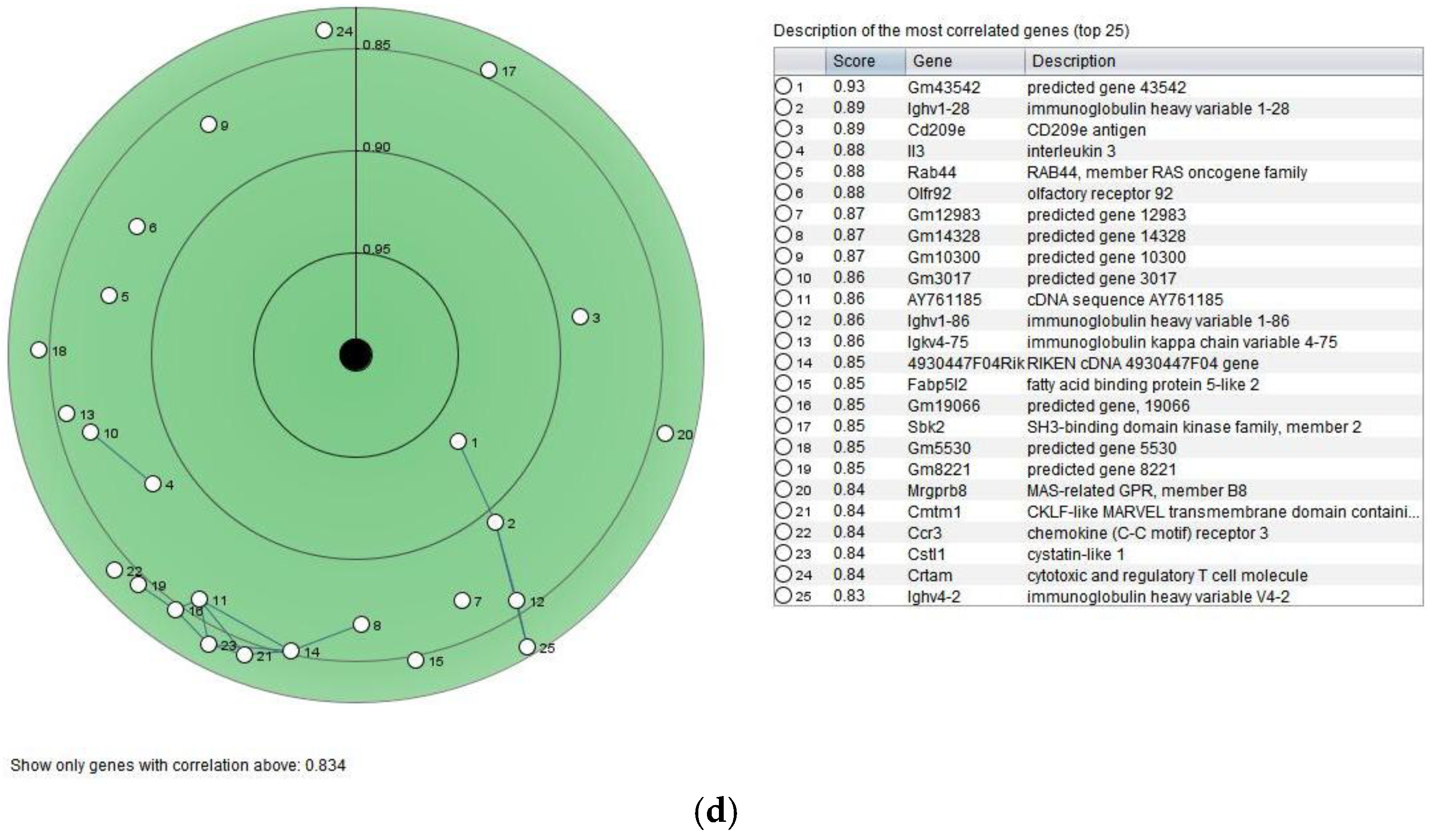
3.6. Gene Expression

Figure 6a–d show the scatter plots of IL-5 (a, b) and IL-13 (c, d) expression across anatomical parts in humans (a, c) and mice (b, d), while Figure 7a–d show circular views of positively correlated genes with IL-5 (a, b) and IL-13 (c, d) across various cancer categories in Homo sapiens (a, c), and mice (b, d) using Pearson’s correlation coefficient. IL-5 shows medium regulation for most of the anatomical parts considered such as alimentary, circulatory, integumentary and endocrine systems, both in humans and mice. The top-most positively correlated genes with IL-5 across various cancer categories in humans are MTND4P32, AC1184651, DOX3P1, HADHAP1 and MTND2P2. The co-expressed genes with IL-5 across various cancer categories in mice include Gm13557, Rgs, 1700018C11Rik, chma3 and Gm20519.
IL-13 expression in both Homo sapiens and Mus musculus show medium expression across most of the anatomical parts considered. Co-expressed genes with IL-13 across various cancer categories in humans include SHLD2P2, OR5B21, PP2D1, AP000925.1 and AC093270.1, while in mice they include Gm43542, Ighv1-28, Cd209e, interleukin 3 and Rab44.

4. Discussion

As gut commensals, many lactic acid bacteria (LAB) species possess probiotic properties and they play key roles in the maintenance of gut homeostasis, ensuring a balance of beneficial microbes [18]. The benefits of LAB include modulating the immune system [19] and improving the quality of animal products [20], as well as preventing and controlling infectious diseases [21,22]. L. salivarius is an important taxon among the LAB species in the intestinal tracts of mammals. The evaluated bacterial genomes in the present study are probiotics, which aligns with previous findings showing that L. salivarius and other LAB strains are promising probiotic candidates, possessing a wide strain-based spectrum of antibacterial and antifungal activity, supporting natural barrier mechanisms against invading pathogens [23]. Wang [24] also confirmed the probiotic nature of L. salivarius, as reported in the present study. In the present study, L. saerimneri is almost 100% probiotic while L. salivarius is more than 90% probiotic. The bacterial genomes are over 80% lactobacillus, about 85 and 93 percent for L. salivarius and L. saerimneri, respectively.
It has been documented that the Lactobacillaceae family, contains three hundred species and thirty-three genera, two hundred and sixty of which are from the former genus Lactobacillus, which has been recognized as among the most studied microorganisms with probiotic potential. ‘Bio’ means life while ‘pro’ means ‘for’ [23]. Stedman [25] successfully isolated and characterized various LAB strains, highlighting their therapeutic relevance through in vitro immune-related assays and comparative genomic studies. These analyses revealed that specific strains of Pediococcus and Lactobacillus possess probiotic qualities and demonstrate the potential to mitigate infectious diseases in wild animals, likely through enhancing phagocytic immune responses that provide protection against pathogens [18].
L. salivarius is a widely distributed specie. It has the ability to adapt to challenging environments such as the intestine, building resistance against acid and bile stresses, being a prerequisite for the beneficial effects of probiotics [26]. Wang et al. [24] suggested that exploring Lactobacillus salivarius strains could significantly advance the development of probiotic formulations by selecting strains that inhibit pathogens, efficiently convert nutrients, and can be genetically enhanced. This species has been detected in diverse environments, including the gastrointestinal and oral cavities of humans and animals like pigs and chickens [27], as well as in fermented food products [28,29,30,31], although isolation from wild animals has also been reported [24], some of which have been reported to alleviate inflammatory responses [11], inhibit reproduction and growth of pathogenic bacteria [19,22,32], prolong host’s lifespan [33] and improve calcium absorption in the intestinal tract [34], as well as reduce pathogen adhesion to host cells [21].
Animals’ biological systems are both dynamic and complex, accommodating symbiotic and mutualistic host–microbe relationships, thereby promoting host’s development and health [35]. Communication is on-going with gut microbiota and immune system, aimed at sustaining the balance between immunogenicity and immune tolerance [36,37]. Probiotics have been used to manipulate animals’ gut flora as an alternative approach to maintain or improve immune balance [38]. Probiotic organisms have the capacity to influence both humoral and cell-mediated immunity, either directly or indirectly, by modulating immune signaling pathways and mitigating specific inflammatory and infectious conditions [39].
In this study, hypothetical protein-coding genes of L. salivarius and L. saerimneri were explored. The importance of protein-coding genes and immunity cannot be overemphasized, although the molecular mechanisms of immune regulation are not fully understood. Studies (both intra and inter species) of protein-coding genes have aided the identification of proteins rapidly evolving as a result of positive selection. A genome-wide comparison of protein-coding sequences from mouse, human and chimpanzee genomes demonstrated that immunity-related genes are strongly represented among those undergoing adaptive evolution [40]. The protein-coding sequences of receptors that recognize pathogens play important functions in differences in immune function among species.
IL-13, a multifunctional cytokine, is produced by various immune cells such as Th2 lymphocytes, basophils, NK cells, mast cells and eosinophils [41]. It plays a critical role in expelling gastrointestinal parasites [42] and drives isotype switching in naïve B cells, particularly favoring IgE and IgG4 antibody production [43,44], coupled with being a vital mediator in airway inflammation seen in asthma and reactive airway diseases [45]. Th1 and Th17 cells are also known to produce it; it is involved in adaptive immunity (including Th17 and TH1 inflammatory responses) [46].
The role that IL-13 plays in biological processes cannot be overstated. Current research is increasingly focused on discovering new compounds capable of modulating IL-13 activity. Peptide-based therapeutics are gaining traction due to their high specificity and minimal toxicity [47,48], which has intensified interest in identifying and validating peptides that stimulate IL-13 production [12,14,49].
IL-13 gene expression is predominantly controlled by GATA3 transcription factors. While IL-13 shares approximately 25% amino acid similarity with IL-4 and is located at the same chromosomal locus (5q31) [50], it exhibits greater promise as a therapeutic target [51]. Notably, IL-13 receptor overexpression in several cancers makes it an attractive candidate for targeted cancer immunotherapies, underscoring the novel findings in the present study. The evaluated IL-13-inducing peptides from L. salivarius and L. saerimneri are immunogens against tumor peptides.
IL-13 is involved in multiple physiological and pathological processes, such as promoting airway hyperresponsiveness, tissue remodeling, and tumor progression. It has been shown that IL-13 inhibitors can potentially support anti-tumor immune responses by restoring immune surveillance suppressed by IL-13, especially in human cancers [52,53]. Furthermore, its receptors are increasingly being recognized for their prognostic significance in gastrointestinal malignancies due to their interaction with components of the tumor microenvironment [54,55,56,57,58].
Emerging evidence [59] indicates that IL-13 also plays a role in the nervous system, particularly in regulating synaptic activity and protecting neurons following injury. This neuroprotective function, facilitated through its receptor complex, may significantly influence brain plasticity and open up new directions for neurological research, suggesting that of a particular relevance to this are the anti-IL-13 therapeutics developed for allergic conditions and against gliomas [59,60].
IL-5 is secreted by immune cells such as Th2 cells, basophils, mast cells and type 2 innate lymphoid cells, playing various biological roles in eosinophil activation, maturation and survival. In the present study, hypothetical proteins from L. salivarius and L. saerimneri produce IL-5-inducing peptides, according to IL5Pred [15]. IL-5 is produced when the aforementioned secreting cells are stimulated by environmental pollutants, microbes and inhaled allergens [61]. The same cluster harbors IL-3, IL-4 IL-13, granulocyte-macrophage colony-stimulating factor (GM-CSF) and the gene encoding IL-5 [62]. It exerts its biological effects primarily through the IL-5 receptor complex (IL-5R) and is known for its pleiotropic activity, comprising a βc and an α chain. It promotes the activation, maturation and survival of eosinophils as well as the release into the bloodstream and eventually to the airways from the bone marrow [61,62,63,64].
IL-5 plays an important role in a number of diseases including dermatitis, asthma, hyper-eosinophilic syndrome and eosinophilic esophagitis [65]. It has been reported that patients with severe COVID-19 conditions had an elevated level of IL-5 [66]. Research has established the importance of IL-5 in managing diseases involving eosinophils [67]. It also shows promise in tumor suppression by regulating eosinophil migration into the lungs and inhibiting metastatic spread [68]. IL-5 has been suggested as an attractive therapeutic target [69,70,71].
Chicken meat and eggs are widely consumed across the globe and the demand for chicken products keeps increasing, necessitating support and encouragement given to backyard producers in order to boost production, so as to ameliorate the challenges of food insecurity, particularly in sub-Saharan regions. However, outbreaks of infectious diseases result in huge economic losses and threaten food security globally as important protein sources. Infectious diseases are often controlled through vaccination, with its accompanying pros and cons. Identification of chicken cytokines that regulate antibody production has been regarded as being indispensable for enhancing vaccination efficacy [2].
IL-5 signaling has been reported to play an essential role in production of antibody. Experiments using IL-5 receptor alpha-deficient mice demonstrated reduced levels of circulating IgM, IgA and IgG subclasses [72,73], underscoring the assertion that chicken IL-5 regulates antibody-producing cells. In mammals, IL-5 appears to act on a range of immune cell types beyond B cells, including eosinophils and regulatory T cells [74,75,76], and may enhance isotype switching to IgY while downregulating IgA production [2]. It has been suggested that IL-5 may function in the differentiation and maturation of activated B cells in chickens. Watanabe et al. [2] noted that IL-5 preferentially promotes class switching to IgY and differentiation into IgM- or IgY-producing cells, decreasing IgA production, suggesting that chicken IL-5 induce a class switch to IgY and regulate antibody-producing cells.
The importance of cytokines (to animal agriculturalists and intensive livestock industries) as natural immunotherapeutics, improving disease resistance and animal health, cannot be over-emphasized. Before attention began to shift, antibiotics had been used for these purposes in the past, but concerns arising from the use of antibiotics such as the development of antibiotic resistance and chemical residue in animal products have limited its sustainability, necessitating the use of alternatives such as probiotics, the sustainable applications of which are unfolding. In the present study, peptides from hypothetical proteins of probiotic L. salivarius and L. saerimneri induce immunogenic IL-5 and IL-13.
IL-5 has been observed in a number of species to increase eosinophils in tissues and blood as well as improving B cell activity. Administering IL-5 to pigs, either through recombinant protein or DNA-based methods, resulted in prolonged elevation of eosinophil counts in circulation, indicating that IL-5 may produce significant biological effects in commercial pigs [77]. IL-5 plays an important role in eosinophil proliferation, migration and degranulation [78,79] and eosinophils have been linked with manifestation of type 2 asthma, thereby reinforcing IL-5’s relevance as a therapeutic target in immunomodulation [80].
In the present study, IL-5- and IL-13-inducing peptides are also found to be immunogens against tumor peptides and bacteria. These findings were corroborated by the findings of previous studies. For instance, studies have illustrated the effectiveness of IFN-α in suppressing tumor development in mouse models inoculated with various cancer cell lines [81,82] and the anti-tumor activity of some other cytokines for possible use in cancer immunotherapy has been suggested [83].
This study shows that the peptides have pI which ranged between 4 and 12, indicating their ability to endure the gastrointestinal tract, being acidic and alkaline in nature. About 65% of the peptides are stable in nature and the aliphatic index for the peptides indicates that they are thermo-stable over a wide temperature range. These parameters may be essential for drug design because these peptides, in addition to being immunogenic, have IL-5- and IL-13-inducing abilities that indicate their potential as vaccine candidates. This study being in silico, it is strongly recommended that these findings are further subjected to more accurate methods than the existing ones. The current study is intended as a foundational step to identify promising candidates for subsequent wet-lab validation.

5. Conclusions

In the present study, I explored two bacterial genomes sourced from chickens and pigs. I analyzed protein-coding hypothetical proteins from both L. salivarius and L. saerimneri for their potential to induce IL-5 and IL-13. From each hypothetical protein, I selected the top twenty IL-5- and IL-13-inducing peptides based on their scores—twenty from L. salivarius and fourteen from L. saerimneri. The evaluated peptides are immunogens against bacterial and tumor peptides, suggesting their roles in immune functionality pathways and probiotic sustainability and their potential as therapeutic feed additives, food supplements and candidates for vaccine development.

Funding

This research received no external funding.

Institutional Review Board Statement

Not applicable.

Informed Consent Statement

Not applicable.

Data Availability Statement

The datasets used in this manuscript are included or are available in public databases and appropriate links are provided where necessary.

Acknowledgments

The author appreciates Olawumi Adejumo’s support.

Conflicts of Interest

The author declares no conflicts of interest.

Abbreviations

The following abbreviations are used in this manuscript:
ECextinction coefficient
IIinstability index
AIaliphatic index
IFN-γinterferon-gamma
GM-CSFgranulocyte-macrophage colony-stimulating factor
IL-3interleukin-3
IL-4interleukin-4
IL-5interleukin-5
IL-13interleukin-13
MWmolecular weight
NKnatural killer
pIisoelectric point
Th2T helper 2
NPLnon-probiotic lactobacillus
PLprobiotic lactobacillus
LABlactic acid bacteria

References

  1. Avery, S.; Rothwell, L.; Degen, W.D.; Schijns, V.E.; Young, J.; Kaufman, J.; Kaiser, P. Characterization of the first nonmammalian T2 cytokine gene cluster: The cluster contains functional single-copy genes for IL-3, IL-4, IL-13, and GM-CSF, a gene for IL-5 that appears to be a pseudogene, and a gene encoding another cytokinelike transcript, KK34. J. Interferon Cytokine Res. 2004, 24, 600–610. [Google Scholar]
  2. Watanabe, T.; Terada, T.; Ezaki, R.; Matsuzaki, M.; Furusawa, S.; Horiuchi, H. Chicken Interleukin-5 is expressed in splenic lymphocytes and affects antigen-specific antibody production. J. Poult. Sci. 2024, 61, jpsa.2024002. [Google Scholar] [CrossRef] [PubMed]
  3. Ologhobo, A.D.; Akangbe, E.I.; Adejumo, I.O.; Adeleye, O. Effect of Moringa oleifera leaf meal as replacement for oxytetracycline on carcass characteristics of the diets of broiler chickens. Annu. Res. Rev. Biol. 2014, 4, 423–431. [Google Scholar] [CrossRef]
  4. Ologhobo, A.D.; Adejumo, I.O.; Akangbe, E.I. Comparison effect of Moringa oleifera leaf meal and oxytetracycline on haematology and serum biochemical profile of broiler finishers. Int. Blood Res. Rev. 2014, 2, 29–36. [Google Scholar] [CrossRef]
  5. Adejumo, I.O.; Adetunji, C.O.; Olopade, C.O.; George, K.O. Response of broilers to dietary Moringa oleifera leaf, raw and cooked seed meal and synthetic antibiotics. J. Exp. Agric. Int. 2016, 12, 1–7. [Google Scholar] [CrossRef]
  6. Odukoya, A.A.; Adejumo, I.O.; Ologhobo, A.D. Chemical constituents of crude extracts of Moringa oleifera (Lam) leaf and biochemical response of weanling Wistar rats administered crude extract. Niger. Agric. J. 2022, 53, 351–356. [Google Scholar]
  7. Adejumo, I.O. Hypothetical proteins of chicken-isolated Limosilactobacillus reuteri subjected to in silico analyses induce IL-2 and IL-10. Genes Nutr. 2024, 19, 24. [Google Scholar] [CrossRef]
  8. Adejumo, I.O.; Adebiyi, O.A. Peptides from hypothetical proteins of Lactobacillus acidophilus induce IL-4 and IL-10. Biomed. Res. Ther. 2025, 12, 1–16. [Google Scholar] [CrossRef]
  9. Adejumo, I.O.; Adebiyi, O.A. IL-4-inducing hypothetical proteins of chicken-isolated Limosilactobacillus reuteri. Biology 2025, in press.
  10. Sun, Y.; Li, H.; Zheng, L.; Li, J.; Hong, Y.; Liang, P.; Kwok, L.-Y.; Zuo, Y.; Zhang, W.; Zhang, H. iProbiotics: A machine learning platform for rapid identification of probiotic properties from whole-genome primary sequences. Brief. Bioinform. 2022, 23, 1–12. [Google Scholar] [CrossRef]
  11. Saha, S.; Raghava, G.P.S. BTXpred: Prediction of Bacterial Toxins. Silico Biol. 2007, 7, 405–412. [Google Scholar] [CrossRef]
  12. Dhanda, S.K.; Vir, P.; Raghava, G.P.S. Designing of interferon-gamma inducing MHC class-II binders. Biol Direct. 2013, 8, 30. [Google Scholar] [CrossRef] [PubMed]
  13. Jain, S.; Dhall, A.; Patiyal, S.; Raghava, G.P. IL13Pred: A method for predicting immunoregulatory cytokine IL-13 inducing peptides. Comput. Biol. Med. 2022, 143, 105297. [Google Scholar] [CrossRef]
  14. Jain, S.; Dhall, A.; Patiyal, S.; Raghava, G.P.S. In Silico tool for identification, designing, and searching of IL13-inducing peptides in antigens. Methods Mol. Biol. 2023, 2673, 329–338. [Google Scholar]
  15. Naorem, D.N.; Sharma, N.; Raghava, G.P.S. A web server for predicting and scanning of IL-5 inducing peptides using alignment-free and alignment-based method. Comput. Biol. Med. 2023, 158, 106864. [Google Scholar] [CrossRef]
  16. Dimitrov, I.; Zaharieva, N.; Doytchinova, I. Bacterial immunogenicity prediction by machine learning methods. Vaccines 2020, 8, 709. [Google Scholar] [CrossRef]
  17. Gasteiger, E.; Hoogland, C.; Gattiker, A.; Duvaud, S.; Wilkins, M.R.; Appel, R.D.; Bairoch, A. Protein identification and analysis tools on the ExPASy server. In The Proteomics Protocols Handbook; Walker, J.M., Ed.; Humana Press: Totowa, NJ, USA, 2005; pp. 571–607. [Google Scholar]
  18. Stedman, A.; van Vliet, A.H.M.; Chambers, M.; Gutierrez-Merino, J. Gut commensal bacteria show beneficial properties as wildlife probiotics. Ann. N. Y. Acad. Sci. 2020, 1467, 112–132. [Google Scholar] [CrossRef]
  19. Zhai, Q.; Shen, X.; Cen, S.; Zhang, C.; Tian, F.; Zhao, J.; Zhang, H.; Xue, Y.; Chen, W. Screening of Lactobacillus salivarius strains from the feces of Chinese populations and the evaluation of their effects against intestinal inflammation in mice. Food Funct. 2020, 11, 221–235. [Google Scholar] [CrossRef] [PubMed]
  20. Chaves, B.; Brashears, M.; Nightingale, K. Applications and safety considerations of Lactobacillus salivarius as a probiotic in animal and human health. J. Appl. Microbiol. 2017, 123, 18–28. [Google Scholar] [CrossRef]
  21. O’Hara, A.M.; O’Regan, P.; Fanning, A.; O’Mahony, C.; Macsharry, J.; Lyons, A.; Bienenstock, J.; O’MAhony, L.; Shanahan, F. Functional modulation of human intestinal epithelial cell responses by Bifidobacterium infantis and Lactobacillus salivarius. Immunology 2006, 118, 202–215. [Google Scholar] [CrossRef]
  22. Messaoudi, S.; Manai, M.; Kergourlay, G.; Prévost, H.; Connil, N.; Chobert, J.M.; Dousset, X. Lactobacillus salivarius: Bacteriocin and probiotic activity. Food Microbiol. 2013, 36, 296–304. [Google Scholar] [CrossRef]
  23. Dobreva, L.; Atanasova, N.; Donchev, P.; Krumova, E.; Abrashev, R.; Karakirova, Y.; Mladenova, R.; Tolchkov, V.; Ralchev, N.; Dishliyska, V.; et al. Candidate-probiotic lactobacilli and their postbiotics as health-benefit promoters. Microorganisms 2024, 12, 1910. [Google Scholar] [CrossRef] [PubMed]
  24. Wang, Y.; Xu, X.; Chen, H.; Yang, F.; Xu, B.; Wang, K.; Liu, Q.; Liang, G.; Zhang, R.; Jiao, X.; et al. Assessment of beneficial effects and identification of host adaptation-associated genes of Ligilactobacillus salivarius isolated from badgers. BMC Genom. 2023, 24, 530. [Google Scholar] [CrossRef]
  25. Stedman, A.; Maluquer de Motes, C.; Lesellier, S.; Dalley, D.; Chambers, M.; Gutierrez-Merino, J. Lactic acid bacteria isolated from European badgers (Meles meles) reduce the viability and survival of Bacillus Calmette-Guerin (BCG) vaccine and influence the immune response to BCG in a human macrophage model. BMC Microbiol. 2018, 18, 1–14. [Google Scholar] [CrossRef] [PubMed]
  26. Fang, F.; Li, Y.; Bumann, M.; Raftis, E.J.; Casey, P.G.; Cooney, J.C.; Walsh, M.A.; O’Toole, P.W. Allelic variation of bile salt hydrolase genes in Lactobacillus salivarius does not determine bile resistance levels. J. Bacteriol. 2009, 191, 5743–5757. [Google Scholar] [CrossRef] [PubMed]
  27. Harris, H.M.B.; Bourin, M.J.B.; Claesson, M.J.; O’Toole, P.W. Phylogenomics and comparative genomics of Lactobacillus salivarius, a mammalian gut commensal. Microb. Genom. 2017, 3, e000115. [Google Scholar] [CrossRef]
  28. Guo, X.H.; Kim, J.M.; Nam, H.M.; Park, S.Y.; Kim, J.M. Screening lactic acid bacteria from swine origins for multistrain probiotics based on in vitro functional properties. Anaerobe 2010, 16, 321–326. [Google Scholar] [CrossRef]
  29. Messaoudi, S.; Madi, A.; Prévost, H.; Feuilloley, M.; Manai, M.; Dousset, X.; Connil, N. In vitro evaluation of the probiotic potential of Lactobacillus salivarius SMXD51. Anaerobe 2012, 18, 584–589. [Google Scholar] [CrossRef]
  30. Raftis, E.J.; Forde, B.M.; Claesson, M.J.; O’Toole, P.W. Unusual genome complexity in Lactobacillus salivarius JCM1046. BMC Genom. 2014, 15, 771. [Google Scholar] [CrossRef]
  31. Kang, C.H.; Han, S.H.; Kim, Y.; Paek, N.S.; So, J.S. In vitro probiotic properties of Lactobacillus salivarius MG242 isolated from human vagina. Probiotics Antimicrob. Proteins 2018, 10, 343–349. [Google Scholar] [CrossRef]
  32. Kang, M.S.; Lim, H.S.; Oh, J.S.; Lim, Y.J.; Wuertz-Kozak, K.; Harro, J.M.; Shirtliff, M.E.; Achermann, Y. Antimicrobial activity of Lactobacillus salivarius and Lactobacillus fermentum against Staphylococcus aureus. Pathog. Dis. 2017, 75, ftx009. [Google Scholar] [CrossRef] [PubMed]
  33. Zhao, Y.; Zhao, L.; Zheng, X.; Fu, T.; Guo, H.; Ren, F. Lactobacillus salivarius strain FDB89 induced longevity in Caenorhabditis elegans by dietary restriction. J. Microbiol. 2013, 51, 183–188. [Google Scholar] [CrossRef] [PubMed]
  34. Gilman, J.; Cashman, K.D. The effect of probiotic bacteria on transepithelial calcium transport and calcium uptake in human intestinal-like Caco-2 cells. Curr. Issues Intest. Microbiol. 2006, 7, 1–5. [Google Scholar]
  35. Charizani, E.; Dushku, E.; Karathodorou, A.; Kyritsi, M.; Amanetidou, E.; Metallinou, E.T.; Kokkaleniou, M.M.; Passalis, N.; Tefas, A.; Staikou, A.; et al. Predicting the immunomodulatory activity of probiotic lactic acid bacteria using supervised machine learning in a Cornu aspersum snail model. Fish Shellfish Immunol. 2024, 152, 109788. [Google Scholar] [CrossRef]
  36. Zheng, D.; Liwinski, T.; Elinav, E. Interaction between microbiota and immunity in health and disease. Cell Res. 2020, 30, 492–506. [Google Scholar] [CrossRef]
  37. Mazziotta, C.; Tognon, M.; Martini, F.; Torreggiani, E.; Rotondo, J.C. Probiotics mechanism of action on immune cells and beneficial effects on human health. Cells 2023, 12, 184. [Google Scholar] [CrossRef] [PubMed]
  38. Hill, C.; Guarner, F.; Reid, G.; Gibson, G.R.; Merenstein, D.J.; Pot, B.; Morelli, L.; Canani, R.B.; Flint, H.J.; Salminen, S.; et al. The International Scientific Association for Probiotics and Prebiotics consensus statement on the scope and appropriate use of the term probiotic. Nat. Rev. Gastroenterol. Hepatol. 2014, 11, 506–514. [Google Scholar] [CrossRef]
  39. Wells, J.M. Immunomodulatory mechanisms of lactobacilli. Microb. Cell Fact. 2011, 10, S17. [Google Scholar] [CrossRef]
  40. Clark, A.G.; Glanowski, S.; Nielsen, R.; Thomas, P.D.; Kejariwal, A.; Todd, M.A.; Tanenbaum, D.M.; Civello, D.; Lu, F.; Murphy, B.; et al. Inferring nonneutral evolution from human-chimp-mouse orthologous gene trios. Science 2003, 302, 1960–1963. [Google Scholar] [CrossRef]
  41. Junttila, I.S. Tuning the cytokine responses: An update on interleukin (IL)-4 and IL-13 receptor complexes. Front. Immunol. 2018, 9, 888. [Google Scholar] [CrossRef]
  42. McKenzie, A.N.; Culpepper, J.A.; de Waal Malefyt, R.; Brière, F.; Punnonen, J.; Aversa, G.; Sato, A.; Dang, W.; Cocks, B.G.; Menon, S. Interleukin 13, a T-cell-derived cytokine that regulates human monocyte and B-cell function. Proc. Natl. Acad. Sci. USA 1993, 90, 3735–3739. [Google Scholar] [CrossRef] [PubMed]
  43. McKenzie, G.J.; Bancroft, A.; Grencis, R.K.; McKenzie, A.N. A distinct role for interleukin-13 in Th2-cell-mediated immune responses. Curr. Biol. 1998, 8, 339–342. [Google Scholar] [CrossRef]
  44. Punnonen, J.; Aversa, G.; Cocks, B.G.; McKenzie, A.N. Interleukin 13 induces interleukin 4-independent IgG4 and IgE synthesis and CD23 expression by human B cells. Proc. Natl. Acad. Sci. USA 1993, 90, 3730–3734. [Google Scholar] [CrossRef] [PubMed]
  45. Li, L.; Xia, Y.; Nguyen, A.; Lai, Y.H.; Feng, L.; Mosmann, T.R.; Lo, D. Effects of Th2 cytokines on chemokine expression in the lung: IL-13 potently induces eotaxin expression by airway epithelial cells. J. Immunol. 1999, 162, 2477–2487. [Google Scholar] [CrossRef]
  46. Gallo, E.; Katzman, S.; Villarino, A.V. IL-13-producing Th1 and Th17 cells characterize adaptive responses to both self and foreign antigens. Eur. J. Immunol. 2012, 42, 2322–2328. [Google Scholar] [CrossRef]
  47. Muttenthaler, M.; King, G.F.; Adams, D.J.; Alewood, P.F. Trends in peptide drug discovery. Nat. Rev. Drug Discov. 2021, 20, 309–325. [Google Scholar] [CrossRef]
  48. Wang, L.; Wang, N.; Zhang, W.; Cheng, X.; Yan, Z.; Shao, G.; Wang, X.; Wang, R.; Fu, C. Therapeutic peptides: Current applications and future directions. Signal Transduct. Target. Ther. 2022, 7, 1–27. [Google Scholar] [CrossRef] [PubMed]
  49. Arora, P.; Periwal, N.; Goyal, Y.; Sood, V.; Kaur, B. iIL13Pred: Improved prediction of IL-13 inducing peptides using popular machine learning classifiers. BMC Bioinform. 2023, 24, 141. [Google Scholar] [CrossRef]
  50. Mannon, P.; Reinisch, W. Interleukin 13 and its role in gut defence and inflammation. Gut 2012, 61, 1765–1773. [Google Scholar] [CrossRef]
  51. Wyn, T.A. IL-13 effector functions. Annu. Rev. Immunol. 2003, 21, 425–456. [Google Scholar] [CrossRef]
  52. Terabe, M.; Park, J.M.; Berzofsky, J.A. Role of IL-13 in regulation of anti-tumor immunity and tumor growth. Cancer Immunol. Immunother. 2004, 53, 79–85. [Google Scholar] [CrossRef]
  53. Su, T.; Mi, Y.; Zhang, L.; Wang, S.; Lu, H.; Shi, L.; Sun, H.; Wu, X.; Zhang, W.; Zuo, L.; et al. Association between IL13 gene polymorphisms and susceptibility to cancer: A meta-analysis. Gene 2013, 515, 56–61. [Google Scholar] [CrossRef]
  54. Ya, C.; Kim, J. Association of IL4, IL13, and IL4R polymorphisms with gastrointestinal cancer risk: A meta-analysis. J. Epidemiol. 2017, 27, 215–220. [Google Scholar]
  55. Liu, H.; Antony, S.; Roy, K.; Juhasz, A.; Wu, Y.; Lu, J.; Meitzler, J.L.; Jiang, G.; Polley, E.; Doroshow, J.H. Interleukin-4 and interleukin-13 increase NADPH oxidase 1-related proliferation of human colon cancer cells. Oncotarget 2017, 8, 38113–38135. [Google Scholar] [CrossRef] [PubMed]
  56. Traub, B.; Sun, L.; Ma, Y.; Xu, P.; Lemke, J.; Paschke, S.; Henne-Bruns, D.; Knippschild, U.; Kornmann, M. Endogenously expressed IL-4Rα promotes the malignant phenotype of human pancreatic cancer in vitro and in vivo. Int. J. Mol. Sci. 2017, 18, 716. [Google Scholar] [CrossRef]
  57. Song, X.; Traub, B.; Shi, J.; Kornmann, M. Possible roles of interleukin-4 and -13 and their receptors in gastric and colon cancer. Int. J. Mol. Sci. 2021, 22, 727. [Google Scholar] [CrossRef]
  58. Ostrand-Rosenberg, S. Immune surveillance: A balance between protumor and antitumor immunity. Curr. Opin. Genet. Dev. 2008, 18, 11–18. [Google Scholar] [CrossRef] [PubMed]
  59. Li, S.; Heuvel, F.O.; Rehman, R.; Aousji, O.; Froehlich, A.; Li, Z.; Jark, R.; Zhang, W.; Conquest, A.; Woelfle, S.; et al. Interleukin-13 and its receptor are synaptic proteins involved in plasticity and neuroprotection. Nat. Commun. 2023, 14, 200. [Google Scholar] [CrossRef] [PubMed]
  60. Debinski, W.; Gibo, D.M.; Hulet, S.W.; Connor, J.R.; Gillespie, G.Y. Receptor for interleukin 13 is a marker and therapeutic target for human high-grade gliomas. Clin. Cancer Res. 1999, 5, 985–990. [Google Scholar]
  61. Pelaia, C.; Paoletti, G.; Puggioni, F.; Racca, F.; Pelaia, G.; Canonica, G.W.; Heffler, E. Interleukin-5 in the pathophysiology of severe asthma. Front. Physiol. 2019, 10, 1514. [Google Scholar] [CrossRef]
  62. Le Beau, M.M.; Lemons, R.S.; Espinosa, R.; Larson, R.A.; Arai, N.; Rowley, J.D. Interleukin-4 and interleukin-5 map to human chromosome 5 in a region encoding growth factors and receptors and are deleted in myeloid leukemias with a del(5q). Blood 1989, 73, 647–650. [Google Scholar]
  63. Adachi, T.; Alam, R. The mechanism of IL-5 signal transduction. Am. J. Physiol. 1998, 275, C623–C633. [Google Scholar] [CrossRef]
  64. Moore, M.L.; Peebles, R.S., Jr. Interleukins |IL-5. Encycl. Respir. Med. 2006, 359–363. [Google Scholar]
  65. Garcia, G.; Taillé, C.; Laveneziana, P.; Bourdin, A.; Chanez, P.; Humbert, M. Anti-interleukin-5 therapy in severe asthma. Eur. Respir. Rev. 2013, 22, 251–257. [Google Scholar] [CrossRef]
  66. Lucas, C.; Wong, P.; Klein, J.; Castro, T.B.R.; Silva, J.; Sundaram, M.; Ellingson, M.K.; Mao, T.; Oh, J.E.; Israelow, B.; et al. Longitudinal analyses reveal immunological misfiring in severe COVID-19. Nature 2020, 584, 463–469. [Google Scholar] [CrossRef] [PubMed]
  67. Roufosse, F. Targeting the interleukin-5 pathway for treatment of eosinophilic conditions other than asthma. Front. Med. 2018, 5, 49. [Google Scholar] [CrossRef] [PubMed]
  68. Ikutani, M.; Yanagibashi, T.; Ogasawara, M.; Tsuneyama, K.; Yamamoto, S.; Hattori, Y.; Kouro, T.; Itakura, A.; Nagai, Y.; Takaki, S.; et al. Identification of innate IL-5–producing cells and their role in lung eosinophil regulation and antitumor immunity. J. Immunol. 2012, 188, 703–713. [Google Scholar] [CrossRef]
  69. Bagnasco, D.; Ferrando, M.; Varricchi, G.; Puggioni, F.; Passalacqua, G.; Canonica, G.W. Anti-Interleukin 5 (IL-5) and IL-5Ra biological drugs: Efficacy, safety, and future perspectives in severe eosinophilic asthma. Front. Med. 2017, 4, 135. [Google Scholar] [CrossRef]
  70. Harish, A.; Schwartz, S.A. Targeted anti-IL-5 therapies and future therapeutics for hypereosinophilic syndrome and rare eosinophilic conditions. Clin. Rev. Allergy Immunol. 2020, 59, 231–247. [Google Scholar] [CrossRef]
  71. Nagase, H.; Ueki, S.; Fujieda, S. The roles of IL-5 and anti-IL-5 treatment in eosinophilic diseases: Asthma, eosinophilic granulomatosis with polyangiitis, and eosinophilic chronic rhinosinusitis. Allergol. Int. 2020, 69, 178–186. [Google Scholar] [CrossRef]
  72. Yoshida, T.; Ikuta, K.; Sugaya, H.; Maki, K.; Takagi, M.; Kanazawa, H.; Sunaga, S.; Kinashi, T.; Yoshimura, K.; Miyazaki, J.-I.; et al. Defective B-1 cell development and impaired immunity against Angiostrongylus cantonensis in IL-5R α-deficient mice. Immunity 1996, 4, 483–494. [Google Scholar] [CrossRef] [PubMed]
  73. Moon, B.; Takaki, S.; Miyake, K.; Takatsu, K. The role of IL-5 for mature B-1 cells in homeostatic proliferation, cell survival, and Ig production. J. Immunol. 2004, 172, 6020–6029. [Google Scholar] [CrossRef]
  74. Takatsu, K.; Nakajima, H. IL-5 and eosinophilia. Curr. Opin. Immunol. 2008, 20, 288–294. [Google Scholar] [CrossRef]
  75. Verma, N.D.; Plain, K.M.; Nomura, M.; Tran, G.T.; Robinson, C.; Boyd, R.; Hodgkinson, S.J.; Hall, B.M. CD4 + CD25 + T cells alloactivated ex vivo by IL-2 or IL-4 become potent alloantigen-specific inhibitors of rejection with different phenotypes, suggesting separate pathways of activation by Th1 and Th2 responses. Blood 2009, 113, 479–487. [Google Scholar] [CrossRef] [PubMed]
  76. Tran, G.T.; Hodgkinson, S.J.; Carter, N.M.; Verma, N.D.; Plain, K.M.; Boyd, R.; Robinson, C.M.; Nomura, M.; Killingsworth, M.; Hall, B.M. IL-5 promotes induction of antigen-specific CD4 + CD25 + T regulatory cells that suppress autoimmunity. Blood 2012, 119, 4441–4450. [Google Scholar] [CrossRef] [PubMed]
  77. Andrew, M.; Morris, K.; Bruce, M.; O’Neil, T.; Jansen, E.; Coupar, B.; Strom, D. Sustained biological effects of porcine interleukin 5 delivered to pigs as recombinant protein or via a DNA vector. Cytokine 2007, 40, 193–200. [Google Scholar] [CrossRef]
  78. Wen, T.; Rothenberg, M.E.; Gordon, S. The regulatory function of eosinophils. Microbiol. Spectr. 2016, 4, 257–269. [Google Scholar] [CrossRef]
  79. Dougan, M.; Dranoff, G.; Dougan, S.K. GM-CSF, IL-3, and IL-5 family of cytokines: Regulators of inflammation. Immunity 2019, 50, 796–811. [Google Scholar] [CrossRef]
  80. Massey, O.W.; Suphioglu, C. Taking a breather: Advances in interleukin 5 inhibition for asthma relief. Int. J. Mol. Sci. 2022, 23, 11166. [Google Scholar] [CrossRef]
  81. Gresser, I.; Bourali, C. Antitumor effects of interferon preparations in mice. J. Natl. Cancer Inst. 1970, 45, 365–376. [Google Scholar]
  82. Spaapen, R.M.; Leung, M.Y.; Fuertes, M.B.; Kline, J.P.; Zhang, L.; Zheng, Y.; Fu, Y.X.; Luo, X.; Cohen, K.S.; Gajewski, T.F. Therapeutic activity of high-dose intratumoral IFN-β requires direct effect on the tumor vasculature. J. Immunol. 2014, 193, 4254–4260. [Google Scholar] [CrossRef] [PubMed]
  83. Berraondo, P.; Sanmamed, M.F.; Ochoa, M.C.; Etxeberria, I.; Aznar, M.A.; Pérez-Gracia, J.L.; Rodriguez-Ruiz, M.E.; Ponz-Sarvise, M.; Castañón, E.; Melero, I. Cytokines in clinical cancer immunotherapy. Br. J. Cancer 2019, 120, 6–15. [Google Scholar] [CrossRef] [PubMed]
Figure 1. The probiotic features of L. salivarius and L. saerimmeri, showing the probiotic and non-probiotic proportion of the genomes; the proportion of Bifidobacterium, Lactobacillus and others; the proportion of probiotic and non-probiotic Lactobacillus for the two genomes. NPL = non-probiotic Lactobacillus; PL = probiotic Lactobacillus.
Biologics 05 00017 g001
Figure 2. (ad). The group distribution of IL-5-inducing capacity and IL-13-inducing capacity of peptides from protein-coding hypothetical proteins of L. salivarius (a,c) and L. saerimneri (b,d) for IL-5 (a,b) and IL-13 (c,d). The letters represent the protein groups.
Biologics 05 00017 g002
Figure 3. (ad). The Duncan grouping for means for IL-5- and IL-13-inducing capacity for peptides obtained from hypothetical proteins of L. salivarius (a,c) and L. saerimneri (b,d) for IL-5 (a,b) and IL-13 (c,d); α = 0.05; means covered by the same bars (same colours) are not significantly different.
Biologics 05 00017 g03aBiologics 05 00017 g03b
Figure 4. (ad). The diffograms showing comparisons for groups of IL-5- and IL-13-inducing peptides obtained from hypothetical proteins of L. salivarius (a,c) and L. saerimneri (b,d) for IL-5 (a,b) and IL-13 (c,d). The letters represent the protein groups.
Biologics 05 00017 g004
Figure 5. (ad). The charts showing immunogenic peptides derived from hypothetical proteins of L. salivarius (a,b) and L. saerimneri (c,d) using bacteria and tumor peptide as target organisms. (a) IL-5-inducing capacity for L. salivarius. (c) L. saeriimmeri; (b) IL-13-inducing capacity for L. salivarius and (d) IL-13-inducing capacity for L. saeriimmeri.
Biologics 05 00017 g005
Figure 6. (ad). The scatter plots of IL-5 (a,b) and IL-13 (c,d) expression across anatomical parts in humans, Homo sapien (a,c), and mice, Mus musculus (b,d).
Biologics 05 00017 g006
Figure 7. (ad). Circular views of positively correlated genes with IL-5 (a,b) and IL-13 (c,d) across various cancer categories in humans, Homo sapiens (a,c), and Mus musculus, mice (b,d), using Pearson’s correlation coefficient.
Biologics 05 00017 g007aBiologics 05 00017 g007b
Table 1. The description of the evaluated selected protein-coding hypothetical proteins from chicken-isolated L. salivarius and pig-isolated L. saerimneri.
Sequence Id
DesignationL. saerimneriL. salivarius
A* 258409048 +* 003705713 +
B* 155824516 +* 035162675 +
C* 009553931 +* 035162669 +
D* 040533884 +* 192044553 +
E* 009555546 +* 003707278 +
F* 338434469 +* 003705087 +
G* 338434518 +* 003709061 +
H* 338433807 +* 003699688 +
I* 278838030 +* 003699582 +
J* 278838849 +* 003699525 +
K* 338433893 +* 255320006 +
L* 009553726 +* 192044603 +
M* 338433902 +* 255320007 +
N* 338433903 +* 095758380 +
O * 081537403 +
P * 162853508 +
Q * 147759279 +
R * 003708122 +
S * 192044672 +
T * 192044673 +
L. saerimneri = Ligilactobacillus saerimneri; L. salivarius = Ligilactobacillus salivarius; * = WP_; + = .1.
Table 2. Statistical analysis result of IL-5- and IL-13-inducing capacity of peptides derived from protein-coding hypothetical proteins of L. salivarius and L. saerimneri.
L. salivariusL. saerimneri
Protein GroupML ScoreIL-13 ScoreML ScoreIL-13 Score
A0.473 ± 0.083 cd0.267 ± 0.150 bcd0.432 ± 0.033 d0.212 ± 0.072 bcd
B0.504 ± 0.054 bc0.207 ± 0.099 def0.434 ± 0.060 d0.240 ± 0.204 bc
C0.390 ± 0.068 h0.264 ± 0.133 bcd0.501 ± 0.030 ab0.170 ± 0.110 cde
D0.542 ± 0.033 a0.196 ± 0.041 def0.512 ± 0.045 ab0.184 ± 0.075 bcde
E0.468 ± 0.040 cde0.294 ± 0.149 bc0.479 ± 0.067 b0.212 ± 0.085 bcd
F0.463 ± 0.021 def0.255 ± 0.098 bcd0.476 ± 0.023 bc0.218 ± 0.112 bcd
G0.478 ± 0.048 cd0.212 ± 0.127 cdef0.508 ± 0.048 ab0.253 ± 0.091 bc
H0.436 ± 0.071 efg0.166 ± 0.053 ef0.528 ± 0.039 a0.151 ± 0.070 de
I0.481 ± 0.031 cd0.226 ± 0.085 cde0.476 ± 0.013 bc0.353 ± 0.148 a
J0.523 ± 0.023 ab0.315 ± 0.120 b0.441 ± 0.053 d0.260 ± 0.090 b
K0.532 ± 0.058 ab0.298 ± 0.128 bc0.444 ± 0.092 cd0.193 ± 0.066 bcde
L0.430 ± 0.027 fg0.136 ± 0.062 f0.502 ± 0.054 ab0.197 ± 0.119 bcde
M0.497 ± 0.044 bcd0.193 ± 0.104 def0.500 ± 0.035 ab0.232 ± 0.161 bcd
N0.534 ± 0.030 ab0.148 ± 0.066 ef0.493 ± 0.049 ab0.123 ± 0.0438 e
O0.481 ± 0.074 cd0.168 ± 0.090 ef
P0.469 ± 0.057 cde0.256 ± 0.211 bcd
Q0.429 ± 0.042 fg0.398 ± 0.097 a
R0.531 ± 0.029 ab0.152 ± 0.071 ef
S0.505 ± 0.040 bc0.334 ± 0.129 ab
T0.405 ± 0.084 gh0.296 ± 0.142 bc
Values are means ± std. dev.; ML score is for IL-5; std. dev = standard deviation. abc… Means with different superscripts are significantly (p < 0.05) different.
Table 3. Selected IL-5- and IL-13-inducing immunogenic peptides from protein-coding hypothetical proteins of L. salivarius and L. saerimneri.
IL-5-Inducing Immunogenic Peptides
L. salivariusL. saerimneri
GroupStart to EndSequencesGroupStart to EndSequences
D15 to 35FSKEVADRANVENIEPGLIRC10 to 30GVGNDRRPVNAKNIKKRRAQ
J15 to 35TLDKLEINTEEFMDFQKAFMG21 to 41GFDTDRYFEENKNEYDWGKP
N18 to 38VRAKNYNAAETQVKVSVIANH24 to 44EKYHLIEAEGIKRVTEEFIW
R2 to 22LYLVEYFINNKLHNMIVRAKL1 to 21DNKVPVHVKGVEYAANAEDS
IL-13-inducing immunogenic peptides
E2 to 22QRVHITNLYGLSGVAGLAQKB5 to 26PLVLGVLFIATGYISYATYR
J6 to 26ALLDELREGTLDKLEINTEEG21 to 41GFDTDRYFEENKNEYDWGKP
Q33 to 53RKFIREIANEKQLNELEILII10 to 30DVGSLLINHVLTSTLVMKQA
S35 to 55SEKIKKEILDAAEKGKMNLKM15 to 35QDPAITAEEERKIIRDFRKK
T25 to 45MKAYDRKQKENPRPKGWVPW
IL-5 = interleukin 5; IL-13 = interleukin 13.
Table 4. Physicochemical properties of selected immunogenic IL-5- and IL-13-inducing peptides derived from hypothetical proteins of L. salivarius and L. saerimneri.
IL-5
L salivarius
GroupMWpI#−ve#+veFormulaECIIAIGRAVY
D2257.534.8743C98H161N29O32nd55.0597.50−0.455
J2450.804.1852C110H168N24O35S2nd28.5563.50−0.380
N2175.479.7013C94H159N29O301490 *−3.7397.50−0.205
R2478.989.5313C116H184N30O28S12980 *24.69131.500.160
L. saerimneri
C2277.6212.0117C94H169N39O27nd107.8158.50−1.655
G2510.614.3063C113H152N28O38848027.390.00−2.065
H2490.845.0253C116H176N28O33699067.1497.50−0.510
L2142.314.7542C92H144N26O331490 *61.7773.00−0.695
IL-13
L salivarius
E2125.469.9902C94H157N29O271490 *19.71117.000.090
J2301.534.0072C98H165N25O38nd37.71122.00−0.645
Q2469.916.2944C111H189N31O32nd50.23141.50−0.365
S2273.729.3046C99H177N27O31S1nd33.4788.00−1.000
T2514.9310.1726C115H176N34O28S112,49039.7019.50−1.950
L. saerimneri
B2217.648.9001C108H165N23O274470 *4.39136.501.110
G2510.614.3063C113H152N28O38848027.390.00−2.065
I2139.546.7411C94H163N25O29S1nd17.09146.000.750
M2443.798.5056C106H179N33O33nd53.3568.50−1.430
MW = molecular weight; pI = theoretical isoelectric point; #−ve = total number of negatively charged residues (Asp + Glu); #+ve = total number of positively charged residues (Arg + Lys); EC (M-1 cm-1) = extinction coefficients; II = instability index; AI = aliphatic index; nd = not detected; GRAVY = grand average of hydropathicity; * Does not contain any Trp residues.
Disclaimer/Publisher’s Note: The statements, opinions and data contained in all publications are solely those of the individual author(s) and contributor(s) and not of MDPI and/or the editor(s). MDPI and/or the editor(s) disclaim responsibility for any injury to people or property resulting from any ideas, methods, instructions or products referred to in the content.

