Review

Is the Gut Microbiome a Target for Adjuvant Treatment of COVID-19?

Institute of Infection and Immunology, St. George’s, University of London, London SW17 0RE, UK
Biologics 2021, 1(3), 285-299; https://doi.org/10.3390/biologics1030017
Submission received: 17 August 2021 / Revised: 10 September 2021 / Accepted: 22 September 2021 / Published: 30 September 2021
(This article belongs to the Special Issue Anti-SARS-CoV-2/COVID-19 Drugs and Vaccines)

Abstract

:
High expression of the transmembrane protein angiotensin I converting enzyme 2 (ACE2), more than 100-times higher as in the lung, and transmembrane serine protease 2 (TMPRSS2) in the gastrointestinal tract leads to infection with SARS-CoV-2. According to meta-analysis data, 9.8–20% of COVID-19 patients experience gastrointestinal symptoms, where diarrhoea is the most frequent, and about 50% shed viruses with high titre through their faeces, where a first faecal transmission was reported. Furthermore, gut inflammation, intestinal damage, and weakening of the gut mucosal integrity that leads to increased permeability has been shown in different studies for COVID-19 patients. This can lead to increased inflammation and bacteraemia. Low mucosal integrity combined with low intestinal damage is a good predictor for disease progression and submission to the intensive care unit (ICU). Several pilot studies have shown that the gut microbiome of COVID-19 patients is changed, microbial richness and diversity were lower, and opportunistic pathogens that can cause bacteraemia were enriched compared to a healthy control group. In a large proportion of these patients, dysbiosis was not resolved at discharge from the hospital and one study showed dysbiosis is still present after 3 months post COVID-19. Consequently, there might be a link between dysbiosis of the gut microbiome in COVID-19 patients and chronic COVID-19 syndrome (CCS). Various clinical trials are investigating the benefit of probiotics for acute COVID-19 patients, the majority of which have not reported results yet. However, two clinical trials have shown that a certain combination of probiotics is beneficial and safe for acute COVID-19 patients. Mortality was 11% for the probiotic treatment group, and 22% for the control group. Furthermore, for the probiotic group, symptoms cleared faster, and an 8-fold decreased risk of developing a respiratory failure was calculated. In conclusion, evidence is arising that inflammation, increased permeability, and microbiome dysbiosis in the gut occur in COVID-19 patients and thus provide new targets for adjuvant treatments of acute and chronic COVID-19. More research in this area is needed.

1. Introduction

With nearly 200 million confirmed cases and more than 4 million deaths (WHO, July 2021), the COVID-19 pandemic has had a huge impact on our society, economy, and way of life. Since the discovery of COVID-19/SARS-CoV-2 in December 2019, a staggering 1,608,720 scientific articles are now available on PubMed (28 July 2021). Huge and impressive progress has been made with vaccination; more than 3.5 billion doses have been administered so far (WHO, July 2021). Variants of SARS-CoV-2 are a potential threat and other treatment options would still be advantageous.
Here, in this review, the focus is on the infection of the gut by SARS-CoV-2 and possible treatment and prevention options. To present this in context, the interaction of SARS-CoV-2 with the gastrointestinal tract will be briefly introduced, including gastrointestinal symptoms, shedding of the virus in stool, probability of faecal transmission, expression of receptor and host protease, inflammation, changes in mucosal integrity, and dysbiosis in the gut microbiome. In an opinion paper by the same author, the connection between changes in the gut microbiome and chronic COVID-19 syndrome (CCS) is discussed in detail [1].

2. Gastrointestinal Symptoms Caused by SARS-CoV-2

SARS-CoV-2 is an RNA virus that belongs to the genera Betacoronavirus [2]. Coronaviruses (CoVs) are known for causing respiratory and gastrointestinal diseases in animals and humans. Young piglets are often infected by porcine epidemic diarrhoea virus (PEDV) or porcine transmissible gastroenteritis virus (TGEV), causing acute diarrhoea, vomiting, and dehydration [3]. PEDV and TEV show about 100% morbidity and 50–100% mortality [4,5]. In cows, the bovine coronavirus (betacoronavirus) causes pneumonia and diarrhoea in calves and adult cows [6]. In humans, 15–30% of the respiratory tract infections each year are caused by the CoVs 229E, OC43, NL63, and HKU1 [7,8].
SARS-CoV-2 induces gastrointestinal symptoms in 9.8–20% of hospitalized patients, according to several meta-analyses of COVID-19 patients; see Table 1. Most studies show diarrhoea as the main syndrome, followed by nausea/vomiting and a lower frequency of abdominal pain. Anorexia varies very widely between studies, from 1 to 79% [9]. It is difficult to judge to what percentage of these reported symptoms is indeed a direct consequence of the SARS-CoV-2 infection in the gut. The virus can influence the vagus nerve and also create a cytokine storm that can cause nausea and diarrhoea [10]. Some studies report symptoms after hospitalization and therefore the use of antibiotics, antivirals, enteral feeding, proton pump inhibitors, and other medications can cause gastrointestinal symptoms as well.
Two studies showed that there were no significant differences observed in critical care patients vs non-severe patients in regards to their gastrointestinal symptoms [11,12,13]. The opposite observations were reported by Wang et al., showing statistical differences in the gastrointestinal symptoms (anorexia and abdominal pain) between ICU patients and non-ICU patients, where ICU patients had a higher percentage of these symptoms [14]. In general, the studies are often difficult to compare, since various parameters were used, for example, the type of symptoms reported, symptoms reported at the onset of illness, or during the hospital stay. It remains to be seen if there are regional differences or differences concerning the SARS-CoV-2 strains.

3. Detection of SARS-CoV-2 Infection of the Gastrointestinal Tract via Swabs or Stool Samples

Early on in this pandemic, several routes of transmission were tested, including the possibility of faecal transmission; see Table 2. Already in this early state, authors were giving a cautious warning that “2019-nCoV may be transmitted through multiple routes” [21]. Later on, Xiao et al. strongly recommended routinely testing for SARS-CoV-2 RNA from faeces in patients with COVID-19 and transmission-based precautions for hospitalised patients if faeces test results were positive [22]. The same authors showed viral nucleocapsid protein expressed in gastric, duodenal, and rectum glandular epithelial cells by immunohistologic staining.
A study by Wu et al. reported that disease severity was not associated with an extended duration of faecal sample viral RNA positivity. Faecal sample positivity lagged behind that of respiratory tract samples. The authors recommended faecal testing to determine the release of recovered patients. In addition, they stated that potential faecal-oral transmission might pose an increased risk in contained living premises, such as hostels, dormitories, trains, buses, and cruise ships [24].
Jiang et al. reported a patient (8-year-old girl) who was asymptomatic and negative in all nasopharyngeal samples but positive at 42 days by anal swab, showing that asymptomatic SARS-CoV-2 infections can still lead to a long period of transmission risk by the oral-faecal transmission route [30]. The authors stressed that for monitoring potential COVID-19 contact, not only nasopharyngeal but also anal swabs should be recommended. During a close contact screening at an outbreak site, 745 children were screened using nasopharyngeal swabs, and 1.3% were positive [31]. All of these children showed mild or no symptoms. The viral load was monitored over time by nasopharyngeal and rectal swabs, showing that 80% were positive for viral RNA using rectal swabs. For all of these patients, the faecal RNA shed continued after the nasopharyngeal swabs became negative. Similarly, Zhang et al. reported the case of three children with mild COVID-19 symptoms, where all three cases tested SARS-CoV-2 positive in their faecal specimens within 10 days despite negative throat swabs [32].
Chen et al. showed that the occurrence rate of gastrointestinal symptoms was not different in patients with stool-positive vs. stool-negative samples. [25]. A study by Lin et al. showed that from the patients with gastrointestinal symptoms, 52.4% were positive for viral RNA compared to 39.1% of the patients without symptoms. In two patients, SARS-CoV-2 RNA was detected in oesophagus, stomach, duodenum, and rectum specimens [28].
Byrne et al. performed a rapid scoping review on the infectious period of SARS-CoV-2 and concluded that the maximum duration of detection ranged from approximately 20–49 days, with the longest duration associated with faecal samples [33].

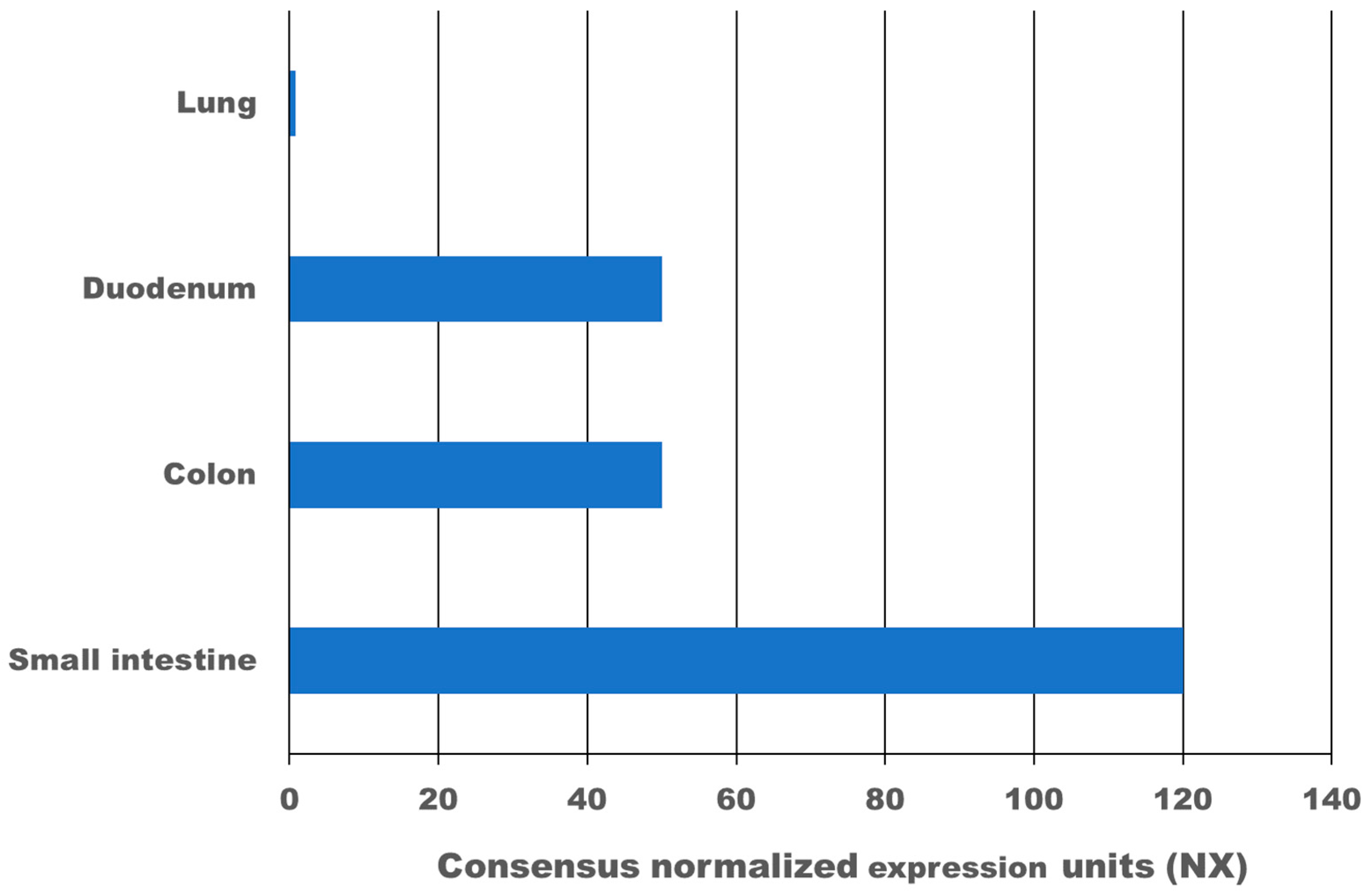
4. Expression of ACE2 and TMPRSS2 in the Gastrointestinal Tract

The virus SARS-CoV-2 enters human cells via the transmembrane protein angiotensin I converting enzyme 2 (ACE2) as a receptor. In addition, host proteases are required to prime the spike protein, especially transmembrane serine protease 2 (TMPRSS2] [34]. ACE2 is highly expressed in the small intestine, colon, and duodenum, compared to very little expression in the lung; see Figure 1 [35]. Immunohistochemistry on human tissue confirms the data, showing high expression of ACE2 protein in the gastrointestinal tract but minimal expression in the lung [22,35].
Using RNAseq, the expression of TMPRSS2 was determined to be high in the small intestine, colon, stomach, and oesophagus; however, it was also high in the lungs [36]. Co-expression of the cell receptor ACE II (ACE2) and the transmembrane serine protease 2 (TMPRSS2) in oesophageal upper epithelial cells, glandular cells, and cells from the ileum and colon was confirmed by single-cell transcriptomic analysis [37]. Given the high expression levels of both ACE2 and TMPRSS2 in the gastrointestinal tract, it seems surprising that gastrointestinal symptoms are relatively mild and low in occurrence.

5. Faecal Transmission of SARS-CoV-2

Over 100 years ago, the ability of an infectious agent to transmit via faeces was demonstrated by Horrocks, showing airborne transport from one hospital building to another by the sewer drains [38]. More than 60 years ago, Jessen reported bioaerosol during toilet flushing [39]. In many studies, it was shown that toilet flushing induces bioaerosols that contaminate the lid and the floor (settled particles), and also the air via droplet nuclei that remain when the water in a droplet evaporates. These droplet nuclei float with the natural air current [40,41,42,43,44,45,46,47].
As described above, SARS-CoV-2 RNA could be detected in either anal swabs or stool specimens in 29–80% of tested patients. Several studies showed consistently that SARS-CoV-2 RNA can be detected in stool for up to 49 days after the onset of symptoms. Since SARS-CoV-2 RNA is present in stool for many patients, the following question arises: Is there live virus present and in what concentration?
Isolation of live virus from stool samples, with the ability to infect monkey kidney VERO cells and demonstrating typical virus formation using electron microscopy (EM), was shown in early 2020, underlining the possibility of faecal–nasal/oral transmission [48]. Xiao et al. showed the expression of viral nucleocapsid protein in gastric, duodenal, and rectal epithelial cells, indicating active replication of the virus [22]. In addition, the authors wrote, “Recently, we and others have isolated infectious SARS-CoV-2 from stool (unpublished data), confirming the release of the infectious virions to the gastrointestinal tract”. Xu et al. reported, “We also isolated alive viral strains from feces, indicating potential infectiousness of feces”. [49]. Wölfle et al. isolated the virus SARS-CoV-2 from samples derived from the throat or lung but could not isolate it from stool samples [50]. Wang et al. reported the isolation of live SARS-CoV-2 from stool samples (non-diarrhoea) from two patients [27]. Zhang et al. demonstrated that live virus can be isolated from stool samples and is able to infect and replicate in monkey kidney VERO cells [26]. The authors concluded, “The live virus in faeces could be an important source of contamination, which may lead to infection and further spread in areas with poor sanitary conditions”.
In a study following 23 patients in a hospital in Beijing, 66.7% tested positive using nasal swabs but 83.3% tested positive using faeces samples. The virus shedding was observed for 10 days (interquartile range (IQR) 8.0–17.0) in nasal-throat mixed swabs and 22 days (IQR 15.5–23.5) in faeces [29]. The authors also reported peak viral titres for respiratory samples of 6–9 days (106.3 copies/mL mean 2535 copies/mL) and for faeces of 14–18 days (105.8 copies/mL mean 5623 copies/mL) after the onset of illness. Wölfe et al. reported about 3 × 10⁶ viral particles per mL in a single faecal sample [50,51]. Infectious virus is present in the stool; thus, could it be a silent route of transfection through faecal–nasal/oral transmission?
Liu et al. studied the potential aerosol transmission of SARS-CoV-2 in two Wuhan hospitals [52]. The authors reported elevated levels of viral RNA in the patient toilet areas and recommended proper disinfection and ventilation. They also showed that the concentration of detectable viral RNA dropped down to undetectable levels when rigorous sanitation procedures were implemented. Van Doremalen et al. showed that viable virus (SARS-CoV-2) could be detected in aerosols up to 3 h post aerosolization, and up to 2–3 days on plastic and stainless steel [53]. Ong et al. followed various patients and their environment in an outbreak centre in Singapore, sampling the air, highly touched areas in the isolation room, and the bathroom [54]. The authors concluded that, “Toilet bowl and sink samples were positive, suggesting that viral shedding in stool could be a potential route of transmission”.
Similar to the Amoy Garden SARS-CoV-1 outbreak in Hong Kong [55], new evidence for infectious faecal aerosols transmitting SARS-CoV-2 in a high-rise apartment building in Guangzhou (China) has been presented. Three families that live in that building were infected with SARS-CoV-2. One family reported travel history into SARS-CoV-2 hotspots; however, the other two families did not. Despite extensive testing in the air and surfaces at various areas, the authors could not find any other traces except the connecting pipes of the drainage system. The authors concluded that faecal aerosol transmission may have caused the community outbreak of COVID-19 in this circumstance [56].
The evidence is strong that faecal transmission is possible and given the fact that there are more infectious SARS-CoV-2 variants around, more awareness of this route should be fostered. Especially in hospitals and care homes, personal protection in dealing with faeces and cleaning procedures for bathrooms are important. Furthermore, shared toilets at workplaces and public toilets in trains, buses, planes, and ships can become a site of contamination. Frequent cleaning with the right additions can mitigate this risk very well.

6. Gut Inflammation in COVID-19 Patients

In a small study describing 40 COVID-19 patients, where 18 reported no diarrhoea, 13 where diarrhoea had ceased, and 9 with active diarrhoea [57], the group with diarrhoea, especially the active diarrhoea, showed a strong increase in faecal calprotectin concentration, a marker for inflammation. This increase correlated with serum interleukin-6 (IL-6) concentration but not C-reactive protein (CRP) or ferritin. These results were underpinned by a study with 26 patients by Reuken et al. [58]. Both studies indicate that inflammation in the gut may occur in COVID-19 patients with diarrhoea and/or positive rectal swabs/faeces.
The influence of COVID-19 on microbial translocation and intestinal damage was investigated [59]. Microbial translocation is the ability of microbes or their products to translocate across the normally very tight epithelial layer into the extraintestinal space and systemic circulation. It occurs when the gut mucosal integrity is weakened. Oliva et al. used three different blood markers to measure microbial translocation and intestinal damage (lipopolysaccharide binding protein, EndoCab IgM, and intestinal fatty acid binding protein) [59]. The cohort was comprised of 45 COVID-19 patients, where 46.6% were admitted to ICU. The data were compared to a healthy donor group. Blood samples taken from day 0 and day 7 showed that COVID-19 patients had both higher microbial translocation and intestinal damage that was maintained over the 7 days. Patients with more severe symptoms showed higher microbial translocation but low intestinal damage compared to patients with mild symptoms. This pattern was a good predictor of disease progression and submission to the ICU. A different study (not yet reviewed) provides the same conclusions. In the 16 COVID-19 patients investigated, the mean levels of lipopolysaccharide (LPS), peptidoglycan (PGN), and fatty acid-binding protein-2 (FABP2) were all increased by about 2 fold compared to healthy controls [60]. All three markers indicate increased gut permeability, and the authors concluded that it may be a source of inflammation, bacteriemia, and consequently worsening of the disease.

7. SARS-CoV-2 and the Gut Microbiome

It has been shown that SARS-CoV-2 infection alters the microbiome of the lung and leads to reduced diversity and in some cases to community collapse [61], shows different bacterial diversity and fewer commensals compared to non-COVID-19 pneumonia [62], and on a functional analysis “decreased potential for lipid metabolism and glycan biosynthesis and metabolism pathways, and increased potential for carbohydrate metabolism pathways” [63]. There is crosstalk between the gut and the lung, often referred to as the “gut-lung axis”. This crosstalk is bidirectional, and the effects of the microbiome in chronic obstructive pulmonary disease (COPD) and inflammatory bowel disease (IBD) has been studied [64]. For a review on the lung-gut axis in respiratory diseases, see Dumas et al. [65]. Infection with SARS-CoV-2 and consequently inflammation in the lung could also lead to changes in the gut microbiota that can further drive the inflammation response. In addition, SARS-CoV-2 infections in the gastrointestinal tract could lead to further changes in the microbiome. It is speculated that the composition and diversity of the “pre-infection” microbiome and post-infection changes, and crosstalk of the gut and lung microbiome could influence the outcome of clinical manifestation [66,67]. In principle, “optimizing” the gut microbiome, especially in the elderly or people with diabetes type II, could positively affect the outcome of SARS-CoV-2 infections.
Honarmand Ebrahimi performed bioinformatic analysis and concluded that members of the microbiome (especially Proteobacteria) of the upper respiratory tract produce ACE2 homologues as well as homologues of TMPRSS2. These could reduce the infectivity of SARS-CoV-2 since the virus would bind to bacteria instead of lung cells. Proteobacteria are reduced in the elderly and will therefore provide less protection [68]. Proteobacteria are also part of the human gut microbiome, but whether the same effect occurs in this more complex environment is unclear. In addition, infection will already occur in the upper gastrointestinal tract where a very different microbiome exists compared to the lower part. Similar to the lung microbiome, diversity in the gut microbiome decreases with age and protective effects may be reduced in the elderly.
The first pilot studies of the gut microbiome of COVID-19 patients have been performed, and examples are given in Table 3. These small studies have their limitations; however, they all have shown for the majority of patients that dysbiosis caused by COVID-19 was not resolved after COVID-19 symptoms eased and patients were discharged. Treatment with antibiotics can also change the gut microbiota; however, in some studies, the described changes in the microbiome were independent of antibiotic treatment. There are various descriptions of dysbiosis. For example, according to [69], “dysbiosis is any change to the composition of resident commensal communities relative to the community found in healthy individuals” and according to [70], “Dysbiosis (also called dysbacteriosis) is characterized as a disruption to the microbiota homeostasis caused by an imbalance in the microflora, changes in their functional composition and metabolic activities, or a shift in their local distribution”. The changes that cause dysbiosis in the microbiome seem to be specific to COVID-19 and can be used as predictors of disease severity. Larger and more systematic studies are urgently needed to understand the impact of SARS-CoV-2 on the gut microbiome, especially long-term effects.
Gou et al. discovered blood proteomic biomarkers that can predict the severity of COVID-19 [75]. Gut microbial features like the relative abundance of Bacteroides genus, Streptococcus genus, Lactobacillus genus, Ruminococcaceae family, Lachnospiraceae family, and Clostridiales order will drive these biomarkers. The faecal metabolome was investigated and showed that 45 faecal metabolites, mainly within the categories of amino acids, fatty acids, and bile acids, can provide a link between the identified core gut microbiota, inflammation, and COVID-19 susceptibility.

8. Targeting the Gut Microbiome as Adjunctive Therapy for COVID-19

The gastrointestinal tract does not just have a digestive function but is also responsible for achieving immune system homeostasis. The gut-associated lymphoid tissue harbours about 70% of the entire immune system [76]. The gut microbiome, its metabolites, and miRNAs influence this homeostasis and also impact mucosal integrity. Weakening of this integrity can result in further inflammation and bacteraemia. As described above, COVID-19 leads to dysbiosis of the gut microbiome, gut inflammation, and weakening of mucosal integrity.
According to the Food and Agriculture Organization of the United Nations World Health Organization, probiotics are defined as “live microorganisms which when administered in adequate amounts confer a health benefit on the host.” Probiotics have been shown to enforce mucosal integrity [77] and be beneficial for influenza virus clearing [78]. Probiotics could therefore in theory support patients with COVID-19 to lessen inflammation, prevent/reduce the very dangerous cytokine storm, and support clearing of the virus. Several clinical trials are underway to study the impact of probiotics on COVID-19 as an adjunctive therapy.
A study from Italy [79] enrolled 70 COVID-19 patients with moderate symptoms (>37.5 °C fever, need of non-invasive oxygen therapy, and according to imaging more than 50% lung involvement) who were treated with hydroxychloroquine (HCQ) 200 mg twice a day, antibiotics (ABX) (azithromycin 500 mg), and Tocilizumab (TCZ), the dosage of which was 8 mg/kg (up to a maximum of 800 mg per dose) with an interval of 12 h two times. A group of randomly picked 28 patients (mean age 59) received probiotics as adjunctive therapy while the remaining 48 patients (mean age 60.5) formed the control group. In this study, Sivomixx® (SivoBiome®, Rockville, MD, USA) was used, consisting of Streptococcus thermophilus DSM 32345, Lactobaccilus acidophilus DSM 32241, Lactobacillus helveticus DSM 32242, Lacticaseibacillus paracasei DSM 32243, Lactobaccilus plantarum DSM 32244, Lactobacillus brevis DSM 27961, Bifidobacterium lactis DSM 32246, and Bifidobacterium lactis DSM 32247. Patients received three equal doses per day (sum of 2400 billion bacteria), for 14 days. Diarrhoea was resolved for 92.9% of the patients in the probiotic group within three days, whereas in the control group, less than 10% after three days and only about 35% after 7 days. Other symptoms like fever, asthenia, headache, myalgia, and dyspnoea resolved in 100% of the patients after 7 days, but only in about 50% of the control group. The author stated, “After 7 days of treatment, the calculated model showed an 8-fold significantly decreased risk to evolve a respiratory failure” [79]. In the probiotic group, 0% of the patients were transferred to the ICU or had a lethal outcome, compared to 4.8% and 9.5%, respectively, in the control group.
The same probiotic Sivomixx® was used by the same group to enlarge the study [80]. This time, 200 patients were enrolled, where 88 received the probiotic at the same dose (3 times daily, a total of 2400 billion bacteria). A similar treatment was provided, including hydroxychloroquine (200 mg twice a day for 7 days), azithromycin (500 mg once a day for 7 days), lopinavir-ritonavir (400/100 mg twice a day), or darunavir–cobicistat (800/150 mg once a day) for 14 days. The risk to be transferred to the ICU was similar in both the control, 21.4% (mean age 64), and probiotic treatment group, 18.1% (mean age 62). There was a significant difference in the mortality between both groups, being 22% in the control group vs. 11% in the probiotic treatment group, clearly demonstrating the potential of this adjuvant treatment.
Currently, Sivomixx® is being tested in another clinical trial in Italy in conjunction with ozone therapy and the recommend best treatment [81]. Systemic autohemotherapy (twice a day) will be combined with 200 billion CFU/day of Sivomixx® (six sachets twice a day). An estimated 152 participants will be enrolled, and various outcome measures determined, with the primary outcome being the number of patients requiring orotracheal intubation.
A clinical trial in Mexico sponsored by AB Biotics, SA, has finished (May 2021), but no data have been published yet [82]. In this intervention study, 300 COVID-19 patients with mild symptoms were enrolled. To a randomly selected group, a probiotic was given (Lactobacillus plantarum CECT 30292, Lactobacillus plantarum CECT 7484, Lactobacillus plantarum CECT 7485 y Pediococcus acidilactici CECT 7483) once a day for 30 days. Various primary and secondary outcomes were determined, for example, severity progression, stay at ICU (frequency and length), mortality, and changes in the faecal microbiome.
A Canadian clinical trial has now finished (June 2021) as well, but no data have been published yet [83]. Nasal irrigation with Probiorinse (2.4 billion colony forming units (CFU) of Lactococcus Lactis W136, (NPN: 80085895)) twice daily for 14 days was used as an intervention. A total of 23 COVID-19 patients were enrolled and changes in severity were monitored for up to 28 days. Another Canadian clinical trial monitored the duration of symptoms, severity, and changes in the oral and faecal microbiome of an estimated 84 patients [84]. A probiotic (undefined) was given to the treatment group for 25 days.
An interventional multi-centre clinical study in Spain is evaluating the probiotic Lactobacillus Coryniformis K8, using a dose of 3 × 10⁹ CFU/day for 2 months, on health care workers [85]. The estimated enrolment is 314 participants, and the incidence and severity of COVID-19 will be measured. In the United States at Duke University, a study is being performed looking at the microbiome of exposed household members from COVID-19 patients. The intervention will be made by providing a probiotic consisting of Lactobacillus rhamnosus GG. In total, 182 participants are expected to enrol [86,87]. A clinical trial in Austria aims to use Omni-Biotic® 10 AAD (Bifidobacterium bifidum W23, Bifidobacterium lactis W51, Enterococcus faecium W54, Lactobacillus acidophilus W37, Lactobacillus acidophilus W55, Lactobacillus paracasei W20, Lactobacillus plantarum W1, Lactobacillus plantarum W62, Lactobacillus rhamnosus W71, and Lactobacillus salivarius W24) as an invention for COVID-19-related diarrhoea [88]. It is planned that an estimated 120 patients will be enrolled.
Another way to influence the gut microbiome more radically is faecal microbiota transplant (FMT). Two cases were reported, from an 80- and a 19-year-old man, who received an FMT to treat a Clostridioides difficile infection (CDI) [89]. Both had severe comorbidities. Unknowingly, both were at the onset of developing COVID-19 at the time point of the FMT. Despite the risk factors of both patients in developing severe COVID-19 symptoms, both experienced rather mild symptoms. This gave rise to a hypothesis that FMT can be used to reduce the risk of severe illness progression. A clinical trial was started to investigate this hypothesis [90].

9. Is There a Link between Changes in the Gut Microbiome in COVID-19 Patients and Chronic COVID-19 Symptoms?

Evidence is accumulating that the gut microbiome is changing for COVID-19 patients and as described above, in a large proportion of patients, these changes (dysbiosis) seem to last. There is compelling evidence that gut microbial dysbiosis can lead to or drive various health problems and is associated with a lower quality of life. It might be no coincidence that more reports are being published describing the long-term effects of COVID-19. Lopez-Leon et al. showed in a meta-analysis that about 80% of COVID-19 patients developed at least one symptom [91]. The main symptoms of this chronic COVID-19 syndrome (CCS) (1) were fatigue (58%), headache (44%), attention disorder (27%), hair loss (25%), and dyspnoea (24%); however, joint pain, sleeping problems, depression, and diarrhoea were reported as well. Fatigue, headache, attention disorders, joint pain, headaches, sleeping problems, depression, and diarrhoea have been linked to dysbiosis in the gut microbiome [92,93,94,95,96,97,98,99,100]. There seems to be an intriguing overlap between these symptoms and more research in this area might reveal new treatment options for CCS. For a more detailed discussion, see [1]. A not yet peer-reviewed article (MedRxiv) shows a loss of diversity in the gut microbiome in chronic COVID-19 patients who experienced severe acute COVID-19 symptoms [101], underpinning the importance of studying this potential connection between gut dysbiosis and chronic COVID-19.

10. Conclusions

SARS-CoV-2 is infecting the gut in a portion of COVID-19 patients, as about 20% develop gastrointestinal symptoms, and about 50% test positive using faecal samples or anal swabs. ACE2 and TMPRSS2 are highly expressed in the gut and explain the reproduction of viruses there. The first studies have shown that the lung, oral, as well as the gut microbiome changes in COVID-19 patients and for a large proportion of patients, the changes do not resolve after discharge from the hospital. Since the gut is also a place to maintain immune homeostasis, changes in the gut can cause or accelerate an inflammation response, weakening of mucosal stability, and a cytokine storm, as seen in critically ill patients. Therefore, it was hypothesized that probiotics or other interventions to favourably change the microbiome or address increased permeability in the gut could reduce the immune answer and be beneficial for the COVID-19 patient. Two clinical trials have now shown the benefits of probiotics, reducing the time to symptom clearing, reducing mortality, and decreasing the risk of developing respiratory failure by 8-fold. Several other clinical trials are underway and will give more insight into the benefits. There also might be a link between changes in the gut microbiome and chronic COVID-19 syndrome (CCS). More research is needed to investigate the potential of this adjuvant treatment.

Funding

This research received no external funding.

Acknowledgments

KH thanks Kathleen Kirmer and Renate Hilpert for their support. KH thanks life/the great mystery for the possibility to live on despite the odds.

Conflicts of Interest

KH is a founder and director of TiKa Diagnostics Ltd.

References

  1. Hilpert, K.; Mikut, R. Is there a connection between gut microbiome dysbiosis occurring in COVID-19 patients and post-COVID-19 symptoms? Front. Microbiol. 2021, 12, 2564. [Google Scholar] [CrossRef]
  2. Fehr, A.R.; Perlman, S. Coronaviruses: An overview of their replication and pathogenesis. In Coronaviruses Methods and Protocols; Springer: New York, NY, USA, 2015; pp. 1–23. [Google Scholar]
  3. Wood, E.N. An apparently new syndrome of porcine epidemic diarrhoea. Veter. Rec. 1977, 100, 243–244. [Google Scholar] [CrossRef]
  4. Jung, K.; Annamalai, T.; Lu, Z.; Saif, L.J. Comparative pathogenesis of US porcine epidemic diarrhea virus (PEDV) strain PC21A in conventional 9-day-old nursing piglets vs. 26-day-old weaned pigs. Veter. Microbiol. 2015, 178, 31–40. [Google Scholar] [CrossRef]
  5. Saif, L.J.; Wang, Q.; Vlasova, A.N.; Jung, K.; Xiao, S. Coronaviruses. In Diseases of Swine; Wiley: Hoboken, NJ, USA, 2019; pp. 488–523. [Google Scholar]
  6. Okur, G.S.; Yazici, Z.; Albayrak, H.; Meral, Y. Rotavirus and Coronavirus Prevalances in Healthy Calves and Calves with Diarrhoea. Available online: https://www.researchgate.net/publication/287890228 (accessed on 29 April 2020).
  7. Rehman, S.U.; Shafique, L.; Ihsan, A.; Liu, Q. Evolutionary trajectory for the emergence of novel coronavirus SARS-CoV-2. Pathogens 2020, 9, 240. Available online: https://www.mdpi.com/2076-0817/9/3/240 (accessed on 28 April 2020). [CrossRef] [Green Version]
  8. Su, S.; Wong, G.; Shi, W.; Liu, J.; Lai, A.C.K.; Zhou, J.; Liu, W.; Bi, Y.; Gao, G.F. Epidemiology, genetic recombination, and pathogenesis of coronaviruses. Trends Microbiol. 2016, 24, 490–502. [Google Scholar] [CrossRef] [Green Version]
  9. Chen, Z.-R.; Liu, J.; Liao, Z.-G.; Zhou, J.; Peng, H.-W.; Gong, F.; Hu, J.-F.; Zhou, Y. COVID-19 and gastroenteric manifestations. World J. Clin. Cases 2021, 9, 4990–4997. [Google Scholar] [CrossRef]
  10. Perisetti, A.; Goyal, H.; Gajendran, M.; Boregowda, U.; Mann, R.; Sharma, N. Prevalence, mechanisms, and implications of gastrointestinal symptoms in COVID-19. Front. Med. 2020, 7, 588711. [Google Scholar] [CrossRef]
  11. Fang, D.; Ma, J.; Guang, J.; Wang, M.; Song, Y.; Tian, D. Manifestations of digestive system in hospitalized patients with novel coronavirus pneumonia in Wuhan, China: A single-center, descriptive study. Chin. J. Dig. 2020, 40, E005. [Google Scholar] [CrossRef]
  12. Guan, W.-J.; Ni, Z.-Y.; Hu, Y.; Liang, W.-H.; Ou, C.-Q.; He, J.-X.; Liu, L.; Shan, H.; Lei, C.-L.; Hui, D.S.; et al. Clinical characteristics of coronavirus disease 2019 in China. N. Engl. J. Med. 2020, 382, 1708–1720. [Google Scholar] [CrossRef]
  13. Tian, Y.; Rong, L.; Nian, W.; He, Y. Review article: Gastrointestinal features in COVID-19 and the possibility of faecal transmission. Aliment. Pharmacol. Ther. 2020, 51, 843–851. [Google Scholar] [CrossRef]
  14. Wang, D.; Hu, B.; Hu, C.; Zhu, F.; Liu, X.; Zhang, J.; Wang, B.; Xiang, H.; Cheng, Z.; Xiong, Y.; et al. Clinical characteristics of 138 hospitalized patients with 2019 novel coronavirus-infected pneumonia in Wuhan, China. JAMA 2020, 323, 1061–1069. [Google Scholar] [CrossRef]
  15. Kumar, V.C.S.; Mukherjee, S.; Harne, P.S.; Subedi, A.; Ganapathy, M.K.; Patthipati, V.S.; Sapkota, B. Novelty in the gut: A systematic review and meta-analysis of the gastrointestinal manifestations of COVID-19. BMJ Open Gastroenterol. 2020, 7, 417. Available online: http://bmjopengastro.bmj.com/ (accessed on 5 October 2020).
  16. Cheung, K.S.; Hung, I.F.; Chan, P.P.; Lung, K.C.; Tso, E.; Liu, R.; Ng, Y.Y.; Chu, M.Y.; Chung, T.W.; Tam, A.R.; et al. Gastrointestinal manifestations of SARS-CoV-2 infection and virus load in fecal samples from the Hong Kong cohort and systematic review and meta-analysis. Gastroenterology 2020, 159, 81–95. [Google Scholar] [CrossRef]
  17. Parasa, S.; Desai, M.; Chandrasekar, V.T.; Patel, H.K.; Kennedy, K.F.; Roesch, T.; Spadaccini, M.; Colombo, M.; Gabbiadini, R.; Artifon, E.L.; et al. Prevalence of gastrointestinal symptoms and fecal viral shedding in patients with coronavirus disease 2019: A systematic review and meta-analysis. JAMA Netw. Open 2020, 3, e2011335. Available online: https://jamanetwork.com/journals/jamanetworkopen/fullarticle/2767009 (accessed on 27 August 2020). [CrossRef]
  18. Rokkas, T. Gastrointestinal involvement in COVID-19: A systematic review and meta-analysis. Ann. Gastroenterol. 2020, 33, 355–365. [Google Scholar] [CrossRef]
  19. Dorrell, R.D.; Dougherty, M.K.; Barash, E.L.; Lichtig, A.E.; Clayton, S.B.; Jensen, E.T. Gastrointestinal and hepatic manifestations of COVID-19: A systematic review and meta-analysis. JGH Open 2021, 5, 107–115. [Google Scholar] [CrossRef]
  20. Silva, F.A.F.D.; Brito, B.B.D.; Santos, M.L.C.; Marques, H.S.; Silva, R.T.D.; Carvalho, L.S.D.; Vieira, E.S.; Oliveira, M.V.; Melo, F.F.D. COVID-19 gastrointestinal manifestations: A systematic review. Rev. Soc. Bras. Med. Trop. 2020, 53, 1–11. [Google Scholar] [CrossRef]
  21. Zhang, W.; Du, R.-H.; Li, B.; Zheng, X.-S.; Yang, X.-L.; Hu, B.; Wang, Y.-Y.; Xiao, G.-F.; Yan, B.; Shi, Z.-L.; et al. Molecular and serological investigation of 2019-nCoV infected patients: Implication of multiple shedding routes. Emerg. Microbes Infect. 2020, 9, 386–389. [Google Scholar] [CrossRef] [Green Version]
  22. Xiao, F.; Tang, M.; Zheng, X.; Liu, Y.; Li, X.; Shan, H. Evidence for gastrointestinal infection of SARS-CoV-2. Gastroenterology 2020, 6, 1831–1833. [Google Scholar] [CrossRef]
  23. Lo, I.L.; Lio, C.F.; Cheong, H.H.; Lei, C.I.; Cheong, T.H.; Zhong, X.; Tian, Y.; Sin, N.N. Evaluation of SARS-CoV-2 RNA shedding in clinical specimens and clinical characteristics of 10 patients with COVID-19 in Macau. Int. J. Biol. Sci. 2020, 16, 1698–1707. Available online: http://www.ijbs.com//creativecommons.org/licenses/by/4.0/ (accessed on 3 September 2020). [CrossRef] [Green Version]
  24. Wu, Y.; Guo, C.; Tang, L.; Hong, Z.; Zhou, J.; Dong, X.; Yin, H.; Xiao, Q.; Tang, Y.; Qu, X.; et al. Prolonged presence of SARS-CoV-2 viral RNA in faecal samples. Lancet Gastroenterol. Hepatol. 2020, 5, 434–435. [Google Scholar] [CrossRef]
  25. Chen, Y.; Chen, L.; Deng, Q.; Zhang, G.; Wu, K.; Ni, L.; Yang, Y.; Liu, B.; Wang, W.; Wei, C.; et al. The presence of SARS-CoV-2 RNA in feces of COVID-19 patients. J Med. Virol. 2020, 92, 833–840. [Google Scholar] [CrossRef] [Green Version]
  26. Zhang, Y.; Chen, C.; Song, Y.; Zhu, S.; Wang, D.; Zhang, H.; Han, G.; Weng, Y.; Xu, J.; Xu, J.; et al. Excretion of SARS-CoV-2 through faecal specimens. Emerg. Microbes. Infect. 2020, 9, 2501–2508. [Google Scholar] [CrossRef]
  27. Wang, W.; Xu, Y.; Gao, R.; Lu, R.; Han, K.; Wu, G.; Tan, W. Detection of SARS-CoV-2 in different types of clinical specimens. JAMA J. Am. Med. Assoc. 2020, 323, 1843–1844. [Google Scholar] [CrossRef] [Green Version]
  28. Lin, L.; Jiang, X.; Zhang, Z.; Huang, S.; Zhang, Z.; Fang, Z.; Gu, Z.; Gao, L.; Shi, H.; Mai, L.; et al. Gut immunity gastrointestinal symptoms of 95 cases with SARS-CoV-2 infection. Gut 2020, 69, 997–1001. Available online: http://gut.bmj.com/ (accessed on 4 May 2020). [CrossRef] [PubMed]
  29. Zhang, N.; Gong, Y.; Meng, F.; Bi, Y.; Yang, P. Virus Shedding Patterns in Nasopharyngeal and Fecal Specimens of COVID-19 Patients. Sci. China Life Sci. 2021, 64, 486–488. Available online: https://doi.org/10.1101/2020.03.28.20043059 (accessed on 12 May 2020). [CrossRef] [PubMed]
  30. Jiang, X.; Luo, M.; Zou, Z.; Wang, X.; Chen, C.; Qiu, J. Asymptomatic SARS-CoV-2 infected case with viral detection positive in stool but negative in nasopharyngeal samples lasts for 42 days. J. Med. Virol. 2020, 92, 1807–1809. [Google Scholar] [CrossRef]
  31. Xu, Y.; Li, X.; Zhu, B.; Liang, H.; Fang, C.; Gong, Y.; Guo, Q.; Sun, X.; Zhao, D.; Shen, J.; et al. Characteristics of pediatric SARS-CoV-2 infection and potential evidence for persistent fecal viral shedding. Nat. Med. 2020, 26, 502–505. [Google Scholar] [CrossRef] [Green Version]
  32. Zhang, T.; Cui, X.; Zhao, X.; Wang, J.; Zheng, J.; Zheng, G.; Guo, W.; Cai, C.; He, S.; Xu, Y. Detectable SARS-CoV-2 viral RNA in feces of three children during recovery period of COVID-19 pneumonia. J. Med. Virol. 2020, 92, 909–914. [Google Scholar] [CrossRef]
  33. Byrne, A.W.; McEvoy, D.; Collins, A.B.; Hunt, K.; Casey, M.; Barber, A.; Butler, F.; Griffin, J.; Lane, E.A.; McAloon, C.; et al. Inferred duration of infectious period of SARS-CoV-2: Rapid scoping review and analysis of available evidence for asymptomatic and symptomatic COVID-19 cases. BMJ Open 2020, 10, e039856. Available online: http://bmjopen.bmj.com/ (accessed on 3 September 2020). [CrossRef]
  34. Hoffmann, M.; Kleine-Weber, H.; Schroeder, S.; Krüger, N.; Herrler, T.; Erichsen, S.; Schiergens, T.S.; Herrler, G.; Wu, N.H.; Nitsche, A.; et al. SARS-CoV-2 cell entry depends on ACE2 and TMPRSS2 and is blocked by a clinically proven protease inhibitor. Cell 2020, 181, 271–280. Available online: https://pubmed.ncbi.nlm.nih.gov/32142651/ (accessed on 14 June 2021). [CrossRef]
  35. Hikmet, F.; Méar, L.; Edvinsson, Å.; Micke, P.; Uhlén, M.; Lindskog, C. The protein expression profile of ACE2 in human tissues. Mol. Syst. Biol. 2020, 16, e9610. [Google Scholar] [CrossRef]
  36. TMPRSS2 Gene—GeneCards. TMPS2 Protein TMPS2 Antibody. Available online: https://www.genecards.org/cgi-bin/carddisp.pl?gene=TMPRSS2 (accessed on 9 September 2021).
  37. Zhang, H.; Kang, Z.; Gong, H.; Xu, D.; Wang, J.; Li, Z.; Li, Z.; Cui, X.; Xiao, J.; Zhan, J.; et al. Digestive system is a potential route of COVID-19: An analysis of single-cell coexpression pattern of key proteins in viral entry process. Gut 2020, 69, 1010–1018. [Google Scholar] [CrossRef]
  38. Horrocks, W.H. Experiments made to determine the conditions under which “specific” bacteria derived from sewage may be present in the air of ventilating pipes, drains, inspection chambers, and sewers. In Proceedings of the Royal Society of London. Series B, Containing Papers of a Biological Character; The Royal Society: London, UK, 1907; Volume 79, pp. 255–266. [Google Scholar]
  39. Jessen, C.U. Airborne Microorganisms: Occurrance and Control; GEC Gad: Copenhagen, Denmark, 1955. [Google Scholar]
  40. Wells, W.F. On air-borne infection: Study II. Droplets and droplet nuclei. Am. J. Epidemiol. 1934, 20, 611–618. [Google Scholar] [CrossRef]
  41. Darlow, H.M.; Bale, W.R. Infective hazards of water-closets. Lancet 1959, 273, 1196–1200. [Google Scholar] [CrossRef]
  42. Blair, M. Ceramic Water Closets; Bloomsbury Publishing: New York, NY, USA, 2008. [Google Scholar]
  43. Gerba, C.P.; Wallis, C.; Melnick, J.L. Microbiological hazards of household toilets: Droplet production and the fate of residual organisms. Appl. Microbiol. 1975, 30, 229–237. [Google Scholar] [CrossRef]
  44. Barker, J.; Vipond, I.; Bloomfield, S. Effects of cleaning and disinfection in reducing the spread of Norovirus contamination via. environmental surfaces. J. Hosp. Infect. 2004, 58, 42–49. [Google Scholar] [CrossRef] [PubMed]
  45. Barker, J.; Jones, M.V. The potential spread of infection caused by aerosol contamination of surfaces after flushing a domestic toilet. J. Appl. Microbiol. 2005, 99, 339–347. [Google Scholar] [CrossRef]
  46. Best, E.; Sandoe, J.; Wilcox, M. Potential for aerosolization of Clostridium difficile after flushing toilets: The role of toilet lids in reducing environmental contamination risk. J. Hosp. Infect. 2012, 80, 1–5. [Google Scholar] [CrossRef] [PubMed]
  47. Newsom, S.W.B. Microbiology of hospital toilets. Lancet 1972, 300, 700–703. [Google Scholar] [CrossRef]
  48. Zhang, Y.; Chen, C.; Zhu, S.; Shu, C.; Wang, D.; Song, J.; Song, Y.; Zhen, W.; Feng, Z.; Wu, G.; et al. Isolation of 2019-nCoV from a stool specimen of a laboratory-confirmed case of the coronavirus disease 2019 (COVID-19). China CDC Wkly. 2020, 2, 123–124. [Google Scholar] [CrossRef]
  49. Xu, K.; Cai, H.; Shen, Y.; Ni, Q.; Chen, Y.; Hu, S.; Li, J.; Wang, H.; Yu, L.; Huang, H.; et al. Management of corona virus disease-19 (COVID-19): The Zhejiang experience. J. Zhejiang Univ. 2020, 49, 147–157. [Google Scholar]
  50. Wölfel, R.; Corman, V.M.; Guggemos, W.; Seilmaier, M.; Zange, S.; Müller, M.A.; Niemeyer, D.; Jones, T.C.; Vollmar, P.; Rothe, C.; et al. Virological assessment of hospitalized patients with COVID-2019. Nature 2020, 581, 465–469. [Google Scholar] [CrossRef] [Green Version]
  51. Wu, F.; Xiao, A.; Zhang, J.; Gu, X.; Lee, W.L.; Kauffman, K.; Hanage, W.; Matus, M.; Ghaeli, N.; Endo, N. SARS-CoV-2 titers in wastewater are higher than expected from clinically confirmed cases. medRxiv 2020, 5, e00614–e00620. [Google Scholar] [CrossRef]
  52. Liu, Y.; Ning, Z.; Chen, Y.; Guo, M.; Liu, Y.; Gali, N.K.; Sun, L.; Duan, Y.; Cai, J.; Westerdahl, D.; et al. Aerodynamic analysis of SARS-CoV-2 in two Wuhan hospitals. Nature 2020, 582, 557–560. [Google Scholar] [CrossRef]
  53. Van Doremalen, N.; Lloyd-Smith, J.O.; Munster, V.J.; Bushmaker, T.; Morris, D.H.; Gamble, A.; Williamson, B.N.; Tamin, A.; Harcourt, J.L.; Thornburg, N.J.; et al. Aerosol and surface stability of HCoV-19 (SARS-CoV-2) compared to SARS-CoV-1. N. Engl. J. Med. 2020, 382, 1564–1567. Available online: https://www.nejm.org/doi/10.1056/NEJMc2004973 (accessed on 17 April 2020). [CrossRef] [PubMed]
  54. Ong, S.W.X.; Tan, Y.K.; Chia, P.Y.; Lee, T.H.; Ng, O.T.; Wong, M.S.Y.; Marimuthu, K. Air, surface environmental, and personal protective equipment contamination by severe acute respiratory syndrome coronavirus 2 (SARS-CoV-2) from a symptomatic patient. JAMA J. Am. Med. Assoc. 2020, 323, 1610–1612. Available online: https://jamanetwork.com/journals/jama/fullarticle/2762692 (accessed on 28 April 2020). [CrossRef] [PubMed] [Green Version]
  55. Yu, I.T.; Li, Y.; Wong, T.W.; Tam, W.; Chan, A.; Lee, J.H.; Leung, D.Y.; Ho, T. Evidence of airborne transmission of the severe acute respiratory syndrome virus. N. Engl. J. Med. 2004, 350, 1731–1739. [Google Scholar] [CrossRef] [PubMed] [Green Version]
  56. Kang, M.; Wei, J.; Yuan, J.; Guo, J.; Zhang, Y.; Hang, J.; Qu, Y.; Qian, H.; Zhuang, Y.; Chen, X.; et al. Probable evidence of fecal aerosol transmission of SARS-CoV-2 in a high-rise building. Ann. Intern. Med. 2020, 173, 974–980. [Google Scholar] [CrossRef]
  57. Effenberger, M.; Grabherr, F.; Mayr, L.; Schwaerzler, J.; Nairz, M.; Seifert, M.; Hilbe, R.; Seiwald, S.; Scholl-Buergi, S.; Fritsche, G.; et al. Faecal calprotectin indicates intestinal inflammation in COVID-19. Gut 2020, 69, 1543–1544. [Google Scholar] [CrossRef] [PubMed] [Green Version]
  58. Reuken, P.A.; Wüst, M.; Löffler, B.; Bauer, M.; Stallmach, A. Letter: SARS-CoV-2-induced gastrointestinal inflammation. Aliment. Pharmacol. Ther. 2020, 52, 1748–1749. Available online: https://www.ncbi.nlm.nih.gov/pmc/articles/PMC7753696/pdf/APT-52-1750.pdf (accessed on 14 June 2021).
  59. Oliva, A.; Miele, M.C.; Di Timoteo, F.; De Angelis, M.; Mauro, V.; Aronica, R.; Al Ismail, D.; Ceccarelli, G.; Pinacchio, C.; D’Ettorre, G.; et al. Persistent systemic microbial translocation and intestinal damage during coronavirus disease-19. Front. Immunol. 2021, 12, 2810. [Google Scholar] [CrossRef]
  60. Prasad, R.; Patton, M.J.; Floyd, J.L.; Vieira, C.P.; Fortmann, S.; DuPont, M.; Harbour, A.; See, J.R.C.; Wright, J.; Lamendella, R.; et al. Plasma microbiome in COVID-19 subjects: An indicator of gut barrier defects and dysbiosis. bioRxiv 2021. Available online: https://www.biorxiv.org/content/10.1101/2021.04.06.438634v1 (accessed on 9 September 2021).
  61. Tsitsiklis, A.; Shoshana, Z.B.; Byrne, A.; Devoe, C.; Levan, S.; Rackaityte, E.; Sunshine, S.; Mick, E.; Ghale, R.; Jauregui, A.; et al. Impaired immune signaling and changes in the lung microbiome precede secondary 1 bacterial pneumonia in COVID-19. Res. Sq. 2021, 1, rs-3. [Google Scholar]
  62. Gaibani, P.; Viciani, E.; Bartoletti, M.; Lewis, R.E.; Tonetti, T.; Lombardo, D.; Castagnetti, A.; Bovo, F.; Horna, C.S.; Ranieri, M.; et al. The lower respiratory tract microbiome of critically ill patients with COVID-19. Sci. Rep. 2021, 11, 10103. [Google Scholar] [CrossRef] [PubMed]
  63. Haiminen, N.; Utro, F.; Seabolt, E.; Parida, L. Functional profiling of COVID-19 respiratory tract microbiomes. Sci. Rep. 2021, 11, 6433. [Google Scholar] [CrossRef] [PubMed]
  64. Keely, S.; Talley, N.J.; Hansbro, P.M. Pulmonary-intestinal cross-talk in mucosal inflammatory disease. Mucosal Immunol. 2012, 5, 7–18. Available online: www.nature.com/mi (accessed on 3 September 2020). [CrossRef] [Green Version]
  65. Dumas, A.; Bernard-Raichon, L.; Poquet, Y.; Lugo, G.; Neyrolles, O. The role of the lung microbiota and the gut-lung axis in respiratory infectious diseases. Cell. Microbiol. 2018, 20, e12966. [Google Scholar] [CrossRef] [Green Version]
  66. Dhar, D.; Mohanty, A. Gut microbiota and Covid-19 possible link and implications. Virus Res. 2020, 285, 198018. [Google Scholar] [CrossRef]
  67. Baud, D.; Dimopoulou Agri, V.; Gibson, G.R.; Reid, G.; Giannoni, E. Using probiotics to flatten the curve of coronavirus disease COVID-2019 pandemic. Front. Public Health 2020, 8, 186. [Google Scholar] [CrossRef]
  68. Ebrahimi, K.H. SARS-CoV-2 spike glycoprotein-binding proteins expressed by upper respiratory tract bacteria may prevent severe viral infection. FEBS Lett. 2020, 594, 1651–1660. [Google Scholar] [CrossRef] [PubMed]
  69. Petersen, C.; Round, J.L. Defining dysbiosis and its influence on host immunity and disease. Cell. Microbiol. 2014, 16, 1024–1033. [Google Scholar] [CrossRef] [PubMed]
  70. Dysbiosis—Wikipedia. Available online: https://en.wikipedia.org/wiki/Dysbiosis (accessed on 9 September 2021).
  71. Zuo, T.; Zhang, F.; Lui, G.C.; Yeoh, Y.K.; Li, A.Y.; Zhan, H.; Wan, Y.; Chung, A.C.; Cheung, C.P.; Chen, N.; et al. Alterations in gut microbiota of patients with covid-19 during time of hospitalization. Gastroenterology 2020, 159, 944–955. [Google Scholar] [CrossRef] [PubMed]
  72. Gu, S.; Chen, Y.; Wu, Z.; Chen, Y.; Gao, H.; Lv, L.; Guo, F.; Zhang, X.; Luo, R.; Huang, C.; et al. Alterations of the gut microbiota in patients with coronavirus disease 2019 or H1N1 influenza. Clin. Infect. Dis. 2020, 71, 2669–2678. [Google Scholar] [CrossRef] [PubMed]
  73. Zuo, T.; Zhan, H.; Zhang, F.; Liu, Q.; Tso, E.Y.; Lui, G.C.; Chen, N.; Li, A.; Lu, W.; Chan, F.K.; et al. Alterations in fecal fungal microbiome of patients with covid-19 during time of hospitalization until discharge. Gastroenterology 2020, 159, 1302–1310. [Google Scholar] [CrossRef] [PubMed]
  74. Ren, Z.; Wang, H.; Cui, G.; Lu, H.; Wang, L.; Luo, H.; Chen, X.; Ren, H.; Sun, R.; Liu, W.; et al. Alterations in the human oral and gut microbiomes and lipidomics in COVID-19. Gut 2021, 70, 1253–1265. [Google Scholar] [CrossRef] [PubMed]
  75. Gou, W.; Fu, Y.; Yue, L.; Chen, G.-D.; Cai, X.; Shuai, M.; Xu, F.; Yi, X.; Chen, H.; Zhu, Y.; et al. Gut microbiota, inflammation and molecular signatures of host response to infection. J. Genet. Genom. 2021, in press. [Google Scholar] [CrossRef]
  76. Vighi, G.; Marcucci, F.; Sensi, L.; DI Cara, G.; Frati, F. Allergy and the gastrointestinal system. Clin. Exp. Immunol. 2008, 153, 3–6. [Google Scholar] [CrossRef]
  77. Hummel, S.; Veltman, K.; Cichon, C.; Sonnenborn, U.; Schmidt, M.A. Differential targeting of the E-cadherin/β-catenin complex by gram-positive probiotic lactobacilli improves epithelial barrier function. Appl. Environ. Microbiol. 2012, 78, 1140–1147. [Google Scholar] [CrossRef] [Green Version]
  78. Zelaya, H.; Alvarez, S.; Kitazawa, H.; Villena, J. Respiratory antiviral immunity and immunobiotics: Beneficial effects on inflammation-coagulation interaction during influenza virus infection. Front. Immunol. 2016, 7, 633. [Google Scholar] [CrossRef] [Green Version]
  79. D’Ettorre, G.; Ceccarelli, G.; Marazzato, M.; Campagna, G.; Pinacchio, C.; Alessandri, F.; Ruberto, F.; Rossi, G.; Celani, L.; Scagnolari, C.; et al. Challenges in the management of SARS-CoV2 infection: The role of oral bacteriotherapy as complementary therapeutic strategy to avoid the progression of COVID-19. Front Med. 2020, 7, 389. [Google Scholar] [CrossRef] [PubMed]
  80. Ceccarelli, G.; Borrazzo, C.; Pinacchio, C.; Santinelli, L.; Innocenti, G.P.; Cavallari, E.N.; Celani, L.; Marazzato, M.; Alessandri, F.; Ruberto, F.; et al. Oral bacteriotherapy in patients with COVID-19: A retrospective cohort study. Front Nutr. 2021, 7, 341. [Google Scholar] [CrossRef] [PubMed]
  81. Oxygen-Ozone as Adjuvant Treatment in Early Control of COVID-19 Progression and Modulation of the Gut Microbial Flora—Full Text View—ClinicalTrials.gov. Available online: https://clinicaltrials.gov/ct2/show/NCT04366089 (accessed on 13 August 2021).
  82. Efficacy of L. Plantarum and P. Acidilactici in Adults with SARS-CoV-2 and COVID-19—Full Text View—ClinicalTrials.gov. Available online: https://clinicaltrials.gov/ct2/show/study/NCT04517422 (accessed on 13 August 2021).
  83. Efficacy of Intranasal Probiotic Treatment to Reduce Severity of Symptoms in COVID19 Infection—Full Text View—ClinicalTrials.gov. Available online: https://clinicaltrials.gov/ct2/show/NCT04458519 (accessed on 13 August 2021).
  84. Efficacy of Probiotics in Reducing Duration and Symptoms of COVID-19—Full Text View—ClinicalTrials.gov. Available online: https://clinicaltrials.gov/ct2/show/NCT04621071 (accessed on 13 August 2020).
  85. Evaluation of the Probiotic Lactobacillus Coryniformis K8 on COVID-19 Prevention in Healthcare Workers—Full Text View—ClinicalTrials.gov. Available online: https://clinicaltrials.gov/ct2/show/study/NCT04366180 (accessed on 12 August 2021).
  86. Tang, H.; Bohannon, L.; Lew, M.; Jensen, D.; Jung, S.-H.; Zhao, A.; Sung, A.D.; Wischmeyer, P.E. Randomised, double-blind, placebo-controlled trial of probiotics to eliminate COVID-19 transmission in exposed household contacts (PROTECT-EHC): A Clinical trial protocol. BMJ Open 2021, 11, e047069. [Google Scholar] [CrossRef] [PubMed]
  87. Effect of Lactobacillus on the Microbiome of Household Contacts Exposed to COVID-19—Full Text View—ClinicalTrials.gov. Available online: https://clinicaltrials.gov/ct2/show/NCT04399252 (accessed on 12 August 2021).
  88. Synbiotic Therapy of Gastrointestinal Symptoms During Covid-19 Infection—Full Text View—ClinicalTrials.gov. Available online: https://clinicaltrials.gov/ct2/show/NCT04420676 (accessed on 13 August 2021).
  89. Biliński, J.; Winter, K.; Jasiński, M.; Szczęś, A.; Bilinska, N.; Mullish, B.H.; Małecka-Panas, E.; Basak, G.W. Rapid resolution of COVID-19 after faecal microbiota transplantation. Gut 2021, 1–2. [Google Scholar] [CrossRef]
  90. The Impact of Fecal Microbiota Transplantation as an Immunomodulation on the Risk Reduction of COVID-19 Disease Progression with Escalating Cytokine Storm and Inflammatory Parameter—Full Text View—ClinicalTrials.gov. Available online: https://clinicaltrials.gov/ct2/show/NCT04824222 (accessed on 16 August 2021).
  91. Lopez-Leon, S.; Wegman-Ostrosky, T.; Perelman, C.; Sepulveda, R.; Rebolledo, P.A.; Cuapio, A.; Villapol, S. More than 50 long-term effects of COVID-19: A systematic review and meta-analysis. Sci. Rep. 2021, 11, 16144. [Google Scholar] [CrossRef]
  92. Arzani, M.; Jahromi, S.R.; Ghorbani, Z.; Vahabizad, F.; Martelletti, P.; Ghaemi, A.; Sacco, S.; Togha, M. Gut-brain Axis and migraine headache: A comprehensive review. J. Headache Pain 2020, 21, 1–2. [Google Scholar] [CrossRef] [PubMed] [Green Version]
  93. Galland, L. The gut microbiome and the brain. J. Med. Food. 2014, 17, 1261–1272. Available online: https://pubmed.ncbi.nlm.nih.gov/25402818/ (accessed on 19 June 2021). [CrossRef]
  94. Matenchuk, B.A.; Mandhane, P.J.; Kozyrskyj, A.L. Sleep, circadian rhythm, and gut microbiota. Sleep Med. Rev. 2020, 53, 101340. Available online: https://pubmed.ncbi.nlm.nih.gov/32668369/ (accessed on 19 June 2021). [CrossRef] [PubMed]
  95. Molina-Torres, G.; Rodriguez-Arrastia, M.; Roman, P.; Labraca, M.N.S.; Cardona, D. Stress and the gut microbiota-brain axis. Behav. Pharmacol. 2019, 30, 187–200. [Google Scholar] [CrossRef]
  96. Ogawa, Y.; Miyoshi, C.; Obana, N.; Yajima, K.; Hotta-Hirashima, N.; Ikkyu, A.; Kanno, S.; Soga, T.; Fukuda, S.; Yanagisawa, M. Gut microbiota depletion by chronic antibiotic treatment alters the sleep/wake architecture and sleep EEG power spectra in mice. Sci. Rep. 2020, 10, 19554. [Google Scholar] [CrossRef] [PubMed]
  97. Peirce, J.M.; Alviña, K. The role of inflammation and the gut microbiome in depression and anxiety. J. Neurosci. Res. 2019, 97, 1223–1241. [Google Scholar] [CrossRef] [PubMed] [Green Version]
  98. Poroyko, V.A.; Carreras, A.; Khalyfa, A.; Khalyfa, A.A.; Leone, V.; Peris, E.; Almendros, I.; Gileles-Hillel, A.; Qiao, Z.; Hubert, N.; et al. Chronic sleep disruption alters gut microbiota, induces systemic and adipose tissue inflammation and insulin resistance in mice. Sci. Rep. 2016, 6, 35405. [Google Scholar] [CrossRef] [PubMed] [Green Version]
  99. Smith, R.P.; Easson, C.; Lyle, S.M.; Kapoor, R.; Donnelly, C.P.; Davidson, E.J.; Parikh, E.; Lopez, J.V.; Tartar, J.L. Gut microbiome diversity is associated with sleep physiology in humans. PLoS ONE 2019, 14, e0222394. [Google Scholar] [CrossRef] [PubMed]
  100. Boehme, M.; Guzzetta, K.E.; Bastiaanssen, T.F.S.; van de Wouw, M.; Moloney, G.M.; Gual-Grau, A.; Spichak, S.; Olavarría-Ramírez, L.; Fitzgerald, P.; Morillas, E.; et al. Microbiota from young mice counteracts selective age-associated behavioral deficits. Nat. Aging 2021, 1, 666–676. [Google Scholar] [CrossRef]
  101. Vestad, B.; Ueland, T.; Lerum, T.V.; Dahl, T.B.; Holm, K.; Barratt-Due, A.; Kasine, T.; Dyrhol-Riise, A.M.; Stiksrud, B.; Tonby, K.; et al. Gut microbiota alterations in patients with persistent respiratory dysfunction three months after severe COVID-19. medRxiv 2021. Available online: https://www.medrxiv.org/content/10.1101/2021.07.13.21260412v1 (accessed on 9 September 2021).
Figure 1. Expression of the angiotensin I converting enzyme 2 (ACE2) in different human organs, based on data by Hikmet et al. [35]. The expression of ACE2 was determined by transcriptomics by three different independent consortia.
Biologics 01 00017 g001
Table 1. Overview of six meta-analyses of COVID-19 patient studies and their reported gastrointestinal symptoms.
Number of COVID-19 PatientsGastro-Intestinal SymptomsDiarrhoeaNausea/VomitingAbdominal PainNumber of Studies Used in Meta-AnalysisReference
247713%7.8%5.5%2.7%17[15]
424317.6%12.5%10.2%9.2%60[16]
4805Not reported7.4%4.6%Not reported29[17]
56019.8%10.4%7.7%6.9%37[18]
17,77620%13%8%4%108[19]
18,246Not reported11.5%6.3%2.3%43[20]
Table 2. Overview of examples of studies investigating SARS-CoV-2 detection in faeces.
Number of COVID-19 PatientsPositive in Faeces/Anal SwabsDuration of Positivity (Days)CommentsReference
1526.7% 53.3% positive in oral swabs[21]
1625% (Day 0)
37.5% (Day 5)
[21]
10100%3–19 [23]
7353%1–1223% stool positive and negative in respiratory samples[22]
7455%Mean of 27.9 (SD 10.7) [24]
4267%Mean 7 (6–10) [25]
25836% Virus isolated and sequenced from stool, infectious in monkey kidney VERO cells, and confirmation by electron microscopy (EM)[26]
205 (153 tested for faecal samples)29% [27]
5915.3% [16]
4243 (Meta-analysis)48.1% 70% of stool positive and negative in respiratory samples[16]
2348% [28]
2383.3%Mean 22 [29]
Table 3. Examples of pilot studies investigating the changes in the gut microbiome in COVID-19 patients.
Number of COVID-19 PatientsHealthy ControlAge (Median)Microbiome InvestigatedEnrichmentLossReference
COVID-19Control
1515 (6 with community-acquired pneumonia)5548 (50 for Pneumonia)Gut (faecal sample)opportunistic pathogens that can cause bacteraemia, including Clostridium hathewayi, Actinomyces viscosus, and Bacteroides nordiiCommensals decreased, for example, Eubacterium, Faecalibacterium prausnitzii, Roseburia, and Lachnospiraceae taxa 1*[71]
3030 (24 with H1N1)5553.5 (48.5 for H1N1)Gut (faecal sample)Streptococcus, Rothia, Veillonella, Erysipelatoclostridium, and Actinomycesmean community richness and microbial diversity were significantly lower in COVID-19 and H1N1 patients 2*[72]
30304634Gut (faecal sample)Diversity 2.5-fold higher, for example, Candida albicans, Candida auris, and Aspergillus flavus [73]
24484948Oral cavitiy and gut (saliva and facecal sample)Lipopolysaccharide producing bacteria increasedMicrobial diversity decreased, butyric acid-producing bacteria decreased[74]
141663.340.5Plasma (from blood)65% of COVID-19 patients showed atypical plasma microbiome 3* [60]
1* Antibiotic treatment led to further depletion of multiple symbionts beneficial to host immunity, including Faecalibacterium prausnitzii, Lachnospiraceae bacterium 5_1_63FAA, Eubacterium rectale, Ruminococcus obeum, and Dorea formicigenerans. 2* for COVID-19 patients loss of Ruminococcaceae family and several genera from the Lachnospiraceae family (Fusicatenibacter, Anaerostipes, Agathobacter, unclassified Lachnospiraceae, and Eubacterium hallii group). 3* abundance of Gram-negative bacteria.
Publisher’s Note: MDPI stays neutral with regard to jurisdictional claims in published maps and institutional affiliations.

