
Use of Instrumented Timed Up and Go in Adults with Traumatic Brain Injury

 ,
,
Abstract
1. Introduction
2. Materials and Methods
2.1. Participants
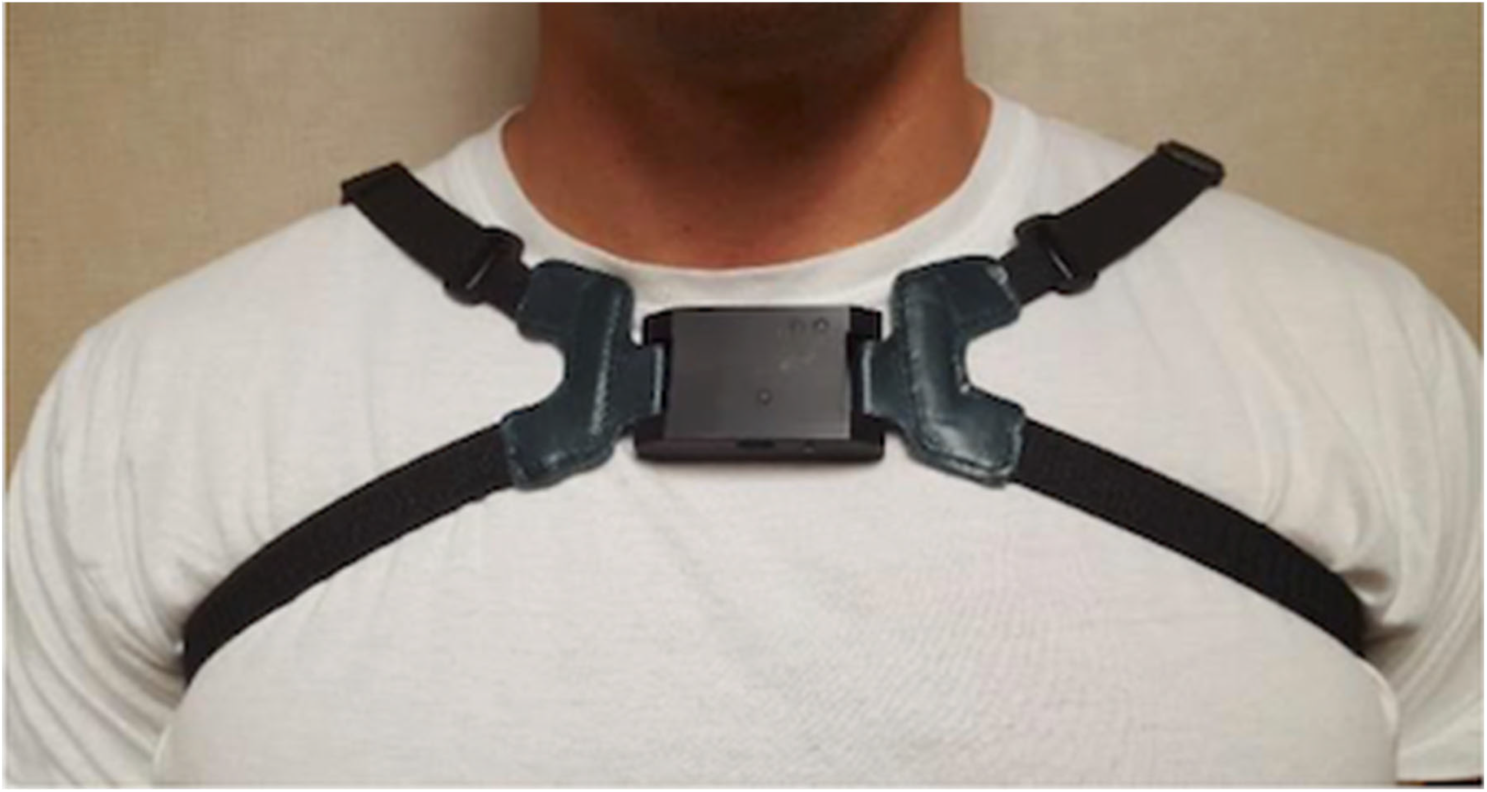
2.2. IMU Sensor
2.3. Instrumented iTUG Protocol
2.4. Data Analyses
2.5. Statistical Analyses
3. Results
3.1. Participants
3.2. Comparison of Performance for iTUG Subcomponents Between Adults with TBI and HCs
3.3. Within-Session Reliability
4. Discussion
Limitations and Future Directions
5. Conclusions
Author Contributions
Funding
Institutional Review Board Statement
Informed Consent Statement
Data Availability Statement
Acknowledgments
Conflicts of Interest
References
- Chou, L.S.; Kaufman, K.R.; Walker-Rabatin, A.E.; Brey, R.H.; Basford, J.R. Dynamic instability during obstacle crossing following traumatic brain injury. Gait Posture 2004, 20, 245–254. [Google Scholar] [CrossRef]
- Williams, G.; Morris, M.E.; Schache, A.; McCrory, P.R. Incidence of gait abnormalities after traumatic brain injury. Arch. Phys. Med. Rehabil. 2009, 90, 587–593. [Google Scholar] [CrossRef]
- Williams, G. A Schache and ME Morris. Running abnormalities after traumatic brain injury. Brain Inj. 2013, 27, 434–443. [Google Scholar] [CrossRef]
- Rahman, R.A.A.; Hanapiah, F.A.; Nikmat, A.W.; Ismail, N.A.; Manaf, H. Effects of Concurrent Tasks on Gait Performance in Children with Traumatic Brain Injury Versus Children with Typical Development. Ann. Rehabil. Med. 2021, 45, 186–196. [Google Scholar] [CrossRef] [PubMed]
- Katz-Leurer, M.; Rotem, H.; Keren, O.; Meyer, S. Effect of concurrent cognitive tasks on gait features among children post-severe traumatic brain injury and typically-developed controls. Brain Inj. 2011, 25, 581–586. [Google Scholar] [CrossRef] [PubMed]
- Newman, M.A.; Hirsch, M.A.; Peindl, R.D.; Habet, N.A.; Tsai, T.J.; Runyon, M.S.; Huynh, T.; Zheng, N.; Carolinas Trauma Network Research Group. Reliability of the sub-components of the instrumented timed up and go test in ambulatory children with traumatic brain injury and typically developed controls. Gait Posture 2018, 63, 248–253. [Google Scholar] [CrossRef] [PubMed]
- Flansbjer, U.B.; Holmbäck, A.M.; Downham, D.; Patten, C.; Lexell, J. Reliability of gait performance tests in men and women with hemiparesis after stroke. J. Rehabil. Med. 2005, 37, 75–82. [Google Scholar] [CrossRef]
- Rabelo, M.; Fachin-Martins, E. Inter-rater and test/retest reliabilities of the isokinetic measurements: Assessing strength and endurance of the trunk muscles in two different protocols for able-bodied and post-stroke hemiparesis. Top Stroke Rehabil. 2018, 25, 424–431. [Google Scholar] [CrossRef]
- Podsiadlo, D.; Richardson, S. The timed “Up & Go”: A test of basic functional mobility for frail elderly persons. J. Am. Geriatr. Soc. 1991, 39, 142–148. [Google Scholar] [CrossRef]
- Iwata, A.; Higuchi, Y.; Kimura, D.; Okamoto, K.; Arai, S.; Iwata, H.; Fuchioka, S. Quick lateral movements of the trunk in a seated position reflect mobility and activities of daily living (ADL) function in frail elderly individuals. Arch. Gerontol. Geriatr. 2013, 56, 482–486. [Google Scholar] [CrossRef]
- McCrory, P.; Meeuwisse, W.; Dvorak, J.; Aubry, M.; Bailes, J.; Broglio, S.; Cantu, R.C.; Cassidy, D.; Echemendia, R.J.; Castellani, R.J.; et al. Consensus statement on concussion in sport—The 5th international conference on concussion in sport held in Berlin, October 2016. Br. J. Sports Med. 2017, 51, 838–847. [Google Scholar] [CrossRef]
- Makdissi, M.; Patricios, J. Concussion in sport: Best from Berlin, direction from Dublin and gems from gridiron. Br. J. Sports Med. 2018, 52, 903–904. [Google Scholar] [CrossRef] [PubMed]
- Herman, T.; Weiss, A.; Brozgol, M.; Giladi, N.; Hausdorff, J.M. Identifying axial and cognitive correlates in patients with Parkinson’s disease motor subtype using the instrumented Timed Up and Go. Exp. Brain Res. 2014, 232, 713–721. [Google Scholar] [CrossRef] [PubMed]
- Weiss, A.; Mirelman, A.; Giladi, N.; Barnes, L.L.; Bennett, D.A.; Buchman, A.S.; Hausdorff, J.M. Transition Between the Timed up and Go Turn to Sit Subtasks: Is Timing Everything? J. Am. Med. Dir. Assoc. 2016, 17, e9–e864. [Google Scholar] [CrossRef]
- Onder, H.; Ozyurek, O. The impact of distinct cognitive dual-tasks on gait in Parkinson’s disease and the associations with the clinical features of Parkinson’s disease. Neurol. Sci. 2021, 42, 2775–2783. [Google Scholar] [CrossRef] [PubMed]
- Weiss, A.; Herman, T.; Mirelman, A.; Shiratzky, S.S.; Giladi, N.; Barnes, L.L.; Bennett, D.A.; Buchman, A.S.; Hausdorff, J.M. The transition between turning and sitting in patients with Parkinson’s disease: A wearable device detects an unexpected sequence of events. Gait Posture 2019, 67, 224–229. [Google Scholar] [CrossRef]
- Poole, V.N.; Dawe, R.J.; Lamar, M.; Esterman, M.; Barnes, L.; Leurgans, S.E.; Bennett, D.A.; Hausdorff, J.M.; Buchman, A.S. Dividing attention during the Timed Up and Go enhances associations of several subtask performances with MCI and cognition. PLoS ONE. 2022, 17, e0269398. [Google Scholar] [CrossRef]
- Mirelman, A.; Weiss, A.; Buchman, A.S.; Bennett, D.A.; Giladi, N.; Hausdorff, J.M. Association between performance on Timed Up and Go subtasks and mild cognitive impairment: Further insights into the links between cognitive and motor function. J. Am. Geriatr. Soc. 2014, 62, 673–678. [Google Scholar] [CrossRef]
- Salarian, A.; Horak, F.B.; Zampieri, C.; Carlson-Kuhta, P.; Nutt, J.G.; Aminian, K. iTUG, a sensitive and reliable measure of mobility. IEEE Trans. Neural. Syst. Rehabil. Eng. 2010, 18, 303–310. [Google Scholar] [CrossRef]
- Newman, M.A.; Hirsch, M.A.; Peindl, R.D.; Habet, N.A.; Tsai, T.J.; Runyon, M.S.; Huynh, T.; Phillips, C.; Zheng, N.; Carolinas Trauma Network Research Group. Use of an instrumented dual-task timed up and go test in children with traumatic brain injury. Gait Posture 2020, 76, 193–197. [Google Scholar] [CrossRef]
- Caronni, A.; Sterpi, I.; Antoniotti, P.; Aristidou, E.; Nicolaci, F.; Picardi, M.; Pintavalle, G.; Redaelli, V.; Achille, G.; Sciumè, L.; et al. Criterion validity of the instrumented Timed Up and Go test: A partial least square regression study. Gait Posture 2018, 61, 287–293. [Google Scholar] [CrossRef]
- Balasubramanian, C.K. The community balance and mobility scale alleviates the ceiling effects observed in the currently used gait and balance assessments for the community-dwelling older adults. J. Geriatr. Phys. Ther. 2015, 38, 78–89. [Google Scholar] [CrossRef]
- Swart, E.; Peindl, R.; Zheng, N.; Habet, N.; Churchill, C.; Ruder, J.A.; Seymour, R.; Karunakar, M.; Kellam, J.; Sims, S. Electronically augmented gait abnormality assessment following lower extremity trauma. OTA Int. 2019, 2, e032. [Google Scholar] [CrossRef]
- Meng, L.; Zhang, X.; Shi, Y.; Li, X.; Pang, J.; Chen, L.; Zhu, X.; Xu, R.; Ming, D. Inertial-Based Dual-Task Gait Normalcy Index at Turns: A Potential Novel Gait Biomarker for Early-Stage Parkinson’s Disease. IEEE Trans. Neural. Syst. Rehabil. Eng. 2025, 33, 687–695. [Google Scholar] [CrossRef] [PubMed]
- LeBrun, D.G.; Tran, T.; Wypij, D.; Kocher, M.S. How Often Do Orthopaedic Matched Case-Control Studies Use Matched Methods? A Review of Methodological Quality. Clin. Orthop. Relat. Res. 2019, 477, 655–662. [Google Scholar] [CrossRef] [PubMed]
- Steffen, T.M.; Hacker, T.A.; Mollinger, L. Age- and Gender-Related Test Performance in Community-Dwelling Elderly People: Six-Minute Walk Test, Berg Balance Scale, Timed Up & Go Test, and Gait Speeds. Phys. Ther. 2002, 82, 128–137. [Google Scholar] [CrossRef] [PubMed]
- Baque, E.; Barber, L.; Sakzewski, L.; Boyd, R.N. Test-re-test reproducibility of activity capacity measures for children with an acquired brain injury. Brain Inj. 2016, 30, 1143–1149. [Google Scholar] [CrossRef]
- Diebel, J. Representing Attitude: Euler Angles, Unit Quaternions, and Rotation Vectors. Matrix 2006, 58, 1–35. [Google Scholar]
- Ryzhkov, L. Quaternion Attitude Determination by Vector Measurement. Int. Appl. Mech. 2021, 57, 613–617. [Google Scholar] [CrossRef]
- Niven, D.J.; Berthiaume, L.R.; Fick, G.H.; Laupland, K.B. Matched case-control studies: A review of reported statistical methodology. Clin. Epidemiol. 2012, 76, 99–110. [Google Scholar] [CrossRef]
- Fleiss, J.L. The Design and Analysis of Clinical Experiments. Biom. J. 1986, 30, 304. [Google Scholar]
- D’Silva, L.J.; Chalise, P.; Obaidat, S.; Rippee, M.; Devos, H. Oculomotor Deficits and Symptom Severity Are Associated with Poorer Dynamic Mobility in Chronic Mild Traumatic Brain Injury. Front. Neurol. 2021, 12, 642457. [Google Scholar] [CrossRef] [PubMed]
- Dibilio, V.; Nicoletti, A.; Mostile, G.; Toscano, S.; Luca, A.; Raciti, L.; Sciacca, G.; Vasta, R.; Cicero, C.E.; Contrafatto, D.; et al. Dopaminergic and non-dopaminergic gait components assessed by instrumented timed up and go test in Parkinson’s disease. J. Neural. Transm. 2017, 124, 1539–1546. [Google Scholar] [CrossRef] [PubMed]
- Odonkor, C.A.; Thomas, J.C.; Holt, N.; Latham, N.; Vanswearingen, J.; Brach, J.S.; Leveille, S.G.; Jette, A.; Bean, J. A Comparison of Straight- and Curved-Path Walking Tests Among Mobility-Limited Older Adults. J. Gerontol. Ser. A Biol. Sci. Med. Sci. 2013, 68, 1532–1539. [Google Scholar] [CrossRef] [PubMed]
- Salarian, A.; Zampieri, C.; Horak, F.B.; Carlson-Kuhta, P.; Nutt, J.G.; Aminian, K. Analyzing 180 degrees; turns using an inertial system reveals early signs of progression of Parkinson’s disease. In Proceedings of the 2009 Annual International Conference of the IEEE Engineering in Medicine and Biology Society, Minneapolis, MN, USA, 3–6 September 2009; pp. 224–227. [Google Scholar] [CrossRef]
- Huang, S.L.; Hsieh, C.L.; Wu, R.M.; Tai, C.H.; Lin, C.H.; Lu, W.S. Minimal Detectable Change of the Timed “Up & Go” Test and the Dynamic Gait Index in People with Parkinson Disease. Phys. Ther. 2011, 91, 114–121. [Google Scholar] [CrossRef]
- Steffen, T.; Seney, M. Test-Retest Reliability and Minimal Detectable Change on Balance and Ambulation Tests, the 36-Item Short-Form Health Survey, and the Unified Parkinson Disease Rating Scale in People With Parkinsonism. Phys. Ther. 2008, 88, 733–746. [Google Scholar] [CrossRef]
- Sample, R.B.; Kinney, A.L.; Jackson, K.; Diestelkamp, W.; Bigelow, K.E. Identification of key outcome measures when using the instrumented timed up and go and/or posturography for fall screening. Gait Posture 2017, 57, 168–171. [Google Scholar] [CrossRef]
- Katz-Leurer, M.; Rotem, H.; Lewitus, H.; Keren, O.; Meyer, S. Functional Balance Tests for Children with Traumatic Brain Injury: Within-Session Reliability. Pediatr. Phys. Ther. 2008, 20, 254–258. [Google Scholar] [CrossRef]
- Weiss, A.; Herman, T.; Plotnik, M.; Brozgol, M.; Giladi, N.; Hausdorff, J.M. An instrumented timed up and go: The added value of an accelerometer for identifying fall risk in idiopathic fallers. Physiol. Meas. 2011, 32, 2003–2018. [Google Scholar] [CrossRef]

| Demographic Variable | TBI | HC | Difference (HC–TBI) | p-Value |
|---|---|---|---|---|
| Age (years) | 28.00 [21.00–41.00] | 28.00 [25.00–37.00] | 0.00 [−2.00–4.00] | 0.269 |
| Female sex | 4 (26.67%) | 4 (26.67%) | — | 1.000 |
| BMI (kg/m2) | 23.13 [19.15–25.92] | 25.79 [22.80–26.62] | 2.45 [−0.09–5.49] | 0.0637 |
| Left leg length (cm) | 87.00 [86.00–93.00] | 92.50 [88.50–96.50] | 3.40 (6.82) | 0.0740 |
| Right leg length (cm) | 87.50 [86.00–93.00] | 93.00 [88.00–96.75] | 3.50 (6.55) | 0.0572 |
| GCS at admission | 7.27 (3.03) | — | — | — |
| Days taken for PTA to clear | 23 [15,16,17,18,19,20,21,22,23,24,25,26,27,28,29,30] | — | — | — |
| Days from injury to test | 27 [17,18,19,20,21,22,23,24,25,26,27,28,29,30,31,32,33,34,35,36,37,38,39] | — | — | — |
| iTUG Variable | TBI | HC | Difference (HC–TBI) | p-Value |
|---|---|---|---|---|
| Total iTUG Completion Time a | 14.50 (2.36) | 9.85 (1.71) | −4.65 (2.75) | 0.0002 |
| 1st Transition: Sit-to-Stand | ||||
| — Total Time Taken to Stand (s) b | 2.66 (0.39) | 2.18 (0.52) | −0.47 (0.70) | 0.0325 |
| — Max Ang Displacement (deg) b | 42.14 (12.86) | 39.46 (12.76) | −2.68 (19.51) | 0.6290 |
| — Max Chest FlexVel (deg/s) b | 63.52 (23.25) | 88.19 (29.20) | 24.67 (40.54) | 0.0486 |
| — Max Chest ExtVel (deg/s) c | −60.89 (20.16) | −75.57 (22.26) | −14.68 (30.71) | 0.0971 |
| — Max Vert Accel (m/s2) b | 2.22 [1.23–2.74] | 3.89 [3.36–5.02] | 2.12 (1.63) | 0.0005 |
| Turning Around | ||||
| — Total Time Taken to Turn (s) b | 3.73 (0.47) | 3.09 (0.47) | −0.65 (0.66) | 0.0041 |
| — Mean Turn Ang Vel (deg/s) b | 51.63 [48.97–55.06] | 64.17 [57.10–71.17] | 11.52 (10.87) | 0.0024 |
| — Pk Turn Ang Vel (deg/s) b | 107.48 (21.59) | 136.77 (24.01) | 29.30 (36.83) | 0.0142 |
| 2nd Transition: Stand-to-Sit | ||||
| — Total Time Taken to Sit (s) d | 2.68 (0.42) | 2.30 (0.85) | −0.38 (1.09) | 0.2545 |
| — Max Chest FlexVel (deg/s) d | 45.07 (16.16) | 55.57 (21.41) | 10.50 (22.86) | 0.1399 |
| — Max Chest ExtVel (deg/s) d | −68.63 (26.95) | −73.91 (24.43) | −5.28 (34.71) | 0.6087 |
| iTUG Variable | TBI | HC |
|---|---|---|
| Total iTUG Completion Time a | 0.906 (0.700–0.973) | 0.940 (0.841–0.978) |
| 1st Transition: Sit-to-Stand | ||
| — Total Time Taken to Stand (s) b | 0.267 (−0.295–0.692) | 0.671 (0.290–0.869) |
| — Max Ang Displacement (deg) b | 0.825 (0.532–0.941) | 0.847 (0.623–0.942) |
| — Max Chest FlexVel (deg/s) b | 0.740 (0.355–0.910) | 0.917 (0.783–0.970) |
| — Max Chest ExtVel (deg/s) c | 0.710 (0.321–0.894) | 0.762 (0.451–0.908) |
| — Max Vert Accel (m/s2) b | 0.846 (0.580–0.949) | 0.836 (0.601–0.938) |
| Turning Around b | ||
| — Total Time Taken to Turn (s) | 0.356 (−0.203–0.740) | 0.757 (0.442–0.906) |
| — Mean Turn Ang Vel (deg/s) | 0.464 (−0.076–0.794) | 0.772 (0.469–0.912) |
| — Pk Turn Ang Vel (deg/s) | 0.880 (0.663–0.961) | 0.727 (0.387–0.893) |
| 2nd Transition: Stand-to-Sit d | ||
| — Total Time Taken to Sit (s) | 0.769 (0.391–0.925) | 0.687 (0.316–0.876) |
| — Max Chest FlexVel (deg/s) | 0.722 (0.298–0.908) | 0.677 (0.299–0.871) |
| — Max Chest ExtVel (deg/s) | 0.833 (0.530–0.947) | 0.804 (0.533–0.925) |
Disclaimer/Publisher’s Note: The statements, opinions and data contained in all publications are solely those of the individual author(s) and contributor(s) and not of MDPI and/or the editor(s). MDPI and/or the editor(s) disclaim responsibility for any injury to people or property resulting from any ideas, methods, instructions or products referred to in the content. |
© 2025 by the authors. Licensee MDPI, Basel, Switzerland. This article is an open access article distributed under the terms and conditions of the Creative Commons Attribution (CC BY) license (https://creativecommons.org/licenses/by/4.0/).
Share and Cite
Pinto, S.M.; Habet, N.A.; Roomian, T.C.; Williams, K.M.; Duemmler, M.; Werts, K.A.; Sims, S.H.; Newman, M.A. Use of Instrumented Timed Up and Go in Adults with Traumatic Brain Injury. BioMed 2025, 5, 16. https://doi.org/10.3390/biomed5030016
Pinto SM, Habet NA, Roomian TC, Williams KM, Duemmler M, Werts KA, Sims SH, Newman MA. Use of Instrumented Timed Up and Go in Adults with Traumatic Brain Injury. BioMed. 2025; 5(3):16. https://doi.org/10.3390/biomed5030016
Chicago/Turabian StylePinto, Shanti M., Nahir A. Habet, Tamar C. Roomian, Kathryn M. Williams, Marc Duemmler, Kelly A. Werts, Stephen H. Sims, and Mark A. Newman. 2025. "Use of Instrumented Timed Up and Go in Adults with Traumatic Brain Injury" BioMed 5, no. 3: 16. https://doi.org/10.3390/biomed5030016
APA StylePinto, S. M., Habet, N. A., Roomian, T. C., Williams, K. M., Duemmler, M., Werts, K. A., Sims, S. H., & Newman, M. A. (2025). Use of Instrumented Timed Up and Go in Adults with Traumatic Brain Injury. BioMed, 5(3), 16. https://doi.org/10.3390/biomed5030016