Entry

Multi-Modal Approach to Mitigating Hamstring Injuries in Division I College Football Athletes

1
Department of Athletics, University of Miami, Coral Gables, FL 33146, USA
2
Miami Heat, National Basketball Association, Miami, FL 33132, USA
3
Department of Physical Therapy, University of Miami, Coral Gables, FL 33146, USA
4
Uhealth Sports Medicine Institute, University of Miami, Coral Gables, FL 33146, USA
5
Department of Athletics, Duke University, Durham, NC 27708, USA
6
Department of Kinesiology & Sports Management, Texas A&M University, College Station, TX 77845, USA
*
Author to whom correspondence should be addressed.
Encyclopedia 2024, 4(4), 1482-1495; https://doi.org/10.3390/encyclopedia4040096
Submission received: 6 August 2024 / Revised: 13 September 2024 / Accepted: 24 September 2024 / Published: 29 September 2024
(This article belongs to the Section Medicine & Pharmacology)

Definition

:
Hamstring injuries (HSIs) are prevalent in sports that involve changes in direction, kicking, and sprinting. These injuries are a major cause of time lost from competition, practice, and training, as well as increased healthcare costs. In a Division I collegiate football program, the authors implemented a multifactorial approach that included repeated performance assessments, detailed data analysis, and a flexible strength and conditioning regimen. Over a three-year period, this resulted in no game time loss due to HSI. This model can be adapted and implemented across sports settings.

1. Introduction or History

Hamstring injuries (HSIs) are common in sports requiring kicking, high-speed running, and sprinting, and are a significant reason for time loss in training, practice, and competition [1,2,3,4,5,6,7]. In 2015, in an epidemiological review across 25 National Collegiate Athletic Association (NCAA) sports, there were a reported 1142 HSIs [3]. Most (72.3%) HSIs were due to non-contact mechanisms, with 12.6% being recurrent in nature, and 6.3% resulting in time loss of greater than 3 weeks [3]. Additionally, pre-season rates of HSI (5.00 per 10,000 AEs) were greater than the regular season/postseason rates (2.34 per 10,000 AEs; RR = 2.14; 95% CI, 1.90–2.40) [3]. Lastly, men’s football contributed the greatest proportion (35.3%) of HSIs among all NCAA sports [3]. A recent epidemiological study spanning several seasons of collegiate football found that HSI was one of the most commonly reported injuries, with increasing rates in the later years of the study [2]. HSI placed the second highest burden on National Football League (NFL) players, accounting for approximately 5800 player-days lost each season, with only anterior cruciate ligament tears having a higher burden, with approximately 12,400 total player-days lost per season [6]. The majority of HSIs occur in skilled-position players such as running backs, defensive backs, and wide receivers, as they have more athletic exposure to higher-speed deceleration actions and quick changes in direction during training, practice, and games [6].
In the NCAA and NFL, time loss due to HSI places a significant burden on the players, as it affects playing time, and subsequently could potentially affect name, image, and likeness rights at the collegiate level, as well current and future employment at the professional level. The nature of HSI for collegiate and NFL football players is multifactorial, with contributing factors including age, training loads, training stimulus, peak quadriceps torque, inter- and intralimb asymmetries, and previous injury [7,8].
Strikingly, the incidence of muscle injuries, like HSI, remains unchanged despite prospective interventional studies [4]. Ekstand et al. retrospectively reviewed 18 years of injury rates in European soccer and found that the incidence of muscle injury had not changed over time, either in training or in games [4]. This suggests that a priority for sports medicine clinicians and researchers is identifying risk-causative factors for HSI, the strategic implementation of objective measures to assess risk, and establishing injury reduction strategies that are continuous, actionable, and efficient, so that a reduction in the incidence and time loss due to HSI can be achieved.
The approach taken by the authors using an interdisciplinary framework of engaging athletic trainers, engineers, programmers, researchers, strength and conditioning coaches, sports board-certified physical therapists, and sports medicine physicians has been a successful in addressing HSI. This collaboration has included, but is not limited to, GPS-directed load analysis and management, a comprehensive strength and conditioning program, and the repeated administration of performance-based outcome measures with sensor metrics for injury profile risk assessment. This interdisciplinary approach resulted in no game time loss due to HSI over a consecutive three-year period. This approach was particularly successful when considering that an American collegiate football roster consists of 85 scholarship players, with an additional number of walk-ons, totaling between 100 and 120 players. Roster turnover occurs annually as a result of early departures for professional leagues, eligibility limitations, graduation, and transfers. The underlying premise of this approach was that continuous risk factor monitoring, load management, and an evidence-based strength and conditioning program reduced time loss due to HSI.
The intention of this work is to present a multi-modal approach to assessing and improving the management of HSI that can be implemented across a variety of settings. By combining practical low-cost tools, such as wearable sensors, smartphone-based apps, and common balance or strength tests, with more advanced technologies when available, this approach aims to provide accessible strategies for the management of HSI. The purpose of this manuscript is to provide a broad overview of HSI in terms of anatomy and injury description, theoretical models of injury severity mitigation, and a comprehensive rehabilitation model that was implemented. The model called for daily programmatic re-evaluation, which included risk factor identification and monitoring using various technologies, load analysis, management during the various training seasons, and the strategic implementation of strength and conditioning programs to maximize adaptability while preventing physiological plateauing.

2. Description of HSI, Injury Prevention, and HSI Rehabilitation Intervention

2.1. Nature of HSI

The hamstrings are susceptible to injury due to their anatomic arrangement and their mechanism of action over two joints (knee and hip). The mechanism of action occurs synchronously with opposing effects on hamstring length, especially during deceleration at the hip, and in rapid transition periods. Negative work or eccentric action of the hamstring with repeated strides and disturbances of the pelvic muscular coordination can induce excessive hamstring stretching in a single stride [9]. The most common phase of running gait where HSI occurs is in terminal swing, where the hamstring is activated eccentrically at a high speed, actively decelerating the limb in space, preparing the foot for ground contact [10,11]. It was demonstrated that peak hamstring musculotendon stretch occurred at approximately 90% of the running gait cycle (i.e., late swing) and was independent of speed [9]. Musculotendon work and eccentric force increased significantly with faster speeds [9]. The hamstrings play a key role in deceleration while sprinting and during high-speed changes in direction, like those experienced in American-style football.
Among the three hamstring muscles, the biceps femoris muscle is the most commonly injured of the group because the anatomical variance in the attachment of the two heads and the dual nerve supply of the two heads of the biceps femoris leads to asynchronous neural stimulation. The idea that anatomical variations could lead to asynchronous nerve firing in the hamstring muscles is more of a theoretical consideration than a well-documented, extensively researched fact. While there is established literature on anatomical variations in muscle and nerve innervation, including variations in the sciatic nerve’s branching pattern and its innervation of the hamstrings, the specific relationship between these variations and asynchronous nerve firing is not explicitly referenced in studies.
While anatomical differences in innervation are recognized, their role in causing significant asynchronous firing is not a prominent topic in the current literature. The possibility exists, based on current clinical assumptions about nerve branching and muscle activation, but it remains largely speculative without direct empirical support. There is a risk for HSI recurrence, with the sensitive period being the first two weeks of return to participation. It is therefore essential to implement a criterion-based rehabilitation program and forward-looking systematic approach using preventative measures [12].

2.2. Preventative Considerations

Preventative interventions need to encompass strengthening the hamstrings throughout various angles, loads, and velocities. Evidence-based interventions that are angle- and load-sensitive will provide adequate stress to replicate the physical demands in football [9]. Of particular use is the incorporation of a strengthening program focusing on hip extension-dominant exercises, targeting the long head of the biceps femoris and semimembranosus [13,14]. Additionally, knee flexion-based exercises targeting the semitendinosus, as well as the short head of the biceps femoris, have been identified as components in mitigating HSI [13,14].
The goal of a comprehensive strength and conditioning program is to allow the various training cycles to build the work capacity of the hamstrings by implementing specific exercises executed through a full range of motion and at varying velocities. The utilization of Nordic hamstring exercises and its variations and accompanied sprinting have been found to be beneficial as preventative measures for HSI [1,15]. Negating the implementation of the training variables can increase the risk of HSI.

3. Evidence-Based Rehabilitation Interventions/Clinical Practice Guidelines

A multifactorial injury prevention and rehabilitative program is superior to solely using isolated exercises that purport injury mitigation [16,17]. Researchers have recognized specific interventions, such as eccentric strengthening throughout lengthened positions, as well as using various isometric and concentric loading in a progressive fashion, to help mitigate HSI [16,17]. It has also been demonstrated that neuromuscular control training, the strengthening of adjacent muscle groups, and the incorporation of a progressive agility and trunk stabilization (PATS) program throughout all rehabilitation phases contribute to reducing the risk of both initial injuries and subsequent re-injury episodes [18,19].
In a clinical practice guideline by Martin et al., high levels of evidence suggested that clinicians should use eccentric training to the patient’s tolerance, as well as stretching, (core) stabilization exercises, and a specified strengthening series for hamstring health [18]. Practitioners should also necessarily implement progressive running programs, ranging through all velocities, in order to prepare the athlete for a safe return to sport [18]. In football, the velocities and speeds placed on the body are not just from sprinting as they include high-speed decelerations, transitions, and collisions. Therefore, it is necessary to create a program that prepares the muscles, joints, and tendons to have the mobility, strength, and tensile capacity to absorb these high forces following HSI. When either dealing with a later-phase patient in rehabilitation from HSI or a healthy athlete, it is imperative to incorporate loaded high-velocity exercises emphasizing eccentric lengthening while also not negating isometric components throughout a variety of ranges [18,20,21]. Enabling the athlete to stabilize and reproduce force in a seamless fashion with multi-joint, complex movements can assist with their preparation for changes in direction and the absorption of ground reaction forces from both non-contact and collision-based activities. High-force and high-velocity exercises under various loads may be able to prepare athletes in the weight room to transfer onto the field safely, as the specificity of exercise is essential to decrease the risk of re-injury [18,20,21]. Recognition of the demands of reaccelerating and generating force from an unstable position helps assist with the real demands of American football, such as receiving contact and having to perform high-speed multi-planar movements.

3.1. Theoretical Models and Factors to Mitigate the Impact of HSI

The primary step to address HSI is to prevent injury. Baroni and Costa identified several important factors that are crucial to implementing an evidence-based approach to HSI, such as assessing both modifiable and non-modifiable factors, and developing a strategic approach to the continuous tracking of athlete outcomes throughout all seasons [22]. The risk of HSI outcomes is influenced by a combination of modifiable and non-modifiable factors (Table 1). For example, Green et al. demonstrated that the strongest risk factors for HSI were non-modifiable in nature (older age, previous HSI, anterior cruciate ligament injury, or a prior calf muscle strain) [5]. In addition, although strength and flexibility measures were the most examined modifiable variables, neither was found to be a consistent or robust predictor of HSI, perhaps due to the lack of continuous measurement and assessment over time.
Bittencourt et al. developed a new theoretical model that accounts for the complexity, nonlinearity, and interaction of modifiable (fatigue, training load, injury event, strength measures, etc.) and non-modifiable (age, sex, previous injury) variables that contribute to injury risk [23]. They emphasized the importance of continuous measurement because modifiable variables change during the season and capturing those changes can identify patterns that trigger when a patient is at risk for injury [23]. These principles were applied to target the modifiable risk factors with the football team. The following sections describe multi-modal approach to HSI injury management.

3.2. Mobile Health Technologies

Over the last 10 years, the use of mobile health (mHealth) technologies and body-worn devices for the assessment of movement, overall fitness, and workload has become a central part of strength and conditioning and rehabilitation care [24,25,26,27,28,29,30,31,32,33,34,35]. Clinicians and researchers from the United States Department of Defense (DoD) and Veterans Affairs (VA) Administration have been collaborating for over 15 years to develop performance-based outcome measures and mHealth technology to assess high-level mobility and develop evidence-based rehabilitation approaches for Service Members and Veterans with lower limb amputation [28,29,30,32,33]. These clinicians and researchers have translated these measures and technologies for use with collegiate athletes, leading to a new mHealth platform directed at preventing lower limb musculoskeletal injuries and assisting with return-to-play decision making [24,25,26,27,31].
The Rehabilitative Lower Limb Orthopaedic Assistive Device for Sport or ReLOAD Sport™ uses a custom mobile application (app) with wearable technology inertial measurement units to quantify lower limb stability. Published and pilot research support the use of ReLOAD Sport™ to categorize student athletes during healthy pre-season testing who are at risk for lower limb injury resulting in surgery, detect balance impairment following concussion, quantify knee joint stability throughout the rehabilitation process for those with an ACL injury, and identify those student athletes who present with lingering lower limb instability with dynamic balance and agility activity after discharge from rehabilitation and return to sport [34,35].
The ReLOAD Sport™ system (Figure 1) is worn on both lower limbs, with each sleeve comprising two small, wireless IMUs attached via an elastic knee sleeve. Each sleeve holds two IMUs positioned at the medial tibial flare and the lateral proximal thigh, just anterior to the insertion of the iliotibial band. The custom-designed wireless IMUs and software are compact (2.5 × 2.5 × 1.25 cm each) and incorporate an accelerometer, a gyroscope, and a magnetometer. Kinematic data, including three-axis linear accelerations, velocities, and magnetic fields, are sampled at 50 Hz and transmitted to a custom iOS mobile app on a tablet. The data are securely stored in a secure cloud server [36,37,38].
To reduce errors in ReLOAD Sport™ that are inherent in sensor placement variability, algorithms are implemented to determine both virtual axis alignment and tilt compensation [39,40]. In addition, two methods are implemented to control for static and dynamic drift during data collection [39,40]. Lastly, once the knee sleeves are donned, the athlete undergoes an alignment procedure which consists of a sit-to-stand activity followed by walking 10 steps at a comfortable self-selected speed. The alignment procedure virtually rotates and aligns the IMUs with the athlete’s body’s frame of reference and knee joint axis for all sensor-based tests [35,38]. The sensor metrics collected through ReLOAD Sport™ include the following: the Region of Limb Stability (ROLS) and Transitional Angular Displacement of Segments (TADS).

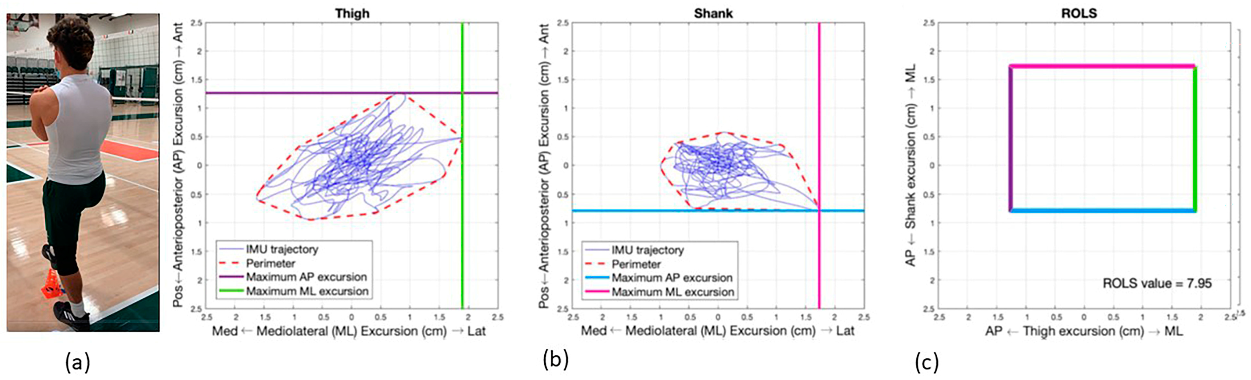
3.3. The Region of Limb Stability (ROLS)

The ROLS metric is a novel measurement of segmental excursion that quantifies lower limb stability during static balance activities, specifically in a single-limb stance (SLS) and can be used to assess injury risk. ROLS measures the segmental excursion of the thigh and shank (cm2) in the anterior–posterior and medial–lateral directions during SLS (Figure 2). This metric is calculated using acceleration data from two IMUs positioned proximally and distally to the knee joint line. The ROLS symmetry index (SI) (%) represents the ratio of lower limb ROLS values as a percentage, with 100% indicating perfect symmetry. A lower ROLS SI value reflects greater asymmetry, with the limb exhibiting a larger ROLS value, demonstrating greater excursion [34,35,39,40].
Previous research supports ROLS as a reliable and valid measure of segmental excursion, which is associated with knee joint stability. Greater excursion indicates a reduced ability to maintain static balance. In a pilot study involving 20 healthy young adults, the ROLS metric demonstrated excellent test–retest reliability over a 48-h period (right leg Interclass Correlation Coefficient (ICC) = 0.94; left leg ICC = 0.98) [35]. The ROLS value for the injured limb and ROLS SI has greater sensitivity and specificity to assess the presence of lower limb instability during MSK rehabilitation than a single sacral IMU [35]. The ROLS value and ROLS SI were more sensitive in detecting balance impairment than the gold standard Balance Error Scoring System in Division I collegiate football players post-concussion [25]. The ROLS values and ROLS SI were able to detect female student athletes at risk for lower limb injury and suggested a need for an off-season training program [31]. For collegiate football players, the ROLS SI demonstrated good diagnostic accuracy (0.80) in detecting healthy players at risk for lower limb injury in the same season of measurement resulting in season-ending surgery (i.e., hamstring tear, avulsion fracture (semitendinosus), ACL tear, knee dislocation, popliteofibular ligament tear, quadriceps tendon tear, meniscus tear, medial collateral ligament tear). Data were collected just prior to the commencement of pre-season training and fall practice. The ROLS injury classification demonstrated accuracy, sensitivity, and specificity of 0.92, 0.94, and 0.91, respectively, in identifying those at risk for lower limb injury resulting in surgery [24].

3.4. Alternative Technologies for Assessing Limb Stability

Several technologies are commonly used to assess single-limb balance, each offering distinct advantages. Force plates, such as Bertec or Hawkin Dynamic, measure ground reaction forces to track postural sway and stability during single-limb stance, providing detailed insights into center-of-pressure movement(s). Balance platforms, like the Biodex Balance System, assess weight distribution and control, often incorporating different biofeedback cues to enhance balance training. Wearable sensors, including IMUs like DynaPort or XSENS, offer portable options to monitor motion, acceleration, and angular velocity in real time. Motion capture systems, such as Vicon or OptiTrack, track body movements in 3D, enabling the precise analysis of body control during single-limb tasks. Pressure mat systems, like Tekscan’s atScan, evaluate pressure distribution under the foot to analyze postural control. Smartphone applications such as PhysiApp, myAnkle, and Sway Medical leverage the smartphone’s inertial measurement units to provide gross balance assessments. Each technology provides unique data, supporting applications in clinical, research, and performance settings.

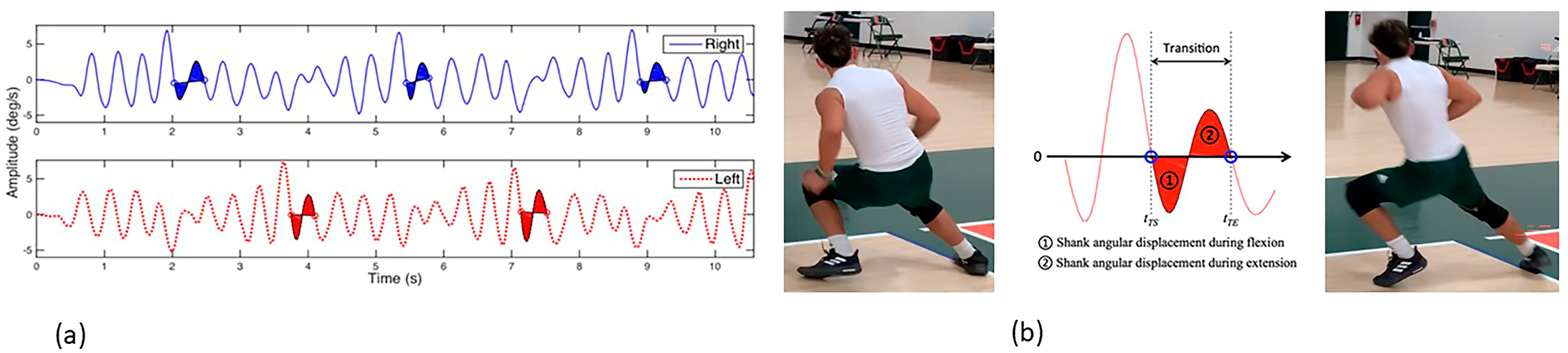
3.5. Transitional Angular Displacement of Segments (TADS)

TADS is a novel measure for quantifying movements of the lower limb segments during a dynamic activity during lateral plane movement to assess injury risk [38]. TADS utilizes the shank sagittal and frontal angular velocity obtained with an IMU sensor to quantify the knee joint motion when changing directions during side-stepping. TADS captures the time and angular velocities of the loading/deceleration and unloading/acceleration phases when the left and right limbs are the lead limbs that are initiating the change in direction. Even though ROLS and TADS measure different constructs, they are assessed using similar metrics. The TADS SI is the percent difference calculated for TADS values between lower limbs, where 100% suggests absolute symmetry. The lower the TADS SI value, the greater the asymmetry [34]. The TADS SI has been used to identify the student athletes who present with significant deceleration/acceleration asymmetry (<80%) between limbs and modify their strength and conditioning program.
The four-meter sidestep test (FmSST) assesses unidirectional frontal plane agility and body control by calculating the time to complete sidestepping to the right and then to the left four meters as quickly as possible a total of three times. TADS is measured during each period of change in direction in the FmSST (Figure 3) [34,39]. Figure 3a illustrates the shank angular displacement for each limb recorded during the FmSST (right limb = blue solid line and left limb = red dotted line) with each step and during the change in direction. In the enlarged view (Figure 3b), knee flexion and extension occurring during the FmSST are represented by (1) negative displacement and (2) positive displacement. Five transitions are marked during one trial of the FmSST. The two middle transitions (indicated by the two vertical black dotted lines in Figure 3a), corresponding to the second and third transitions for TADS left and TADS right, are used to compute the TADS SI [34].
TADS values from the FmSST have been shown to be a reliable and valid measure of dynamic knee motion and stability [31]. The test–retest ICC values for TADS and its symmetry index (SI) range from 0.81 to 0.89. In a group of student athletes cleared to return to sport (RTS), the TADS values were significantly different from the pre-season baseline healthy values (85.82 ± 4.84% vs. 76.44 ± 13.22%, p = 0.046). However, no significant differences were observed between pre-season healthy and RTS FmSST times (9.26 ± 1.31 s vs. 9.79 ± 1.99 s, p = 0.32) [34].
Lastly, the ReLOAD Sport was developed to be user-friendly and time-efficient, displaying results in real-time for coaches, athletic trainers, and physical therapists, as demanding schedules require efficiency in time, resource management, and immediate results. Based on previous studies, it takes less than 60 s to place the knee sleeves on both lower limbs. While the sensor sleeves are donned, the clinician finds the patient’s record on the iPad and the sensors are paired to the iPad once the athlete stands. The athlete performs a walking calibration test which takes 10 s to complete. The SLS test takes 30 s per limb, with a 30 s rest between trials, for 90 s. The mean time to complete the FmSST is 9.5 s per trial with 60 s of rest between the two trials for a total of 80 s. The athlete then removes the knee sleeves. The total time to complete the testing according to the mobile app time stamps, from the initial encounter to exit, is about 4 min. Lastly, all the data can be accessed immediately on a secure web portal where the coaches can view data streaming in real time and print a report summarizing individual athlete results.

3.6. Alternative Technologies for Assessing Limb Stability

Several technologies are available for quantifying lower limb movements during dynamic activities, particularly in the frontal plane. Motion capture systems, such as Vicon and Optitrack, are widely used to track and record 3D joint kinematics by utilizing high-speed cameras and reflective markers on the lower limbs. These systems provide detailed data on segmental movements during frontal plane activities like cutting or side-stepping. IMUs, such as those from XSENS and Delsys, offer portable alternatives by capturing the motion, acceleration, and angular velocity of the lower limb segments, making them suitable for lab and field settings. Additionally, force plates can be used in conjunction with these systems to measure ground reaction forces and assess movement efficiency, especially in the frontal plane. Pressure mapping systems such as Teckscan’s F-Scan, can be used to analyze foot pressure and force distribution during high-speed multi-planar movements, providing insight into lower limb mechanics. Smartphone apps such as DorsaVi ViPerform and Kinetic Balance provide gross assessments for tracking agility, acceleration, and biomechanics.

3.7. GPS Tracking

Player monitoring has become commonplace among professional and collegiate sports teams. Metrics such as speed, distance, acceleration, deceleration, and distance traveled can be collected and stored on an IMU or accelerometer on the athlete and transmitted locally to a computer for the assessment of work. GPS systems have developed proprietary algorithms to harness the motion data collected from their sensors to give the athlete and coaches information about the magnitude and direction of accelerations and decelerations and changes in direction. The GPS systems collect data at 100 Hz, thereby permitting high-speed movements to be captured accurately.
The authors used the Catapult GPS system as a predictive metric for injury prevention. The primary GPS parameters used were the (1) total speed, (2) number of high-speed efforts, accelerations, and decelerations, (3) distance traveled, (4) acute-to-chronic workload ratio, (5) player load, and (6) player load per minute. Each of these primary metrics is assessed within the seasonal training cycles, and threshold levels are established using position-specific reference values to ensure optimal running and sport-specific performance.
The acute/chronic workload ratio (ACWR) is adaptable, and the variation in ACWR tolerances between players further reinforces the need to develop personalized, player-specific risk thresholds [41,42,43,44,45,46]. Therefore, more important than monitoring player loads, the high-speed efforts and decelerations, as well as total yards, should account for each athlete’s demands and personal limitations. The recognition of significant variance in load for an athlete can flag an injury risk, allowing the multidisciplinary team to implement a strategy to either increase or decrease the number of high-speed efforts. Constant monitoring allows for an accurate depiction of the workload and the number of efforts in a specific time period.
Gabbett et al. described a ‘paradox’ of high chronic workloads as a potentially preventative effect, if week-to-week load changes are kept within ∼10% and the ACWR is kept in a moderate range [41,42,43]. Hulin et al. found that if the ACWR was kept within a moderate zone (0.85–1.35), high chronic workloads were associated with the lowest risks of injuries, other than in the case of players with very low (<2 SD) ACWR [44]. ACWR monitoring is often considered a necessary component in team sports, allowing the staff to recognize each athlete’s individual short-term loads in comparison to the past 3–4 weeks, and to recognizing the overall intensity or load. GPS technology facilitates managing these spikes in frequency, intensity, and/or volume, which can be pivotal for injury prevention and performance workload [45].
A framework such as the ACWR, which can help monitor as well as recognize players at risk of injury, is optimal and can often be used to flag high-injury-risk cases. It has been found that 5–8 high-speed efforts (>85%) have been effective in preventing non-contact HSI [46,47]. Incorporating GPS is an essential tool in order to recognize key performance indicators such as ranges of top-end speed, as well as quantifying player loads and total distance, numbers of high-speed efforts, accelerations, and decelerations.

4. Force Plate Analysis

Force plate data are used to assess injury risk as well as enhance performance. The correlation between force plate closed-chain measures and the 90% occurrence of hamstring injuries during open-chain terminal stance lies in the ability to detect interlimb asymmetries and eccentric loading capacity. Monitoring force generation at transition points, such as force at zero velocity, highlights deficits in eccentric control, which are key to injury prevention. Neuromotor connections also play a role, as impaired neuromuscular control can increase the risk of injury by reducing the athlete’s ability to handle high-force eccentric loads.
Force plates and the multitude of performance tests from ballistic, isometric, balance, or dynamic assessments can provide a comprehensive and detailed analysis, unlike many other tools. These include but are not limited to limb asymmetries in power production, the rate of force production, and landing strategies during a ballistic movement, including but not limited to the countermovement jump (CMJ), depth jump (DJ), and squat jump (SJ) [48]. Depending on the software available with the force plates, there are a vast number of objective measurements to examine throughout the different phases of a jump. The CMJ is frequently analyzed and is an effective movement to recognize various asymmetries throughout the different phases of the ballistic movement [49,50]. There are six phases of the CMJ that have been recognized by McMahon et al. which can help practitioners to categorize movement impairments [50]. Jump metrics provide a clear depiction of how load is transferred through the entirety of the movement. These metrics include but are not limited to the reactive strength index (RSI), eccentric braking RFD, eccentric mean power, eccentric peak velocity, concentric mean power, and concentric peak velocity [51]. Hart et al. found that despite being cleared to return to sport, athletes had significant inter-limb asymmetries that persisted in the concentric and eccentric phases of the CMJ [52].
The frequent utilization of force plates may help build a profile where there are standard ranges for an athlete that are objectively recognized and can assist with the identification of abnormal biomarkers when compared to their normal movement pattern or force profile. Mitchell et al. found that individuals with a previous injury demonstrated a compensatory movement strategy in a range of force–time history metrics, with significant deficits in the phase duration and overall depth of a bilateral CMJ [49]. Mitchell et al. went on to suggest that a single-leg CMJ test may be more sensitive to the previous injury than a double-limb CMJ [49]. Programs and teams have utilized force plates with a variety of movements and assessments to assist with optimizing performance and injury mitigation.
Prior to the implementation of the multi-modal model, in the previous two seasons, 10 football players had HSIs which resulted in a total time loss of 14 games. Following implementation of the model, there were 11 football players who had HSIs which resulted in 0-game time loss over three seasons. Thus, while the incidence of HSI remained relatively equal, there was a reduction in game time loss and an accelerated return to play. The authors believe that this multi-modal model mitigated the impact of injury. Table 2 presents the tests, technology systems, metrics, and strength and conditioning variables implemented in the multi-modal program. Each column in Table 2 lists the tests, metrics, training approaches, and principles used in the multi-modal program.

5. Hamstring-Based Exercise Program (HBEP)

Manipulation of the force-velocity curve and the application of its principles are key factors in any strength and conditioning or rehabilitation program [53,54,55,56,57,58,59]. A significant portion of work capacity was developed during the off-season and pre-season. This capacity was built through various means, particularly by enhancing overall lower extremity strength, progressively increasing eccentric hamstring strength, and improving the hamstrings’ tolerance to high-velocity accelerations and decelerations. The Hamstring-based exercise program (HBEP) used at by the authors incorporated closed- and open-chain progressive resistive exercises throughout different lengths of the muscle, while also applying varied loads and velocities to the movements. A pillar of the HBEP was to consistently target the hip extensors while challenging the trunk through varying load applications with different hinge and squat movements. A second pillar was to allow for axial loading of the spine when indicated, emphasizing available squat depth, and frequently altering the load volume and intensity after training days that emphasized high volume or high intensity. The rationale of the second pillar was to assist with trunk control, maintaining a rather vertical position, and utilizing the hamstrings in a loaded isotonic contraction throughout the athlete’s full available range of motion. These movements were also coupled with various horizontal hip extension and knee flexion exercises such as Nordic hamstring curls and razor curls. The loading progressions followed an undulating wave pattern throughout various mesocycles, each consisting of four-week blocks. High-speed efforts, including sprinting, were consistently monitored and progressively increased in each cycle. Resisted sprinting and change-in-direction training were incorporated into every mesocycle during the off-season and pre-season. In-season training focused on maintaining a specific number of high-speed efforts per week while continuing to strengthen the lower extremities through undulating wave programs adjusted to align with the demands of the season. The oscillation of movements, volume, and intensity, from both resistance training as well as preparatory field work incorporating base and speed training, change-in-direction training, and reactive agility training were key components of the program.
Although the principle was to incorporate a variety of squatting, hinge, and Olympic movements, certain staple exercises were implemented throughout the entirety of a macrocycle. These exercises included but were not limited to Nordic hamstring curls, razor curls, Romanian deadlifts, overhead squats, back extensions, and various Olympic lifts or derivatives from the hang above or below the knee. The overhead squat was utilized when indicated as it has been found to activate all lower limb muscles, trunk extensors, and anterior abdominal musculature [53,54,55,56,57,58,59].
Each exercise or its derivative was specific to preparing the hamstrings for the extreme force and velocity demands of football. Sprinting and Nordic hamstring exercises have been shown to help reduce the risk of HSI; however, it is known that many other variables need to be taken into consideration [55]. Another key objective of the program was to consistently address movement quality while running or changing direction. The goal of addressing movement quality was to mitigate decrement(s) in performance, thus minimizing the athlete’s exposure to potential HSI. Exercises and drills were administered and modified depending on the training cycle. Weekly set and repetition goals were established for the athletes and recorded by the strength and conditioning staff.

6. Summary/Conclusions

The authors suggest various measures and strategies to help mitigate the detrimental impact of time loss associated with HSI. Force plates and ReLOAD SportTM can be complementary to each other, identifying potential impairments. The multi-modal model incorporated limb-to-limb analysis through kinematic sensors, force plates, and strategic management from GPS monitoring, as well as daily interventions from their physical therapists, athletic trainers, and strength coaches. The combination of a strengthening program, accompanied by a comprehensive pre-habilitation program developed by a physical therapist and the sports medicine staff, helped to prepare our student athletes for the demands of the sport.
In summary, it is the authors’ opinion that a combination of testing and interventional strategies helped to mitigate HSI over a consecutive three-year period. Once an impairment or outlier was identified, the interdisciplinary team developed interventional strategies to address the problem. Through an understanding of the intricacies of all the components discussed in this manuscript, sports medicine and strength and conditioning staff can use multi-modal framework for areas of manipulation or adjustments as needed.

Author Contributions

Conceptualization, J.T.R. and D.F.; methodology, J.T.R. and J.B.M.; software, I.A.G.; investigation, J.T.R. and D.F.; resources, L.A.F.; writing—original draft preparation, J.T.R.; writing—review and editing, L.A.F., I.A.G. and T.M.B.; visualization, I.A.G.; supervision, T.M.B. and L.A.F.; project administration, J.T.R. and D.F. All authors have read and agreed to the published version of the manuscript.

Funding

This research received no external funding.

Institutional Review Board Statement

This study was conducted in accordance with the Declaration of Helsinki and approved by the Institutional Review Board of the University of Miami (20180810, 23 October 2018).

Informed Consent Statement

Informed consent was obtained from all subjects involved in this study.

Data Availability Statement

The datasets presented in this article are not readily available because of data privacy restrictions.

Acknowledgments

The authors would like to acknowledge the support personnel in the University of Miami’s Department of Athletics and Department of Physical Therapy, and the UHealth Sports Medicine Institute.

Conflicts of Interest

Jeffrey T. Ruiz is an employee of Miami Heat, National Basketball Association. The authors declare no conflicts of interest.

References

  1. Al Attar, W.S.A.; Soomro, N.; Sinclair, P.J.; Pappas, E.; Sanders, R.H. Effect of Injury Prevention Programs that Include the Nordic Hamstring Exercise on Hamstring Injury Rates in Soccer Players: A Systematic Review and Meta-Analysis. Sports Med. 2017, 47, 907–916. [Google Scholar] [CrossRef]
  2. Chandran, A.; Morris, S.N.; Powell, J.R.; Boltz, A.J.; Robison, H.J.; Collins, C.L. Epidemiology of Injuries in National Collegiate Athletic Association Men’s Football: 2014–2015 Through 2018–2019. J. Athl. Train. 2021, 56, 643–650. [Google Scholar] [CrossRef]
  3. Dalton, S.L.; Kerr, Z.Y.; Dompier, T.P. Epidemiology of Hamstring Strains in 25 NCAA Sports in the 2009–2010 to 2013–2014 Academic Years. Am. J. Sports Med. 2015, 43, 2671–2679. [Google Scholar] [CrossRef]
  4. Ekstrand, J.; Spreco, A.; Bengtsson, H.; Bahr, R. Injury Rates Decreased in Men’s Professional Football: An 18-Year Prospective Cohort Study of Almost 12,000 Injuries Sustained During 1.8 Million Hours of Play. Br. J. Sports Med. 2021, 55, 1084–1091. [Google Scholar] [CrossRef]
  5. Green, B.; Bourne, M.N.; van Dyk, N.; Pizzari, T. Recalibrating the risk of hamstring strain injury (HSI): A 2020 systematic review and meta-analysis of risk factors for index and recurrent hamstring strain injury in sport. Br. J. Sports Med. 2020, 54, 1081–1088. [Google Scholar] [CrossRef]
  6. Mack, C.D.; Kent, R.W.; Coughlin, M.J.; Shiue, K.Y.; Weiss, L.J.; Jastifer, J.R.; Wojtys, E.M.; Anderson, R.B. Incidence of Lower Extremity Injury in the National Football League: 2015 to 2018. Am. J. Sports Med. 2020, 48, 2287–2294. [Google Scholar] [CrossRef]
  7. Poudel, B.; Pandey, S. Hamstring Injury; StatPearls Publishing: Treasure Island, FL, USA, 2021. [Google Scholar]
  8. Wing, C.E.; Turner, A.N.; Bishop, C.J. Importance of Strength and Power on Key Performance Indicators in Elite Youth Soccer. J. Strength Cond. Res. 2020, 34, 2006–2014. [Google Scholar] [CrossRef] [PubMed]
  9. Chumanov, E.S.; Heiderscheit, B.C.; Thelen, D.G. The Effect of Speed and Influence of Individual Muscles on Hamstring Mechanics During the Swing Phase of Sprinting. J. Biomech. 2007, 40, 3555–3562. [Google Scholar] [CrossRef]
  10. Danielsson, A.; Horvath, A.; Senorski, C.; Alentorn-Geli, E.; Garrett, W.E.; Cugat, R.; Samuelsson, K.; Senorski, E.H. The Mechanism of Hamstring Injuries—A Systematic Review. BMC Musculoskelet Disord. 2020, 21, 641. [Google Scholar] [CrossRef]
  11. Heiderscheit, B.C.; Sherry, M.A.; Silder, A.; Chumanov, E.S.; Thelen, D.G. Hamstring Strain Injuries: Recommendations for Diagnosis, Rehabilitation, and Injury Prevention. J. Orthop. Sports Phys. Ther. 2010, 40, 67–81. [Google Scholar] [CrossRef]
  12. Erickson, L.N.; Sherry, M.A. Rehabilitation and Return to Sport after Hamstring Strain Injury. J. Sport Health Sci. 2017, 6, 262–270. [Google Scholar] [CrossRef] [PubMed]
  13. Bourne, M.N.; Williams, M.D.; Opar, D.A.; Al Najjar, A.; Kerr, G.K.; Shield, A.J. Impact of Exercise Selection on Hamstring Muscle Activation. Br. J. Sports Med. 2017, 51, 1021–1028. [Google Scholar] [CrossRef]
  14. Bourne, M.N.; Duhig, S.J.; Timmins, R.G.; Williams, M.D.; Opar, D.A.; Al Najjar, A.; Kerr, G.K.; Shield, A.J. Impact of the Nordic Hamstring and Hip Extension Exercises on Hamstring Architecture and Morphology: Implications for Injury Prevention. Br. J. Sports Med. 2019, 51, 469–477. [Google Scholar] [CrossRef]
  15. Biz, C.; Nicoletti, P.; Baldin, G.; Bragazzi, N.L.; Crimì, A.; Ruggieri, P. Hamstring Strain Injury (HSI) Prevention in Professional and Semi-Professional Football Teams: A Systematic Review and Meta-Analysis. Int. J. Environ. Res. Public Health. 2021, 18, 8272. [Google Scholar] [CrossRef]
  16. Askling, C.M.; Tengvar, M.; Thorstensson, A. Acute Hamstring Injuries in Swedish Elite Football: A Prospective Randomised Controlled Clinical Trial Comparing Two Rehabilitation Protocols. Br. J. Sports Med. 2013, 47, 953–959. [Google Scholar] [CrossRef] [PubMed]
  17. Tyler, T.F.; Schmitt, B.M.; Nicholas, S.J.; McHugh, M.P. Rehabilitation After Hamstring-Strain Injury Emphasizing Eccentric Strengthening at Long Muscle Lengths: Results of Long-Term Follow-Up. J. Sport Rehabil. 2017, 26, 131–140. [Google Scholar] [CrossRef]
  18. Martin, R.L.; Cibulka, M.T.; Bolgla, L.A.; Koc, T.A.; Loudon, J.K.; Manske, R.C.; Weiss, L.; Christoforetti, J.J.; Heiderscheit, B.C. Hamstring Strain Injury in Athletes. J. Orthop. Sports Phys. Ther. 2022, 52, CPG1–CPG44. [Google Scholar] [CrossRef]
  19. Sherry, M.A.; Best, T.M. A Comparison of 2 Rehabilitation Programs in the Treatment of Acute Hamstring Strains. J. Orthop. Sports Phys. Ther. 2004, 34, 116–125. [Google Scholar] [CrossRef]
  20. Van Hooren, B.; Bosch, F. Is There Really an Eccentric Action of the Hamstrings During the Swing Phase of High-Speed Running? Part I: A Critical Review of the Literature. J. Sports Sci. 2017, 35, 2313–2321. [Google Scholar] [CrossRef] [PubMed]
  21. Van Hooren, B.; Bosch, F. Is There Really an Eccentric Action of the Hamstrings During the Swing Phase of High-Speed Running? Part II: Implications for Exercise. J. Sports Sci. 2017, 35, 2322–2333. [Google Scholar] [CrossRef]
  22. Baroni, B.M.; Costa, L.O.P. Evidence-Based Prevention of Sports Injuries: Is the Sports Medicine Community on the Right Track? J. Orthop. Sports Phys. Ther. 2021, 51, 91–93. [Google Scholar] [CrossRef]
  23. Bittencourt, N.F.N.; Meeuwisse, W.H.; Mendonça, L.D.; Nettel-Aguirre, A.; Ocarino, J.M.; Fonseca, S.T. Complex Systems Approach for Sports Injuries: Moving from Risk Factor Identification to Injury Pattern Recognition—Narrative Review and New Concept. Br. J. Sports Med. 2016, 50, 1309–1314. [Google Scholar] [CrossRef] [PubMed]
  24. Feigenbaum, L.A.; Gaunaurd, I.A.; Kim, K.J.; Raya, M.A.; Ruiz, J.T.; Rapicavoli, J.; Best, T.M.; Kaplan, L.; Gailey, R.S. Accuracy of the Region of Limb Stability in Predicting Risk for Lower Limb Injury. Med. Sci. Sports Exer. 2020, 11, 2483–2488. [Google Scholar] [CrossRef] [PubMed]
  25. Feigenbaum, L.A.; Kim, K.J.; Gaunaurd, I.A.; Kaplan, L.D.; Scavo, V.A.; Bennett, C.; Gailey, R.S. Post-Concussive Changes in Balance and Postural Stability Measured with CaneSense and the Balance Error Scoring System (Bess) in Division I Collegiate Football Players: A Case Series. Int. J. Sports Phys. Ther. 2019, 14, 296–307. [Google Scholar] [CrossRef]
  26. Feigenbaum, L.A.; Kaplan, L.D.; Musto, T.; Gaunaurd, I.A.; Gailey, R.S.; Kelley, W.P.; Alemi, T.J.; Espinosa, B.; Mandler, E.; Scavo, V.A.; et al. A Multidisciplinary Approach in the Rehabilitation of a Collegiate Football Players Following an Ankle Fracture: Case Report. Int. J. Sports Phys. Ther. 2016, 11, 436–449. [Google Scholar] [PubMed]
  27. Gailey, R.; Gaunaurd, I.; Raya, M.; Kirk-Sanchez, N.; Prieto-Sanchez, L.M.; Roach, K. Effectiveness of an Evidence Based-Amputee Rehabilitation (EBAR) Program: A Pilot Randomized Controlled Trial. Phys Ther. 2020, 100, 773–787. [Google Scholar] [CrossRef]
  28. Gailey, R.S.; Gaunaurd, I.A.; Raya, M.A.; Roach, K.E.; Linberg, A.A.; Campbell, S.M.; Jayne, D.M.; Scoville, C. The Development and Reliability Testing of the Comprehensive High-level Activity Mobility Predictor (CHAMP) in Service Members with Traumatic Lower Limb Loss. J. Rehabil. Res. Dev. 2013, 50, 905–918. [Google Scholar] [CrossRef] [PubMed]
  29. Gailey, R.S.; Scoville, C.; Gaunaurd, I.A.; Raya, M.A.; Linberg, A.A.; Stoneman, P.D.; Campbell, S.M.; Roach, K.E. Construct Validity of the Comprehensive High-level Activity Mobility Predictor (CHAMP) for Service Members with Traumatic Lower Limb Loss. J. Rehabil. Res. Dev. 2013, 50, 919–930. [Google Scholar] [CrossRef]
  30. Gaunaurd, I.; Gailey, R.; Springer, B.; Symsack, A.; Clemens, S.; Lucarevic, J.; Kristal, A.; Bennette, C.; Isaacson, B.; Agrawal, V.; et al. The Utility of the DoD/VA Mobile Device Outcomes Based Rehabilitation Program (MDORP) for Higher Functioning Service Members and Veterans with Lower Limb Amputation. Mil. Med. 2020, 185, 480–489. [Google Scholar] [CrossRef] [PubMed]
  31. Gaunaurd, I.; Kim, K.J.; Feigenbaum, L.; Raya, M.; Baraga, M.; Fontela, N.; Gailey, R. Construct validation of lower limb segmental excursion as a measure of potential risk for lower limb injury in Division I women’s basketball players. J. Biomech. 2019, 84, 252–256. [Google Scholar] [CrossRef]
  32. Gaunaurd, I.A.; Gailey, R.S.; Pasquina, P. More than the Final Score: The Development, Application, and Future Research of the Comprehensive High-level Activity Mobility Predictor. J. Rehabil. Res. Dev. 2013, 50, vii–xiii. [Google Scholar] [CrossRef]
  33. Gaunaurd, I.A.; Roach, K.; Raya, M.A.; Hooper, R.; Linberg, A.A.; Laferrier, J.Z.; Campbell, S.M.; Scoville, C.; Gailey, R.S. Factors Related to High Level Mobility in Service Members with Traumatic Lower Limb Loss. J. Rehabil. Res. Dev. 2013, 50, 969–984. [Google Scholar] [CrossRef]
  34. Kim, K.J.; Gailey, R.; Agrawal, V.; Gaunaurd, I.; Feigenbaum, L.; Bennett, C.; Felt, V.; Best, T.M. Quantification of Agility Testing with Inertial Sensors after a Knee Injury. Med. Sci. Sports Exerc. 2020, 52, 244–251. [Google Scholar] [CrossRef]
  35. Kim, K.J.; Agrawal, V.; Bennett, C.; Gaunaurd, I.; Feigenbaum, L.; Gailey, R. Measurement of Lower Limb Segmental Excursion Using Inertial Sensors during Single Limb Stance. J. Biomech. 2018, 71, 151–158. [Google Scholar] [CrossRef]
  36. Allseits, E.K.; Agrawal, V.; Prasad, A.; Bennett, C.; Kim, K.J. Characterizing the Impact of Sampling Rate and Filter Design on the Morphology of Lower Limb Angular Velocities. IEEE Sens. J. 2019, 19, 4115–4122. [Google Scholar] [CrossRef]
  37. Allseits, E.; Kim, K.J.; Bennett, C.; Gailey, R.; Gaunaurd, I.; Agrawal, V. A Novel Method for Estimating Knee Angle Using Two Leg-Mounted Gyroscopes for Continuous Monitoring with Mobile Health Devices. Sensors 2018, 22, 2759. [Google Scholar] [CrossRef]
  38. Allseits, E.; Lučarević, J.; Gailey, R.; Agrawal, V.; Gaunaurd, I.; Bennett, C. The Development and Concurrent Validity of a Real-Time Algorithm for Temporal Gait Analysis Using Inertial Measurement Units. J. Biomech. 2017, 55, 27–33. [Google Scholar] [CrossRef]
  39. Kim, K.J.; Agrawal, V.; Gaunaurd, I.; Gailey, R.S.; Bennett, C.L. Missing Sample Recovery for Wireless Inertial Sensor-Based Human Movement Acquisition. IEEE Trans. Neural. Syst. Rehabil. Eng. 2016, 24, 1191–1198. [Google Scholar] [CrossRef]
  40. Kim, K.J.; Lučarević, J.; Bennett, C.; Gaunaurd, I.; Gailey, R.; Agrawal, V. Testing the Assumption of Normality in Body Sway Area Calculations during Unipedal Stance Tests with an Inertial Sensor. Ann. Int. Conf. IEEE Eng. Med. Biol. Soc. 2016, 4987–4990. [Google Scholar]
  41. Gabbett, T.J.; Whiteley, R. Two Training-Load Paradoxes: Can We Work Harder and Smarter, Can Physical Preparation and Medical Be Teammates? Int. J. Sports Physiol. Perform. 2017, 12, S250–S254. [Google Scholar] [CrossRef]
  42. Gabbett, T.J. The training-injury prevention paradox: Should athletes be training smarter and harder? Br. J. Sports Med. 2016, 50, 273–280. [Google Scholar] [CrossRef]
  43. Windt, J.; Gabbett, T.J. How Do Training and Competition Workloads Relate to Injury? The Workload-Injury Aetiology Model. Br. J. Sports Med. 2017, 51, 428–435. [Google Scholar] [CrossRef]
  44. Hulin, B.T.; Gabbett, T.J.; Lawson, D.W.; Caputi, P.; Sampson, J.A. The Acute:Chronic Workload Ratio Predicts Injury: High Chronic Workload May Decrease Injury Risk in Elite Rugby League Players. Br. J. Sports Med. 2016, 50, 231–236. [Google Scholar] [CrossRef]
  45. Impellizzeri, F.M.; Tenan, M.S.; Kempton, T.; Novak, A.; Coutts, A.J. Acute:Chronic Workload Ratio: Conceptual Issues and Fundamental Pitfalls. Int. J. Sports Physiol. Perform. 2020, 15, 907–913. [Google Scholar] [CrossRef]
  46. Colby, M.J.; Dawson, B.; Peeling, P.; Heasman, J.; Rogalski, B.; Drew, M.K.; Stares, J. Improvement of Prediction of Noncontact Injury in Elite Australian Footballers with Repeated Exposure to Established High-Risk Workload Scenarios. Int. J. Sports Physiol. Perform. 2018, 13, 1130–1135. [Google Scholar] [CrossRef]
  47. Shah, S.; Collins, K.; Macgregor, L.J. The Influence of Weekly Sprint Volume and Maximal Velocity Exposures on Eccentric Hamstring Strength in Professional Football Players. Sports 2022, 10, 125. [Google Scholar] [CrossRef] [PubMed]
  48. Suchomel, T.J.; Nimphius, S.; Stone, M.H. The Importance of Muscular Strength in Athletic Performance. Sports Med. 2016, 46, 1419–1449. [Google Scholar] [CrossRef]
  49. Mitchell, A.; Holding, C.; Greig, M. The Influence of Injury History on Countermovement Jump Performance and Movement Strategy in Professional Soccer Players: Implications for Profiling and Rehabilitation Foci. J. Sport Rehabil. 2021, 30, 768–773. [Google Scholar] [CrossRef]
  50. McMahon, J.J.; Lake, J.P.; Comfort, P. Reliability of and Relationship between Flight Time to Contraction Time Ratio and Reactive Strength Index Modified. Sports 2018, 6, 81. [Google Scholar] [CrossRef]
  51. Merrigan, J.J.; Tufano, J.J.; Jones, M.T. Potentiating Effects of Accentuated Eccentric Loading Are Dependent Upon Relative Strength. J. Strength Cond. Res. 2021, 35, 1208–1216. [Google Scholar] [CrossRef]
  52. Hart, A.S.; Erskine, R.M.; Clark, D.R. The Use of Physical Characteristics to Explain Variation in Ball-Carrying Capability in Elite Rugby Union: A Narrative Review. J. Strength Cond. Res. 2023, 37, 1718–1727. [Google Scholar] [CrossRef] [PubMed]
  53. Aspe, R.; Swinton, P.A. Electromyographic and Kinetic Comparison of the Back Squat and Overhead Squat. J. Strength Cond. Res. 2014, 28, 2827–2836. [Google Scholar] [CrossRef]
  54. Delgado, J.; Drinkwater, E.J.; Banyard, H.G.; Haff, G.G.; Nosaka, K. Comparison between Back Squat, Romanian Deadlift, and Barbell Hip Thrust for Leg and Hip Muscle Activities during Hip Extension. J. Strength Cond. Res. 2019, 33, 2595–2601. [Google Scholar] [CrossRef] [PubMed]
  55. Ishøi, L.; Hölmich, P.; Aagaard, P.; Thorborg, K.; Bandholm, T.; Serner, A. Effects of the Nordic Hamstring Exercise on Sprint Capacity in Male Football Players: Randomized Controlled Trial. J. Sports Sci. 2018, 36, 1663–1672. [Google Scholar] [CrossRef] [PubMed]
  56. Martin-Fuentes, I.; Oliva-Lozano, J.M.; Muyor, J.M. Electromyographic Activity in Deadlift Exercise and Its Variants: A Systematic Review. PLoS ONE 2020, 15, e0229507. [Google Scholar] [CrossRef]
  57. Pollard, C.W.; Opar, D.A.; Williams, M.D.; Bourne, M.N.; Timmins, R.G. Razor Hamstring Curls and Nordic Hamstring Exercise Architectural Adaptations: Impact of Exercise Selection and Intensity. Scand. J. Med. Sci. Sports 2019, 29, 706–715. [Google Scholar] [CrossRef]
  58. Steele, J.; Bruce-Low, S.; Smith, D. A Review of the Specificity of Exercises Designed for Conditioning the Lumbar Extensors. Br. J. Sports Med. 2015, 49, 291–297. [Google Scholar] [CrossRef]
  59. van der Horst, N.; Smits, D.; Petersen, J.; Geodhart, E.A.; Backx, J.G. The Preventive Effects of the Nordic Hamstring Exercise on Hamstring Injuries in Amateur Soccer Players: A Randomized Controlled Trial. Am. J. Sport Med. 2015, 43, 1316–1323. [Google Scholar] [CrossRef]
Figure 1. ReLOAD Sport™ system comprising two knee sleeves with custom inertial measurement units, mobile application, and cloud-based secure storage. Adapted from ref. [35].
Encyclopedia 04 00096 g001
Figure 2. (a) ROLS is calculated during SLS. (b) The movements of the thigh and shank in the supporting limb during SLS are represented by solid blue lines, while the red dashed line outlines the perimeter of each segmental excursion. The colored lines indicate the maximum excursions of the thigh and shank in the medial–lateral (ML) and anterior–posterior (AP) directions. (c) The excursion values for the thigh and shank in the AP and ML directions are combined to determine the total excursion of the lower limb, which is depicted in the ROLS excursion diagram (ED). The area of the ROLS ED represents the ROLS value (in cm2). Adapted from ref. [24].
Encyclopedia 04 00096 g002
Figure 3. TADS was calculated during the FmSST. Using angular velocities, the coupled angular displacements (in radians) of the right and left shanks were determined, as shown in the wave diagram, with the right shank IMU represented by red dotted lines and the left shank IMU by blue solid lines in (a). (b) A graphical interpretation of images (1) and (2), illustrating the athlete’s loading (deceleration) and unloading (acceleration) of the right limb during a change in direction, corresponding with knee flexion and extension during the FmSST. The changes in direction (1) and (2) were matched to (1) negative displacement and (2) positive displacement based on the aligned orientation. Adapted from ref. [34].
Encyclopedia 04 00096 g003
Table 1. Modifiable and non-modifiable variables related to HSI.
Modifiable
Low endurance and strength deficits
Lack of core stability and pelvic control
Tightness of the hamstrings and surrounding musculature
Reduced neuromuscular control due to fatigue
Inadequate dynamic stretching, activation, and warm-up
Muscle strength imbalances (e.g., quadriceps-to-hamstrings)
Incomplete or improper rehab of previous injuries
Inefficient sprint mechanics and improper running form
Excessive intensity or volume
Non-Modifiable
Older age
Anatomical factors (muscle architecture and fascicle length)
Genetic predisposition (collagen properties affecting tissue integrity)
Gender (males have a slightly higher risk)
History of previous HSI
Skeletal structure (pelvic tilt or leg length discrepancies)
Table 2. Performance-based outcome measures (PBOM), sensor technology (ReLOAD Sport and GPS), kinetic metrics, and strength and conditioning variables used in multi-modal program.
PBOMReLOAD SportTMGPSForce PlateStrength and Conditioning
Single-limb stance (s)Lower limb ROLS value (cm2)Top speedEccentric braking RFDDaily exercise sets and repetitions (HBEP)
Four-meter
side-step test (s)
ROLS
Symmetry (%)
Player training loadForce at zero velocity asymmetryWeekly exercise sets and repetitions (HBEP)
Countermovement jumpLower limb TADS (deg/s)High-speed efforts monitored per position groupConcentric impulse
Eccentric impulse
Movement training of running mechanics
Depth jumpTADS
symmetry (%)
Total distance traveled (day and week)Landing asymmetryMovement training of deceleration mechanics
Squat jump Total accelerations and decelerationsReactive strength indexMovement training of change in direction mechanics
Acute: chronic workload ratioPeak powerPreventative mobility/exercises
Disclaimer/Publisher’s Note: The statements, opinions and data contained in all publications are solely those of the individual author(s) and contributor(s) and not of MDPI and/or the editor(s). MDPI and/or the editor(s) disclaim responsibility for any injury to people or property resulting from any ideas, methods, instructions or products referred to in the content.

