Review

Interleukin-33 and Obesity-Related Inflammation and Cancer

by
Cameron Kowitt
1,2 and
Qiuyang Zhang
1,3,4,5,*
1
Department of Structural & Cellular Biology, Tulane University School of Medicine, New Orleans, LA 70112, USA
2
School of Public Health and Tropical Medicine, Tulane University, New Orleans, LA 70112, USA
3
Tulane Center for Aging, Tulane University School of Medicine, New Orleans, LA 70112, USA
4
Tulane Cancer Center, Tulane University School of Medicine, New Orleans, LA 70112, USA
5
Louisiana Cancer Research Center, New Orleans, LA 70112, USA
*
Author to whom correspondence should be addressed.
Encyclopedia 2024, 4(4), 1770-1789; https://doi.org/10.3390/encyclopedia4040117
Submission received: 28 August 2024 / Revised: 24 October 2024 / Accepted: 20 November 2024 / Published: 23 November 2024
(This article belongs to the Section Medicine & Pharmacology)

Abstract

:
Interleukin-33 (IL-33) is a cytokine belonging to the IL-1 family. It is primarily associated with type 2 immune responses. It interacts with a receptor complex on immune cells in reaction to tissue damage or cellular injury. IL-33 is crucial in immune responses and is involved in various autoimmune and inflammatory diseases. Obesity is marked by chronic inflammation and is a known risk factor for several types of cancer. Recent studies have shown that IL-33 and its receptor complex are expressed in adipose (fat) tissue, suggesting they may play a role in obesity. While inflammation connects obesity and cancer, it is not yet clear whether IL-33 contributes to cancer associated with obesity. Depending on the cellular context, inflammatory environment, expression levels, and bioactivity, IL-33 can exhibit both protumorigenic and antitumorigenic effects. This review will explore the various functions of IL-33 in the inflammation linked to obesity and its relationship with cancer.

1. Introduction

Interleukins (ILs), a specific subgroup of cytokines [1,2] mainly produced and secreted by white blood cells (leukocytes) and other types of cells in the body, are essential for regulating inflammation, tissue repair, and immune responses in healthy tissue [3]. The human genome encodes over 50 interleukins and related proteins [4]. ILs also help to maintain the balance and function of normal cells and tissues. Importantly, they serve as vital chemical signals in the immune system, significantly influencing cancer development, progression, and treatment. Based on their structural homology, ILs are categorized into several protein families, including IL-1, IL-2, IL-6, IL-10, IL-12, and IL-17 [4]. The IL-1 family is further divided into three subfamilies (the IL-1 subfamily, the IL-18 subfamily, and the IL-36 subfamily) based on the length of the N-terminal pro-pieces. The IL-1 subfamily consists of IL-1α, IL-1β, the IL-1 receptor antagonist (IL-1Ra), and IL-33 [5].
IL-33, discovered in 2005 as a new member of the IL-1 family [6] and confirmed to be a Nuclear Factor from High Endothelial Venules (NF-HEVs) [7], has been linked to several health conditions. These include asthma, airway inflammatory diseases [8], organ fibrosis [9], kidney injury [10], pediatric heart disease and transplantation [11], rheumatoid arthritis [12,13], and neurodegenerative diseases [14]. Recent research involving knowledge mapping of IL-33 has revealed its essential role as a cytokine in numerous diseases, especially allergic diseases [15].
Overweight and obesity are significant health issues affecting millions of people worldwide [16]. According to the World Health Statistics 2024 [17], approximately 2.5 billion adults aged 18 and older are classified as overweight, including over 890 million individuals living with obesity. This statistic indicates that around 43% of adults (43% of men and 44% of women) are overweight, with 16% facing obesity. Epidemiological studies consistently identify obesity as a significant risk factor for 13 types of cancer [18], which together account for 40% of all cancer diagnoses in the United States each year. Globally, approximately 4–8% of all cancers are linked to obesity [19]. However, the causal relationship between obesity and cancer remains unclear. Research indicates that IL-33 plays a critical role in obesity [20,21,22] and shows promising findings related to IL-33 and various cancers [23,24,25,26,27,28,29,30]. Despite this, there is a lack of research on IL-33 in the context of obesity-related cancer, particularly regarding the mechanisms by which IL-33 influences these cancers.
Gaining insight into the complex connections between IL-33, obesity, and cancer could be crucial for the future management of these conditions, offering hope in the fight against these diseases. The mechanisms may involve IL-33’s regulation of specific immune cells related to obesity that contribute to disease aggressiveness. Understanding these potential pathways and targets could guide future research and therapeutic development. Therefore, the review will discuss the importance of researching obesity-related inflammation and cancer, and potential future directions.

2. The Biology of IL-33 and Its Receptors
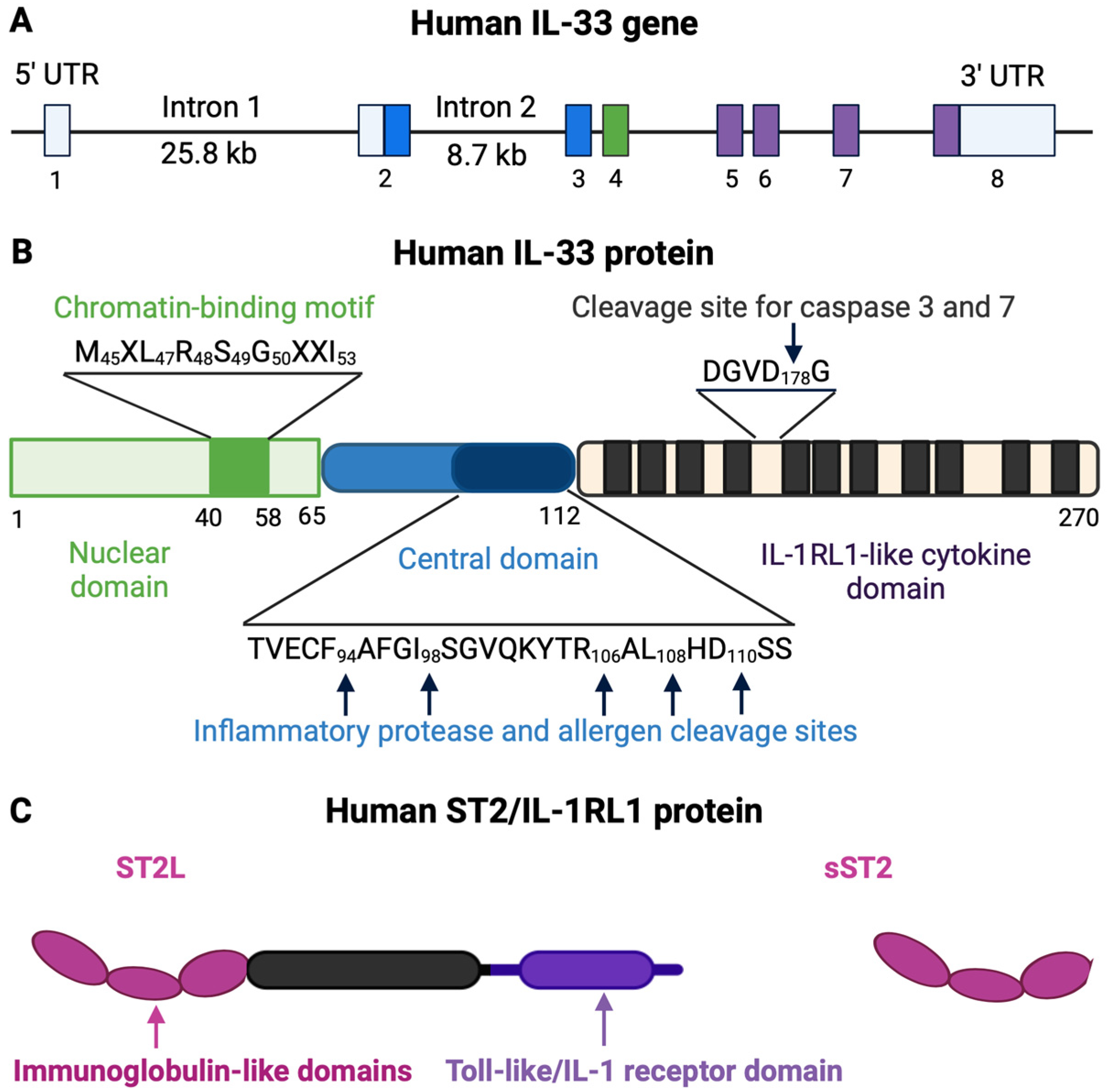
IL-33, also known by several designations, including C9orf26 (Chromosome 9 Open Reading Frame 26), DVS27 (DVS27-Related Protein), NF-HEV (Nuclear Factor from High Endothelial Venules), DKFZp586H0523, NFEHEV, and RP11-575C20.2, is a cytokine that belongs to the IL-1 superfamily, identified explicitly as Interleukin-1 Family Member 11 (IL1F11) [6]. It was initially recognized for inducing type 2 immune responses [31,32], stimulating the production of immune cells involved in allergic reactions, and protecting against parasites. Understanding this function is crucial for comprehending its role in immune system regulation.
As a tissue-derived cytokine, IL-33 has various functions; it operates as a traditional cytokine and an intracellular nuclear factor [32,33]. Its association with tissue damage or necrosis is just one facet of its complexity. In response to harmful events such as infection, injury, or inflammation, barrier tissue cells, including epithelial cells, release IL-33. The released IL-33 plays a significant role in cell communication and regulating inflammation within the body.

2.1. IL-33

The IL-33 gene is located on chromosome 9p24.1 in humans and chromosome 19qC1 in mice. The human IL-33 gene consists of eight exons, with exon 1 being non-coding and exons 2-8 coding for the IL-33 protein, spanning over 42kb of genomic DNA [34]. The IL-33 protein features an N-terminal domain containing a chromatin-binding motif, a nuclear localization signal, a central domain, and a C-terminal IL-1-like cytokine domain that gives IL-33 cytokine-like properties [6,32]. The nuclear localization signal can be found within amino acids 46-67 [35]. The ability to bind to DNA determines the functions of full-length IL-33 in the nucleus as a transcription factor [36]. In mice, two different promoters initiate IL-33 gene transcription, resulting in two IL-33 transcripts (Il33a and Il33b) with differing 5′-untranslated regions but encoding the same protein. The human IL-33 mRNA encodes a protein containing 270 amino acids, while the mouse IL-33 protein consists of 266 amino acids, with a 55% similarity between the two. Research indicates that the IL-33 protein is highly conserved among mammals and is closely related to IL-18 among the IL-1 family members. IL-33 has a three-dimensional structure akin to other family members, comprised of 12 β-strands arranged in a β-trefoil fold [37,38]. The binding area of IL-33 contains a mix of polar and nonpolar regions that facilitate specific interactions between the receptor and ligand [38] (Figure 1).

2.2. IL-33 Receptor

The IL-33 receptor, also known as serum stimulation-2 (ST2), interleukin 1 receptor-like 1 (IL1RL1), or T1, was initially discovered in 1989 and was considered an orphan receptor until the identification of IL-33 [40,41]. There are multiple isoforms of ST2, including the membrane-bound form (ST2L) and soluble form (sST2). ST2L consists of three immunoglobulin-like motifs responsible for extracellular ligand-binding (extracellular domain), an intracellular domain, and a transmembrane domain. The extracellular domain of ST2L binds IL-33 with the assistance of the IL-1 receptor accessory protein (IL-1RAcP), which forms the transmembrane IL-33 receptor complex and recruits downstream signaling components via its Toll/interleukin-1 receptor (TIR) domain [31]. Due to its lack of intracellular and transmembrane domains, but the possession of a C-terminal sequence [42], sST2 may serve as a decoy receptor for IL-33 [43]. The IL-1R8 receptor, known as SIGIRR (single immunoglobulin IL-1R-related receptor) or TIR8 [44], acts as a negative regulator of the IL-33/ST2 signaling pathway [45]. It can dimerize with ST2L to attenuate the biological effects of IL-33 (Figure 1).

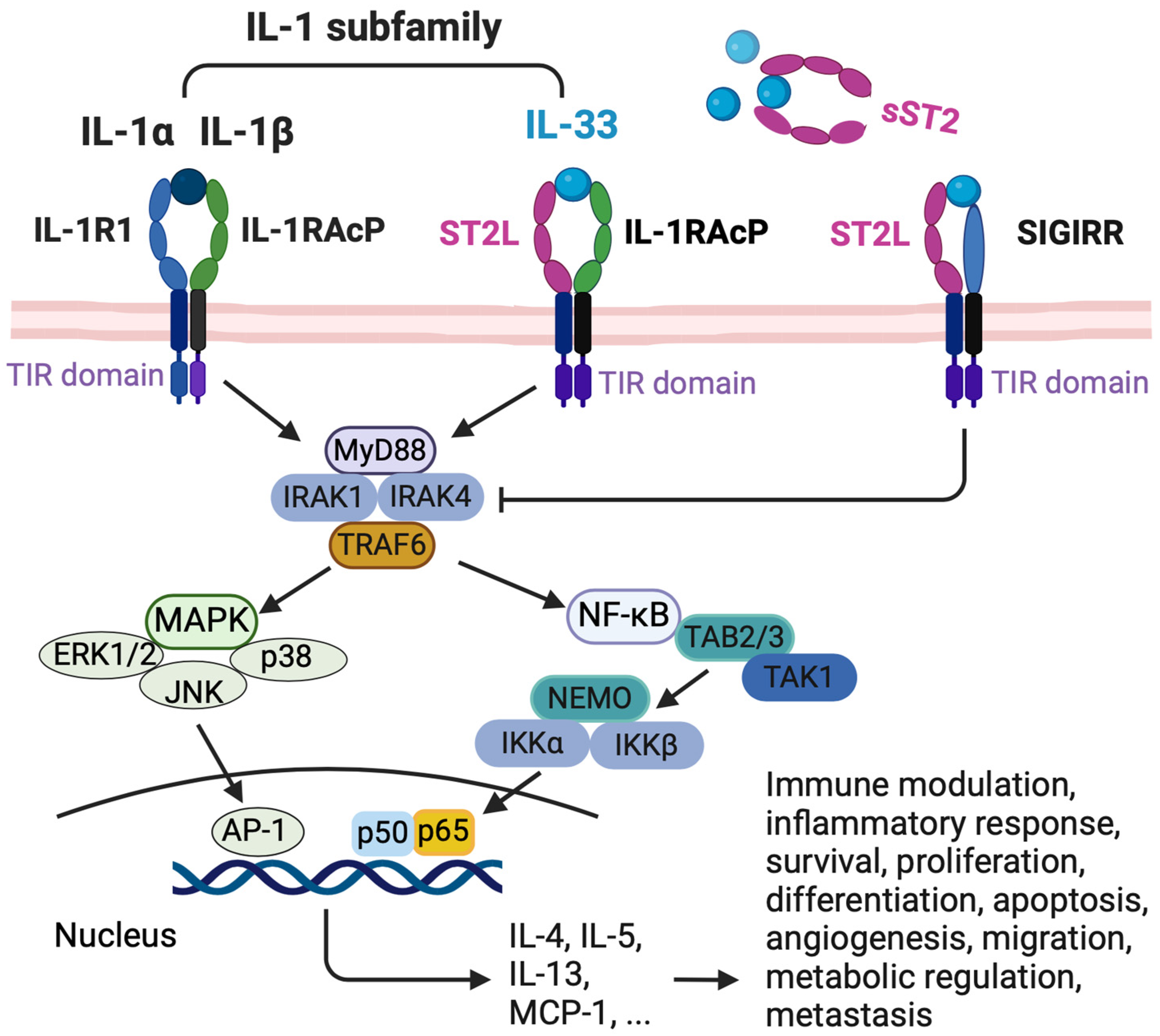
2.3. IL-33/ST2 Signaling Pathway

IL-33 has a dual role, acting as a nuclear factor by binding to chromatin to suppress inflammatory responses and as a cytokine released into the extracellular space in response to cell damage or mechanical injury [33]. This dual functionality underscores its crucial role in immune modulation. IL-33 exerts its cytokine activity by binding to the primary receptor, ST2, and then recruiting the accessory receptor, IL-1RAcP [6,46,47,48].
When IL-33 binds to the IL-33 receptor, forming the ST2L and IL-1RAcP heterodimeric complex, it initiates a signaling pathway. Following receptor binding, the TIR domain dimerization starts a pathway that includes the myeloid differentiation primary response protein 88 (MyD88), IL-1R-associated kinase 1 (IRAK1), IRAK4, tumor necrosis factor (TNF) receptor-associated factor 6 (TRAF6), mitogen-activated protein kinases (MAPKs), or nuclear factor-κB (NF-κB) [34].
The intracellular signaling pathways initiated by IL-33 activate a wide array of downstream effectors. These effectors play a significant role in immune modulation and inflammatory responses [49,50], cell survival [51,52], proliferation [27], differentiation [27], apoptosis [51], angiogenesis [53], migration [54], metabolic regulation [55], and metastasis [56] (Figure 2).

3. The Role of IL-33 in Immune Responses

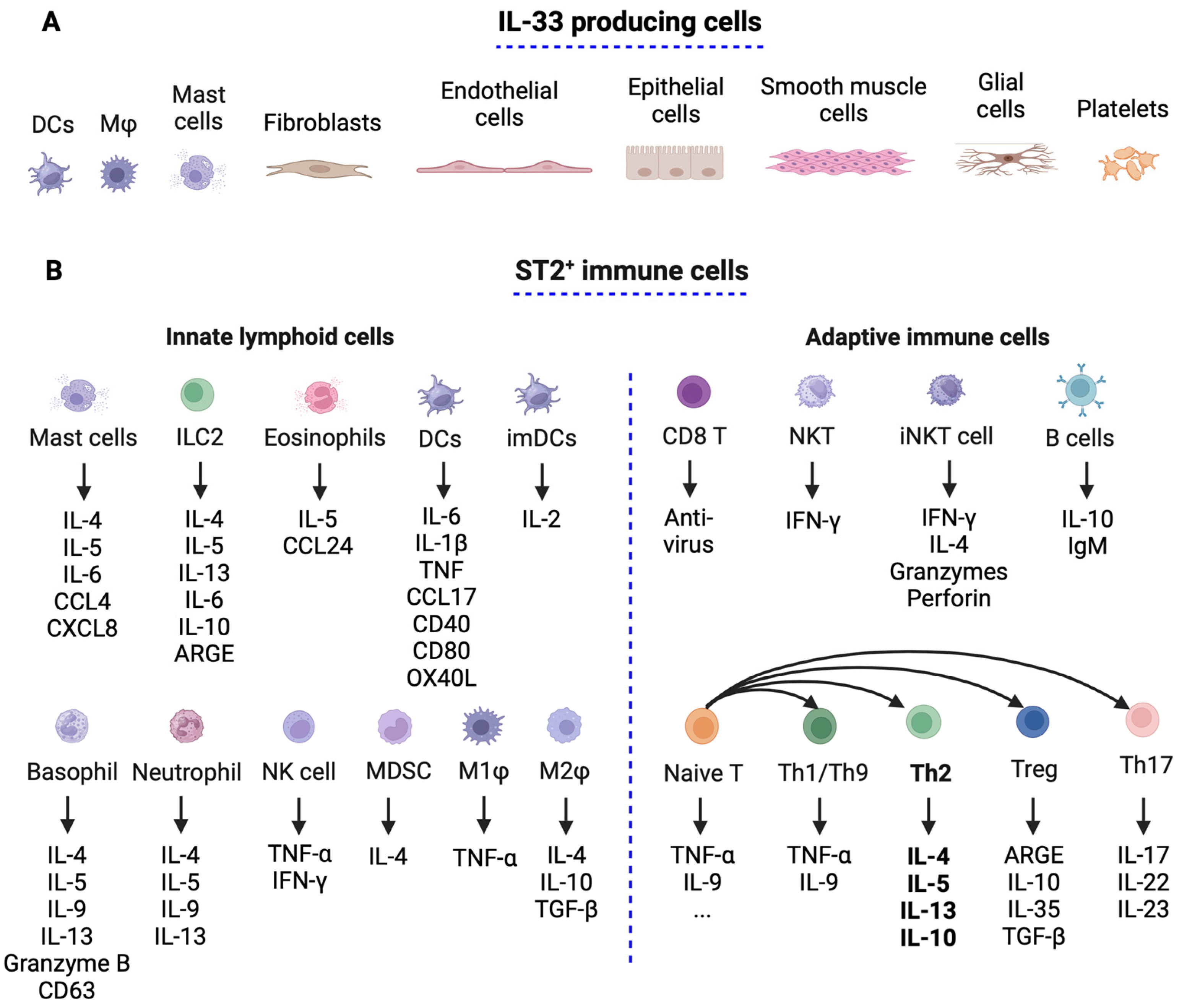
3.1. IL-33 and ST2 Expression in Immune Cells

IL-33 is typically found in the stroma and is released by damaged or necrotic barrier cells such as endothelial and epithelial cells [57]. It is expressed at high levels in the steady-state nuclei of various cell types [34], including immune cells like resting dendritic cells (DCs) [58], activated macrophages [59], monocytes, and regulatory T cells (Tregs), as well as mast cells [59], endothelial cells, epithelial cells of barrier tissues such as the lung, intestine, skin, and fibroblasts, glial cells, astrocytes, smooth muscle cells, and platelets. IL-33 is a ubiquitous and crucial immune modulator that influences various immune cell responses [50]. It acts as an alarm signal (alarmins or danger signals), rapidly released from producing cells in response to cellular stress and injury [49] (Figure 3).
ST2 is expressed in T helper 2 (Th2) cells, mast cells [42,46,60], cardiomyocytes [61], and other immune cells [57,62], including innate lymphoid cell type 2 (ILC2) [63], Tregs [64], CD4+ and CD8+ T cells [65], NKT cells [66,67], and myeloid cells, including basophils, eosinophils, and macrophages [59]. Recently, ST2 was reported to be expressed in Th1 cells [68,69], though the expression level was low and transient upon virus infection [70] (Figure 3).

3.2. Il-33’s Role in Immune Responses

The IL-33/ST2 pathway plays a crucial role in Th2 responses. When IL-33 activates Th2 receptors, it produces IL-4, IL-5, and IL-13. These cytokines, produced by Th2 cells, enhance chemokine production in epithelial cells [71,72,73]. The IL-33 gene and these cytokines are linked to Th2-related diseases, protecting against intracellular pathogens, promoting tissue repair, and contributing to chronic inflammatory diseases [74].
IL-4, a critical Th2 cytokine, is primarily produced by activated T cells, mast cells, basophils, and eosinophils. Its primary function is to transform naive CD4+T cells into Th2 cells, which initiates allergic reactions and triggers immune responses against extracellular parasites [75]. IL-4 also affects various diseases, particularly inflammatory bowel disease (IBD), where it contributes to chronic inflammation and fibrosis by promoting tissue remodeling and scarring, as well as autoimmune diseases [73]. Moreover, IL-4 supports the proliferation and survival of several cancer cells [76], being overexpressed in many human tumor types, including malignant glioma, ovarian, lung, breast, pancreatic, colon, and bladder carcinomas, which also overexpress its receptors (IL-4R) [77,78,79]. While several therapeutics have been developed for asthma and inflammatory diseases, their lack of efficacy has hindered further development. However, most of these treatments have shown tolerance and high safety in humans, making clinical trials for cancer therapy a possibility [76].
IL-5 is significant in developing allergic reactions and inflammation, as it regulates the production, maturation, activation, and survival of eosinophil progenitors in the blood [80]. It stimulates B lymphocytes to produce more antibodies and increases the number of eosinophils in the airways. Its primary role is to promote the activation and longevity of eosinophils, aiding their migration from the blood to the airways [81]. By encouraging the production of antibodies, particularly immunoglobulin A (IgA), which is crucial for mucosal immunity, IL-5 also influences B cell activity [81]. Additionally, IL-5 acts as a biological modifier that can enhance the immune system in cancer therapy.
IL-13 is a critical factor in developing allergic inflammation [82]. It closely interacts with IL-5 and significantly impacts the airways by promoting mucus production [83]. IL-13 encourages goblet cells in the airway to produce mucus, leading to airway blockage and mucus plugging, which directly contribute to asthma [83,84]. Furthermore, IL-13 is overexpressed in various solid tumors and is associated with poor prognosis in conditions such as glioblastoma, colorectal cancer, adrenocortical carcinoma, pancreatic cancer, and breast cancer [85].

4. IL-33 in Obesity

Obesity is linked to chronic inflammation, hormonal imbalances, insulin resistance, high insulin levels, adipokines changes, and immune function alterations [86]. Hormones such as adipokines, produced by adipose tissue, contribute to low-grade inflammation, leading to conditions like insulin resistance, diabetes, high blood pressure, and asthma, all commonly associated with obesity [87]. These adipokines can stimulate cell growth, angiogenesis, and apoptosis, all directly connected to cancer progression. Furthermore, these hormonal functions regulate the appetite, energy metabolism, and inflammation, increasing individuals’ susceptibility to cancer development and progression [88].
Adipose tissue is essential for regulating energy balance and is generally categorized into white adipose tissue (WAT) and brown adipose tissue (BAT) [89,90,91]. Additionally, there is a type of adipose tissue known as brown-in-white or (brite)/beige adipose tissue (BeAT), which shares the characteristics of both WAT and BAT in terms of morphology and function [92]. Research on the role of IL-33 in developing metabolic disorders such as diabetes, obesity, and cardiovascular disease has produced mixed results [22]. However, the IL-33/ST2 pathway is thought to be protective during obesity. Manipulating IL-33 expression to induce Th2 cytokines and promote macrophage polarization may offer a promising therapeutic strategy for treating or preventing type 2 diabetes in obese patients.

4.1. IL-33 and ST2 Immune Cell Distribution in Adipose Tissue

Immune cells and their mediators significantly regulate the metabolism, leading to a new understanding of immune regulation in metabolic processes [21]. Innate immune cells present in adipose tissue include macrophages, neutrophils, eosinophils, dendritic cells (DCs), innate lymphoid cells (ILCs), and natural killer (NK) cells [93]. In lean adipose tissue, the most common immune cells are alternatively activated macrophages (M2-like macrophages), type 2 innate lymphoid cells (ILC2s), eosinophils, Tregs, and Th2 cells [94,95]. These cells contribute to an anti-inflammatory environment.
However, during obesity development and adipocyte hypertrophy, the distribution of immune cell populations changes. Most immune cells increase in adipose tissue, leading to an inflammatory state that supports insulin resistance. Exceptions to this trend include Tregs, eosinophils [21], and Th2 cells [96].
IL-33 and its receptor ST2 are abundant in adipose tissue, including preadipocytes, adipocytes, and endothelial cells [97,98,99]. Additionally, infiltrating immune cells that express IL-33 and ST2 are plentiful in this tissue (Figure 4).

4.2. Effect of IL-33 in Adipose Tissue During Obesity

Despite its increased expression in obesity, IL-33 alone cannot maintain homeostasis in obese adipose tissue. Research indicates that IL-33 treatment can improve the inflammation and metabolic changes related to obesity [22]. The role of IL-33 in immunity and the metabolism is complex, demonstrating both pro- and anti-inflammatory properties [22]. IL-33 is essential for maintaining adipose tissue homeostasis and may help protect against obesity and type 2 diabetes [100,101].
Tissue-resident stromal cells produce IL-33, supporting immune homeostasis in adipose tissue through the involvement of ILCs [101]. Furthermore, IL-33 protects against adipose tissue inflammation during obesity by inducing Th2 cytokines in WAT and promoting the polarization of WAT macrophages toward an M2, an alternatively activated phenotype [100,102,103]. This process reduces adipose mass and lowers fasting glucose levels [100]. In laboratory cultures of WAT, IL-33 stimulates the production of Th2 cytokines and chemokines while increasing the serum levels of Th2 cytokines and the number of Th2 cells in WAT. This shift affects macrophage activity in both adipose and liver tissues, reducing the chronic inflammatory response associated with obesity. Additionally, adiponectin can inhibit IL-33 signaling through AMPK-mediated feedback [104].
As obesity progresses, adipocyte hypertrophy significantly alters immune cell populations. While WAT shows increased IL-33 expression, local ILC2 levels tend to decrease [22]. This imbalance highlights the need to understand the discrepancies in regulating IL-33 at both tissue-specific and systemic levels amid obesity [22]. IFN-γ inhibits the activation and proliferation of ILC2 cells, limiting the functions of IL-33-mediated ILC2 and Tregs while promoting a Th1 immune response [97]. This suppression by IFN-γ likely facilitates inflammatory responses and reallocates metabolic resources necessary for host protection (Figure 4).
Moreover, obesity is linked to an imbalance between Th17 and Treg cells, which can lead to metabolic disorders [104,105,106]. It also disrupts the Th1/Th2 balance, increasing the number of Th1 cells in adipose tissue while decreasing Th2 cells [107]. Although there are currently no direct studies examining how IL-33 contributes to this imbalance, these disruptions may result from reduced levels of IL-33, warranting further investigation (Figure 4).
Figure 4. IL-33 and ST2 immune cells in adipose tissue. During the development of obesity, adipocyte hypertrophy is accompanied by significant changes in immune cell populations. Most immune cells increase in quantity in adipose tissue due to obesity, with a few exceptions, including regulatory T cells (Tregs) and eosinophils. Tregs and eosinophils reduce the inflammatory responses of other immune cells in adipose tissue, particularly adipose tissue macrophages. As obesity progresses, the population of IL-33/ST2 target immune cells in adipose tissue also varies, leading to obesity-related inflammation. Lean adipose tissue primarily contains non-inflammatory cells, such as activated M2 macrophages (M2φ), eosinophils, ILC2 cells, Tregs, and Th2 cells. In contrast, obesity shifts the immune profile of adipose tissue toward a pro-inflammatory state, characterized by an influx of macrophages (M1φ), NK cells, neutrophils, CD4+ T cells (Th1, Th17), and CD8+ T cells. When IL-33 is reduced during obesity, sST2 is increased [108], which can inhibit metabolic health. In addition, the balance between Th1 and Th2 cells, and between Treg and Th17 cells, is disrupted, leading to increased adipose tissue-related inflammation (often referred to as “met-inflammation”) and contributing to the development of metabolic diseases. DC, dendritic cell; M, macrophage; ILC2, innate lymphoid cell type 2; iNKT, invariant natural killer T; NK, natural killer cells; NKT, natural killer T cells; MDSC, myeloid-derived suppressor cells. This figure was created with BioRender.
Encyclopedia 04 00117 g004

5. Effect of Il-33 in the Tumor Microenvironment

The cytokine IL-33 has a dual role in the tumor microenvironment (TME), promoting and inhibiting tumor growth. Its protumor effects involve the accumulation of immune-suppressive cells. Conversely, IL-33 can inhibit tumor growth by interacting with the T cell receptor (TCR) and IL-12 signaling, enhancing the effectiveness of CD8+ T cells [109]. Additionally, IL-33 amplifies the responses of Th1 CD4+ T cells and Th2-type cells by acting on various immune cells, including human basophils, allergen-reactive Th2 cells, iNKT, NK cells [110], eosinophils, and ILC2 [111].

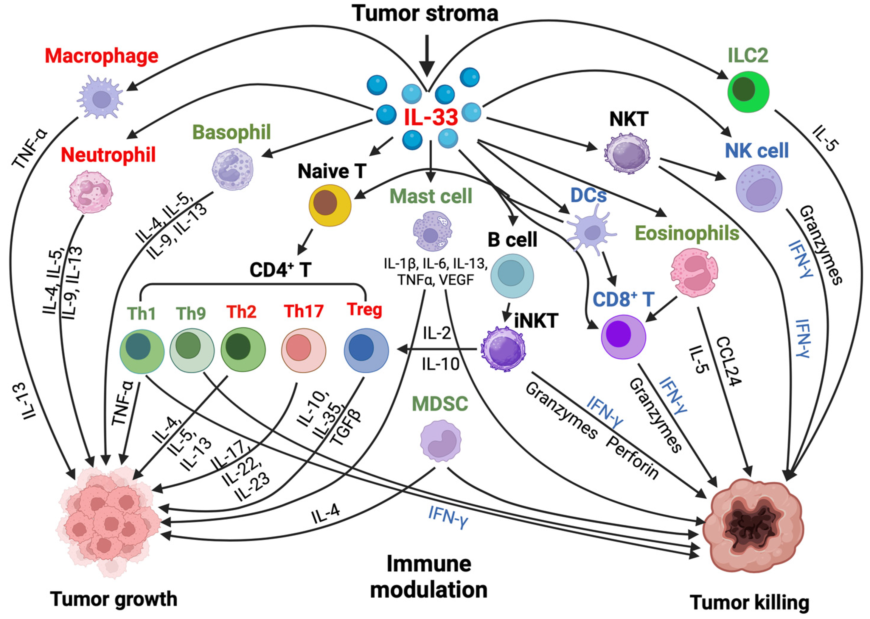
5.1. IL-33 and ST2 Immune Cell Distribution in the Tumor Microenvironment (TME)

The TME varies among different types of tumors but generally includes immune cells, stromal cells, blood vessels, and the extracellular matrix [112], which actively promote cancer progression. Depending on the context, immune cells can play a pivotal role in inhibiting or supporting tumor growth [113]. ST2 is expressed in various immune cells, and IL-33 is released from damaged structural cells. IL-33/ST2 signaling is involved in numerous cancers, exhibiting both protumor and antitumor functions [114].
Understanding the diverse effects of IL-33 on immune responses in the cancer context is crucial [115]. For example, IL-33 can activate ILC2 in the lung, which promotes tumor burden by leading to innate type 2 inflammation and suppressing the production of IFN-γ and the cytotoxic functions of lung NK cells [116]. In melanoma, the overexpression of IL-33 has been shown to inhibit lung metastases by activating CD8+ T cells and NK cells [117]. Additionally, the induced expression of IL-33 in tumors can enhance anti-melanoma immune responses via IFN-γ-producing CD8+ T cells and NK cells [118], presenting potential opportunities for cancer therapy.
Eosinophils [119] and DCs [120] may also mediate the anti-melanoma effects of exogenous IL-33. In breast cancer, IL-33 secreted by cancer-associated fibroblasts enhances ILC2 and Th2 type responses, induces the TCR-independent secretion of IL-13, and recruits immunosuppressive granulocytes [121]. Furthermore, IL-33 activates the intrinsic signaling pathway in Treg cells, which is necessary for their immunosuppressive action in cancer [122]. When combined with the pro-inflammatory cytokine IL-12, IL-33 amplifies the production of the Th1 cytokine IFN-γ [109].
Overall, the IL-33/ST2 signaling pathway significantly influences the TME in cancer by impacting immune effector cells and regulating the recruitment of cells that either promote or inhibit tumor growth [123]. Due to its immunomodulatory properties, IL-33 has the potential to synergize with various cancer therapies, including immune checkpoint blockade and chemotherapy. This information focuses on the effects of IL-33/ST2 on immune cells and stroma cells within the TME [111] (Figure 5).

5.2. Effect of Il-33 on Neutrophils, Eosinophils, Mast Cells, and Basophils

Depending on the specific disease context, IL-33 affects innate immune cells, including neutrophils, eosinophils, mast cells, and basophils [124]. It can promote inflammatory or regulatory responses in neutrophils and eosinophils, contributing to tissue repair or immunosuppression [125]. Studies have demonstrated that IL-33 directly influences murine neutrophils, enhancing their recruitment and activation in inflamed tissues [125]. In cancer, IL-33-driven neutrophils are associated with pro-inflammatory and regulatory functions.
Eosinophils, which play a vital role in the host defense against parasites and infections, are activated by IL-33 for allergic reactions and tissue repair [125]. In the context of cancer, Il-33 is linked to using eosinophils for antitumor effects, such as inhibiting growth and preventing metastasis. IL-33-activated eosinophils express higher effector molecules, including the eosinophil cationic protein (ECP), eosinophil peroxidase (EPX), and granzymes. They also express integrin CD11b/CD18, which forms immune synapses that facilitate the killing of tumor cells in vitro and in vivo [126].
Mast cells, which respond to IL-33, accumulate in tumors and their microenvironment during disease progression [127]. They are involved in tumor growth, with their mediators exhibiting both pro- and antitumorigenic roles in various human cancers [128]. Activating mast cells and basophils by IL-33 may influence tumor progression by modulating cytokine release and triggering immune complex activation. Recent studies indicate that tumor-infiltrating mast cells activated by IL-33 secrete IL-2 and promote the expansion of ICOS+ Tregs, which contributes to gastric cancer progression [129].
Basophils, produced in the bone marrow, are white blood cells needed for maintaining a healthy immune system. Abnormal levels of basophils can indicate conditions such as inflammation and hyperthyroidism. IL-33 can activate both murine and human basophils, increasing the production of histamine and cytokine in vitro and promoting their expansion in vivo [130,131,132,133]. However, a limited understanding of the relationship between basophils, cancer, and IL-33 regulation remains [125,134].

5.3. Effect of Il-33 on Macrophages and Dendritic Cells

Macrophages and DCs play crucial roles in the innate immune system, particularly regarding tumor immunity and immunology [135]. IL-33 is a crucial regulator of the activity of DCs and macrophages within the TME, displaying both pro- and antitumor effects [135]. Notably, IL-33 is essential for promoting M2-like polarization in macrophages, a process associated with immunosuppressive and tumor-promoting properties [135]. This polarization involves the release of anti-inflammatory cytokines and the stimulation of tumor angiogenesis, invasion, and metastasis [134,136].
In addition to its effects on macrophages, IL-33 also influences DCs. DCs activated by IL-33 demonstrate increased cytokine production and enhanced antigen presentation capabilities, which promote T cell activation and bolster antitumor immune responses. However, conflicting studies regarding IL-33’s influence on DC-mediated antitumor immunity suggest that its function in DCs may vary based on its location within the body [134]. This discrepancy highlights the need for further research in this area.
Moreover, with the assistance of dectin1-activated DCs, IL-33 can induce Th9 cell differentiation and enhance the antitumor efficacy [137,138]. While some studies indicate that IL-33 improves T cell activation and DC-mediated antigen presentation, others argue that IL-33 may lead DCs to acquire tolerogenic characteristics, inhibiting the immune system’s ability to combat cancer [134].

5.4. Effect of Il-33 on CD8+ T, NK, and NKT Cells

Several studies have demonstrated that IL-33 expression is positively correlated with the recruitment of CD8+ T cells and NK cells within the TME. CD8+ T, NK, and NKT cells are effective tumor-killer cells with many phenotypes and functions relevant to antitumor immunity [139]. IL-33 directly activates NK and NKT cells, leading to the production of IFN-γ and the promotion of Th1 immunity [140]. It also targets immunoregulatory invariant NKT (iNKT) cells to enhance their activation state [66]. This activation induces the production of IFN-γ and promotes Th1 immunity in both human and mouse NK cells [138].
When combined with IL-12, IL-33 further enhances NK cell activation, improving their ability to destroy tumor cells through cytotoxicity. In preclinical models, IL-33 has been shown to support the recruitment and activation of cytotoxic NK cells in the TME, which hinders the formation of metastasis and suppresses tumor progression [134]. However, it is crucial to note that conflicting reports are suggesting that, in specific tumor microenvironments, IL-33/ST2 signaling may hinder NK cell activation. This underscores the need to consider the context-dependent effects of IL-33 on NK cell-mediated antitumor responses. Such considerations should be a key focus in future research and clinical applications, emphasizing the necessity for further investigation and careful application [27].

5.5. Effect of Il-33 on Myeloid-Derived Suppressor Cells

IL-33 significantly affects myeloid-derived suppressor cells (MDSCs), which are critical mediators of immune suppression within the tumor [23]. MDSCs play a crucial role in cancer by helping tumors evade the immune system and accelerating disease progression [23]. IL-33 partially regulates the function and development of MDSCs within the TME [23]. In this environment, IL-33 reduces apoptosis and supports MDSC survival by inducing autocrine secretion of GM-CSF. This creates a positive feedback loop that promotes MDSC accumulation [23]. The increase and promotion of MDSCs are linked to tumor development and heightened immunosuppression due to IL-33. However, studies have shown that, in some contexts, IL-33 can inhibit the growth of MDSCs and decrease their immunosuppressive capabilities, resulting in enhanced antitumor immune responses and tumor regression [23,134].

5.6. Effect of Il-33 on T Helper Cells

IL-33 plays a significant role in regulating the development and function of CD4+ T cells, including CD4+ T helper (Th) cells and CD4+ Treg cells [124,141]. Early in T cell activation, IL-33 stimulates Th1 differentiation and works alongside IL-12 to enhance Th1 polarization [124,141]. Moreover, IL-33 may promote Th1 cell differentiation depending on the presence of IL-12 and ST2 [141]. MyD88 is essential for IL-33, as well as IL-12-induced Th1 cell development and the production of IFN-γ [141]. Additionally, IL-33 fosters the proliferation of Th cells that produce IL-9 (Th9), strengthening immune responses against tumors.
Conversely, IL-33 signaling encourages the proliferation of suppressive CD4+ Foxp3+ GATA3+ Treg cells, especially in tumor-specific environments high in IL-33 [60,141]. These Treg cells have a complex regulatory role, which may limit antitumor immunity while exhibiting suppressive solid activities [134]. In preclinical models, IL-9-producing Th9 cells are vital for initiating robust immune responses against tumors and promoting tumor regression. Research on T cell differentiation indicates that IL-33 has context-dependent effects on Th1- and Th2-skewing, reflecting the complexities of immune system regulation [134,141]. The unique cues from the tumor microenvironment can influence how these signaling pathway function in Th cell development.
Th17 cells play a significant role in inflammation and are implicated in various autoimmune diseases [142]. Their activity is influenced by different molecular signals within the TME [143], which affects their proliferation, differentiation, metabolic reprogramming, and phenotypic transformation, resulting in a dual impact on tumor progression [143]. Recent studies suggest that Th17 cells accumulating in the small intestine during inflammation express the IL-33 receptor (ST2), with intestinal epithelial cells (IECs) being the primary source of IL-33 [144]. When exposed to IL-33, both mouse and human Th17 cells exhibit reduced expression of pro-inflammatory genes and increased IL-10. This anti-inflammatory cytokine can limit intestinal inflammation and control previously activated pathogenic Th17 cells. The findings indicate that, in response to IL-33, Th17 cells acquire immunosuppressive properties. The IL-33/ST2 axis is likely crucial in regulating pathogenic Th17 cells in the small intestine, helping to maintain homeostasis [144].

5.7. Effect of Il-33 on CD4+ Treg Cells and in Tumor Immunity

IL-33 signaling influences effector Th cells and the growth and activity of Treg cells within the TME. Research shows that suppressive CD4+ Foxp3+ GATA3+ Treg cells proliferate in response to ST2/IL-33 signaling, both in vivo and in vitro, which aids in immunosuppression and tumor immune evasion [134]. IL-33 signaling promotes the generation of IL-2 by DCs and facilitates TGF-β1-mediated Treg cell development, leading to the expansion of ST2+ Treg cells [134,145]. These IL-33-expanded Treg cells are present in both immune and non-immune tissues and exert a strong suppressor effect, creating an immunosuppressive tumor microenvironment [146].
In the lungs, CD4+Foxp3+ Treg cells are expressed the IL-33 receptor ST2 [147]. When exposed to IL-33, Treg cells increase their canonical Th2 transcription factor GATA3 and ST2 expression levels, along with producing type 2 cytokines. This exposure leads to a significant shift in the immune response, causing Treg cells to lose their ability to suppress effector T cells [147]. Furthermore, IL-33 has been implicated in promoting the accumulation and maintenance of ST2+ Treg cells in inflamed tissues, exacerbating immunosuppression and facilitating tumor progression [134].
The complex interaction between IL-33 signaling and CD4+ T cell subsets underscores its significance in supporting antitumor immune responses [134]. By stimulating the growth of suppressive Treg cells and influencing Th cell differentiation, IL-33 regulates adaptive immune responses within the tumor. The crucial role of IL-33 in promoting Treg cell expansion highlights its potential as a target for therapeutic intervention in cancer immunotherapy, emphasizing the importance of understanding and utilizing IL-33 in cancer treatment [138].

5.8. IL-33-Expressing Cells and the Effects of IL-33 on Non-Immune Cells in the TME

The impact of IL-33 is influenced by the types of cells that produce it within the TME. These cells include immune cells, stromal cells, non-tumor epithelial cells, and transformed (epithelial) tumor cells, which are significant sources of IL-33 in the TME [57]. Fibroblasts, in particular, play a crucial role; their dysregulation during cancer creates an environment that fosters tumor growth by remodeling the extracellular matrix and releasing pro-angiogenic factors [148,149]. Research has shown that cancer-associated fibroblasts can induce IL-33 production [150].
Stromal cells have also been identified as a primary source of IL-33 among non-immune cells, and IL-33 derived from these cells has been linked to tumor metastasis [115,148,151,152]. The analysis of The Cancer Genome Atlas (TCGA) data and mRNA sequencing from the TCGA revealed that IL-33, expressed in endothelial and epithelial cells, could further contribute to tumor immune responses [115,151]. Moreover, IL-33 produced by non-tumor epithelial cells has been shown to drive colorectal cancer tumorigenesis in APCMin/+ mice, which serve as an excellent model for colorectal tumors due to a mutation in the APC gene, a significant tumor-suppressor gene in the Wnt signaling pathway [153].
The expression of IL-33 changes during tumor progression. At the same time, it is highly expressed in normal epithelial cells, and tumor development leads to its downregulation in epithelial cells and upregulation in the tumor stroma and serum [114]. Therapeutic overexpression of IL-33 in tumor epithelial cells promotes type 1 antitumor immune responses through CD8+ T cells and NK cells. In contrast, IL-33 from tumor stroma fosters immune tolerance and suppression via Tregs and MDSCs [114]. Overall, IL-33 expression is context-dependent. In some tumors, IL-33 is highly expressed in normal epithelial cells but downregulated in cancer cells in advanced conditions [114,154,155]. Conversely, IL-33 or ST2 protein expression levels in other tumors increase in cancerous lesions compared to matched normal tissues [123,156,157].

6. Discussion of IL-33 and Obesity-Associated Cancer

Recent scientific research has confirmed a clear connection between a high BMI and the risk of developing various types of cancer [19,30]. While obesity itself may not directly cause cancer, it increases the likelihood of developing aggressive, fast-growing forms of cancer that are more challenging to treat. Therefore, addressing obesity is vital for reducing the risk of cancer-related complications [19].
Research suggests that inflammation is connected to obesity and cancer [158]. IL-33 plays multiple roles in adipose tissue and the TME. However, studies explicitly examining the direct effects of IL-33 in obesity-related cancer are limited. Given its significant role in both obesity and cancer, more research is needed to fully understand IL-33’s potential in regulating obesity and decreasing cancer risk. Current findings are promising and may indicate a new pathway for disease management.
Future research should investigate how IL-33 impacts fat tissue function, energy expenditure, and immune modulation to identify potential therapeutic targets. It is also crucial to further explore the influence of immune cells on both antitumor and protumor processes in different types of obesity-related cancers. Targeting IL-33 signaling pathways and their downstream effects could help regulate obesity while exhibiting antitumor properties. Additionally, IL-33’s anti-inflammatory characteristics provide another possible intervention avenue, as chronic inflammation is a critical factor in obesity and a significant contributor to cancer development. Therapies based on IL-33 could reduce obesity-induced inflammation in fat tissue and other affected organs, subsequently lowering cancer risk.
Moreover, conducting clinical trials and utilizing personalized medicine approaches are essential to evaluating the safety and effectiveness of IL-33-based treatments. This process involves developing tailored treatment plans considering each patient’s unique risk factors and metabolic profiles. A comprehensive understanding of IL-33’s role in immune regulation and metabolism is critical for harnessing it as a therapeutic target to manage obesity and mitigate cancer risk. Collaboration among scientists, clinicians, and industry partners is vital for expanding our understanding of IL-33 biology and translating scientific discoveries into practical applications.

7. Conclusions and Prospects

Research into the various roles of IL-33 in immune modulation, obesity, and cancer is essential. The complex interactions between the IL-33/ST2 axis in adipose tissue and the tumor microenvironment (TME) deserve a thorough investigation, especially concerning obesity-related tumors. Given IL-33’s importance in both obesity and cancer, much remains to be explored regarding the roles of the IL-33/ST2 pathway in various types of obesity-related cancers and their responses to treatment. Both animal and human studies are necessary to fully understand how the IL-33/ST2 axis contributes to these cancers.

Author Contributions

Conceptualization, Q.Z.; writing—original draft preparation, C.K. and Q.Z.; writing—review and editing, C.K. and Q.Z.; funding acquisition, Q.Z. All authors have read and agreed to the published version of the manuscript.

Funding

This research was funded by the National Cancer Institute of the National Institutes of Health (NIH), grant number R01CA255802; the National Institute of General Medical Sciences of the National Institutes of Health, grant number P20 GM103629; the University Senate Committee on Research Fellowship Program Award; the Carol Lavin Bernick Faculty Grant; the BoRSF-Research Competitiveness Subprogram (RCS) Award. The funders had no role in the design of the study; in the collection, analyses, or interpretation of data; in the writing of the manuscript; or in the decision to publish the results.

Institutional Review Board Statement

Not applicable.

Informed Consent Statement

Not applicable.

Data Availability Statement

The data presented are available in the references cited in this article.

Acknowledgments

Figures created with BioRender.com.

Conflicts of Interest

The authors declare no conflicts of interest.

References

  1. Takeuchi, T. Cytokines and cytokine receptors as targets of immune-mediated inflammatory diseases-RA as a role model. Inflamm. Regen. 2022, 42, 35. [Google Scholar] [CrossRef] [PubMed]
  2. Turner, M.D.; Nedjai, B.; Hurst, T.; Pennington, D.J. Cytokines and chemokines: At the crossroads of cell signalling and inflammatory disease. Biochim. Biophys. Acta 2014, 1843, 2563–2582. [Google Scholar] [CrossRef] [PubMed]
  3. Samadi, M.; Kamrani, A.; Nasiri, H.; Shomali, N.; Heris, J.A.; Shahabi, P.; Ghahremanzadeh, K.; Mohammadinasab, R.; Sadeghi, M.; Sadeghvand, S.; et al. Cancer immunotherapy focusing on the role of interleukins: A comprehensive and updated study. Pathol. Res. Pract. 2023, 249, 154732. [Google Scholar] [CrossRef] [PubMed]
  4. Brocker, C.; Thompson, D.; Matsumoto, A.; Nebert, D.W.; Vasiliou, V. Evolutionary divergence and functions of the human interleukin (IL) gene family. Hum. Genomics 2010, 5, 30–55. [Google Scholar] [CrossRef] [PubMed]
  5. Tsutsui, H.; Cai, X.; Hayashi, S. Interleukin-1 Family Cytokines in Liver Diseases. Mediat. Inflamm. 2015, 2015, 630265. [Google Scholar] [CrossRef]
  6. Schmitz, J.; Owyang, A.; Oldham, E.; Song, Y.; Murphy, E.; McClanahan, T.K.; Zurawski, G.; Moshrefi, M.; Qin, J.; Li, X.; et al. IL-33, an interleukin-1-like cytokine that signals via the IL-1 receptor-related protein ST2 and induces T helper type 2-associated cytokines. Immunity 2005, 23, 479–490. [Google Scholar] [CrossRef]
  7. Baekkevold, E.S.; Roussigné, M.; Yamanaka, T.; Johansen, F.E.; Jahnsen, F.L.; Amalric, F.; Brandtzaeg, P.; Erard, M.; Haraldsen, G.; Girard, J.P. Molecular characterization of NF-HEV, a nuclear factor preferentially expressed in human high endothelial venules. Am. J. Pathol. 2003, 163, 69–79. [Google Scholar] [CrossRef]
  8. Gaurav, R.; Poole, J.A. Interleukin (IL)-33 immunobiology in asthma and airway inflammatory diseases. J. Asthma 2022, 59, 2530–2538. [Google Scholar] [CrossRef]
  9. Di Carmine, S.; Scott, M.M.; McLean, M.H.; McSorley, H.J. The role of interleukin-33 in organ fibrosis. Discov. Immunol. 2022, 1, kyac006. [Google Scholar] [CrossRef]
  10. Chen, W.Y.; Li, L.C.; Wu, Y.H.; Yang, J.L.; Tzeng, H.T. Emerging Roles of Interleukin-33-responsive Kidney Group 2 Innate Lymphoid Cells in Acute Kidney Injury. Int. J. Mol. Sci. 2020, 21, 1544. [Google Scholar] [CrossRef]
  11. Brunetti, G.; Barile, B.; Nicchia, G.P.; Onorati, F.; Luciani, G.B.; Galeone, A. The ST2/IL-33 Pathway in Adult and Paediatric Heart Disease and Transplantation. Biomedicines 2023, 11, 1676. [Google Scholar] [CrossRef] [PubMed]
  12. De la Fuente, M.; MacDonald, T.T.; Hermoso, M.A. The IL-33/ST2 axis: Role in health and disease. Cytokine Growth Factor Rev. 2015, 26, 615–623. [Google Scholar] [CrossRef] [PubMed]
  13. Yi, X.M.; Lian, H.; Li, S. Signaling and functions of interleukin-33 in immune regulation and diseases. Cell Insight 2022, 1, 100042. [Google Scholar] [CrossRef] [PubMed]
  14. Jia, Z.; Guo, M.; Ge, X.; Chen, F.; Lei, P. IL-33/ST2 Axis: A Potential Therapeutic Target in Neurodegenerative Diseases. Biomolecules 2023, 13, 1494. [Google Scholar] [CrossRef] [PubMed]
  15. Sun, J.; Xia, Y.; Zhang, D.; Yu, Z.; Ning, Y.; Tan, Z. Knowledge mapping of interleukin-33: A bibliometric study. Am. J. Transl. Res. 2023, 15, 914–931. [Google Scholar]
  16. Okunogbe, A.; Nugent, R.; Spencer, G.; Powis, J.; Ralston, J.; Wilding, J. Economic impacts of overweight and obesity: Current and future estimates for 161 countries. BMJ Glob. Health 2022, 7, e009773. [Google Scholar] [CrossRef]
  17. World Health Organization. World Health Statitiscs 2024: Monitoring Health for the SDGs, Sustainable Development Goals; WHO: Geneva, Switzerland, 2024. [Google Scholar]
  18. United States Cancer Statistics. 2024. Available online: https://www.cdc.gov/cancer/dataviz (accessed on 28 August 2024).
  19. Arnold, M.; Pandeya, N.; Byrnes, G.; Renehan, P.A.G.; Stevens, G.A.; Ezzati, P.M.; Ferlay, J.; Miranda, J.J.; Romieu, I.; Dikshit, R.; et al. Global burden of cancer attributable to high body-mass index in 2012: A population-based study. Lancet Oncol. 2015, 16, 36–46. [Google Scholar] [CrossRef]
  20. Hasan, A.; Kochumon, S.; Al-Ozairi, E.; Tuomilehto, J.; Ahmad, R. Association between Adipose Tissue Interleukin-33 and Immunometabolic Markers in Individuals with Varying Degrees of Glycemia. Dis. Markers 2019, 2019, 7901062. [Google Scholar] [CrossRef]
  21. Ferrante, A.W. The immune cells in adipose tissue. Diabetes Obes. Metab. 2013, 15 (Suppl. S3), S34–S38. [Google Scholar] [CrossRef]
  22. de Oliveira, M.F.A.; Talvani, A.; Rocha-Vieira, E. IL-33 in obesity: Where do we go from here? Inflamm. Res. 2019, 68, 185–194. [Google Scholar] [CrossRef]
  23. Xiao, P.; Wan, X.; Cui, B.; Liu, Y.; Qiu, C.; Rong, J.; Zheng, M.; Song, Y.; Chen, L.; He, J.; et al. Interleukin 33 in tumor microenvironment is crucial for the accumulation and function of myeloid-derived suppressor cells. Oncoimmunology 2016, 5, e1063772. [Google Scholar] [CrossRef] [PubMed]
  24. Allegra, A.; Innao, V.; Tartarisco, G.; Pioggia, G.; Casciaro, M.; Musolino, C.; Gangemi, S. The ST2/Interleukin-33 Axis in Hematologic Malignancies: The IL-33 Paradox. Int. J. Mol. Sci. 2019, 20, 5226. [Google Scholar] [CrossRef] [PubMed]
  25. Song, M.; Yang, J.; Di, M.; Hong, Y.; Pan, Q.; Du, Y.; Xiang, T.; Liu, J.; Tang, Y.; Wang, Q.; et al. Alarmin IL-33 orchestrates antitumoral T cell responses to enhance sensitivity to 5-fluorouracil in colorectal cancer. Theranostics 2023, 13, 1649–1668. [Google Scholar] [CrossRef] [PubMed]
  26. Chatrabnous, N.; Jafarzadeh, A.; Ghaderi, A.; Ariafar, A.; Aminizadeh, N.; Ghassabi, F.; Nemati, M. Association of elevated interleukin-33 serum levels with tumorstages in patients with prostate cancer. Eur. Cytokine Netw. 2019, 30, 144–150. [Google Scholar] [CrossRef]
  27. Stojanovic, B.; Gajovic, N.; Jurisevic, M.; Stojanovic, M.D.; Jovanovic, M.; Jovanovic, I.; Stojanovic, B.S.; Milosevic, B. Decoding the IL-33/ST2 Axis: Its Impact on the Immune Landscape of Breast Cancer. Int. J. Mol. Sci. 2023, 24, 14026. [Google Scholar] [CrossRef]
  28. Liu, N.; Chen, J.; Zhao, Y.; Zhang, M.; Piao, L.; Wang, S.; Yue, Y. Role of the IL-33/ST2 receptor axis in ovarian cancer progression. Oncol. Lett. 2021, 22, 504. [Google Scholar] [CrossRef]
  29. Borovcanin, M.M.; Vesic, K. Breast cancer in schizophrenia could be interleukin-33-mediated. World J. Psychiatry 2021, 11, 1065–1074. [Google Scholar] [CrossRef]
  30. Pati, S.; Irfan, W.; Jameel, A.; Ahmed, S.; Shahid, R.K. Obesity and Cancer: A Current Overview of Epidemiology, Pathogenesis, Outcomes, and Management. Cancers 2023, 15, 485. [Google Scholar] [CrossRef]
  31. Garlanda, C.; Dinarello, C.A.; Mantovani, A. The interleukin-1 family: Back to the future. Immunity 2013, 39, 1003–1018. [Google Scholar] [CrossRef]
  32. Carriere, V.; Roussel, L.; Ortega, N.; Lacorre, D.A.; Americh, L.; Aguilar, L.; Bouche, G.; Girard, J.P. IL-33, the IL-1-like cytokine ligand for ST2 receptor, is a chromatin-associated nuclear factor in vivo. Proc. Natl. Acad. Sci. USA 2007, 104, 282–287. [Google Scholar] [CrossRef]
  33. Cayrol, C.; Girard, J.P. Interleukin-33 (IL-33): A nuclear cytokine from the IL-1 family. Immunol. Rev. 2018, 281, 154–168. [Google Scholar] [CrossRef] [PubMed]
  34. Liew, F.Y.; Girard, J.P.; Turnquist, H.R. Interleukin-33 in health and disease. Nat. Rev. Immunol. 2016, 16, 676–689. [Google Scholar] [CrossRef] [PubMed]
  35. Luzina, I.G.; Clerman, A.; Fishelevich, R.; Todd, N.W.; Lockatell, V.; Atamas, S.P. Identification of the IL-33 protein segment that controls subcellular localization, extracellular secretion, and functional maturation. Cytokine 2019, 119, 1–6. [Google Scholar] [CrossRef] [PubMed]
  36. Cayrol, C.; Girard, J.P. The IL-1-like cytokine IL-33 is inactivated after maturation by caspase-1. Proc. Natl. Acad. Sci. USA 2009, 106, 9021–9026. [Google Scholar] [CrossRef] [PubMed]
  37. Liu, X.; Hammel, M.; He, Y.; Tainer, J.A.; Jeng, U.S.; Zhang, L.; Wang, S.; Wang, X. Structural insights into the interaction of IL-33 with its receptors. Proc. Natl. Acad. Sci. USA 2013, 110, 14918–14923. [Google Scholar] [CrossRef]
  38. Lingel, A.; Weiss, T.M.; Niebuhr, M.; Pan, B.; Appleton, B.A.; Wiesmann, C.; Bazan, J.F.; Fairbrother, W.J. Structure of IL-33 and its interaction with the ST2 and IL-1RAcP receptors--insight into heterotrimeric IL-1 signaling complexes. Structure 2009, 17, 1398–1410. [Google Scholar] [CrossRef]
  39. Iwahana, H.; Yanagisawa, K.; Ito-Kosaka, A.; Kuroiwa, K.; Tago, K.; Komatsu, N.; Katashima, R.; Itakura, M.; Tominaga, S. Different promoter usage and multiple transcription initiation sites of the interleukin-1 receptor-related human ST2 gene in UT-7 and TM12 cells. Eur. J. Biochem. 1999, 264, 397–406. [Google Scholar] [CrossRef]
  40. Klemenz, R.; Hoffmann, S.; Werenskiold, A.K. Serum- and oncoprotein-mediated induction of a gene with sequence similarity to the gene encoding carcinoembryonic antigen. Proc. Natl. Acad. Sci. USA 1989, 86, 5708–5712. [Google Scholar] [CrossRef]
  41. Tominaga, S. A putative protein of a growth specific cDNA from BALB/c-3T3 cells is highly similar to the extracellular portion of mouse interleukin 1 receptor. FEBS Lett. 1989, 258, 301–304. [Google Scholar] [CrossRef]
  42. Gächter, T.; Werenskiold, A.K.; Klemenz, R. Transcription of the interleukin-1 receptor-related T1 gene is initiated at different promoters in mast cells and fibroblasts. J. Biol. Chem. 1996, 271, 124–129. [Google Scholar] [CrossRef]
  43. Kakkar, R.; Lee, R.T. The IL-33/ST2 pathway: Therapeutic target and novel biomarker. Nat. Rev. Drug Discov. 2008, 7, 827–840. [Google Scholar] [CrossRef] [PubMed]
  44. Mariotti, F.R.; Supino, D.; Landolina, N.; Garlanda, C.; Mantovani, A.; Moretta, L.; Maggi, E. IL-1R8: A molecular brake of anti-tumor and anti-viral activity of NK cells and ILC. Semin. Immunol. 2023, 66, 101712. [Google Scholar] [CrossRef] [PubMed]
  45. Bulek, K.; Swaidani, S.; Qin, J.; Lu, Y.; Gulen, M.F.; Herjan, T.; Min, B.; Kastelein, R.A.; Aronica, M.; Kosz-Vnenchak, M.; et al. The essential role of single Ig IL-1 receptor-related molecule/Toll IL-1R8 in regulation of Th2 immune response. J. Immunol. 2009, 182, 2601–2609. [Google Scholar] [CrossRef] [PubMed]
  46. Xu, D.; Chan, W.L.; Leung, B.P.; Huang, F.; Wheeler, R.; Piedrafita, D.; Robinson, J.H.; Liew, F.Y. Selective expression of a stable cell surface molecule on type 2 but not type 1 helper T cells. J. Exp. Med. 1998, 187, 787–794. [Google Scholar] [CrossRef] [PubMed]
  47. Ali, S.; Huber, M.; Kollewe, C.; Bischoff, S.C.; Falk, W.; Martin, M.U. IL-1 receptor accessory protein is essential for IL-33-induced activation of T lymphocytes and mast cells. Proc. Natl. Acad. Sci. USA 2007, 104, 18660–18665. [Google Scholar] [CrossRef]
  48. Chackerian, A.A.; Oldham, E.R.; Murphy, E.E.; Schmitz, J.; Pflanz, S.; Kastelein, R.A. IL-1 receptor accessory protein and ST2 comprise the IL-33 receptor complex. J. Immunol. 2007, 179, 2551–2555. [Google Scholar] [CrossRef]
  49. Cayrol, C.; Girard, J.P. IL-33: An alarmin cytokine with crucial roles in innate immunity, inflammation and allergy. Curr. Opin. Immunol. 2014, 31, 31–37. [Google Scholar] [CrossRef]
  50. He, P.Y.; Wu, M.Y.; Zheng, L.Y.; Duan, Y.; Fan, Q.; Zhu, X.M.; Yao, Y.M. Interleukin-33/serum stimulation-2 pathway: Regulatory mechanisms and emerging implications in immune and inflammatory diseases. Cytokine Growth Factor Rev. 2024, 76, 112–126. [Google Scholar] [CrossRef]
  51. Seki, K.; Sanada, S.; Kudinova, A.Y.; Steinhauser, M.L.; Handa, V.; Gannon, J.; Lee, R.T. Interleukin-33 prevents apoptosis and improves survival after experimental myocardial infarction through ST2 signaling. Circ. Heart Fail. 2009, 2, 684–691. [Google Scholar] [CrossRef]
  52. Zarezadeh Mehrabadi, A.; Shahba, F.; Khorramdelazad, H.; Aghamohammadi, N.; Karimi, M.; Bagherzadeh, K.; Khoshmirsafa, M.; Massoumi, R.; Falak, R. Interleukin-1 receptor accessory protein (IL-1RAP): A magic bullet candidate for immunotherapy of human malignancies. Crit. Rev. Oncol. Hematol. 2024, 193, 104200. [Google Scholar] [CrossRef]
  53. Choi, Y.S.; Choi, H.J.; Min, J.K.; Pyun, B.J.; Maeng, Y.S.; Park, H.; Kim, J.; Kim, Y.M.; Kwon, Y.G. Interleukin-33 induces angiogenesis and vascular permeability through ST2/TRAF6-mediated endothelial nitric oxide production. Blood 2009, 114, 3117–3126. [Google Scholar] [CrossRef] [PubMed]
  54. Artru, F.; Bou Saleh, M.; Maggiotto, F.; Lassailly, G.; Ningarhari, M.; Demaret, J.; Ntandja-Wandji, L.C.; Pais de Barros, J.P.; Labreuche, J.; Drumez, E.; et al. IL-33/ST2 pathway regulates neutrophil migration and predicts outcome in patients with severe alcoholic hepatitis. J. Hepatol. 2020, 72, 1052–1061. [Google Scholar] [CrossRef] [PubMed]
  55. Zeyda, M.; Wernly, B.; Demyanets, S.; Kaun, C.; Hämmerle, M.; Hantusch, B.; Schranz, M.; Neuhofer, A.; Itariu, B.K.; Keck, M.; et al. Severe obesity increases adipose tissue expression of interleukin-33 and its receptor ST2, both predominantly detectable in endothelial cells of human adipose tissue. Int. J. Obes. 2013, 37, 658–665. [Google Scholar] [CrossRef] [PubMed]
  56. Kwon, J.W.; Seok, S.H.; Kim, S.; An, H.W.; Choudhury, A.D.; Woo, S.H.; Oh, J.S.; Kim, J.K.; Voon, D.C.; Kim, D.Y.; et al. A synergistic partnership between IL-33/ST2 and Wnt pathway through Bcl-xL drives gastric cancer stemness and metastasis. Oncogene 2023, 42, 501–515. [Google Scholar] [CrossRef] [PubMed]
  57. Cayrol, C.; Girard, J.P. Interleukin-33 (IL-33): A critical review of its biology and the mechanisms involved in its release as a potent extracellular cytokine. Cytokine 2022, 156, 155891. [Google Scholar] [CrossRef]
  58. Göpfert, C.; Andreas, N.; Weber, F.; Häfner, N.; Yakovleva, T.; Gaestel, M.; Kamradt, T.; Drube, S. The p38-MK2/3 Module Is Critical for IL-33-Induced Signaling and Cytokine Production in Dendritic Cells. J. Immunol. 2018, 200, 1198–1206. [Google Scholar] [CrossRef]
  59. Ishiguro, N.; Moriyama, M.; Furusho, K.; Furukawa, S.; Shibata, T.; Murakami, Y.; Chinju, A.; Haque, A.S.M.R.; Gion, Y.; Ohta, M.; et al. Activated M2 Macrophages Contribute to the Pathogenesis of IgG4-Related Disease via Toll-like Receptor 7/Interleukin-33 Signaling. Arthritis Rheumatol. 2020, 72, 166–178. [Google Scholar] [CrossRef]
  60. Löhning, M.; Stroehmann, A.; Coyle, A.J.; Grogan, J.L.; Lin, S.; Gutierrez-Ramos, J.C.; Levinson, D.; Radbruch, A.; Kamradt, T. T1/ST2 is preferentially expressed on murine Th2 cells, independent of interleukin 4, interleukin 5, and interleukin 10, and important for Th2 effector function. Proc. Natl. Acad. Sci. USA 1998, 95, 6930–6935. [Google Scholar] [CrossRef]
  61. Weinberg, E.O.; Shimpo, M.; De Keulenaer, G.W.; MacGillivray, C.; Tominaga, S.; Solomon, S.D.; Rouleau, J.L.; Lee, R.T. Expression and regulation of ST2, an interleukin-1 receptor family member, in cardiomyocytes and myocardial infarction. Circulation 2002, 106, 2961–2966. [Google Scholar] [CrossRef]
  62. Griesenauer, B.; Paczesny, S. The ST2/IL-33 Axis in Immune Cells during Inflammatory Diseases. Front. Immunol. 2017, 8, 475. [Google Scholar] [CrossRef]
  63. Neill, D.R.; Wong, S.H.; Bellosi, A.; Flynn, R.J.; Daly, M.; Langford, T.K.; Bucks, C.; Kane, C.M.; Fallon, P.G.; Pannell, R.; et al. Nuocytes represent a new innate effector leukocyte that mediates type-2 immunity. Nature 2010, 464, 1367–1370. [Google Scholar] [CrossRef] [PubMed]
  64. Schiering, C.; Krausgruber, T.; Chomka, A.; Fröhlich, A.; Adelmann, K.; Wohlfert, E.A.; Pott, J.; Griseri, T.; Bollrath, J.; Hegazy, A.N.; et al. The alarmin IL-33 promotes regulatory T-cell function in the intestine. Nature 2014, 513, 564–568. [Google Scholar] [CrossRef] [PubMed]
  65. Pastille, E.; Wasmer, M.H.; Adamczyk, A.; Vu, V.P.; Mager, L.F.; Phuong, N.N.T.; Palmieri, V.; Simillion, C.; Hansen, W.; Kasper, S.; et al. The IL-33/ST2 pathway shapes the regulatory T cell phenotype to promote intestinal cancer. Mucosal Immunol. 2019, 12, 990–1003. [Google Scholar] [CrossRef] [PubMed]
  66. Bourgeois, E.; Van, L.P.; Samson, M.; Diem, S.; Barra, A.; Roga, S.; Gombert, M.; Schneider, E.; Dy, M.; Gourdy, P.; et al. The pro-Th2 cytokine IL-33 directly interacts with invariant NKT and NK cells to induce IFN-γ production. Eur. J. Immunol. 2009, 39, 1046–1055. [Google Scholar] [CrossRef]
  67. Zou, L.; Dang, W.; Tao, Y.; Zhao, H.; Yang, B.; Xu, X.; Li, Y. THE IL-33/ST2 Axis Promotes Acute Respiratory Distress Syndrome by Natural Killer T Cells. Shock 2023, 59, 902–911. [Google Scholar] [CrossRef]
  68. Baumann, C.; Bonilla, W.V.; Fröhlich, A.; Helmstetter, C.; Peine, M.; Hegazy, A.N.; Pinschewer, D.D.; Löhning, M. T-bet- and STAT4-dependent IL-33 receptor expression directly promotes antiviral Th1 cell responses. Proc. Natl. Acad. Sci. USA 2015, 112, 4056–4061. [Google Scholar] [CrossRef]
  69. Dwyer, G.K.; D’Cruz, L.M.; Turnquist, H.R. Emerging Functions of IL-33 in Homeostasis and Immunity. Annu. Rev. Immunol. 2022, 40, 15–43. [Google Scholar] [CrossRef]
  70. Brunner, T.M.; Serve, S.; Marx, A.F.; Fadejeva, J.; Saikali, P.; Dzamukova, M.; Durán-Hernández, N.; Kommer, C.; Heinrich, F.; Durek, P.; et al. A type 1 immunity-restricted promoter of the IL-33 receptor gene directs antiviral T-cell responses. Nat. Immunol. 2024, 25, 256–267. [Google Scholar] [CrossRef]
  71. Milovanovic, M.; Volarevic, V.; Radosavljevic, G.; Jovanovic, I.; Pejnovic, N.; Arsenijevic, N.; Lukic, M.L. IL-33/ST2 axis in inflammation and immunopathology. Immunol. Res. 2012, 52, 89–99. [Google Scholar] [CrossRef]
  72. O’Donnell, C.; Mahmoud, A.; Keane, J.; Murphy, C.; White, D.; Carey, S.; O’Riordain, M.; Bennett, M.W.; Brint, E.; Houston, A. An antitumorigenic role for the IL-33 receptor, ST2L, in colon cancer. Br. J. Cancer 2016, 114, 37–43. [Google Scholar] [CrossRef]
  73. Kurowska-Stolarska, M.; Kewin, P.; Murphy, G.; Russo, R.C.; Stolarski, B.; Garcia, C.C.; Komai-Koma, M.; Pitman, N.; Li, Y.; Niedbala, W.; et al. IL-33 induces antigen-specific IL-5+ T cells and promotes allergic-induced airway inflammation independent of IL-4. J. Immunol. 2008, 181, 4780–4790. [Google Scholar] [CrossRef] [PubMed]
  74. Kokubo, K.; Onodera, A.; Kiuchi, M.; Tsuji, K.; Hirahara, K.; Nakayama, T. Conventional and pathogenic Th2 cells in inflammation, tissue repair, and fibrosis. Front. Immunol. 2022, 13, 945063. [Google Scholar] [CrossRef] [PubMed]
  75. Stark, J.M.; Tibbitt, C.A.; Coquet, J.M. The Metabolic Requirements of Th2 Cell Differentiation. Front. Immunol. 2019, 10, 02318. [Google Scholar] [CrossRef] [PubMed]
  76. Setrerrahmane, S.; Xu, H. Tumor-related interleukins: Old validated targets for new anti-cancer drug development. Mol. Cancer 2017, 16, 153. [Google Scholar] [CrossRef] [PubMed]
  77. Joshi, B.H.; Leland, P.; Lababidi, S.; Varrichio, F.; Puri, R.K. Interleukin-4 receptor alpha overexpression in human bladder cancer correlates with the pathological grade and stage of the disease. Cancer Med. 2014, 3, 1615–1628. [Google Scholar] [CrossRef]
  78. Prokopchuk, O.; Liu, Y.; Henne-Bruns, D.; Kornmann, M. Interleukin-4 enhances proliferation of human pancreatic cancer cells: Evidence for autocrine and paracrine actions. Br. J. Cancer 2005, 92, 921–928. [Google Scholar] [CrossRef]
  79. Todaro, M.; Lombardo, Y.; Francipane, M.G.; Alea, M.P.; Cammareri, P.; Iovino, F.; Di Stefano, A.B.; Di Bernardo, C.; Agrusa, A.; Condorelli, G.; et al. Apoptosis resistance in epithelial tumors is mediated by tumor-cell-derived interleukin-4. Cell Death Differ. 2008, 15, 762–772. [Google Scholar] [CrossRef]
  80. Brightling, C.E.; Desai, D.; Pavord, I.D. Cytokine-Specific Therapy in Asthma. In Middleton’s Allergy, 8th ed.; Elsevier: Amsterdam, The Netherlands, 2014; pp. 1491–1502. [Google Scholar]
  81. Pelaia, C.; Paoletti, G.; Puggioni, F.; Racca, F.; Pelaia, G.; Canonica, G.W.; Heffler, E. Interleukin-5 in the Pathophysiology of Severe Asthma. Front. Physiol. 2019, 10, 1514. [Google Scholar] [CrossRef]
  82. Gour, N.; Wills-Karp, M. IL-4 and IL-13 signaling in allergic airway disease. Cytokine 2015, 75, 68–78. [Google Scholar] [CrossRef]
  83. Seibold, M.A. Interleukin-13 Stimulation Reveals the Cellular and Functional Plasticity of the Airway Epithelium. Ann. Am. Thorac. Soc. 2018, 15 (Suppl. S2), S98–S102. [Google Scholar] [CrossRef]
  84. Doran, E.; Cai, F.; Holweg, C.T.J.; Wong, K.; Brumm, J.; Arron, J.R. Interleukin-13 in Asthma and Other Eosinophilic Disorders. Front. Med. 2017, 4, 139. [Google Scholar] [CrossRef] [PubMed]
  85. Knudson, K.M.; Hwang, S.; McCann, M.S.; Joshi, B.H.; Husain, S.R.; Puri, R.K. Recent Advances in IL-13Rα2-Directed Cancer Immunotherapy. Front. Immunol. 2022, 13, 878365. [Google Scholar] [CrossRef] [PubMed]
  86. Kirichenko, T.V.; Markina, Y.V.; Bogatyreva, A.I.; Tolstik, T.V.; Varaeva, Y.R.; Starodubova, A.V. The Role of Adipokines in Inflammatory Mechanisms of Obesity. Int. J. Mol. Sci. 2022, 23, 14982. [Google Scholar] [CrossRef] [PubMed]
  87. Schmidt, F.M.; Weschenfelder, J.; Sander, C.; Minkwitz, J.; Thormann, J.; Chittka, T.; Mergl, R.; Kirkby, K.C.; Faßhauer, M.; Stumvoll, M.; et al. Inflammatory Cytokines in General and Central Obesity and Modulating Effects of Physical Activity. PLoS ONE 2015, 10, e0121971. [Google Scholar] [CrossRef]
  88. Kim, J.W.; Kim, J.H.; Lee, Y.J. The Role of Adipokines in Tumor Progression and Its Association with Obesity. Biomedicines 2024, 12, 97. [Google Scholar] [CrossRef]
  89. Gesta, S.; Tseng, Y.H.; Kahn, C.R. Developmental origin of fat: Tracking obesity to its source. Cell 2007, 131, 242–256. [Google Scholar] [CrossRef]
  90. Frontini, A.; Cinti, S. Distribution and development of brown adipocytes in the murine and human adipose organ. Cell Metab. 2010, 11, 253–256. [Google Scholar] [CrossRef]
  91. Rosen, E.D.; Spiegelman, B.M. What we talk about when we talk about fat. Cell 2014, 156, 20–44. [Google Scholar] [CrossRef]
  92. Lee, M.J.; Wu, Y.; Fried, S.K. Adipose tissue heterogeneity: Implication of depot differences in adipose tissue for obesity complications. Mol. Asp. Med. 2013, 34, 1–11. [Google Scholar] [CrossRef]
  93. Michailidou, Z.; Gomez-Salazar, M.; Alexaki, V.I. Innate Immune Cells in the Adipose Tissue in Health and Metabolic Disease. J. Innate Immun. 2022, 14, 4–30. [Google Scholar] [CrossRef]
  94. Man, K.; Kallies, A.; Vasanthakumar, A. Resident and migratory adipose immune cells control systemic metabolism and thermogenesis. Cell Mol. Immunol. 2022, 19, 421–431. [Google Scholar] [CrossRef] [PubMed]
  95. Mathis, D. Immunological goings-on in visceral adipose tissue. Cell Metab. 2013, 17, 851–859. [Google Scholar] [CrossRef] [PubMed]
  96. Deiuliis, J.; Shah, Z.; Shah, N.; Needleman, B.; Mikami, D.; Narula, V.; Perry, K.; Hazey, J.; Kampfrath, T.; Kollengode, M.; et al. Visceral adipose inflammation in obesity is associated with critical alterations in tregulatory cell numbers. PLoS ONE 2011, 6, e16376. [Google Scholar] [CrossRef] [PubMed]
  97. Molofsky, A.B.; Van Gool, F.; Liang, H.E.; Van Dyken, S.J.; Nussbaum, J.C.; Lee, J.; Bluestone, J.A.; Locksley, R.M. Interleukin-33 and Interferon-γ Counter-Regulate Group 2 Innate Lymphoid Cell Activation during Immune Perturbation. Immunity 2015, 43, 161–174. [Google Scholar] [CrossRef]
  98. Odegaard, J.I.; Lee, M.W.; Sogawa, Y.; Bertholet, A.M.; Locksley, R.M.; Weinberg, D.E.; Kirichok, Y.; Deo, R.C.; Chawla, A. Perinatal Licensing of Thermogenesis by IL-33 and ST2. Cell 2016, 166, 841–854. [Google Scholar] [CrossRef]
  99. Li, Q.; Li, D.; Zhang, X.; Wan, Q.; Zhang, W.; Zheng, M.; Zou, L.; Elly, C.; Lee, J.H.; Liu, Y.C. E3 Ligase VHL Promotes Group 2 Innate Lymphoid Cell Maturation and Function via Glycolysis Inhibition and Induction of Interleukin-33 Receptor. Immunity 2018, 48, 258–270.e5. [Google Scholar] [CrossRef]
  100. Miller, A.M.; Asquith, D.L.; Hueber, A.J.; Anderson, L.A.; Holmes, W.M.; McKenzie, A.N.; Xu, D.; Sattar, N.; McInnes, I.B.; Liew, F.Y. Interleukin-33 induces protective effects in adipose tissue inflammation during obesity in mice. Circ. Res. 2010, 107, 650–658. [Google Scholar] [CrossRef]
  101. Mahlakõiv, T.; Flamar, A.L.; Johnston, L.K.; Moriyama, S.; Putzel, G.G.; Bryce, P.J.; Artis, D. Stromal cells maintain immune cell homeostasis in adipose tissue via production of interleukin-33. Sci. Immunol. 2019, 4, eaax0416. [Google Scholar] [CrossRef]
  102. Brestoff, J.R.; Kim, B.S.; Saenz, S.A.; Stine, R.R.; Monticelli, L.A.; Sonnenberg, G.F.; Thome, J.J.; Farber, D.L.; Lutfy, K.; Seale, P.; et al. Group 2 innate lymphoid cells promote beiging of white adipose tissue and limit obesity. Nature 2015, 519, 242–246. [Google Scholar] [CrossRef]
  103. Lee, M.W.; Odegaard, J.I.; Mukundan, L.; Qiu, Y.; Molofsky, A.B.; Nussbaum, J.C.; Yun, K.; Locksley, R.M.; Chawla, A. Activated type 2 innate lymphoid cells regulate beige fat biogenesis. Cell 2015, 160, 74–87. [Google Scholar] [CrossRef]
  104. Wang, L.; Luo, Y.; Luo, L.; Wu, D.; Ding, X.; Zheng, H.; Wu, H.; Liu, B.; Yang, X.; Silva, F.; et al. Adiponectin restrains ILC2 activation by AMPK-mediated feedback inhibition of IL-33 signaling. J. Exp. Med. 2021, 218, e20191054. [Google Scholar] [CrossRef] [PubMed]
  105. Zhang, S.; Gang, X.; Yang, S.; Cui, M.; Sun, L.; Li, Z.; Wang, G. The Alterations in and the Role of the Th17/Treg Balance in Metabolic Diseases. Front. Immunol. 2021, 12, 678355. [Google Scholar] [CrossRef] [PubMed]
  106. Wang, Q.; Wang, Y.; Xu, D. The roles of T cells in obese adipose tissue inflammation. Adipocyte 2021, 10, 435–445. [Google Scholar] [CrossRef] [PubMed]
  107. Schmidt, V.; Hogan, A.E.; Fallon, P.G.; Schwartz, C. Obesity-Mediated Immune Modulation: One Step Forward, (Th)2 Steps Back. Front. Immunol. 2022, 13, 932893. [Google Scholar] [CrossRef] [PubMed]
  108. Zhao, X.Y.; Zhou, L.; Chen, Z.; Ji, Y.; Peng, X.; Qi, L.; Li, S.; Lin, J.D. The obesity-induced adipokine sST2 exacerbates adipose T. Sci. Adv. 2020, 6, eaay6191. [Google Scholar] [CrossRef]
  109. Yang, Q.; Li, G.; Zhu, Y.; Liu, L.; Chen, E.; Turnquist, H.; Zhang, X.; Finn, O.J.; Chen, X.; Lu, B. IL-33 synergizes with TCR and IL-12 signaling to promote the effector function of CD8+ T cells. Eur. J. Immunol. 2011, 41, 3351–3360. [Google Scholar] [CrossRef]
  110. Smithgall, M.D.; Comeau, M.R.; Yoon, B.R.; Kaufman, D.; Armitage, R.; Smith, D.E. IL-33 amplifies both Th1- and Th2-type responses through its activity on human basophils, allergen-reactive Th2 cells, iNKT and NK cells. Int. Immunol. 2008, 20, 1019–1030. [Google Scholar] [CrossRef]
  111. Arrizabalaga, L.; Risson, A.; Ezcurra-Hualde, M.; Aranda, F.; Berraondo, P. Unveiling the multifaceted antitumor effects of interleukin 33. Front. Immunol. 2024, 15, 1425282. [Google Scholar] [CrossRef]
  112. Anderson, N.M.; Simon, M.C. The tumor microenvironment. Curr. Biol. 2020, 30, R921–R925. [Google Scholar] [CrossRef]
  113. Truffi, M.; Sorrentino, L.; Corsi, F. Fibroblasts in the Tumor Microenvironment. Adv. Exp. Med. Biol. 2020, 1234, 15–29. [Google Scholar] [CrossRef]
  114. Lu, B.; Yang, M.; Wang, Q. Interleukin-33 in tumorigenesis, tumor immune evasion, and cancer immunotherapy. J. Mol. Med. 2016, 94, 535–543. [Google Scholar] [CrossRef] [PubMed]
  115. Peng, L.; Sun, W.; Wei, F.; Chen, L.; Wen, W. Interleukin-33 modulates immune responses in cutaneous melanoma in a context-specific way. Aging 2021, 13, 6740–6751. [Google Scholar] [CrossRef] [PubMed]
  116. Schuijs, M.J.; Png, S.; Richard, A.C.; Tsyben, A.; Hamm, G.; Stockis, J.; Garcia, C.; Pinaud, S.; Nicholls, A.; Ros, X.R.; et al. ILC2-driven innate immune checkpoint mechanism antagonizes NK cell antimetastatic function in the lung. Nat. Immunol. 2020, 21, 998–1009. [Google Scholar] [CrossRef] [PubMed]
  117. Gao, K.; Li, X.; Zhang, L.; Bai, L.; Dong, W.; Shi, G.; Xia, X.; Wu, L. Transgenic expression of IL-33 activates CD8(+) T cells and NK cells and inhibits tumor growth and metastasis in mice. Cancer Lett. 2013, 335, 463–471. [Google Scholar] [CrossRef] [PubMed]
  118. Gao, X.; Wang, X.; Yang, Q.; Zhao, X.; Wen, W.; Li, G.; Lu, J.; Qin, W.; Qi, Y.; Xie, F.; et al. Tumoral expression of IL-33 inhibits tumor growth and modifies the tumor microenvironment through CD8+ T and NK cells. J. Immunol. 2015, 194, 438–445. [Google Scholar] [CrossRef]
  119. Lucarini, V.; Ziccheddu, G.; Macchia, I.; La Sorsa, V.; Peschiaroli, F.; Buccione, C.; Sistigu, A.; Sanchez, M.; Andreone, S.; D’Urso, M.T.; et al. IL-33 restricts tumor growth and inhibits pulmonary metastasis in melanoma-bearing mice through eosinophils. Oncoimmunology 2017, 6, e1317420. [Google Scholar] [CrossRef]
  120. Dominguez, D.; Ye, C.; Geng, Z.; Chen, S.; Fan, J.; Qin, L.; Long, A.; Wang, L.; Zhang, Z.; Zhang, Y.; et al. Exogenous IL-33 Restores Dendritic Cell Activation and Maturation in Established Cancer. J. Immunol. 2017, 198, 1365–1375. [Google Scholar] [CrossRef]
  121. Briukhovetska, D.; Dörr, J.; Endres, S.; Libby, P.; Dinarello, C.A.; Kobold, S. Interleukins in cancer: From biology to therapy. Nat. Rev. Cancer 2021, 21, 481–499. [Google Scholar] [CrossRef]
  122. Hatzioannou, A.; Banos, A.; Sakelaropoulos, T.; Fedonidis, C.; Vidali, M.S.; Köhne, M.; Händler, K.; Boon, L.; Henriques, A.; Koliaraki, V.; et al. An intrinsic role of IL-33 in T. Nat. Immunol. 2020, 21, 75–85. [Google Scholar] [CrossRef]
  123. Yeoh, W.J.; Vu, V.P.; Krebs, P. IL-33 biology in cancer: An update and future perspectives. Cytokine 2022, 157, 155961. [Google Scholar] [CrossRef]
  124. Alvarez, F.; Fritz, J.H.; Piccirillo, C.A. Pleiotropic Effects of IL-33 on CD4+ T Cell Differentiation and Effector Functions. Rontiers Immunol. 2019, 10, 522. [Google Scholar] [CrossRef] [PubMed]
  125. Zhou, Z.; Yan, F.; Liu, O. Interleukin (IL)-33: An orchestrator of immunity from host defence to tissue homeostasis. Clin. Transl. Immunol. 2020, 9, e1146. [Google Scholar] [CrossRef] [PubMed]
  126. Andreone, S.; Spadaro, F.; Buccione, C.; Mancini, J.; Tinari, A.; Sestili, P.; Gambardella, A.R.; Lucarini, V.; Ziccheddu, G.; Parolini, I.; et al. IL-33 Promotes CD11b/CD18-Mediated Adhesion of Eosinophils to Cancer Cells and Synapse-Polarized Degranulation Leading to Tumor Cell Killing. Cancers 2019, 11, 1664. [Google Scholar] [CrossRef] [PubMed]
  127. Marichal, T.; Tsai, M.; Galli, S.J. Mast cells: Potential positive and negative roles in tumor biology. Cancer Immunol. Res. 2013, 1, 269–279. [Google Scholar] [CrossRef] [PubMed]
  128. Ribatti, D. Mast Cells and Resistance to Immunotherapy in Cancer. Arch. Immunol. Ther. Exp. 2023, 71, 11. [Google Scholar] [CrossRef]
  129. Lv, Y.; Tian, W.; Teng, Y.; Wang, P.; Zhao, Y.; Li, Z.; Tang, S.; Chen, W.; Xie, R.; Lü, M.; et al. Tumor-infiltrating mast cells stimulate ICOS. J. Adv. Res. 2024, 57, 149–162. [Google Scholar] [CrossRef]
  130. Andreone, S.; Gambardella, A.R.; Mancini, J.; Loffredo, S.; Marcella, S.; La Sorsa, V.; Varricchi, G.; Schiavoni, G.; Mattei, F. Anti-Tumorigenic Activities of IL-33: A Mechanistic Insight. Front. Immunol. 2020, 11, 571593. [Google Scholar] [CrossRef]
  131. Schneider, E.; Petit-Bertron, A.F.; Bricard, R.; Levasseur, M.; Ramadan, A.; Girard, J.P.; Herbelin, A.; Dy, M. IL-33 activates unprimed murine basophils directly in vitro and induces their in vivo expansion indirectly by promoting hematopoietic growth factor production. J. Immunol. 2009, 183, 3591–3597. [Google Scholar] [CrossRef]
  132. Pecaric-Petkovic, T.; Didichenko, S.A.; Kaempfer, S.; Spiegl, N.; Dahinden, C.A. Human basophils and eosinophils are the direct target leukocytes of the novel IL-1 family member IL-33. Blood 2009, 113, 1526–1534. [Google Scholar] [CrossRef]
  133. Rivellese, F.; Suurmond, J.; de Paulis, A.; Marone, G.; Huizinga, T.W.; Toes, R.E. IgE and IL-33-mediated triggering of human basophils inhibits TLR4-induced monocyte activation. Eur. J. Immunol. 2014, 44, 3045–3055. [Google Scholar] [CrossRef]
  134. Afferni, C.; Buccione, C.; Andreone, S.; Galdiero, M.R.; Varricchi, G.; Marone, G.; Mattei, F.; Schiavoni, G. The Pleiotropic Immunomodulatory Functions of IL-33 and Its Implications in Tumor Immunity. Front. Immunol. 2018, 9, 2601. [Google Scholar] [CrossRef] [PubMed]
  135. Prete, A.D.; Salvi, V.; Soriani, A.; Laffranchi, M.; Sozio, F.; Bosisio, D.; Sozzani, S. Dendritic cell subsets in cancer immunity and tumor antigen sensing. Cell. Mol. Immunol. 2023, 20, 432–447. [Google Scholar] [CrossRef] [PubMed]
  136. Xu, H.; Li, D.; Ma, J.; Zhao, Y.; Xu, L.; Tian, R.; Liu, Y.; Sun, L.; Su, J. The IL-33/ST2 axis affects tumor growth by regulating mitophagy in macrophages and reprogramming their polarization. Cancer Biol. Med. 2021, 18, 172–183. [Google Scholar] [CrossRef] [PubMed]
  137. Chen, J.; Zhao, Y.; Jiang, Y.; Gao, S.; Wang, Y.; Wang, D.; Wang, A.; Yi, H.; Gu, R.; Yi, Q.; et al. Interleukin-33 Contributes to the Induction of Th9 Cells and Antitumor Efficacy by Dectin-1-Activated Dendritic Cells. Front. Immunol. 2018, 9, 1787. [Google Scholar] [CrossRef] [PubMed]
  138. Zhang, X.; Chen, W.; Zeng, P.; Xu, J.; Diao, H. The Contradictory Role of Interleukin-33 in Immune Cells and Tumor Immunity. Cancer Manag. Res. 2020, 12, 7527–7537. [Google Scholar] [CrossRef]
  139. Liu, X.; Li, L.; Si, F.; Huang, L.; Zhao, Y.; Zhang, C.; Hoft, D.F.; Peng, G. NK and NKT cells have distinct properties and functions in cancer. Oncogene 2021, 40, 4521–4537. [Google Scholar] [CrossRef]
  140. Choi, M.R.; Sosman, J.A.; Zhang, B. The Janus Face of IL-33 Signaling in Tumor Development and Immune Escape. Cancers 2021, 13, 3281. [Google Scholar] [CrossRef]
  141. Komai-Koma, M.; Wang, E.; Kurowska-Stolarska, M.; Li, D.; McSharry, C.; Xu, D. Interleukin-33 promoting Th1 lymphocyte differentiation dependents on IL-12. Immunobiology 2016, 221, 412–417. [Google Scholar] [CrossRef]
  142. Wu, X.; Tian, J.; Wang, S. Insight Into Non-Pathogenic Th17 Cells in Autoimmune Diseases. Front. Immunol. 2018, 9, 1112. [Google Scholar] [CrossRef]
  143. Xing, J.; Man, C.; Liu, Y.; Zhang, Z.; Peng, H. Factors impacting the benefits and pathogenicity of Th17 cells in the tumor microenvironment. Front. Immunol. 2023, 14, 1224269. [Google Scholar] [CrossRef]
  144. Pascual-Reguant, A.; Bayat Sarmadi, J.; Baumann, C.; Noster, R.; Cirera-Salinas, D.; Curato, C.; Pelczar, P.; Huber, S.; Zielinski, C.E.; Löhning, M.; et al. TH17 cells express ST2 and are controlled by the alarmin IL-33 in the small intestine. Mucosal Immunol. 2017, 10, 1431–1442. [Google Scholar] [CrossRef] [PubMed]
  145. Guo, H.; Bossila, E.A.; Ma, X.; Zhao, C.; Zhao, Y. Dual Immune Regulatory Roles of Interleukin-33 in Pathological Conditions. Cells 2022, 11, 3237. [Google Scholar] [CrossRef] [PubMed]
  146. Lei, S.; Jin, J.; Zhao, X.; Zhou, L.; Qi, G.; Yang, J. The role of IL-33/ST2 signaling in the tumor microenvironment and Treg immunotherapy. Exp. Biol. Med. 2022, 247, 1810–1818. [Google Scholar] [CrossRef] [PubMed]
  147. Chen, C.C.; Kobayashi, T.; Iijima, K.; Hsu, F.C.; Kita, H. IL-33 dysregulates regulatory T cells and impairs established immunologic tolerance in the lungs. J. Allergy Clin. Immunol. 2017, 140, 1351–1363.e1357. [Google Scholar] [CrossRef]
  148. Shani, O.; Vorobyov, T.; Monteran, L.; Lavie, D.; Cohen, N.; Raz, Y.; Tsarfaty, G.; Avivi, C.; Barshack, I.; Erez, N. Fibroblast-Derived IL33 Facilitates Breast Cancer Metastasis by Modifying the Immune Microenvironment and Driving Type 2 Immunity. Cancer Res. 2020, 80, 5317–5329. [Google Scholar] [CrossRef]
  149. Monteran, L.; Erez, N. The Dark Side of Fibroblasts: Cancer-Associated Fibroblasts as Mediators of Immunosuppression in the Tumor Microenvironment. Front. Immunol. 2019, 10, 1835. [Google Scholar] [CrossRef]
  150. Chen, S.F.; Nieh, S.; Jao, S.W.; Wu, M.Z.; Liu, C.L.; Chang, Y.C.; Lin, Y.S. The paracrine effect of cancer-associated fibroblast-induced interleukin-33 regulates the invasiveness of head and neck squamous cell carcinoma. J. Pathol. 2013, 231, 180–189. [Google Scholar] [CrossRef]
  151. Peng, L.; Sun, W.; Chen, L.; Wen, W.P. The Role of Interleukin-33 in Head and Neck Squamous Cell Carcinoma Is Determined by Its Cellular Sources in the Tumor Microenvironment. Front. Oncol. 2020, 10, 588454. [Google Scholar] [CrossRef]
  152. Andersson, P.; Yang, Y.; Hosaka, K.; Zhang, Y.; Fischer, C.; Braun, H.; Liu, S.; Yu, G.; Beyaert, R.; Chang, M.; et al. Molecular mechanisms of IL-33-mediated stromal interactions in cancer metastasis. JCI Insight 2018, 3, e122375. [Google Scholar] [CrossRef]
  153. He, Z.; Chen, L.; Souto, F.O.; Canasto-Chibuque, C.; Bongers, G.; Deshpande, M.; Harpaz, N.; Ko, H.M.; Kelley, K.; Furtado, G.C.; et al. Epithelial-derived IL-33 promotes intestinal tumorigenesis in Apc. Sci. Rep. 2017, 7, 5520. [Google Scholar] [CrossRef]
  154. Chen, L.; Sun, R.; Xu, J.; Zhai, W.; Zhang, D.; Yang, M.; Yue, C.; Chen, Y.; Li, S.; Turnquist, H.; et al. Tumor-Derived IL33 Promotes Tissue-Resident CD8. Cancer Immunol. Res. 2020, 8, 1381–1392. [Google Scholar] [CrossRef] [PubMed]
  155. Yang, M.; Feng, Y.; Yue, C.; Xu, B.; Chen, L.; Jiang, J.; Lu, B.; Zhu, Y. Lower expression level of IL-33 is associated with poor prognosis of pulmonary adenocarcinoma. PLoS ONE 2018, 13, e0193428. [Google Scholar] [CrossRef] [PubMed]
  156. Liu, J.; Shen, J.X.; Hu, J.L.; Huang, W.H.; Zhang, G.J. Significance of interleukin-33 and its related cytokines in patients with breast cancers. Front. Immunol. 2014, 5, 141. [Google Scholar] [CrossRef] [PubMed]
  157. Kim, J.Y.; Kim, G.; Lim, S.C.; Choi, H.S. IL-33-Induced Transcriptional Activation of LPIN1 Accelerates Breast Tumorigenesis. Cancers 2021, 13, 2174. [Google Scholar] [CrossRef]
  158. Kolb, R.; Sutterwala, F.S.; Zhang, W. Obesity and cancer: Inflammation bridges the two. Curr. Opin. Pharmacol. 2016, 29, 77–89. [Google Scholar] [CrossRef]
Figure 1. The human IL-33 gene and the IL-33 and ST2 proteins. (A) The human IL33 gene is located on the short arm of chromosome 9 at 9p24.1. A large intron (25.8 kb; intron 1) separates the first non-coding exon (exon 1, also designated exon 1a) from the first coding exon (exon 2). An alternative exon 1b is located 4.6 kb upstream of exon 2. (B) Structure of the human IL-33 protein. It comprises two evolutionary conserved domains (the nuclear and IL-1-like cytokine domains) separated by a highly divergent linker region in the center (the central domain). Chromatin-binding motif and cleavage sites for caspases and inflammatory proteases are indicated. (C) The IL1RL1 gene encodes the ST2 protein. In humans, ST2 was identified in 3 splicing variants (not shown), but only the proteins ST2L and sST2 were identified in human cells [39].
Encyclopedia 04 00117 g001
Figure 2. The IL-33/ST2 signaling pathway. When stromal cells experience damage or mechanical injury, they undergo necrosis and release IL-33. This cytokine activates the heterodimeric ST2/IL-1RAcP receptor complex on various immune cells. IL-33 can also bind to a decoy receptor composed of a soluble form of ST2 (sST2) or SIGIRR, which leads to inactivation. Upon activation of the ST2L, TIR initiates the pathway by first recruiting MyD88, which induces recruitment of IRAK1 and IRAK4, similar to the binding process seen with other interleukin-1 family members, such as Il-1α and IL-1β. This recruitment activates the transcription factor nuclear factor-κB (NF-κB) and the mitogen-activated protein kinase (MAPK) pathways. The activation is mediated by the MAPKs, including extracellular signal-regulated kinase (ERK), p38, and JUN N-terminal kinase (JNK), ultimately producing Th2 cytokines and chemokines. The activity of MAPK pathways and canonical NF-κB is regulated at multiple levels. ST2L, suppression of tumorigenicity 2 ligand; IL-1RAcP, IL-1 receptor accessory protein; MyD88, myeloid differentiation primary response protein 88; IRAK1, interleukin receptor-associated kinase 1; IRAK4, interleukin receptor-associated kinase 4; TRAF6, tumor necrosis factor receptor-associated factor 6; MAPK, mitogen-activated protein kinase; ERK1/2, extracellular signal-regulated kinases 1/2; JNK, c-Jun N-terminal kinase; p38, the subgroup of MAP kinases; AP-1, transcription factor; NF-κB, nuclear factor κB; TAK1, transforming growth factor (TGF)-β-activated protein kinase 1; NEMO, NF-κB essential modulator; IKKβ, inhibitor of κB (IκB) kinase β. This figure was created with BioRender.
Encyclopedia 04 00117 g002
Figure 3. The production of IL-33 and the distribution of ST2. (A) Various immune cells release IL-33 in response to cell stress and injury. Under pathological conditions, IL-33 is released by endothelial and epithelial cells of barrier tissues such as the lung, intestine, skin, and fibroblasts, as well as glial cells, astrocytes, smooth muscle cells, platelets, and several types of immune cells, including macrophages (Mφ), dendritic cells (DCs), immature dendritic cells (imDCs), and mast cells. (B)Active IL-33 signals through ST2, expressed in different types of immune cells, including innate lymphoid cells and adaptive immune cells, generate different cytokines or polarize into the corresponding phenotypes in different pathological conditions. This figure was created with BioRender.
Encyclopedia 04 00117 g003
Figure 5. Role of IL-33 in the tumor microenvironment: IL-33 influences immune regulation and can have both protumor and antitumor effects. The population of immune cells targeted by IL-33/ST2 in the TME changes depending on the context. This results in opposing effects in different tumors. Along with tumor development, IL-33 is likely downregulated in epithelial cells but upregulated in the tumor microenvironment. The increased IL-33 expression in stroma either maintains or activates suppressor immune cells such as macrophages, Tregs, and CD4+ Th2 or Th17 cells, thus contributing to tumor growth and metastasis. However, IL-33 may also have an antitumor effect by activating innate natural killer (NK) cells and adaptive (CD4+ Th1 or CD8+ T cells) immune responses. IL-33/ST2 signaling can lead to a dual role on other cell types such as eosinophils, basophils, group 2 innate lymphoid cells (ILC2s), and myeloid-derived suppressor cells (MDSCs) either directly or through interaction with other cell types, depending on cancer type [123]. Cell names in red indicate that their primary function is a protumor effect; cell names in blue indicate that their primary function is an antitumor effect; cell names in green indicate dual roles. The cells are named in black, and their primary functions are on the corresponding side. This figure was created with BioRender.
Encyclopedia 04 00117 g005
Disclaimer/Publisher’s Note: The statements, opinions and data contained in all publications are solely those of the individual author(s) and contributor(s) and not of MDPI and/or the editor(s). MDPI and/or the editor(s) disclaim responsibility for any injury to people or property resulting from any ideas, methods, instructions or products referred to in the content.

