
Photoremovable Protecting Groups


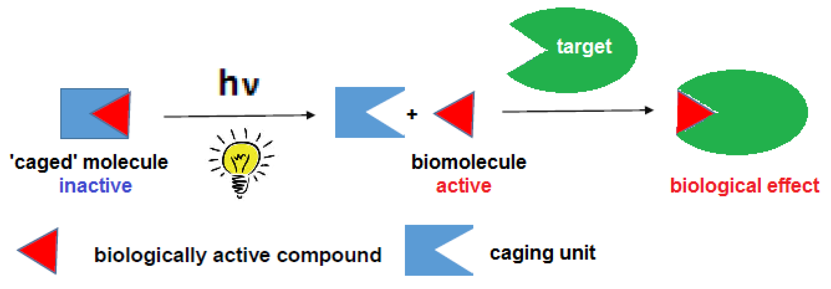
Definition
1. Introduction
2. Design and Applications of PPGs
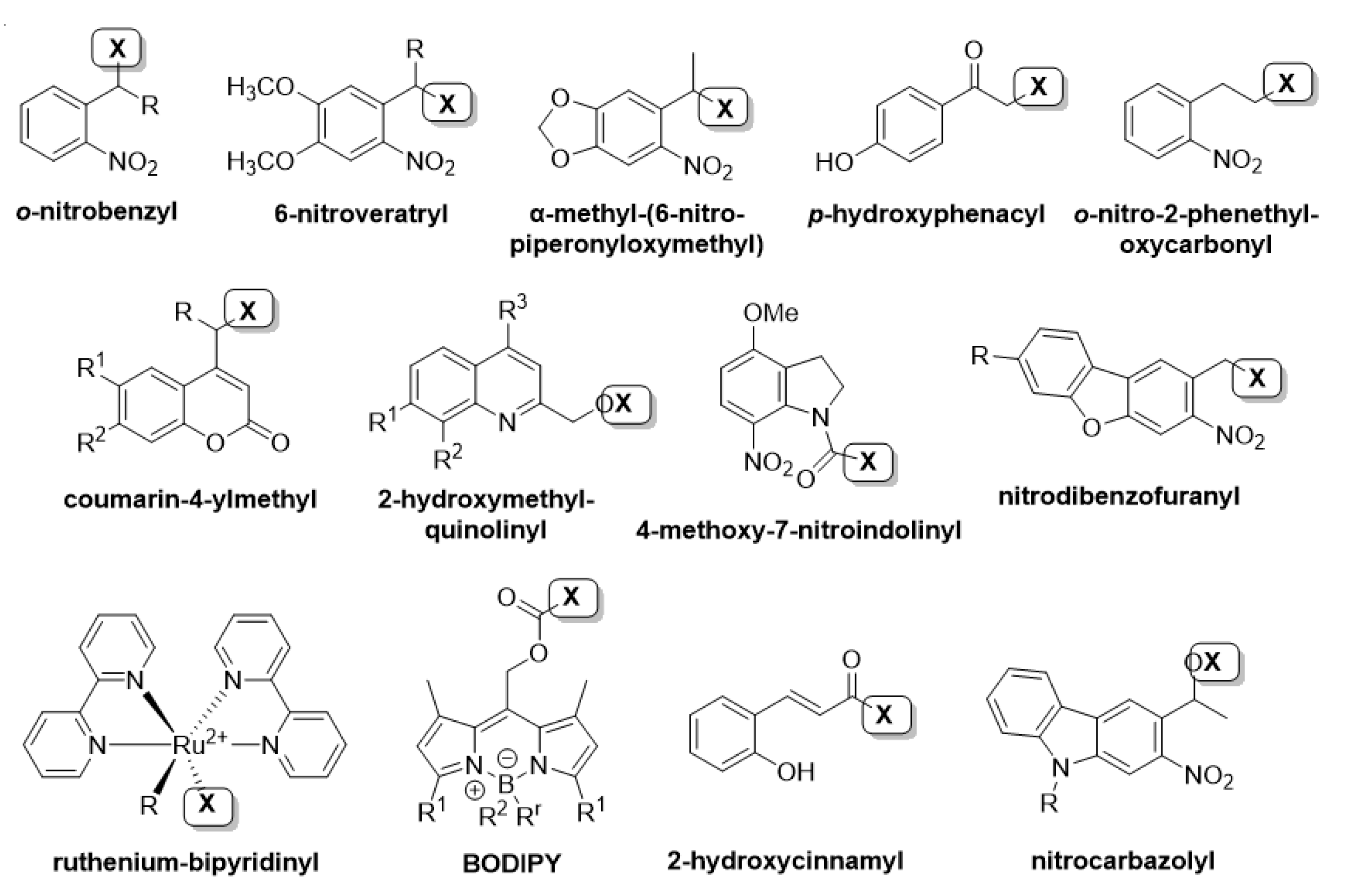
2.1. The Substrate Scope of PPGs
2.2. Preparation of PPG-Substrate Constructs
2.3. Application Criteria for PPGs
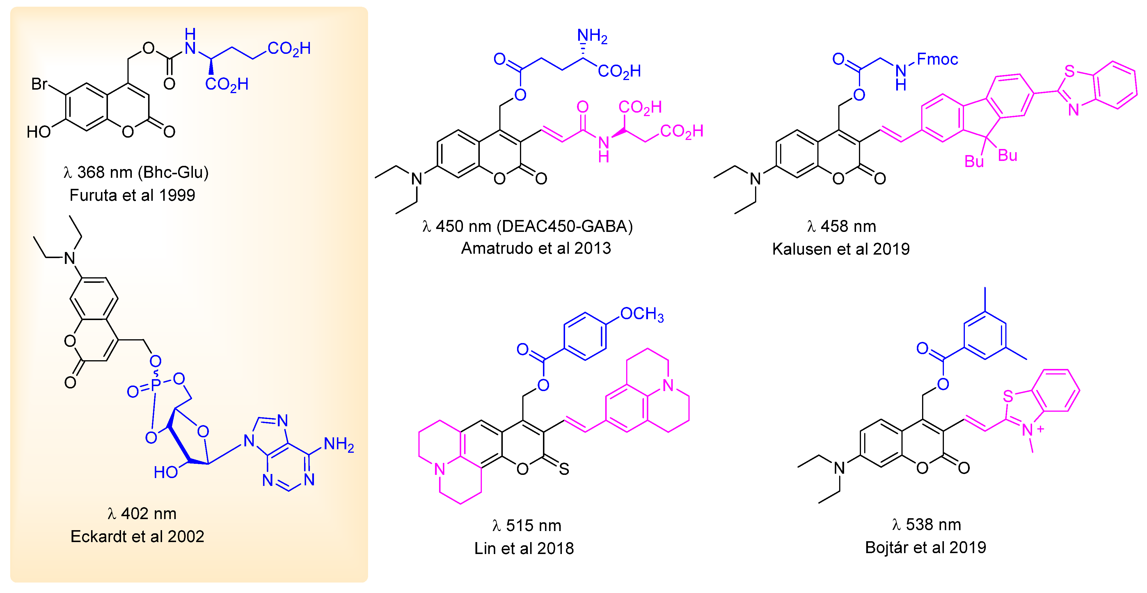
2.4. UV and VIS (One-Photon) Activation of PPGs, Design Aspects
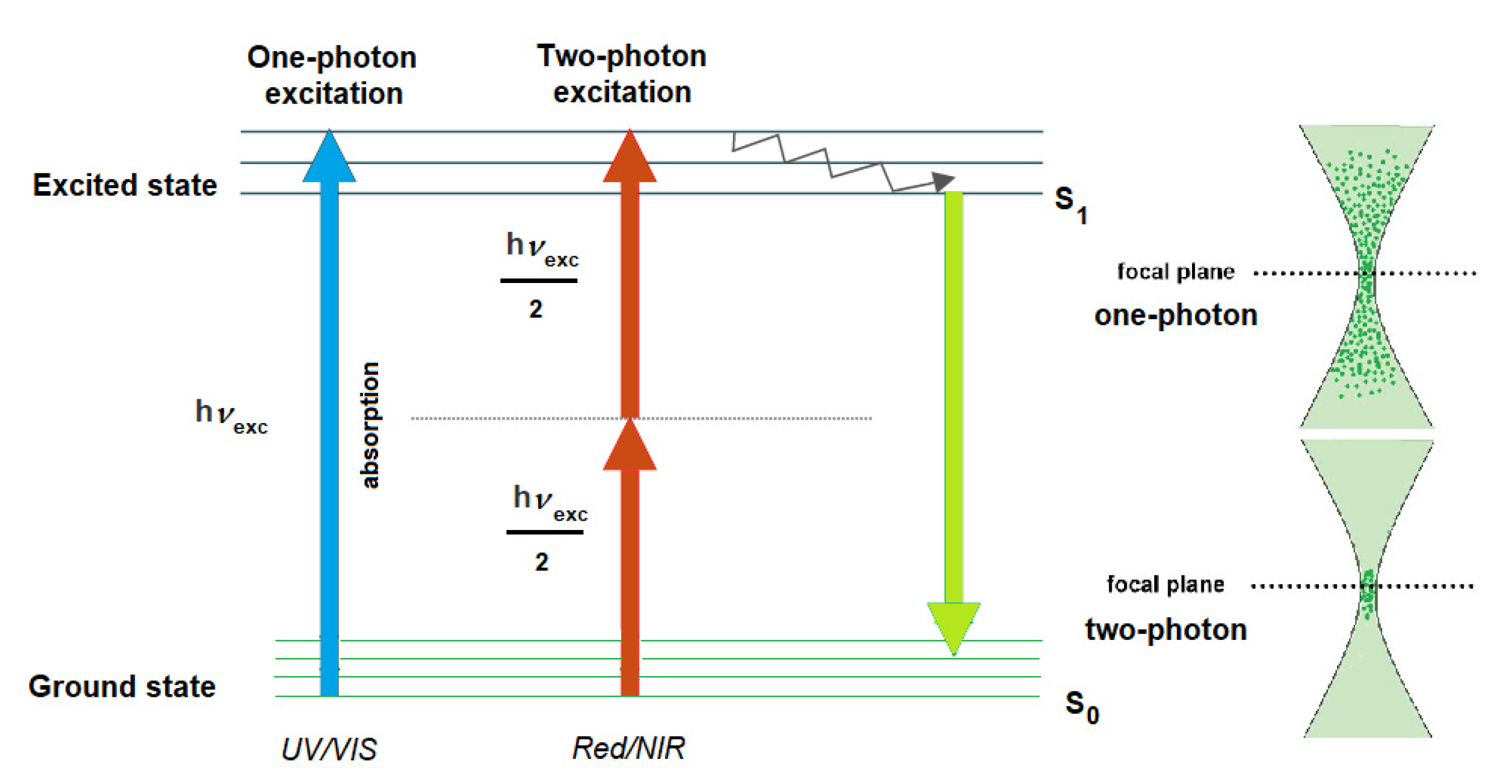
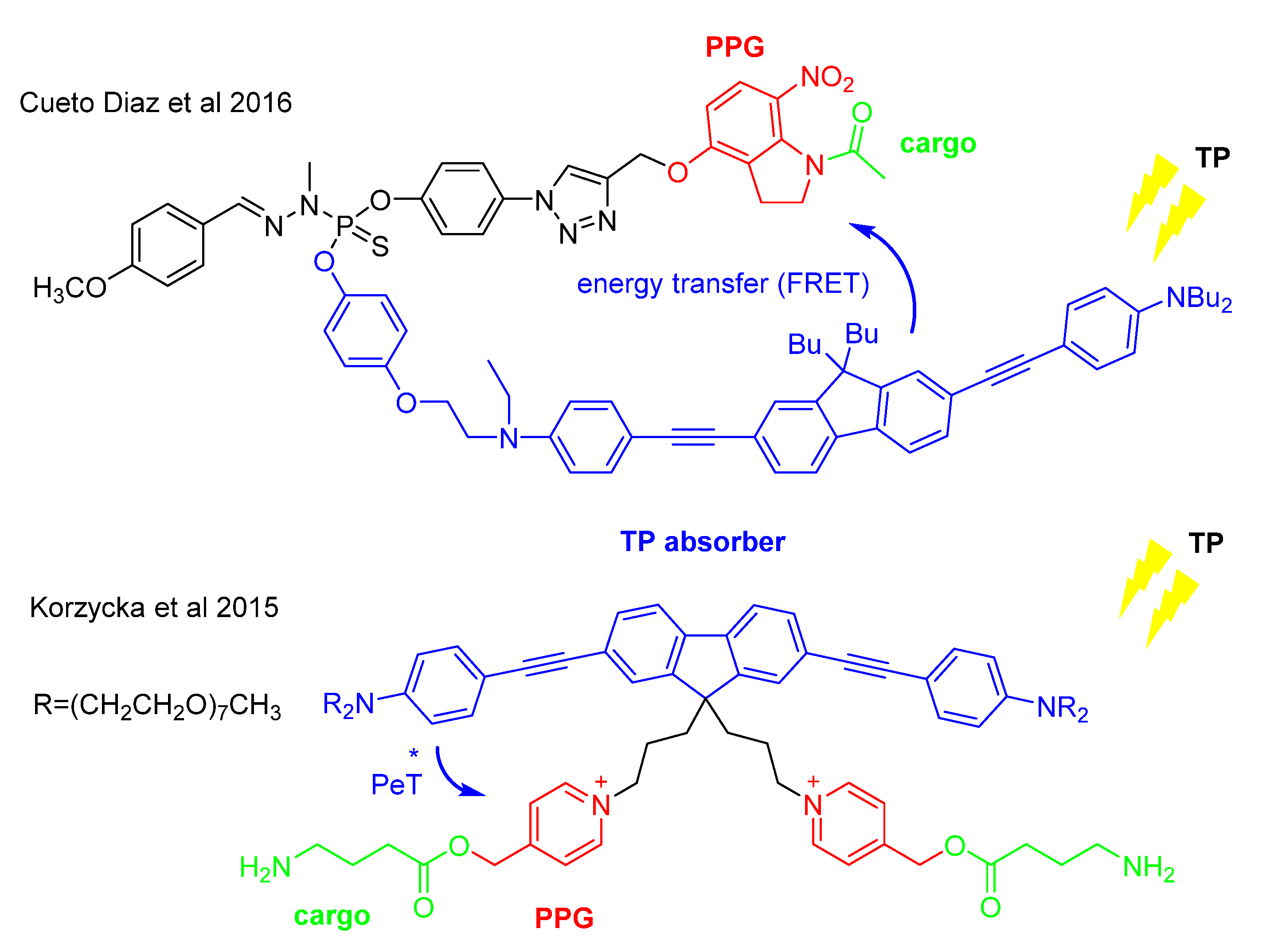
2.5. NIR (Two-Photon) Activation of PPGs, Design Aspects. Characterization of PPGs
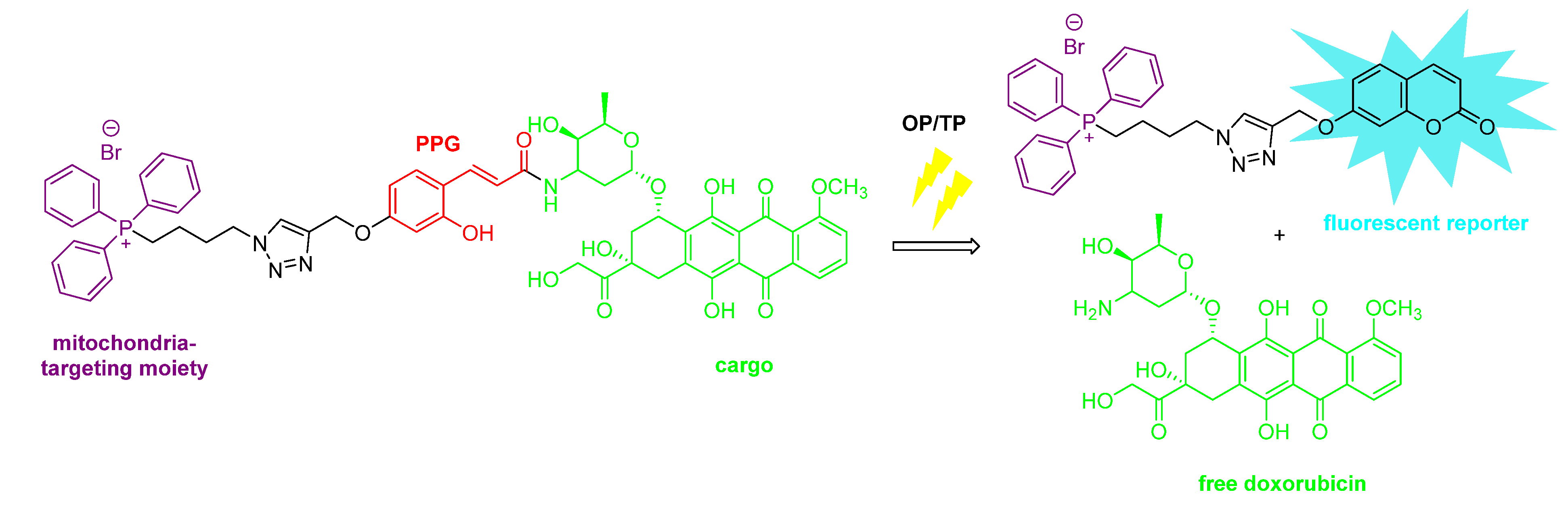
2.6. Selected Applications of PPGs
3. Practical Issues and Future Directions
Funding
Conflicts of Interest
References
- Raza, A.; Rasheed, T.; Nabeel, F.; Hayat, U.; Bilal, M.; Iqbal, H.M.N. Endogenous and Exogenous Stimuli-Responsive Drug Delivery Systems for Programmed Site-Specific Release. Molecules 2019, 24, 1117. [Google Scholar] [CrossRef] [PubMed]
- Monteiro, D.C.F.; Amoah, E.; Rogers, C.; Pearson, A.R. Using photocaging for fast time-resolved structural biology studies. Acta Crystallogr. Sect. D Struct. Biol. 2021, 77, 1218–1232. [Google Scholar] [CrossRef] [PubMed]
- Ellis-Davies, G.C.R. Caged compounds: Photorelease technology for control of cellular chemistry and physiology. Nat. Methods 2007, 4, 619–628. [Google Scholar] [CrossRef] [PubMed]
- Klán, P.; Solomek, T.; Bochet, C.G.; Blanc, A.; Givens, R.; Rubina, M.; Popik, V.; Kostikov, A.; Wirz, J. Photoremovable Protecting Groups in Chemistry and Biology: Reaction Mechanisms and Efficacy. Chem. Rev. 2013, 113, 119–191. [Google Scholar] [CrossRef]
- Szymański, W.; Beierle, J.M.; Kistemaker, H.A.V.; Velema, W.A.; Feringa, B.L. Reversible Photocontrol of Biological Systems by the Incorporation of Molecular Photoswitches. Chem. Rev. 2013, 113, 6114–6178. [Google Scholar] [CrossRef]
- Hüll, K.; Morstein, J.; Trauner, D. In Vivo Photopharmacology. Chem. Rev. 2018, 118, 10710–10747. [Google Scholar] [CrossRef]
- Fuchter, M.J. On the Promise of Photopharmacology Using Photoswitches: A Medicinal Chemist’s Perspective. J. Med. Chem. 2020, 63, 11436–11447. [Google Scholar] [CrossRef]
- Korzycka, K.A.; Bennett, P.M.; Cueto-Diaz, E.J.; Wicks, G.; Drobizhev, M.; Blanchard-Desce, M.; Rebane, A.; Anderson, H.L. Two-photon sensitive protecting groups operating via intramolecular electron transfer: Uncaging of GABA and tryptophan. Chem. Sci. 2015, 6, 2419–2426. [Google Scholar] [CrossRef]
- Piant, S.; Bolze, F.; Specht, A. Two-photon uncaging, from neuroscience to materials. Opt. Mater. Express 2016, 6, 1679. [Google Scholar] [CrossRef]
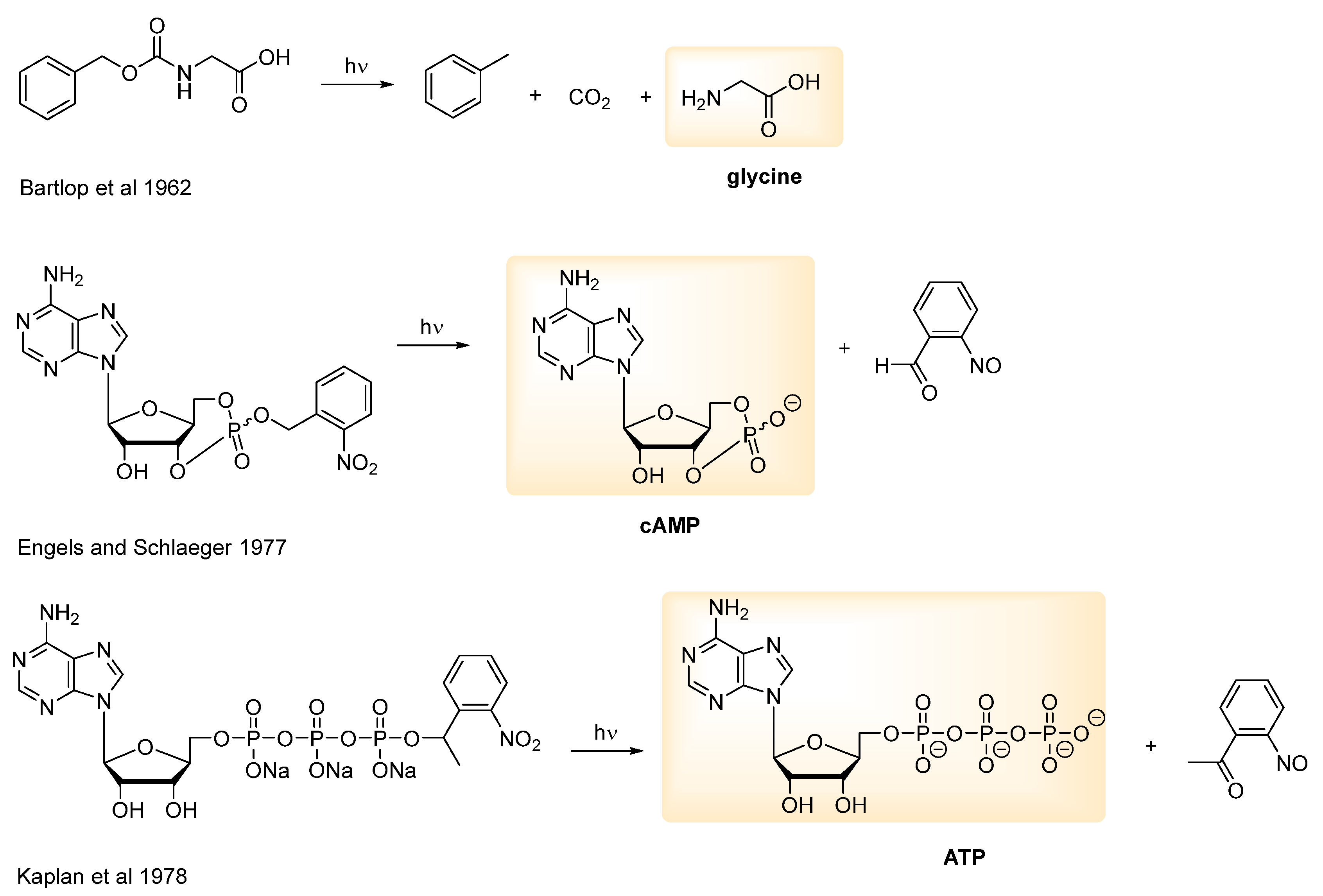
- Engels, J.; Schlaeger, E.J. Synthesis, structure, and reactivity of adenosine cyclic 3′,5′-phosphate-benzyltriesters. J. Med. Chem. 1977, 20, 907–911. [Google Scholar] [CrossRef]
- Kaplan, J.H.; Forbush, I.B.; Hoffman, J.F. Rapid photolytic release of adenosine 5′-triphosphate from a protected analog: Utilization by the sodium:potassium pump of human red blood cell ghosts. Biochemistry 1978, 17, 1929–1935. [Google Scholar] [CrossRef] [PubMed]
- Barltrop, J.; Schofield, P. Photosensitive Protecting Groups. Tetrahedron Lett. 1962, 3, 697–699. [Google Scholar] [CrossRef]
- Hurevich, M.; Samarasimhareddy, M.; Alshanski, I.; Mervinetsky, E. Photodeprotection of up to Eight Photolabile Protecting Groups from a Single Glycan. Synlett 2018, 29, 880–884. [Google Scholar] [CrossRef]
- Kessler, M.; Glatthar, R.; Giese, B.; Bochet, C.G. Sequentially Photocleavable Protecting Groups in Solid-Phase Synthesis. Org. Lett. 2003, 5, 1179–1181. [Google Scholar] [CrossRef]
- Agarwal, H.K.; Janicek, R.; Chi, S.-H.; Perry, J.W.; Niggli, E.; Ellis-Davies, G.C.R. Calcium Uncaging with Visible Light. J. Am. Chem. Soc. 2016, 138, 3687–3693. [Google Scholar] [CrossRef]
- Basa, P.N.; Barr, C.A.; Oakley, K.M.; Liang, X.; Burdette, S.C. Zinc Photocages with Improved Photophysical Properties and Cell Permeability Imparted by Ternary Complex Formation. J. Am. Chem. Soc. 2019, 141, 12100–12108. [Google Scholar] [CrossRef]
- Venkatesh, Y.; Vangala, V.; Mengji, R.; Chaudhuri, A.; Bhattacharya, S.; Datta, P.K.; Banerjee, R.; Jana, A.; Singh, N.D.P. One- and Two-Photon Uncaging of Carbon Monoxide (CO) with Real-Time Monitoring: On-Demand Carbazole-Based Dual CO-Releasing Platform to Test over Single and Combinatorial Approaches for the Efficient Regression of Orthotopic Murine Melanoma In Vivo. J. Med. Chem. 2022, 65, 1822–1834. [Google Scholar] [CrossRef]
- Fraix, A.; Parisi, C.; Seggio, M.; Sortino, S. Nitric Oxide Photoreleasers with Fluorescent Reporting. Chem. Eur. J. 2021, 27, 12714–12725. [Google Scholar] [CrossRef]
- Štacko, P.; Muchová, L.; Vítek, L.; Klán, P. Visible to NIR Light Photoactivation of Hydrogen Sulfide for Biological Targeting. Org. Lett. 2018, 20, 4907–4911. [Google Scholar] [CrossRef]
- Li, W.-H.; Llopis, J.; Whitney, M.A.; Zlokarnik, G.; Tsien, R.Y. Cell-permeant caged InsP3 ester shows that Ca2+ spike frequency can optimize gene expression. Nature 1998, 392, 936–941. [Google Scholar] [CrossRef]
- Ellis-Davies, G.C.R. Useful Caged Compounds for Cell Physiology. Accounts Chem. Res. 2020, 53, 1593–1604. [Google Scholar] [CrossRef]
- Chiovini, B.; Pálfi, D.; Majoros, M.; Juhász, G.; Szalay, G.; Katona, G.; Szőri, M.; Frigyesi, O.; Haveland, L.C.; Szabó, G.; et al. Theoretical Design, Synthesis, and In Vitro Neurobiological Applications of a Highly Efficient Two-Photon Caged GABA Validated on an Epileptic Case. ACS Omega 2021, 6, 15029–15045. [Google Scholar] [CrossRef] [PubMed]
- Agarwal, H.K.; Zhai, S.; Surmeier, D.J.; Ellis-Davies, G.C.R. Intracellular Uncaging of cGMP with Blue Light. ACS Chem. Neurosci. 2017, 8, 2139–2144. [Google Scholar] [CrossRef] [PubMed]
- Mangubat-Medina, A.E.; Ball, Z.T. Triggering biological processes: Methods and applications of photocaged peptides and proteins. Chem. Soc. Rev. 2021, 50, 10403–10421. [Google Scholar] [CrossRef] [PubMed]
- Silva, J.M.; Silva, E.; Reis, R.L. Light-triggered release of photocaged therapeutics—Where are we now? J. Control. Release 2019, 298, 154–176. [Google Scholar] [CrossRef]
- Shchelik, I.S.; Tomio, A.; Gademann, K. Design, Synthesis, and Biological Evaluation of Light-Activated Antibiotics. ACS Infect. Dis. 2021, 7, 681–692. [Google Scholar] [CrossRef]
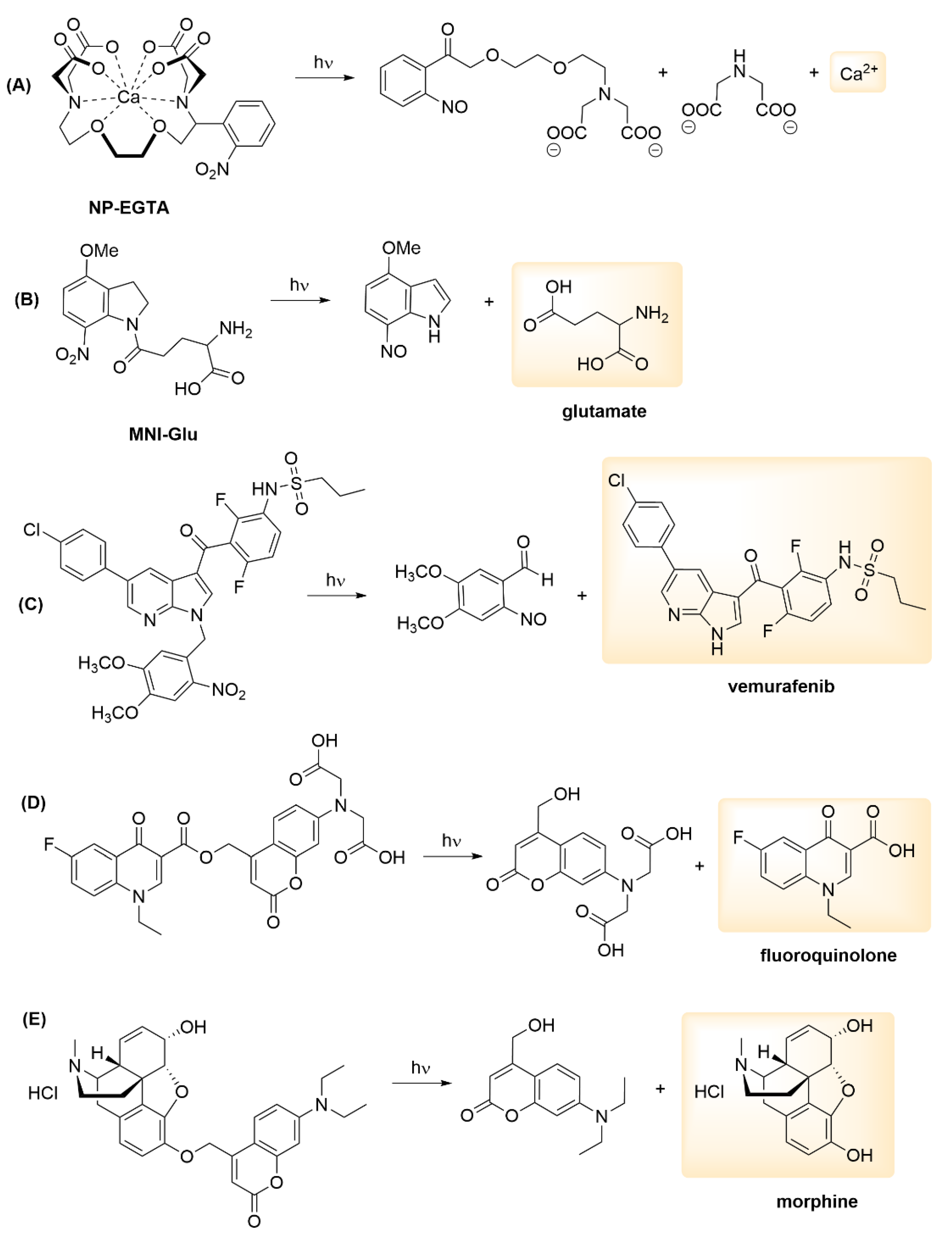
- López-Cano, M.; Font, J.; Aso, E.; Sahlholm, K.; Cabré, G.; Giraldo, J.; De Koninck, Y.; Hernando, J.; Llebaria, A.; Fernández-Dueñas, V.; et al. Remote local photoactivation of morphine produces analgesia without opioid-related adverse effects. Br. J. Pharmacol. 2021. [Google Scholar] [CrossRef]
- Dunkel, P.; Ilaš, J. Targeted Cancer Therapy Using Compounds Activated by Light. Cancers 2021, 13, 3237. [Google Scholar] [CrossRef]
- Wang, X.; Feng, M.; Xiao, L.; Tong, A.; Xiang, Y. Postsynthetic Modification of DNA Phosphodiester Backbone for Photocaged DNAzyme. ACS Chem. Biol. 2015, 11, 444–451. [Google Scholar] [CrossRef]
- Casey, J.P.; Blidner, R.A.; Monroe, W.T. Caged siRNAs for Spatiotemporal Control of Gene Silencing. Mol. Pharm. 2009, 6, 669–685. [Google Scholar] [CrossRef]
- Vaníková, Z.; Hocek, M. Polymerase Synthesis of Photocaged DNA Resistant against Cleavage by Restriction Endonucleases. Angew. Chem. Int. Ed. 2014, 53, 6734–6737. [Google Scholar] [CrossRef] [PubMed]
- Ellis-Davies, G.C.; Kaplan, J.H. Nitrophenyl-EGTA, a photolabile chelator that selectively binds Ca2+ with high affinity and releases it rapidly upon photolysis. Proc. Natl. Acad. Sci. USA 1994, 91, 187–191. [Google Scholar] [CrossRef] [PubMed]
- Horbert, R.; Pinchuk, B.; Davies, P.; Alessi, D.; Peifer, C. Photoactivatable Prodrugs of Antimelanoma Agent Vemurafenib. ACS Chem. Biol. 2015, 10, 2099–2107. [Google Scholar] [CrossRef]
- Velema, W.A.; van der Berg, J.P.; Szymanski, W.; Driessen, A.J.M.; Feringa, B.L. Orthogonal Control of Antibacterial Activity with Light. ACS Chem. Biol. 2014, 9, 1969–1974. [Google Scholar] [CrossRef]
- Ellis-Davies, G.C. A chemist and biologist talk to each other about caged neurotransmitters. Beilstein J. Org. Chem. 2013, 9, 64–73. [Google Scholar] [CrossRef] [PubMed]
- Peterson, J.A.; Wijesooriya, C.; Gehrmann, E.J.; Mahoney, K.M.; Goswami, P.P.; Albright, T.R.; Syed, A.; Dutton, A.S.; Smith, E.A.; Winter, A.H. Family of BODIPY Photocages Cleaved by Single Photons of Visible/Near-Infrared Light. J. Am. Chem. Soc. 2018, 140, 7343–7346. [Google Scholar] [CrossRef] [PubMed]
- Wang, Z.; Martin, S.F. Design, Synthesis and Evaluation of Novel Carbazole-Derived Photocages. Chem. Eur. J. 2022, 28, e202200311. [Google Scholar] [CrossRef]
- Lester, H.A.; Nerbonne, J.M. Physiological and Pharmacological Manipulations with Light Flashes. Annu. Rev. Biophys. Bioeng. 1982, 11, 151–175. [Google Scholar] [CrossRef] [PubMed]
- Pauff, S.M.; Miller, S.C. A Trifluoroacetic Acid-labile Sulfonate Protecting Group and Its Use in the Synthesis of a Near-IR Fluorophore. J. Org. Chem. 2012, 78, 711–716. [Google Scholar] [CrossRef][Green Version]
- McMillan, T.J.; Leatherman, E.; Ridley, A.; Shorrocks, J.; Tobi, S.E.; Whiteside, J.R. Cellular effects of long wavelength UV light (UVA) in mammalian cells. J. Pharm. Pharmacol. 2008, 60, 969–976. [Google Scholar] [CrossRef]
- Cheong, W.-F.; Prahl, S.A.; Welch, A.J. A review of the optical properties of biological tissues. IEEE J. Quantum Electron. 1990, 26, 2166–2185. [Google Scholar] [CrossRef]
- Weissleder, R. A clearer vision for in vivo imaging. Nat. Biotechnol. 2001, 19, 316–317. [Google Scholar] [CrossRef] [PubMed]
- Gorka, A.P.; Nani, R.R.; Zhu, J.; Mackem, S.; Schnermann, M.J. A Near-IR Uncaging Strategy Based on Cyanine Photochemistry. J. Am. Chem. Soc. 2014, 136, 14153–14159. [Google Scholar] [CrossRef] [PubMed]
- Josa-Culleré, L.; Llebaria, A. In the Search for Photocages Cleavable with Visible Light: An Overview of Recent Advances and Chemical Strategies. ChemPhotoChem 2020, 5, 296–314. [Google Scholar] [CrossRef]
- Amatrudo, J.M.; Olson, J.P.; Lur, G.; Chiu, C.Q.; Higley, M.J.; Ellis-Davies, G.C.R. Wavelength-Selective One- and Two-Photon Uncaging of GABA. ACS Chem. Neurosci. 2013, 5, 64–70. [Google Scholar] [CrossRef]
- Klausen, M.; Dubois, V.; Clermont, G.; Tonnelé, C.; Castet, F.; Blanchard-Desce, M. Dual-wavelength efficient two-photon photorelease of glycine by π-extended dipolar coumarins. Chem. Sci. 2019, 10, 4209–4219. [Google Scholar] [CrossRef]
- Lin, Q.; Yang, L.; Wang, Z.; Hua, Y.; Zhang, D.; Bao, B.; Bao, C.; Gong, X.; Zhu, L. Coumarin Photocaging Groups Modified with an Electron-Rich Styryl Moiety at the 3-Position: Long-Wavelength Excitation, Rapid Photolysis, and Photobleaching. Angew. Chem. Int. Ed. 2018, 57, 3722–3726. [Google Scholar] [CrossRef]
- Bojtár, M.; Kormos, A.; Kis-Petik, K.; Kellermayer, M.; Kele, P. Green-Light Activatable, Water-Soluble Red-Shifted Coumarin Photocages. Org. Lett. 2019, 21, 9410–9414. [Google Scholar] [CrossRef]
- Furuta, T.; Wang, S.S.-H.; Dantzker, J.L.; Dore, T.M.; Bybee, W.J.; Callaway, E.M.; Denk, W.; Tsien, R.Y. Brominated 7-hydroxycoumarin-4-ylmethyls: Photolabile protecting groups with biologically useful cross-sections for two photon photolysis. Proc. Natl. Acad. Sci. USA 1999, 96, 1193–1200. [Google Scholar] [CrossRef]
- Eckardt, T.; Hagen, V.; Schade, B.; Schmidt, R.; Schweitzer, C.; Bendig, J. Deactivation Behavior and Excited-State Properties of (Coumarin-4-yl)methyl Derivatives. 2. Photocleavage of Selected (Coumarin-4-yl)methyl-Caged Adenosine Cyclic 3‘,5‘-Monophosphates with Fluorescence Enhancement. J. Org. Chem. 2002, 67, 703–710. [Google Scholar] [CrossRef]
- Klausen, M.; Blanchard-Desce, M. Two-photon uncaging of bioactive compounds: Starter guide to an efficient IR light switch. J. Photochem. Photobiol. C Photochem. Rev. 2021, 48, 100423. [Google Scholar] [CrossRef]
- Specht, A.; Bolze, F.; Nicoud, J.F.; Goeldner, M. Characterization of One- and Two-Photon Photochemical Uncaging Efficiency. Methods Mol. Biol. 2013, 995, 79–87. [Google Scholar] [CrossRef] [PubMed]
- Bort, G.; Gallavardin, T.; Ogden, D.; Dalko, P.I. From One-Photon to Two-Photon Probes: “Caged” Compounds, Actuators, and Photoswitches. Angew. Chem. Int. Ed. 2013, 52, 4526–4537. [Google Scholar] [CrossRef]
- Klausen, M.; Dubois, V.; Verlhac, J.; Blanchard-Desce, M. Tandem Systems for Two-Photon Uncaging of Bioactive Molecules. ChemPlusChem 2019, 84, 589–598. [Google Scholar] [CrossRef] [PubMed]
- Diaz, E.J.C.; Picard, S.; Klausen, M.; Hugues, V.; Pagano, P.; Genin, E.; Blanchard-Desce, M. Cooperative Veratryle and Nitroindoline Cages for Two-Photon Uncaging in the NIR. Chem. Eur. J. 2016, 22, 10848–10859. [Google Scholar] [CrossRef] [PubMed]
- Dcona, M.; Mitra, D.; Goehe, R.W.; Gewirtz, D.A.; Lebman, D.A.; Hartman, M.C.T. Photocaged permeability: A new strategy for controlled drug release. Chem. Commun. 2012, 48, 4755–4757. [Google Scholar] [CrossRef]
- Liu, P.; Li, B.; Zhan, C.; Zeng, F.; Wu, S. A two-photon-activated prodrug for therapy and drug release monitoring. J. Mater. Chem. B 2017, 5, 7538–7546. [Google Scholar] [CrossRef]
- Singh, N.; Gupta, A.; Prasad, P.; Sah, R.K.; Singh, A.; Kumar, S.; Singh, S.; Gupta, S.; Sasmal, P.K. Mitochondria-Targeted Photoactivatable Real-Time Monitoring of a Controlled Drug Delivery Platform. J. Med. Chem. 2021, 64, 17813–17823. [Google Scholar] [CrossRef]
- Bochet, C.G. Two Decades of Chromatic Orthogonality. Isr. J. Chem. 2021, 61, 486–495. [Google Scholar] [CrossRef]
- Hansen, M.J.; Velema, W.A.; Lerch, M.M.; Szymanski, W.; Feringa, B.L. Wavelength-selective cleavage of photoprotecting groups: Strategies and applications in dynamic systems. Chem. Soc. Rev. 2015, 44, 3358–3377. [Google Scholar] [CrossRef]
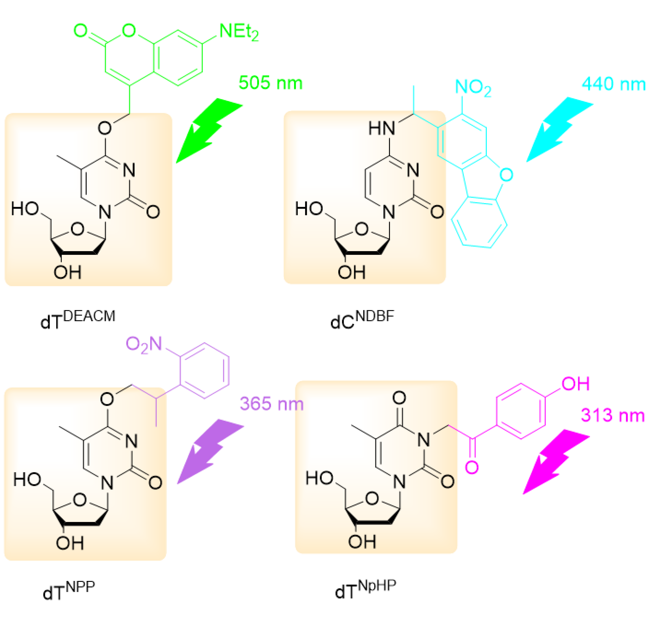
- Rodrigues-Correia, A.; Weyel, X.M.M.; Heckel, A. Four Levels of Wavelength-Selective Uncaging for Oligonucleotides. Org. Lett. 2013, 15, 5500–5503. [Google Scholar] [CrossRef] [PubMed]
- Liu, J.; Kang, W.; Wang, W. Photocleavage-based Photoresponsive Drug Delivery. Photochem. Photobiol. 2021, 98, 288–302. [Google Scholar] [CrossRef] [PubMed]
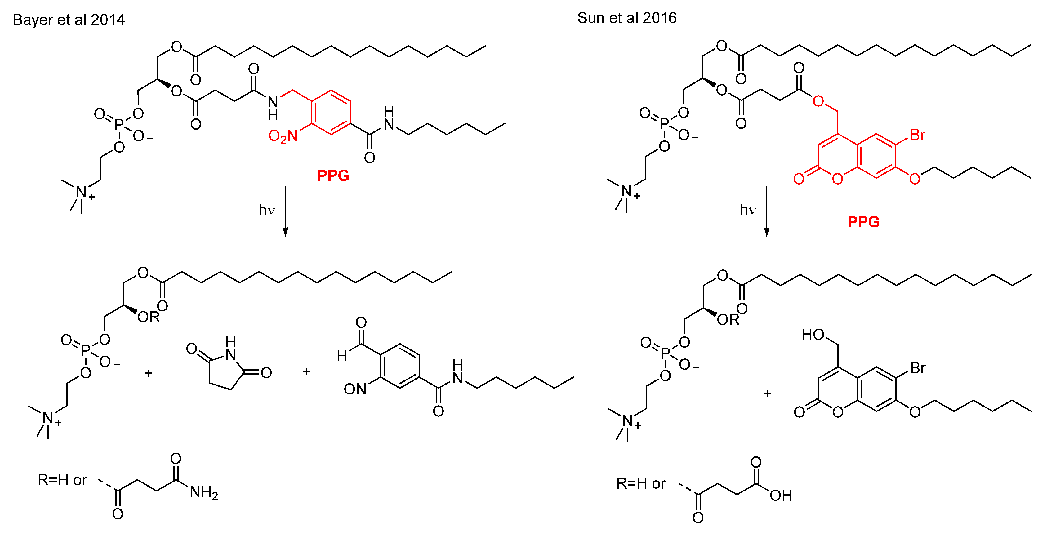
- Bayer, A.M.; Alam, S.; Mattern-Schain, S.I.; Best, M.D. Triggered Liposomal Release through a Synthetic Phosphatidylcholine Analogue Bearing a Photocleavable Moiety Embedded within thesn-2 Acyl Chain. Chem. Eur. J. 2014, 20, 3350–3357. [Google Scholar] [CrossRef]
- Sun, Y.; Ji, Y.; Yu, H.; Wang, D.; Cao, M.; Wang, J. Near-infrared light-sensitive liposomes for controlled release. RSC Adv. 2016, 6, 81245–81249. [Google Scholar] [CrossRef]
- Lerch, M.M.; Hansen, M.J.; Van Dam, G.M.; Szymanski, W.; Feringa, B.L. Emerging Targets in Photopharmacology. Angew. Chem. Int. Ed. 2016, 55, 10978–10999. [Google Scholar] [CrossRef]
- Said, S.S.; Campbell, S.; Hoare, T. Externally Addressable Smart Drug Delivery Vehicles: Current Technologies and Future Directions. Chem. Mater. 2019, 31, 4971–4989. [Google Scholar] [CrossRef]
- Yang, Y.; Shao, Q.; Deng, R.; Wang, C.; Teng, X.; Cheng, K.; Cheng, Z.; Huang, L.; Liu, Z.; Liu, X.; et al. In Vitro and In Vivo Uncaging and Bioluminescence Imaging by Using Photocaged Upconversion Nanoparticles. Angew. Chem. Int. Ed. 2012, 51, 3125–3129. [Google Scholar] [CrossRef]
- Ran, C.; Zhang, Z.; Hooker, J.; Moore, A. In Vivo Photoactivation Without “Light”: Use of Cherenkov Radiation to Overcome the Penetration Limit of Light. Mol. Imaging Biol. 2012, 14, 156–162. [Google Scholar] [CrossRef]
- Yang, Y.; Wu, M.; Vázquez-Guardado, A.; Wegener, A.J.; Grajales-Reyes, J.G.; Deng, Y.; Wang, T.; Avila, R.; Moreno, J.A.; Minkowicz, S.; et al. Wireless multilateral devices for optogenetic studies of individual and social behaviors. Nat. Neurosci. 2021, 24, 1035–1045. [Google Scholar] [CrossRef]
- Park, S.I.; Brenner, D.S.; Shin, G.; Morgan, C.D.; Copits, B.A.; Chung, H.U.; Pullen, M.Y.; Noh, K.N.; Davidson, S.; Oh, S.J.; et al. Soft, stretchable, fully implantable miniaturized optoelectronic systems for wireless optogenetics. Nat. Biotechnol. 2015, 33, 1280–1286. [Google Scholar] [CrossRef]
- Yoon, I.; Li, J.Z.; Shim, Y.K. Advance in Photosensitizers and Light Delivery for Photodynamic Therapy. Clin. Endosc. 2013, 46, 7–23. [Google Scholar] [CrossRef] [PubMed]


Publisher’s Note: MDPI stays neutral with regard to jurisdictional claims in published maps and institutional affiliations. |
© 2022 by the author. Licensee MDPI, Basel, Switzerland. This article is an open access article distributed under the terms and conditions of the Creative Commons Attribution (CC BY) license (https://creativecommons.org/licenses/by/4.0/).
Share and Cite
Dunkel, P. Photoremovable Protecting Groups. Encyclopedia 2022, 2, 1225-1236. https://doi.org/10.3390/encyclopedia2030082
Dunkel P. Photoremovable Protecting Groups. Encyclopedia. 2022; 2(3):1225-1236. https://doi.org/10.3390/encyclopedia2030082
Chicago/Turabian StyleDunkel, Petra. 2022. "Photoremovable Protecting Groups" Encyclopedia 2, no. 3: 1225-1236. https://doi.org/10.3390/encyclopedia2030082
APA StyleDunkel, P. (2022). Photoremovable Protecting Groups. Encyclopedia, 2(3), 1225-1236. https://doi.org/10.3390/encyclopedia2030082