Article

Effect of Diazepam Premedication on Acute Kidney Injury Due to Ischemia-Reperfusion in Rats

by
Piotr Wichary
1,
Wojciech Wystrychowski
2,*,
Mirosław Śnietura
3,
Szymon Białka
1,
Hanna Misiołek
1,
Antoni Wystrychowski
4,† and
Grzegorz Wystrychowski
5
1
Department of Anesthesiology and Intensive Care, Faculty of Medical Sciences in Zabrze, Medical University of Silesia in Katowice, 40-055 Katowice, Poland
2
Department of General, Vascular and Transplant Surgery, Faculty of Medical Sciences in Katowice, Medical University of Silesia in Katowice, 40-055 Katowice, Poland
3
Department of Pathomorphology and Molecular Diagnostics, Faculty of Medical Sciences in Katowice, Medical University of Silesia in Katowice, 40-055 Katowice, Poland
4
Department of Nephrology, Transplantation and Internal Medicine, Faculty of Medical Sciences in Katowice, Medical University of Silesia in Katowice, 40-055 Katowice, Poland
5
Department of Nephrology, Regional Specialized Hospital No. 4, 41-902 Bytom, Poland
*
Author to whom correspondence should be addressed.
Deceased author.
Kidney Dial. 2026, 6(2), 30; https://doi.org/10.3390/kidneydial6020030
Submission received: 15 February 2026 / Revised: 29 April 2026 / Accepted: 2 May 2026 / Published: 8 May 2026

Abstract

Background: Ischemia-reperfusion injury (IRI) impairs kidney transplants. Diazepam can reduce IRI through peripheral benzodiazepine receptors. We aimed to evaluate the effect of diazepam premedication on the IRI of the rat kidney. Methods: Fourteen days after unilateral nephrectomy, male Sprague-Dawley rats underwent a 45 min sole kidney ischemia. Sixty minutes prior to ischemia, the animals were randomly assigned to a subcutaneous injection of 0.75 mg diazepam (n = 28) or 0.5 mL 0.9% NaCl (n = 31). Results: After 48 h, serum creatinine of diazepam-administered rats was lower and creatinine clearance was higher than in controls (119.8 ± 73.3 vs. 217.5 ± 105.3 µmol/L, p < 0.01 and 0.14 ± 0.07 vs. 0.08 ± 0.05 mL/min/100 g BM, p < 0.01, respectively). Moreover, the former had lower urinary losses of sodium and potassium (fractional excretions of 1.24 ± 1.39% vs. 2.87 ± 3.66%, p = 0.02 and 111.1 ± 95.7% vs. 199.0 ± 143.3%, p < 0.01, respectively). After 7 days, diazepam-treated rats remained superior vs. controls, regarding serum creatinine (53.7 ± 12.7 vs. 77.6 ± 21.3 µmol/L, p < 0.01), creatinine clearance (0.22 ± 0.08 vs. 0.17 ± 0.06 mL/min/100 g BM, p < 0.01), potassium sparing (50.2 ± 31.7% vs. 73.4 ± 38.7% excretion, p < 0.01), and renal edema (1.92 ± 0.45 vs. 2.30 ± 0.61 g of kidney mass, p < 0.01). Furthermore, their 24 h proteinuria was marginally reduced (4.03 ± 2.62 vs. 5.06 ± 2.74 mg, p = 0.06). Conclusions: Administration of diazepam preceding renal ischemia attenuates subsequent kidney injury in rats. Benzodiazepines may be beneficial prior to kidney transplantation.

1. Introduction

One of the factors contributing to the delayed recovery of transplanted kidney function is ischemia-reperfusion injury (IRI). IRI is characterized by a transient interruption in blood flow to an organ, followed by its subsequent restoration. Clinically, this situation may be encountered when arterial hypotension, hypovolemia, or arterial clamping are reversed, or during the process of organ transplantation. It is important to note that, during this process, there is a possibility of cell death and irreversible tissue damage. Ischemia is associated with tissue edema or necrosis, and thus restoration of blood flow to all parts of the organ may be incomplete. Furthermore, paradoxically, reperfusion and subsequent tissue re-oxygenation have been shown to exacerbate ischemic damage through the generation of reactive oxygen free radicals [1]. Ischemia-reperfusion has also been demonstrated to induce damage to distant organs due to the release of neutrophil-activating factors [2].
Renal ischemia-reperfusion injury (IRI) is currently recognized as a multifactorial process involving mitochondrial dysfunction, oxidative stress, inflammatory signaling, endothelial damage and complement activation. During reperfusion, disruption of the mitochondrial electron-transport chain and succinate accumulation trigger a burst of reactive oxygen species (ROS), including superoxide anions, hydroxyl radicals and hydrogen peroxide. Furthermore, disturbances in the function of the Na+/K+-ATPase, Na+/H+-ATPase and Ca2+-ATPase pumps have been observed [3,4]. Injured cells release damage-associated molecular patterns (DAMPs) that activate the alternative complement pathway. This leads to formation of the membrane attack complex (C5b9) and liberation of anaphylatoxins C3a and C5a, which recruit leukocytes and exacerbate tubular injury. Oxidative stress can further trigger complement activation and signaling through C3a/C3aR, and C5a/C5aR impairs mitochondrial function, sensitizing cells to ROS-induced damage [5,6]. The early inflammatory response in IRI features up-regulation of pro-inflammatory cytokines—TNF alpha, interleukin-1β and interleukin 6 that enhance adhesion molecule expression and recruit neutrophils and monocytes [4]. Experimental models also implicate interleukin-17 family cytokines in amplifying inflammation during renal IRI [7]. This intricate interplay between oxidative stress, complement activation, and cytokine-driven inflammation underpins the pathogenesis of renal IRI and guides the search for targeted therapeutic strategies.
The mechanisms underlying ischemia-reperfusion injury are common to the heart, brain, liver, intestine, and skeletal muscle, which has greatly intensified the search for methods of its prevention. A number of pharmacological treatment strategies have been identified as showing potential, including the modulation of cell survival pathways, the evasion of oxidative damage, the preservation of cell membrane integrity and the improvement in cellular energetic status [8]. The protective effect of caloric dietary restriction on reducing ischemia-reperfusion injury has been demonstrated in rats and is attributed to improvement in mitochondria-related pathways [9]. A low-calorie diet has been demonstrated to attenuate renal IRI in humans; however, its utilization prior to surgery is not recommended. Efforts to utilize a pharmaceutical agent that exhibits a comparable effect to that of starvation failed to offer protection against renal IRI in mice [10]. Renal ischemic conditioning is defined as the induction of brief periods of ischemia and reperfusion of the kidney prior to the actual renal IRI, or the applying of such an intervention in a remote organ before, during, or shortly after renal IRI, or implementing pauses in kidney reperfusion. These approaches have been demonstrated to exert protective effects in experimental models in rats and other animals by some, but not all authors [11,12,13,14,15,16], but none has been introduced into clinical practice. Pharmacological postconditioning, which utilizes pharmaceuticals as an alternative to vascular closure, may be a viable option. Postconditioning with an alpha-2 adrenoceptor agonist has been found to have a beneficial effect in urologic patients undergoing surgeries requiring closure of the blood supply to the kidney, alleviating renal ischemia-reperfusion injury [17]. A promising field has opened with the mesenchymal stem cell, extracellular vesicles, and microRNA-based therapies; however, methodological issues remain to be optimized and clinical verification to be carried out [3,18].
Research has demonstrated that prolonged cold renal ischemia and the utilization of organs from deceased versus living donors are significant risk factors for kidney graft rejection. Conversely, other variables such as organ preservation method, HLA antigen mismatch, age at transplantation, gender, and diabetic status have been shown to have no effect on graft survival [19,20]. Despite extensive and promising research [21], there is currently no standard pre-transplant management to prevent or treat renal graft IRI [3,8].
Perioperative stress may contribute to inflammatory and sympathetic activation [22,23,24], which could plausibly modulate susceptibility to ischemia-reperfusion injury. However, this link remains hypothetical in the context of renal IRI and served as a rationale for the objective of this study: to examine the impact of pre-ischemic anxiolytic premedication with diazepam on the development of acute post-ischemic renal failure in rats.

2. Materials and Methods

The experiment was conducted on male Sprague-Dawley rats, aged approximately four weeks. The animals were obtained from the Experimental Medicine Centre at the Medical University of Silesia in Katowice, Poland. All rats were provided with standard laboratory chow and water ad libitum in a temperature-controlled environment (23 °C) with a 12 h light–dark cycle.

2.1. The Model of IRI-Induced AKI

The rats underwent unilateral right-sided nephrectomy, which was followed by induction of a 45 min long ischemia of the remaining left kidney after 14 days. Both surgical procedures were performed via a flank approach under inhalation anesthesia.
The animals were anesthetized with diethyl ether vapor generated with an open-drop method. Briefly, the animal was placed in a closed jar containing gauze moistened with pure diethyl ether liquid. Within a few minutes, the rat was simultaneously losing consciousness and muscular tone, at which point it was immediately removed from the jar and placed on the surgical table.
Both surgeries involved a small 1 cm long incision into flank integuments and dissection of the renal pedicle. In the case of nephrectomy, the renal pedicle was ligated, and kidney was cut away and removed through the incision. The procedure lasted 1–2 min.
Ischemia of the solitary kidney was induced by means of a vascular clip placed on the renal artery, with the compression intensity 0.3–0.4 N being adapted to the caliber of the vessel (Aesculap AG, Tuttlingen, Germany). This stage took up to 5 min, starting from the skin incision. Following the induction of ischemia, the flank abdominal integuments were temporarily closed using a metal clamp for a period of 45 min, during which the animal was not anesthetized. Subsequently, the procedure for reversing ischemia by clamp removal and skin suturing was performed under short ether anesthesia, as described above.
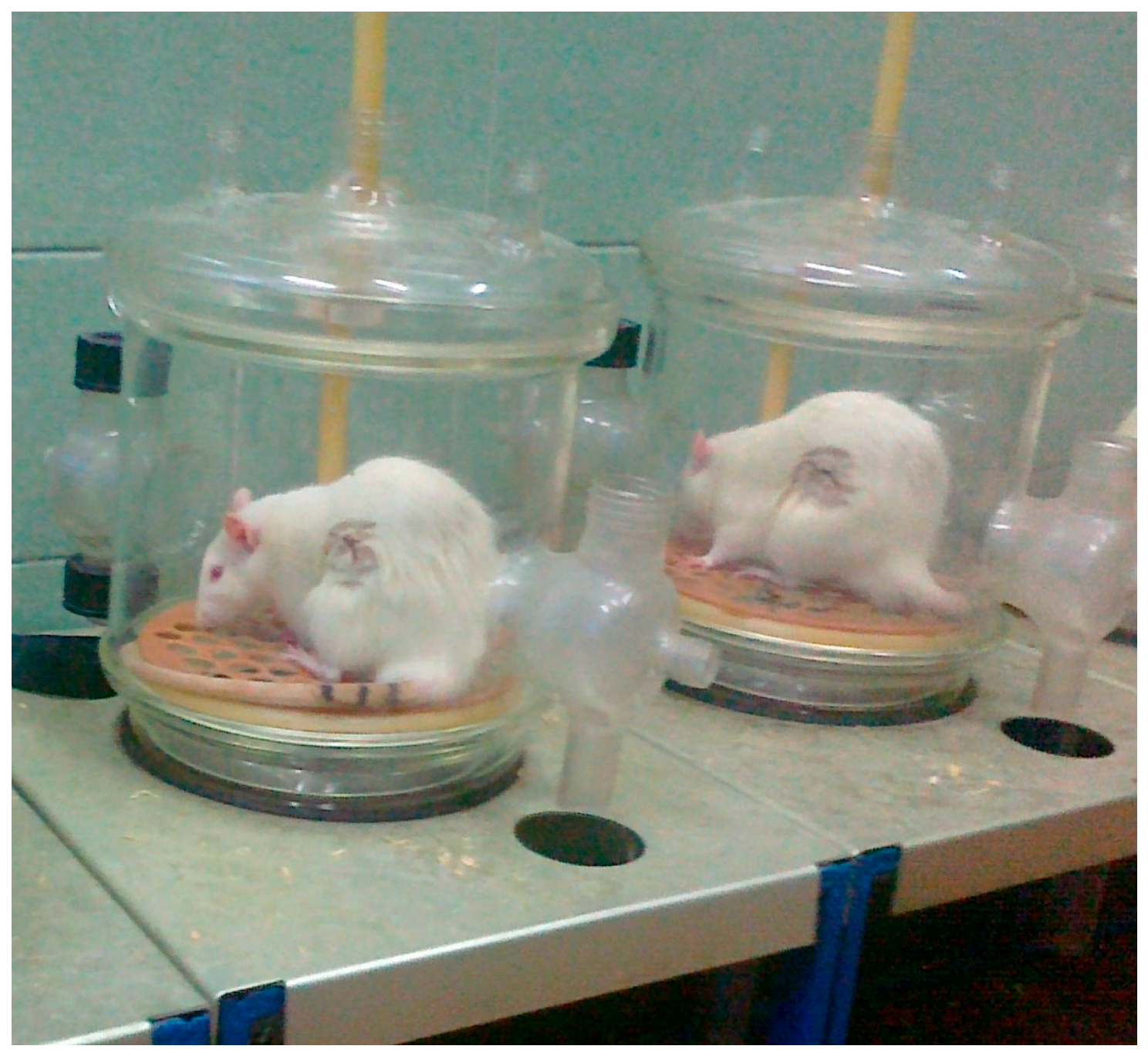
The inflicted wounds healed within several days with no inflammatory or infectious complications. In addition, their dorsolateral position bore no risk of self-inflicted wound damage (Figure 1).

2.2. Experimental Intervention

Diazepam is used in experiments on rats at doses of 0.5–10 mg/kg body mass. Smaller doses have an anxiolytic effect, while higher doses induce sedation [25]. In our study, ~50% of the animals were randomly assigned a single subcutaneous injection of an intermediate dose of 0.75 mg of diazepam (Polfa Warszawa, Warsaw, Poland), equivalent to 2.40 ± 0.23 mg/kg BM. The injection was performed 60 min prior to renal ischemia. Animals in the control group were injected with 0.5 mL of 0.9% sodium chloride solution at the same time. The experiment was carried out concurrently in both groups of rats, in a head-to-head manner, with a small concluding surplus of the control animals in case of possibly worse survival (which did not take place). One operator was involved in the injections, and the other took care of the ischemia-reperfusion procedure, unaware of the applied pretreatment. No adverse events were noted. The humane endpoints included 20% body weight loss, wound infection, and lethargy, but were not met in any studied animal.

2.3. Outcome Measures

The severity of the ischemia-reperfusion injury was assessed on the basis of biochemical parameters of renal function on the second and seventh days following the induction of ischemia. Two days after renal ischemia, the following were measured: rat mass; daily diuresis; serum and urine concentrations of creatinine, sodium, and potassium; and urine protein concentration. Endogenous creatinine clearance, the ratio of urinary protein and creatinine concentrations, and fractional urinary excretions of sodium and potassium were estimated. These parameters were assessed again seven days after renal ischemia, with an additional measurement of the explanted kidney mass after the animal was euthanized. The extracted kidneys were preserved in formalin and used to prepare histology specimens. Prior to blood collection from the retro-orbital plexus, the rats were kept in metabolic cages for 24 h with chow deprivation and continuous urine collection. Serum creatinine level was estimated using a spectrophotometer (PZ Cormay S.A., Łomianki, Poland); plasma and urine electrolyte concentrations were measured using a flame photometer (Eppendorf, Hamburg, Germany); urine creatinine concentration was estimated using a colorimetric picric acid method spectrophotometer (Carl Zeiss, Jena, Germany); and urine protein concentration was assessed with the trichloroacetic acid precipitation method. The degree of ischemia-reperfusion injury sequelae was comparatively assessed in the renal tissue of animals injected with either diazepam or saline, using hematoxylin and eosin (HE), Periodic acid–Schiff (PAS), and Jones stains. The analysis of the histological images was corroborated with the use of artificial intelligence (ChatGPT-5.2.).
All measurements and histological preparatory works were performed without knowledge of the group allocation of the samples.

2.4. Statistical Analysis

Statistical comparisons between the two studied groups were carried out with the t-test or Mann–Whitney U test in case of a non-normal distribution of any given dataset. Normality of the distribution of the variables was assessed with the Shapiro–Wilk W-test. Statistica 14.0.1.25 (TIBCO Software Inc.) was used for the computations.
Group allocations were known at the stage of the outcome interpretations, i.e., statistical analysis and self-conducted histological assessment. On the other hand, the prompts used for the histology analysis by ChatGPT did not reveal any details of the experiment or group identifications, but comprised simple requests to compare histological images of the rat kidney tissue.

3. Results

Twenty-eight rats were administered 0.75 mg diazepam prior to IRI, whereas thirty-one were given saline and thus constituted the control group.

3.1. Kidney Function at 48 h Post-IRI

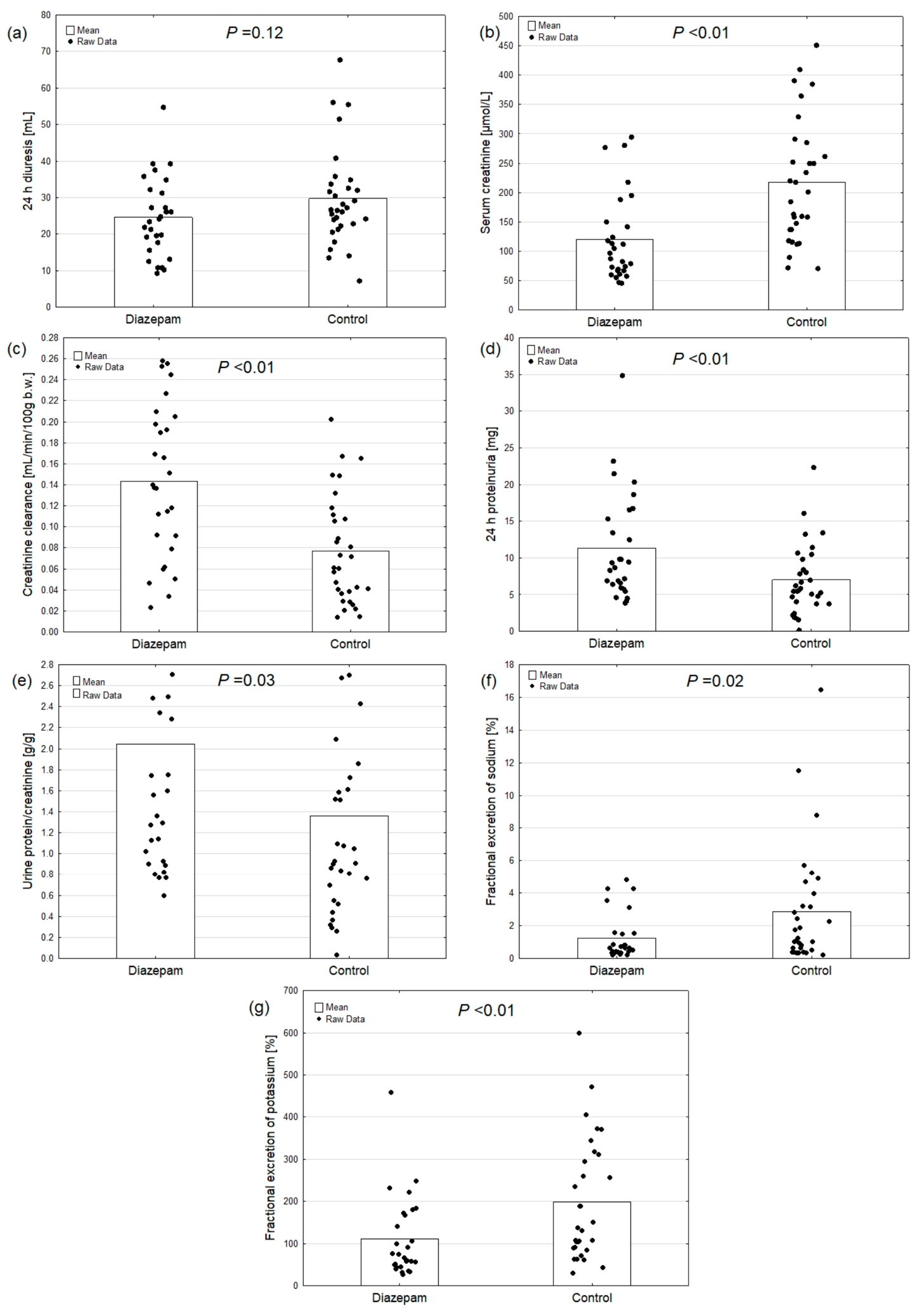
Following a 48 h period of renal IRI, rats that received diazepam demonstrated enhanced preservation of glomerular and tubular kidney function. In comparison with saline-injected controls, the serum creatinine concentration was reduced by 45%, creatinine clearance was elevated by 86%, and urinary losses of sodium and potassium were diminished by 57% and 44%, respectively. Surprisingly, the degree of proteinuria was elevated to a greater extent in animals that were treated with diazepam, reaching approximately 150% of the maximum level observed in the control group (see Table 1 for the means and Figure 2 for the raw data).

3.2. Kidney Function at 7 Days Post-IRI

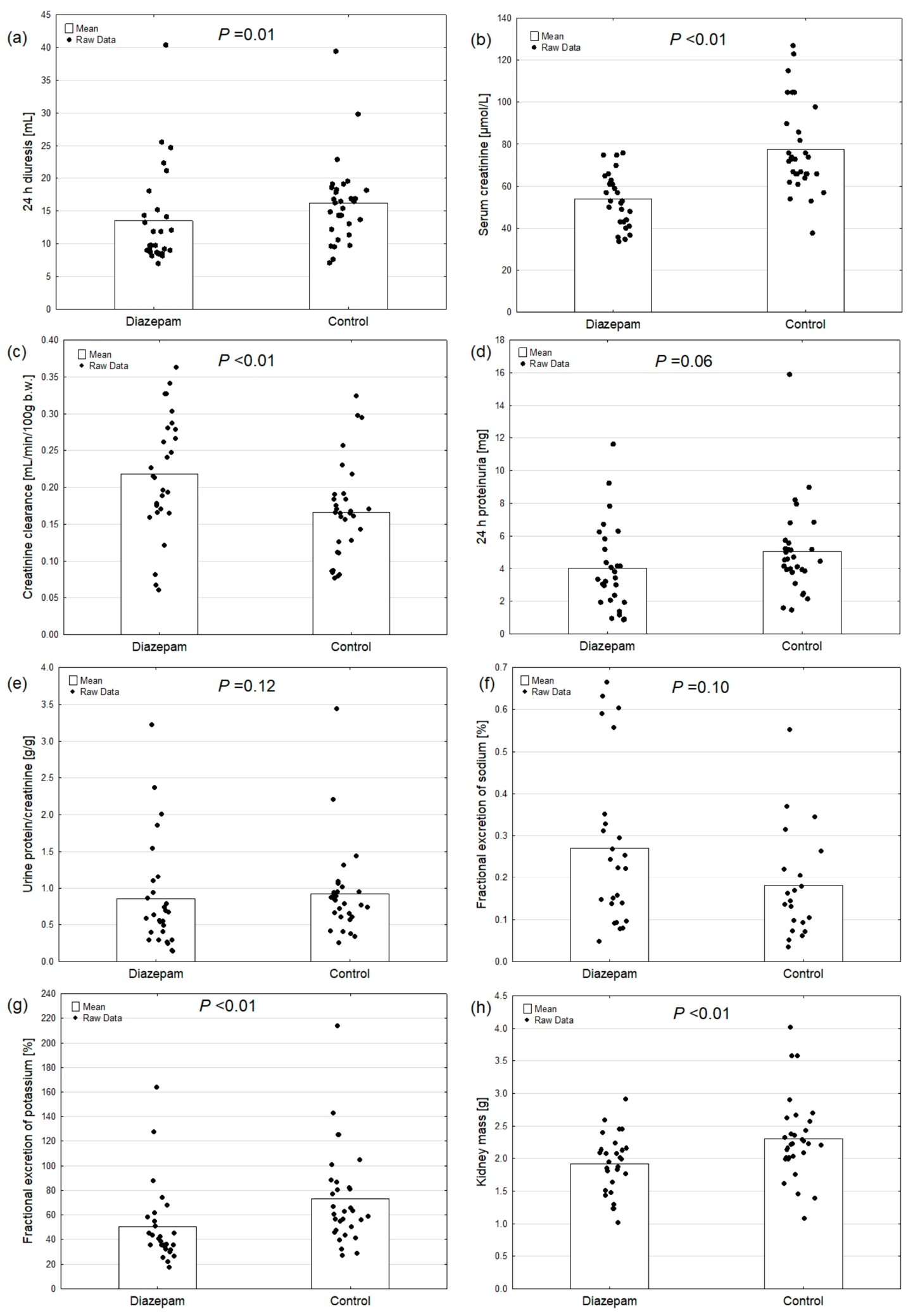
Following a period of renal vasoconstriction and reperfusion, a marked improvement in renal function was evident in both study groups after seven days. Nevertheless, the diazepam-treated rats demonstrated superiority over the control group with regard to serum creatinine (lower by 31%), creatinine clearance (higher by 31%), and potassium sparing (excretion lower by 31%). Moreover, a 16% decrease in kidney mass was observed in the experimental animals in comparison to the control group. Concurrently, proteinuria exhibited a 20% reduction, which approached statistical significance (see Table 2 for the means and Figure 3 for the raw data).

3.3. Kidney Histology at 7 Days Post-IRI

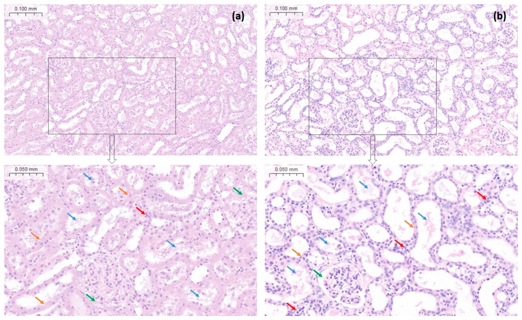
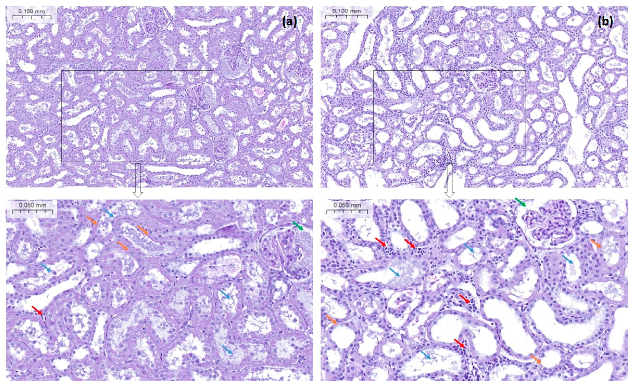
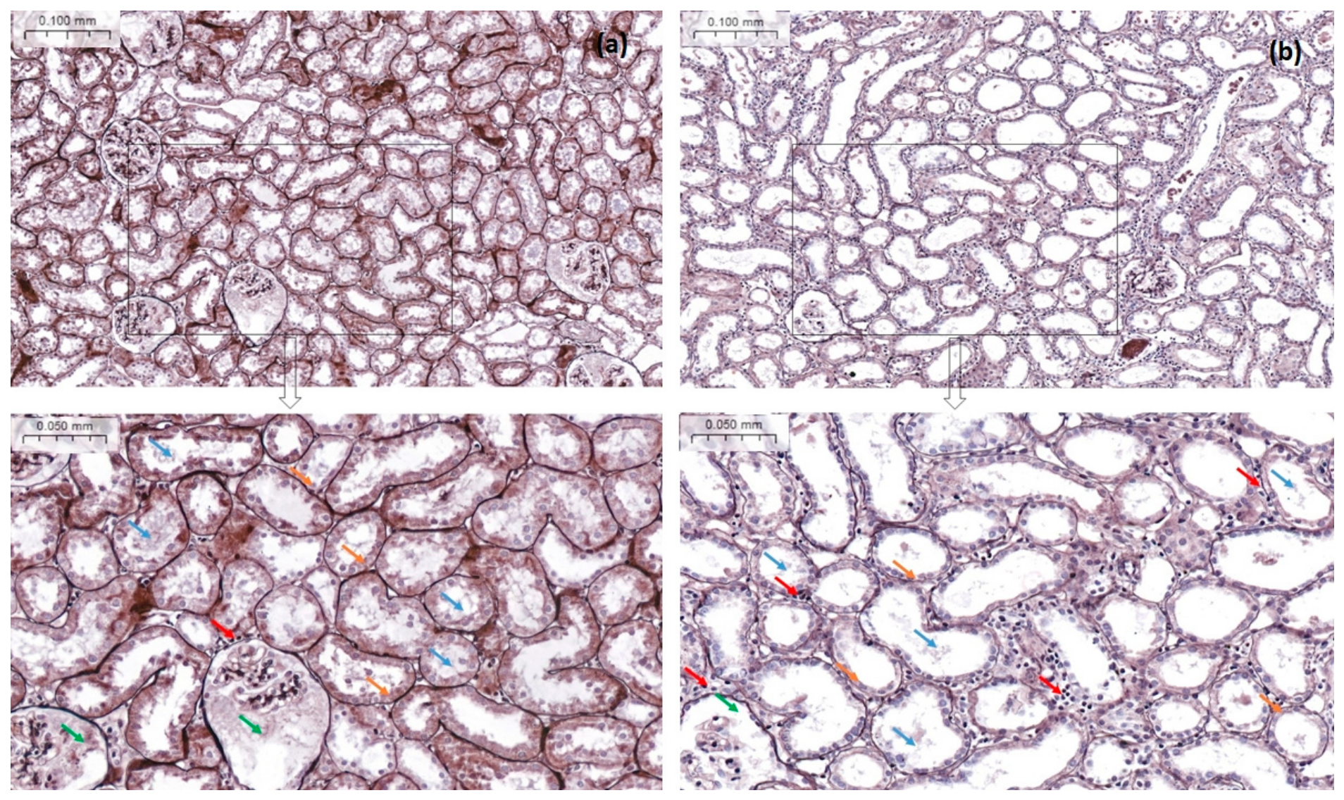
The microscopic analysis of kidney cortex specimens at seven days from ischemia-reperfusion confirmed a noticeable attenuation of the inflicted injury in the diazepam-treated rats. In comparison with the control animals, no significant flattening of the tubular cells or brush border loss were observed. Moreover, the proximal tubules were less dilated, and interstitial inflammatory infiltrates were negligible. Overall, the tubulointerstitial inflammatory process was less advanced and less active in the diazepam group. Furthermore, a slight glomerular inflammation, that was discerned in the controls, was absent in the diazepam-treated rats. In contrast, the glomeruli of rats administered diazepam exhibited a higher prevalence of intracapsular debris compared to those given saline (see Figure 4, Figure 5 and Figure 6). The relevant compartmental injury scores were moderately lower in the diazepam than in the control group (Table 3).

4. Discussion

4.1. Revealed Diazepam Effects

The results of the study demonstrate that the administration of diazepam prior to the induction of renal ischemia-reperfusion injury can attenuate the subsequent development of acute kidney injury. This is evidenced by an enhancement in glomerular filtration and a preservation of tubular electrolyte sparing, along with a reduction in polyuria and kidney edema during the week following IRI. Conversely, renal protein loss appears augmented by diazepam early after IRI, while it diminished in the later phase. In line with these outcomes, kidney histology at seven days from IRI differed substantially between both studied groups. Renal tissue in the diazepam-pretreated animals exhibited milder tubular injury, significantly less interstitial inflammation, and was protected from mild glomerulitis, compared to the control group.
Renal parameters in the interventional and control groups at 48 h from IRI were indicative of severe acute kidney injury. Specifically, the elevated urine potassium loss, which exceeded its glomerular filtration rate, was indicative of substantial tubular necrosis. We also observed that kidney function underwent significant enhancement over time, thereby validating the findings of Basile et al. who demonstrated the restoration of renal function by the seventh day following transient kidney ischemia in rats. However, from the 16th week after renal ischemia, these authors observed permanent impairment of urine concentrating ability, fibrosis of the renal interstitium, and significant proteinuria. Furthermore, a reduction in renal microvessels, particularly in the outer layer of the medulla, was observed [26]. The result of these changes can be the development of chronic renal failure and, in clinical settings, this process can particularly affect the transplanted kidney’s survival. The persistence of kidney function improvement with lower proteinuria in diazepam-treated rats at one week after IRI in our experimental model suggests a long-term nephroprotective effect of the diazepam premedication.
The precise etiology of the initially significantly elevated proteinuria observed in animals treated with diazepam remains to be elucidated. This abnormality could be attributed to excessive tubular protein secretion, which is corroborated by the high and beneficial expression of uromodulin in renal IRI, as demonstrated by El-Achkar et al. [27]. Conversely, in the ensuing phase of IRI in our study, Bowman’s capsules in rats administered diazepam were found to be filled with acellular proteinaceous material, which was discernible until the proximal tubular lumens but was predominantly resorbed from the distal tubules. However, the observation of reduced proteinuria at this stage indicates that the observed glomerular exudate is likely to be necrotic material, rather than protein leak. It is evident that further research is necessary in order to investigate this issue.

4.2. The Possible Mechanism of Diazepam Action

The significant renal protection observed with diazepam in the context of IRI suggests that the mechanism is not exclusively attributable to its anxiolytic/stress-reducing effects on the central nervous system. A predominant pathway of diazepam’s nephroprotective action may be through its interaction with the mitochondrial 18 kDa translocator protein (TSPO), also known as the peripheral benzodiazepine receptor. This protein co-forms a protein complex in the outer mitochondrial membrane of 200–240 kDa, which is known as the mitochondrial permeability transition pore, or mitochondrial megachannel. Its expression is found to be higher in tissues characterized by increased steroidogenesis. In a healthy kidney, the expression level of this protein is low, with the majority being detected in the epithelium of the thick part of the ascending arm of the loop of Henle and in the collecting duct [28]. Overexpression of TSPO has been observed in a variety of diseases, including cancer, brain injury, neurodegeneration, and post-ischemic reperfusion processes [29,30]. In the context of renal IRI, TSPO expression has been observed in the proximal tubule, Bowman’s capsule, the endothelium of damaged vessels, and infiltrating macrophages [29,31,32].

4.3. The Role of TSPO and Its Ligands in IRI

Cellular damage during IRI, in addition to other pathomechanisms, may result from an uncontrolled influx of cholesterol into the mitochondria across TSPO during the reperfusion phase. During periods of ischemia, a decline in cholesterol levels has been observed within both the mitochondrial matrix and mitochondrial membranes. The subsequent increase in cholesterol within the inner mitochondrial membrane during the reperfusion phase promotes its autooxidation into toxic oxysterols, which in turn cause the opening of the mitochondrial megachannel [33].
TSPO ligands have been demonstrated to modulate immune cell function, either enhancing or diminishing it [28,34,35,36], whilst concurrently impeding the generation of oxygen free radicals [37]. In a rat model of acute myocardial ischemia, the TSPO ligand, 4′chlorodiazepam, administered before ischemia, blocked the intramyocardial transport of cholesterol and thus prevented mitochondrial damage during the reperfusion phase [33,38]. This translates into a cardioprotective effect of 4′chlorodiazepam, which reduced cardiomyocyte damage and improved myocardial contractile function in rats after an ischemic incident [39].
The results of studies of TSPO ligands in the models of renal IRI are inconclusive. An increase in TSPO mRNA expression levels was observed following IRI in the rabbit kidney, which was associated with increased local steroidogenesis [40]. It has also been demonstrated that an augmentation in TSPO expression exerts a nephroprotective effect in a porcine renal autograft model by improving the maintenance of mitochondrial integrity. Furthermore, the identical localization of TSPO in human and porcine kidneys was demonstrated, which suggests the translatability of these findings to clinical conditions [41]. In contrast, Kunduzova et al. achieved a nephroprotective effect in a rat kidney IRI model using an irreversible TSPO antagonist (SSR180575). The administration of this compound prior to renal ischemia resulted in a reduction in oxidative stress and apoptosis consequent to reperfusion and accelerated the return of renal function [42].

4.4. Diazepam and IRI

Diazepam has been shown to act as a TSPO agonist, inducing the opening of mitochondrial megachannels [43]. In accordance with our observations made in the renal IRI model, the results of other studies suggest a protective effect of diazepam and its analogs in the IRI of heart and brain [44,45,46]. Jiang et al. observed that the administration of diazepam (2.5 mg/kg and 5 mg/kg BM) for a period of 14 days prior to cardiac ischemia in rats resulted in a positive outcome. Diazepam demonstrated cardioprotective and anti-inflammatory properties, evidenced by a reduction in cardiac expressions of troponin I, C-C chemokine receptor type 2, tumor necrosis factor alpha, interleukins IL-1β and IL-6, as well as Bax and caspase-3. This translated into reduced cardiomyocyte apoptosis [44]. Furthermore, in a study of rat brain IRI, diazepam (10 mg/kg BM) has been shown to exert a protective effect on neurons through a number of complex mechanisms that regulate the synthesis and release of excitatory/inhibitory amino acids (e.g., glutamate, aspartate, MDA, GABA) and free radicals [45]. A novel diazepam derivative, PJM-20, which exhibits a comparable anxiolytic profile, was administered orally to rats at a dose of 4 or 8 mg/kg BM, one hour following brain IRI. This treatment resulted in a significant reduction in neurological deficits, edema formation, and total infarct area. The protective effect on the brain is hypothesized to have occurred via the prevention of Ca2+ accumulation in mitochondria and the reduction in glutamate and aspartate concentrations in the cerebrospinal fluid. The neuroprotective effect was observed even when the drug was administered 8 h after the restoration of blood flow [46].
Our literature search has not revealed any clinical interventional studies of diazepam in IRI.

4.5. Diazepam and Inflammation

Literature reports conflict on the impact of diazepam on immune cell migration and phagocytosis [47,48,49,50,51,52]. Covelli et al. demonstrated that diazepam inhibited the ability of human multinucleated cells and monocytes to phagocytose and kill pathogens, and this effect was dose-dependent [47]. Other researchers showed that both diazepam and a ligand of the peripheral benzodiazepine receptor inhibit the chemotaxis of human neutrophils and their production of superoxides [50]. A reduction in peritoneal macrophage phagocytosis was also demonstrated after oral administration of diazepam for 4 days in chickens [48]. In mouse models of Ehrlich tumors, the administration of diazepam over a period of one week, at a dose of no less than 3 mg/kg, resulted in an augmentation of tumor size. Concurrently, this treatment led to a decline in the infiltration of macrophages into the tumor, as well as reduction in their capacity to synthesize nitric oxide [52]. Marino et al. presented contradictory results in their study of neutrophils collected from healthy blood donors. They demonstrated that diazepam stimulates both migration and phagocytosis of these cells through the activation of the peripheral benzodiazepine receptor. The induction of phagocytosis was found to be attributable to an increase in the cytosolic calcium concentration [49]. Interestingly, it has been established by other researchers that rat neutrophil phagocytosis is increased by acute treatment with diazepam, but is decreased by long-term treatment [53]. A recent study has revealed that diazepam can inhibit LPS-induced pyroptosis and subsequent inflammation in a murine model of pulmonary fibrosis by upregulating the let-7a-5p microRNA [54].

4.6. Diazepam and AKI

Diazepam has not been thoroughly studied with regard to its possible renal effects. Of note, Zhang et al. have recently reported an association between the use of diazepam and the risk of acute kidney injury in hospitalized children in a retrospective cross-sectional analysis. However, this finding is undermined by not taking into account diazepam dosing and limiting the adjustment for clinical context just to co-morbidities. Furthermore, the revealed association was primarily with benign (stage I) AKI, which weakens its clinical importance [55]. Another study that implicated a negative impact of diazepam on the kidneys was conducted by Setiawan et al. in rats. Still, the dosing of diazepam was very large (62.25–124.5 mg/kg BM orally) and long (daily for 28 days), making the study more toxicology- and CKD- than AKI-relevant. Moreover, beside a limited kidney histological scoring, the conclusion was arguably based on the increased urine concentrations of creatinine and urea after diazepam administration, with no blood measurements of these markers [56].

4.7. AKI Study Model Particularities

Our model of a sole kidney IRI preceded by a contralateral nephrectomy 2 weeks earlier was developed to achieve an AKI setting with two short and minimally invasive procedures. This provided an excellent survival of the animals. In addition, the interval between the two procedures enabled an adaptation of the animal to the reduced renal tissue and minimized the known bias of an increased blood flow through the remaining kidney in the post-nephrectomy phase [57].
The relatively large sample size (fifty-nine animals in total) was based on our previous studies in the AKI model with other compounds and the expected mild effect of diazepam. Statistical estimation was not employed as it seemed problematic, given the unknown action of diazepam and multiple outcome measures.
The applied 45 min long ischemia of the rat kidney constitutes a model of moderate AKI. As opposed to a ≥60 min long ischemia, the survival of animals is preserved, and there occurs a gradual and significant restoration of the renal tissue functions after the reperfusion [58,59]. The 45 min long ischemia of the sole kidney was used by other groups of researchers alike [60,61,62].
The intermediate dose of diazepam used in the study provided an anxiolytic effect, which more relevantly than a higher sedative dose resembled the concept of the study (stress alleviation) and the current clinical settings of diazepam use in the peri-transplant period (most commonly given as a tranquilizer prior to surgery). Furthermore, a higher dose would bear an increased risk of respiratory depression due to synergism with anesthesia.
For perioperative anesthesia, both in this and our previous experiments, we used diethyl ether vapor generated with an open-drop method. Our experience with this classical anesthetic with regard to animal survival was superior to intraperitoneal ketamine + xylazine, which is in line with other researchers [63]. Of note, the experimental phase of this study was designed and executed when the experimental use of ether was more prevalent. The American Veterinary Medical Association recommendations issued in 2007, which were in operation at that time, accepted ether under the condition of necessary caution in its handling [64]. Furthermore, contrary to the main argument against ether, we did not observe overt signs of respiratory tract irritation in our animals.
An unwanted influence of anesthesia on the course of study can never be ruled out, regardless of the applied method. However, unlike methoxyflurane or sevoflurane, diethyl ether has not been reported to affect renal function directly [65,66]. In addition, its interaction with benzodiazepines was found to concern primarily central nervous systems with a higher risk of respiratory depression, similar to other inhaled anesthetics [67]. Of note, diethyl ether does not affect the activity of liver cytochrome p450 in rats [68], which takes part in the diazepam catabolism.

4.8. Study Strengths and Implications

The assets of our study include: novelty of the demonstrated nephroprotective effect of diazepam, its relevance based on relatively large groups of studied rats, and potential for a significant clinical impact if confirmed in human subjects.
It is evident from the findings of this study that diazepam has a role to play in the management of renal IRI in clinical settings where its occurrence is predictable. This indicates that kidney transplantation or major surgeries should be considered as the most suitable for future clinical studies and applications.
It will be interesting to test whether premedication with diazepam is beneficial if used in the kidney graft donor or recipient, or both. It is imperative to conduct further research to ascertain whether the nephroprotective efficacy of diazepam is contingent upon the dosage or the time of its administration with respect to the induction of IRI.
Our methodology limits the assumptions of clinical benefits of diazepam to anxiolytic doses, i.e., 1–5 mg, depending on age, body mass, and clinical profile of the patient. However, it can be deduced that higher doses may not necessarily be more beneficial, but, on the contrary, harmful. Diazepam exerts a small systemic vasodilatory effect with slight blood pressure decrease [69], which could become clinically significant with regard to the perfusion of the renal graft with a higher dosing of the drug.

4.9. Study Limitations

The study involved only male rats, which is in line with the vast majority of research with use of the rat models of kidney injury. Only a few early studies included both sexes and showed that the female rat kidney is not as prone to damage from ischemia-reperfusion as the male one. This is owing to female sex hormones [70], likely alleviating the sympathetic nervous system activation [71]. Consequently, detecting effects of an intervention in female rats would require larger numbers of studied animals, and would be questionable on both bioethical and budgetary grounds in a pilot study, like this one.
Regrettably, the present study lacks insight into the pathophysiological mechanisms of the revealed clinical and histological effects. However, the results demonstrate that the nephroprotective actions of diazepam occur both at the glomerular level (e.g., maintenance of filtration, no inflammatory infiltrates, and reduced proteinuria in the long-term) and the tubulointerstitial compartment (e.g., reduced inflammatory infiltrates, tubular regeneration, water and electrolyte sparing, attenuated kidney edema). Interaction of the drug with TSPO appears to be a plausible mechanism, given the mentioned widespread TSPO expression across renal tissue undergoing IRI [29,31,32]. Owing to the achieved enhancement of mitochondrial function and cellular energetic balance, renal tissue would be able to maintain a greater proportion of its structural and functional integrity during the period of ischemia, and would be better equipped to manage oxidative stress during reperfusion. Furthermore, it can be deduced that studying any of the multiple elements of the inflammatory reaction, complement activation, or oxidative stress would show some diazepam-induced alleviation. However, to discern which of the pathways is more or less affected would require an excessively broad and comprehensive spectrum of investigation that exceeds the scope of a preliminary study.
On the side of the shortcomings, neither had we capacity to analyze systemic or renal hemodynamics. Nevertheless, we believe that the high numbers of tested animals reduced the possibility of a significant bias associated with a hypothetical variability in the inflicted renal ischemia and reperfusion. With the revealed beneficial action of diazepam, it is also unlikely that any significant systemic blood pressure decrease took place in the diazepam-treated animals.

5. Conclusions

In conclusion, the present study demonstrates that the administration of an anxiolytic dose of diazepam to rats prior to renal ischemia attenuates the subsequent ischemia-reperfusion injury of the kidney. The tubulointerstitial inflammatory and degenerative morphological sequelae are alleviated and glomerular inflammation is prevented. This translates into almost twice-higher glomerular filtration and twice-lower tubular electrolyte losses in the early phase, and similar improvements by roughly 1/3 in the later phase post-injury. The latter ameliorations align with the 15–20% reductions in proteinuria and kidney edema.
Premedication with benzodiazepine derivatives may be of benefit in situations of anticipated renal ischemia, such as in renal transplantation. This area calls for further experimental and clinical studies.

Author Contributions

Conceptualization, A.W., W.W. and G.W.; methodology, A.W., W.W. and P.W.; software, G.W.; formal analysis, G.W. and M.Ś.; investigation, A.W., W.W. and P.W.; resources, S.B. and H.M.; data curation, M.Ś., S.B. and H.M.; writing—original draft preparation, P.W.; writing—review and editing, G.W.; visualization, G.W., W.W. and M.Ś.; supervision, A.W. and H.M.; project administration, A.W. and G.W.; funding acquisition, A.W. Author A.W. passed away prior to the publication of this manuscript. All other authors have read and agreed to the published version of the manuscript.

Funding

This research received no external funding.

Institutional Review Board Statement

This animal study protocol was approved by the Local Ethical Committee for Animal Experiments at the Medical University of Silesia in Katowice, Poland—Consent No. KNW-002/LKE-1-15/2011, 12 January 2011.

Informed Consent Statement

Not applicable.

Data Availability Statement

The original contributions presented in this study are included in the article. Further inquiries can be directed to the corresponding author.

Acknowledgments

We are grateful to Ewa Obuchowicz and Andrzej Więcek for providing the laboratory facilities essential for executing this research. During the preparation of this manuscript, the authors used ChatGPT-5.2 for the purposes of the analysis of the histological images. The authors have reviewed and edited the output and take full responsibility for the content of this publication.

Conflicts of Interest

The authors declare no conflicts of interest.

Abbreviations

The following abbreviations are used in this manuscript:
IRIIschemia-reperfusion injury
HLAHuman leukocyte antigen
HEHematoxylin and eosin
PASPeriodic acid–Schiff
TSPO18kDa translocator protein
AMPKAMP-activated protein kinase

References

  1. Boratyńska, M.; Kamińska, D.; Mazanowska, O. Pathophysiology of ischemia-reperfusion injury in renal transplantation. Postep. Hig. Med. Dosw. 2004, 58, 1–8. [Google Scholar]
  2. Fuller, B.J. Ischaemia/Reperfusion Injury and Inflammation. Transplantation 2000, 69, 327–328. [Google Scholar] [CrossRef]
  3. Huang, A.J.; Sharma, G.K.; Parikh, R.; Jin, Z.; Darras, F.S.; Bergese, S.D. Molecular Mechanisms and Potential Therapeutic Targets of Ischemia-Reperfusion Injury in Kidney Transplantation. Curr. Issues Mol. Biol. 2025, 47, 282. [Google Scholar] [CrossRef]
  4. Nørgård, M.Ø.; Svenningsen, P. Acute Kidney Injury by Ischemia/Reperfusion and Extracellular Vesicles. Int. J. Mol. Sci. 2023, 24, 15312. [Google Scholar] [CrossRef]
  5. Peng, Q.; Li, K.; Smyth, L.A.; Xing, G.; Wang, N.; Meader, L.; Lu, B.; Sacks, S.H.; Zhou, W. C3a and C5a Promote Renal Ischemia-Reperfusion Injury. JASN J. Am. Soc. Nephrol. 2012, 23, 1474–1485. [Google Scholar] [CrossRef]
  6. Troise, D.; Allegra, C.; Cirolla, L.A.; Mercuri, S.; Infante, B.; Castellano, G.; Stallone, G. Exploring Potential Complement Modulation Strategies for Ischemia-Reperfusion Injury in Kidney Transplantation. Antioxidants 2025, 14, 66. [Google Scholar] [CrossRef]
  7. Wang, F.; Yin, J.; Lin, Y.; Zhang, F.; Liu, X.; Zhang, G.; Kong, Y.; Lu, Z.; Wu, R.; Wang, N.; et al. IL-17C Has a Pathogenic Role in Kidney Ischemia/Reperfusion Injury. Kidney Int. 2020, 97, 1219–1229. [Google Scholar] [CrossRef]
  8. Soares, R.O.S.; Losada, D.M.; Jordani, M.C.; Évora, P.; Castro-E-Silva, O. Ischemia/Reperfusion Injury Revisited: An Overview of the Latest Pharmacological Strategies. Int. J. Mol. Sci. 2019, 20, 5034. [Google Scholar] [CrossRef]
  9. Andrianova, N.V.; Zorova, L.D.; Pevzner, I.B.; Kolosova, N.G.; Plotnikov, E.Y.; Zorov, D.B. Calorie Restriction Provides Kidney Ischemic Tolerance in Senescence-Accelerated OXYS Rats. Int. J. Mol. Sci. 2022, 23, 15224. [Google Scholar] [CrossRef]
  10. van den Akker, E.; Dor, F.J.M.F.; IJzermans, J.N.M.; de Bruin, R.W.F. Rapamycin Does Not Act as a Dietary Restriction Mimetic in the Protection against Ischemia Reperfusion Injury. Eur. Surg. Res. Eur. Chir. Forsch. Rech. Chir. Eur. 2023, 64, 261–267. [Google Scholar] [CrossRef]
  11. Gholampour, F.; Bagheri, A.; Barati, A.; Masoudi, R.; Owji, S.M. Remote Ischemic Perconditioning Modulates Apelin Expression After Renal Ischemia-Reperfusion Injury. J. Surg. Res. 2020, 247, 429–437. [Google Scholar] [CrossRef]
  12. Xue, J.; Qin, Z.; Li, X.; Cao, P.; Jia, R. Protective Effects of Ischemic Preconditioning-Mediated Homing of Endothelial Progenitor Cells on Renal Acute Ischemia and Reperfusion Injury in Male Rats. Ann. Transplant. 2017, 22, 66–74. [Google Scholar] [CrossRef]
  13. Jin, H.; Lin, X.; Liu, Z.; Wang, J.; Wang, J.; Zhang, Y.; Cao, C.; Chai, Y.; Shou, S. Remote Ischemic Postconditioning Protects against Crush-Induced Acute Kidney Injury via down-Regulation of Apoptosis and Senescence. Eur. J. Trauma Emerg. Surg. Off. Publ. Eur. Trauma Soc. 2022, 48, 4585–4593. [Google Scholar] [CrossRef]
  14. Jiang, H.; Chen, R.; Xue, S.; Zhu, H.; Sun, X.; Sun, X. Protective Effects of Three Remote Ischemic Conditioning Procedures against Renal Ischemic/Reperfusion Injury in Rat Kidneys: A Comparative Study. Ir. J. Med. Sci. 2015, 184, 647–653. [Google Scholar] [CrossRef]
  15. Kierulf-Lassen, C.; Kristensen, M.L.V.; Birn, H.; Jespersen, B.; Nørregaard, R. No Effect of Remote Ischemic Conditioning Strategies on Recovery from Renal Ischemia-Reperfusion Injury and Protective Molecular Mediators. PLoS ONE 2015, 10, e0146109. [Google Scholar] [CrossRef]
  16. van den Akker, E.K.; Manintveld, O.C.; Hesselink, D.A.; de Bruin, R.W.F.; Ijzermans, J.N.M.; Dor, F.J.M.F. Protection against Renal Ischemia-Reperfusion Injury by Ischemic Postconditioning. Transplantation 2013, 95, 1299–1305. [Google Scholar] [CrossRef]
  17. Jiang, L.; Zhang, T.; Zhang, Y.; Yu, D.; Zhang, Y. Dexmedetomidine Postconditioning Provides Renal Protection in Patients Undergoing Laparoscopic Partial Nephrectomy: A Randomized Controlled Trial. Front. Pharmacol. 2022, 13, 988254. [Google Scholar] [CrossRef]
  18. Miceli, V.; Bulati, M.; Gallo, A.; Iannolo, G.; Busà, R.; Conaldi, P.G.; Zito, G. Role of Mesenchymal Stem/Stromal Cells in Modulating Ischemia/Reperfusion Injury: Current State of the Art and Future Perspectives. Biomedicines 2023, 11, 689. [Google Scholar] [CrossRef]
  19. Troppmann, C.; Gillingham, K.J.; Benedetti, E.; Almond, P.S.; Gruessner, R.W.; Najarian, J.S.; Matas, A.J. Delayed Graft Function, Acute Rejection, and Outcome after Cadaver Renal Transplantation. The Multivariate Analysis. Transplantation 1995, 59, 962–968. [Google Scholar] [CrossRef]
  20. Ounissi, M.; Cherif, M.; Abdallah, T.B.; Bacha, M.; Hedri, H.; Abderrahim, E.; Goucha, R.; Kheder, A.; Slama, R.B.; Derouiche, A.; et al. Risk Factors and Consequences of Delayed Graft Function. Saudi J. Kidney Dis. Transplant. Off. Publ. Saudi Cent. Organ Transplant. Saudi Arab. 2013, 24, 243–246. [Google Scholar] [CrossRef]
  21. Thuillier, R. Molecular Frontiers in Transplantation-Induced Ischemia-Reperfusion Injury. Int. J. Mol. Sci. 2023, 24, 3450. [Google Scholar] [CrossRef]
  22. Rossaint, J.; Margraf, A.; Zarbock, A. Perioperative inflammation. Anaesthesist 2019, 68, 421–427. [Google Scholar] [CrossRef]
  23. Kotlęga, D.; Gołąb-Janowska, M.; Masztalewicz, M.; Ciećwież, S.; Nowacki, P. The Emotional Stress and Risk of Ischemic Stroke. Neurol. Neurochir. Pol. 2016, 50, 265–270. [Google Scholar] [CrossRef]
  24. Hori, H.; Kim, Y. Inflammation and Post-Traumatic Stress Disorder. Psychiatry Clin. Neurosci. 2019, 73, 143–153. [Google Scholar] [CrossRef]
  25. Swenson, J.; Carpenter, J.W. Select Topics for the Exotic Animal Veterinarian. In Exotic Animal Formulary, 5th ed.; Carpenter, J.W., Marion, C., Eds.; Saunders: Philadelphia, PA, USA, 2017; pp. 636–663. [Google Scholar] [CrossRef]
  26. Basile, D.P.; Donohoe, D.; Roethe, K.; Osborn, J.L. Renal Ischemic Injury Results in Permanent Damage to Peritubular Capillaries and Influences Long-Term Function. Am. J. Physiol. Ren. Physiol. 2001, 281, F887–F899. [Google Scholar] [CrossRef]
  27. El-Achkar, T.M.; Wu, X.-R.; Rauchman, M.; McCracken, R.; Kiefer, S.; Dagher, P.C. Tamm-Horsfall Protein Protects the Kidney from Ischemic Injury by Decreasing Inflammation and Altering TLR4 Expression. Am. J. Physiol. Ren. Physiol. 2008, 295, F534–F544. [Google Scholar] [CrossRef]
  28. Kołodziejczyk, A. 18 kDa translocator protein—Implications in cell’s functions. Postep. Hig. Med. Dosw. 2015, 69, 34–50. [Google Scholar] [CrossRef]
  29. Batarseh, A.; Papadopoulos, V. Regulation of Translocator Protein 18 kDa (TSPO) Expression in Health and Disease States. Mol. Cell. Endocrinol. 2010, 327, 1–12. [Google Scholar] [CrossRef]
  30. Denora, N.; Natile, G. An Updated View of Translocator Protein (TSPO). Int. J. Mol. Sci. 2017, 18, 2640. [Google Scholar] [CrossRef]
  31. Thuillier, R.; Hauet, T. Role of Translocator Protein in Renal Ischemia Reperfusion, Renal Preservation and Acute Kidney Injury. Curr. Mol. Med. 2012, 12, 413–425. [Google Scholar]
  32. Casellas, P.; Galiegue, S.; Basile, A.S. Peripheral Benzodiazepine Receptors and Mitochondrial Function. Neurochem. Int. 2002, 40, 475–486. [Google Scholar] [CrossRef]
  33. Paradis, S.; Leoni, V.; Caccia, C.; Berdeaux, A.; Morin, D. Cardioprotection by the TSPO Ligand 4’-Chlorodiazepam Is Associated with Inhibition of Mitochondrial Accumulation of Cholesterol at Reperfusion. Cardiovasc. Res. 2013, 98, 420–427. [Google Scholar] [CrossRef]
  34. Mokrov, G.V.; Deeva, O.A.; Gudasheva, T.A. The Ligands of Translocator Protein: Design and Biological Properties. Curr. Pharm. Des. 2021, 27, 217–237. [Google Scholar] [CrossRef]
  35. Woods, M.J.; Williams, D.C. Multiple Forms and Locations for the Peripheral-Type Benzodiazepine Receptor. Biochem. Pharmacol. 1996, 52, 1805–1814. [Google Scholar] [CrossRef]
  36. Papadopoulos, V.; Baraldi, M.; Guilarte, T.R.; Knudsen, T.B.; Lacapère, J.-J.; Lindemann, P.; Norenberg, M.D.; Nutt, D.; Weizman, A.; Zhang, M.-R.; et al. Translocator Protein (18kDa): New Nomenclature for the Peripheral-Type Benzodiazepine Receptor Based on Its Structure and Molecular Function. Trends Pharmacol. Sci. 2006, 27, 402–409. [Google Scholar] [CrossRef]
  37. Laghi Pasini, F.; Ceccatelli, L.; Capecchi, P.L.; Orrico, A.; Pasqui, A.L.; Di Perri, T. Benzodiazepines Inhibit in Vitro Free Radical Formation from Human Neutrophils Induced by FMLP and A23187. Immunopharmacol. Immunotoxicol. 1987, 9, 101–114. [Google Scholar] [CrossRef]
  38. Bréhat, J.; Leick, S.; Musman, J.; Su, J.B.; Eychenne, N.; Giton, F.; Rivard, M.; Barel, L.-A.; Tropeano, C.; Vitarelli, F.; et al. Identification of a Mechanism Promoting Mitochondrial Sterol Accumulation during Myocardial Ischemia-Reperfusion: Role of TSPO and STAR. Basic Res. Cardiol. 2024, 119, 481–503. [Google Scholar] [CrossRef]
  39. Xiao, J.; Liang, D.; Zhang, H.; Liu, Y.; Li, F.; Chen, Y.-H. 4’-Chlorodiazepam, a Translocator Protein (18 kDa) Antagonist, Improves Cardiac Functional Recovery during Postischemia Reperfusion in Rats. Exp. Biol. Med. 2010, 235, 478–486. [Google Scholar] [CrossRef]
  40. Rupprecht, R.; Papadopoulos, V.; Rammes, G.; Baghai, T.C.; Fan, J.; Akula, N.; Groyer, G.; Adams, D.; Schumacher, M. Translocator Protein (18 kDa) (TSPO) as a Therapeutic Target for Neurological and Psychiatric Disorders. Nat. Rev. Drug Discov. 2010, 9, 971–988. [Google Scholar] [CrossRef]
  41. Hauet, T.; Han, Z.; Wang, Y.; Hameury, F.; Jayle, C.; Gibelin, H.; Goujon, J.M.; Eugene, M.; Papadopoulos, V. Modulation of Peripheral-Type Benzodiazepine Receptor Levels in a Reperfusion Injury Pig Kidney-Graft Model. Transplantation 2002, 74, 1507–1515. [Google Scholar] [CrossRef]
  42. Kunduzova, O.R.; Escourrou, G.; De La Farge, F.; Salvayre, R.; Séguélas, M.-H.; Leducq, N.; Bono, F.; Herbert, J.-M.; Parini, A. Involvement of Peripheral Benzodiazepine Receptor in the Oxidative Stress, Death-Signaling Pathways, and Renal Injury Induced by Ischemia-Reperfusion. JASN J. Am. Soc. Nephrol. 2004, 15, 2152–2160. [Google Scholar] [CrossRef]
  43. Chelli, B.; Falleni, A.; Salvetti, F.; Gremigni, V.; Lucacchini, A.; Martini, C. Peripheral-Type Benzodiazepine Receptor Ligands: Mitochondrial Permeability Transition Induction in Rat Cardiac Tissue. Biochem. Pharmacol. 2001, 61, 695–705. [Google Scholar] [CrossRef]
  44. Jiang, T.; Ma, X.; Chen, H.; Jia, H.; Xiong, Y. Diazepam Ameliorated Myocardial Ischemia-Reperfusion Injury via Inhibition of C-C Chemokine Receptor Type 2/Tumor Necrosis Factor-Alpha/Interleukins and Bcl-2-Associated X Protein/Caspase-3 Pathways in Experimental Rats. J. Vet. Med. Sci. 2021, 83, 1965–1976. [Google Scholar] [CrossRef]
  45. Hu, B.; Mei, Y.; Wei, G.; Qiu, X.; Sun, S.; Tong, E. Effect of Diazepam on the Contents of Amino Acids and Free Radical during Ischemia/Reperfusion Injury. Curr. Med. Sci. 2001, 21, 102–104. [Google Scholar] [CrossRef]
  46. Nuñez-Figueredo, Y.; Ramírez-Sánchez, J.; Hansel, G.; Simões Pires, E.N.; Merino, N.; Valdes, O.; Delgado-Hernández, R.; Parra, A.L.; Ochoa-Rodríguez, E.; Verdecia-Reyes, Y.; et al. A Novel Multi-Target Ligand (JM-20) Protects Mitochondrial Integrity, Inhibits Brain Excitatory Amino Acid Release and Reduces Cerebral Ischemia Injury In Vitro and In Vivo. Neuropharmacology 2014, 85, 517–527. [Google Scholar] [CrossRef]
  47. Covelli, V.; Decandia, P.; Altamura, M.; Jirillo, E. Diazepam Inhibits Phagocytosis and Killing Exerted by Polymorphonuclear Cells and Monocytes from Healthy Donors. In Vitro Studies. Immunopharmacol. Immunotoxicol. 1989, 11, 701–714. [Google Scholar] [CrossRef]
  48. Morgulis, M.S.; Rodrigues, P.M.; Palermo-Neto, J. Benzodiazepine Receptors and Avian Macrophage Activity: Diazepam Decreases Spreading and Phagocytosis. Immunopharmacol. Immunotoxicol. 1999, 21, 787–802. [Google Scholar] [CrossRef]
  49. Marino, F.; Cattaneo, S.; Cosentino, M.; Rasini, E.; Di Grazia, L.; Fietta, A.M.; Lecchini, S.; Frigo, G. Diazepam Stimulates Migration and Phagocytosis of Human Neutrophils: Possible Contribution of Peripheral-Type Benzodiazepine Receptors and Intracellular Calcium. Pharmacology 2001, 63, 42–49. [Google Scholar] [CrossRef]
  50. Finnerty, M.; Marczynski, T.J.; Amirault, H.J.; Urbancic, M.; Andersen, B.R. Benzodiazepines Inhibit Neutrophil Chemotaxis and Superoxide Production in a Stimulus Dependent Manner; PK-11195 Antagonizes These Effects. Immunopharmacology 1991, 22, 185–193. [Google Scholar] [CrossRef]
  51. de Lima, C.B.; Sakai, M.; Latorre, A.O.; Moreau, R.L.d.M.; Palermo-Neto, J. Effects of Different Doses and Schedules of Diazepam Treatment on Lymphocyte Parameters in Rats. Int. Immunopharmacol. 2010, 10, 1335–1343. [Google Scholar] [CrossRef]
  52. Sakai, M.; Fonseca, E.S.M.; Dagli, M.L.Z.; Palermo-Neto, J. Diazepam Effects on Ehrlich Tumor Growth and Macrophage Activity in Mice. Life Sci. 2006, 78, 1777–1783. [Google Scholar] [CrossRef] [PubMed]
  53. da Silva, F.R.; Lazzarini, R.; de Sá-Rocha, L.C.; Morgulis, M.S.F.A.; de Oliveira Massoco, C.; Palermo-Neto, J. Effects of Acute and Long-Term Diazepam Administrations on Neutrophil Activity: A Flow Cytometric Study. Eur. J. Pharmacol. 2003, 478, 97–104. [Google Scholar] [CrossRef]
  54. Song, D.; Tang, X.; Du, J.; Tao, K.; Li, Y. Diazepam Inhibits LPS-Induced Pyroptosis and Inflammation and Alleviates Pulmonary Fibrosis in Mice by Regulating the Let-7a-5p/MYD88 Axis. PLoS ONE 2024, 19, e0305409. [Google Scholar] [CrossRef]
  55. Zhang, M.; Huang, L.; Zhu, Y.; Zeng, L.; Cheng, G.; Li, H.; Zhang, L. Diazepam Exposure Associated with an Increased Risk of Acute Kidney Injury in Children: An Observational Cohort Study. BMC Pediatr. 2025, 25, 159. [Google Scholar] [CrossRef]
  56. Setiawan, P.G.M.; Tunjung, W.A.S.; Nurhidayat, L. Effect of Diazepam on Kidney Function and Histological Structure of White Rat’s Kidney. J. Biol. Res. 2017, 22, 1–6. [Google Scholar] [CrossRef]
  57. Kierulf-Lassen, C.; Nielsen, P.M.; Qi, H.; Damgaard, M.; Laustsen, C.; Pedersen, M.; Krag, S.; Birn, H.; Nørregaard, R.; Jespersen, B. Unilateral Nephrectomy Diminishes Ischemic Acute Kidney Injury through Enhanced Perfusion and Reduced Pro-Inflammatory and pro-Fibrotic Responses. PLoS ONE 2017, 12, e0190009. [Google Scholar] [CrossRef]
  58. Decuypere, J.-P.; Hutchinson, S.; Monbaliu, D.; Martinet, W.; Pirenne, J.; Jochmans, I. Autophagy Dynamics and Modulation in a Rat Model of Renal Ischemia-Reperfusion Injury. Int. J. Mol. Sci. 2020, 21, 7185. [Google Scholar] [CrossRef]
  59. Irazu, C.E.; Ruidera, E.; Singh, I.; Orak, J.K.; Fitts, C.T.; Rajagopalan, P.R. Effect of Ischemia and 24 Hour Reperfusion on ATP Synthesis in the Rat Kidney. J. Exp. Pathol. 1989, 4, 29–36. [Google Scholar]
  60. Au-Yeung, K.K.W.; Shang, Y.; Wijerathne, C.U.B.; Madduma Hewage, S.; Siow, Y.L.; O, K. Acute Kidney Injury Induces Oxidative Stress and Hepatic Lipid Accumulation through AMPK Signaling Pathway. Antioxidants 2023, 12, 883. [Google Scholar] [CrossRef] [PubMed]
  61. Shokeir, A.A.; Hussein, A.M.; Awadalla, A.; Samy, A.; Abdelaziz, A.; Khater, S.; Barakat, N. Protection against Renal Ischaemia/Reperfusion Injury: A Comparative Experimental Study of the Effect of Ischaemic Preconditioning vs. Postconditioning. Arab J. Urol. 2012, 10, 418–424. [Google Scholar] [CrossRef] [PubMed]
  62. Shang, Y.; Madduma Hewage, S.; Wijerathne, C.U.B.; Siow, Y.L.; Isaak, C.K.; O, K. Kidney Ischemia-Reperfusion Elicits Acute Liver Injury and Inflammatory Response. Front. Med. 2020, 7, 201. [Google Scholar] [CrossRef]
  63. Aledani, A.H.E.; Khudhair, N.A.; Alrafas, H.R. Effect of Different Methods of Anesthesia on Physio-Biochemical Parameters in Laboratory Male Rats. Basrah J. Vet. Res. 2020, 19, 206–214. [Google Scholar]
  64. American Veterinary Medical Association. AVMA Guidelines on Euthanasia (Formerly Report of the AVMA Panel on Euthanasia); American Veterinary Medical Association: Schaumburg, IL, USA, 2007. Available online: https://www.supremecourt.gov/opinions/URLs_Cited/OT2007/07-5439/07-5439_8.pdf (accessed on 1 May 2026).
  65. Eger, E.I.; Koblin, D.D.; Bowland, T.; Ionescu, P.; Laster, M.J.; Fang, Z.; Gong, D.; Sonner, J.; Weiskopf, R.B. Nephrotoxicity of Sevoflurane versus Desflurane Anesthesia in Volunteers. Anesth. Analg. 1997, 84, 160–168. [Google Scholar] [CrossRef]
  66. National Academy of Science-National Research Council Washington, DC. Methoxyflurane Nephrotoxicity. JAMA 1971, 217, 958–959. [Google Scholar] [CrossRef]
  67. Christensen, L.Q.; Bonde, J.; Kampmann, J.P. Drug Interactions with Inhalational Anaesthetics. Acta Anaesthesiol. Scand. 1993, 37, 231–244. [Google Scholar] [CrossRef]
  68. Plate, A.Y.A.; Crankshaw, D.L.; Gallaher, D.D. The Effect of Anesthesia by Diethyl Ether or Isoflurane on Activity of Cytochrome P450 2E1 and P450 Reductases in Rat Liver. Anesth. Analg. 2005, 101, 1063–1064. [Google Scholar] [CrossRef]
  69. Kitajima, T.; Kanbayashi, T.; Saito, Y.; Takahashi, Y.; Ogawa, Y.; Sugiyama, T.; Kaneko, Y.; Aizawa, R.; Shimizu, T. Diazepam Reduces Both Arterial Blood Pressure and Muscle Sympathetic Nerve Activity in Human. Neurosci. Lett. 2004, 355, 77–80. [Google Scholar] [CrossRef] [PubMed]
  70. Robert, R.; Ghazali, D.A.; Favreau, F.; Mauco, G.; Hauet, T.; Goujon, J.-M. Gender Difference and Sex Hormone Production in Rodent Renal Ischemia Reperfusion Injury and Repair. J. Inflamm. 2011, 8, 14. [Google Scholar] [CrossRef]
  71. Tanaka, R.; Tsutsui, H.; Ohkita, M.; Takaoka, M.; Yukimura, T.; Matsumura, Y. Sex Differences in Ischemia/Reperfusion-Induced Acute Kidney Injury Are Dependent on the Renal Sympathetic Nervous System. Eur. J. Pharmacol. 2013, 714, 397–404. [Google Scholar] [CrossRef] [PubMed]
Figure 1. Rats at 48 h post-IRI of the solitary left kidney.
Kidneydial 06 00030 g001
Figure 2. Raw data of renal function parameters: (a) 24 h diuresis, (b) serum creatinine concentration, (c) creatinine clearance, (d) 24 h proteinuria, (e) urine protein/creatinine ratio, (f) fractional excretion of sodium, (g) fractional excretion of potassium 48 h after induction of ischemia-reperfusion injury of the sole kidney in rats that were administered 0.75 mg diazepam (Diazepam) or 0.9% NaCl (Control) 60 min before IRI.
Kidneydial 06 00030 g002
Figure 3. Raw data of renal function parameters: (a) 24 h diuresis, (b) serum creatinine concentration, (c) creatinine clearance, (d) 24 h proteinuria, (e) urine protein/creatinine ratio, (f) fractional excretion of sodium, (g) fractional excretion of potassium, (h) kidney mass 7 days after induction of ischemia-reperfusion injury (IRI) of the sole kidney in rats that were administered 0.75 mg diazepam (Diazepam) or 0.9% NaCl (Control) 60 min before IRI.
Kidneydial 06 00030 g003
Figure 4. Renal cortex at seven days post ischemia-reperfusion injury in rats that were administered (a) 0.75 mg diazepam vs. (b) 0.9% NaCl 60 min before IRI, 250 and 500 magnified; HE stain: tubular cell height and brush border (yellow arrows), tubular lumen width and debris (blue arrows), interstitial inflammatory infiltrates (red arrows), Bowman’s spaces (green arrows).
Kidneydial 06 00030 g004
Figure 5. Renal cortex at seven days post ischemia-reperfusion injury in rats that were administered (a) 0.75 mg diazepam vs. (b) 0.9% NaCl 60 min before IRI, 250 and 500 magnified; PAS stain: tubular cell height and brush border (yellow arrows), tubular lumen width and debris (blue arrows), interstitial inflammatory infiltrates (red arrows), Bowman’s spaces (green arrows).
Kidneydial 06 00030 g005
Figure 6. Renal cortex at seven days post ischemia-reperfusion injury in rats that were administered (a) 0.75 mg diazepam vs. (b) 0.9% NaCl 60 min before IRI, 250 and 500 magnified; Jones stain: tubular cell height and brush border (yellow arrows), tubular lumen width and debris (blue arrows), interstitial inflammatory infiltrates (red arrows), Bowman’s spaces (green arrows).
Kidneydial 06 00030 g006
Table 1. Renal function parameters 48 h after induction of ischemia-reperfusion injury (IRI) of the sole kidney in rats that were administered 0.75 mg diazepam or 0.9% NaCl 60 min before IRI.
Diazepam
0.75 mg
60′ Before IRI
(n = 28)
Control (0.9% NaCl
60′ Before IRI)
(n = 31)
Statistical
Significance
- Body mass [g]314.7 ± 29.1
(260–358)
318.9 ± 22.8
(274–364)
0.53
- 24 h diuresis [mL]24.5 ± 10.9
(9.3–54.8)
29.8 ± 13.3
(7.3–67.8)
0.12
- Serum creatinine concentration [µmol/L]119.8 ± 73.3
(46–295)
217.5 ± 105.3
(71–451)
<0.01
- Creatinine clearance [mL/min/100 g BM]0.14 ± 0.07
(0.02–0.26)
0.08 ± 0.05
(0.01–0.20)
<0.01
- Serum sodium [mmol/L]148.8 ± 3.5147.1 ± 4.10.11
- Serum potassium [mmol/L]3.72 ± 0.333.90 ± 0.530.16
- Fractional excretion of sodium [%]1.24 ± 1.39
(0.22–4.82)
2.87 ± 3.66
(0.22–16.5)
0.02
- Fractional excretion of potassium [%]111.1 ± 95.7
(26.1–457.6)
199.0 ± 143.3
(30.2–598.2)
<0.01
- 24 h proteinuria [mg]11.3 ± 7.3
(3.9–34.9)
7.0 ± 4.8
(0.19–22.4)
<0.01
- Urine protein/creatinine [g/g]2.05 ± 1.73
(0.59–8.38)
1.36 ± 1.24
(0.03–6.44)
0.03
Means ± standard deviations (ranges); t-test (body mass) or Mann–Whitney U test (else), p < 0.05 in bold.
Table 2. Renal function 7 days after induction of ischemia-reperfusion injury (IRI) of the sole kidney in rats that were administered 0.75 mg diazepam or 0.9% NaCl 60 min before IRI.
Diazepam
0.75 mg
60′ Before IRI
(n = 28)
Control (0.9% NaCl
60′ Before IRI)
(n = 31)
Statistical
Significance
- Body mass [g]327.3 ± 24.7
(253–380)
319.3 ± 29.9
(248–390)
0.20
- 24 h diuresis [mL]13.5 ± 7.5
(7.1–40.4)
16.2 ± 6.3
(7.1–39.5)
0.01
- Serum creatinine concentration [µmol/L]53.7 ± 12.7
(34–76)
77.6 ± 21.3
(38–127)
<0.01
- Creatinine clearance [mL/min/100 g BM]0.22 ± 0.08
(0.06–0.36)
0.17 ± 0.06
(0.08–0.32)
<0.01
- Serum sodium [mmol/L]149.0 ± 4.40149.7 ± 4.380.60
- Serum potassium [mmol/L]3.67 ± 0.363.72 ± 0.490.67
- Fractional excretion of sodium [%]0.27 ± 0.19
(0.05–0.66)
0.18 ± 0.13
(0.03–0.55)
0.10
- Fractional excretion of potassium [%]50.2 ± 31.7
(17.7–163.7)
73.4 ± 38.7
(27.4–213.6)
<0.01
- 24 h proteinuria [mg]4.03 ± 2.62
(0.9–11.6)
5.06 ± 2.74
(1.5–15.9)
0.06
- Urine protein/creatinine [g/g]0.85 ± 0.73
(0.14–3.22)
0.92 ± 0.61
(0.26–3.44)
0.12
- Mass of the explanted kidney [g]1.92 ± 0.45
(1.02–2.91)
2.30 ± 0.61
(1.09–4.02)
<0.01
Means ± standard deviations (ranges); t-test (body mass) or Mann–Whitney U test (else), p < 0.05 in bold.
Table 3. Comparative analysis of kidney histology 7 days after induction of ischemia-reperfusion injury (IRI) of the sole kidney in rats that were administered 0.75 mg diazepam or 0.9% NaCl 60 min before IRI (ChatGPT-supported).
Diazepam 0.75 mg
60′ Before IRI
Control (0.9% NaCl
60′ Before IRI)
Renal tubules
moderate cytoplasmic vacuolization
focal loss of brush border
mild epithelial cell sloughing
moderate tubular dilatation
more pronounced epithelial flattening
more extensive loss of brush border
frequent sloughed cells and cellular debris
presence of intraluminal casts
more diffuse tubular dilatation
Tubular Injury Score: 2–33
Interstitial inflammatory infiltrate
sparse, focal infiltrate
scattered inflammatory cells
increased inflammatory cell density
focally aggregated
(peritubular accentuation)
still not diffuse or severe
Interstitial Inflammatory Score: 12
Glomeruli
preserved architecture
no inflammatory cells in capillary loops
no active lesions
overall preserved structure
occasional inflammatory cells within capillaries
very mild glomerulitis
Glomerular Score:01
Banff g:g0g1
Overall disease pattern
predominantly tubular injury
pattern consistent with early/milder acute injury
more diffuse and active process
added inflammatory component
early glomerular involvement
Disclaimer/Publisher’s Note: The statements, opinions and data contained in all publications are solely those of the individual author(s) and contributor(s) and not of MDPI and/or the editor(s). MDPI and/or the editor(s) disclaim responsibility for any injury to people or property resulting from any ideas, methods, instructions or products referred to in the content.