Review

Eryptosis in Peritoneal and Hemodialysis: Pathophysiology, Mechanisms, Triggers, and Translational Perspectives


1
International Renal Research Institute of Vicenza (IRRIV) Foundation, 36100 Vicenza, Italy
2
Department of Nephrology, Universidad de Antioquia, Medellin 050001, Colombia
3
Department of Nephrology, Dialysis and Transplant, St Bortolo Hospital, 36100 Vicenza, Italy
*
Author to whom correspondence should be addressed.
Kidney Dial. 2026, 6(2), 29; https://doi.org/10.3390/kidneydial6020029
Submission received: 17 February 2026 / Revised: 22 April 2026 / Accepted: 28 April 2026 / Published: 6 May 2026

Abstract

Eryptosis is a programmed cellular death that leads to the removal of defective red blood cells (RBCs). It is driven by convergent intracellular pathways centered on cytosolic Ca2+ overload, ceramide formation, caspase and calpain activation, disruption of membrane phospholipid asymmetry, and the externalization of phosphatidylserine on the cell surface, which marks the cell for clearance by macrophages. In hemodialysis (HD), intermittent extracorporeal circulation exposes erythrocytes to mechanical stress, bio-incompatible membrane surfaces, and rapid osmotic and ionic shifts. Experimental evidence indicates that osmotic shock induces eryptosis through synergistic Ca2+ influx and sphingomyelinase-dependent ceramide generation, providing a mechanistic framework for intradialytic erythrocyte injury. Clinical studies report heterogeneous eryptotic responses during HD, reflecting the balance between toxin removal and procedure-related stress. In contrast, peritoneal dialysis (PD) imposes sustained exposure to hyperosmolar, glucose-based solutions and is strongly influenced by inflammation and residual kidney function. Clinical and experimental data consistently demonstrate increased eryptosis in PD patients, with marked amplification during peritonitis and close associations with inflammatory mediators. This review integrates mechanistic and clinical evidence on eryptosis in HD and PD, highlights modality-specific triggers converging on shared downstream pathways and discusses translational implications and research priorities for improving dialysis biocompatibility and anemia management.

1. Introduction

For decades, apoptosis was considered exclusive to nucleated cells and therefore irrelevant to anucleate erythrocytes. This view shifted in the early 2000s when Berg, Bratosin, and colleagues demonstrated that red blood cells can indeed undergo a form of programmed cell death [1,2]. Their work introduced the concept of an apoptosis-like death of anucleate cells, later termed eryptosis, establishing that erythrocytes had a regulated death program despite lacking nuclei and mitochondria [3]. Eryptosis serves as a physiological mechanism enabling the removal of damaged or infected erythrocytes without triggering hemolysis. Furthermore, it is increasingly recognized in pathological conditions, including sepsis, inherited blood disorders, toxic exposures to heavy metals, and chronic diseases like diabetes, hepatic disease, autoimmune conditions, and chronic kidney disease (CKD) [4,5].
In CKD, multiple uremia-related factors promote eryptosis, such as circulating toxins, hyperphosphatemia, chronic inflammation, hypoxia, and oxidative stress [4,6]. In addition, dialysis adds further specific stressors. Hemodialysis (HD) drives circulating erythrocytes through extracorporeal circuits where they face contact with bio-incompatible membrane surfaces, rapid osmotic and ionic fluctuations, and intense mechanical shear stress. Peritoneal dialysis (PD), conversely, subjects erythrocytes indirectly to a chronically altered biochemical environment characterized by hyperosmolar, acidic, glucose-rich solutions and their degradation products, creating sustained oxidative and metabolic stress [7].
Despite the biological and clinical relevance of eryptosis in kidney failure, the current literature predominantly examines eryptosis in the broad context of CKD, with limited focus on how HD and PD differentially shape erythrocyte susceptibility to injury. This review aims to provide the cellular and molecular mechanisms of eryptosis, delineate modality-specific triggers, integrate evidence from clinical and experimental studies, and propose a translational framework that highlights potential therapeutic targets and research directions.

2. Background: Biology of Eryptosis

Mature Red Blood Cells (RBCs) can undergo two distinct forms of regulated cell death, eryptosis and necroptosis, depending on incoming life/death signals. Eryptosis is a regulated cell death critically dependent on Ca2+ signaling. Increased cytosolic Ca2+ activates Ca2+-sensitive K+ channels (Gardos channels), leading to K+ efflux, membrane hyperpolarization, Cl exit, and osmotic water loss, resulting in cell shrinkage. Concurrently, Ca2+ activates Calpain-1, which cleaves cytoskeletal components such as spectrin and Band-3, contributing to cytoskeletal destabilization, membrane blebbing, and microvesicle release. Ca2+ also inhibits flippases and activates scramblases, generating PS externalization on the outer membrane leaflet and marking RBCs for macrophage-mediated clearance [3,8].
Eryptosis can be triggered and amplified by oxidative stress. Increases in reactive oxygen species oxidize hemoglobin and membrane proteins, promote additional Ca2+ entry, and activate sphingomyelinases, leading to ceramide formation. Accumulated ceramide within the erythrocyte membrane induces lipid microdomain reorganization, membrane rigidification, and enhanced scramblase activity, thereby facilitating PS exposure [9]. Importantly, oxidative stress and ceramide enrichment also sensitize erythrocytes to extrinsic death signals, including Fas receptor engagement. Although erythrocytes lack nuclei and mitochondria, they express functional Fas receptors, and Fas ligation can activate Caspase-8 and downstream proteases in a non-canonical manner. This caspase-associated pathway acts synergistically with Ca2+ and ceramide signaling, integrating multiple stress inputs into a unified programmed cell death response [5,10].
Morphologically, eryptotic RBCs exhibit externalization of phosphatidylserine (PS) on the outer membrane and can release PS-rich microvesicles that propagate inflammation and promote thrombosis. These microvesicles have been shown to induce blood clotting through activation of factor XII. In addition, PS-exposing RBCs can adhere to endothelial cells, possibly through direct interactions with matrix thrombospondin, thereby exacerbating endothelial dysfunction, microvascular injury, and the pro-thrombotic milieu characteristic of CKD and dialysis populations [1,11,12].
In CKD, eryptosis becomes markedly accelerated, and uremic toxins such as indoxyl sulfate, p-cresyl sulfate, acrolein, methylglyoxal, vanadate, and phosphate induce eryptosis through increased reactive oxygen species generation, depletion of intracellular ATP, activation of sphingomyelinases, and promotion of Ca2+ influx [1,7,13]. Based on this evidence, Van Spitzenbergen et al. described that the uremic solute 3-carboxy-4-methyl-5-propyl-2-furanpropionate (CMPF) at concentrations observed in dialysis patients induces eryptosis, presumably through potential activation of PIEZO1 located on RBCs. A conformational change in PIEZO1 increases its conductivity for Ca2+, and a rise in intracellular Ca2+, the central convergence point for eryptosis [14]. Persistent and chronic activation of these pathways accelerates erythrocyte turnover, ultimately leading to a marked shortening of erythrocyte lifespan. In advanced CKD, RBCs’ lifespan decreases from approximately 120 days in healthy individuals to 60–90 days and is further reduced in dialysis (both HD and PD) populations [15]. In these patients, eryptosis was associated with increased erythropoietin requirements and higher reticulocyte counts, indicating that accelerated erythrocyte clearance contributes substantially to dialysis-related anemia in both modalities [16,17]. Although accelerated eryptosis and reduced erythrocyte survival are shared features of advanced CKD and dialysis-dependent patients, the upstream triggers and stressors driving erythrocyte injury differ substantially between dialysis modalities. Hemodialysis and peritoneal dialysis expose circulating erythrocytes to distinct biochemical, mechanical, and inflammatory environments, which may differentially shape eryptotic susceptibility, magnitude, and clinical impact.
Necroptosis represents an alternative form of regulated cell death in erythrocytes, characterized by a lytic and pro-inflammatory phenotype distinct from eryptosis. This pathway is classically mediated by the activation of receptor signaling, typically through CD59 or Fas ligand and membrane pore formation, receptor-interacting protein kinases RIPK1 and RIPK3, which assemble into the necrosome and subsequently phosphorylate the executioner protein MLKL [10,18,19,20]. Activated MLKL oligomerizes and translocate to the plasma membrane, where it induces pore formation, membrane permeabilization, and eventual cell rupture with release of intracellular contents, including damage-associated molecular patterns (DAMPs), thereby promoting inflammation [10,20,21]. Emerging evidence suggests that erythrocytes can switch between eryptosis and erythronecroptosis depending on the balance of intracellular stressors and regulatory inputs; Fas signaling and residual caspase-8/caspase-3 may mediate crosstalk between these pathways, suggesting tightly controlled regulation rather than random mechanical disintegration [19]. Functionally, the lytic nature of necroptosis may contribute to inflammation, endothelial activation, and microvascular injury, processes that are particularly relevant in chronic kidney disease and dialysis settings, where necroinflammatory pathways are increasingly recognized as drivers of tissue damage and disease progression [10,20].

3. Hemodialysis and Eryptosis

In the context of hemodialysis, eryptosis arises from a distinct combination of extracorporeal stressors that interact with the uremic milieu, creating a dynamic environment in which both pro-eryptotic stimuli and toxin removal shape erythrocyte behavior. Table 1 summarizes studies on HD patients (Table 1).
Bonomini et al. demonstrated that patients on HD exhibit a markedly increased proportion of PS-exposing RBCs (3.1%) compared with healthy controls (0.68%), a finding that correlates strongly with the degree of uremia and is reversible after replacement of uremic plasma with normal plasma [22]. Hemodialysis may both exacerbate and modulate eryptosis in patients with chronic kidney disease, as the procedure imposes a constellation of biochemical, mechanical, and inflammatory stressors that promote erythrocyte injury, while the concomitant removal of circulating uremic toxins can transiently attenuate the eryptotic stimulus.
From a mechanistic perspective, the extracorporeal circulation intrinsic to HD exposes erythrocytes to non-physiological conditions that favor eryptosis. Passage through the blood pump, dialyzer fibers, connectors, and pressure gradients generates shear forces capable of destabilizing the erythrocyte membrane and cytoskeleton [23]. Different studies have investigated levels of eryptosis pre- and post-HD sessions, consistently highlighting the complex interplay between dialytic stress and toxin removal. Abed et al. showed that the hemodialysis procedure itself significantly increases eryptosis, with PS-exposing erythrocytes rising from 0.84% pre-HD to 1.35% post-HD. This rise was paralleled by increases in intracellular Ca2+, oxidative stress, and ceramide abundance, indicating that HD imposes acute stress on circulating erythrocytes. Also, in vitro experiments demonstrated that predialytic plasma strongly induces eryptosis in healthy RBCs, but not post-dialytic plasma, confirming that uremic pro-eryptotic factors are dialyzable [24]. In contrast, Meyring-Wösten et al. observed that eryptosis decreased during HD, a finding that likely reflects a fundamentally different biochemical and clinical context. Their patients displayed lower baseline inflammation, were treated exclusively via arteriovenous fistula, underwent conventional single-pass HD using ultrapure dialysate (endotoxin levels less than 0.3 EU/mL), and had controlled dialysate sodium profile conditions associated with minimal complement activation and reduced membrane injury; also, the absence of catheter-related inflammatory load may have limited acute osmotic and mechanical stress on RBCs [25].
Improvements in dialyzer biocompatibility and depurative performance have progressively reshaped the intradialytic environment, potentially mitigating mechanical and inflammatory erythrocyte injury and modulating the net eryptotic response. In particular, Caprara et al. reported the immunological and erythrocyte effects of a single expanded hemodialysis (HDx) session using a medium cut-off membrane (Theranova). They found no significant intradialytic changes in the percentage of PS-exposing erythrocytes, nor in the distribution of major immune cell populations (CD3+, CD4+, CD8+ lymphocytes, NK cells, or total monocytes). Only modest reductions in monocyte HLA-DR expression and in the absolute number of regulatory T cells were observed. These findings suggest that HDx with an MCO membrane does not aggravate eryptosis or broadly alter immune cell composition during a single treatment, attenuates inflammatory activation, and reduces eryptosis-related immune signaling [26]. Our group reported a similarly favorable biocompatibility in a small clinical trial where we combined conventional HD with hemadsorption using the HA130 cartridge. Preliminary results show that eryptosis remained stable before and after treatment, while substantial reductions in β2-microglobulin and parathyroid hormone confirmed enhanced removal of middle-molecule and protein-bound toxins [27]. Together, these findings suggest that newer dialytic strategies designed to address the biochemical and mechanical limitations of conventional HD may enhance the efficiency and spectrum of toxin clearance while avoiding additional RBC stress, thereby preventing exacerbation of intradialytic RBC injury and potentially improving overall hemocompatibility.
Rapid intradialytic changes in plasma osmolality represent a biologically relevant but underappreciated trigger of erythrocyte injury. Important experimental work by Lang et al. demonstrated that acute hyperosmotic shock (≈950 mOsm) induces eryptosis through a concerted mechanism involving Ca2+ entry via cation channels and activation of sphingomyelinase with subsequent ceramide generation. Notably, osmotic shock-induced phosphatidylserine exposure was only partially prevented by Ca2+ removal, while genetic or pharmacologic inhibition of sphingomyelinase significantly blunted eryptosis, identifying ceramide as a critical parallel pathway [28]. In the HD setting, where RBCs are exposed to rapid osmotic and ionic shifts rather than sustained hyperosmolarity [29,30], this mechanism provides a compelling framework linking intradialytic solute gradients to Ca2+ overload, ceramide accumulation, and membrane scrambling. Such acute perturbations may synergize with mechanical shear stress, oxidative insults, and inflammatory mediators inherent to the extracorporeal circuit, collectively amplifying eryptotic susceptibility and contributing to the premature clearance of erythrocytes during dialysis sessions. Although direct in vivo confirmation in HD patients is limited, the observation that intradialytic eryptosis parallels increases in intracellular Ca2+, oxidative stress, and ceramide supports the translational relevance of the osmotic shock–ceramide axis. These findings raise the possibility that rapid solute and osmotic shifts during hemodialysis may contribute to erythrocyte membrane destabilization, pro-eryptotic signaling, and premature RBC clearance in clinical settings, suggesting potential avenues for interventions aimed at mitigating intradialytic erythrocyte injury.

4. Peritoneal Dialysis and Eryptosis

Peritoneal dialysis exposes RBC to a biochemical environment fundamentally different from that of hemodialysis. Rather than intermittent extracorporeal stress, PD is characterized by sustained intraperitoneal exposure to hyperosmolar, acidic, glucose-based solutions and their degradation products, which impose continuous metabolic and oxidative challenges on circulating erythrocytes. This persistent exposure imposes ongoing metabolic and oxidative challenges, which may gradually compromise erythrocyte membrane integrity, alter red cell metabolism, and increase susceptibility to eryptosis over time. Unlike the acute mechanical and osmotic insults of HD, the stressors in PD are chronic and subtle, potentially leading to cumulative erythrocyte injury that is less immediately apparent but clinically relevant in shaping anemia and overall RBC lifespan in PD patients. Table 2 summarize studies on HD patients (Table 2).
Previous comparative data from Bonomini et al. demonstrated increased PS exposure in both modalities of dialysis, confirming that dialysis patients exhibit significantly higher eryptosis than controls, while PS exposure was numerically higher in HD than PD in that cohort [22]. In the analysis by Bissinger et al., higher levels of eryptotic erythrocytes were reported in patients undergoing PD compared with those on HD [16]. However, the study did not identify a single dominant mechanism responsible for eryptosis in PD, nor did it establish a direct head-to-head comparison that would allow firm conclusions regarding whether eryptosis is more pronounced in one dialysis modality versus the other. Consequently, while the findings highlight multiple biochemical and metabolic stressors that may contribute to erythrocyte injury in PD, the relative contribution of these factors—and how they compare to the acute mechanical and osmotic stresses of HD—remains uncertain and warrants further investigation.
This mechanistic gap has been addressed by more recent experimental and clinical studies, which provide direct evidence that eryptosis is profoundly altered in PD patients and is closely linked to inflammation, peritoneal stress, and clinical events such as peritonitis. In a previous study, our group provided direct evidence that eryptosis is significantly altered in PD patients. In a cohort of 46 PD patients compared with healthy controls, eryptosis levels were significantly higher in PD patients (p < 0.001), independently of major comorbid conditions such as diabetes, hypertension, cardiovascular disease, dialysis modality (CAPD vs. APD), dialysis adequacy (Kt/V urea), or history of peritonitis. Importantly, eryptosis was significantly lower in PD patients with preserved weekly creatinine clearance (≥45 L/week/1.73 m2) and in those with residual diuresis. These findings support the concept that eryptosis in PD patients is strongly influenced by the gradual accumulation of uremic toxins due to reduced clearance, while the progressive loss of residual renal function may further exacerbate erythrocyte stress. Such conditions could promote RBC shrinkage, membrane destabilization, and phosphatidylserine externalization—key hallmarks of suicidal erythrocyte death. Over time, the persistent exposure to hyperosmolar, acidic, and glucose-rich dialysate solutions may act synergistically with these metabolic and uremic stressors, subtly but chronically enhancing eryptotic susceptibility and contributing to the shortened RBC lifespan observed in PD populations [31].
The impact of acute inflammatory stress on eryptosis in PD was subsequently investigated in vitro and in vivo assessments of peritoneal inflammation in PD-related peritonitis. In this study, 22 PD patients with acute peritonitis and 17 healthy controls were studied. Eryptosis, assessed by annexin-V binding in freshly isolated RBCs, was significantly higher in PD patients with peritonitis than in controls (7.7%, IQR 4.3–14.2 vs. 0.8%, IQR 0.7–1.3; p < 0.001). Moreover, eryptosis levels showed significant positive correlations with all analyzed peritoneal effluent biomarkers of peritonitis, including peritoneal white blood cell count, pNGAL, IL-6, and IL-1β. Concomitantly, in vitro experiments, incubation of healthy RBCs with plasma from PD patients with peritonitis resulted in a significant increase in eryptosis, with higher eryptosis observed after longer exposure times. Together, these in vivo and in vitro findings demonstrate that PD-related peritonitis is associated with a marked increase in systemic eryptosis and support a close relationship between peritoneal inflammatory activity and erythrocyte membrane scrambling, a feature of potential relevance for both diagnostic assessment and therapeutic monitoring in PD patients [32].
Inflammation has emerged as a dominant amplifier of systemic eryptosis during PD-related peritonitis, acting through multiple interconnected pathways. Pro-inflammatory cytokines, oxidative stress, and complement activation can synergistically destabilize erythrocyte membranes, increase intracellular Ca2+, and promote phosphatidylserine externalization. In a cross-sectional cohort of 65 PD patients (31 with acute peritonitis, 34 stable controls), eryptosis was markedly higher during peritonitis, and correlated positively with CRP, IL-1β, and IL-6. Complementing the clinical signal, in vitro exposure of healthy RBCs to IL-1β, IL-6, and IL-18 induced a dose- and time-dependent increase in eryptosis, supporting a mechanistic link between cytokine load and erythrocyte membrane scrambling during PD inflammation [33].
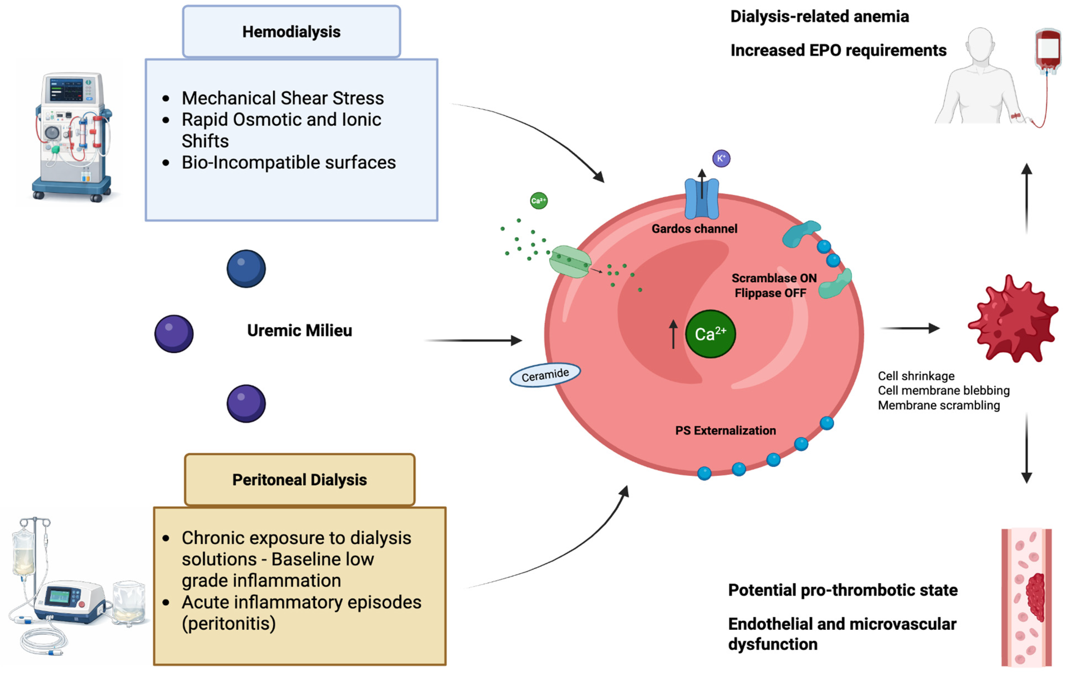
Taken together, available evidence indicates that although eryptosis is a shared feature of advanced CKD and dialysis-dependent patients, the nature, timing, and dominant upstream triggers differ substantially between hemodialysis and peritoneal dialysis. HD is characterized by intermittent mechanical and osmotic stress superimposed on the chronic burden of uremic toxins, with acute intradialytic solute shifts, shear forces, and complement activation directly destabilizing erythrocyte membranes. In contrast, PD imposes a sustained metabolic and inflammatory burden, shaped by continuous exposure to hyperosmolar, acidic, glucose-based dialysate solutions, progressive loss of residual kidney function, and episodes of peritoneal inflammation, which together subtly but persistently compromise RBC integrity. Despite these modality-specific differences, erythrocyte injury in both HD and PD converges on common downstream pathways involving intracellular Ca2+ overload, oxidative stress, and sphingolipid remodeling, ultimately promoting membrane scrambling, phosphatidylserine exposure, and premature erythrocyte clearance. Understanding these shared and divergent mechanisms provides a conceptual framework for targeted strategies aimed at mitigating anemia and preserving RBC lifespan across dialysis populations.
Figure 1 provides an integrated conceptual framework highlighting distinct upstream stressors and shared eryptotic pathways in HD and PD (Figure 1).

5. Translational Perspectives

The recognition of eryptosis as a regulated and biologically meaningful form of erythrocyte death provides a unifying framework to reinterpret several endpoints in CKD patients. The clinical significance is primarily the exacerbation of anemia, and the challenge this poses to achieving target hemoglobin levels, even with erythropoiesis-stimulating agents and iron supplementation [17,34]. Rather than replacing erythropoietin deficiency as the primary driver of renal anemia, eryptosis should be considered a complementary mechanism that accelerates erythrocyte loss. The combination of reduced erythropoiesis and increased erythrocyte clearance may better explain the severity and persistence of anemia, particularly in advanced CKD and dialysis settings [17,35,36]. Additionally, eryptosis is related to inflammation and potential increased thrombogenicity, as PS-exposing RBCs can promote coagulation cascades. Rather than representing passive RBC destruction, eryptosis emerges as an active, stress-integrating process driven by convergent Ca2+, oxidative, and lipid-mediated signaling pathways.
In HD, the translational relevance of eryptosis lies in the balance between toxin removal and procedure-related stress. Advances in membrane biocompatibility, ultrapure dialysate, optimized sodium and bicarbonate profiling, and extracorporeal circuit design appear capable of mitigating oxidative, inflammatory, and mechanical stress, as well as intradialytic erythropoietic signaling. The observation that expanded hemodialysis and hemoadsorption strategies can enhance middle-molecule and protein-bound toxin clearance while maintaining stable eryptosis levels supports the feasibility of dialysis prescriptions that are erythrocyte-protective [26,27].
In this context, the improvement in anemia observed in patients undergoing extended or more frequent dialysis schedules may not be solely explained by enhanced erythropoietin responsiveness [37,38]. Improved clearance of uremic toxins reduced oxidative stress, and attenuation of chronic inflammation could plausibly decrease eryptotic signaling, thereby prolonging erythrocyte lifespan. Although direct evidence remains limited, this hypothesis suggests that modulation of eryptosis may represent an additional mechanism contributing to improved anemia control in optimized dialysis regimens.
In PD, eryptosis assumes additional translational importance as a systemic reflection of peritoneal and inflammatory stress. The consistent association between eryptosis, loss of residual kidney function, and peritonitis-related inflammation suggests that erythrocyte membrane scrambling may serve as an indirect marker of global biocompatibility and inflammatory burden in PD patients. The strong correlation between eryptosis and inflammatory mediators such as IL-1β and IL-6 further highlights the intersection between red blood cell biology and immune activation, particularly during acute inflammatory episodes.
Across both dialysis modalities, these observations support the concept that eryptosis integrates multiple upstream stressors into a common downstream pathway with clinically relevant consequences. Evidence from observational and experimental studies shows that targeting upstream eryptotic triggers such as oxidative stress, inflammation, and biocompatibility-related factors represents a potential adjunctive strategy to improve patient outcomes in kidney replacement therapy.
Eryptosis can be assessed using flow cytometry-based techniques, most commonly through the detection of phosphatidylserine exposure on the erythrocyte surface using fluorescently labeled annexin V, which represents the hallmark of membrane scrambling. Additional parameters, as described in cytofluorometric approaches, include cell volume changes estimated by forward scatter, intracellular Ca2+ levels, oxidative stress markers, and ceramide abundance. Together, these measurements provide a robust experimental framework to identify eryptotic erythrocytes and characterize the underlying signaling mechanisms [39]. However, despite the availability of these robust techniques, their application remains largely confined to research settings. These assays are not standardized for routine clinical use, lack validated thresholds for clinical decision-making, and are not widely accessible [40,41].

6. Future Directions

Despite substantial progress since the first description of the eryptosis mechanism, several important gaps remain in our understanding, particularly in dialysis patients. First, prospective studies integrating standardized erythrocyte phenotyping with detailed dialysis prescription parameters are needed to better discriminate between uremia-related effects and procedure-induced stress, particularly in the hemodialysis setting. Such approaches would help clarify the relative contribution of dialytic stressors versus toxin removal to erythrocyte injury.
Second, although the osmotic shock ceramide axis is well established in experimental erythrocyte models, its clinical relevance in dialysis patients remains largely inferential. Carefully designed studies evaluating the impact of controlled intradialytic osmotic and ionic modulation may provide valuable insights into whether these pathways contribute meaningfully to eryptosis in vivo.
Third, in PD, longitudinal investigations are required to determine whether eryptosis may serve as a biomarker of peritoneal stress, inflammation, or progressive loss of residual kidney function. The combined assessment of systemic eryptosis and peritoneal effluent inflammatory markers could improve risk stratification and facilitate earlier identification of patients at increased risk of inflammation-driven anemia.
Finally, future translational research should explore both pharmacologic and non-pharmacologic strategies aimed at mitigating excessive eryptosis, including optimization of dialysis biocompatibility, modulation of oxidative stress, and careful targeting of eryptotic pathways. Importantly, any intervention designed to attenuate eryptosis must preserve its physiological role in the clearance of senescent or damaged RBCs, emphasizing the need for balanced and individualized therapeutic approaches.

7. Conclusions

Eryptosis, together with inflammation and oxidative stress, has emerged as a clinically meaningful pathway of erythrocyte death in chronic kidney disease and dialysis patients [42]. By accelerating red blood cell turnover, it contributes directly to refractory anemia and vascular complications [17,35,36]. Although increased eryptosis is observed in both hemodialysis and peritoneal dialysis, the dominant upstream triggers differ between modalities [10]. Hemodialysis is characterized by a dynamic balance between uremic toxin removal and procedure-related mechanical and osmotic stress, whereas peritoneal dialysis imposes a sustained metabolic and inflammatory burden strongly influenced by residual kidney function and peritonitis. Despite these modality-specific differences, erythrocyte injury in both settings converges on shared Ca2+-, oxidative-, and sphingolipid-dependent pathways. Improved understanding of these mechanisms may support the development of more biocompatible dialysis strategies and contribute to more personalized approaches to anemia management.

Author Contributions

Conceptualization, M.E. and G.M.V.; writing—original draft preparation, G.M.V., M.M. and M.E.; writing—review and editing, G.M.V., C.R. and M.Z.; supervision, C.R. and M.Z. All authors have read and agreed to the published version of the manuscript.

Funding

This research received no external funding.

Institutional Review Board Statement

Not applicable.

Informed Consent Statement

Not applicable.

Data Availability Statement

No new data were created or analyzed in this study.

Conflicts of Interest

The authors declare no conflicts of interest.

References

  1. Bratosin, D.; Estaquier, J.; Petit, F.; Arnoult, D.; Quatannens, B.; Tissier, J.P.; Slomianny, C.; Sartiaux, C.; Alonso, C.; Huart, J.J.; et al. Programmed cell death in mature erythrocytes: A model for investigating death effector pathways operating in the absence of mitochondria. Cell Death Differ. 2001, 8, 1143–1156. [Google Scholar] [CrossRef]
  2. Berg, C.P.; Engels, I.H.; Rothbart, A.; Lauber, K.; Renz, A.; Schlosser, S.F.; Schulze-Osthoff, K.; Wesselborg, S. Human mature red blood cells express caspase-3 and caspase-8, but are devoid of mitochondrial regulators of apoptosis. Cell Death Differ. 2001, 8, 1197–1206. [Google Scholar] [CrossRef]
  3. Repsold, L.; Joubert, A.M. Eryptosis: An Erythrocyte’s Suicidal Type of Cell Death. BioMed Res. Int. 2018, 2018, 9405617. [Google Scholar] [CrossRef]
  4. Dias, G.F.; Grobe, N.; Rogg, S.; Jörg, D.J.; Pecoits-Filho, R.; Moreno-Amaral, A.N.; Kotanko, P. The Role of Eryptosis in the Pathogenesis of Renal Anemia: Insights from Basic Research and Mathematical Modeling. Front. Cell Dev. Biol. 2020, 8, 598148. [Google Scholar] [CrossRef] [PubMed]
  5. Tkachenko, A.; Alfhili, M.A.; Alsughayyir, J.; Attanzio, A.; Bhuyan, A.A.M.; Bukowska, B.; Cilla, A.; Quintanar-Escorza, M.A.; Föller, M.; Havranek, O.; et al. Current understanding of eryptosis: Mechanisms, physiological functions, role in disease, pharmacological applications, and nomenclature recommendations. Cell Death Dis. 2025, 16, 467. [Google Scholar] [CrossRef] [PubMed]
  6. Clementi, A.; Virzì, G.M.; Milan Manani, S.; Battaglia, G.G.; Ronco, C.; Zanella, M. Eryptosis in Patients with Chronic Kidney Disease: A Possible Relationship with Oxidative Stress and Inflammatory Markers. J. Clin. Med. 2022, 11, 7167. [Google Scholar] [CrossRef] [PubMed]
  7. Virzì, G.M.; Clementi, A.; Ronco, C.; Zanella, M. Red Cell Death in Renal Disease: The Role of Eryptosis in CKD and Dialysis Patients. Cells 2025, 14, 967. [Google Scholar] [CrossRef]
  8. Alghareeb, S.A.; Alfhili, M.A.; Fatima, S. Molecular Mechanisms and Pathophysiological Significance of Eryptosis. Int. J. Mol. Sci. 2023, 24, 5079. [Google Scholar] [CrossRef]
  9. Lang, E.; Bissinger, R.; Gulbins, E.; Lang, F. Ceramide in the regulation of eryptosis, the suicidal erythrocyte death. Apoptosis Int. J. Program. Cell Death 2015, 20, 758–767. [Google Scholar] [CrossRef]
  10. Tkachenko, A.; Havranek, O. Cell death signaling in human erythron: Erythrocytes lose the complexity of cell death machinery upon maturation. Apoptosis Int. J. Program. Cell Death 2025, 30, 652–673. [Google Scholar] [CrossRef]
  11. Bonomini, M.; Sirolli, V.; Gizzi, F.; Di Stante, S.; Grilli, A.; Felaco, M. Enhanced adherence of human uremic erythrocytes to vascular endothelium: Role of phosphatidylserine exposure. Kidney Int. 2002, 62, 1358–1363. [Google Scholar] [CrossRef][Green Version]
  12. Manodori, A.B.; Barabino, G.A.; Lubin, B.H.; Kuypers, F.A. Adherence of phosphatidylserine-exposing erythrocytes to endothelial matrix thrombospondin. Blood 2000, 95, 1293–1300. [Google Scholar] [CrossRef]
  13. Ahmed, M.S.E.; Abed, M.; Voelkl, J.; Lang, F. Triggering of suicidal erythrocyte death by uremic toxin indoxyl sulfate. BMC Nephrol. 2013, 14, 244. [Google Scholar] [CrossRef]
  14. Van Spitzenbergen, B.A.K.; Bohnen Andrade, G.; Sousa Dias, E.; Bacarin Monte Alegre, J.; Ferreira Dias, G.; Grobe, N.; Moreno-Amaral, A.N.; Kotanko, P. The uremic solute 3-carboxy-4-methyl-5-propyl-2-furanpropionate (CMPF) may enhance eryptosis and increase erythrocyte osmotic fragility through potential activation of PIEZO1. Nephrol. Dial. Transplant. 2025, 40, 1342–1349. [Google Scholar] [CrossRef]
  15. Vos, F.E.; Schollum, J.B.; Coulter, C.V.; Doyle, T.C.A.; Duffull, S.B.; Walker, R.J. Red blood cell survival in long-term dialysis patients. Am. J. Kidney Dis. 2011, 58, 591–598. [Google Scholar] [CrossRef]
  16. Bissinger, R.; Artunc, F.; Qadri, S.M.; Lang, F. Reduced Erythrocyte Survival in Uremic Patients Under Hemodialysis or Peritoneal Dialysis. Kidney Blood Press. Res. 2016, 41, 966–977. [Google Scholar] [CrossRef] [PubMed]
  17. Deng, X.; Gong, X.; Huang, Y.; Zhou, J.; Ren, S. Eryptosis in renal anemia: Mechanisms, clinical implications, and therapeutic targeting. Front. Pharmacol. 2025, 16, 1718803. [Google Scholar] [CrossRef] [PubMed]
  18. Tkachenko, A.; Havránek, O. Redox Status of Erythrocytes as an Important Factor in Eryptosis and Erythronecroptosis. Folia Biol. 2023, 69, 116–126. [Google Scholar] [CrossRef]
  19. Lotfi, M.S.; Rassouli, F.B. Navigating the complexities of cell death: Insights into accidental and programmed cell death. Tissue Cell 2024, 91, 102586. [Google Scholar] [CrossRef] [PubMed]
  20. Tkachenko, A.; Havranek, O. Erythronecroptosis: An overview of necroptosis or programmed necrosis in red blood cells. Mol. Cell. Biochem. 2024, 479, 3273–3291. [Google Scholar] [CrossRef]
  21. Kolbrink, B.; von Samson-Himmelstjerna, F.A.; Murphy, J.M.; Krautwald, S. Role of necroptosis in kidney health and disease. Nat. Rev. Nephrol. 2023, 19, 300–314. [Google Scholar] [CrossRef]
  22. Bonomini, M.; Sirolli, V.; Settefrati, N.; Dottori, S.; Di Liberato, L.; Arduini, A. Increased erythrocyte phosphatidylserine exposure in chronic renal failure. J. Am. Soc. Nephrol. 1999, 10, 1982–1990. [Google Scholar] [CrossRef]
  23. Ávila, E.; Sepúlveda, R.A.; Retamal, J.; Hachim, D. Biocompatibility in hemodialysis: Artificial membrane and human blood interactions. BMC Nephrol. 2025, 26, 482. [Google Scholar] [CrossRef] [PubMed]
  24. Abed, M.; Artunc, F.; Alzoubi, K.; Honisch, S.; Baumann, D.; Föller, M.; Lang, F. Suicidal erythrocyte death in end-stage renal disease. J. Mol. Med. 2014, 92, 871–879. [Google Scholar] [CrossRef] [PubMed]
  25. Meyring-Wösten, A.; Kuntsevich, V.; Campos, I.; Williams, S.; Ma, J.; Patel, S.; Ornillo, C.; Thijssen, S.; Kotanko, P. Erythrocyte Sodium Sensitivity and Eryptosis in Chronic Hemodialysis Patients. Kidney Blood Press. Res. 2017, 42, 314–326. [Google Scholar] [CrossRef] [PubMed]
  26. Caprara, C.; Virzì, G.M.; Chieregato, K.; Marchionna, N.; Corradi, V.; Brendolan, A.; Ronco, C.; Zanella, M. Immunomodulation Driven by Theranova Filter during a Single HD Session. J. Clin. Med. 2024, 13, 2147. [Google Scholar] [CrossRef]
  27. Marcello, M.; Virzì, G.M.; Cantaluppi, V.; Marengo, M.; Brendolan, A.; Ronco, C.; Zanella, M. #1573 Effects of haemodialysis coupled with hemadsorption on uremic toxins removal, oxidative stress and cellular death. Nephrol. Dial. Transplant. 2024, 39, gfae069-1528. [Google Scholar] [CrossRef]
  28. Lang, K.S.; Myssina, S.; Brand, V.; Sandu, C.; Lang, P.A.; Berchtold, S.; Huber, S.M.; Lang, F.; Wieder, T. Involvement of ceramide in hyperosmotic shock-induced death of erythrocytes. Cell Death Differ. 2004, 11, 231–243. [Google Scholar] [CrossRef]
  29. Singh, A.T.; McCausland, F.R. Osmolality and blood pressure stability during hemodialysis. Semin. Dial. 2017, 30, 509–517. [Google Scholar] [CrossRef]
  30. Griveas, I.; Gompou, A.; Kyritsis, I.; Papatheodorou, G.; Agroyannis, I.; Tsakoniatis, M.; Kapeleris, E.; Vrachnes, S. Osmolal gap in hemodialyzed uremic patients. Artif. Organs 2012, 36, 16–20. [Google Scholar] [CrossRef]
  31. Virzì, G.M.; Milan Manani, S.; Clementi, A.; Castegnaro, S.; Brocca, A.; Riello, C.; de Cal, M.; Giuliani, A.; Battaglia, G.G.; Crepaldi, C.; et al. Eryptosis Is Altered in Peritoneal Dialysis Patients. Blood Purif. 2019, 48, 351–357. [Google Scholar] [CrossRef]
  32. Virzì, G.M.; Morisi, N.; Marturano, D.; Milan Manani, S.; Tantillo, I.; Ronco, C.; Zanella, M. Peritoneal Inflammation in PD-Related Peritonitis Induces Systemic Eryptosis: In Vitro and In Vivo Assessments. Int. J. Mol. Sci. 2024, 25, 4284. [Google Scholar] [CrossRef]
  33. Virzì, G.M.; Milan Manani, S.; Marturano, D.; Clementi, A.; Lerco, S.; Tantillo, I.; Giuliani, A.; Battaglia, G.G.; Ronco, C.; Zanella, M. Eryptosis in Peritoneal Dialysis-Related Peritonitis: The Potential Role of Inflammation. J. Clin. Med. 2022, 11, 6918. [Google Scholar] [CrossRef]
  34. Bissinger, R.; Qadri, S.M.; Artunc, F. Eryptosis: A driver of anemia in chronic kidney disease. Curr. Opin. Nephrol. Hypertens. 2024, 33, 220–225. [Google Scholar] [CrossRef] [PubMed]
  35. Basak, B.; Biswas, P.; Dey, S.; Paul, A.; Chakraborty, I.; Ray, A.; Dolai, T.K.; Haldar, R. Iron deficiency anemia perturbs erythrocyte morphology and may promote eryptosis. Int. J. Hematol. 2026, 123, 521–534. [Google Scholar] [CrossRef] [PubMed]
  36. Bissinger, R.; Schaefer, L.; Bohnert, B.N.; Schork, A.; Hoerber, S.; Peter, A.; Qadri, S.M.; Birkenfeld, A.L.; Heyne, N.; Bakchoul, T.; et al. GFR is a Key Determinant of Red Blood Cell Survival in Anemia Associated with Progressive CKD. Kidney Int. Rep. 2024, 10, 730–742. [Google Scholar] [CrossRef] [PubMed]
  37. Wong, B.; Collister, D.; Muneer, M.; Storie, D.; Courtney, M.; Lloyd, A.; Campbell, S.; Pauly, R.P. In-center nocturnal hemodialysis versus conventional hemodialysis: A systematic review of the evidence. Am. J. Kidney Dis. 2017, 70, 218–234. [Google Scholar] [CrossRef] [PubMed]
  38. Ornt, D.B.; Larive, B.; Rastogi, A.; Rashid, M.; Daugirdas, J.T.; Hernandez, A.; Tamura, M.K.; Suri, R.S.; Levin, N.W.; Kliger, A.S. Impact of frequent hemodialysis on anemia management: Results from the Frequent Hemodialysis Network (FHN) Trials. Nephrol. Dial. Transplant. 2013, 28, 1888–1898. [Google Scholar] [CrossRef]
  39. Jemaà, M.; Fezai, M.; Bissinger, R.; Lang, F. Methods employed in cytofluorometric assessment of eryptosis, the suicidal erythrocyte death. Cell. Physiol. Biochem. 2017, 43, 431–444. [Google Scholar] [CrossRef]
  40. Tkachenko, A.; Onishchenko, A.; Myasoedov, V.; Yefimova, S.; Havranek, O. Assessing regulated cell death modalities as an efficient tool for in vitro nanotoxicity screening: A review. Nanotoxicology 2023, 17, 218–248. [Google Scholar] [CrossRef] [PubMed]
  41. Turpin, C.; Meilhac, O.; Bourdon, E.; Canonne-Hergaux, F.; Rondeau, P. Methodologies and tools to shed light on erythrophagocytosis. Biochimie 2022, 202, 166–179. [Google Scholar] [CrossRef] [PubMed]
  42. Gok, M.G.; Paydas, S.; Boral, B.; Onan, E.; Kaya, B. Evaluation of eryptosis in patients with chronic kidney disease. Int. Urol. Nephrol. 2022, 54, 2919–2928. [Google Scholar] [CrossRef] [PubMed]
Figure 1. Mechanisms of eryptosis in HD and PD. HD exposes erythrocytes to mechanical shear stress, rapid osmotic and ionic shifts, and bio-incompatible surfaces, while PD imposes chronic metabolic stress from dialysate exposure and episodic inflammation during peritonitis. Both pathways converge on intracellular Ca2+ overload, ceramide accumulation, and phosphatidylserine (PS) externalization, leading to cell shrinkage, membrane blebbing, and membrane scrambling. These processes contribute to dialysis-related anemia, increased erythropoietin requirements, endothelial dysfunction, and a potential pro-thrombotic state.
Kidneydial 06 00029 g001
Table 1. Schematic report of studies on eryptosis and HD.
AuthorPrimary FindingMechanisms & StressorsClinical Context/Outcome
Bonomini et al.Elevated PS-exposure in HD vs. healthy controls.Uremic toxins; reversible when RBCs are placed in normal plasma.Direct correlation between uremia severity and eryptosis levels.
Abed et al.Intradialytic increase in eryptosis.High intracellular Ca2+, oxidative stress, and ceramide abundance.HD procedure itself acts as an acute stressor; pre-HD plasma is pro-eryptotic.
Meyring-Wösten et al.Decrease in eryptosis during the HD session.Use of AV fistulas, ultrapure dialysate, and controlled sodium profiles.High biocompatibility and lower baseline inflammation can attenuate eryptosis.
Caprara et al.Stable eryptosis levels.Medium Cut-Off (MCO) membrane (Theranova) in expanded HD (HDx).Enhanced biocompatibility; no aggravation of RBC injury or immune cell shifts.
Marcello et al.Stable eryptosis levels pre- and post-treatment.Conventional HD combined with HA130 hemadsorption cartridge.Effective removal of middle-molecules (β2-microglobuline, PTH) without additional RBC stress.
Lang et al.Osmotic shock as a trigger for RBC “suicide.”Cation channel activation and sphingomyelinase (ceramide) pathway.Identified ceramide as a critical parallel pathway to calcium in membrane scrambling.
Table 2. Schematic report of studies on eryptosis and PD.
StudyPopulationDialysis ModalityKey FindingsMechanistic Insights
Bonomini et al. HD and PD patients vs. healthy controlsHD & PDIncreased PS exposure in both modalities; numerically higher in HDConfirms dialysis patients exhibit higher eryptosis than controls
Bissinger et al. Dialysis patientsPD vs. HDHigher levels of eryptotic RBCs in PD compared with HDHighlights modality-specific differences, mechanism not fully elucidated
Virzì et al.46 PD patients vs. healthy controlsPDEryptosis significantly higher in PD; lower in patients with preserved residual kidney functionAccumulation of uremic toxins and loss of residual diuresis promote RBC shrinkage, membrane destabilization, PS exposure
Virzì et al.22 PD patients with acute peritonitis vs. 17 healthy controlsPDEryptosis markedly higher during peritonitis Positive correlation with WBC, pNGAL, IL-6, IL-1β; plasma from peritonitis patients induced eryptosis in vitro
Virzì et al.31 PD with peritonitis, 34 stable PD subjectsPDEryptosis higher in peritonitis; correlated with CRP, IL-1β, IL-6Link between inflammation and membrane scrambling in eryptosis
Disclaimer/Publisher’s Note: The statements, opinions and data contained in all publications are solely those of the individual author(s) and contributor(s) and not of MDPI and/or the editor(s). MDPI and/or the editor(s) disclaim responsibility for any injury to people or property resulting from any ideas, methods, instructions or products referred to in the content.