Article

Anemia Is a Predictor of Withdrawal from Peritoneal Dialysis in Stable Peritoneal Dialysis Patients

1
Department of Nephrology, Nagasaki University Hospital, Nagasaki 852-8501, Japan
2
Department of Respiratory Medicine, Nagasaki University Graduate School of Biomedical Sciences, Nagasaki 852-8523, Japan
*
Author to whom correspondence should be addressed.
Kidney Dial. 2025, 5(2), 15; https://doi.org/10.3390/kidneydial5020015
Submission received: 10 March 2025 / Revised: 10 April 2025 / Accepted: 11 April 2025 / Published: 14 April 2025

Abstract

:
Introduction: The association between anemia and peritoneal dialysis (PD) withdrawal in patients with stable PD remains poorly established. Herein, we investigated the relationship between anemia and PD withdrawal in patients with stable PD. Materials and Methods: We included 43 patients undergoing PD for at least 6 months between October 2011 and December 2022. Patients were categorized based on their hemoglobin (Hb) levels at the time of their first peritoneal equilibration test (PET) during the study period as follows: Hb ≥ 11 g/dL and Hb < 11 g/dL. The PD withdrawal rates were compared between these groups. Patients were followed up until death or 31 July 2023. Results: During the follow-up, 36 patients discontinued treatment. Patients with Hb < 11 g/dL had a significantly higher PD withdrawal rate than those with Hb ≥ 11 g/dL. Cox proportional hazards analysis identified Hb level as a risk factor for PD withdrawal. Furthermore, Hb levels negatively correlated with the annual decline in urine volume. Conclusions: Our findings suggest anemia as a predictor of PD withdrawal in patients with stable PD. The negative correlation between Hb levels and the annual decline in urine volume implies that anemia may contribute to PD withdrawal via the deterioration of residual renal function.

Graphical Abstract

1. Introduction

Peritoneal dialysis (PD) is a renal replacement therapy for patients with end-stage renal diseases (ESRDs). There are differences between hemodialysis (HD) and PD, such as technical demands, frequency of hospital visits, and effects on hemodynamics. PD patients preserve residual renal function compared with HD patients and have a better quality of life [1].
Despite its numerous advantages, PD withdrawal remains a common issue among patients. Common causes of PD withdrawal include peritonitis, encapsulating peritoneal sclerosis, inadequate small-solute clearance and/or ultrafiltration, and social reasons [2]. Approximately 10% of patients in Japan withdraw from PD annually [3]. PD withdrawal is often unexpected and leads to failure to continue treatment according to patients’ desires. Therefore, it is important to identify the risk factors for PD withdrawal. To achieve longer-term PD continuation and enhance patient satisfaction, it is crucial to identify the risks associated with PD withdrawal and develop targeted intervention strategies.
Previous studies have shown that some patient characteristics, such as age, sex, Charlson Comorbidity Index (CCI), PD vintage, and urinary volume, are risk factors for PD withdrawal [4]. However, there are no established risk factors for PD withdrawal.
Among the various complications associated with PD patients, anemia stands out as a significant concern. It has been linked to all-cause mortality and cardiovascular events [5]. In addition to the factors mentioned, mortality remains a significant cause of PD withdrawal, and anemia may also play a role. However, data on the relationship between anemia and PD withdrawal, as well as the mechanisms involved, are limited. Previous reports have shown that anemia present at the initiation of PD is associated with an increased risk of early PD withdrawal [6,7]. However, the relationship between anemia and PD withdrawal in patients with stable PD following initiation is unclear. Furthermore, the mechanisms underlying the relationship between anemia and PD withdrawal remain unclear.
Therefore, we investigated the relationship between anemia and PD withdrawal in patients undergoing stable maintenance PD following initiation. In addition, we examined the potential mechanisms by which anemia could affect PD withdrawal.

2. Materials and Methods

This retrospective longitudinal observational cohort study included 43 PD patients. All patients with PD underwent at least two peritoneal equilibration tests (PETs), including 24 h urine sample collection, between October 2011 and December 2022. Patients receiving combined PD and HD therapy at the time of the first PET scan during the study period or those receiving PD for less than 6 months were excluded. Patients with PD were followed up to determine the PD continuation rate and prognosis. This study was conducted in accordance with the Declaration of Helsinki and the ethical guidelines for medical and health research involving human subjects and was approved by the Ethics Review Board of Nagasaki University Hospital (Approval number: 24082908).

2.1. Data Collection

Baseline patient data were collected during the first PET scan of the study period. The PET results were calculated using PD ADEQUEST software (version 2.0; Baxter International Inc., Deerfield, IL, USA). Patients were followed up until their death or 31 July 2023. If patients withdrew from PD, the causes of withdrawal were collected. PD withdrawal was defined as transition to HD (including combined PD and HD therapy) or death.

2.2. Statistical Analysis

Categorical variables are presented as numerical values (%), and continuous variables are presented as mean ± standard deviation. Non-normally distributed data are presented as medians and interquartile ranges. Continuous variables were compared using the t-test or Mann–Whitney U test and categorical variables were evaluated using the chi-square test. PD withdrawal rates and patient mortality were assessed using Kaplan–Meier analysis and compared using the log-rank test between patients with high and low Hb levels. The Hb cut-off was Hb 11 g/dL as specified in the guidelines of the Japanese Society for Dialysis Therapy (JSDT) [8]; Hb ≥ 11 g/dL was defined as high and Hb < 11 g/dL as low. Risk factors for PD withdrawal were analyzed using a multivariate Cox proportional regression model. Defined as risk factors for PD withdrawal, Hb level, age, sex, PD duration, CCI, and daily urinary volume were used as independent variables in the multivariable model [4]. To investigate the relationship between anemia and the decrease in urinary volume, we calculated the annual decrease using the urinary volumes from the first and second PET scans during the study period. The correlation between Hb levels from the first PET scan and the annual decrease in urinary volume was analyzed using Pearson’s correlation test and multiple linear regression analysis. In multiple linear regression analysis, the same variables used in the Cox proportional hazards model were selected.
Statistical analyses were performed using JMP version 17.2.0 software (SAS Institute Inc., Cary, NC, USA). Statistical significance was set at p < 0.05.

3. Results

3.1. Patient Characteristics

Table 1 lists patient characteristics at the first PET scan. The mean age was 60.9 ± 12.0 years, and 26 of 43 patients were males. The median duration of PD was 14 months (range: 11–24). The leading cause of ESRD was nephrosclerosis (30.2%), and the median CCI was 2 (range: 2–4). The median urinary volume was 14.5 (range: 10.0–17.0) dL/day and the median total Kt/V was 1.96 ± 0.52. The Hb was 11 (range: 10.1–11.8) g/dL. Next, we divided patients into two groups, Hb < 11 and Hb ≥ 11 g/dL, and compared the patient characteristics. As shown in Table 1, compared with the high Hb patients, low Hb patients showed high erythropoiesis-stimulating agent (ESA) does [120 (62.5–150) vs. 32.5 (0–50) μg/4 weeks, p < 0.01), high P [5.8 ± 0.3 vs. 4.9 ± 0.2 mg/dL, p = 0.01], low renal Kt/V [0.64 (0.41–0.82) vs. 0.96 (0.75–1.52), p = 0.01], and high urinary protein [1.13 (0.75–2.25) vs. 0.75 (0.43–1.05), p = 0.048].

3.2. Survival Analysis by Hemoglobin

Next, we investigated the effect of Hb levels on PD withdrawal. During the follow-up period, 36 patients withdrew from the PD. As shown in Figure 1, patients with Low Hb levels had a higher risk of PD withdrawal than those with high Hb levels (log-rank test, p = 0.02).
Furthermore, univariate and multivariate Cox proportional hazard analyses were performed to investigate the risk factors for PD withdrawal. As shown in Table 2, Hb (HR [hazard ratio] 0.71; 95%CI [confidence interval] 0.56–0.92; p < 0.01), albumin (HR 0.14; 95% CI 0.04–0.46; p < 0.01), urinary volume (HR 0.90; 95% CI 0.85–0.97; p < 0.01), and urinary protein (HR 1.24; 95% CI 1.06–1.41; p < 0.01) were associated with PD withdrawal in univariate analysis. In multivariate analysis, Hb (HR 0.73; 95% CI 0.54–0.99; p = 0.04) was independently associated with PD withdrawal. Sensitivity analysis with a model incorporating the number of peritonitis episodes into the original multivariate analysis showed Hb (HR 0.73; 95% CI 0.53–0.98; p = 0.04) was associated with PD withdrawal (Table S1). These results suggest that a low Hb level is a risk factor for PD withdrawal.

3.3. Cause of PD Withdrawal

Table 3 lists the causes of PD withdrawal in patients with low and high Hb levels. The primary cause of PD withdrawal in low and high urinary Hb patients was inadequate dialysis (uremia or volume overload). However, inadequate dialysis was more common in patients with low Hb levels than those with high Hb levels (72.2% vs. 50.0%).

3.4. Association Between Anemia and Residual Renal Function Decline

Residual renal function decline is a common cause of uremia and volume overload in patients with PD. We investigated the correlation between anemia and residual renal function decline. Daily urine volume was used to assess the residual renal function. Table 4 lists the correlation between the baseline patient characteristics and the subsequent annual decline in urine volume. Simple regression analysis showed that age (standard beta = −0.37, p = 0.01) and Hb (standard beta = −0.34, p = 0.02) negatively correlated with the subsequent annual decline in urine volume, while urinary protein (standard beta = 0.32, p = 0.04) positively correlated. Furthermore, multiple regression analysis showed that Hb was independently negatively correlated with the annual decline in urine volume (standard beta = −0.35, p = 0.03). These results suggest that anemia is associated with a subsequent decline in urine volume.

4. Discussion

In this study, we determined whether anemia predicts PD withdrawal in patients with PD and showed that a lower Hb level (Hb < 11 g/dL) is a risk factor for PD withdrawal. PD withdrawal is attributed to various factors, including inappropriate dialysis, peritonitis, and death. The causes of PD withdrawal vary based on the timing of withdrawal; peritonitis is particularly common early following PD initiation, and withdrawal due to insufficient dialysis increases as PD duration increases [9]. In our study, the median PD duration was 13 months; therefore, patients who withdrew from PD early following initiation were excluded. The main cause of PD withdrawal in this study was inappropriate dialysis, consistent with the findings of previous reports [9].
PD withdrawal due to inadequate dialysis occurs when a decline in a patient’s residual renal function renders PD insufficient to fully compensate for this loss. In this study, hemoglobin levels negatively correlated with the subsequent annual decrease in urine output, suggesting an association between anemia and the deterioration of residual renal function. Therefore, anemia may accelerate the decline in residual renal function in patients with PD, ultimately leading to PD withdrawal. Anemia affects renal function in patients with non-dialysis chronic kidney disease (CKD), with implicated mechanisms that include renal hypoxia and oxidative stress [10,11]. Because renal anemia typically arises as a consequence of impaired kidney function, hypoxia and oxidative stress may be both causes and consequences of declining residual renal function. Moreover, factors such as inflammation and malnutrition—known contributors to renal anemia—should also be considered when interpreting the association between anemia and renal function. Nevertheless, it remains plausible that anemia itself plays a contributory role in the progression of residual renal dysfunction. Similarly, in patients undergoing PD, anemia may contribute to a decline in residual renal function through similar pathways, potentially leading to PD withdrawal. Previous reports have linked anemia to decreased urine output and anuria in patients undergoing PD [12], consistent with our findings.
The factors contributing to anemia in dialysis patients are multifactorial and include nutritional status, inflammation, vitamin deficiency, and ESA resistance [13,14,15,16]. Nutritional status and inflammation are also associated with decreased residual renal function [17,18]. Although our study incorporated the CCI into the multivariate analysis to account for various comorbidities, it remains unclear whether anemia independently and directly contributes to PD withdrawal, separate from factors such as nutritional status or inflammation. Our results suggest that improving anemia may enable a longer PD duration; however, this conclusion requires further verification. Furthermore, further research is needed to elucidate the detailed mechanisms underlying the relationship between anemia and PD withdrawal and confirm whether anemia correction can extend PD duration. Our results suggest a relationship between anemia and PD withdrawal in patients with stable PD.
This study has some limitations. First, the sample size was small and retrospective. Therefore, unrecognized confounding factors could not be completely ruled out. Furthermore, the small sample size limited the number of independent variables in the multivariate analysis. A decline in residual renal function is well recognized as contributing to erythropoietin resistance and anemia in patients undergoing PD [12,19]. In our analysis, urine output was incorporated into the Cox proportional hazards model as a surrogate for residual renal function; however, it should be noted that residual renal function can be evaluated using various parameters. Therefore, anemia may have plausibly arisen as a consequence of declining residual renal function rather than serving as a direct cause of PD discontinuation. While further studies are warranted to elucidate this causal relationship, our findings at least suggest that the presence of anemia in patients with PD serves as an indicator of subsequent early withdrawal from PD therapy. Second, since the baseline median duration of PD in our study was 13 months, patients who withdrew from PD early following initiation were excluded. Therefore, the findings of this study may only apply to patients undergoing PD for at least one year. Third, although we defined anemia as Hb < 11 g/dL in accordance with the recommendation of the JSDT, it should be noted that the target Hb levels for anemia management in PD patients vary considerably across guidelines, and the appropriateness of this threshold remains uncertain. For example, the Kidney Disease: Improving Global Outcomes (KDIGO) guidelines suggest initiating anemia treatment when Hb levels fall to 9–10 g/dL, to avoid levels below 9.0 g/dL [20]. One reason for this variation is that KDIGO applies the same target range to both HD and PD patients, and the underlying evidence is largely derived from studies involving HD populations. In contrast, the JSDT provides hemoglobin targets for PD patients based on data from non-dialysis CKD populations. This discrepancy reflects the scarcity of robust data specific to anemia management in PD patients. As our study involved a Japanese cohort, we adopted the JSDT-recommended cutoff of <11 g/dL. However, should future evidence establish more appropriate hemoglobin targets tailored to PD patients, it may become necessary to re-evaluate the criteria used in this study. Finally, in our study, the decision regarding PD withdrawal was made at the discretion of each attending physician. Therefore, among patients who underwent PD withdrawal due to inadequate dialysis, no standardized criteria for uremia or fluid overload may have influenced the present results.

5. Conclusions

Among the patients with stable PD, those with anemia were at a higher risk of subsequent PD withdrawal. Moreover, patients with anemia experience a greater decline in urine output over time. Our results suggest that anemia leads to PD withdrawal through the progressive deterioration of residual renal function. Clinicians encountering anemia in patients undergoing PD should be aware of the potentially increased risk of future PD withdrawal. Efforts to investigate the underlying causes of anemia and to initiate appropriate corrective measures may help reduce the risk of early PD discontinuation. However, further studies remain warranted to elucidate the underlying mechanisms linking anemia and PD withdrawal and determine whether correcting anemia can enable longer PD durations.

Supplementary Materials

The following supporting information can be downloaded at: https://www.mdpi.com/article/10.3390/kidneydial5020015/s1, Table S1. Cox proportional hazard model for PD withdrawal (sensitivity analysis).

Author Contributions

Conceptualization, K.T. (Kenta Torigoe); methodology, K.T. (Kenta Torigoe); formal analysis, K.T. (Kenta Torigoe); data curation, K.T. (Kenta Torigoe), E.O., K.T. (Kiyokazu Tsuji), A.Y., M.K., T.T., N.S. and K.M.; validation, E.O., K.T. (Kiyokazu Tsuji), A.Y., M.K., T.T., N.S. and K.M.; writing—original draft preparation, K.T. (Kenta Torigoe); writing—review and editing, E.O, K.T. (Kiyokazu Tsuji), A.Y., M.K., T.T., N.S., K.M., H.M. and T.N.; visualization, K.T. (Kenta Torigoe); supervision, H.M. and T.N.; project administration, K.T. (Kenta Torigoe). All authors have read and agreed to the published version of the manuscript.

Funding

This research received no external funding.

Institutional Review Board Statement

The study was conducted according to the guidelines of the Declaration of Helsinki and approved by the Institutional Review Board (IRB approval number 24082908; date of approval, 29 August 2024).

Informed Consent Statement

Patient consent was waived due to the retrospective nature of the study.

Data Availability Statement

The data presented in this study are available on request from the corresponding author.

Conflicts of Interest

The authors declare no conflicts of interest.

Abbreviations

PDPeritoneal dialysis
PETPeritoneal equilibration test
HbHemoglobin
ESAErythropoiesis-stimulating agent
ESRDEnd-stage renal disease
HDHemodialysis
CCICharlson Comorbidity Index
CIConfidence interval
HRHazard ratio

References

  1. Tokgoz, B. Clinical advantages of peritoneal dialysis. Perit. Dial. Int. 2009, 29, S59–S61. [Google Scholar] [CrossRef] [PubMed]
  2. Banno, T.; Shima, H.; Kawahara, K.; Okada, K.; Minakuchi, J. Risk factors for peritoneal dialysis withdrawal due to peritoneal dialysis-related peritonitis. Nephrol. Ther. 2021, 17, 108–113. [Google Scholar] [CrossRef] [PubMed]
  3. Kawaguchi, Y.; Ishizaki, T.; Imada, A.; Oohira, S.; Kuriyama, S.; Nakamoto, H.; Nakamoto, M.; Hiramatu, M.; Maeda, K.; Ota, K.; et al. Searching for the reasons for drop-out from peritoneal dialysis: A nationwide survey in Japan. Perit. Dial. Int. 2003, 23, S175–S177. [Google Scholar] [PubMed]
  4. Nagasaka, T.; Washida, N.; Uchiyama, K.; Hama, E.Y.; Kusahana, E.; Nakayama, T.; Yasuda, I.; Morimoto, K.; Itoh, H. Health-Related quality of life sleep score predicts transfer to hemodialysis among patients on peritoneal dialysis. Healthcare 2022, 10, 1030. [Google Scholar] [CrossRef] [PubMed]
  5. Molnar, M.Z.; Mehrotra, R.; Duong, U.; Kovesdy, C.P.; Kalantar-Zadeh, K. Association of hemoglobin and survival in peritoneal dialysis patients. Clin. J. Am. Soc. Nephrol. 2011, 6, 1973–1981. [Google Scholar] [CrossRef] [PubMed]
  6. Pulliam, J.; Li, N.C.; Maddux, F.; Hakim, R.; Finkelstein, F.O.; Lacson, E., Jr. First-year outcomes of incident peritoneal dialysis patients in the United States. Am. J. Kidney Dis. 2014, 64, 761–769. [Google Scholar] [CrossRef] [PubMed]
  7. Luo, Q.; Xia, X.; Lin, Z.; Lin, J.; Yang, X.; Huang, F.; Yu, X. Very early withdrawal from treatment in patients starting peritoneal dialysis. Ren. Fail. 2018, 40, 8–14. [Google Scholar] [CrossRef] [PubMed]
  8. Yamamoto, H.; Nishi, S.; Tomo, T.; Masakane, I.; Saito, K.; Nangaku, M.; Hattori, M.; Suzuki, T.; Morita, S.; Ashida, A.; et al. 2015 Japanese Society for Dialysis Therapy: Guidelines for Renal Anemia in Chronic Kidney Disease. Ren. Replace Ther. 2017, 3, 36. [Google Scholar] [CrossRef]
  9. Mizuno, M.; Ito, Y.; Tanaka, A.; Suzuki, Y.; Hiramatsu, H.; Watanabe, M.; Tsuruta, Y.; Matsuoka, T.; Ito, I.; Tamai, H.; et al. Peritonitis is still an important factor for withdrawal from peritoneal dialysis therapy in the Tokai area of Japan. Clin. Exp. Nephrol. 2011, 15, 727–737. [Google Scholar] [CrossRef] [PubMed]
  10. Kuriyama, S.; Tomonari, H.; Yoshida, H.; Hashimoto, T.; Kawaguchi, Y.; Sakai, O. Reversal of anemia by erythropoietin therapy retards the progression of chronic renal failure, especially in nondiabetic patients. Nephron 1997, 77, 176–185. [Google Scholar] [CrossRef] [PubMed]
  11. Buliga-Finis, O.N.; Ouatu, A.; Tanase, D.M.; Gosav, E.M.; Seritean Isac, P.N.; Richter, P.; Rezus, C. Managing anemia: Point of convergence for heart failure and chronic kidney disease? Life 2023, 13, 1311. [Google Scholar] [CrossRef] [PubMed]
  12. Imaizumi, T.; Hasegawa, T.; Kosugi, T.; Nishiwaki, H.; Abe, M.; Hanafusa, N.; Honda, H.; Tsuruya, K.; Ito, Y.; Kuragano, T. Renal anemia and hyporesponsiveness to ESA for preservation of residual kidney function in patients undergoing peritoneal dialysis. Sci. Rep. 2025, 15, 2689. [Google Scholar] [CrossRef] [PubMed]
  13. Radić, J.; Bašić-Jukić, N.; Vujičić, B.; Klarić, D.; Radulović, G.; Jakić, M.; Jurić, K.; Altabas, K.; Grđan, Z.; Kovačević-Vojtušek, I.; et al. Anemia is correlated with malnutrition and inflammation in croatian peritoneal dialysis patients: A multicenter nationwide study. Perit. Dial. Int. 2017, 37, 472–475. [Google Scholar] [CrossRef] [PubMed]
  14. Pérez-Flores, I.; Coronel, F.; Cigarrán, S.; Herrero, J.A.; Calvo, N. Relationship between residual renal function, inflammation, and anemia in peritoneal dialysis. Adv. Perit. Dial. 2007, 23, 140–143. [Google Scholar] [PubMed]
  15. Zhang, C.; Wang, J.; Xie, X.; Sun, D. Low serum vitamin D concentration is correlated with anemia, microinflammation, and oxidative stress in patients with peritoneal dialysis. J. Transl. Med. 2021, 19, 411. [Google Scholar] [CrossRef] [PubMed]
  16. Dai, S.; Chen, Y.; Hao, C.; Ge, X.; Xie, Q.; Shang, D.; Zhu, T. Addition of roxadustat to erythropoiesis-stimulating agent (ESA) effectively corrects ESA-hyporesponsive anaemia in patients on peritoneal dialysis. J. Clin. Pharm. Ther. 2022, 47, 1525–1530. [Google Scholar] [CrossRef] [PubMed]
  17. Li, L.; Liang, W.; Ye, T.; Chen, Z.; Zuo, X.; Du, X.; Qian, K.; Zhang, C.; Hu, X.; Li, J.; et al. The association between nutritional markers and biochemical parameters and residual renal function in peritoneal dialysis patients. PLoS ONE 2016, 11, e0156423. [Google Scholar] [CrossRef] [PubMed]
  18. Chung, S.H.; Heimbürger, O.; Stenvinkel, P.; Bergström, J.; Lindholm, B. Association between inflammation and changes in residual renal function and peritoneal transport rate during the first year of dialysis. Nephrol. Dial. Transplant. 2001, 16, 2240–2245. [Google Scholar] [CrossRef] [PubMed]
  19. Wang, A.Y.; Woo, J.; Wang, M.; Sea, M.M.; Sanderson, J.E.; Lui, S.F.; Li, P.K. Important differentiation of factors that predict outcome in peritoneal dialysis patients with different degrees of residual renal function. Nephrol. Dial. Transplant. 2005, 20, 396–403. [Google Scholar] [CrossRef] [PubMed]
  20. KDIGO Anemia Work Group. KDIGO Clinical Practice Guideline for Anemia in Chronic Kidney Disease. Kidney Int. Suppl. 2012, 2, 279–335. [Google Scholar]
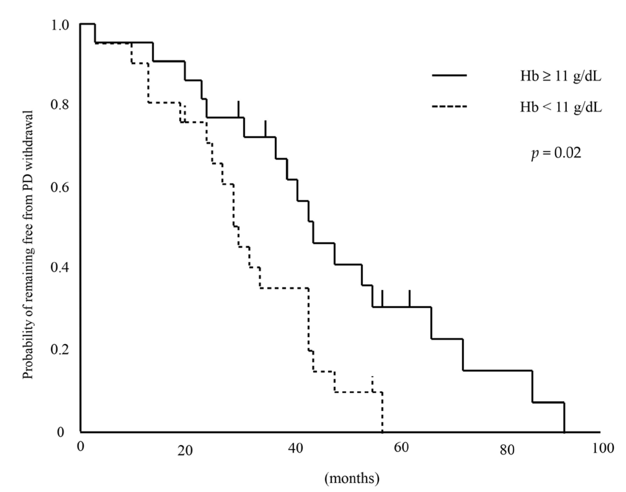
Figure 1. Kaplan-Meier curve for PD withdrawal. Hb—hemoglobin; PD—peritoneal dialysis.
Kidneydial 05 00015 g001
Table 1. Baseline characteristics of PD patients.
All
(n = 43)
Hb < 11 g/dL
(n = 21)
Hb ≥ 11 g/dL
(n = 22)
p-Value
Age (years)60.9 ± 12.059.0 ± 2.662.7 ± 2.60.32
Sex (Male—Female)26:1711:1015:70.36
BMI22.6 (20.5–25.2)22.6 (20.5–25.4)22.7 (20.3–25.1)0.91
Systolic BP (mmHg)130.6 ± 26.3135.2 ± 5.7126.2 ± 5.60.27
Diastolic BP (mmHg)76.3 ± 16.080.3 ± 3.472.4 ± 3.40.11
Duration of PD (months)14 (11–24)12 (8.5–25)14(12–24.5)0.13
Diabetes mellitus 25.628.622.70.74
Charlson Comorbidity Index2 (2–4)2 (2–4)2.5 (2–3.25)>0.99
Number of peritonitis episodes (n)
03718190.71
1312
2321
>3000
ARB/ACE-I62.847.677.30.06
CCB62.861.963.6>0.99
Diuretic83.785.781.80.77
Statin27.923.831.80.40
ESA does (μg/4 weeks)50 (25–120)120 (62.5–150)32.5 (0–50)<0.01
ERI (μg/kg/g/dL/4 weeks)0.082 (0.032–0.204)0.200 (0.101–0.232)0.052 (0–0.083)<0.01
Iron supplement23.333.313.60.16
CAPD—APD28:1513:815:70.75
Hb (g/dL)11 (10.1–11.8)10.1 (10.0–10.5)11.8 (11.1–12.8)<0.01
Alb (g/dL)3.2 ± 0.43.1 ± 0.13.3 ± 0.10.11
Corrected Ca (mg/dL)9.2 ± 0.69.1 ± 0.19.3 ± 0.10.47
P (mg/dL)5.3 ± 1.25.8 ± 0.34.9 ± 0.20.01
Intact PTH (pg/mL)171.9 (97–260.2)153 (104.1–268.6)179.2 (91.6–250.3)0.95
Ferritin (ng/mL)156 (107–252)156 (80–252)157.5 (111.3–258.3)>0.99
TSAT (%)35.5 ± 17.431.7 ± 3.939.1 ± 3.80.17
CRP (mg/dL)0.06 (0.03–0.22)0.06 (0.02–0.22)0.07 (0.04–0.24)0.54
Urinary volume (dL/day)14.5 (10.0–17.0)12.0 (6.7–16.5)15.0 (12.0–17.0)0.07
Peritoneal Kt/V1.03 ± 0.371.11 ± 0.080.96 ± 0.080.17
Renal Kt/V0.79 (0.62–1.23)0.64 (0.41–0.82)0.96 (0.75–1.52)<0.01
Total Kt/V1.96 ± 0.521.84 ± 0.112.08 ± 0.110.13
4-h D/P Cr0.6 (0.56–0.68)0.6 (0.58–0.67)0.61 (0.53–0.69)0.89
nPCR0.86 ± 0.180.82 ± 0.040.90 ± 0.040.12
Urinary protein (g/gCr)0.89 (0.54–1.35)1.13 (0.75–2.25)0.75 (0.43–1.05)0.048
Urinary protein (g/day)0.51 (0.28–0.78)0.51 (0.31–0.77)0.51 (0.27–0.83)0.92
ACE-I—angiotensin-converting enzyme inhibitor; Alb—albumin; APD—automated peritoneal dialysis; ARB—angiotensin II receptor blocker; BMI—body mass index; BP—blood pressure; Ca—calcium; CAPD—continuous ambulatory peritoneal dialysis; CCB—calcium channel blocker; CRP—C-reactive protein; ESA—erythropoietin stimulating agents; ESRD—end-stage renal disease; ERI—erythropoietin resistance index; GFR—glomerular filtration rate; Hb—hemoglobin; HbA1c—hemoglobin A1c; nPCR—normalized protein catabolic rate; P—phosphorus; PD—peritoneal dialysis; PTH—parathyroid hormone; TSAT—transferrin saturation; 4 h-D/P Cr—4 h dialysate/plasma creatinine.
Table 2. Cox proportional hazard model for PD withdrawal.
UnivariateMultivariate
HR (95% CI)p-ValueHR (95% CI)p-Value
Age (years)1.00 (0.97–1.03)0.781.00 (0.96–1.04)0.99
Sex (Male)1.20 (0.62–2.34)0.591.15 (0.43–3.04)0.78
BMI0.79 (0.90–1.07)0.81
Systolic BP (mmHg)1.01 (0.99–1.02)0.22
Diastolic BP (mmHg)1.00 (0.98–1.03)0.66
Duration of PD (months)1.00 (0.97–1.03)0.991.00 (0.96–1.03)0.81
Diabetes mellitus 1.53 (0.73–3.21)0.27
Charlson Comorbidity Index1.04 (0.77–1.34)0.790.95 (0.67–1.30)0.77
Number of peritonitis episodes1.16 (0.62–1.92)0.60
ARB/ACE-I0.94 (0.47–1.86)0.85
CCB1.05 (0.52–2.13)0.89
Diuretic1.76 (0.67–4.58)0.22
Statin0.76 (0.35–1.64)0.47
ESA does (μg/4 weeks)1.00 (1.00–1.01)0.11
ERI (μg/kg/g/dL/4 weeks)13.18 (0.76–149.53)0.07
Iron supplement1.35 (0.63–2.91)0.46
CAPD—APD0.62 (0.30–1.27)0.20
Hb (g/dL)0.71 (0.56–0.92)0.0090.73 (0.54–0.99)0.04
Alb (g/dL)0.14 (0.04–0.46)0.001
Corrected Ca (mg/dL)0.76 (0.45–1.26)0.29
P (mg/dL)2.81 (0.87–1.62)0.28
Intact PTH (pg/mL)1.00 (0.99–1.00)0.52
Ferritin (ng/mL)1.00 (0.99–1.00)0.94
TSAT (%)0.99 (0.97–1.02)0.56
CRP (mg/dL)1.60 (0.42–4.32)0.44
Urinary volume (dL/day)0.90 (0.85–0.97)0.0060.93 (0.86–1.01)0.09
Peritoneal Kt/V1.20 (0.51–2.74)0.66
Renal Kt/V0.51 (0.22–1.04)0.06
Total Kt/V0.56 (0.25–1.15)0.12
4-h D/P Cr7.09 (0.31–191.09)0.23
nPCR0.27 (0.03–2.60)0.26
Urinary protein (g/gCr)1.24 (1.06–1.41)0.009
Urinary protein (g/day)1.44 (0.87–2.11)0.14
ACE-I—angiotensin-converting enzyme inhibitor; Alb—albumin; APD—automated peritoneal dialysis; ARB—angiotensin II receptor blocker; BMI—body mass index; BP—blood pressure; Ca—calcium; CAPD—continuous ambulatory peritoneal dialysis; CCB—calcium channel blocker; CI—confidence interval; CRP—C-reactive protein; ESA—erythropoietin stimulating agents; ESRD—end-stage renal disease; ERI—erythropoietin resistance index; GFR—glomerular filtration rate; Hb—hemoglobin; HbA1c—hemoglobin A1c; HR—hazard ratio; nPCR—normalized protein catabolic rate; P—phosphorus; PD—peritoneal dialysis; PTH—parathyroid hormone; TSAT—transferrin saturation; 4 h-D/P cr—4 h dialysate/plasma creatinine.
Table 3. Cause of PD withdrawal.
Hb < 11 g/dL (n = 18)Hb ≧ 11 g/dL (n = 18)p-Value
Inadequate dialysis
(uremia or volume overload)
13 (72.2%)9 (50%)0.03
Death2 (11.1%)7 (39.9%)
PD-related infection3 (16.7%)0 (%)
Others0 (0%)2 (11.1%)
Total1818
PD—peritoneal dialysis.
Table 4. Pearson’s correlation and multiple regression analyses for the annual decline of urine volume in peritoneal dialysis patients.
UnivariateMultivariate
Standard βp-ValueStandard βp-Value
Age−0.370.01−0.220.18
Sex (Female)0.360.020.090.61
BMI−0.020.88
Systolic BP (mmHg)0.260.09
Diastolic BP (mmHg)0.280.07
Duration of PD (months)0.080.63−0.020.92
Diabetes mellitus −0.090.55
Charlson Comorbidity Index−0.230.13−0.130.39
Number of peritonitis episodes0.150.95
ARB/ACE-I−0.200.19
CCB0.030.84
Diuretic−0.220.16
Statin0.110.48
ESA does (μg/4 weeks)0.270.08
ERI (μg/kg/g/dL/4 weeks)0.240.12
Iron supplement0.410.01
CAPD—APD0.130.41
Hb (g/dL)−0.340.02−0.350.03
Alb (g/dL)−0.160.31
Corrected Ca (mg/dL)−0.130.41
P (mg/dL)0.050.74
Intact PTH (pg/mL)−0.110.50
Ferritin (ng/mL)−0.100.57
TSAT−0.090.58
CRP (mg/dL)0.060.70
Urinary volume (dL/day)0.220.170.220.16
Peritoneal Kt/V−0.130.41
Renal Kt/V0.050.77
Total Kt/V−0.040.78
4-h D/P Cr−0.100.55
nPCR−0.170.28
Urinary protein (g/gCr)0.270.08
Urinary protein (g/day)0.320.04
ACE-I—angiotensin-converting enzyme inhibitor; Alb—albumin; APD—automated peritoneal dialysis; ARB—angiotensin II receptor blocker; BMI—body mass index; BP—blood pressure; Ca—calcium; CAPD—continuous ambulatory peritoneal dialysis; CCB—calcium channel blocker; CRP—C-reactive protein; ESA—erythropoietin stimulating agents; ESRD—end-stage renal disease; ERI—erythropoietin resistance index; GFR—glomerular filtration rate; Hb—hemoglobin; HbA1c—hemoglobin A1c; nPCR—normalized protein catabolic rate; P—phosphorus; PD—peritoneal dialysis; PTH—parathyroid hormone; TSAT—transferrin saturation; 4 h-D/P cr—4 h dialysate/plasma creatinine.
Disclaimer/Publisher’s Note: The statements, opinions and data contained in all publications are solely those of the individual author(s) and contributor(s) and not of MDPI and/or the editor(s). MDPI and/or the editor(s) disclaim responsibility for any injury to people or property resulting from any ideas, methods, instructions or products referred to in the content.