Article

Computational Discovery of Novel Imidazole Derivatives as Inhibitors of SARS-CoV-2 Main Protease: An Integrated Approach Combining Molecular Dynamics and Binding Affinity Analysis

by
Benjamin Ayodipupo Babalola
1,* and
Abayomi Emmanuel Adegboyega
2
1
Department of Chemistry, Purdue University, West Lafayette, IN 47907, USA
2
Department of Biology, Purdue University, West Lafayette, IN 47907, USA
*
Author to whom correspondence should be addressed.
COVID 2024, 4(6), 672-695; https://doi.org/10.3390/covid4060046
Submission received: 24 April 2024 / Revised: 7 May 2024 / Accepted: 16 May 2024 / Published: 23 May 2024

Abstract

:
One of the most pressing challenges associated with SARS treatment is the emergence of new variants that may be transmissible, causing more severe disease or being resistant to the current standard of treatment. This study aimed to identify potential drug candidates from novel imidazole derivatives against SARS-CoV-2 main protease (Mpro), a crucial drug target for treating viral infection, using a computational approach that integrated molecular docking and dynamics simulation. In this study, we utilized AutoDock Vina within the PyRx workspace for molecular docking analysis to explore the inhibitory effects of the compounds on the Mpro, a drug target for SARS-CoV-2. The ADMET properties of these compounds, including absorption, distribution, metabolism, excretion, and toxicity, were evaluated using the SwissADME and ADMETLab servers. Each of the 18 compounds that were tested demonstrated strong binding affinities towards Mpro, with imidazolyl–methanone C10 showing the most significant binding affinity. Moreover, pyridyl–imidazole C5, thiophenyl–imidazole C1, and quinoline–imidazole C14 displayed binding affinities of −8.3, −8.2, and −7.7 Kcal/mol, respectively. These compounds interacted with specific amino acid residues (HIS A:41—CYS A:145) within the Mpro protein. To assess the stability of the ligand with the best binding affinity, molecular dynamics (MD) simulations were conducted using Schrodinger software, which revealed its stability over the simulation period. The study provides valuable insights into the potential of imidazole derivatives as SARS-CoV-2 Mpro inhibitors. All compounds including C10 display promising characteristics and hold potential as drug candidates for SARS-CoV-2. However, further optimization and experimental validation of these compounds are necessary to advance their development as effective therapeutics against viral infections.

1. Introduction

In the 21st century, a pandemic has not been as severe as the recent outbreak of severe acute respiratory syndrome coronavirus 2 (SARS-CoV-2). The World Health Organization first identified the outbreak in China in February 2003, which then spread to over 24 countries across North and South America, Europe, and Asia before being contained [1,2]. SARS resulted in 8098 cases and 774 deaths worldwide [2]. Similarly, in late December 2019, a new outbreak was identified that was airborne [3]. The outbreak originated in Wuhan, China, and rapidly spread worldwide, resulting in 775,364,261 confirmed cases and 7,046,320 deaths [1]. This led to the implementation of policies across the globe to curb the spread of the virus.
One of the most pressing challenges associated with SARS treatment is the emergence of new variants that may be transmissible, causing more severe disease or being resistant to the current standard of treatment. In a recent publication, we conducted an in silico investigation on the therapeutic possibilities of new heterocyclic derivatives targeting SARS-CoV-2 [4]. In this study, we reported 17 compounds with better binding affinity relative to remdesivir and N3. In the study, we reported the results of this study against SARS-CoV-2 drug targets: main protease (Mpro), spike protein (Spro), and RNA-dependent RNA polymerase (RdRp). However, in this present study, we are reporting the results of 18 compounds against the main protease. The main protease is also known as 3 chymotrypsin-like proteases, and it plays a critical role in the maturation cleavage of amino acid units connected by peptide bonds during the process of virus reproduction [5]. This main protease exists as a homodimer, composed of two promoters: papain-like cysteine protease (PL pro) and 3 chymotrypsin-like proteases (3CLpro) [6]. It consists of three domains: domain I (residue 8–101), domain II (residue 102–184), and domain III (residue 201–303). Domains I and II consist of six antiparallel β-barrels, while domain III, comprising a cluster of five α-helices, indirectly interacts with substrates, which is crucial for the enzymatic activity of proteins by removing the inactive protease [7]. The main protease is an upstream enzyme involved in the replication and transcription of SARS-CoV-2 [4]. Through the use of computational models and in silico screening, it becomes possible to identify potential inhibitory compounds against the protein target [8,9].
The conventional approach to developing medications and treatment protocols is often characterized by time-consuming and expensive processes, which frequently yield unsatisfactory clinical outcomes [4]. In this investigation, an alternative approach employing computational tools is employed, eschewing the traditional methodologies for the discovery and development of novel therapeutic agents [4]. The latter typically entails intricate scientific processes and may be time-intensive. However, utilizing the bioinformatics approach offers a promising pathway for formulating and producing novel pharmaceutical substances with biomedical significance. This method allows for the prediction of binding affinities and ADMET (absorption, distribution, metabolism, excretion, and toxicity) properties of test compounds towards protein receptors and domains.
Imidazoles represent a class of heterocyclic compounds characterized by notable pharmacological properties and potent biological activity [10]. These compounds, like other natural medicinal products, exhibit a wide array of desirable attributes, encompassing anti-parasitic, anti-inflammatory, anti-cancer, anti-fungal, anti-bacterial, anti-malarial, anti-tubercular, and antiviral actions [11,12]. Moreover, imidazoles have demonstrated inhibitory effects on copper erosion, as well as diverse enzymes, including aromatase, phosphatidylinositol 3-kinase alpha (PI3Kα), cholinesterase, HSP90, and Topo II [13,14,15,16]. The pivotal role played by the imidazole ring has offered versatile synthetic routes, and the incorporation of the imidazole nucleus and electron-rich imidazole ring into the design of therapeutics is recognized as crucial by the pharmaceutical industry. Notably, imidazole derivatives are currently being synthesized and employed in antiviral research due to their potential antiproliferative effects [17]. Their effectiveness as antiviral agents is greatly augmented by their ability to selectively target vital viral enzymes and proteins pivotal to the viral life cycle.
The emergence of new variants of SARS-CoV-2 presents a formidable challenge in the landscape of SARS-CoV-2 treatment, as these variants may exhibit heightened transmissibility, increased pathogenicity, or resistance to currently available interventions, including therapeutics and vaccines. The global impact of the initial emergence of SARS-CoV-2 has been nothing short of catastrophic, resulting in a staggering number of fatalities worldwide. Consequently, it becomes imperative to undertake further comprehensive investigations, akin to the present study, in order to equip the scientific community with robust strategies to effectively combat future outbreaks or occurrences. Within the purview of this investigation, a computational framework was employed to comprehensively evaluate the therapeutic potential of specific novel imidazole derivatives against SARS-CoV-2.

2. Methodology

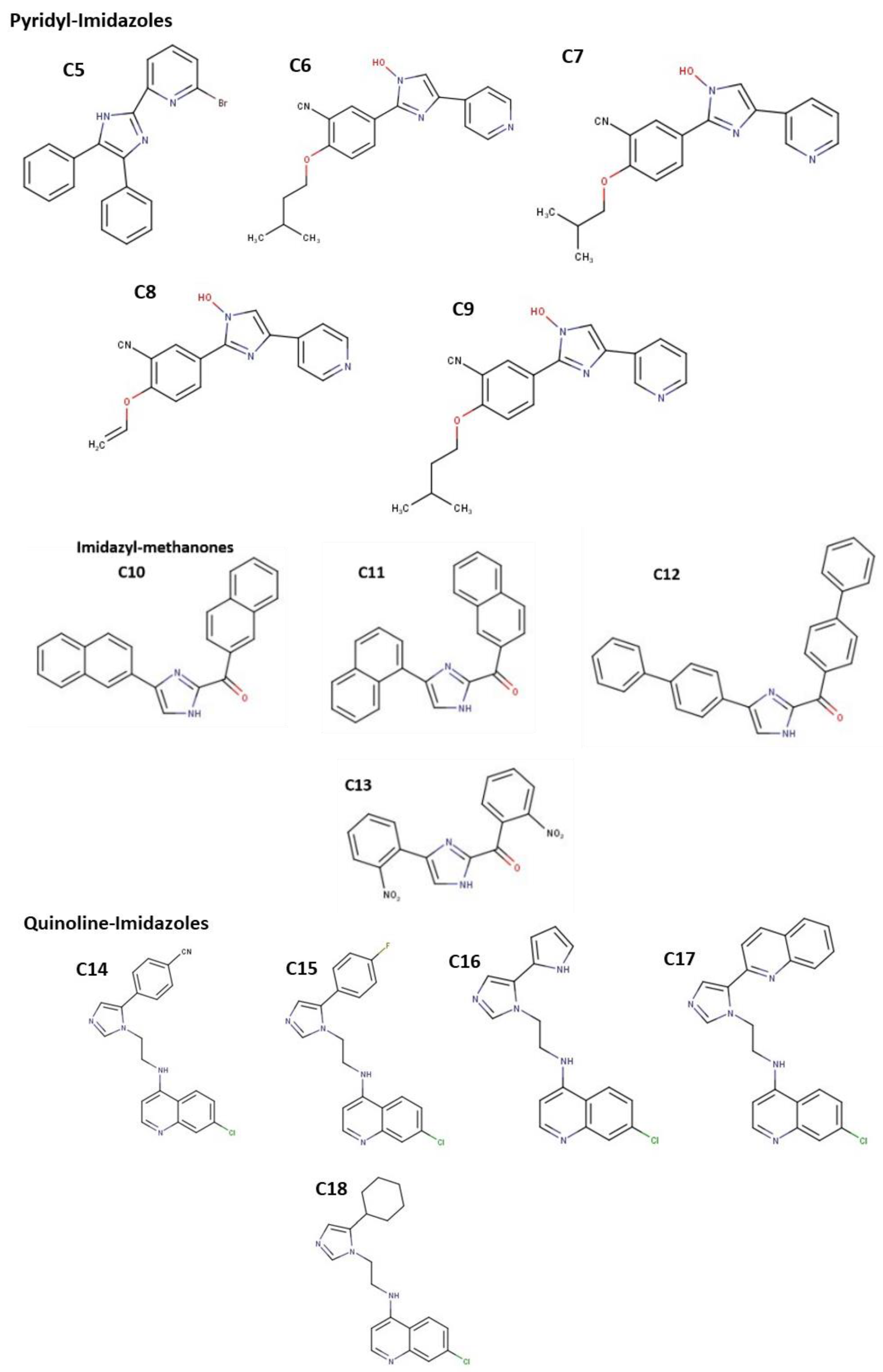
2.1. Imidazole Derivatives

Imidazoles represent a class of heterocyclic compounds characterized by notable pharmacological properties and potent biological activity [10]. The rationale behind the design of these compounds lies in the fact that different studies have reported the potential of imidazole derivatives against SARS-CoV-2, and interaction with important residues of the main protease of SARS-CoV-2 [11,18]. The test compounds employed in this study predominantly encompass imidazole derivatives, as depicted in Figure 1. Specifically, compounds C1 to C4 correspond to thiophenyl-substituted imidazoles, compounds C5 to C9 represent pyridyl-substituted imidazoles, compounds C10 to C14 pertain to imidazolyl–methanone derivatives, and compounds C14 to C18 are classified as quinoline-substituted imidazoles.

2.2. Ligand Preparation

The canonical Simplified Molecular Input Line Entry System (SMILES) representations of compounds C1 to C18 were converted into MOL Structure-Data File (SDF) format using OpenBabel [19] and Chemdes [20] software tools. For the creation of a reference ligand, the co-crystallized ligand (PDB: K36) taken from the main protease was chosen as the standard ligand (Figure 1). Following this, the MOL SDF files of both the compounds and the standard ligand were imported into PyRx software and transformed into the Protein Data Bank (PDB) QT (PDBQT) format with the aid of the OpenBabel plug-in. Subsequently, the resulting files underwent energy minimization employing the Universal Force Field (UFF) to achieve the ligands’ minimum energy conformation suitable for docking. Molecular docking analysis revealed that the 18 compounds evaluated exhibited significantly higher binding affinities compared to the standard ligand (Figure 2).

2.3. Preparation of Protein

The main protease (Mpro: 7C6U) of SARS-CoV-2 was chosen as the main target protein for this study. The crystallographic data for the Mpro protein structures were obtained from the RCSB Protein Data Bank (PDB) repository (https://www.rcsb.org/, accessed on 29 July 2022). The Mpro structures were co-crystallized with the ligand K36, which was utilized as a reference ligand throughout the study. In order to ready the protein structures for further analysis, the PDB format files were brought into the PyMol visualization tool workspace. Following pre-established criteria, the elimination of non-standard residues, including ions, water molecules, and bound ligands situated within the active binding pocket, was carried out. The processed protein structures were then imported into the PyRx software to facilitate the ensuing molecular docking analysis.

2.4. Molecular Docking

After adequately preparing the target proteins and ligands, molecular docking analysis was performed using AutoDock Vina 1.1.2, which was integrated into the PyRx 0.9.9 workspace tool. Established scoring functions [21] were employed during the process. Prior to docking, the ligand conformations were energetically minimized and converted to the Protein Data Bank with Charges (PDBQT) format. For the docking simulations, specific grid box resolutions were chosen for both the ligands and receptors. The dimensions of the grid boxes were precisely set as follows: 51.3737 × 66.9738 × 58.6069 Å for the main protease. These dimensions were meticulously determined to accurately define the binding sites of the target receptors, optimize the search space, and encompass the complete three-dimensional active sites of the protein targets. As a result, the accurate placement of the ligands in the active sites of their respective targets was confirmed. The first step involved docking the standard ligands against each receptor, and the interactions were then compared to those of the lead-like analogs in the same binding site, with the use of identical grid box dimensions. Furthermore, BIOVIA Discovery Studio21 was employed to visualize the complexes resulting from the docking poses of the ligands and receptors, enabling a detailed analysis of the interactions and bonds formed between them.

2.5. Molecular Dynamics Simulation

Top-docking poses of the selected hit molecules and Mpro were used in all-atom classical MD simulations using the Desmond package, developed by Schrodinger LLC. The simulations were carried out for a duration of 10.012 nanoseconds. To visualize the pose of ligands within the protein binding pocket, static views of the molecular docking results were examined. In the initial preprocessing of the protein–ligand complex, the Protein Preparation Wizard tool in Maestro was utilized. This involved complex optimization and minimization, employing default parameters. The System Builder tool in Maestro was then employed to prepare all the systems. TIP3P (Transferable Intermolecular Interaction Potential 3 Points) was chosen as the solvent model, and an orthorhombic box with dimensions of 10 × 10 × 10 Å was employed. To achieve electrical neutrality, counter ions (Na+ or Cl) were added to the models. The OPLS_2005 force field and RESPA integrator parameters were used for the simulation of the protein–ligand complexes. Before the simulation, the models were subjected to a relaxation process. During all molecular dynamics simulations, the NPT ensemble was used with a temperature of 300 K and a pressure of 1 atm. To mimic physiological conditions, a salt concentration of 1.661 mM NaCl was included. Trajectories were saved at intervals of 100 ps for subsequent analysis, and the stability of the simulations was assessed by measuring the root mean square deviation (RMSD) of the protein and ligand over time. Also, the protein RMSF, protein secondary structure elements, ligand RMSF, protein–ligand contacts, radius of gyration, intramolecular hydrogen bond, molecular surface area, solvent accessible surface area, and polar surface area were determined.

2.6. ADMET Predictions

In the ADMET evaluation, the compounds’ absorption, distribution, metabolism, excretion, and toxicity were predicted using both the SwissADME [4] and ADMETLab server [22].

3. Results

3.1. Binding Affinities and Stability of Test Compounds with SARS-CoV-2 Drug Targets

The tested compounds demonstrated a range of binding affinities, as indicated by Gibbs free energy (∆G kcal/mol), within the Mpro (7C6U) protein target. The imidazole–methanone derivatives within the compound classes exhibited notable binding affinities, with compounds C10, C11, C12, and C13 displaying the highest levels of affinity (−9.2, −8.9, −8.9, and −8.2 kcal/mol, respectively). Among the thiophenyl–imidazoles, compound C1 demonstrated the highest binding affinity for Mpro (−8.2 kcal/mol). Within the pyridyl–imidazole group, compound C5 exhibited the highest binding affinity for Mpro (−8.3 kcal/mol), while compound C14 demonstrated the highest binding affinity among the quinoline–imidazoles (−7.7 kcal/mol). C10 showed best binding affinity. Remarkably, all the test compounds displayed binding affinities surpassing those of the standard inhibitors, as summarized in Table 1.

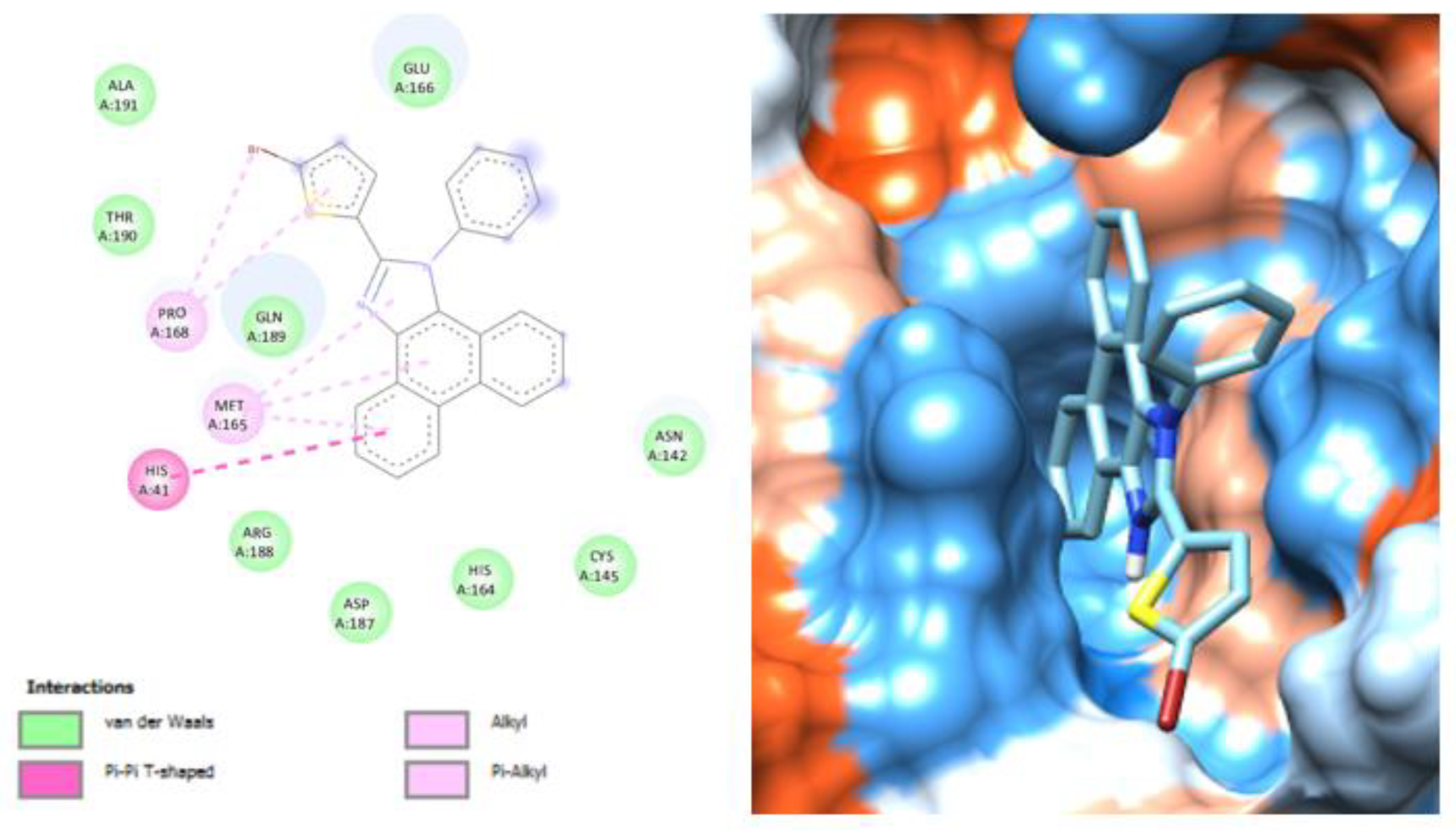
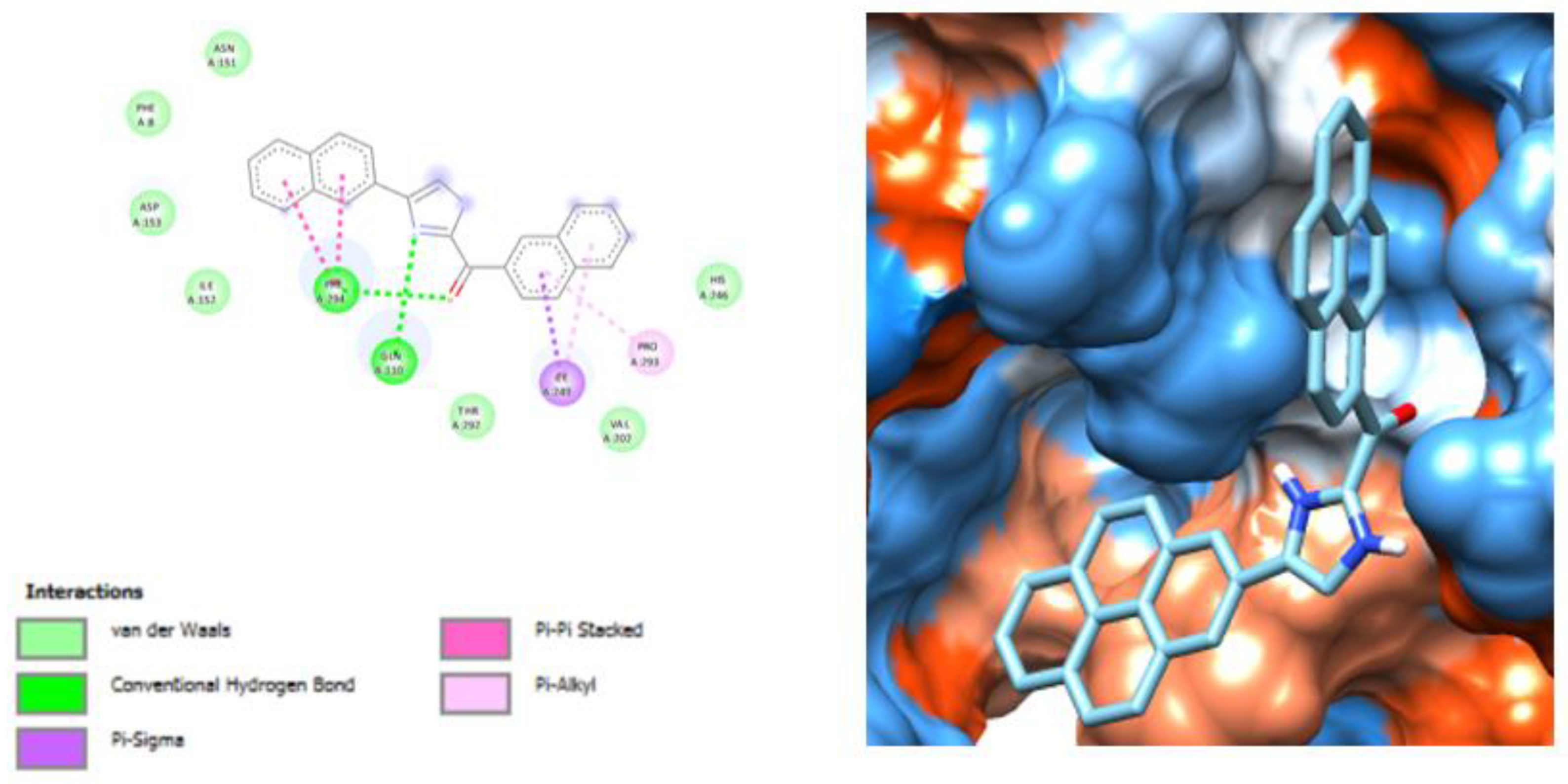
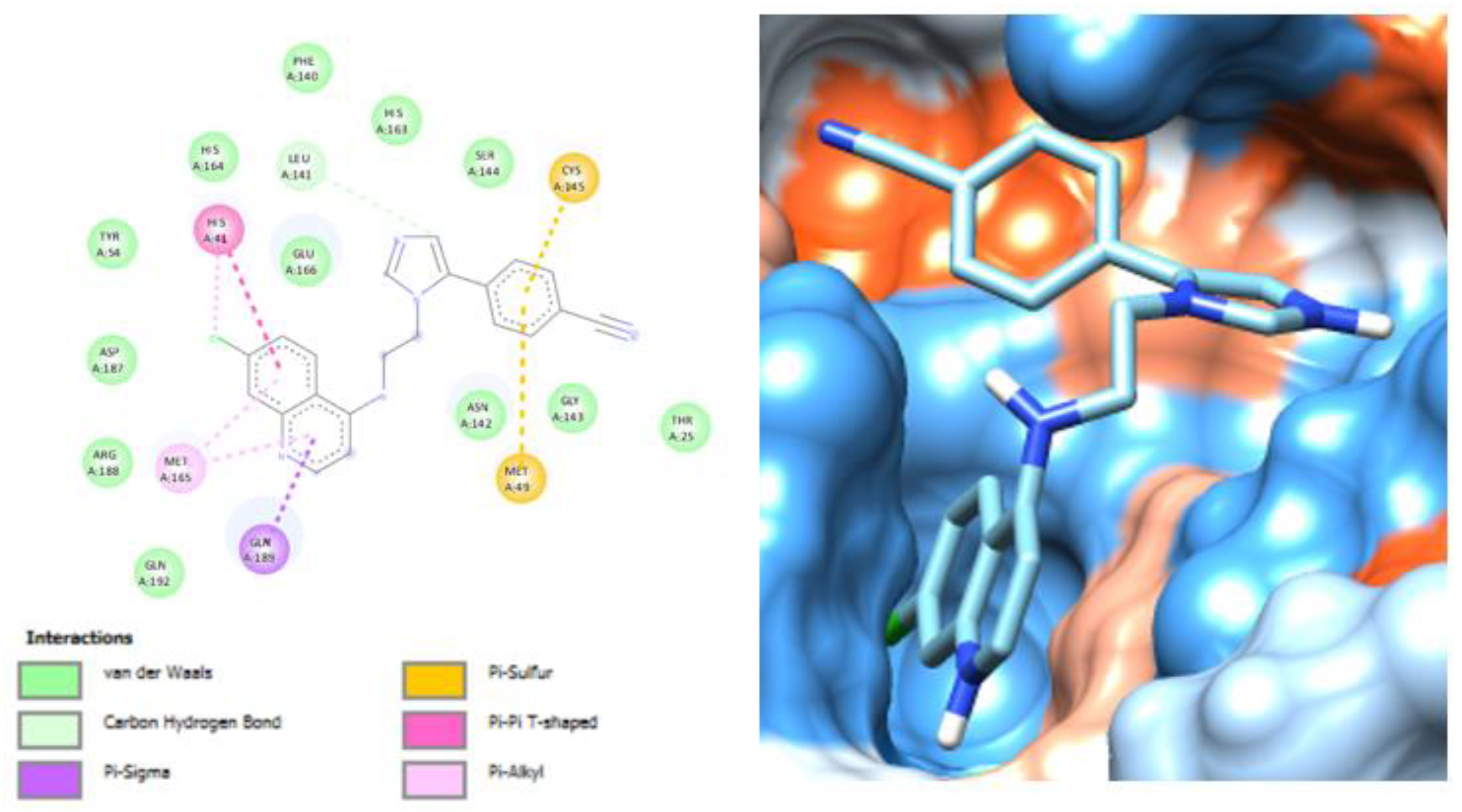
3.2. Molecular Docking Analysis of Selected Test Compounds

Figure 3, Figure 4, Figure 5, Figure 6 and Figure 7 showcase the 3D and 2D structures of the SARS-CoV-2 main protease (Mpro) in complex with the four compounds and standard inhibitors (C1, C5, C10, and C14) that displayed the highest affinities for Mpro.
Compound C1 established interactions involving п–alkyl bonds, п–alkyl bonds, and п–п bonds with amino acid residues PRO A:168, MET A:165, HIS A:41, and other residues within the inhibitor (K36) binding site of the SARS-CoV-2 Mpro (Figure 3). Compound C5 formed a complex with the SARS-CoV-2 Mpro, interacting with particular amino acid residues at the binding site in various ways (Figure 4). Residues GLU A:106, CYS A:145, MET A:165, MET A:49, and HIS A:41, among others, established п–anion, п–sulfur, п–alkyl, and п–п bond interactions (Figure 4). Likewise, compound C10 interacted with amino acid residues PHE A:294, GLN A:110, PRO A:293, and ILE A:243 within the binding pocket of the SARS-CoV-2 Mpro, employing hydrogen interactions, п–alkyl interactions, and п–sigma bonds (Figure 5). Similarly, compound C14 exhibited interactions with amino acid residues HIS A:41, MET A:169, GLN A:189, LEU A:141, MET A:49, and CYS A:145 within the Mpro binding pocket, forming carbon–hydrogen interactions, п–sigma interactions, п–sulfur interactions, п–п interactions, and п–alkyl interactions (Figure 6). Moreover, the standard inhibitor K36 exhibited interactions with amino acid residues ASP A:176, GLY A:379, PHE A:381, and ARG A:105 at the binding pocket of the SARS-CoV-2 Mpro. These interactions involved both covalent bonding and conventional hydrogen bonding interactions (Figure 7).

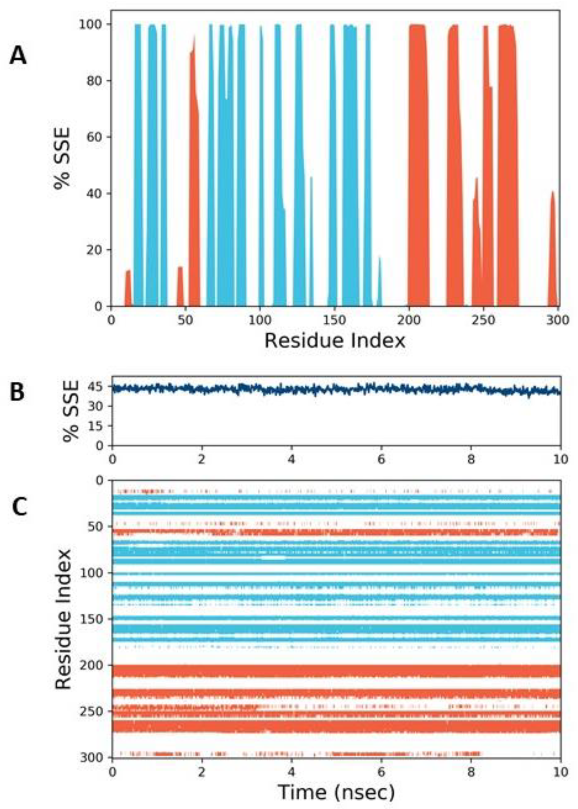
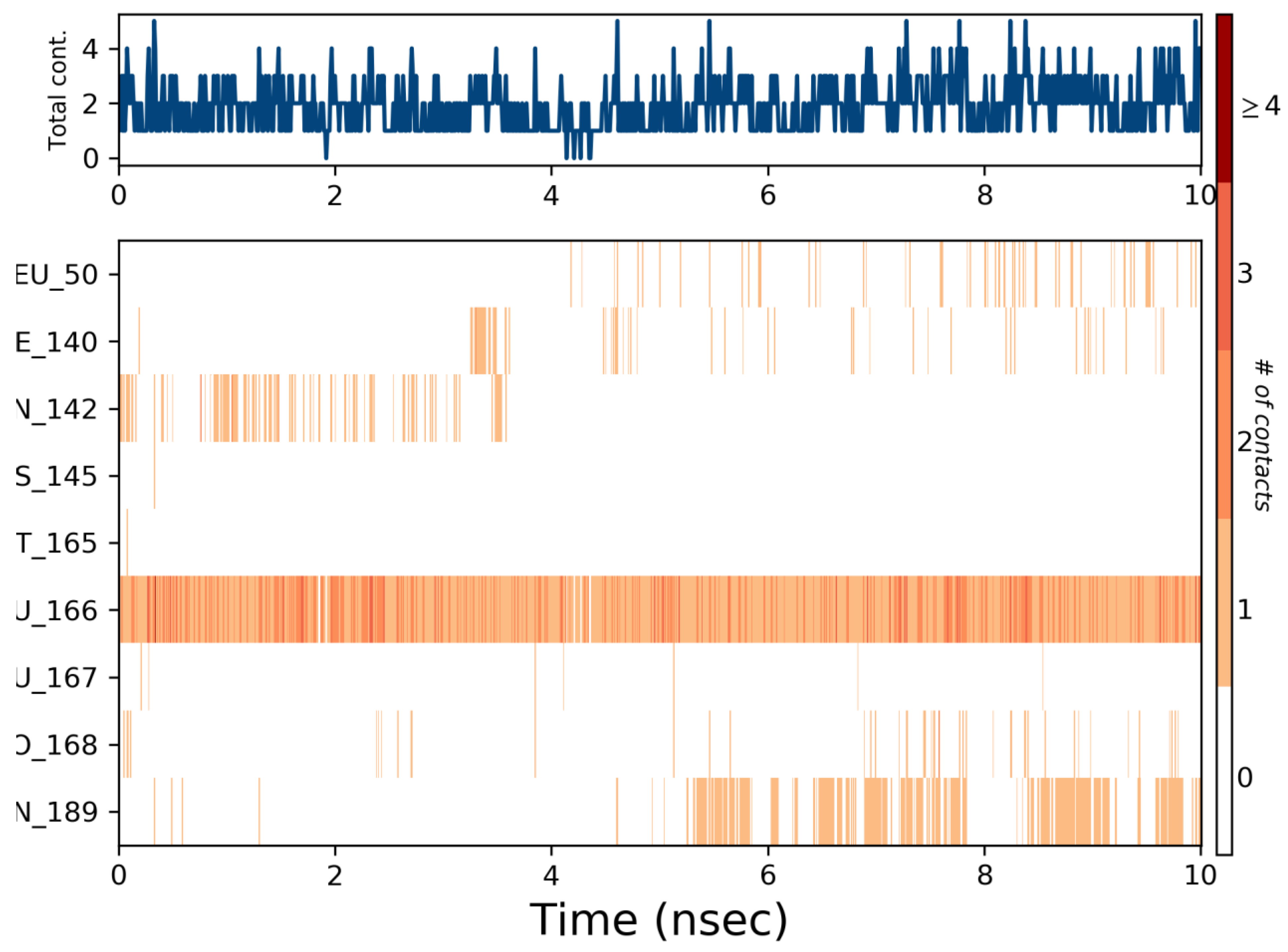
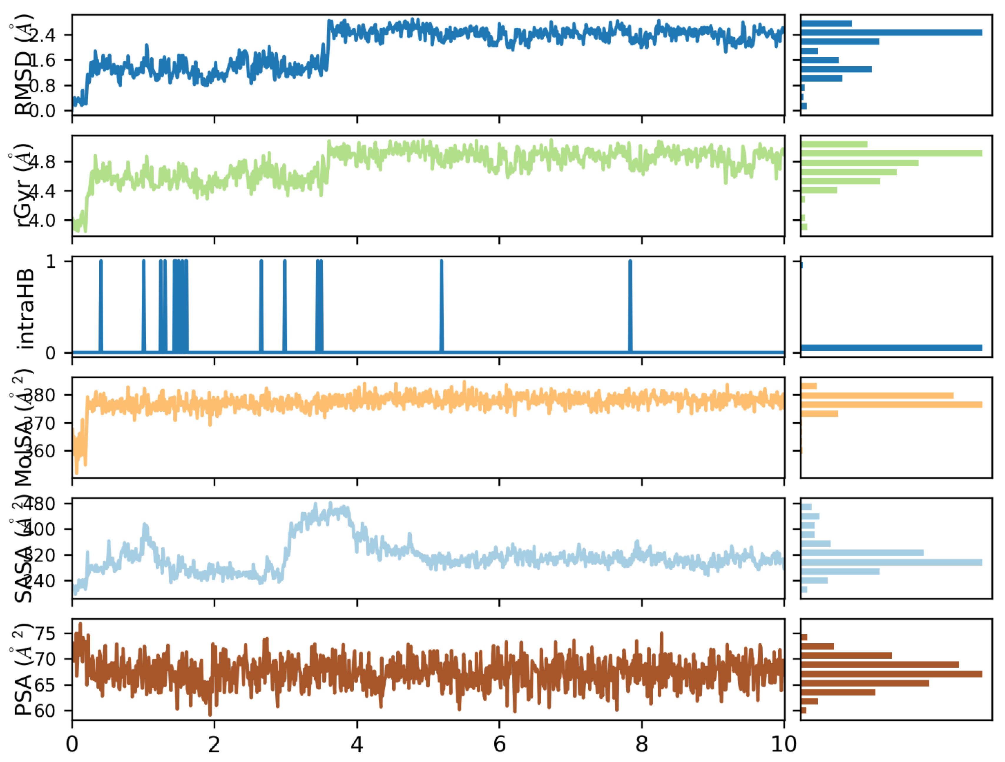
3.3. Molecular Dynamics Simulation

Figure 8 presents the root mean square deviation (RMSD) profiles of SARS-CoV-2 Mpro and imidazolyl–methanone C10. The protein exhibits small fluctuations in RMSD, ranging from 1 to 2.25 Å, indicating overall stability throughout the simulation. C10, on the other hand, shows lower RMSD values compared to the protein, suggesting a relatively stable binding to the protein. The red spectrum in the plot represents the RMSD of the ligand, where the protein–ligand complex is initially aligned based on the protein backbone of the reference structure, and then the RMSD of the ligand’s heavy atoms is measured. The ligand’s RMSD remains stable in the beginning until approximately 3.8 ns, followed by some fluctuations in the subsequent nanoseconds, and eventually stabilizes again at around 4.4 ns.
In Figure 9A,B, the root mean square fluctuation (RMSF) profiles of the protein target and C10 inhibitor are shown, respectively. The protein exhibits minimum fluctuations at around 0.46 and maximum fluctuations at 2.0, indicating regions of higher flexibility. Conversely, the ligand demonstrates minimum fluctuations at 1.5 and maximum fluctuations at 3.4, suggesting variations in its conformational behavior.
Figure 10 provides insights into the secondary structure elements (SSEs) of Mpro in the presence of the ligand. The plot displays the distribution of SSEs across the protein structure throughout the simulation. It also summarizes the composition of SSEs for each trajectory frame, revealing the time-dependent changes in SSE assignments for individual residues.
Figure 11 depicts a timeline representation of the interactions between C10 and Mpro. The top panel illustrates the total number of specific contacts formed between the protein and the ligand over the simulation time, which amounts to approximately 5 contacts. The bottom panel highlights GLU 166, along with other residues mentioned in Figure 5, as it interacts with the ligand residues across the trajectory. Several residues, including LEU 50, PHE 140, ASN 142, CYS 145, and MET 165, establish multiple specific contacts with the ligand.
Figure 12 showcases the torsional potential energies of different rotatable bonds in C10. The blue-encoded bond exhibits a torsional potential energy of 9.03 kcal/mol, primarily occurring between 0° and 90°, and 135° and 180°. The green-encoded bond shows a torsional potential energy of 15.04 kcal/mol, concentrated in the range of 0° to 90°. The peach-encoded bond demonstrates a torsional potential energy of 1.48 kcal/mol, mainly distributed between −180° and 0°. Lastly, the yellow-encoded bond displays a torsional potential energy of 11.09 kcal/mol, predominantly observed between 0° and 90°. The histogram and torsion potential relationships provide insights into the conformational strain experienced by the ligand to maintain a stable protein-bound conformation.
Figure 13 presents several molecular properties of C10. The radius of gyration (ROG) remains stable around 4.9 Å after an initial stability phase between 4.4 Å and 4.8 Å. The hydrogen bonding analysis indicates the presence of approximately 14 hydrogen bonds between C10 and Mpro. The solvent-accessible surface area (SASA) of the compound within the protein binding region ranges from 370 Å to 390 Å. Furthermore, the minimum and maximum SASA values are observed at 240 Å and 340 Å, respectively. The SASA shows stability at around 5 ns, with a value of 300 Å.

3.4. ADMET Profile

Table 2 presents the lipophilicity, drug-likeness, skin permeability, and bioavailability scores of the compounds, as predicted by SwissADME. The thiophenyl–imidazole compounds exhibit high Log p values, with C1, C3, and C2 having values of 6.06, 5.94, and 5.86, respectively, while C4 has the lowest Log p value of 4.9. The Log p values of the pyridyl–imidazoles range from 2.39 to 4.64, while the imidazolyl–methanones exhibit values between 1.29 and 4.76. The quinoline–imidazoles display Log p values ranging from 0.63 to 4.87. Regarding drug-likeness prediction, all thiophenyl–imidazole compounds, except C4, violate one Lipinski rule. On the other hand, none of the compounds from the remaining three classes violate any Lipinski rules, except for C18, which violates one Lipinski rule. Furthermore, all compounds adhere to Veber’s rule, except for C18, which has two violations. The table also displays the skin permeation values (log Kp in cm/s) of the test compounds, ranging from −8.76 (highly permeable) to −3.64 (less permeable). Among all the compounds, C18 and C12 demonstrate the highest skin permeability, while the range of values for each test compound indicates that they are all permeable. In terms of bioavailability prediction, the compounds, except for C18, exhibit a score of 0.55, while C18 scores 0.11.
Table 3 presents the results of the pharmacokinetics prediction for the test compounds. As depicted in the table, the majority of the compounds demonstrate significant gastrointestinal (GI) absorption potential; however, compounds C1, C2, C3, C11, and C18 exhibit lower GI absorption potential. Significantly, six compounds (C5, C9, C10, C13, C16, and C17) demonstrate the ability to cross the blood–brain barrier. All test compounds are P-glycoprotein (Pgp) substrates, except for C1, C6, C7, C8, and C11. Additionally, a considerable number of the test compounds are projected to inhibit CYP1A2, CYP2C19, CYP2C9, CYP2D6, and CYP3A4, except for C18, which lacks inhibitory activity. Specifically, compounds C6, C8, C11, C13, C14, C15, and C17 demonstrate inhibitory effects on all of the aforementioned cytochrome P450 enzymes (CYP1A2, CYP2C19, CYP2C9, CYP2D6, and CYP3A4).
In Table 4, the evaluation of various toxicological parameters is presented. None of the test compounds exhibit carcinogenic tendencies, except for compound C13. Additionally, cytotoxicity is not observed for any of the test compounds, with the exception of C1, C4, C5, C13, and C17. Compounds C1, C4, C5, C11, and C12 exhibit nephrotoxicity. Regarding skin sensitization, none of the test compounds display such potential. Moreover, eye irritation is absent for most compounds, with the exception of C4, C5, C11, and C12. None of the compounds cause eye corrosion, except for C5.

4. Discussion

The main protease (Mpro) plays a crucial role in the replication of coronaviruses and has emerged as a prominent target for antiviral drug development [5]. Within the 14 open reading frames (ORFs) of SARS-CoV-2, the ORF 1ab encodes two overlapping polyproteins (pp1a and pp1ab) that undergo precise cleavage events, leading to the generation of 16 non-structural proteins (nsp1–16) [23]. Among the various viral proteases involved in this intricate process, Mpro holds a central position. Inhibiting the enzymatic activity of Mpro has proven to be a crucial therapeutic strategy in the fight against SARS-CoV-2. The use of heterocyclic compounds as templates for antiviral drug development has shown great promise, demonstrating potent activity against various viral infections, including hepatitis C (HCV), hepatitis B (HBV), human immunodeficiency virus (HIV), herpes simplex (HSV), rotavirus, adenoviruses, and coxsackie viruses [24,25]. Notably, antiviral imidazole compounds have been reported to exert their activity by targeting viral proteases and other critical proteins that are essential to the viral life cycle [25]. Hence, in this study, we employed computational techniques to identify potential SARS-CoV-2 inhibitors from a specific set of distinct imidazole derivatives.
The imidazole compounds examined in this study displayed strong binding affinities to the target proteins of SARS-CoV-2, indicating their potential as effective therapeutic agents against the virus. Imidazoles, owing to their structural resemblance to the histidine amino acid, have the ability to interact with crucial protein molecules, thereby influencing their functions [25]. In this study, it was observed that the imidazolyl–methanones possess a higher binding affinity for all the target proteins than the other three classes of imidazoles. This could be linked to the imidazole rings, the methanone group, and several other aromatic rings contained in the structure of these compounds. Additionally, the spatial conformation and flexibility of imidazolyl–methanones might be favorable for optimal binding to the target proteins. The distinct structure and adaptability of the imidazolyl–methanone scaffold may enable improved accommodation and fitting within the main protease’s binding pocket, leading to enhanced interactions and a stronger binding affinity. Furthermore, the electronic and physicochemical properties of imidazolyl–methanones may contribute to their superior binding affinity. Analysis of the protein–ligand interaction of the highest-affinity compound, C10, revealed that all functional groups, including the aromatic rings, contribute to its binding to the target proteins. Aromatic interactions play a critical role in biological recognition, including protein–ligand interactions, as approximately 20% of amino acids possess aromatic properties [4]. Leveraging aromatic interactions is of great importance in drug design to enhance efficacy and optimize leads [4]. Overall, the results suggest that the imidazole derivatives, particularly the imidazolyl–methanones, hold promise as potential therapeutics against SARS-CoV-2. Their strong binding affinities, driven by their structural features and interactions, highlight their potential for further development and optimization in the fight against this global health threat.
The molecular docking analysis provided insights into the binding orientations of the compounds from the four imidazole classes with the best binding affinities compared to the standard ligands. Examination of the 3D and 2D structures of the docked complexes between the test compounds and SARS-CoV-2 target proteins confirmed their inhibitory potential. Specifically, compounds C1, C5, C10, and C14 from the thiophenyl–imidazole, pyridyl–imidazole, imidazolyl–methanone, and quinoline–imidazole classes, respectively, interacted with specific amino acid residues within the binding pocket of SARS-CoV-2 Mpro. These interactions involved various types of chemical bonds and interactions, such as carbon–hydrogen interactions, π–sulfur interactions, π–alkyl interactions, π–anion interactions, π–π bond interactions, hydrogen interactions, and π–sigma bonds. In contrast, the standard ligand formed covalent bonds and conventional hydrogen bonds. Notably, interactions with CYS A:145 and HIS A:41 were observed for compounds C1, C5, and C14, which are amino acids crucial for catalysis at the catalytic site of the SARS-CoV-2 main protease [4,26]. Dimerization and mutations in Mpro, leading to reduced enzymatic activity, are associated with interactions involving residues around GLU 288, ASP 289, and GLU 290 [27]. However, the tested compounds did not show interactions with GLU 288 and ASP 289, suggesting that their mechanism of inhibiting SARS-CoV-2 replication is more likely through direct elimination of Mpro via CYS 145 and HIS 41 [28,29], rather than by inducing mutations or dimerization. Overall, the findings suggest that the tested imidazole compounds have the potential to inhibit SARS-CoV-2 replication by directly targeting the catalytic site of Mpro through interactions with specific amino acid residues. Further experimental studies are warranted to validate these findings and explore the therapeutic potential of these compounds as antiviral agents.
The top-docking poses of the selected hit molecule C10 and Mpro were used in all-atom classical MD simulations using the Desmond package, developed by Schrodinger LLC. Molecular dynamics (MD) is a computational simulation method used to study the dynamic behavior of biomolecular systems over time. It involves modeling the interactions between individual atoms and molecules using physical laws and equations of motion.
The determination of root mean square deviation (RMSD) is a standard approach in computer-aided drug design to assess the structural changes in a macromolecule during molecular dynamics simulations [30]. In our study, we applied RMSD analysis to investigate the stability and movement of imidazolyl–methanone C10, the compound with the highest binding affinity, within the hydrated environment of the active pocket of SARS-CoV-2 Mpro throughout the simulation. Figure 8 illustrates the RMSD profiles of the protein, showing changes in the range of 1–2.25 Å. The protein demonstrates stability after approximately 3 ns, reaching an equilibrium state. Monitoring the protein’s RMSD provides valuable insights into its conformational dynamics during the simulation [31]. Fluctuations within the range of 1–3 Å are considered acceptable for small, globular proteins [32]. Larger changes indicate significant conformational transitions during the simulation. Based on our results, the observed changes in RMSD fall within the acceptable range, indicating a well-behaved system. The RMSD of C10 is consistently lower than that of the protein, with values ranging from 1 to 2.25 Å. Initially, the ligand remains stable within the protein until around 3.8 ns, after which it experiences fluctuations for a few nanoseconds before stabilizing again at approximately 4.4 ns. The ligand’s RMSD provides insights into its stability within the protein’s binding pocket. If the ligand’s RMSD values are significantly larger than those of the protein, it suggests that the ligand has diffused away from its initial binding site [33]. The observed stability of the ligand within the active pocket indicates that the complexes do not undergo substantial structural shifts, supporting the stability of these ligands at the protein’s binding site. Overall, the RMSD analysis provides valuable information on the structural dynamics and stability of both the protein and the ligand, offering insights into their conformational behavior during the simulation. The results suggest that the studied complexes maintain a favorable interaction within the active pocket, enhancing their potential as promising drug candidates for SARS-CoV-2 inhibition.
Root mean square fluctuation (RMSF) analysis provides valuable insights into the flexibility of different regions within a macromolecule. Figure 9A illustrates the RMSF plot of Mpro, highlighting the regions that exhibit the highest fluctuations during the simulation. The peaks observed indicate that the N- and C-terminal tails of the protein undergo greater fluctuations compared to other parts. This observation is consistent with previous findings, where secondary structure elements such as alpha helices and beta strands tend to be more rigid and exhibit lower fluctuations compared to loop regions [34].
In Figure 10, a comprehensive analysis of secondary structure elements (SSE) in Mpro is presented, tracking their distribution and composition throughout the simulation in relation to the ligand. This plot provides a detailed representation of the SSEs across the protein structure, enabling the monitoring of each residue’s SSE assignment and changes over time. By examining the SSE distribution, it becomes possible to assess the conformational stability and dynamic behavior of the protein–ligand complex. Furthermore, Figure 9B demonstrates the RMSF profile of the inhibitor imidazolyl–methanone C10, offering insights into the changes in ligand atom positions in relation to the protein. The ligand RMSF analysis allows for a characterization of the ligand fragments’ interactions with the protein and their role in the binding event. In both Figure 9A,B, the protein and ligand exhibit varying levels of fluctuation. The protein’s RMSF ranges from a minimum of 0.46 to a maximum of 2.0, while the ligand’s RMSF ranges from a minimum of 1.5 to a maximum of 3.4. These results indicate that the ligand maintains flexibility within the ligand–protein complex throughout the simulation, suggesting favorable dynamics and a potential for efficient binding interactions. Overall, the RMSF analysis provides valuable information on the flexibility and dynamic behavior of the protein and ligand within the complex. The observed fluctuations in specific regions of the protein and ligand shed light on their conformational changes and their entropic contributions to the binding event. These findings contribute to our understanding of the dynamic nature of the protein–ligand complex and its implications for drug design and molecular interactions.
As mentioned earlier and depicted in Figure 5, the interaction between C10 and Mpro involves specific residues, including LEU 50, PHE 140, ASN 142, CYS 145, and MET 165. Further insights into the dynamic nature of this interaction are provided in Figure 11, which reveals that the total number of specific contacts between Mpro and the ligand throughout the trajectory is approximately 5. Notably, GLU 166 and other residues interact with the ligand consistently in each trajectory frame. Notably, residues such as LEU 50, PHE 140, ASN 142, CYS 145, and MET 165 establish multiple specific contacts with the ligand.
In addition, the ligand torsions plot offers a comprehensive overview of the conformational changes in each rotatable bond (RB) within C10 during the simulation trajectory spanning from 0.00 to 10.00 ns. The bar plots in the plot summarize the torsional probability density, which represents the likelihood of observing specific torsional potential energy values within the dataset. The torsional potential energy values themselves reflect the energy required to rotate the rotatable bonds in the ligand. Higher energy values indicate increased strain or resistance to rotation, while lower energy values signify more favorable or stable conformations [35]. Moreover, the probability density reveals the prevalence of certain energy ranges, indicating the frequency of sampling or stability of conformations with those energy values [36]. By analyzing the torsion plot, valuable insights into the conformational dynamics and stability of the ligand can be obtained. The analysis of the histogram and torsion potential relationships provides valuable insights into the conformational strain experienced by the ligand in maintaining a protein-bound conformation. The torsional potential energy values offer a measure of the energy required to rotate the ligand’s rotatable bonds, indicating the stability and strain associated with different conformations. In our study, the blue bond exhibited a torsional potential energy of 9.03 kcal/mol, with preferred conformations observed in the ranges of 0°–90° and 135°–180°. These angles suggest favorable and stable orientations of the bond, indicating lower conformational strain. Conversely, the green bond displayed a higher torsional potential energy of 15.04 kcal/mol within the range of 0°–90°, indicating greater strain and resistance to rotation. On the other hand, the peach bond showed a torsional potential energy of 1.48 kcal/mol within the range of −180°–0°, indicating more favorable and stable conformations. Finally, the yellow bond exhibited a torsional potential energy of 11.09 kcal/mol between 0°–90°, suggesting higher strain and reduced stability. These findings highlight the diverse conformational dynamics of the ligand and provide important insights into its ability to adopt protein-bound conformations. The understanding of such conformational strain is crucial for designing and optimizing ligands with improved binding affinity and pharmacological properties.
In molecular dynamics (MD) simulations, several structural descriptors such as the radius of gyration (ROG), hydrogen bond analysis, solvent-accessible surface area (SASA), and polar surface area (PSA) play a crucial role in analyzing the behavior and properties of biomolecules.
The ROG provides valuable insights into the compactness of the system during the simulation, reflecting its performance in a biological context. It quantifies the “extendedness” of a ligand and is mathematically equivalent to its principal moment of inertia [37]. As shown in Figure 13, the ROG spectrum demonstrates that the system maintains a consistent level of compactness throughout the simulation. The histogram’s mode value indicates that the ROG of C10 remains stable at approximately 4.9 Å, with an initial period of stability observed between 4.4 Å and 4.8 Å at around 3.8 ns. This observation suggests that the Mpro protein exhibits a well-folded structure and is likely to engage in effective interaction mechanisms with the ligand hits.
In molecular modeling, the evaluation of a ligand’s ability to bind to a target typically involves molecular docking, which is complemented by visual inspection to understand the nature of the interaction [38]. However, a limitation of molecular docking is its inability to consider the presence of water molecules within the active site. To address this, advanced modeling analyses, such as hydrogen bond computation after molecular dynamics (MD) simulation, are often employed to accurately determine the number of hydrogen bonds involved in ligand–protein interactions. In contrast to the results obtained from docking and visual inspection (Figure 6), the analysis of hydrogen bonding reveals that imidazolyl–methanone C10 forms approximately 14 hydrogen bonds with Mpro. This finding contradicts the absence of hydrogen bonds observed in the initial docking analysis. The presence of hydrogen bonds suggests a significant binding force that mediates the interaction between these compounds and Mpro. This observation further indicates that imidazolyl–methanone has the potential to effectively inhibit the target protein.
Molecular surface area (MolSA) serves as a quantitative measure of the total surface area of a molecule, encompassing both its accessible and inaccessible regions [39]. By examining the SASA (solvent-accessible surface area) of the compound within the protein binding region, we observe a range from 370 Å to 390 Å. A reduction in MolSA implies a compacting effect, surface burial, or the closure of pockets [40]. Conversely, a significant increase in MolSA may indicate structural expansion, surface exposure, or the opening of cavities or binding sites. These alterations in MolSA can be associated with various molecular phenomena such as flexibility, ligand binding, protein–protein interactions, or solvent interactions.
Considering that the drug–target binding process takes place within a dynamic fluid environment of a biological system, it is crucial to compute the solvent-accessible surface area (SASA) after conducting molecular dynamics simulations. This analysis helps assess the degree to which the simulated system accurately represents the intracellular milieu. SASA represents the surface area of a molecule that is accessible to solvent molecules and actively interacts with them [41]. In this study, the mean SASA values for each complex were computed and plotted over the course of the simulation (Figure 13). The calculated SASA values ranged from 240 Å to 340 Å, with a stable SASA observed around 300 Å at approximately 5 ns. These results indicate that the studied compounds exhibit a comparable degree of exposure to water, suggesting their potential for biological stability in contrast to the other examined complexes.
Polar surface area (PSA) is a parameter that quantifies the extent of the molecule’s surface area occupied by polar atoms or polar groups. The analysis of the graph provides insights into the ability of C10 to engage in hydrogen bonding and interact with other polar molecules. Throughout the 10 ns simulation, the PSA values of the compounds range from 60 Å to 75 Å. In the field of drug discovery, PSA is frequently employed as a descriptor to predict a molecule’s permeability and its capacity to traverse biological membranes. Higher PSA values generally indicate increased polarity and a greater potential for hydrogen bonding, both of which can influence the molecule’s solubility and membrane permeability characteristics [42].
In addition to their demonstrated inhibitory potentials towards the drug targets, the test compounds exhibit favorable ADMET properties of moderate magnitude. However, it is imperative to acknowledge that certain compounds may necessitate further optimization to enhance their overall ADMET profile while concurrently preserving or augmenting their binding affinity. ADMET analysis encompasses critical parameters encompassing absorption, distribution, metabolism, elimination, and toxicity, which collectively govern the pharmacokinetic behavior of a compound. This evaluation seeks to ascertain the compound’s ability to be readily absorbed, reach the intended site of action, undergo biotransformation without compromising its activity, be efficiently eliminated from the organism, and avert the manifestation of adverse toxicological effects [4]. The ideal drug candidate is characterized by not only its efficacy against the therapeutic target but also its favorable ADMET properties within the therapeutic dose range [43]. The recent emergence of computational models for in silico prediction of chemical ADMET properties has presented a significant advantage in drug discovery and development. These computational approaches offer a valuable tool for early identification of potential pharmacokinetic liabilities and drug failures, enabling informed decision making during the drug design process prior to costly and time-consuming clinical trials [4]. Therefore, leveraging computational techniques for the comprehensive evaluation of ADMET properties holds tremendous potential in the identification and optimization of promising drug candidates, facilitating the rational design of efficacious and safe therapeutic agents.
Lipophilicity, a crucial physicochemical parameter, serves as the pivotal link between solubility, membrane permeability, and, consequently, the processes of drug absorption, distribution, and clearance [4]. Lipophilic compounds exhibit lower solubility in aqueous environments but possess favorable solubility in oils and lipids, thereby facilitating their permeation across biological membranes. However, it is noteworthy that compounds with high lipophilicity (Log p > 5) often exhibit elevated metabolic turnover, reduced solubility, and poor oral absorption. Furthermore, highly lipophilic compounds have a heightened propensity to interact with hydrophobic targets other than the intended target, leading to an increased risk of promiscuity and toxicity [44]. The lipophilicity of a compound is quantitatively characterized by its Log p value, whereby higher Log p values correspond to greater lipophilicity and lower water solubility [45]. In our study, the test compounds demonstrated Log p values ranging from 0.63 to 6.60 (C1–C18), resulting in moderate IT Log Sw values indicative of their solubility characteristics (Table 2).
Drug-likeness assessment, a vital aspect of drug development, involves evaluating chemical structures and physicochemical properties to qualitatively determine the oral bioavailability of potential drug candidates [4]. The Lipinski filter, a widely utilized approach, enables the screening of compounds for oral drug-likeness based on factors such as molecular weight, hydrogen bond acceptors and donors, and lipophilicity [46]. The rule outlines specific conditions that a compound must fulfill to be regarded as orally active. These conditions include having ≤5 H-bond donors, ≤10 H-bond acceptors, a molecular weight ≤500 g/mol, and a Log p value less than 5.43. If a compound violates two or more of Lipinski’s rules, it is classified as orally inactive [46]. In our study, all test compounds exhibited zero Lipinski violations, except for C1, C2, C3, and C18, which had one violation (Table 2). Thus, with the exception of C18, all compounds are likely to possess oral activity, a conclusion further supported by their oral bioavailability scores. The quinoline–imidazole compound displayed a bioavailability score of 0.11, indicating approximately 11% probability of achieving at least 10% oral bioavailability in rats or measurable permeability in human colon carcinoma (Caco-2) cells. On the contrary, the remaining compounds displayed a bioavailability score of 0.55, implying a 55% chance of achieving at least 10% oral absorption in rats or human colon carcinoma absorptivity, according to Testa and Kraemer [47]. It is noteworthy that all the test compounds adhered to Veber’s rule, with the exception of C18, which did not meet the criteria of having ≤10 rotatable bonds and a polar surface area (TPSA) ≤ 140 Å2, as proposed by Veber et al. [48].
The pharmacokinetic predictions of the test compounds indicate that a significant number of these compounds possess inhibitory activity against several cytochrome P450 enzymes, namely CYP1A2, CYP2C19, CYP2C9, CYP2D6, and CYP3A4, with the exception of C18, which does not exhibit inhibitory effects. Notably, compounds C6, C8, C11, C13, C14, C15, and C17 demonstrate inhibitory activity against all of these cytochrome P450 enzymes (Table 3). Cytochrome P450 (CYP) enzymes are part of an isoenzyme superfamily that plays a vital role in various phase I drug metabolism processes [4]. Inhibiting the five major isoforms, namely CYP1A2, CYP2C19, CYP2C9, CYP2D6, and CYP3A4, can lead to drug–drug interactions related to pharmacokinetics since these enzymes are commonly involved in the metabolism of medications [45,49]. Understanding the inhibitory potential of compounds against these enzymes is essential for assessing their potential for drug–drug interactions and optimizing their pharmacokinetic properties.
With the exception of compounds C1, C6, C7, C8, and C11, all the test compounds chosen for this investigation are anticipated to act as substrates of P-glycoprotein (Pgp). P-glycoprotein is an ATP-binding cassette transporter responsible for actively transporting xenobiotics across biological membranes, which can lead to drug resistance and safeguard the body against foreign toxins [4]. The interaction of compounds with Pgp as substrates has implications for their pharmacokinetics, including absorption, distribution, metabolism, and excretion. Pgp is present in the intestines, where it can efflux drugs back into the intestinal lumen, reducing their absorption into the bloodstream [50]. Consequently, compounds that are Pgp substrates may exhibit lower oral bioavailability. Moreover, Pgp is expressed in the blood–brain barrier and other tissues [51]. Its activity can limit the penetration of Pgp substrate drugs into the brain and other target tissues, potentially impacting their efficacy in reaching specific sites of action. Additionally, Pgp’s role in drug transport can indirectly influence drug metabolism by modulating drug concentrations in organs and tissues where metabolism occurs [52]. Furthermore, Pgp is involved in drug excretion, particularly in the liver and kidneys, by actively pumping drugs out of hepatocytes and renal tubular cells, contributing to their elimination from the body [53]. Overall, the interaction of drugs with Pgp can significantly influence their pharmacokinetics, altering drug absorption, distribution, metabolism, and excretion. Considering the compounds identified as Pgp substrates, it is crucial to take into account potential drug–drug interactions and determine appropriate dosing strategies.
While the toxicity prediction results indicated that compounds C2, C3, C6, C7, C8, C9, C10, C14, C15, C16, and C18 did not exhibit any tendencies towards toxicity based on the parameters tested, it is important to consider experimental validation of all compounds reported in the study. In vitro and in vivo studies, such as cell-based assays or animal models, offer the potential to obtain more precise and reliable information regarding their toxicity profiles. Experimental findings can validate or disprove the predicted toxicities, providing valuable insights into the actual risk associated with the compounds. Additionally, there is potential to repurpose these compounds as antibacterial and anticancer agents, exploring their effectiveness in these applications [54,55,56,57,58]. Thus, it is recommended to conduct experimental studies to further investigate these compounds and explore their potential for development as novel drugs for the treatment of SARS-CoV-2.

5. Conclusions

A comprehensive evaluation was conducted on four distinct classes of recently synthesized imidazole derivatives to assess their therapeutic potential against SARS-CoV-2 Mpro. The results revealed remarkable binding affinities and stability of these compounds with the target proteins. Notably, all 18 compounds exhibited substantial binding affinities towards Mpro, with imidazolyl–methanone C10 displaying the highest affinity. Furthermore, pyridyl–imidazole C5, thiophenyl–imidazole C1, and quinoline–imidazole C14 exhibited binding affinities of −8.3, −8.2, and −7.7 Kcal/mol, respectively. The interaction of these compounds with specific residues such as HIS A:41 and CYS A:145 within the binding pocket of SARS-CoV-2 Mpro further validates their potential as therapeutic candidates against the virus. However, to optimize the ADMET properties of certain compounds while preserving their biological activities, lead optimization strategies may be necessary. It is imperative to validate the findings of this in silico study through wet laboratory experiments to establish the true potential of these novel imidazoles as therapeutics for SARS-CoV-2.

Author Contributions

Conceptualization, B.A.B.; methodology, B.A.B.; software, B.A.B. and A.E.A.; validation, B.A.B.; formal analysis, B.A.B.; investigation, B.A.B. and A.E.A.; resources, B.A.B.; data curation, B.A.B.; writing—original draft preparation, B.A.B.; writing—review and editing, B.A.B.; visualization, B.A.B. and A.E.A.; supervision, B.A.B.; project administration, B.A.B.; funding acquisition, B.A.B. All authors have read and agreed to the published version of the manuscript.

Funding

This study did not receive any dedicated funding from public, commercial, or non-profit organizations.

Acknowledgments

The authors extend their gratitude to the healthcare frontline, scientists, and the World Health Organization for their immense contributions to the fight against coronavirus in Sub-Saharan Africa.

Conflicts of Interest

The authors affirm that they have no known financial conflicts of interest or personal relationships that could have influenced the findings presented in this paper.

References

  1. World Health Organization—WHO. Severe Acute Respiratory Syndrome (SARS). 2024. Available online: https://www.who.int/emergencies/diseases/novel-coronavirus-2019 (accessed on 6 May 2024).
  2. Centers for Disease Control and Prevention—CDC. SARS Basics Fact Sheet. Available online: https://www.cdc.gov/sars/about/fs-sars.html (accessed on 13 July 2022).
  3. Babalola, B.; Akinsuyi, O.S.; Folajimi, E.M.; Olujimi, F.; Otunba, A.A.; Chikere, B.; Adewumagun, I.A.; Adetobi, T.E. Exploring the future of SARS-CoV-2 treatment after the first two years of the pandemic: A comparative study of alternative therapeutics. Biomed. Pharmacotherapy 2023, 165, 115099. [Google Scholar] [CrossRef] [PubMed]
  4. Babalola, B.A.; Adetobi, T.E.; Akinsuyi, O.S.; Adebisi, O.A.; Folajimi, E.O. Computational Study of the Therapeutic Potential of Novel Heterocyclic Derivatives against SARS-CoV-2. Covid 2021, 1, 757–774. [Google Scholar] [CrossRef]
  5. Pang, X.; Xu, W.; Liu, Y.; Li, H.; Chen, L. The research progress of SARS-CoV-2 main protease inhibitors from 2020 to 2022. Eur. J. Med. Chem. 2023, 257, 115491. [Google Scholar] [CrossRef]
  6. Li, Q.; Kang, C. Progress in Developing Inhibitors of SARS-CoV-2 3C-Like Protease. Microorganisms. 2020, 8, 1250. [Google Scholar] [CrossRef]
  7. Hu, Q.; Xiong, Y.; Zhu, G.H.; Zhang, Y.N.; Zhang, Y.W.; Huang, P.; Ge, G.B. The SARS-CoV-2 main protease (Mpro): Structure, function, and emerging therapies for COVID-19. MedComm 2022, 3, e151. [Google Scholar] [CrossRef]
  8. Adetobi, E.T.; Akinsuyi, S.O.; Ahmed, O.A.; Folajimi, E.O.; Babalola, B.A. In silico Evaluation of the Inhibitory Potential of Cymbopogonol from Cymbopogon citratus Towards Falcipain-2 (FP2) Cysteine Protease of Plasmodium falciparum. Trop. J. Nat. Prod. Res. 2022, 6, 1687–1694. [Google Scholar]
  9. Babalola, B.A.; Akinwande, A.I.; Gboyega, A.E.; Otunba, A.A. Extraction, purification and characterization of papain cysteine-proteases from the leaves of Carica papaya. Sci. Afr. 2023, 19, e01538. [Google Scholar] [CrossRef]
  10. Slassi, S.; Aarjane, M.; Amine, A. Synthesis, spectroscopic characterization (FT-IR, NMR, UV-Vis), DFT study, antibacterial and antioxidant in vitro investigations of 4, 6-bis ((E)-1-((3-(1H-imidazol-1-yl) propyl) imino) ethyl) benzene-1, 3-diol. J. Mol. Struct. 2022, 1255, 132457. [Google Scholar] [CrossRef]
  11. Seck, I.; Nguemo, F. Triazole, imidazole, and thiazole-based compounds as potential agents against coronavirus. Results Chem. 2021, 3, 100132. [Google Scholar] [CrossRef] [PubMed]
  12. Batiha, G.E.S.; Teibo, J.O.; Shaheen, H.M.; Babalola, B.A.; Teibo, T.K.A.; Al-Kuraishy, H.M.; Al-Garbeeb, A.I.; Alexiou, A.; Papadakis, M. Therapeutic potential of Lawsonia inermis Linn: A comprehensive overview. Naunyn Schmiedeberg Arch. Pharmacol. 2024, 397, 3525–3540. [Google Scholar] [CrossRef]
  13. Zhang, Q.; Guo, L.; Huang, Y.; Zhang, R.; Ritacca, A.G.; Leng, S.; Zheng, X.; Yang, Y.; Singh, A. Influence of an imidazole-based ionic liquid as electrolyte additive on the performance of alkaline Al-air battery. J. Power Sources 2023, 564, 232901. [Google Scholar] [CrossRef]
  14. Çetiner, G.; Çevik, U.A.; Celik, I.; Bostancı, H.E.; Özkay, Y.; Kaplancıklı, Z.A. New Imidazole Derivatives as Aromatase Inhibitor: Design, Synthesis, Biological Activity, Molecular Docking, and Computational ADME-Tox Studies. J. Mol. Struct. 2023, 1278, 134920. [Google Scholar] [CrossRef]
  15. Poyraz, S.; Döndaş, H.A.; Sansano, J.M.; Belveren, S.; Yamali, C.; Ülger, M.; Döndaş, N.Y.; Sağlık, B.N.; Pask, C.M. N-Benzoylthiourea-pyrrolidine carboxylic acid derivatives bearing an imidazole moiety: Synthesis, characterization, crystal structure, in vitro ChEs inhibition, and antituberculosis, antibacterial, antifungal studies. J. Mol. Struct. 2023, 1273, 134303. [Google Scholar] [CrossRef]
  16. Sadula, A.; Gaddhe, L. Synthesis, computational studies and biological evaluation of novel Acenaphthoquinone-imidazole derivatives as dual inhibitors of HSP90 and Topo II in cancer therapy. Results Chem. 2023, 5, 100796. [Google Scholar] [CrossRef]
  17. Muhammed, M.T.; Mustafa, E.R.; Akkoc, S. Molecular modeling and in vitro antiproliferative activity studies of some imidazole and isoxazole derivatives. J. Mol. Struct. 2023, 1282, 135066. [Google Scholar] [CrossRef]
  18. Chhetri, A.; Chettri, S.; Rai, P.; Sinha, B.; Brahman, D. Exploration of inhibitory action of Azo imidazole derivatives against COVID-19 main protease (Mpro): A computational study. J. Mol. Struct. 2021, 1224, 129178. [Google Scholar] [CrossRef]
  19. O’Boyle, N.M.; Banck, M.; James, C.A.; Morley, C.; Vandermeersch, T.; Hutchison, G.R. Open Babel: An open chemical toolbox. J Cheminform 2011, 3, 33. [Google Scholar] [CrossRef]
  20. Dong, J.; Cao, D.S.; Miao, H.Y.; Liu, S.; Deng, B.C.; Yun, Y.H.; Wang, N.N.; Lu, A.P.; Zeng, W.B.; Chen, A.F. ChemDes: An integrated web-based platform for molecular descriptor and fingerprint computation. J. Cheminform. 2015, 7, 60. [Google Scholar] [CrossRef]
  21. Trott, O.; Olson, A.J. AutoDock Vina: Improving the speed and accuracy of docking with a new scoring function, efficient optimization, and multithreading. J. Comput. Chem. 2010, 31, 455–461. [Google Scholar] [CrossRef]
  22. Dong, J.; Wang, N.N.; Yao, Z.J.; Zhang, L.; Cheng, Y.; Ouyang, D.; Lu, A.P.; Cao, D.S. ADMETlab: A platform for systematic ADMET evaluation based on a comprehensively collected ADMET database. J. Cheminform. 2018, 10, 29. [Google Scholar] [CrossRef]
  23. Ullrich, S.; Nitsche, C. The SARS-CoV-2 main protease as drug target. Bioorganic Med. Chem. Lett. 2020, 30, 127377. [Google Scholar] [CrossRef] [PubMed]
  24. Liu, L.; Hu, Y.; Shen, Y.F.; Wang, G.X.; Zhu, B. Evaluation on antiviral activity of coumarin derivatives against spring viraemia of carp virus in epithelioma papulosum cyprini cells. Antivir. Res. 2017, 144, 173–185. [Google Scholar] [CrossRef]
  25. Kanwal, A.; Ahmad, M.; Aslam, S.; Naqvi, S.A.R.; Saif, M.J. Recent advances in antiviral benzimidazole derivatives: A mini review. Pharm. Chem. J. 2019, 53, 179–187. [Google Scholar] [CrossRef]
  26. Kneller, D.W.; Phillips, G.; Weiss, K.L.; Pant, S.; Zhang, Q.; O’Neill, H.M.; Coates, L.; Kovalevsky, A. Unusual zwitterionic catalytic site of SARS–CoV-2 main protease revealed by neutron crystallography. J. Biol. Chem. 2020, 295, 17365–17373. [Google Scholar] [CrossRef] [PubMed]
  27. Novak, J.; Rimac, H.; Kandagalla, S.; Pathak, P.; Naumovich, V.; Grishina, M.; Potemkin, V. Proposition of a new allosteric binding site for potential SARS-CoV-2 3CL protease inhibitors by utilizing molecular dynamics simulations and ensemble docking. J. Biomol. Struct. Dyn. 2022, 40, 9347–9360. [Google Scholar] [CrossRef] [PubMed]
  28. Lan, J.; Ge, J.; Yu, J.; Shan, S.; Zhou, H.; Fan, S.; Zhang, Q.; Shi, X.; Wang, Q.; Zhang, L.; et al. Structure of the SARS-CoV-2 spike receptor-binding domain bound to the ACE2 receptor. Nature 2020, 581, 215–220. [Google Scholar] [CrossRef] [PubMed]
  29. Yan, R.; Zhang, Y.; Li, Y.; Xia, L.; Guo, Y.; Zhou, Q. Structural basis for the recognition of SARS-CoV-2 by full-length human ACE2. Science 2020, 367, 1444–1448. [Google Scholar] [CrossRef] [PubMed]
  30. Sargsyan, K.; Grauffel, C.; Lim, C. How molecular size impacts RMSD applications in molecular dynamics simulations. J. Chem. Theory Comput. 2017, 13, 1518–1524. [Google Scholar] [CrossRef]
  31. Monhemi, H.; Housaindokht, M.R.; Moosavi-Movahedi, A.A.; Bozorgmehr, M.R. How a protein can remain stable in a solvent with high content of urea: Insights from molecular dynamics simulation of Candida antarctica lipase B in urea: Choline chloride deep eutectic solvent. Phys. Chem. Chem. Phys. 2014, 16, 14882–14893. [Google Scholar] [CrossRef]
  32. Uttarkar, A.; Niranjan, V. Re-profiling of natural inhibitor via combinatorial drug screening: Brefeldin A variant design as an effective antagonist leading to EPAC2 structure modification and antibody design for identification. bioRxiv 2021. [Google Scholar] [CrossRef]
  33. Durdagi, S.; ul Qamar, M.T.; Salmas, R.E.; Tariq, Q.; Anwar, F.; Ashfaq, U.A. Investigating the molecular mechanism of staphylococcal DNA gyrase inhibitors: A combined ligand-based and structure-based resources pipeline. J. Mol. Graph. Model. 2018, 85, 122–129. [Google Scholar] [CrossRef]
  34. Oum, Y.H.; Kell, S.A.; Yoon, Y.; Liang, Z.; Burger, P.; Shim, H. Discovery of novel aminopiperidinyl amide CXCR4 modulators through virtual screening and rational drug design. Eur. J. Med. Chem. 2020, 201, 112479. [Google Scholar] [CrossRef] [PubMed]
  35. Rai, B.K.; Sresht, V.; Yang, Q.; Unwalla, R.; Tu, M.; Mathiowetz, A.M.; Bakken, G.A. Comprehensive assessment of torsional strain in crystal structures of small molecules and protein–ligand complexes using ab initio calculations. J. Chem. Inf. Model. 2019, 59, 4195–4208. [Google Scholar] [CrossRef]
  36. Denning, E.J.; Priyakumar, U.D.; Nilsson, L.; Mackerell Jr, A.D. Impact of 2′-hydroxyl sampling on the conformational properties of RNA: Update of the CHARMM all-atom additive force field for, R.N.A. J. Comput. Chem. 2011, 32, 1929–1943. [Google Scholar] [CrossRef]
  37. Baby, K.; Maity, S.; Mehta, C.H.; Nayak, U.Y.; Shenoy, G.G.; Pai, K.S.R.; Harikumar, K.B.; Nayak, Y. Computational drug repurposing of Akt-1 allosteric inhibitors for non-small cell lung cancer. Sci. Rep. 2023, 13, 7947. [Google Scholar] [CrossRef]
  38. Fischer, A.; Smiesko, M.; Sellner, M.; Lill, M.A. Decision making in structure-based drug discovery: Visual inspection of docking results. J. Med. Chem. 2021, 64, 2489–2500. [Google Scholar] [CrossRef]
  39. Wu, J.; Hu, B.; Sun, X.; Wang, H.; Huang, Y.; Zhang, Y.; Liu, M.; Liu, Y.; Zhao, Y.; Wang, J.; et al. In silico study reveals existing drugs as α-glucosidase inhibitors: Structure-based virtual screening validated by experimental investigation. J. Mol. Struct. 2020, 1218, 128532. [Google Scholar] [CrossRef]
  40. Gyebi, G.A.; Ogunyemi, O.M.; Adefolalu, A.A.; Rodríguez-Martínez, A.; López-Pastor, J.F.; Banegas-Luna, A.J.; Pérez-Sánchez, H.; Adegunloye, A.P.; Ogunro, O.B.; Afolabi, S.O. African derived phytocompounds may interfere with SARS-CoV-2 RNA capping machinery via inhibition of 2′-O-ribose methyltransferase: An in silico perspective. J. Mol. Struct. 2022, 1262, 133019. [Google Scholar] [CrossRef]
  41. Chen, H.; Panagiotopoulos, A.Z. Molecular modeling of surfactant micellization using solvent-accessible surface area. Langmuir 2019, 35, 2443–2450. [Google Scholar] [CrossRef]
  42. Whitty, A.; Zhong, M.; Viarengo, L.; Beglov, D.; Hall, D.R.; Vajda, S. Quantifying the chameleonic properties of macrocycles and other high-molecular-weight drugs. Drug Discov. Today 2016, 21, 712–717. [Google Scholar] [CrossRef]
  43. Babalola, B.A.; Akinwande, A.I.; Otunba, A.A.; Adebami, G.E.; Babalola, O.; Nwufo, C. Therapeutic benefits of Carica papaya: A review on its pharmacological activities and characterization of papain. Arab. J. Chem. 2024, 17, 105369. [Google Scholar] [CrossRef]
  44. Gao, Y.; Gesenberg, C.; Zheng, W. Oral formulations for preclinical studies: Principle, design, and development considerations. In Developing Solid Oral Dosage Forms; Academic Press: Cambridge, MA, USA, 2017; pp. 455–495. [Google Scholar]
  45. Daina, A.; Michielin, O.; Zoete, V. SwissADME: A free web tool to evaluate pharmacokinetics, drug-likeness and medicinal chemistry friendliness of small molecules. Sci. Rep. 2017, 7, 42717. [Google Scholar] [CrossRef]
  46. Lipinski, C.A.; Lombardo, F.; Dominy, B.W.; Feeney, P.J. Experimental and computational approaches to estimate solubility and permeability in drug discovery and development settings. Adv. Drug Deliv. Rev. 2012, 64, 4–17. [Google Scholar] [CrossRef]
  47. Testa, B.; Kraemer, S.D. The biochemistry of drug metabolism—An introduction: Part 5. Metabolism and bioactivity. Chem. Biodivers. 2009, 6, 591–684. [Google Scholar] [CrossRef]
  48. Veber, D.F.; Johnson, S.R.; Cheng, H.Y.; Smith, B.R.; Ward, K.W.; Kopple, K.D. Molecular properties that influence the oral bioavailability of drug candidates. J. Med. Chem. 2002, 45, 2615–2623. [Google Scholar] [CrossRef]
  49. Ursu, O.; Rayan, A.; Goldblum, A.; Oprea, T.I. Understanding drug-likeness. Wiley Interdiscip. Rev. Comput. Mol. Sci. 2011, 1, 760–781. [Google Scholar] [CrossRef]
  50. Lazzaro, S.; West, M.A.; Eatemadpour, S.; Feng, B.; Varma, M.V.; Rodrigues, A.D.; Temesszentandrási-Ambrus, C.; Kovács-Hajdu, P.; Nerada, Z.; Gáborik, Z.; et al. Translatability of in vitro inhibition potency to in vivo P-glycoprotein mediated drug interaction risk. J. Pharm. Sci. 2023, 112, 1715–1723. [Google Scholar] [CrossRef]
  51. Cox, B.; Nicolaï, J.; Williamson, B. The role of the efflux transporter, P-glycoprotein, at the blood–brain barrier in drug discovery. Biopharm. Drug Dispos. 2023, 44, 113–126. [Google Scholar] [CrossRef]
  52. Yoshitomo, A.; Asano, S.; Hozuki, S.; Tamemoto, Y.; Shibata, Y.; Hashimoto, N.; Takahashi, K.; Sasaki, Y.; Ozawa, N.; Kageyama, M.; et al. Significance of basal membrane permeability of epithelial cells in predicting intestinal drug absorption. Drug Metab. Dispos. 2023, 51, 318–328. [Google Scholar] [CrossRef]
  53. Danner, L.; Malard, F.; Valdes, R.; Olivier-Van Stichelen, S. Non-Nutritive Sweeteners Acesulfame Potassium and Sucralose Are Competitive Inhibitors of the Human P-glycoprotein/Multidrug Resistance Protein 1 (PGP/MDR1). Nutrients 2023, 15, 1118. [Google Scholar] [CrossRef]
  54. Femi-Olabisi, F.J.; Ishola, A.A.; Faokunla, O.; Agboola, A.O.; Babalola, B.A. Evaluation of the inhibitory potentials of selected compounds from Costus spicatus (Jacq.) rhizome towards enzymes associated with insulin resistance in polycystic ovarian syndrome: An in silico study. J. Genet. Eng. Biotechnol. 2021, 19, 176. [Google Scholar] [CrossRef]
  55. Otunba, A.A.; Osuntoki, A.A.; Okunowo, W.; Olukoya, D.K.; Babalola, B.A. Characterization of novel bacteriocin PB2 and comprehensive detection of the pediocin gene ped-A1 from Pediococcus pentosaceus PB2 strain isolated from a sorghum-based fermented beverage in Nigeria. Biotechnol. Rep. 2022, 36, e00772. [Google Scholar] [CrossRef] [PubMed]
  56. Otunba, A.A.; Osuntoki, A.A.; Olukoya, D.K.; Babalola, B.A. Genomic, biochemical and microbial evaluation of probiotic potentials of bacterial isolates from fermented sorghum products. Heliyon 2021, 7, e08536. [Google Scholar] [CrossRef]
  57. Adebami, G.E.; Fasiku, S.A.; Babalola, B.A. Physicochemical and microbial evaluations of different fish ponds’waste waters and the antibiotics profiles of isolated bacteria. Ethiop. J. Environ. Stud. Manag. 2020, 13. [Google Scholar]
  58. Babalola, B.A.; Adebami, G.E.; Akinsuyi, S.E. Mechanistic basis for Cancer Immune Evasion role of immune checkpoint blockades in Immuno-Oncology Glob. J. Cancer Ther. 2021, 7, 35–42. [Google Scholar]
Figure 1. Structure of K36.
Covid 04 00046 g001
Figure 2. Structure of the imidazole derivatives.
Covid 04 00046 g002aCovid 04 00046 g002b
Figure 3. Three-dimensional (left) and 2D (right) views of the molecular interactions of amino acid residues of Mpro (7C6U) with C1 (thiophenyl–imidazole).
Covid 04 00046 g003
Figure 4. Three-dimensional (left) and 2D (right) views of the molecular interactions of amino acid residues of Mpro (7C6U) with C5 (pyridyl–imidazole).
Covid 04 00046 g004
Figure 5. Three-dimensional (left) and 2D (right) views of the molecular interactions of amino acid residues of Mpro (7C6U) with C10 (imidazolyl–methanones).
Covid 04 00046 g005
Figure 6. Three-dimensional (left) and 2D (right) views of the molecular interactions of amino acid residues of Mpro (7C6U) with C14 (quinoline–imidazole).
Covid 04 00046 g006
Figure 7. Three-dimensional (left) and 2D (right) views of the molecular interactions of amino acid residues of Mpro (7C6U) with the standard ligand, K36.
Covid 04 00046 g007
Figure 8. The RMSD of the backbone atom of the SARS-CoV-2 Mpro and the compound C10 over 10 ns.
Covid 04 00046 g008
Figure 9. The time-evolving root mean square fluctuation of (A) the residues present in the protein and (B) the ligand.
Covid 04 00046 g009
Figure 10. Secondary structural analysis of SARS-CoV-2 Mpro during 10 ns MD simulation: (A) SSE distribution by residue; (B) summary of the SSE composition for each trajectory frame; (C) residue and its SSE assignment over time.
Covid 04 00046 g010
Figure 11. A timeline representation of the interactions and contacts (H-bonds, hydrophobic, ionic, water bridges) between Mpro and C10. The top panel shows the total number of specific contacts the protein makes with the ligand over the course of the trajectory. The bottom panel shows which residues interact with the ligand in each trajectory frame.
Covid 04 00046 g011
Figure 12. The torsion profiles of rotatable bonds of imidazolyl–methanone C10 throughout the simulation trajectory. The upper panel shows the 2D schematic of a ligand with color-coded rotatable bonds. Each rotatable bond torsion is accompanied by a dial plot and bar plots of the same color. The beginning of the simulation is in the center of the radial plot and the time evolution is plotted radially outwards.
Covid 04 00046 g012
Figure 13. Ligand properties: RMSD, radius of gyration, intramolecular hydrogen bonding, molecular surface area (MolSA), solvent-accessible surface area (SASA), polar surface area (PSA).
Covid 04 00046 g013
Table 1. The docking scores (∆G in kcal/mol) of the test compounds against SARS-CoV-2 Mpro.
Compounds∆G Energy (Kcal/mol)
Thiophenyl–imidazole
C1−8.2
C2−8.1
C3−8.0
C4−7.9
Pyridyl–imidazole
C5−8.3
C6−7.6
C7−7.3
C8−7.2
C9−7.2
Imidazolyl–methanones
C10−9.2
C11−8.9
C12−8.9
C13−8.2
Quinoline–imidazole
C14−7.7
C15−7.6
C16−7.4
C17−7.6
C18−7.2
Standard ligand
K36−7.1
Table 2. Predicted lipophilicity (Log p), water solubility (Log Sw), drug-likeness, and bioavailability scores.
MoleculeMWConsensus
Log p
Silicos-IT LogSwlog Kp (cm/s)Lipinski
#violations
Veber
#violations
Bioavailability
Score
C1457.386.06−8.66−4.31100.55
C2436.525.86−9.88−4.05100.55
C3376.475.94−9.68−3.64100.55
C4316.424.9−8.42−4.57000.55
C5376.254.64−9.21−5.02000.55
C6348.43.33−5.92−5.78000.55
C7304.32.39−4.76−6.37000.55
C8334.372.94−5.52−5.95000.55
C9348.43.25−5.92−5.78000.55
C10348.44.72−9.53−4.24000.55
C11348.44.76−9.53−4.24000.55
C12338.271.29−4.95−6.19000.55
C13351.833.21−7.78−6.05000.55
C14440.924.87−10.37−4.94000.55
C15378.853.61−8.29−5.66000.55
C16398.894.64−9.82−4.88000.55
C17364.833.3−7.6−5.81000.55
C18485.550.63−3.75−8.76120.11
Table 3. Pharmacokinetics prediction results for the test compounds.
MoleculeGI
Absorption
BBB
Permeant
Pgp
Substrate
CYP1A2
Inhibitor
CYP2C19
Inhibitor
CYP2C9
Inhibitor
CYP2D6
Inhibitor
CYP3A4
Inhibitor
C1LowNoNoYesYesNoNoNo
C2LowNoYesYesYesNoNoYes
C3LowNoYesNoYesNoNoNo
C4HighNoYesYesYesYesNoYes
C5HighYesYesYesYesNoYesYes
C6HighNoNoYesYesYesYesYes
C7HighNoNoYesNoYesYesYes
C8HighNoNoYesYesYesYesYes
C9HighYesYesYesYesNoNoNo
C10HighYesYesYesYesNoNoNo
C11LowNoNoYesYesYesYesYes
C12HighNoYesYesYesNoNoYes
C13HighYesYesYesYesYesYesYes
C14HighNoYesYesYesYesYesYes
C15HighYesYesYesYesYesYesYes
C16HighYesYesYesYesNoYesYes
C17HighYesYesYesYesYesYesYes
C18LowNoYesNoNoNoNoNo
Table 4. Toxicity profiles of imidazole derivatives.
CompoundLD50 (mg/kg)Toxicity
Class
CarcinogenicityCytotoxicityEye
Irritation
Eye
Corrosion
Skin
Sensitization
Nephrotoxicity
C150005InactiveInactiveInactiveInactive InactiveActive
C234205InactiveInactiveInactiveInactive InactiveInactive
C37704InactiveInactiveInactiveInactive InactiveInactive
C43003InactiveInactiveActiveInactive InactiveActive
C520004InactiveInactiveActiveActiveInactiveActive
C628005InactiveInactiveInactiveInactive InactiveInactive
C720004InactiveInactiveInactiveInactive InactiveInactive
C820004InactiveInactiveInactiveInactive InactiveInactive
C928005InactiveInactiveInactiveInactive InactiveInactive
C103003InactiveInactiveInactiveInactiveInactiveInactive
C113003InactiveInactiveActiveInactiveInactiveInactive
C127204InactiveInactiveActiveInactiveInactiveInactive
C132923ActiveInactiveInactiveInactiveInactiveActive
C146404InactiveInactiveInactiveInactiveInactiveInactive
C157804InactiveInactiveInactiveInactiveInactiveInactive
C1611504InactiveInactiveInactiveInactiveInactiveInactive
C175904InactiveInactiveInactiveInactiveInactiveActive
C186404InactiveInactiveInactiveInactiveInactiveInactive
Disclaimer/Publisher’s Note: The statements, opinions and data contained in all publications are solely those of the individual author(s) and contributor(s) and not of MDPI and/or the editor(s). MDPI and/or the editor(s) disclaim responsibility for any injury to people or property resulting from any ideas, methods, instructions or products referred to in the content.

