Review

Appropriate Human Serum Albumin Fluid Therapy and the Alleviation of COVID-19 Vulnerabilities: An Explanation of the HSA Lymphatic Nutrient Pump

1
Department of Biology, University of Napoli Federico II, 80138 Naples, Italy
2
Wrocław Medical University, 50-367 Wroclaw, Poland
3
Institute of Ageing and Chronic Diseases, University of Liverpool, Liverpool L69 3BX, UK
*
Author to whom correspondence should be addressed.
COVID 2022, 2(10), 1379-1395; https://doi.org/10.3390/covid2100099
Submission received: 6 September 2022 / Accepted: 27 September 2022 / Published: 30 September 2022

Abstract

:
COVID-19 and long COVID-19 vulnerabilities may be caused indirectly by albumin binding deficiency (ABD), which can be corrected by the correct administration of human serum albumin (HSA). The liver is the primary site of nutrient regulation and fluid volume maintenance; control of both is by changes to albumin concentration. In healthy subjects, the HSA lymphatic nutrient pump (HSALNP) ensures continual pumping of nutrients from the liver and that nutrients are appropriately distributed to organs. Nutrients are delivered to cells according to the availability of binding to HSA. The HSALNP, therefore, maintains the correct nutrient and colloidal pressure balance in all tissues independently. In unhealthy tissues, following COVID-19 infection, the passage of HSA/nutrients through the interstitial spaces and lymph will be impeded. Fluid therapy into the periphery leads to the dilution of essential nutrients attached to the protein carriers such as albumin. The levels of albumin being charged by the liver with nutrients is critical in maintaining immune stability by maintaining nutrient support and colloidal pressure of the cellular structures. The site of HSA binding by the liver is of great importance, and direct infusion of albumin into the hepatic portal vein is the most appropriate method of maintaining colloid pressure and cellular nutrient levels.

1. Introduction

HSA is the main transporter of endogenous and exogenous ligands and the main component in regulating interstitial pressure. Although some drugs are known to be preferentially bound to prealbumin and gamma globulins, HSA comprises 50% of plasma protein content and regulates 80% of normal plasma colloidal pressure in the small capillaries and endothelial cells [1,2], as demonstrated in studies with iodinated albumin. Other nutrient binders can have a similar, though lesser, role.
HSA is a very large transport protein that binds to nutrient ligands in the intestine and liver, transporting ligands to the small capillaries and interstitial spaces, where HSA has a 7 h half-life. In our previous paper [3], we showed that HSA and other transport proteins increase the levels of nutrients available to cells and increase fluid reabsorption. Ligands attached to HSA are transported as a hydrated solid HSA–ligand complex. HSA nutrient ligands held in this manner are in equilibrium with the same nutrient ligands in solution, resulting in the plasma containing a concentration of nutrients many times that of the solution alone. The HSA–ligand complex then both transports the nutrients and increases the reabsorption of fluids from the interstitial spaces and cells. Molecules with similar molecular weights compete for dissolution in the plasma and lymph. Therefore, changes in the binding of HSA have consequences for every molecule in the plasma [2,4]. HSA also binds to COVID-19 virions and corresponding antibodies [3] as well as many of the drugs, including those used to treat COVID-19 patients [3,5]. Most HSA remains within the cellular spaces, forming a ‘pool’ of HSA. Pool-HSA is an effective carrier permitting plasma to transport many more nutrients to the cells. HSA is produced, and the levels are maintained by pressure catalysis of precursors by the hepatocytes of the liver [1].
HSA is created by liver hepatocytes and rapidly excreted into the bloodstream, where it has a half-life of about 18 days. After 2 h, 90% of secreted albumin remains within the intravascular space. Serum albumin functions as a significant modulator of plasma oncotic pressure and as a transporter of endogenous and exogenous (virions and drugs) ligands. Albumin secretion is not controlled directly by the liver. Secretion is a direct result of a change in oncotic pressure in the hepatocytes of the liver; as albumin is responsible for up to 80% of oncotic pressure, a normative rate is maintained. Factors in increasing pressure in the hepatocytes can be cardiovascular and colloidal pressure [1,3].
Normal levels of HSA vary considerably between individuals according to physiology age and build (Figure 1). Hypoalbuminemia is defined as 3.5 g per decilitre from a normative value of 4.5, suggesting that a 20% drop from normal is sufficient to cause physiological damage for the average person. This corresponds to illness from hypoalbuminemia in half the population, concurrent with the 20% drop in HSA binding sites. For individuals in which albumin is impaired below 4.0 g/dL, such as over 50 s, or those with vulnerabilities, a small decrease in albumin-binding sites will precipitate stress in endothelia and localised pressure changes. The albumin molecule is many times smaller than the COVID-19 virion and more than one molecule of albumin may bind to a virion depleting available nutrient-albumin binding [1,3]. In addition, albumin carries ions such as Ca2+ and haem. Low in vitro albumin exhibits a coagulant action [6] and thromboembolic events [7].
The binding of HSA to nutrient ligands can be due to covalent bonds, hydrogen bonds, or van de Waals forces. For some ligands such as glucose, binding at low concentrations is by hydrogen bonding, and at higher concentration, glycation takes place, forming covalent bonds. In many cases, bonding is reversible either with or without enzymatic behaviour [8]. Albumin-binding permits more nutrients to be transported by the plasma by the formation of bonds, which then effectively ‘entrap’ nutrients by removing them from solution and allowing other similar-sized and charged molecules into the solution. Nutrient ligands are therefore transported as a part of the albumin complex, which exhibits discrete colloidal pressure according to hydration. Large nutrients attached to albumin exert a higher pressure than smaller waste molecules (Figure 2c).
Figure 1. Illustrative profile match of albumin decrease with age and risk of COVID-19. Levels of albumin binding changes during ageing, making older males more vulnerable after 50 years and females after 65. Serum albumin levels of males (a) decrease with age earlier than those of females (b) (derived from [9]). SARS-CoV-2 virions, antibodies, excess waste, and factors from other illnesses reduce the tolerance of unbound albumin further (c). When the number of ligands caused by COVID-19 (c) exceeds that of either the male HSA binding tolerance (a) or the female HSA binding tolerance (b), the ability of HSA to transport nutrients is exhausted. The implication is that, as SARS-CoV-2 virions enter the system, they and the consequential antibodies and other created ligands block the natural ability of HSA to bind the correct nutrients, causing cellular stress and crisis in the systemic system and affecting all organs, leading to excess death rates in both males and females as illustrated (curves derived from data [10]). Human figures designed by Tartila/Freepik. Reprinted from [3] with permission under Creative Commons licence CC BY-NC.
Covid 02 00099 g001

2. Long COVID-19

HSA is known to act as a receptacle in protecting bacterial microbiomes [11]. A microbiome formation is possible between COVID-19 virions [12]. Due to the long half-life of HSA both in the interstitial spaces and the body, any bound COVID-19 virions, therefore, may be trapped for weeks or months. This entrapment of COVID-19 virions may be an explanation for long COVID-19 and recurrences of COVID-19 symptoms. HSA levels, therefore, maintain the healthy levels of nutrients to all organs of the body and control is by the liver.

Evidence That Low HSA Is Responsible for COVID-19 and Long-COVID-19 Vulnerabilities

There is now overwhelming evidence that a low level of human serum albumin (HSA) in patients is a predictor of vulnerabilities to COVID-19 and long COVID-19. Furthermore, HSA levels decline with age and are correlated with vulnerabilities to COVID-19 infection (Figure 1), as discussed in [3,5]. In Figure 2a, we illustrate the timing logistic of HSA production. HSA is bound to nutrient ligands in competition in the liver, which is provided with 20% of blood flow in a circulation that recharges every 2 min. The liver is a highly complex organ that both produces HSA and modifies nutrient binding according to its own feedback. Control of HSA concentration and, thus, nutrient moderation takes place during each circulation, corresponding to a half-life of 7 h. HSA then passes to the heart and the lungs.
Figure 2. (a) An illustration of the HSA plasma lymphatic pump. HSA is charged with nutrient ligands for every 2 min circulation through the heart–lungs–liver circulation using 20% of cardiac output. The rest of the cardiac output travels to the periphery, where 30% passes into the interstitial cells and remains with a half-life of about 7 h depending upon organ and fluid flow. It is the albumin flowing through the interstitial fluid that facilitates nutrient support, colloidal pressure, and waste removal. (b) An illustration of the effect on albumin binding behaviour during infection, showing a decrease in binding potential. (c) An illustration of the colloidal pressure and nutrient changes due to albumin binding of nutrients and waste at different points in lung HSA Lymphatic Nutrient Pump.
Covid 02 00099 g002
Cellular nutrition is achieved through the circulation of HSA through the heart–lungs–lymph circulation, taking 7 h or longer in infection. Figure 2b is an illustration of an infection with COVID-19 showing a reduction in albumin binding as the blood virus particle waste, antibodies, and other ligands reduce albumin binding. As the disease increases in severity, HSA binding is reduced by diminution of HSA and by the ligand binding caused by the disease. Figure 2c illustrates colloidal pressure decreasing across the capillaries due to the exchange of nutrients.

3. Vaccines Revisited

Just 14% of people in low-income countries have received at least one vaccine dose, compared with about 80% in high- and upper middle-income countries (WHO). Vaccines were discovered in the late 19th century, and their mechanisms were elucidated not long after. Although technical advances to preparations have been made over the last 150 years, their method of action remains as was for Pasteur and Jenner. It is important to briefly describe and repeat the mechanism of vaccines and their limitations in relation to the body’s own immune system to place some context on their effectiveness to combat disease in vulnerable individuals. Vaccines are not medicines; they bear no relation to drugs that have defined physiological roles. A vaccine acts as a foreign body to encourage a body’s own physiology to produce antibodies. The active molecules are not the vaccine but the corresponding antibodies produced by the response of the body to a foreign object (the vaccine). For the vaccine to be effective, the antibody must precisely match the foreign body (vaccine) and the vaccine must precisely represent the formation of the virus particle or other target. The vaccine itself has no direct effect in combatting the disease but works indirectly through the normal immune system of the body. Timing is critical as is the efficiency in manufacture when transcribing COVID-19 virion variants to a “keyed” vaccine and subsequently the body’s effectiveness of transcribing the vaccine to “keyed” antibodies. Advances have been made to produce vaccines accurate enough to produce antibodies of varying specificity to the new COVID-19 variants. The available evidence suggests that vaccines may become less effective [13] over time.
The second process producing antibodies is performed by the body’s own immune system, and its efficiency depends upon the physiological state and adaptations of the host. For most individuals, COVID-19 is asymptomatic. Therefore, in a healthy state, our bodies find no difficulty in eradicating COVID-19 virions. This paper is concerned with aligning vulnerabilities to the systemic mechanism of nutrient transport to cells in relation to albumin binding of COVID-19, and concomitant antibodies and waste and with describing the common factor albumin.

4. Generation of Vulnerabilities That Spread Systemically to Cause Complications

4.1. Albumin-Binding Deficiency

Figure 2b illustrates the passage of time during COVID-19 in relation to an infection and shows the rise in multiplying COVID-19 virions, antibodies, and waste products, any and all of which reduce the capacity of albumin for binding. As COVID-19 virions replicate, their concentration in the systemic system increases, followed by antibodies produced from immune response; these all bind to albumin. The immune response also creates waste ligands. The relative ascent of the curves is a variable of infection and immune responses. COVID-19 virion increase, and antibody immune response depends upon factors that affect immune response such as vaccination status or health issues. However, the timing always produces a decrease in albumin binding. All three types of ligands—virions, antibodies, and waste—compete for transport with the protein carrier system, the most abundant of which is HSA. As the levels of ligands increase the albumin-binding potential is reduced for the nutrient ligands such as glycolates that maintain the integrity of the endothelial capillaries [5,14]. As competition increases between different ligands and HSA binding sites in the plasma, interstitial fluids, and lymph, the intracellular concentration of ligands also changes. These changes in ligand-nutrient concentration are a result of the different binding opportunities afforded by ligands during capillary exchange when albumin binding becomes deficient and has implications for patients with COVID-19 vulnerabilities. This capillary exchange may also affect colloidal pressure. Albumin-binding deficiency can occur because of insufficient albumin and/or insufficient binding sites; both are relevant to COVID-19 and long COVID-19. Albumin binding deficiency has been shown in chronic kidney disease [15]. Oxidative damage may also impair the binding properties of albumin. In advanced liver disease, a reduced binding capacity of albumin site II has been found mainly related to impaired liver function [16].

4.2. Common Damage to Epithelial Cells

In a recent publication [3], we discussed systemic septic shock and defined sepsis as the “systemic decrease in available albumin-protein binding sites for glycolates”, leading to a decrease in the endothelial glycocalyx. The layer provides the integral support mechanisms for cellular adhesion, while a decrease will lead to cellular instability. Endothelial permeability appears quite early in the progress of aging. Aortas of 30-month-old rats had a two-fold increase in endothelial permeability to albumin compared with 10-month-old rats [17]. Endothelial cells lining blood vessels form a continuous layer that constrains proteins and blood elements to the vascular lumen. An increase in endothelial cell isometric tension (contraction) may disrupt the continuous endothelial barrier, leading to an increase in permeability and development of oedema, a hallmark of acute and chronic inflammation [18]. Recently, a comprehensive COVID-19 treatment protocol has been suggested involving the need to preserve the glycocalyx involving N-acetylcysteine (NAC) and other sulphur donors by optimising inorganic sulphate availability and, therefore, sulfation [19]. During COVID-19, any albumin-nutrient deficit may result in weakening of the endothelial cells, for example due to dissolution of the glycocalyx layer [5,14]. This leads to infection across barriers such as the gut and blood–brain barriers. The glycocalyx layer also becomes undersulphated in COVID-19. “The undersulphated glycocalyx may not only increase susceptibility to SARS-CoV-2 infection, but would also result in a hyperinflammatory response, vascular permeability, and shedding of the glycocalyx components, giving rise to a procoagulant and antifibrinolytic state and eventual multiple organ failure” [20]. Once the barrier function is compromised, large molecules such as albumin pass through, changing both pressure and nutrient support. The weakening of this layer changes cellular structural integrity, leading to secondary infections, loosening of intercellular adhesion, and direct changes to the nutritional medium of the cell and thrombosis. The effects of sepsis are, therefore, cellular in nature and are dependent on the type of cell and function not necessarily organ based. That many organs are affected concurrently indicates the common factor of albumin binding deficiency (ABD) and is linked to the lymph nutrient–albumin pump.

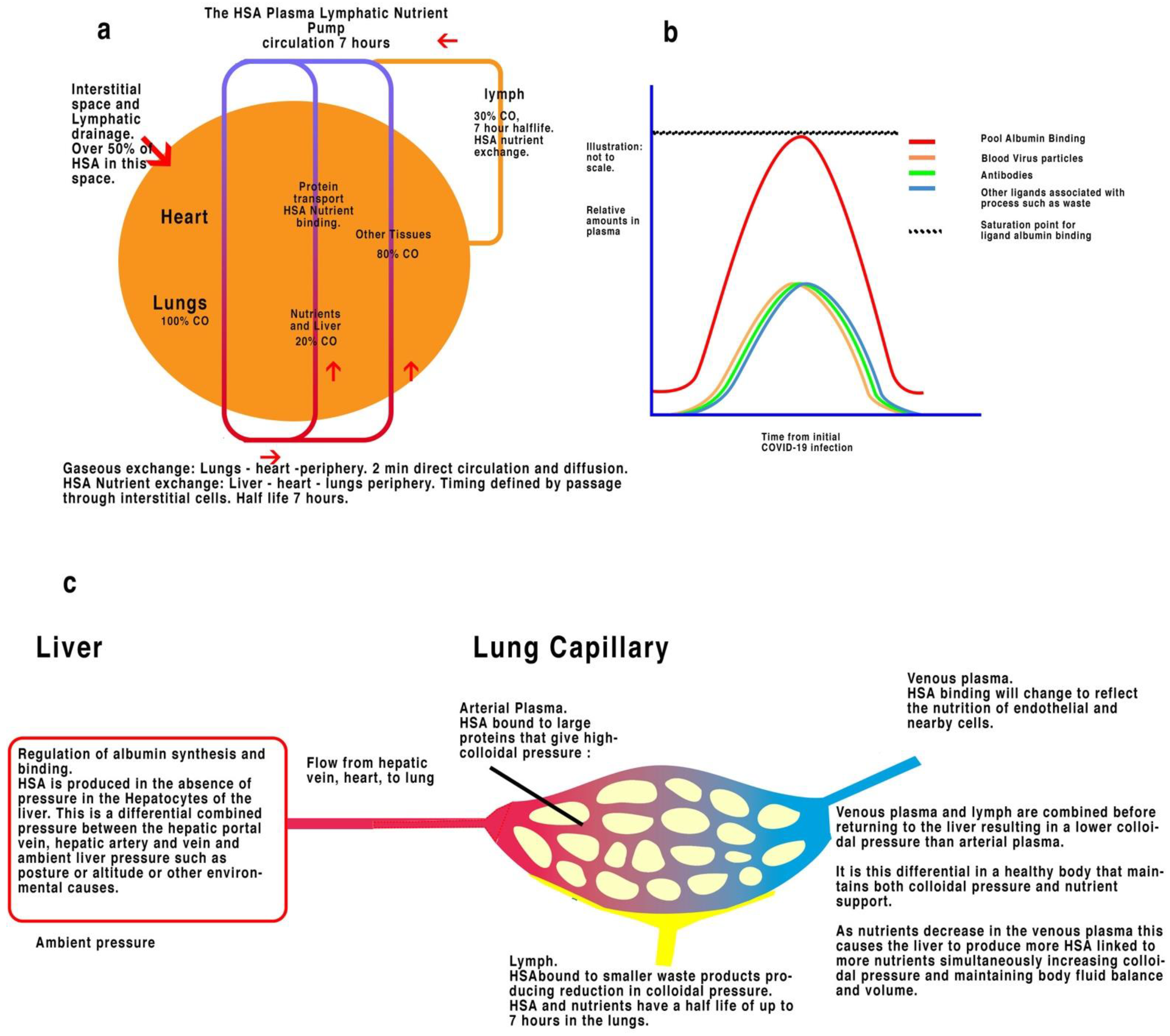
4.3. The Lymphatic System and Plasma–Lymph Nutrient–Albumin Pump

How is the albumin charged with nutrients? The lymphatic system maintains tissue interstitial pressure by collecting protein-rich fluid that is extracted from capillaries. The lymphatic system is also a critical component of the immune system (Figure 2a).
The lymphatic system is conventionally regarded as part of the immune system, in part because of the changes that take place during infection. A closer look reveals a system that can be considered being made of channels starting as the leakage of fluid from capillaries through endothelial cell gaps into interstitial spaces. This fluid initially resembling plasma and containing the full protein–ligand complements of plasma infuses the interstitial spaces around cells, providing membrane surface area access to cells. Flow is determined by the mechanical action of movement and muscle rather than from cardiac activity, with the lymph formed flowing back into the venous system and eventually the vena cava and heart. The circulation of albumin is complementary to the cardiac circulatory system to which it returns extracellular fluids [2]. Transport of nutrients in the blood, therefore, follows a separate circulatory pattern to that of the gaseous respiratory system, and rather than a few minutes, it may take hours or weeks for an albumin molecule to circulate in a normal healthy subject due to the half-life of albumin within the interstitial fluid (Figure 2a).
Nutrients enter through the stomach and are transferred via the hepatic portal vein (HPV) into the liver. Hepatocytes in the liver perform an enzymatic modulation of HPV and hepatic arterial plasma, responding to hormones such as insulin to maintain glycogen. The liver can store and metabolise molecular structures. It can both metabolise and manufacture albumin according to pressure. Organ body fluid concentrations of nutrients and colloidal pressure are, therefore, moderated and controlled by the liver. Both released and circulating albumin bind to the moderated concentrations of molecular nutrients until equilibrium is formed by mixing. Nutrient bound albumin then passes through the heart to the lungs.
In the lungs, 60–80% of the nutrient–albumin complex passes into the interstitial spaces and remains there for many hours before returning to the vena cava through the lymph, the rest remaining in circulation. Only about 10% will be recharged by the liver. In the lungs, some nutrients on the albumin complex are exchanged for waste products because of metabolism. In healthy patients, the moderation of nutrients with albumin is assumed to be minimal; however, any illness or damage to the lungs will cause this equilibrium to change. In COVID-19-infected lungs, COVID-19 virion antibodies and waste replace nutrient ligands, changing both nutrients and colloidal pressure [21]. The aerated plasma is then pumped by the heart to the organs and periphery. Only about 10% of the lymph returns to the liver to be recharged with ligands. In a healthy subject, the continual mixing and rapid supply of re-bound albumin through the capillary circulation is enough to prevent albumin-binding deficiency. During COVID-19 infection in the lungs, there will be a rise in COVID-19 virions, corresponding antibodies, and detritus. In addition, secondary infections will have the additive effect of creating further antibodies.

4.4. Liver

The liver is the main control of nutrient ligands and waste in the body and the site of albumin synthesis [22]. The feedback process to evaluate nutrients and controlled supply is indirectly provided by the ligand–albumin complex. Albumin synthesis is directly linked to the reduction in pressure in hepatocytes caused by insufficient HPV blood pressure. Albumin provides 80% of oncotic pressure and the addition of albumin increases blood volume and pressure in the hepatocytes self-regulating overall blood volume and pressure and concurrent nutrients. A lack of nutrients in the periphery changes colloidal pressure by changing the binding of nutrients [21], metabolism, and hydration, thereby reducing the amount of albumin in the blood because of low lymph flow and metabolism of albumin. When nutrients are bound to albumin, they are removed from solution but maintain most of the hydrogen bonds responsible for their oncotic pressure. Bound albumin removes ligands from solution but retains the ligand’s hydrophilic ability, increasing osmotic pressure. This causes low pressure in the hepatocytes and instigates the production of albumin. Oncotic pressure reduction will occur across the system, including on the hepatocytes, which produce more nutrient bound albumin.

4.5. Obesity

The most prevalent vulnerability to COVID-19 is obesity. Obesity is a worldwide major public health problem affecting many organs, including the heart, where it can cause heart disease, stroke, high blood pressure, diabetes, cancers, and dermatological complaints. Obesity has metabolic effects, such as causing hyperandrogenism and gout, which in turn are associated with cutaneous manifestations [23]. Dermatological manifestations of a systemic disease, such as gout, must have a systemic common factor.
Fatty acid concentrations in the plasma, interstitial fluids, and lymph have been shown to reflect the concentrations in adjacent fat cells. More fat in cells create a corresponding higher level of fatty acids in the interstitial spaces [3]. Fatty acids are transported by albumin, and greater obesity leads to a higher concentration of fatty acids in the blood and bound to albumin, causing binding deficiency. As fatty acids rise in the interstitial fluids, lymph, and plasma, more remain bound to albumin in circulation. This reduces the number of available binding sites on albumin for other nutrients to supply endothelial and cellular structures. A reduction in binding sites sufficient to affect nutrients eventually destabilises cell integrity.

4.6. Diabetes

Both insulin and glucose are transported by albumin as well as competing fatty acids. Restricting insulin access to albumin-binding sites in COVID-19 reduces the concentration of insulin concentration delivered to the liver with the subsequent elevation of glucose. This excess glucose may result in glycosylation of albumin further reducing binding sites. A reduction in albumin predicts type 2 diabetes [24]. This process is promoted by the presence of elevated blood glucose concentrations in diabetes and occurs with various proteins [25]. Glycated albumin also suppresses glucose-induced insulin secretion by impairing glucose metabolism in rats [26] and pancreatic β-cells dysfunction through autophagy [27].
Glycated albumin has a greater affinity for virions than albumin, and the ability of bacteria and viruses to surround themselves with serum proteins is a recognised immune evasion and pathogenic process [28]. SARS-CoV-2 spike binding protein binds to glycated serum albumin [28]. Long-term binding of virions in interstitial spaces would slow the flow and isolate COVID-19 virions shielded by albumin for many weeks and may be an explanation for Long-COVID-19.

4.7. Arthritic Pain

Biological activity regulation by protein post-translational modification (PTM) is critical for cell function, development, differentiation, and survival. Dysregulation of PTM proteins is present in various pathological conditions, including rheumatoid arthritis (RA) [29]. A decreased albumin/globulin ratio in RA patients significantly correlates with dyslipidemia and ARDs, implicating the albumin binding limits of fats concurrently changing [30].

4.8. Lungs

An unusual feature of the COVID-19 disease is microthrombosis and localised disruption of the osmotic potential with pulmonary microvascular dilation, a commonality in sepsis-induced ARDS [28]. In COVID-19, there is a greater risk of thrombosis [31] and coagulation [32]. Most patients are asymptomatic, with only a few patients severely affected. The resultant stagnation of HSA and ABD will reduce the levels of waste and distort the action of cellular structures, providing a possible mechanism for the “ground glass lung opacity”, seen in COVID-19.

4.9. Heart

The risk of cardiovascular problems, such as a heart attack or stroke, remains high even many months after a SARS-CoV-2 infection clears up [33] and can affect even those with mild symptoms. The heart is a dynamic organ in continuous movement regulating pressure and flow of blood to the whole body; importantly, the movement of the heart also determines heart lymph flow, determining the amount of nutrients transported to essential heart cells. In disease, the first limiting factor is usually oxygen supply, where deprivation can produce stress within seconds; for medical practitioners, this is usually the first concern. Secondary to this, to maintain functional stability, the heart cells must be infused with nutrients. This occurs over a longer time-period, with albumin-charged nutrients lining the endothelial glycocalyx, protecting the stability of both capillary walls, and maintaining the correct supply of nutrients. This is dependent upon the albumin lymphatic pump over a much longer timescale, as heart movement and lymphatic flow become restricted and nutrients slowly cease to be delivered. As discussed above, the heart is secondary to the lungs; a change in nutrient metabolism due to lung disease, producing a deficit in nutrients over time, will inevitably lead to further degradation of the heart and its function.

4.10. The Blood Brain, Placental Barriers, and Albumin Transport in the Kidney

A common factor for the blood–brain barrier (BBB), the placental barrier (PB), and the kidney is that normal movement of albumin is restricted, and in each case, albumin is controlled by clathrin enabled endocytosis [34,35]. Infection with COVID-19 leads to a reduction in albumin binding sites, including that used by clathrin to initiate endocytosis of the albumin–nutrient complex. This blocks the albumin from entering the cell and passing the barrier in each case.

4.11. The Central Nervous System and the Blood–Brain Barrier (BBB)

Severe COVID-19 and long COVID19 are both associated with cognitive defects [36]. In healthy subjects, both nutrients and pressure are kept stable within the cerebral spinal fluid (CSF) by the action of the blood–brain barrier, which stabilises and regulates albumin, intercranial pressure, and bound nutrients. Both pressure and nutrient support are therefore maintained within controlled limits within the CSF in a separate environment to the cardiac circulation. In the CSF, where 95% of amyloid b is bound to albumin [37], any decrease in binding levels will have a direct effect on amyloid beta (Aβ) concentration, potentially increasing plaque formation [38]. Studies have shown that the possibility that patients with COVID-19-associated neurological syndromes exhibit impaired amyloid processing [39]. There is therefore evidence of a connection between neurological damage due to plaque formation, with a direct link to the control of Aβ by albumin and albumin binding levels [37].
During initial COVID-19 systemic infection, COVID-19 virions interfere with entry of a proportion of the albumin by occupying the binding site for clathrin. This leads to gradual nutrient deficit within the CNS. There will also be weakening of the capillary walls due to lack of glycolates [2], thrombosis [40], and disturbances of synapse connectivity as transmitter vesicles decay. As the disease progresses the blood–brain barrier becomes weaker, and rupture may ensue, allowing larger bacteria, in addition to viruses, to enter from the systemic system leading to meningitis. There may, therefore, be more than one action occurring during COVID-19 infection in the brain in regard to albumin:
(I)
Virions attached to HSA may affect the transport of vital nutrients across the BBB. This nutrient deficit will depend upon the state of has-binding deficiency. This level of binding deficiency will alter the levels of transmitter and affect transmission of action potential affecting cognition.
(II)
Depletion of nutrients will also affect the capillaries of the brain, for example, the endothelial glycocalyx layer (EGL) already described [3,5]. A reduction in the EGL will eventually cause leakage, thrombosis [40], and rupture. Rupture may promote secondary infection, leading to symptoms of meningitis [41].

4.12. Kidney

Pathology of COVID-19 in the kidney indicates symptoms of nephrotic syndrome, numerous glomerulonephritides, microscopic polyangiitis vasculitis and collapsing glomerulopathy, and thrombotic microangiopathies, such as atypical haemolytic uremic syndrome (aHUS) [42].
In healthy individuals, there is minimum albumin loss from the kidneys and any albumin is reabsorbed by the peritubular capillaries by phagocytosis [43,44]. The glomerulus, the filtering unit of the kidney, is a unique bundle of capillaries lined by delicate fenestrated endothelia [45]. A large percentage of COVID-19 affected patients present with acute kidney injury (AKI); most cases of CoV-AKI are driven by a form that can cause impairment in tubular reabsorption of filtered proteins [46]. Reabsorption of albumin is usually by clathrin-mediated endocytosis, as described above. This necessitates the binding of albumin to clathrin. Any ligand that competes with clathrin will change this equilibrium and permit albumin to pass into the urine. This also correlates with evidence that urinary excretion of uric acid is negatively associated with albuminuria in patients with chronic kidney disease [47]. The association between albuminuria and serum uric acid may not be interrelated via renal handling of uric acid [47] but by the levels of albumin-binding available [47].

4.13. Pregnancy

Previously [48], we noted that albumin is entirely metabolised by the foetus and is not therefore circulated by the liver. Pregnancy therefore removes bound albumin–nutrient complexes for the metabolism of the foetus, leaving a deficit that may be one cause of the adverse symptoms in preeclampsia [48]. A lack of albumin due to metabolism by the foetus is a plausible explanation for the stresses some pregnant women have experienced in the third trimester. For the same reason, in COVID-19, both the foetus and mother may experience reduced albumin-binding caused by both the permanent exclusion of returning albumin from mother to foetus and COVID-19 disease.

4.14. Skin: Distribution of Albumin in the Adult and Child and Infant Body

The human foetus metabolises albumin passed from the mother in the form of the albumin–nutrient complex. In the young child, albumin is concentrated in the periphery and the muscle and skin; this may be caused by children having a larger surface area to volume ratio. In the adult, the lungs and organs contain relatively larger proportions. There are great variations between individuals and ages. There have been many reported instances of dermatologically significant issues [49], including, thrombosis, chilblains [40,49,50,51,52], mucocutaneous disease [53], purpura [54], and rashes. The frequency and timing of cutaneous manifestations of COVID-19 are difficult to ascertain; also unclear is the association of certain skin manifestations with the illness severity. Moreover, it cannot be excluded that, in some patients, the observed skin findings may represent cutaneous reactions to the treatments used for COVID-19.
Obese COVID-19 patients have a high occurrence of dermatological problems. Increased body mass index affects skin physiology, skin barrier, collagen structure, and wound healing. Obesity also affects sebaceous and sweat glands and causes circulatory and lymphatic changes. Furthermore, obesity is associated with an increased incidence of bacterial and Candida skin infections, as well as onychomycosis; inflammatory skin diseases; and chronic dermatoses such as hidradenitis suppurativa, psoriasis, and rosacea. Obesity is also related to rare skin conditions [23]. Obese children have a higher prevalence of skin lesions than normal weight children [55].
In COVID-19 infection, the dermatological signs are diverse and the timing is irregular. There is a greater resting pool of albumin during COVID-19, remaining in the interstitial spaces for longer; a lack of movement during illness reduces the activity of the HSALNP HSA lymphatic nutrient pump, isolating the stagnating albumin and causing albumin binding deficiency in associated areas depending upon flow. For the skin of a child, therefore, dermatological nutrients bound albumin and, therefore, nutrients will be decreased in relation to the percentage of albumin flow, with changes in colloidal pressure. The timing of this is dependent upon the nutrient-bound albumin flow into the interstitial spaces. The pooling of albumin may be provoking dermatological reactions independently from the COVID-19 infection sites. Dermatological conditions, therefore, will vary according to localised albumin pooling, timing, and binding deficiency. A lack of available albumin binding may therefore instigate systemic nutrient deficiency, leading to symptoms of multisystem inflammatory disease, where apart from obesity (25.3%), comorbidities are rare [56].
Hypercortisolaemia is a condition involving prolonged excess serum levels of cortisol that can develop as a result of disregulatory abnormalities in the hypothalamic-pituitary–adrenal axis or from exogenous-source steroids. Hypercortisolaemia induces a state of immunocompromise that predisposes the patient to various bacterial, viral, fungal, and parasitic infections [57]. Low serum albumin levels in patients with ischemic stroke are associated with higher serum cortisol levels and predisposes to hypercortisolaemia [58]. High serum total cortisol concentrations are associated with high mortality from COVID-19 [59].
Inflammatory markers and acute phase reactants (“markers”) are also associated with COVID-19 infection and may be able to predict disease severity [37,60,61]. Negative acute phase reactants are downregulated, and their concentrations decrease during inflammation. Positive acute phase reactants include procalcitonin, C-reactive protein, ferritin, fibrinogen, hepcidin, and serum amyloid A. Negative acute phase reactants include albumin, prealbumin, transferrin, retinol-binding protein, and antithrombin. A reduction in albumin binding will have a concurrent effect on marker concentration.

4.15. Fluid Therapy

The use of fluid therapy is ubiquitous in medicine, with all medical staff, doctors, nurses, and many ancillary staff trained in infusion techniques. “Intravenous fluid therapy is one of the most common interventions in acutely ill patients. Each day, over 20% of patients in intensive care units (ICUs) receive intravenous fluid resuscitation, and more than 30% receive fluid resuscitation during their first day in the ICU. Virtually all hospitalized patients receive intravenous fluid to maintain hydration and as diluents for drug administration. Until recently, the amount and type of fluids administered were based on a theory described over 100 years ago, much of which is inconsistent with current physiological data and emerging knowledge. Despite their widespread use, various fluids for intravenous administration have entered clinical practice without a robust evaluation of their safety and efficacy. The belief that dehydration and hypovolaemia can cause or worsen kidney and other vital organ injury has resulted in liberal approaches to fluid therapy and the view that fluid overload and tissue oedema are ‘normal’ during critical illness; this is quite possibly harming patients. Increasing evidence indicates that restrictive fluid strategies might improve outcomes.” [62]. Attempts at albumin infusion have been inconclusive [63,64], but there is ongoing discussion of its merits [62,65].

5. Discussion

Nutrient support in cells is an essential method for maintaining both physiological stasis and immune response. In a healthy body, albumin is charged with nutrients in the liver and exhibits discrete osmotic pressure according to nutrient bonding. Pumped by the heart in circulation and muscle in the lymph, organs divert at least half of the nutrient bound albumin into the interstitial spaces and lymph; the rest flows through the capillaries and is a part of cardiovascular lung systemic circulation. The diverted albumin that passes into the interstitial spaces exchanges nutrients and binds to waste products. This changes both the nutrient bound to albumin and the colloid osmotic pressure exerted by the changing of ligands on the albumin molecule. Albumin then passes back via the lymph into the systemic circulation. Less than 10% of albumin will be recharged with nutrients on each circulation. A decrease in mean albumin levels by 10% is sufficient to create symptoms of hypoalbuminemia. For individuals aged 50+, albumin level decreases significantly (see Figure 1). Any excess fatty acids such as in obesity further reduces albumin binding.

5.1. Physiology

The liver is the central control organ for cellular nutrition, colloidal pressure, and waste removal by a dual circulation. The capillary circulation and the lymphatic circulation of HSA, which it feeds, are directly linked to the pressure of the liver. Changes to arterial or venous pressure, or ambient pressure will affect the stability of HSA production, binding, and nutrient and colloidal support. The production of albumin is affected by all pressure changes to the body, including outside ambient pressure (Figure 2c). This may account for the increase in HSA measured during ascension to altitude [66] and may have importance for acclimatisation.

5.2. Why Is Present Fluid Therapy Inappropriate in COVID-19?

The present purpose of fluid therapy (FT) is to correct colloidal pressures in the cellular structures. Saline provides a change in pressure but dilutes nutrient ligands with unpredictable results. HSA’s effect on oncotic pressure is well known and is partially down to the nutrients bound. Changes to colloidal pressure by HSA FT can therefore only be predicted knowing the correct relevant ligand–albumin binding for each organ (pulmonary artery, aorta, and vena cava will have differing albumin bindings with differences enhanced as HSA passes into cellular spaces). In a healthy body, this difference between ligand–albumin binding in the lungs and that delivered to the periphery will be minimal. However, in COVID-19, damage to the lungs causes HSA to be restricted, thus reducing the amount of nutrients with resultant immune reactions. Furthermore, contemporary FT infusion is given via a peripheral vein such as the brachial vein (Figure 3). The lungs receive 100% cardiac output, but the liver only receives about 15% of cardiac output; 85% of HSA infused to the vena cava through the periphery will not be charged with nutrients during circulation to the liver and will not be providing either the correct nutrients or oncotic pressure to the cells and interstitial spaces. HSA has a half-life in intracellular spaces of about 17 h so that any immediate replenishment will be compromised, and nutrient deficiency will take place during this time. Furthermore, the serum albumin content determined by peripheral vein analysis may not represent the content of pool albumin in the interstitial spaces, especially during illness and poor blood flow.

5.3. Saline

Saline initially rehydrates the cellular structures; however, this reduces the levels of nutrients in the plasma and intracellular spaces. Diluting available albumin may also result in waste deposits not being removed. The addition of saline also results in a rise in pressure in the hepatocytes, which will reduce albumin production, binding to nutrients, and subsequent nutrient output into the systemic system. Between individuals, there is great variation in the levels used natively to regulate pressure and nutrients. Saline FT can, therefore, only safely be used to rehydrate a patient to a hydrated state when it is known that nutrient support is maintained, and organ damage is minimal. Continual use of saline deprives cellular structures of nutrients.

5.4. HSA Infusion

The infusion of colloids into the periphery was described in Figure 3. Manufactured HSA preparations contain no nutrients, and colloidal pressure will differ from that of HSA in situ. Infusions of HSA into the periphery flow through the heart and into the lungs, where more than 50% rests in the interstitial spaces for many hours before re-joining the venous flow to the heart. From the heart, only 10% of the total albumin in the plasma will flow to the liver, with the remainder passing to the peripheral capillaries to be divided again between the capillary flow and the interstitial spaces. Any further HSA addition will lead to an increasing nutrient deficit within the extracellular spaces (Figure 2a).
Where the main concern is the liver, for example to treat cirrhosis, albumin infusion into the periphery has been shown to be highly beneficial [67]. We suggest that its success is because liver function is the main therapeutic target organ. The liver has the ability to bind the albumin or, at higher infusion rates, to metabolise HSA to provide further metabolites. At low rates of infusion, the liver will recover, but there will be little or no effect on other systemic organs unless they have independent vulnerabilities; at higher rates or longer application, both nutrition and pressure in the interstitial spaces will change, causing depletion of nutrients and stress, limiting this method. Higher rates of infusion to produce benefit to the periphery and CNS is probably only achievable directly through infusion to the liver via the portal vein, therefore producing nutrient bound HSA at the correct colloidal pressure.
Albumin-binding deficiency (ABD) occurs when insufficient binding sites remain on the albumin molecule to sufficient supply cellular and intracellular structures with the required nutrients. This may occur with any ligand that binds to albumin or other molecules in the plasma. This occurs due to insufficient albumin and/or insufficient binding sites. COVID-19 virions, antibodies, and vaccines are carried on albumin. A lack of albumin causes energy depletion from glucose, mitochondria failure, and cell death, as shown in Table 1.
Cellular nutrient support is dependent upon the flow of nutrients from the liver and must be adequately supplied from the hepatic portal vein from the stomach. Molecules compete for dissolution and relative concentration in fluids irrespective of protein binding so that most molecules carried in the blood are affected by albumin binding. Nutrient concentrations in the cells are defined by the levels of nutrients’ capability to bind to albumin, which therefore affects other molecules of similar size and charge. Because albumin is the largest carrier of ligands in the blood and defines and regulates 80% of oncotic pressure, it is the main determinant of nutrient balance to the cells.
Although this review was created for an understanding of COVID-19 and long COVID-19, the theoretical details may be applied to many different abnormalities, each of which is linked through albumin (HSA) binding mechanisms and their deterioration, where HSA binding levels are a common factor. Any disease or injury that decreases the levels of albumin binding such as secondary infections will also increase the risk of albumin binding deficiency and sepsis.

6. Conclusions

The appropriate method of fluid therapy is to infuse albumin to the liver directly through the hepatic portal vein. Both nutrient support for the cells and oncotic pressure are regulated by the liver by HSA concentration. Long-term saline or peripheral albumin infusion results in degradation of nutrients to the periphery.
COVID-19 attacks albumin binding and charging of nutrients by reducing blood and lymph flow; by causing excretion of albumin through the kidneys; by degrading normal liver function; and by decreasing the binding potential of HSA caused by excess COVID-19 virions, antibodies, and waste.
We have provided evidence that albumin binding deficit may be responsible for COVID-19 and long COVID-19. The obvious solution is to raise albumin flow to the liver to raise nutrients. The liver is a fast and adaptable moderator of nutrients that can supply itself from body stores almost without limitation.
Infusing albumin directly will lead to a fall in albumin creation by the liver and a possible reversal into metabolism of albumin to provide other nutrients, which will benefit the liver. Infusion to the liver also binds albumin to the correct nutrients for the systemic system and periphery within healthy limits; liver limitations are few. As the liver is the centre of control, unlike other remedies, the amount of albumin entering will be proportionate to the amount of nutrient-bound albumin exiting. Infusion into the liver should allow for proportional control over both colloidal pressure and nutrients.
Albumin must therefore be administered directly to the liver via the portal vein. There are tributaries to the HPV, such as the mesenteric and umbilical veins, that are only a few cm deep and could be ultrasound guided cannulated.
The concentrations of all systemic ligands transported in the blood including all drugs are affected by albumin binding, and there has been little consideration of the interactions between ligands and transporters.

Author Contributions

A.S.J. provided the conceptual framework for review and worked with W.W. to write the paper. Further ideas and inputs were provided by G.P. and M.J. All authors have read and agreed to the published version of the manuscript.

Funding

The authors received no external funding to support this work.

Conflicts of Interest

We have no conflict of interest, financial, or otherwise.

Abbreviations

HSA, human serum albumin; ABD, albumin binding deficiency; HSALNP, HSA lymphatic nutrient pump; LHLL, liver heart lungs liver; Ligand, any molecule capable of binding to albumin both nutrient and waste; HPV, hepatic portal vein.

References

  1. Moman, R.N.; Gupta, N.; Varacallo, M. Physiology, Albumin. In StatPearls; StatPearls Publishing: Treasure Island, FL, USA, 2022. Available online: https://www.ncbi.nlm.nih.gov/books/NBK459198/ (accessed on 4 January 2022).
  2. Sherwood, L. Human Physiology: From Cells to Systems, 8th ed.; Brooks/Cole, Cengage Learning: Boston, MA, USA, 2013; ISBN 9781111577438. (In English) [Google Scholar]
  3. Johnson, A.S.; Winlow, W. COVID-19 vulnerabilities are intensified by declining human serum albumin levels. Exp. Physiol. 2022, 107, 674–682. [Google Scholar] [CrossRef] [PubMed]
  4. Swartz, M.A. The physiology of the lymphatic system. Adv. Drug Deliv. Rev. 2001, 50, 3–20. [Google Scholar] [CrossRef]
  5. Johnson, A.S.; Fatemi, R.; Winlow, W. SARS-CoV-2 Bound Human Serum Albumin and Systemic Septic Shock. Front. Cardiovasc. Med. 2020, 7, 153. [Google Scholar] [CrossRef] [PubMed]
  6. Paar, M.; Rossmann, C.; Nusshold, C.; Wagner, T.; Schlagenhauf, A.; Leschnik, B.; Oettl, K.; Koestenberger, M.; Cvirn, G.; Hallström, S. Anticoagulant action of low, physiologic, and high albumin levels in whole blood. PLoS ONE 2017, 12, e0182997. [Google Scholar] [CrossRef]
  7. Gyamlani, G.; Molnar, M.; Lu, J.; Sumida, K.; Kalantar-Zadeh, K.; Kovesdy, C. Association of serum albumin level and venous thromboembolic events in a large cohort of patients with nephrotic syndrome. Nephrol. Dial. Transplant. 2017, 32, 157–164. [Google Scholar] [CrossRef]
  8. Goncharov, N.V.; Belinskaya, D.A.; Razygraev, A. On the enzymatic activity of albumin. Russ. J. Bioorg. Chem. 2015, 41, 113–124. [Google Scholar] [CrossRef]
  9. Weaving, G.; Batstone, G.F.; Jones, R.G. Age and sex variation in serum albumin concentration: An observational study. Ann. Clin. Biochem. 2016, 53, 106–111. [Google Scholar] [CrossRef]
  10. Islam, N.; Shkolnikov, V.M.; Acosta, R.J.; Klimkin, I.; Kawachi, I.; Irizarry, R.A.; Alicandro, G.; Khunti, K.; Yates, T.; Jdanov, D.A.; et al. Excess deaths associated with COVID-19 pandemic in 2020: Age and sex disaggregated time series analysis in 29 high income countries. BMJ 2021, 373, n1137. [Google Scholar] [CrossRef]
  11. Egesten, A.; Frick, I.M.; Mörgelin, M.; Olin, A.I.; Björck, L. Binding of albumin promotes bacterial survival at the epithelial surface. J. Biol. Chem. 2011, 286, 2469–2476. [Google Scholar] [CrossRef]
  12. De, R.; Dutta, S. Role of the Microbiome in the Pathogenesis of COVID-19. Front. Cell. Infect. Microbiol. 2022, 12, 736397. [Google Scholar] [CrossRef]
  13. Altmann, D.M.; Boyton, R.J. COVID-19 vaccination: The road ahead. Science 2022, 375, 1127–1132. [Google Scholar] [CrossRef]
  14. Yamaoka-Tojo, M. Endothelial glycocalyx damage as a systemic inflammatory microvascular endotheliopathy in COVID-19. Biomed. J. 2020, 43, 399–413. [Google Scholar] [CrossRef] [PubMed]
  15. Klammt, S.; Wojak, H.J.; Mitzner, A.; Koball, S.; Rychly, J.; Reisinger, E.; Mitzner, S. Albumin-binding capacity (ABiC) is reduced in patients with chronic kidney disease along with an accumulation of protein-bound uraemic toxins. Nephrol. Dial. Transplant. 2012, 27, 2377–2383. [Google Scholar] [CrossRef]
  16. Oettl, K.; Birner-Gruenberger, R.; Spindelboeck, W.; Stueger, H.P.; Dorn, L.; Stadlbauer, V.; Putz-Bankuti, C.; Krisper, P.; Graziadei, I.; Vogel, W.; et al. Oxidative albumin damage in chronic liver failure: Relation to albumin binding capacity, liver dysfunction and survival. J. Hepatol. 2013, 59, 978–983. [Google Scholar] [CrossRef] [PubMed]
  17. Belmin, J.; Corman, B.; Merval, R.; Tedgui, A. Age-related changes in endothelial permeability and distribution volume of albumin in rat aorta. Am. J. Physiol. 1993, 264 Pt 2, H679–H685. [Google Scholar] [CrossRef] [PubMed]
  18. Shehadeh, L.A.; Webster, K.A.; Hare, J.M.; Vazquez-Padron, R.I. Dynamic regulation of vascular myosin light chain (MYL9) with injury and aging. PLoS ONE 2011, 6, e25855. [Google Scholar] [CrossRef]
  19. Du Preez, H.N.; Aldous, C.; Kruger, H.G.; Lin, J. N-acetylcysteine and other sulfur-donors as a preventative and adjunct therapy for COVID-19. Adv. Pharmacol. Pharm. Sci. J. 2022; preprint. [Google Scholar] [CrossRef]
  20. Du Preez, H.N.; Aldous, C.; Hayden, M.R.; Kruger, H.G.; Lin, J. Pathogenesis of COVID-19 described through the lens of an undersulfated and degraded epithelial and endothelial glycocalyx. FASEB J. Off. Publ. Fed. Am. Soc. Exp. Biol. 2022, 36, e22052. [Google Scholar] [CrossRef]
  21. Curry, F.E.; Michel, C.C. The Colloid Osmotic Pressure Across the Glycocalyx: Role of Interstitial Fluid Sub-Compartments in Trans-Vascular Fluid Exchange in Skeletal Muscle. Front. Cell Dev. Biol. 2021, 9, 729873. [Google Scholar] [CrossRef]
  22. Ridruejo, E.; Soza, A. The liver in times of COVID-19: What hepatologists should know. Ann. Hepatol. 2020, 19, 353–358. [Google Scholar] [CrossRef]
  23. Hirt, P.A.; Castillo, D.E.; Yosipovitch, G.; Keri, J.E. Skin changes in the obese patient. J. Am. Acad. Dermatol. 2019, 81, 1037–1057. [Google Scholar] [CrossRef] [PubMed]
  24. Chang, D.C.; Xu, X.; Ferrante, A.W.; Krakoff, J. Reduced plasma albumin predicts type 2 diabetes and is associated with greater adipose tissue macrophage content and activation. Diabetol. Metab. Syndr. 2019, 11, 14. [Google Scholar] [CrossRef] [PubMed]
  25. Anguizola, J.; Matsuda, R.; Barnaby, O.S.; Hoy, K.S.; Wa, C.; DeBolt, E.; Koke, M.; Hage, D.S. Glycation of human serum albumin. Clin. Chim. Acta Int. J. Clin. Chem. 2013, 425, 64–76. [Google Scholar] [CrossRef]
  26. Shiraki, T.; Miura, Y.; Sawada, T. Glycated albumin suppresses glucose-induced insulin secretion by impairing glucose metabolism in rat pancreatic β-cells. Nutr. Metab. 2011, 8, 20. [Google Scholar] [CrossRef] [PubMed]
  27. Song, Y.M.; Song, S.O.; You, Y.-H.; Yoon, K.-H.; Kang, E.S.; Cha, B.S.; Lee, H.C.; Kim, J.-W.; Lee, B.-W. Glycated albumin causes pancreatic β-cells dysfunction through autophagy dysfunction. Endocrinology 2013, 154, 2626–2639. [Google Scholar] [CrossRef] [PubMed]
  28. Iles, J.K.; Zmuidinaite, R.; Sadee, C.; Gardiner, A.E.; Lacey, J.C.; Harding, S.; Ule, J.; Roblett, D.; Heeney, J.L.; Baxendale, H.; et al. SARS-CoV-2 Spike Protein Binding of Glycated Serum Albumin—Its Potential Role in the Pathogenesis of the COVID-19 Clinical Syndromes and Bias towards Individuals with Pre-Diabetes/Type 2 Diabetes and Metabolic Diseases. Int. J. Mol. Sci. 2022, 23, 4126. [Google Scholar] [CrossRef]
  29. Taldaev, A.; Rudnev, V.; Kulikova, L.; Nikolsky, K.; Efimov, A.; Malsagova, K.; Kaysheva, A. Molecular Dynamics Study of Citrullinated Proteins Associated with the Development of Rheumatoid Arthritis. Proteomes 2022, 10, 8. [Google Scholar] [CrossRef]
  30. Chen, Y.; Zhao, L.; He, H.; Wei, L.; Lai, W.; Yuan, J.; Hong, X.; Liu, L.; Wang, B.; Nandakumar, K.S.; et al. Albumin/Globulin Ratio as Yin-Yang in Rheumatoid Arthritis and Its Correlation to Inflamm-Aging Cytokines. J. Inflamm. Res. 2021, 14, 5501–5511. [Google Scholar] [CrossRef]
  31. Katsoularis, I.; Fonseca-RodrÃguez, O.; Farrington, P.; Jerndal, H.; Lundevaller, E.H.; Sund, M. Risks of deep vein thrombosis, pulmonary embolism, and bleeding after covid-19: Nationwide self-controlled cases series and matched cohort study. BMJ 2022, 377, e069590. [Google Scholar] [CrossRef]
  32. Asakura, H.; Ogawa, H. COVID-19-associated coagulopathy and disseminated intravascular coagulation. Int. J. Hematol. 2021, 113, 45–57. [Google Scholar] [CrossRef]
  33. Sidik, S.M. Heart Disease after COVID: What the Data Say. Nature 2022, 608, 26–28. [Google Scholar] [CrossRef] [PubMed]
  34. Lambot, N.; Lybaert, P.; Boom, A.; Delogne-Desnoeck, J.; Vanbellinghen, A.M.; Graff, G.; Lebrun, P.; Meuris, S. Evidence for a clathrin-mediated recycling of albumin in human term placenta. Biol. Reprod. 2006, 75, 90–97. [Google Scholar] [CrossRef] [PubMed]
  35. Kaksonen, M.; Roux, A. Mechanisms of clathrin-mediated endocytosis. Nat. Rev. Mol. Cell Biol. 2018, 19, 313–326. [Google Scholar] [CrossRef] [PubMed]
  36. Hampshire, A.; Chatfield, D.A.; MPhil, A.M.; Jolly, A.; Trender, W.; Hellyer, P.J.; Giovane, M.D.; Newcombe, V.; Outtrim, J.G.; Warne, B.; et al. Cambridge NeuroCOVID Group, the NIHR COVID-19 BioResource, and Cambridge NIHR Clinical Research Facility (2022). Multivariate profile and acute-phase correlates of cognitive deficits in a COVID-19 hospitalised cohort. EClinicalMedicine 2022, 47, 101417. [Google Scholar] [CrossRef]
  37. Stanyon, H.F.; Viles, J.H. Human serum albumin can regulate amyloid-β peptide fiber growth in the brain interstitium: Implications for Alzheimer disease. J. Biol. Chem. 2012, 287, 28163–28168. [Google Scholar] [CrossRef]
  38. Zhao, M.; Guo, C. Multipronged Regulatory Functions of Serum Albumin in Early Stages of Amyloid-β Aggregation. ACS Chem. Neurosci. 2021, 12, 2409–2420. [Google Scholar] [CrossRef]
  39. Ziff, O.J.; Ashton, N.J.; Mehta, P.R.; Brown, R.; Athauda, D.; Heaney, J.; Heslegrave, A.J.; Benedet, A.L.; Blennow, K.; Checkley, A.M.; et al. Amyloid processing in COVID-19-associated neurological syndromes. J. Neurochem. 2022, 161, 146–157. [Google Scholar] [CrossRef]
  40. Gorog, D.A.; Storey, R.F.; Gurbel, P.A. Current and novel biomarkers of thrombotic risk in COVID-19: A Consensus Statement from the International COVID-19 Thrombosis Biomarkers Colloquium. Nat. Rev. Cardiol. 2022, 19, 475–495. [Google Scholar] [CrossRef] [PubMed]
  41. Mondal, R.; Ganguly, U.; Deb, S.; Shome, G.; Pramanik, S.; Bandyopadhyay, D.; Lahiri, D. Meningoencephalitis associated with COVID-19: A systematic review. J. Neurovirology 2021, 27, 12–25. [Google Scholar] [CrossRef]
  42. Wu, H.L.; Shenoy, M.; Kalra, P.A.; Chinnadurai, R. Intrinsic Kidney Pathology Following COVID-19 Infection in Children and Adolescents: A Systematic Review. Children 2022, 9, 3. [Google Scholar] [CrossRef]
  43. Birn, H.; Christensen, E.I. Renal albumin absorption in physiology and pathology. Kidney Int. 2006, 69, 440–449. [Google Scholar] [CrossRef] [PubMed]
  44. Castrop, H.; Schießl, I.M. Novel routes of albumin passage across the glomerular filtration barrier. Acta Physiol. 2017, 219, 544–553. [Google Scholar] [CrossRef] [PubMed]
  45. Pollak, M.R.; Quaggin, S.E.; Hoenig, M.P.; Dworkin, L.D. The glomerulus: The sphere of influence. Clin. J. Am. Soc. Nephrol. CJASN 2014, 9, 1461–1469. [Google Scholar] [CrossRef] [PubMed]
  46. Muner, M.B.; Velez, J. Proteinuria in COVID-19. Clin. Kidney J. 2021, 14 (Suppl. 1), i40–i47. [Google Scholar] [CrossRef]
  47. Li, F.; Guo, H.; Zou, J.; Chen, W.; Lu, Y.; Zhang, X.; Fu, C.; Xiao, J.; Ye, Z. Urinary excretion of uric acid is negatively associated with albuminuria in patients with chronic kidney disease: A cross-sectional study. BMC Nephrol. 2018, 19, 95. [Google Scholar] [CrossRef] [PubMed]
  48. Johnson, A.; Winlow, W. Pre-Eclampsia, Hypoalbuminaemia and Albumin Therapy. Eur. J. Biomed. Pharm. Sci. 2021, 8, 75–78. [Google Scholar]
  49. Agnihothri, R.; Fox, L.P. Clinical Patterns and Morphology of COVID-19 Dermatology. Dermatol. Clin. 2021, 39, 487–503. [Google Scholar] [CrossRef]
  50. Baeck, M.; Hoton, D.; Marot, L.; Herman, A. Chilblains and COVID-19: Why SARS-CoV-2 endothelial infection is questioned. Br. J. Dermatol. 2020, 183, 1152–1153. [Google Scholar] [CrossRef]
  51. Colmenero, I.; Santonja, C.; Alonso-Riaño, M.; Noguera-Morel, L.; Hernández-Martín, A.; Andina, D.; Wiesner, T.; Rodríguez-Peralto, J.L.; Requena, L.; Torrelo, A. SARS-CoV-2 endothelial infection causes COVID-19 chilblains: Histopathological, immunohistochemical and ultrastructural study of seven paediatric cases. Br. J. Dermatol. 2021, 183, 729–737. [Google Scholar] [CrossRef]
  52. Genovese, G.; Moltrasio, C.; Berti, E.; Marzano, A.V. Skin Manifestations Associated with COVID-19: Current Knowledge and Future Perspectives. Dermatology 2021, 237, 1–12. [Google Scholar] [CrossRef]
  53. Rekhtman, S.; Tannenbaum, R.; Strunk, A.; Birabaharan, M.; Wright, S.; Garg, A. Mucocutaneous disease and related clinical characteristics in hospitalized children and adolescents with COVID-19 and multisystem inflammatory syndrome in children. J. Am. Acad. Dermatol. 2021, 84, 408–414. [Google Scholar] [CrossRef] [PubMed]
  54. Khan, I.A.; Karmakar, S.; Chakraborty, U.; Sil, A.; Chandra, A. Purpura fulminans as the presenting manifestation of COVID-19. Postgrad. Med. J. 2021, 97, 473. [Google Scholar] [CrossRef] [PubMed]
  55. Hirschler, V. Skin and obesity in childhood: An update. AIMS Med. Sci. 2021, 8, 311–323. [Google Scholar] [CrossRef]
  56. Hoste, L.; Van Paemel, R.; Haerynck, F. Multisystem inflammatory syndrome in children related to COVID-19: A systematic review. Eur. J. Pediatr. 2021, 180, 2019–2034. [Google Scholar] [CrossRef]
  57. Fareau, G.G.; Vassilopoulou-Sellin, R. Hypercortisolemia and infection. Infect. Dis. Clin. N. Am. 2007, 21, 639–657. [Google Scholar] [CrossRef]
  58. Dziedzic, T.; Pera, J.; Wnuk, M.; Szczudlik, A.; Slowik, A. Serum albumin as a determinant of cortisol release in patients with acute ischemic stroke. Atherosclerosis 2012, 221, 212–214. [Google Scholar] [CrossRef]
  59. Tan, T.; Khoo, B.; Mills, E.G.; Phylactou, M.; Patel, B.; Eng, P.C.; Thurston, L.; Muzi, B.; Meeran, K.; Prevost, A.T.; et al. Association between high serum total cortisol concentrations and mortality from COVID-19. Lancet Diabetes Endocrinol. 2020, 8, 659–660. [Google Scholar] [CrossRef]
  60. Sakthivadivel, V.; Bohra, G.K.; Maithilikarpagaselvi, N.; Khichar, S.; Meena, M.; Palanisamy, N.; Gaur, A.; Garg, M.K. Association of Inflammatory Markers with COVID-19 Outcome among Hospitalized Patients: Experience from a Tertiary Healthcare Center in Western India. Maedica 2021, 16, 620–627. [Google Scholar] [CrossRef]
  61. Chen, C.-H.; Lin, S.-W.; Shen, C.-F.; Hsieh, K.-S.; Cheng, C.-M. Biomarkers during COVID-19: Mechanisms of Change and Implications for Patient Outcomes. Diagnostics 2022, 12, 509. [Google Scholar] [CrossRef]
  62. Finfer, S.; Myburgh, J.; Bellomo, R. Intravenous fluid therapy in critically ill adults. Nat. Rev. Nephrol. 2018, 14, 541–557. [Google Scholar] [CrossRef]
  63. Dubois, M.J.; Orellana-Jimenez, C.; Melot, C.; De Backer, D.; Berre, J.; Leeman, M.; Brimioulle, S.; Appoloni, O.; Creteur, J.; Vincent, J.L. Albumin administration improves organ function in critically ill hypoalbuminemic patients: A prospective, randomized, controlled, pilot study. Crit. Care Med. 2006, 34, 2536–2540. [Google Scholar] [CrossRef] [PubMed]
  64. Caironi, P.; Tognoni, G.; Masson, S.; Fumagalli, R.; Pesenti, A.; Romero, M.; Fanizza, C.; Caspani, L.; Faenza, S.; Grasselli, G.; et al. Albumin replacement in patients with severe sepsis or septic shock. N. Engl. J. Med. 2014, 370, 1412–1421. [Google Scholar] [CrossRef] [PubMed]
  65. Ramadori, G. Albumin Infusion in Critically Ill COVID-19 Patients: Hemodilution and Anticoagulation. Int. J. Mol. Sci. 2021, 22, 7126. [Google Scholar] [CrossRef]
  66. Imoberdorf, R.; Garlick, P.J.; McNurlan, M.A.; Casella, G.A.; Peheim, E.; Turgay, M.; Bärtsch, P.; Ballmer, P.E. Enhanced synthesis of albumin and fibrinogen at high altitude. J. Appl. Physiol. 2001, 2, 528–537. [Google Scholar] [CrossRef] [PubMed]
  67. Ramadori, G. Hypoalbuminemia: An underestimated, vital characteristic of hospitalized COVID-19 positive patients? Hepatoma Res. 2020, 6, 28. [Google Scholar] [CrossRef]
Figure 3. Illustration of the liver and application of fluid therapy. The bulk HSA added to the periphery does not enter the liver to be appropriately charged with nutrients but remains in the interstitial fluid and lymph for many hours, leading to nutrient deprivation and sepsis. (a) Inappropriate application of fluid therapy to periphery leading to nutrient and pressure deformations in the lungs. (b) Appropriate application of fluid therapy to portal vein providing correct pressure and nutrients to all target organs. Circulation modified from pikisuperstar/Freepik with permission under Creative Commons licence CC BY-NC.
Covid 02 00099 g003
Table 1. Key deductions on albumin binding and its appropriate therapy.
There is repeated evidence of a connection between hypoalbuminemia and COVID-19 for each symptom of and vulnerability to COVID-19. HSA binding deficiency is a common factor.
There is evidence that raising HSA concentration in the liver may alleviate some of the vulnerabilities to COVID-19 by reducing any HSA binding deficiencies.
A mechanism for albumin involvement in long COVID-19 also exists and could be removed by appropriate HSA therapy, given that the liver precisely modulates nutrients in the plasma and maintains HSA levels.
Present fluid therapy, either saline or colloid, applied to a peripheral vein, results in a destabilisation of nutrient transport, leading to nutrient deficits in cells and cellular components because of albumin-binding deficiency.
Publisher’s Note: MDPI stays neutral with regard to jurisdictional claims in published maps and institutional affiliations.

