Article

Simultaneous Detection of Seven Human Coronaviruses by Multiplex PCR and MALDI-TOF MS

1
Hebei Key Laboratory of Animal Physiology, Biochemistry and Molecular Biology, College of Life Sciences, Hebei Normal University, Shijiazhuang 050024, China
2
State Key Laboratory of Pathogen and Biosecurity, Beijing Institute of Microbiology and Epidemiology, Academy of Military Medical Sciences (AMMS), Beijing 100071, China
3
Rongzhi Biological Technology (Qingdao) Co., Ltd., Qingdao 266109, China
*
Authors to whom correspondence should be addressed.
COVID 2022, 2(1), 5-17; https://doi.org/10.3390/covid2010002
Submission received: 25 November 2021 / Revised: 18 December 2021 / Accepted: 27 December 2021 / Published: 29 December 2021

Abstract

:
Human coronaviruses (HCoVs) are associated with a range of respiratory symptoms. The discovery of severe acute respiratory syndrome (SARS)-CoV, Middle East respiratory syndrome, and SARS-CoV-2 pose a significant threat to human health. In this study, we developed a method (HCoV-MS) that combines multiplex PCR with matrix-assisted laser desorption/ionization-time of flight mass spectrometry (MALDI-TOF MS), to detect and differentiate seven HCoVs simultaneously. The HCoV-MS method had high specificity and sensitivity, with a 1–5 copies/reaction detection limit. To validate the HCoV-MS method, we tested 163 clinical samples, and the results showed good concordance with real-time PCR. Additionally, the detection sensitivity of HCoV-MS and real-time PCR was comparable. The HCoV-MS method is a sensitive assay, requiring only 1 μL of a sample. Moreover, it is a high-throughput method, allowing 384 samples to be processed simultaneously in 30 min. We propose that this method be used to complement real-time PCR for large-scale screening studies.

1. Introduction

Coronaviruses (CoVs) are large, enveloped, positive-sense RNA viruses that cause respiratory diseases in a range of animals, including humans [1]. CoVs are divided into four genera, namely δ-CoVs, γ-CoVs, β-CoVs, and α-CoVs, among which β-CoVs and α-CoVs can infect mammals [2]. Seven human CoVs (HCoVs) have been identified, including HCoV-NL63, HCoV-229E, HCoV-OC43, HCoV-HKU1, SARS-CoV, MERS-CoV, and SARS-CoV-2 [1,3]. Respiratory diseases caused by HCoV infection range from mild to severe. Approximately 15–30% of respiratory tract infections worldwide each year are caused by HCoV-229E, HCoV-OC43, HCoV-NL63, and HCoV-HKU1. They are mild and self-healing diseases that do not pose a major threat to public health [4]. In November 2002, severe acute respiratory syndrome (SARS) appeared in southern China. It quickly spread to other countries, with more than 8000 confirmed cases in less than a year and a mortality rate of 9.6%. It took several months for researchers to determine that the pathogen was SARS-CoV. Later on, MERS broke out in Saudi Arabia and South Korea in 2012 and 2015, respectively, with more than 2000 confirmed cases worldwide and a mortality rate of 35%. MERS was found to be associated with respiratory diseases similar to SARS. The MERS outbreak once again highlighted the hazards of HCoVs to human health [3]. Moreover, many tourists traveling to the Middle East increase the risk of MERS-CoV transmission to other areas [3]. The COVID-19 outbreak in 2019 has spread rapidly across the globe, becoming a major public health emergency of international concern [5]. The World Health Organization revealed that more than 257 million people had been infected with SARS-CoV-2 worldwide, with more than five million deaths as of 23 November 2021 (https://covid19.who.int/; accessed on 15 December 2021). Although no SARS-related cases have been reported since, and MERS-related cases have been sparse, the related viruses have not disappeared [4]. Furthermore, SARS-CoV-2 is still spreading all over the world. All three HCoVs are highly pathogenic zoonotic diseases that have detrimental effects on the lives of people.
Studies have shown that HCoV most probably originated in wild animal hosts such as bats. A rich gene pool of SARS-related CoVs was found in bats in a cave in Yunnan, China [6]. Related viruses may reappear at any time and possibly mutate to produce more pathogenic CoV variants. Effective treatment methods are lacking for SARS-CoV, MERS-CoV, and SARS-CoV-2. Moreover, the initial symptoms of HCoVs infection are similar, but the treatment methods are different. The rapid, accurate detection and diagnosis of HCoV will help treat and block the spread, minimizing the loss of life caused by an epidemic. Therefore, it is important to develop a sensitive, high-throughput detection method for HCoV.
The traditional method for detecting HCoVs involves cell culture isolating the virus from clinical specimens. However, this approach is time-consuming, and most of the common cell lines are not suitable for the growth of HCoV. In addition to being time-consuming, these contributing factors do not constitute a conventional diagnostic method [7]. Next-generation sequencing technology can obtain whole-genome information of HCoVs, which contributes to our knowledge of HCoVs and helps the discovery of unknown HCoVs. However, this technology requires sophisticated bioinformatic analysis and is expensive and time-consuming; hence, it is unsuitable for large-scale population screening [8]. In recent years, real-time multiplex PCR (RT-PCR) and mass spectrometry technologies have gradually been developed into established pathogen detection methods, which are widely used today [9,10,11]. Multiplex RT-PCR is highly specific and sensitive and short detection time, making it a rapid and reliable diagnostic tool. Unfortunately, this methodology has some drawbacks. The types of fluorescence and light sources are limited, and although it is highly sensitive, a correspondingly larger sample size is required [12]. Multiplex RT-PCR theoretically detects more than a dozen viruses [13]. RT-PCR has become the gold standard for HCoV detection [14]. However, when multiple RT-PCRs are run, the sensitivity decreases as the detection factor increases [13,15].
Nucleic acid mass spectrometry analysis, where multiplex PCR (mPCR) is combined with matrix-assisted laser desorption/ionization-time of flight mass spectrometry (MALDI-TOF MS), designs sequence targets based on single nucleotide polymorphism (SNP) sites to achieve multiple detections [16,17]. Nucleic acid mass spectrometry technology allows multi-target high-throughput screening and can, in principle, reach about 40 detections. This is not possible with other multiplex detection methods. Moreover, MALDI-TOF MS is known for its strong specificity and high sensitivity. At present, nucleic acid mass spectrometry has been widely used for multiple detection and typing of bacteria and viruses. For example, it has been used for the simultaneous detection of 10 duck viruses [18], multi-site typing of Mycoplasma pneumoniae, simultaneous detection of drug resistance [19], and simultaneous detection of 21 common respiratory viruses [20]. There are many detection methods for respiratory viruses, each with different detection specificities, sensitivities, and detection limits [21,22]. At present, no methodologies for the simultaneous testing of seven HCoVs by nucleic acid mass spectrometry technology exist to the best of our knowledge. Moreover, there is no construction of a nucleic acid mass spectrometry system that achieves higher detection sensitivity (less than five copies/reaction).
This study developed HCoV-MS to detect and identify seven HCoVs simultaneously. The detection sensitivity of HCoV-MS was continuously optimized by adjusting the proportion of reaction components. The system was validated using plasmids and subsequently applied to 163 clinical samples. Finally, 26 clinical samples were used to compare the detection sensitivities of HCoV-MS and real-time PCR.

2. Materials and Methods

2.1. Target Gene Selection

The genome sequences of the seven HCoV strains used in this study were obtained from GenBank (http://www.ncbi.nlm.nih.gov/genbank/) (Table S1). N and RdRp genes were chosen as targets for the detection and differentiation of HCoV-229E, HCoV-NL63, HCoV-OC43, and HCoV-HKU1; the N, E, and ORF1b genes for SARS-CoV; the N, RdRp, E, and ORF1b genes for MERS-CoV; and the N1, N2, ORF1b, and S genes for SARS-CoV-2. Target gene sequences were compared and analyzed using Clustalx2 and DNAMAN. The conserved sequences within each target gene and species-specific fragments were selected as target sequences.

2.2. Primer Design

mPCR primers were designed using Primer Premier 6 software. Primers were 19–21 bp in length, had a Tm value of 55–65 ℃, and a GC content of 40–60%. The universal sequence ACGTTGGATG was added to all mPCR primers. The gene locus analysis and design system (Rongzhi Biotechnology Co., Ltd., Qingdao, China) was used to design a MPE primer according to the base quality and position of the mPCR primers (Table 1). All primers were synthesized by Sangon Biotech Co., Ltd., Shanghai, China.

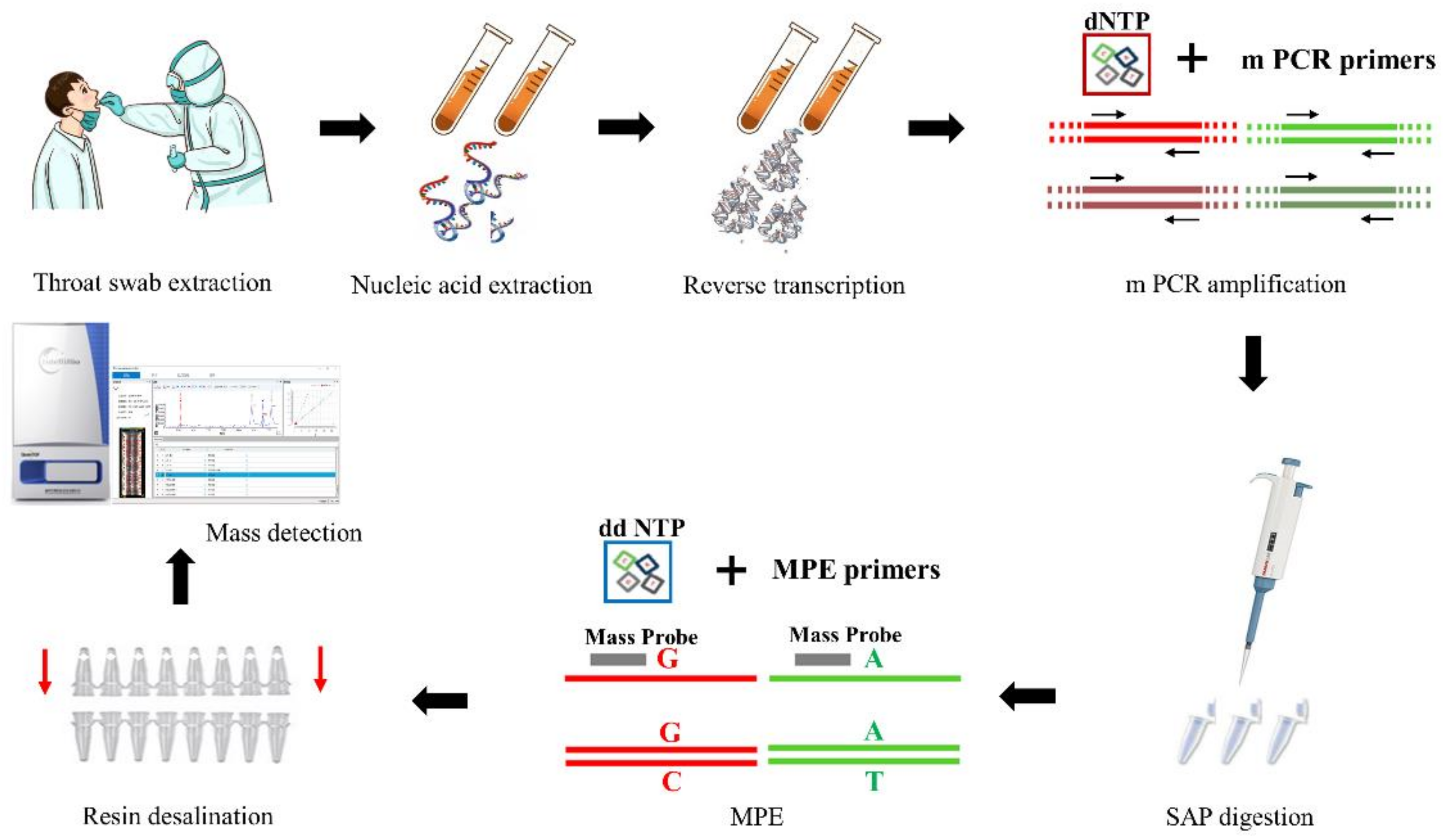
2.3. HCoV-MS Method Establishment

In this study, the plasmids that were used to establish the method included human RNase P, HCoV-229E, HCoV-OC43, HCoV-NL63, HCoV-HKU1, SARS-CoV, MERS-CoV, and SARS-CoV-2. Among these plasmids, HCoV-HKU1 showed more conservative sequence mutation sites within the species, so two plasmids, HCoV-HKU1-1, and HCoV-HKU1-2, were constructed according to mutation frequency selection and synthesis. Each plasmid contained all target genes. All plasmids were constructed on the pUC57 backbone and synthesized by Sangon Biotech Co., Ltd., Shanghai, China.
Plasmids were extracted from Escherichia coli cultures using the TIANprep Mini Plasmid Kit (TIANGEN Biotech, Beijing, China). Plasmid extracts were diluted to a concentration of 102 copies/μL, and deionized water was used as a non-template control. The amplification efficiency of the mixed mPCR and MPE primers was determined, and the ratio of primers was adjusted accordingly. Due to non-specific amplification or the formation of primer-dimers, the amplification efficiency of a single primer would be low. The amplification efficiency was improved by either changing the primer or adjusting the concentration of the primer in the mixed primer.

2.4. Protocol for the HCoV-MS Method

For detection of RNA viruses, viral RNA was extracted using the QIAamp Viral RNA kit (Qiagen, Hilden, Germany), and reverse-transcribed to cDNA using the InRcute IncRNA First-Strand cDNA Kit (TIANGEN Biotech, Beijing, China), in accordance with the recommendations of the manufacturer. All samples were reverse transcribed immediately after RNA extraction. The obtained cDNA was immediately transferred to −80 °C. For mPCR, 5 µL reaction mixes contained 2 μL PCR master mix, 1 μL deionized water, 1 μL PCR primer mix, and 1 μL DNA template. The PCR conditions involved a pre-denaturation step at 95 °C for 15 min, 45 cycles at 95 °C for 15 s, 59 °C for 30 s, and 72 °C for 30 s, and a final extension at 60 °C for 10 min. Shrimp alkaline phosphatase was directly added to the reaction system and incubated at 37 °C for 40 min followed by 85 °C for 5 min to dephosphorylate the mPCR mixture. Next, the MPE reaction mix (1 μL E-ddNTPmix, 1.4 μL MPE buffer, 0.6 μL MPE enzyme, 1 μL mixed mass probe) was added. The reaction conditions involved a pre-denaturation step at 95 °C for 30 s, 5 cycles at 95 °C for 5 s, 52 °C for 5 s, and 80 °C for 5 s, followed by 40 cycles of 95 °C for 5 s and 80 °C for 5 s. A final extension step at 72 °C for 3 min concluded the reaction. The final product was desalinated using a resin column. The SNP typing kit of Rongzhi Biotechnology (Qingdao, China) was used in the previous steps. The last step involved MS detection using a QuanTOF I mass spectrometer (Rongzhi Biotechnology, Qingdao, China). One microliter of α-Cyano-4-hydroxycinnamic acid (10 mg/mL in 50% acetonitrile and 0.1% trifluoroacetic acid) was spotted on the target plate before 0.5 μL of each sample was spotted per well, with three wells per sample. The linear positive ion mode was used as the acquisition mode of the instrument, and the parameters were set as follows: Accelerate Voltage: 20 kV; Mass Range: 3000–1100 Da; Laser Frequency: 3000 Hz; Shots/Spectrum: 800; and Laser energy: 30 uJ, signal-to-noise ratio > 4. The instrument was calibrated using calibrators (4000–10,000 Da) with molecular weights of 4550.0 Da, 5478.6 Da, 6332.2 Da, 7717.0 Da, and 9507.2 Da (Figure 1). The theoretical values of MPE and product masses are shown in Table S2. The allowable deviation of the actual value was ±1 Da.

2.5. Testing the HCoV-MS Method

Nine high-concentration CoV plasmids (105 copies/μL) were used to verify the specificity of the HCoV-MS method. The mPCR cycles were set at 30, 35, 40, or 45 cycles. In addition, the respiratory viruses ADV7, InfB, H1N1, and H3N2 (>105 CFU/mL) were used to test whether other influenza viruses would cross-react.
The HCoV-MS method was used to test the plasmids undergoing concentration gradient dilution to verify the sensitivity of the system. We diluted the plasmid to 1 copy/μL, 2.5 copies/μL, 5 copies/μL, 101 copies/μL, 102 copies/μL, 103 copies/μL, and 104 copies/μL. Plasmids at different concentration gradients were tested when the mPCR was set to 30, 35, 40, or 45 cycles. Each concentration of the gradient plasmid was subjected to six repeated experiments. The lowest concentration that could be detected in all six experiments is considered as the detection limit.

2.6. Application of the HCoV-MS Method

Unknown samples of throat or nasal swabs collected from 151 patients who had travelled abroad (collected between February and April 2021). All the clinical samples (oropharyngeal swabs) were collected in a virus transport medium and stored at −80 °C. Twelve known clinical samples were from an external quality assessment exercise (UN-CoV-2020) organized by the Robert Koch Institute in the UNSGM project RefBio. These samples include four SARS-CoV-2 samples, two SARS-CoV samples, and one HCoV-NL63 sample. The cDNAs were frozen and thawed only once, when tested by the HCoV-MS method and RT-PCR. The RT-PCR test results were used as the gold standard for comparison. All samples were previously well established using both qPCR and whole-genome sequencing; therefore, the true positives/negatives were well established. The RT-PCR was performed using the Novel Coronavirus (2019-nCoV) Nucleic Acid Diagnostic Kit (PCR-Fluorescence Probing) (Shengxiang Biotechnology, Hunan, China) in accordance with the recommendations of the manufacturer. The Novel Coronavirus (2019-nCoV) Nucleic Acid Diagnostic Kit (PCR-Fluorescence Probing) is a fully validated kit, which was approved by the National Medical Products Administration of China on 28 January 2020, obtained European Union CE certification on March 3, and also obtained emergency use authorization from the U.S. Food and Drug Administration on May 5. The kit is widely used in diagnosing COVID-19 worldwide. The limit of detection of the kit was 200 copies/mL. In addition, the kit had passed 27 kinds of virus, bacteria, or parasite samples for cross-reaction verification, and the results showed that the specificity was good.

2.7. Comparison of the HCoV-MS and Real-Time PCR Methods

We performed 5 four-fold gradient dilutions on 26 clinical samples with confirmed positive SARS-CoV-2, simultaneously detected them using the HCoV-MS method and RT-PCR, and compared the sensitivity difference between the two using the Novel Coronavirus (2019-nCoV) Nucleic Acid Diagnostic Kit (PCR-Fluorescence Probing) (Shengxiang Biotechnology, Hunan, China). The cDNAs of the 26 selected clinical samples were frozen and thawed twice.

3. Results

3.1. Performance of the Hcov-MS Method

The initial concentration of the mPCR primers for all HCoVs target genes was 0.5 μM. Mixing of the mass probes extension (MPE) primers was based on equalizing the mass spectrum signal intensity of each primer; hence, the amount added was slightly different according to the molecular weight. None of the primers and probes participated in an extension reaction when deionized water was used as the template for detection. When the mixed plasmid (102 copies/μL) was used as the template for detection, target genes of other plasmids could be detected to form specific product peaks by MPE, except NL63-RdRp, which had low amplification efficiency. In this case, the concentration of the NL63-RdRp mPCR primer in the mixed primer (reaction concentration was 4 μM) was used to optimize the system. After optimization, each primer was specifically amplified at 45 cycles.

3.2. Specificity of the HCoV-MS Method

Nine high-concentration (105 copies/μL) plasmids containing target genes were used to verify the specificity of the system, with the number of mPCR cycles set at 30, 35, 40, or 45. The results showed that high-concentration plasmids amplified well in the detection system and that all target genes could be specifically amplified in 45 cycles (Figure S1). Respiratory samples containing high concentrations of ADV7, InfB, H1N1, and H3N2 viruses were used to evaluate the specificity of the HCoV-MS method. The results showed no cross-reactivity, suggesting that the specificity of the HCoV-MS method is good.

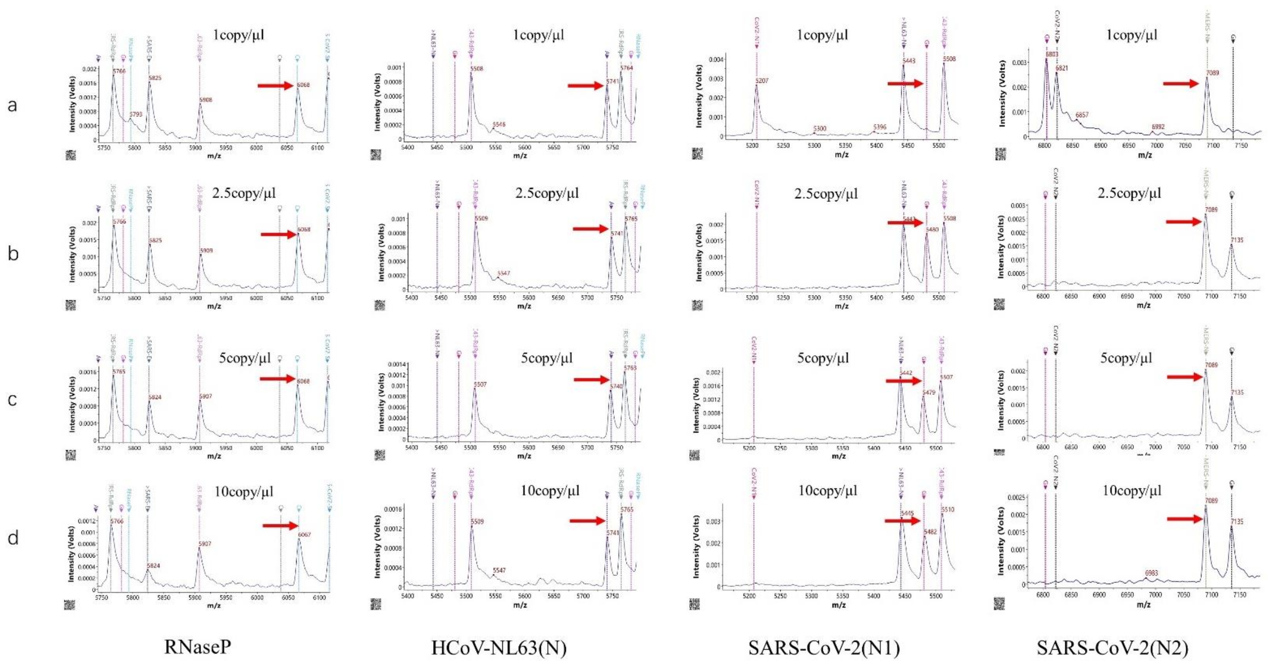
3.3. Sensitivity of the HCoV-MS Method

Serial plasmid dilutions were used to evaluate the sensitivity of the HCoV-MS method. The detection limits of part of the target genes are shown in Figure 2, and listed in Table 2. The detection limit of the HCoV-MS method was found to be 1–5 copies/reaction.

3.4. Screening Clinical Samples by the HCoV-MS Method

A total of 163 clinical samples were analyzed using the HCoV-MS method, and the results were compared with those obtained by RT-PCR (Table 3). Of the 163, 99 were positive samples, and 64 were negative samples. Both methods detected 1 case of HCoV-229E, 1 case of HCoV-NL63, 2 cases of SARS-CoV, and 95 cases of SARS-CoV-2. The detection sensitivity of the HCoV-MS method was 100% (99/99), and the detection specificity was also 100% (64/64). HCoV-OC43, HCoV-HKU1, and MERS-CoV were not detected in any samples.

3.5. Sensitivity Comparisons of HCoV-MS and RT-PCR

A total of 26 SARS-CoV-2 clinical samples were serially diluted, and the HCoV-MS and RT-PCR methods were used for simultaneous detection to compare their detection sensitivities. The detection limit gradient of the 26 SARS-CoV-2 clinical samples is shown in Figure 3 and Table 4. Results for the majority of the clinical samples were the same for the two methods or differed by only 1–2 gradients. Furthermore, only a few samples showed a significantly different detection gradient in the experiment. Evidently, the detection sensitivity of the HCoV-MS method is almost the same as that of RT-PCR.

4. Discussion

In the past 20 years, we have witnessed 3 outbreaks of highly pathogenic HCoVs. These three HCoVs realized that efficient and accurate identification methods are crucial during an epidemic [4]. At present, some research groups have successfully performed nucleic acid mass spectrometry technologies to detect six kinds of HcoVs, except for SARS-CoV-2. However, the sensitivity of this method was only 101–102 copies/reaction [6]. Here, a HCoV-MS method was established to simultaneously detect seven HcoVs, which can be used as a detection system when new HCoVs appear. Notably, the detection sensitivity of HCoV-MS is 1–5 copies/reaction. Except for SARS-CoV, other HCoVs could be detected with 1–2.5 copies/reaction. HCoV-NL63 was even detected with 1 copy/reaction. This sensitivity of this method outperforms that of other detection methods [21,22,23,24,25].
When testing 151 unknown clinical samples, the specificity and sensitivity of the HCoV-MS method reached 100%, surpassing other methods [26,27,28]. This could, however, be due to insufficient clinical samples. Samples of human/animal throat swabs or cell cultures were obtained in cooperation with UN-CoV-2020. Seven samples were positive, including four SARS-CoV-2 samples, two SARS-CoV samples, and one HCoV-NL63 sample. The results obtained with the HCoV-MS method were consistent with the official answers (Table S3). Moreover, the concentration of individual positive samples was low, but could still be accurately identified, which further highlights the detection ability of the HCoV-MS method.
Twenty-six clinical samples of SARS-CoV-2 were used to compare the detection sensitivities of HCoV-MS and RT-PCR. The latter has contributed to the fight against the epidemic [29]. We found that the detection sensitivities of the two methods were comparable and that the detection gradient was almost ±1. Furthermore, sample volumes for the RT-PCR kit were 20 μL, while the HCoV-MS method required only 1 μL, highlighting a clear advantage of using the HCoV-MS method.
The HCoV-MS method is high throughput, as reflected in the detection of multiple target genes and the requirement of small sample sizes. The HCoV-MS method could simultaneously detect 384 targets in one run spanning 30 min, with results automatically determined by the relevant software. Moreover, the reagent cost of the HCoV-MS method is relatively low, making this method ideal for large-scale population screening.
SARS-CoV-2 rapidly spread worldwide, and novel variants have gradually appeared since December 2019. The new variants were more contagious and caused serious harm. As of December 2021, the U.S. Centers for Disease Control and Prevention and the European Centre for Disease Prevention and Control have identified multiple SARS-CoV-2 variant lineages and have designated a few as variants of concern or variants of interest. The lineages include the Alpha (B.1.1.7 and Q.1–Q.8), Beta (B.1.351, B.1.351.2, and B.1.351.3), Gamma (P.1, P.1.1, and P.1.2), Epsilon (B.1.427 and B.1.429), Eta (B.1.525), Iota (B.1.526), Kappa (B.1.617.1), B.1.617.3, Mu (B.1.621 and B.1.621.1), Zeta (P.2), Delta (B.1.617.2 and AY.1 sublineages), and Omicron (B.1.1.529) [30,31]. Most variants evolve due to many base changes in the viral genome. To test whether the variant impacts our detection system, we compared the gene sequences of the currently emerging variants related to the detection target in the HCoV-MS method. We found that only Omicron’s T547K was located at the 5′ of the forward primer. The second base at the 5′ of the primer has a mutation, which theoretically may not affect the detection system. Other mutation sites do not affect HCoV-MS. In addition, we designed four detection targets for SARS-CoV-2, and the detection limits of these four targets were all 2.5 copies/reaction. Mutations are happening all the time. Even if a mutant strain affects a certain detection target in the future, other targets can accurately identify SARS-CoV-2.
The HCoV-MS that we established also has some limitations. This method is difficult to identify new HCoV because it is based on comparing and analyzing known HCoV sequences, selecting gene fragments with conserved intraspecies specificity. In conclusion, the HCoV-MS method has the characteristics of high throughput, speed, and sensitivity, only requiring a small number of samples. Therefore, it is expected to be a supplement to real-time PCR technology.

Supplementary Materials

The following are available online at https://www.mdpi.com/article/10.3390/covid2010002/s1, Figure S1: Amplification results of each plasmid target gene when 45 multiplex PCR (mPCR) cycles were used, Table S1: Strain number of the selected human coronaviruses (HCoVs), Table S2: Related values of mass probe extension (MPE) amplification, Table S3: Sample information and identification results of United Nations Coronavirus (UN-CoV)-2020.

Author Contributions

Conceptualization, B.Z., J.W. (Jinglin Wang) and W.X.; methodology, B.Z. and W.X.; software, Z.Z., H.S. and X.Z.; validation, T.L., L.K., Y.L., Z.Z., H.S. and X.Z.; formal analysis, T.L., Z.G., J.H. and J.X.; investigation, J.W. (Jing Wang), S.G. and J.L.; resources, Y.H., X.K., T.J. and H.L.; data curation, Y.H., X.K., T.J. and H.L.; writing—original draft preparation, T.L., J.X. and L.K.; writing—review and editing, Y.Y. and S.G.; visualization, Z.G., J.H. and J.L.; supervision, J.W. (Jing Wang) and Y.L.; project administration, B.Z., J.W. (Jing Wang), J.W. (Jinglin Wang) and W.X.; funding acquisition, B.Z., J.W. (Jinglin Wang) and W.X. All authors have read and agreed to the published version of the manuscript.

Funding

This work was supported by project of State Key Laboratory of Pathogen and Biosecurity (SKLPBS2102) and the project to strengthen the reference laboratories in the UNSGM.

Institutional Review Board Statement

This study was carried out following the recommendations of national ethics regulations and approved by the ethics committee of the Academy of Military Medical Sciences with written informed consent from all subjects. All subjects gave written informed consent following the Declaration of Helsinki. The protocol was approved by the ethics committee of the Academy of Military Medical Sciences.

Informed Consent Statement

This study was carried out in accordance with the recommendations of national ethics regulations and approved by the ethics committee of Academy of Military Medical Sciences. Informed consent was obtained from all subjects involved in the study. Written informed consent has been obtained from the patients to publish this paper.

Data Availability Statement

The data that support the findings of this study are available within the article and its Supplementary Information files, or are available from the corresponding author upon reasonable request.

Acknowledgments

The authors thank Yujun Cui from the Beijing Institute of Microbiology and Epidemiology, Academy of Military Medical Sciences, for providing the sequencing result analysis.

Conflicts of Interest

Author Zhixiang Zhai, Hexing Song, and Xiaoguang Zhou are employed by Rongzhi Biological Technology (Qingdao) Co., Ltd. The remaining authors declare that the research was conducted in the absence of any commercial or financial relationships that could be construed as a potential conflict of interest.

References

  1. Xiu, L.; Zhang, C.; Wu, Z.; Peng, J. Establishment and Application of a Universal Coronavirus Screening Method Using MALDI-TOF Mass Spectrometry. Front. Microbiol. 2017, 8, 1510. [Google Scholar] [CrossRef] [PubMed]
  2. Ezhilan, M.; Suresh, I.; Nesakumar, N. SARS-CoV, MERS-CoV and SARS-CoV-2: A Diagnostic Challenge. Measurement 2021, 168, 108335. [Google Scholar] [CrossRef]
  3. Fung, T.S.; Liu, D.X. Human Coronavirus: Host-Pathogen Interaction. Annu. Rev. Microbiol. 2019, 73, 529–557. [Google Scholar] [CrossRef]
  4. Miller, K.; McGrath, M.E.; Hu, Z.; Ariannejad, S.; Weston, S.; Frieman, M.; Jackson, W.T. Coronavirus interactions with the cellular autophagy machinery. Autophagy 2020, 16, 2131–2139. [Google Scholar] [CrossRef] [PubMed]
  5. Younes, N.; Al-Sadeq, D.W.; Al-Jighefee, H.; Younes, S.; Al-Jamal, O.; Daas, H.I.; Yassine, H.M.; Nasrallah, G.K. Challenges in Laboratory Diagnosis of the Novel Coronavirus SARS-CoV-2. Viruses 2020, 12, 582. [Google Scholar] [CrossRef]
  6. Xiao-Shuang, Z.; Zeng, L.-P.; Yang, X.-L.; Ge, X.-Y.; Zhang, W.; Lin-Fa, W.; Xie, J.-Z.; Dong-Sheng, L.; Zhang, Y.-Z.; Wang, N.; et al. Discovery of a rich gene pool of bat SARS-related coronaviruses provides new insights into the origin of SARS coronavirus. PLoS Pathog. 2017, 13, e1006698. [Google Scholar] [CrossRef]
  7. Loeffelholz, M.J.; Tang, Y.-W. Laboratory diagnosis of emerging human coronavirus infections–the state of the art. Emerg. Microbes Infect. 2020, 9, 747–756. [Google Scholar] [CrossRef]
  8. Gu, W.; Miller, S.; Chiu, C.Y. Clinical Metagenomic Next-Generation Sequencing for Pathogen Detection. Annu. Rev. Pathol. Mech. Dis. 2019, 14, 319–338. [Google Scholar] [CrossRef]
  9. Gao, J.; Quan, L. Current Status of Diagnostic Testing for SARS-CoV-2 Infection and Future Developments: A Review. Med. Sci. Monit. 2020, 26. [Google Scholar] [CrossRef]
  10. Appak, Ö.; Duman, M.; Belet, N.; Sayiner, A.A. Viral respiratory infections diagnosed by multiplex polymerase chain reaction in pediatric patients. J. Med. Virol. 2019, 91, 731–737. [Google Scholar] [CrossRef]
  11. Gilsenan-Reed, C.; Higgins, G.; Langlois, N. Determining a sampling regime for PCR detection of respiratory tract viral infection at coronial post-mortem examinations. Forensic Sci. Med. Pathol. 2020, 16, 457–462. [Google Scholar] [CrossRef]
  12. Pabbaraju, K.; Wong, A.A.; Ma, R.; Zelyas, N.; Tipples, G.A. Development and validation of a multiplex reverse transcriptase-PCR assay for simultaneous testing of influenza A, influenza B and SARS-CoV-2. J. Virol. Methods 2021, 293, 114151. [Google Scholar] [CrossRef]
  13. Li, J.; Mao, N.-Y.; Zhang, C.; Yang, M.-J.; Wang, M.; Xu, W.-B.; Ma, X.-J. The development of a GeXP-based multiplex reverse transcription-PCR assay for simultaneous detection of sixteen human respiratory virus types/subtypes. BMC Infect. Dis. 2012, 12, 189. [Google Scholar] [CrossRef]
  14. Vogels, C.B.F.; Brito, A.F.; Wyllie, A.L.; Fauver, J.R.; Ott, I.M.; Kalinich, C.C.; Petrone, M.E.; Casanovas-Massana, A.; Muenker, M.C.; Moore, A.J.; et al. Analytical sensitivity and efficiency comparisons of SARS-CoV-2 RT–qPCR primer–probe sets. Nat. Microbiol. 2020, 5, 1299–1305. [Google Scholar] [CrossRef]
  15. Nörz, D.; Hoffmann, A.; Aepfelbacher, M.; Pfefferle, S.; Lütgehetmann, M. Clinical evaluation of a fully automated, laboratory-developed multiplex RT-PCR assay integrating dual-target SARS-CoV-2 and influenza A/B detection on a high-throughput platform. J. Med. Microbiol. 2021, 70, 001295. [Google Scholar] [CrossRef] [PubMed]
  16. Settanni, L.; Corsetti, A. The use of multiplex PCR to detect and differentiate food- and beverage-associated microorganisms: A review. J. Microbiol. Methods 2007, 69, 1–22. [Google Scholar] [CrossRef]
  17. Tsuchida, S.; Umemura, H.; Nakayama, T. Current Status of Matrix-Assisted Laser Desorption/Ionization–Time-of-Flight Mass Spectrometry (MALDI-TOF MS) in Clinical Diagnostic Microbiology. Molecules 2020, 25, 4775. [Google Scholar] [CrossRef] [PubMed]
  18. Liu, N.; Wang, L.; Cai, G.; Zhang, D.; Lin, J. Establishment of a simultaneous detection method for ten duck viruses using MALDI-TOF mass spectrometry. J. Virol. Methods 2019, 273, 113723. [Google Scholar] [CrossRef]
  19. Zhao, F.; Zhang, J.; Wang, X.; Liu, L.; Gong, J.; Zhai, Z.; He, L.; Meng, F.; Xiao, D. A multisite SNP genotyping and macrolide susceptibility gene method for Mycoplasma pneumoniae based on MALDI-TOF MS. iScience 2021, 24, 102447. [Google Scholar] [CrossRef]
  20. Zhang, C.; Xiao, Y.; Du, J.; Ren, L.; Wang, J.; Peng, J.; Jin, Q. Application of Multiplex PCR Coupled with Matrix-Assisted Laser Desorption Ionization–Time of Flight Analysis for Simultaneous Detection of 21 Common Respiratory Viruses. J. Clin. Microbiol. 2015, 53, 2549–2554. [Google Scholar] [CrossRef] [PubMed]
  21. Leung, E.C.-M.; Chow, V.C.-Y.; Lee, M.K.-P.; Tang, K.P.-S.; Li, D.K.-C.; Lai, R.W.-M. Evaluation of the Xpert Xpress SARS-CoV-2/Flu/RSV Assay for Simultaneous Detection of SARS-CoV-2, Influenza A and B Viruses, and Respiratory Syncytial Virus in Nasopharyngeal Specimens. J. Clin. Microbiol. 2021, 59, e02965-20. [Google Scholar] [CrossRef]
  22. Paradis, S.; Lockamy, E.; Cooper, C.K.; Young, S. Clinical evaluation of the molecular-based BD SARS-CoV-2/flu for the BD MAX™ System. J. Clin. Virol. 2021, 143, 104946. [Google Scholar] [CrossRef]
  23. Xi, Y.; Xu, C.-Z.; Xie, Z.-Z.; Zhu, D.-L.; Dong, J.-M.; Xiao, G. Development of a reverse transcription recombinase polymerase amplification assay for rapid detection of human respiratory syncytial virus. Mol. Cell. Probes 2019, 45, 8–13. [Google Scholar] [CrossRef]
  24. Zhang, W.S.; Pan, J.; Li, F.; Zhu, M.; Xu, M.; Zhu, H.; Yu, Y.; Su, G. Reverse Transcription Recombinase Polymerase Amplification Coupled with CRISPR-Cas12a for Facile and Highly Sensitive Colorimetric SARS-CoV-2 Detection. Anal. Chem. 2021, 93, 4126–4133. [Google Scholar] [CrossRef] [PubMed]
  25. Lin, Y.; Fu, Y.; Xu, M.; Su, L.; Cao, L.; Xu, J.; Cheng, X. Evaluation of a PCR/ESI-MS platform to identify respiratory viruses from nasopharyngeal aspirates. J. Med Virol. 2015, 87, 1867–1871. [Google Scholar] [CrossRef]
  26. Aoki, A.; Mori, Y.; Okamoto, Y.; Jinno, H. Development of a genotyping platform for SARS-CoV-2 variants using high-resolution melting analysis. J. Infect. Chemother. 2021, 27, 1336–1341. [Google Scholar] [CrossRef]
  27. Lope, P.; Maribel, H.; Egma, M.; Henri, B.; Carlos, P. Characterization of influenza A(H1N1)pdm09 isolates of Peru using HRM, a post PCR molecular biology method. Bioinformation 2019, 15, 640–645. [Google Scholar] [CrossRef] [PubMed]
  28. Zhou, H.; Zhao, M.; Li, X.; Zhang, D.; Chen, C.; Feng, Z.; Ma, X. Clinical evaluation of the isothermal amplification assays for the detection of four common respiratory viruses in children with pneumonia. Arch. Virol. 2017, 162, 1311–1318. [Google Scholar] [CrossRef]
  29. Chau, C.H.; Strope, J.; Figg, W.D. COVID-19 Clinical Diagnostics and Testing Technology. Pharmacother. J. Hum. Pharmacol. Drug Ther. 2020, 40, 857–868. [Google Scholar] [CrossRef] [PubMed]
  30. U.S. CDC. SARS-CoV-2 Variant Classifications and Definitions. Available online: https://www.cdc.gov/coronavirus/2019-ncov/variants/variant-classifications.html (accessed on 15 December 2021).
  31. ECDC. SARS-CoV-2 Variants of Concern as of 15 December 2021. Available online: https://www.ecdc.europa.eu/en/covid-19/variants-concern (accessed on 15 December 2021).
Figure 1. The human coronavirus-mass spectrometry (HCoV-MS) method protocol. Samples could be nasopharyngeal swabs, sputum, or alveolar lavage fluid.
Covid 02 00002 g001
Figure 2. The detection limit of part of target genes: (a) 1 copy/reaction, (b) 2.5 copies/reaction, (c) 5 copies/reaction, and (d) 10 copies/reaction. The red arrow in each figure refers to extended or unextended primer.
Covid 02 00002 g002
Figure 3. Comparison of the gradient of detection limit of 26 severe acute respiratory syndrome coronavirus type 2 (SARS-CoV-2) clinical samples. The concentration of the gradient 1–5 is gradually increasing.
Covid 02 00002 g003
Table 1. Multiplex PCR (mPCR) and mass probes extension (MPE) primers.
Target GenemPCR Primer (F) 1mPCR Primer (R) 1MPE Primer
Human RNase PAGATTTGGACCTGCGAGCGGAGCGGCTGTCTCCACAAGTCAGTAGCTGTTTCTGAACT
HCoV-229E-NACGGTGTTAGGCGCAAGAATAGGAGCACGGGAGTCAGGTTCCAACCAGAGATACCACACTTCAA
HCoV-229E-RdRpGGACCACGAGCAGTCCATGTGTTCTGCCCTCATGCCAAGTCCCCATGTATAACTTACTTAAAGG
HCoV-NL63-NGTTGCTGCTGTTACTTTGGCTCCCTGGGTTGAGAAAGAGGCTTACCAGTCGAAGTCACCT
HCoV-NL63-RdRpCACTTGTTACAACTGCTGGTTTGTCAACCTAACTGARTGTGTGAAAGCAATTAGGTTTGGT
HCoV-OC43-NAGCAACCAGGCTGATGTCAAGGCGGAAACCTAGTCGGAATATTCGCTACTTGGGTCCCGAT
HCoV-OC43-RdRpCCCAGGATGTGGTGTTGCTATCGCAATCCAATGCATGACACACGCATGACACATGGTCAG
HCoV-HKU1-NACTCCCGGTCATYATGCTGGTTCGYTCAGATTGGTCARCCGGAGAAGTTTTCTTGAGGATT
HCoV-HKU1-RdRpACACACCGYTATCGTTTGTCTCAAGCAGAGCACTAGCAGATGGGGTGCATAGCAGGATCTGCTGCATA
MERS-CoV-NTTGGCGGAGACAGGACAGAAGGAATGGGAGTGCTGCTTCGCCAAAATTAATACCGGGAATGGA
MERS-CoV-EACACTCTTGGTGTGTATGGCTGCGGGCTGAACTAACAGGGTACCGGCTACTAGATTATGTGTGCAAT
MERS-CoV-RdRpGGAGAACGTGTACGCCAAGCAGCACACCGACTAAACCAGCAGCCAAGCTATCTTAAACA
MERS-CoV-ORF1bGCTGCTCTTCTTGCCGGTTCTGGTCAAGGGCTGTGCATCACCCCACAGGGTCATCAACAAT
SARS-CoV-NTGATGAAGCTCAGCCTTTGCCAATCATCCATGTCAGCCGCAGGGGCAGAGACAAAAGAAGCAGCCC
SARS-CoV-EGCCATCCTTACTGCGCTTCGACGCGAGTAGACGTAAACCGTCTTGTTAACGTGAGTTTA
SARS-CoV-ORF1bAGAAACGCCCGTAATGGTGTCTAGCTTGTGCTGGTCCCTTGGGTTTTAATAACAGAAGGTTCAGT
SARS-CoV2-ORF1bTGCTGTAGATGCTGCTAAAGCGCCTGACCAGTACCAGTGTGCCCATCTTAACACAATTAGTGATTGG
SARS-CoV2-N1AGAATGGAGAACGCAGTGGGCGGTGAACCAAGACGCAGTACGACGTTGTTTTGATCG
SARS-CoV2-N2CAACTCCAGGCAGCAGTAGGTGTCAAGCAGCAGCAAAGCATCTGGCTGGCAATGGCGGTGAT
SARS-CoV2-SACAGGCACAGGTGTTCTTACTTGGATCACGGACAGCATCAGTCTCAATTTGGCAGAGACATT
1 The primers used in this study contained a 5′ 10-base extension (ACGTTGGATG).
Table 2. Detection limit of the human coronavirus-mass spectrometry (HCoV-MS) method.
AssaysTargetDetection Limit (Copies/Reaction)
Human RNase PHuman RNase P1
HCoV-NL63N1
RdRp1
HCoV-229EN2.5
RdRp2.5
HCoV-OC43N2.5
RdRp2.5
HCoV-HKU1N1
RdRp2.5
MERS-CoVN1
RdRp2.5
E2.5
ORF1b2.5
SARS-CoVE5
N5
ORF1b5
SARS-CoV-2N12.5
N22.5
S2.5
ORF1b2.5
Table 3. Comparison of the results of human coronavirus-mass spectrometry (HCoV-MS) method and real time PCR (RT-PCR).
NameHCoV-MSRT-PCR
pos 1F-pos 2neg 3F-neg 4posneg
HCoV-229E100010
HCoV-NL63100010
SARS-CoV200020
SARS-CoV-295000950
——--64--64
1 pos: positive. 2 F-pos: False-positive. 3 neg: negative. 4 F-neg: False-negative.
Table 4. The detection limit gradient of 26 severe acute respiratory syndrome coronavirus 2 (SARS-CoV-2) clinical samples.
SampleGradient
12345
RT-PCR *HCoV-MS *RT-PCRHCoV-MSRT-PCRHCoV-MSRT-PCRHCoV-MSRT-PCRHCoV-MS
1No Ct/No Ct1No Ct/No Ct40.74/No Ct+ 236.87/No Ct+34.17/No Ct+
2No Ct/No CtNo Ct/No Ct40.65/No Ct36.30/No Ct+34.36/No Ct+
335.94/No Ct32.84/No Ct33.35/No Ct32.56/No Ct+30.16/No Ct+
436.88/34.01+35.96/32.03+32.92/29.77+31.24/27.63+28.52/25.92+
539.64/33.68+37.01/32.01+34.79/30.05+33.03/27.92+30.6/25.83+
637.33/35.04+36.67/32.81+35.45/31.17+34.28/29.21+32.9/27.14+
740.63/34.79+40.11/32.60+38.66/30.97+35.83/28.46+33.83/26.86+
8No Ct/No CtNo Ct/39.0239.35/36.4536.3/34.71+35.15/32.30+
9No Ct/No CtNo Ct/No CtNo Ct/No Ct39.33/36.50+36.32/34.60+
10No Ct/No Ct37.23/37.4337.06/34.76+35.16/32.90+33.24/30.99+
1139.02/36.7836.35/33.84+34.35/31.44+32.36/30.16+30.66/28.16+
12No Ct/No Ct+No Ct/No Ct+No Ct/38.39+35.95/35.15+34.61/32.06+
13No Ct/No Ct+No Ct/No Ct+No Ct/No Ct+34.99/No Ct+33.94/38.01+
14No Ct/No CtNo Ct/No CtNo Ct/38.16+37.12/35.66+34.52/33.22+
15No Ct/No Ct+37.15/36.10+37.04/33.67+35.26/31.76+33.01/30.57+
16No Ct/No Ct+40.81/No Ct+No Ct/38.45+39.89/36.62+37.84/35.04+
17No Ct/No CtNo Ct/39.7339.31/37.82+37.87/36.36+36.43/34.64+
18No Ct/No CtNo Ct/40.08No Ct/39.4842.18/38.98+40.41/36.18+
19No Ct/No Ct+37.22/36.00+36.90/34.46+34.81/33.69+32.92/31.26+
2037.80/32.63+36.22/30.56+33.91/28.28+32.24/26.46+29.53/23.98+
2139.93/34.69+36.10/32.38+32.28/30.01+30.51/27.60+27.69/25.37+
22No Ct/No CtNo Ct/38.56+38.56/39.32+37.10/34.75+36.13/33.54+
23No Ct/No Ct35.15/No Ct34.93/No Ct34.15/38.7230.79/35.09+
24No Ct/No Ct39.17/36.95+36.61/34.35+35.95/31.70+33.32/29.73+
25No Ct/No CtNo Ct/No Ct+No Ct/38.88+36.66/37.20+33.94/35.70+
26No Ct/No Ct36.46/No Ct+34.97/38.94+35.39/35.67+33.65/34.16+
The first RT-PCR value represents RT-PCR-ROX-N, while the second represents RT-PCR-FAM-ORF1ab. The concentration of the gradient 1–5 is gradually increasing. 1 “−” represents a negative HCoV-MS result. 2 “+” represents a positive HCoV-MS result. * RT-PCR: real time PCR * HCoV-MS: human coronavirus-mass spectrometry.
Publisher’s Note: MDPI stays neutral with regard to jurisdictional claims in published maps and institutional affiliations.

