Article

Microwave-Assisted Carbonization Processing for Carbon Dot-like Nanomaterials with Antimicrobial Properties

by
Buta Singh
1,
Audrey F. Adcock
2,
Simran Dumra
1,
Jordan Collins
1,
Liju Yang
2,*,
Christopher E. Bunker
3,*,
Haijun Qian
4,
Mohammed J. Meziani
5 and
Ya-Ping Sun
1
1
Department of Chemistry, Clemson University, Clemson, SC 29634, USA
2
Department of Pharmaceutical Sciences and Biomanufacturing Research Institute and Technology Enterprise, North Carolina Central University, Durham, NC 27707, USA
3
Air Force Research Laboratory, Aerospace Systems Directorate, Combustion Branch, Turbine Engine Division, Wright-Patterson Air Force Base, Dayton, OH 45433, USA
4
Electron Microscopy Facility, Clemson University, Clemson, SC 29634, USA
5
Department of Natural Sciences, Northwest Missouri State University, Maryville, MO 64468, USA
*
Authors to whom correspondence should be addressed.
Micro 2025, 5(1), 14; https://doi.org/10.3390/micro5010014
Submission received: 3 December 2024 / Revised: 25 December 2024 / Accepted: 10 March 2025 / Published: 17 March 2025
(This article belongs to the Special Issue Advances in Micro- and Nanomaterials: Synthesis and Applications)

Abstract

:
Carbon dots (CDots) are classically defined as small carbon nanoparticles with effective surface passivation, which, in the classical synthesis, has been accomplished by surface organic functionalization. CDot-like nanostructures could also be produced by the thermal carbonization processing of selected organic precursors, in which the non-molecular nanocarbons resulting from the carbonization are embedded in the remaining organic species, which may provide the passivation function for the nanocarbons. In this work, a mixture of oligomeric polyethylenimine and citric acid in the solid state was used for efficient thermal carbonization processing with microwave irradiation under various conditions to produce dot samples with different nanocarbon content. The samples were characterized in terms of their structural and morphological features regarding their similarity or equivalency to those of the classical CDots, along with their significant divergences. Also evaluated were their optical spectroscopic properties and their photoinduced antimicrobial activity against selected bacterial species. The advantages and disadvantages of the thermal carbonization processing method and the resulting dot samples with various features and properties mimicking those of classically synthesized CDots are discussed.

1. Introduction

In the development of carbon-derived/-based nanomaterials for technological applications, carbon “quantum” dots [1,2], which are more appropriately named carbon dots [1,3] due to the lack of classical quantum confinement effects in these zero-dimensional carbon nanomaterials, have attracted much recent attention [3,4,5,6,7,8,9,10,11,12,13,14,15,16,17,18,19,20]. The search for carbon dots was inspired by surprising experimental results showing that carbon nanotubes purified by the well-established oxidative acid treatment protocol and then chemically functionalized with organic species could exhibit bright and colorful fluorescence emissions, which were attributed to the organic functionalized surface defects of the nanotubes [3,21,22]. A logical extrapolation at the time was that, since small carbon nanoparticles (CNPs) are populated with surface defects, their organic functionalization should result in similar fluorescence emissions, which led to the finding of carbon dots [1,2,3]. Because of the original synthesis, which is now considered as the deliberate chemical functionalization method for carbon dots, the classical definition of carbon dots (denoted simply as CDots, Figure 1) is “CNPs with surface organic functionalization” or, even more broadly, “CNPs with effective surface passivation” [3], of which a recent example is the capping of CNPs with ZnS shells [23].
CNPs can be produced directly by processing neat carbon materials in laser ablation or arc discharge under controlled conditions [1,3,24]. They can also be harvested from commercially available neat carbon soot (often marketed as carbon nanopowder) products containing CNPs [25]. Since carbon soot is typically produced from the thermal carbonization of organic materials, it is conceivable that the formation of CNPs and their surface passivation with organic species could be accomplished in a single process by carbonizing selected organic matter under controlled conditions. In fact, the thermal carbonization processing of organic molecules or materials has been a quite popular preparation method for dot samples, which are referred to in various ways (“carbon dots”, “carbon nanodots”, “carbonized polymer dots”, etc.) [25,26,27,28,29,30,31,32,33,34,35,36,37,38,39,40]; some of these have exhibited similar excitation-wavelength-dependent fluorescence emissions to those of classically synthesized CDots. However, the optical spectroscopic similarities are not necessarily indicative of their similar sample structures and morphologies due to the fact that the light wavelengths used in the optical spectroscopy span hundreds of nanometers, and we lack the required resolution to identify the samples’ structural and morphological differences in the length scale of a few nanometers or even sub-nanometers [41]. It is also conceivable that, in the preparation of such dot samples, the thermal carbonization must be incomplete in order to preserve enough organic species for the required surface passivation of the CNP-like entities produced during carbonization and also to ensure the desired solubility characteristics of the dot samples. Since the thermal processing for partial carbonization is intrinsically difficult to control, analogous to the difficulty in overcooking vegetables in a controllable fashion, one may expect the resulting products to be more like composites consisting of the CNP-like entities mixed with the organic species that have survived the thermal carbonization processing conditions. Therefore, the dot samples thus produced, with special composite-like characteristics, may be considered as “nanocarbon/organic hybrids” (denoted simply as hybrids, Figure 1) [41]. In such hybrids, the nanoscale structural elements (in the order of 10 nm in length) may each contain one or more CNP-like entities immersed in organic matter, thus being analogous or equivalent to the nanoscale configuration in the classically defined CDots (Figure 1) [41].
Among the thermal processing tools used for carbonization and synthesis, the microwave irradiation of solid-state organic precursors is an efficient and effective option; it also has the important advantage of not being subject to the operational temperature limit commonly found in commercial microwave reactors. The high-temperature conditions in the solid-state precursors created by microwave irradiation ensures sufficient carbonization in the resulting dot samples and eliminates the potential for contamination by organic molecular dyes that could form as intermediate products and survive if the thermal processing conditions are too mild [41].
In this work, designed with the aim of developing a production method for dot samples with structural domains that are equivalent to those of classically defined CDots, a mixture of oligomeric polyethylenimine and citric acid in the solid state was used for efficient thermal carbonization processing with microwave irradiation under various conditions, aiming to produce dot samples with different non-molecular nanocarbon content. The samples were characterized in terms of their structural and morphological features regarding their similarity or equivalency to those of the classical CDots, along with their significant divergences. Also evaluated were their optical spectroscopic properties and their associated photoinduced antimicrobial activity against selected bacterial species. The advantages and disadvantages of the thermal carbonization processing method and the resulting hybrids, as dot samples with various features and properties mimicking those of classically synthesized CDots, are discussed.

2. Experimental Section

Materials. Citric acid was purchased from Alfa Aesar, and oligomeric polyethylenimine (PEI, average molecular weight ~600) was obtained from Polysciences, Inc. Dialysis membrane tubing (molecular weight cut-off ~1000) was supplied by Spectrum Laboratories. Water was deionized and purified by passing it through the Nanopure water purification system.
Measurement. UV/vis absorption spectra were recorded on a Shimadzu UV2501-PC spectrophotometer. Fluorescence spectra were measured on a Jobin-Yvon emission spectrometer equipped with a 450 W xenon source, Gemini-180 excitation and Triax-550 emission monochromators, and a photon counting detector (Hamamatsu R928P PMT at 950 V). 9,10-Bis(phenylethynyl)-anthracene in cyclohexane was used as a standard in the determination of fluorescence quantum yields by the relative method (matching the absorbance at the excitation wavelength between the sample and standard solutions and comparing their corresponding integrated total fluorescence intensities). Thermal gravimetric analysis (TGA) was performed on a Mettler-Toledo TGA/SDTA851e system. Powder X-ray diffraction measurements were obtained on a Scintag XDS-2000 powder diffraction system. Transmission electron microscopy (TEM) imaging was carried out on a Hitachi H-9500 high-resolution TEM system, and scanning electron microscopy (SEM) imaging was performed on a Hitachi SU9000 ultra-high-resolution SEM system. Atomic force microscopy (AFM) images were acquired on a Nanosurf Core AFM instrument.
Dot Samples with Different Nanocarbon Content. PEI (1 g) and citric acid (CA, 300 mg) were mixed in water (1 mL) in a small scintillation vial, followed by the removal of the water via purging with nitrogen gas. Separately, a silica crucible casting dish (about 8 cm in diameter and 2.5 cm in height) containing silicon carbide (150 g) was pre-heated in a conventional microwave oven at 500 W for 3 min. The small scintillation vial containing the PEI–CA mixture was immersed in the pre-heated silicon carbide bath for processing in the same microwave oven. The initial treatment was at 200 W for about 4 min until no bubbles were observed in the precursor sample and the sample color turned dark orange. Three such initially prepared samples were further treated with microwave heating for 4 min under different power conditions, 300 W, 500 W, and 700 W, with the resulting samples denoted as low-C, mid-C, and high-C, respectively, based on the expectation that the treatment with higher microwave power would yield a sample with higher content of nanocarbons. The three samples thus prepared were cooled to ambient temperature and dispersed in ethanol (20 mL each) for three sample solutions. The solutions of the as-prepared samples were dialyzed in membrane tubing (molecular weight cut-off ~1000) against fresh water for 24 h to obtain aqueous solutions of the dot samples.
Antibacterial Evaluations. The visible light-activated antibacterial activity of the dot samples was evaluated against Gram-positive drug-resistant Listeria monocytogenes 10403S cells and laboratory model bacterium Bacillus subtilis (Gram-positive) cells. Briefly, bacterial cells were harvested from an overnight-grown culture and centrifuged in an Eppendorf 5424 microcentrifuge at 4000× g for 5 min and then washed twice with phosphate-buffered saline (PBS). The cell pellet was resuspended in PBS and diluted to the desired cell concentration for the antibacterial tests.
Treatments of the cells with the dot samples were performed in 96-well plates. Aliquots of 100 μL of bacterial cell suspensions were placed into the wells, and then aqueous solutions of the dot samples were added to obtain the desired final concentrations, ranging from 50 to 200 μg/mL. The bacterial cell concentration in each well was about 107–108 CFU/mL. The control samples were bacterial suspensions in PBS without any dot samples. The plates were placed on an orbital shaker (BT Lab Systems) at 350 rpm, with exposure to visible light from a commercial LED lamp (CREE, omnidirectional 815 lumens) for 1 h. After the treatments, the viable cell numbers in the treated and the control samples were determined using 10 μL dot plating on Luria–Bertani (LB) agar plates. Briefly, the samples were 1:10 serially diluted with PBS, and aliquots of 10 μL of appropriate dilutions were carefully deposited onto the plate. The plates were incubated at 37 °C overnight, and the colonies were counted and calculated in colony-forming units per mL (CFU/mL) for the viable cell numbers in all samples. The reductions in the logarithmic viable cell number in the treated samples, compared to those in the control samples, were used as a measure of the visible-light-activated antibacterial effects of the dot samples at the given concentration.

3. Results and Discussion

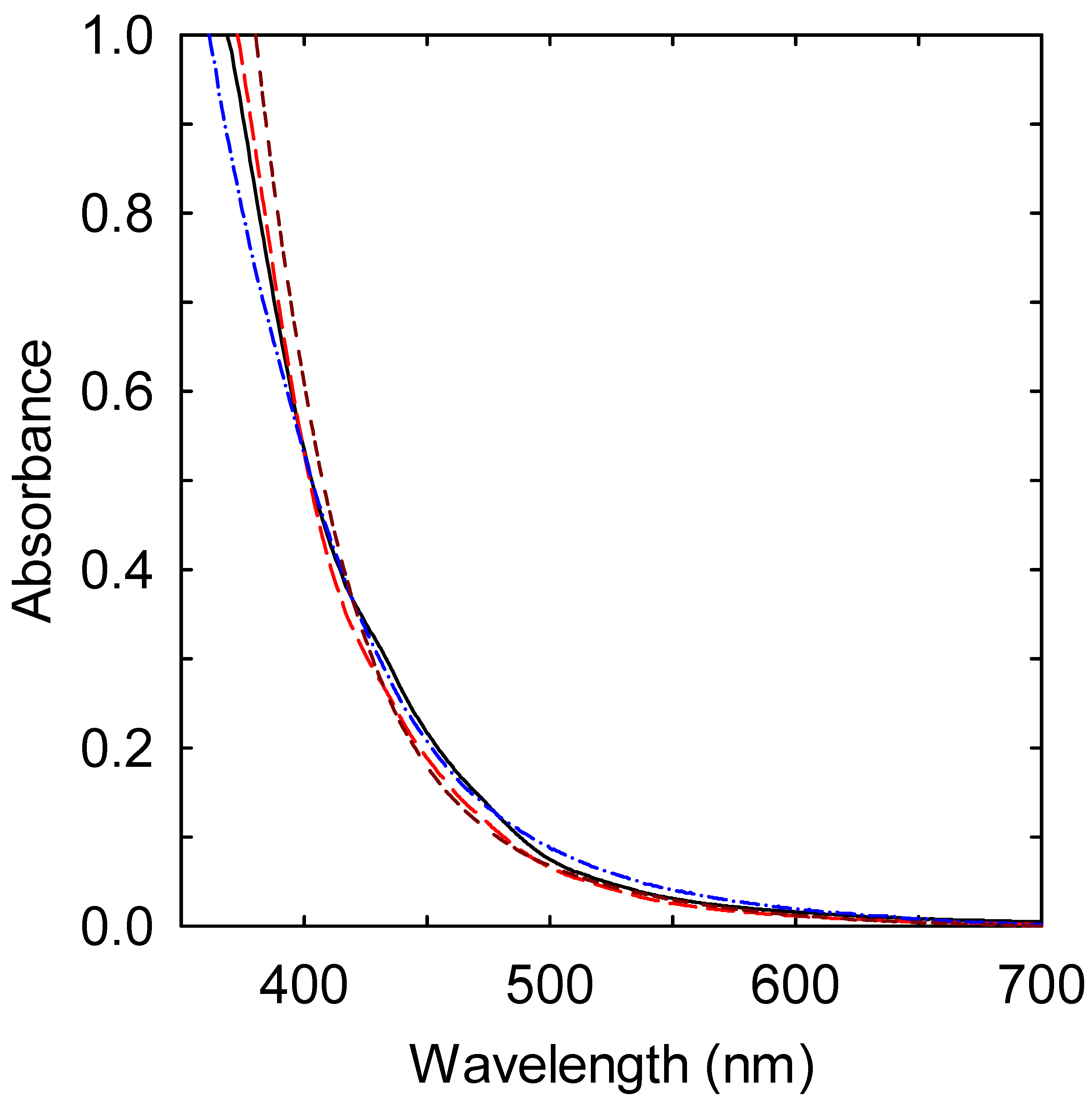
Mixtures of oligomeric PEI and citric acid (CA) have been popular precursors for thermal carbonization processing to prepare CDot-like hybrids. Their popularity may be due to the possible advantage of the precursor mixtures in terms of the interactions between the amine moieties in PEI and the carboxylic acid groups in CA. The resulting zwitterionic pairs might contribute to the more homogeneous distribution and mixing of PEI and CA, even in the solid states of their mixtures. The solid-state precursors enable thermal processing via highly efficient microwave heating, without some of the limitations associated with microwave reactors. In this study, the heating of the solid-state precursor mixtures of PEI and CA was carried out in a conventional microwave oven, with significant carbonization achieved in a few minutes for the resulting samples with varying nanocarbon (non-molecular carbon) content [42]. Experimentally, the same PEI–CA mixture as a precursor was heated in a microwave oven for the same processing time but with different microwave power to obtain the low-C, mid-C, and high-C dot samples. All of these samples were soluble in ethanol and yielded colored solutions, where the color was likely due to the optical absorption in the visible spectrum (Figure 2) by the nanocarbons in these samples produced via microwave-assisted thermal carbonization processing.
By using the previously estimated optical absorptivity of CNPs at 550 nm of 11.6 MC−1cm−1 [43], where MC is the molar concentration of non-molecular carbon atoms (namely those in the carbonization-produced nanocarbons) in the sample solution, and assuming that the absorptivity was insensitive to the organic matrix and/or functionalization, the content of nanocarbons in the low-C, mid-C, and high-C dot samples was estimated to be ~6%, 18%, and 37% by weight, respectively.
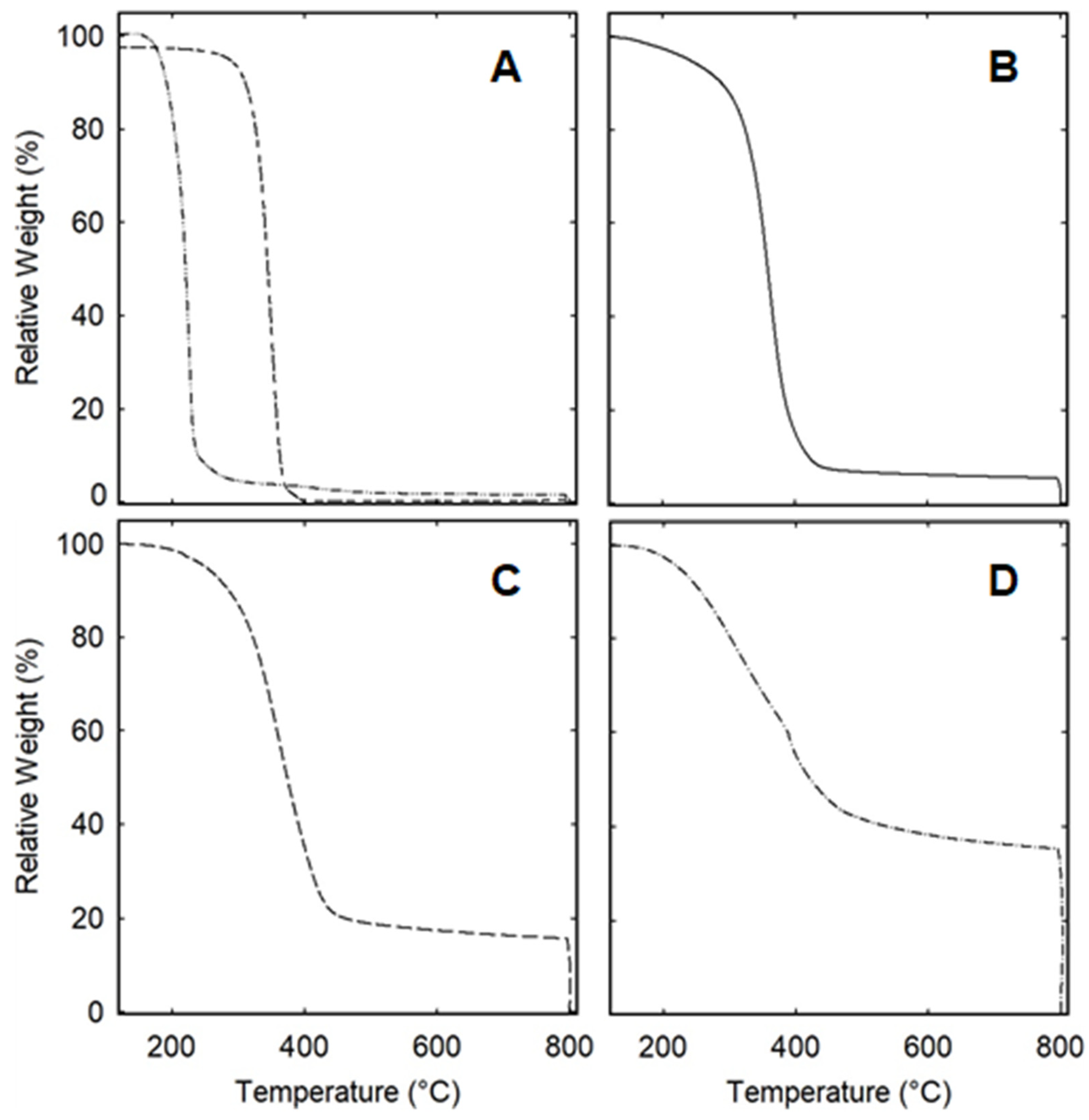
Independently of the above-estimated nanocarbon content based on optical absorption, the thermal gravimetric analysis (TGA) technique was employed to determine the nanocarbon content in the dot samples, with the understanding that carbon materials, including CNPs, in the absence of oxidants are stable up to very high temperatures, far exceeding those used for the decomposition and evaporation of organic materials. In the TGA measurements, in a flow of pure nitrogen gas, each sample was heated to 800 °C at a rate of 10 °C/min, during which the weight loss reached a plateau at approximately 450 °C in all samples (Figure 3). The attribution of the residual weight at the plateau to the nanocarbons in the dot samples was confirmed by switching the gas flow from pure nitrogen to air at 800 °C, thus converting the remaining sample fraction into carbon dioxide, without any meaningful residue (Figure 3). Based on the weight at the mid-point of the plateau at around 600 °C for each sample in the TGA results, the estimated nanocarbon content in the low-C, mid-C, and high-C dot samples was ~5%, 16%, and 35% by weight, respectively, in reasonably good agreement with the optical-absorption-derived results.
The significantly different quantities of the carbonization-produced nanocarbons in the three dot samples obtained using the same solid-state precursor, with the same microwave processing time but seemingly not so dramatically different microwave power (varied from 300 W to 700 W), are striking, suggesting the high sensitivity of carbonization to the applied microwave energy. The results may be rationalized considering the unique feature of microwave heating, such that the heating is selective and much more efficient for materials with a high microwave absorption cross-section, which is more accurately defined as the “dissipation factor” [44]. CNPs (and likely the nanocarbons in the carbonization-produced hybrids as well) are known for their larger dissipation factor compared to those of typical organic molecules [45,46], being nearly an order of magnitude larger at the operational microwave frequency of 2.45 GHz commonly used in commercial microwave ovens [44,46]. In the microwave-assisted thermal carbonization of the organic precursor, the initially produced nanocarbons (essentially the “nanocarbon seeds”) would be heated by the microwave irradiation much more efficiently than the organic materials, with carbonization around the nanocarbon seeds preferred for more and better-defined nanocarbon entities in the sample. This represents a major advantage of microwave heating over simple thermal or hydrothermal processing, coupled with the advantage of using solid-state precursors over those in solution or the liquid phase.
For the preparation of dot samples via the thermal carbonization processing of organic precursors, an interesting question that is relevant to some applications is the source of molecular carbons for the CNP-like domains in the produced dot samples. In addition to the mixture with PEI used in this study, CA has been a popular choice in precursors for thermal carbonization processing, probably due to its molecular structure, which is characterized by three carboxylic acid groups. These are relatively less stable and more susceptible to thermal decarboxylation; thus, CA is considered as the carbon source for the nanocarbons in the resulting dot samples. Regarding the precursor mixture used in this study for the three dot samples, i.e., 300 mg CA and 1 g PEI, the maximum amount of molecular carbons in CA was ~112 mg, corresponding to the maximum nanocarbon content by weight in the resulting dot sample of ~8.6% under the assumption that nothing evaporated or was lost. It would be ~11% if all other elements besides the carbons in CA evaporated and there was no loss of PEI at all, and it would be up to 37.5% if only the complete loss of PEI occurred, which is not realistic because PEI (molecular weight ~600) is nonvolatile and relatively stable. It is more likely that the majority of the nanocarbons in the dot samples (or at least the high-C sample) were sourced from CA, for which carbonization must be accompanied by the evaporation of some other elements. However, in order to obtain a high-C sample containing about 36% in weight of nanocarbons, the carbonization of PEI must occur, which would similarly be accompanied by the evaporation of some other elements. Thus, it can be concluded that both CA and PEI in the precursor mixture contributed molecular carbons for the creation of nanocarbons in the carbonization-produced dot samples.
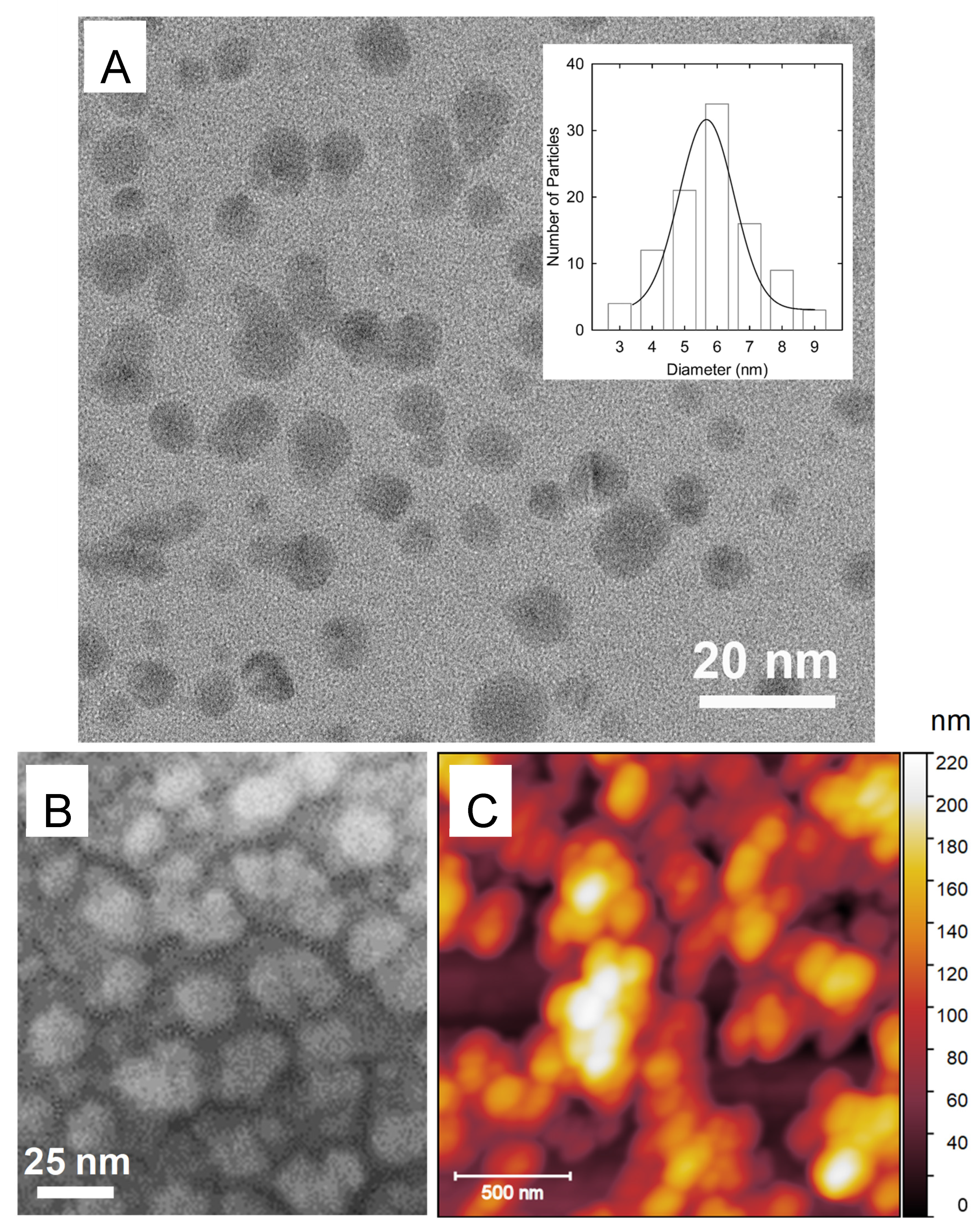
The mass balance calculation and discussion above are consistent with the TGA results, suggesting that the dot samples contained substantial amounts of organic species that survived the thermal carbonization processing conditions. These samples are considered as hybrids (Figure 1), with the sample structure and morphology characterized as being analogous to those of nanocomposites, such that the CNP-like nanocarbons are dispersed in the organic matrix. Also consistent with the sample structure and morphology are the results of the X-ray diffraction and microscopy characterization. As expected, and as confirmed by the powder X-ray diffraction (XRD) analysis, the CNP-like nanocarbons and the dot samples as a whole were amorphous. Nevertheless, the amorphous nanocarbon entities in the hybrids could be identified under a high-resolution transmission electron microscope (TEM). For the TEM imaging, a highly dilute sample solution was prepared, and a few drops of the solution were deposited onto a silicon-oxide-coated copper grid, followed by evaporation to remove the solvent. In the TEM images of the low-C sample, there was a smaller population of CNP-like entities, averaging roughly 3 nm in diameter, with a mostly low-contrast background (making the results qualitative estimates at best), consistent with the low content of nanocarbons. For tshe high-C sample, the TEM images showed more defined nanoparticles (Figure 4). Multiple images were used for a statistical size analysis of the randomly dispersed nanoparticles, from which particle sizes of 6 ± 5 nm in diameter were obtained.
Since TEM is, by design, a tool used to exploit the different electron densities of materials in mixtures, it is capable of identifying the higher-electron-density CNPs and nanocarbons in the matrices of organic species. For the hybrids, however, their composite-like sample structure and morphology became evident in the imaging performed via scanning electron microscopy (SEM). The substantial presence of electrically insulating organic species in all three dot samples introduced severe charging effects that hindered normal SEM imaging. To mitigate the charging effects, the sample specimens were coated with an ultra-thin layer of platinum metal. Nevertheless, the SEM imaging of even the high-C sample (with the lowest content of organic species) with the platinum coating was still not immune to charging effects, yielding images consistent with composites of nanocarbon domains dispersed in the matrices of abundant organic materials (Figure 4). Further supporting the composite-like structure and morphology in the dot samples, namely the hybrids (Figure 1), were the results derived from the atomic force microscopy imaging (Figure 4).
The results of the mass balance analysis discussed above suggest that a significant portion of the PEI in the precursor mixture could survive the carbonization processing conditions and be present in the dot samples. In the classical synthesis of CDots by the deliberate chemical functionalization of preexisting CNPs, organic species carrying primary and secondary amines for functionalization are preferred to ensure that the resulting dot samples are more fluorescent [3]. For the hybrids from thermal carbonization processing, amine molecules have also been popular choices as precursors or in precursor mixtures for similar purposes [47]. In this study, the abundant PEI species in the dot samples, especially those in direct contact with the nanocarbons, might have created localized nanoscale domains (Figure 1), which may be considered as being equivalent in configuration to PEI-CDots [25,48]. Indeed, the observed fluorescence quantum yields at 400 nm excitation of 14%, 13%, and 13% for the low-C, mid-C, and high-C dot samples, respectively, are similar to those of typical PEI-CDot samples [48].
In addition to the largely similar optical absorption spectra of the three dot samples to those of PEI-CDots (Figure 2), their fluorescence spectra and excitation wavelength dependencies were similar as well (Figure 5), despite their already revealed structural and morphological differences. In fact, the characteristic excitation wavelength dependence of fluorescence emissions has become a popular and, in many cases, singular justification for the associated samples derived from thermal carbonization processing to be designated as “carbon dots” [3,49]. The reality is that there are domains of organic species with amine groups surrounding some nanocarbon entities in most carbonization-produced dot samples from amine-containing precursors, and such domains are similar in configuration to classically defined CDots, with the same characteristic excitation wavelength dependence of fluorescence emissions. However, these dot samples could have very different compositions, structures, and morphologies, but such differences at the nanoscopic length scale (in the order of 10 nm, for example) would not be identified or reflected by fluorescence spectroscopy because the light wavelengths used span hundreds of nanometers [41]. Moreover, it is common knowledge that conventional optical microscopy cannot be used for imaging at the nanoscale. Nevertheless, the hybrids, including especially the mid-C and high-C dot samples, with bright fluorescence emissions, may compete with PEI-CDots in applications in which the samples’ structural and morphological differences at the nanoscale have no impact.
The low-C and high-C dot samples were evaluated for their visible-light-activated antibacterial activity against L. monocytogene 10403S (drug-resistant) cells and the laboratory model bacterium B. subtilis cells. In the experiments, the final concentrations of bacterial cells were about 107 CFU/mL, and the concentrations of the dot samples varied from 50 to 200 μg/mL. All samples were exposed to visible light for 1 h, and the viable cell numbers were determined after the treatments. As shown in Figure 6A, the high-C sample, at a higher concentration (in terms of that of the nanocarbons in the sample), could kill up to two logs of the drug-resistant bacteria, but, at the equivalent concentration, the low-C sample had little effect on the same bacterial cells, suggesting a significantly weaker antibacterial function. The mechanism of light-activated CDots as antimicrobial agents requires the combined action of the separated redox pairs generated immediately after the photoexcitation of CDots and the classical reactive oxygen species (ROS) produced in the emissive excited states of the CDots at a longer time scale [50]. The former, resembling the “light-activated redox species” (LARS) in the conventional semiconductor QDs [51], are highly reactive and thus short-lived, contributing significantly to the overall antimicrobial activity but requiring the close proximity of the photoexcited CDots (or the photoexcited nanocarbon domains in the hybrids) to the bacterial target [50,52,53]. The same mechanism should be applicable to the antibacterial activity of the CDot-like domains in the hybrids prepared by the thermal carbonization processing of organic precursors. Here, regarding the low-C and high-C dot samples, they both have CDot-like domains that are colored and therefore readily activated by visible light, but these domains in the two dot samples exist in different organic matrix environments, with considerably more organic species per CDot-like domain in the former than in the latter. Thus, the apparently greater activity of the visible-light-activated high-C sample against the drug-resistant L. monocytogene might be attributed to its lower relative content of organic species, facilitating the near-neighbor killing action of the highly reactive redox pairs. The same action is hindered by the much larger population of organic species in the low-C sample. Nevertheless, upon visible light activation, the low-C sample was capable of inactivating the “easier” bacterial target B. subtilis (Figure 6B).

4. Summary and Conclusions

The microwave-assisted thermal carbonization processing of a selected precursor mixture, such as PEI–CA, in the solid state is effective and efficient in producing dot samples with different content of non-molecular nanocarbons. As demonstrated, the solid-state organic precursors offer major advantages such that the carbonization could be performed at high temperatures by microwave heating, far beyond the temperatures possible in hydrothermal or other solution-based processing, even in pressurized vessels. Moreover, the initially created nanocarbon “seeds” could preferentially absorb microwave energy to drive further carbonization and increase the nanocarbon domains. These dot samples are nanocarbon/organic hybrids with a similar structure and morphology to nanocomposites; within them, the localized domains of CNP-equivalent nanocarbons embedded in and passivated by the surrounding organic species are similar to the configuration seen in classically defined CDots. The composite-like sample structure and morphology of the hybrids are consistent with the findings revealed by the comparison of the high-resolution TEM, SEM, and AFM imaging results. The dot samples apparently exhibit the same optical spectroscopic properties as CDots, including the characteristic excitation wavelength dependence of the fluorescence emissions, which is expected given that optical spectroscopy is insensitive to any structural and morphological differences at the nanoscale. On the other hand, the obviously different light-activated antibacterial activity of the dot samples with lower and higher nanocarbon content may be attributed to the correspondingly higher and lower content of organic species in the samples, which hinder the antibacterial action to varying degrees. Further studies to improve our understanding of the carbonization process and to elucidate the CDot-like nanostructures in the hybrids will prove valuable. This effective and efficient preparation method based on simple microwave heating may be further developed for large quantities of dot samples, such as those needed for antimicrobial and other applications.

Author Contributions

Data acquisition and analyses, B.S., A.F.A., S.D., J.C., C.E.B., H.Q. and M.J.M.; Funding acquisition, L.Y. and Y.-P.S.; Investigation, B.S. and A.F.A.; Project administration, M.J.M., C.E.B., L.Y. and Y.-P.S.; Supervision, L.Y. and Y.-P.S.; Validation, B.S. and A.F.A.; Writing—original draft, B.S., A.F.A., L.Y. and Y.-P.S.; Writing—review and editing, L.Y. and Y.-P.S. All authors have read and agreed to the published version of the manuscript.

Funding

USDA (2023-67018-40681) and NSF (2102021 and 2102056).

Institutional Review Board Statement

Not applicable.

Informed Consent Statement

Not applicable.

Data Availability Statement

All data are available from the corresponding author upon request.

Acknowledgments

The financial support of the USDA (2023-67018-40681) and NSF (2102021 and 2102056) is gratefully acknowledged.

Conflicts of Interest

The authors declare no conflicts of interest.

References

  1. Sun, Y.-P.; Zhou, B.; Lin, Y.; Wang, W.; Fernando, K.A.S.; Pathak, P.; Meziani, M.J.; Harruff, B.A.; Wang, X.; Wang, H.; et al. Quantum-Sized carbon dots for bright and colorful photoluminescence. J. Am. Chem. Soc. 2006, 128, 7756–7757. [Google Scholar] [CrossRef] [PubMed]
  2. Sun, Y.-P. Fluorescent Carbon Nanoparticles. U.S. Patent 7,829,772 B2, 9 November 2010. [Google Scholar]
  3. Sun, Y.-P. Carbon Dots—Exploring Carbon at Zero-Dimension; Springer International Publishing: New York, NY, USA, 2020. [Google Scholar]
  4. Ding, C.; Zhu, A.; Tian, Y. Functional Surface Engineering of C-Dots for Fluorescent Biosensing and in Vivo Bioimaging. Acc. Chem. Res. 2014, 47, 20–30. [Google Scholar] [CrossRef]
  5. Lim, S.Y.; Shen, W.; Gao, Z. Carbon Quantum Dots and Their Applications. Chem. Soc. Rev. 2015, 44, 362–381. [Google Scholar] [CrossRef]
  6. Gao, J.; Zhu, M.; Huang, H.; Liu, Y.; Kang, Z. Advances, Challenges and Promises of Carbon Dots. Inorg. Chem. Front. 2017, 4, 1963–1986. [Google Scholar] [CrossRef]
  7. Du, J.; Xu, N.; Fan, J.; Sun, W.; Peng, X. Carbon Dots for In Vivo Bioimaging and Theranostics. Small 2019, 15, 1805087. [Google Scholar] [CrossRef] [PubMed]
  8. Kang, Z.; Lee, S.-T. Carbon Dots: Advances in Nanocarbon Applications. Nanoscale 2019, 11, 19214–19224. [Google Scholar] [CrossRef]
  9. Rosso, C.; Filippini, G.; Prato, M. Carbon Dots as Nano-Organocatalysts for Synthetic Applications. ACS Catal. 2020, 10, 8090–8105. [Google Scholar] [CrossRef]
  10. Liu, J.; Li, R.; Yang, B. Carbon Dots: A New Type of Carbon-Based Nanomaterial with Wide Applications. ACS. Cent. Sci. 2020, 6, 2179–2195. [Google Scholar] [CrossRef]
  11. Feng, T.; Tao, S.; Yue, D.; Zeng, Q.; Chen, W.; Yang, B. Recent Advances in Energy Conversion Applications of Carbon Dots: From Optoelectronic Devices to Electrocatalysis. Small 2020, 16, 2001295. [Google Scholar] [CrossRef]
  12. Li, H.; Yan, X.; Kong, D.; Jin, R.; Sun, C.; Du, D.; Lin, Y.; Lu, G. Recent Advances in Carbon Dots for Bioimaging Applications. Nanoscale Horiz. 2020, 5, 218–234. [Google Scholar] [CrossRef]
  13. Dhenadhayalan, N.; Lin, K.-C.; Saleh, T.A. Recent Advances in Functionalized Carbon Dots toward the Design of Efficient Materials for Sensing and Catalysis Applications. Small 2020, 16, 1905767. [Google Scholar] [CrossRef] [PubMed]
  14. He, C.; Xu, P.; Zhang, X.; Long, W. The Synthetic Strategies, Photoluminescence Mechanisms and Promising Applications of Carbon Dots: Current State and Future Perspective. Carbon 2022, 186, 91–127. [Google Scholar] [CrossRef]
  15. Ðorđević, L.; Arcudi, F.; Cacioppo, M.; Prato, M. A Multifunctional Chemical Toolbox to Engineer Carbon Dots for Biomedical and Energy Applications. Nat. Nanotechnol. 2022, 17, 112–130. [Google Scholar] [CrossRef]
  16. Yadav, P.K.; Chandra, S.; Kumar, V.; Kumar, D.; Hasan, S.H. Carbon Quantum Dots: Synthesis, Structure, Properties, and Catalytic Applications for Organic Synthesis. Catalysts 2023, 13, 422. [Google Scholar] [CrossRef]
  17. Dua, S.; Kumar, P.; Pani, B.; Kaur, A.; Khanna, M.; Bhatt, G. Stability of Carbon Quantum Dots: A Critical Review. RSC Adv. 2023, 13, 13845–13861. [Google Scholar] [CrossRef]
  18. Jing, H.H.; Bardakci, F.; Akgöl, S.; Kusat, K.; Adnan, M.; Alam, M.J.; Gupta, R.; Sahreen, S.; Chen, Y.; Gopinath, S.C.; et al. Green Carbon Dots: Synthesis, Characterization, Properties and Biomedical Applications. J. Funct. Biomater. 2023, 14, 27. [Google Scholar] [CrossRef]
  19. Bhattacharya, T.; Shin, G.H.; Kim, J.T. Carbon Dots: Opportunities and Challenges in Cancer Therapy. Pharmaceutics 2023, 15, 1019. [Google Scholar] [CrossRef] [PubMed]
  20. Zhao, W.-B.; Liu, K.-K.; Wang, Y.; Li, F.-K.; Guo, R.; Song, S.-Y.; Shan, C.-X. Antibacterial Carbon Dots: Mechanisms, Design, and Applications. Adv. Healthc. Mater. 2023, 12, 2300324. [Google Scholar] [CrossRef]
  21. Riggs, J.E.; Guo, Z.; Carroll, D.L.; Sun, Y.-P. Strong Luminescence of Solubilized Carbon Nanotubes. J. Am. Chem. Soc. 2000, 122, 5879–5880. [Google Scholar] [CrossRef]
  22. Guldi, D.M.; Holzinger, M.; Hirsch, A.; Georgakilasc, V.; Prato, M. First Comparative Emission Assay of Single-Wall Carbon Nanotubes—Solutions and Dispersions. Chem. Commun. 2003, 10, 1130–1131. [Google Scholar] [CrossRef]
  23. Liang, W.; Sheriff, K.; Singh, B.; Qian, H.; Dumra, S.; Collins, J.; Yerra, S.; Yang, L.; Sun, Y.-P. On Carbon “Replacing” the Core in Classical Core/ZnS Quantum Dots. Gen. Chem. 2024, 10, 240001. [Google Scholar]
  24. Jiang, H.; Chen, F.; Lagally, M.G.; Denes, F.S. New Strategy for Synthesis and Functionalization of Carbon Nanoparticles. Langmuir 2010, 26, 1991–1995. [Google Scholar] [CrossRef]
  25. Ge, L.; Pan, N.; Jin, J.; Wang, P.; LeCroy, G.E.; Liang, W.; Yang, L.; Teisl, L.R.; Tang, Y.; Sun, Y.-P. Systematic Comparison of Carbon Dots from Different Preparations—Consistent Optical Properties and Photoinduced Redox Characteristics in Visible Spectrum, and Structural and Mechanistic Implications. J. Phys. Chem. C 2018, 122, 21667–21676. [Google Scholar] [CrossRef]
  26. Li, H.; Kang, Z.; Liu, Y.; Lee, S.-T. Carbon Nanodots: Synthesis, Properties and Applications. J. Mater. Chem. 2012, 22, 24230–24253. [Google Scholar] [CrossRef]
  27. Liu, X.; Pang, J.; Xu, F.; Zhang, X. Simple Approach to Synthesize Amino-Functionalized Carbon Dots by Carbonization of Chitosan. Sci. Rep. 2016, 6, 31100. [Google Scholar] [CrossRef] [PubMed]
  28. Xia, C.; Zhu, S.; Feng, T.; Yang, M.; Yang, B. Evolution and Synthesis of Carbon Dots: From Carbon Dots to Carbonized Polymer Dots. Adv. Sci. 2019, 6, 1901316. [Google Scholar] [CrossRef]
  29. de Medeiros, T.V.; Manioudakis, J.; Noun, F.; Macairan, J.-R.; Victoria, F.; Naccache, R. Microwave-Assisted Synthesis of Carbon Dots and Their Applications. J. Mater. Chem. C 2019, 7, 7175–7195. [Google Scholar] [CrossRef]
  30. Semeniuk, M.; Yi, Z.; Poursorkhabi, V.; Tjong, J.; Jaffer, S.; Lu, Z.-H.; Sain, M. Future Perspectives and Review on Organic Carbon Dots in Electronic Applications. ACS Nano 2019, 13, 6224–6255. [Google Scholar] [CrossRef]
  31. Kung, J.-C.; Tseng, I.-T.; Chien, C.-S.; Lin, S.-H.; Wang, C.-C.; Shih, C.-J. Microwave Assisted Synthesis of Negative-Charge Carbon Dots with Potential Antibacterial Activity against Multi-Drug Resistant Bacteria. RSC Adv. 2020, 10, 41202–41208. [Google Scholar] [CrossRef]
  32. Ye, Z.; Li, G.; Lei, J.; Liu, M.; Jin, Y.; Li, B. One-Step and One-Precursor Hydrothermal Synthesis of Carbon dots with Superior Antibacterial Activity. ACS Appl. Bio. Mater. 2020, 3, 7095–7102. [Google Scholar] [CrossRef]
  33. Hasan, M.R.; Saha, N.; Quaid, T.; Reza, M.T. Formation of Carbon Quantum Dots via Hydrothermal Carbonization: Investigate the Effect of Precursors. Energies 2021, 14, 986. [Google Scholar] [CrossRef]
  34. Liao, G.; Luo, J.; Cui, T.; Zou, J.; Xu, M.; Ma, Y.; Shi, L.; Jia, J.; Ma, C.; Li, H.; et al. Microwave-Assisted One-Pot Synthesis of Carbon Dots for Highly Sensitive and Selective Detection of Selenite. Microchem. J. 2022, 179, 107440. [Google Scholar] [CrossRef]
  35. Liu, W.; Gu, H.; Liu, W.; Lv, C.; Du, J.; Fan, J.; Peng, X. NIR-emitting Carbon Dots for Discriminative Imaging and Photo-Inactivation of Pathogenic Bacteria. Chem. Eng. J. 2022, 450, 137384. [Google Scholar] [CrossRef]
  36. Kang, C.; Tao, S.; Yang, F.; Yang, B. Aggregation and Luminescence in Carbonized Polymer Dots. Aggregate 2022, 3, e169. [Google Scholar] [CrossRef]
  37. Saadh, M.J.; Al-dolaimy, F.; Alamir, H.T.A.; Kadhim, O.; Al-Abdeen, S.H.Z.; Sattar, R.; Abid, M.K.; Jetti, R.; Alawadi, A.; Alsalamy, A. Emerging Pathways in Environmentally Friendly Synthesis of Carbon-Based Quantum Dots for Exploring Antibacterial Resistance. Inorg. Chem. Commun. 2024, 161, 112012. [Google Scholar] [CrossRef]
  38. Simões, R.; Rodrigues, J.; Neto, V.; Monteiro, T.; Gonçalves, G. Carbon Dots: A Bright Future as Anticounterfeiting Encoding Agents. Small 2024, 20, 2311526. [Google Scholar] [CrossRef]
  39. Wang, B.; Waterhouse, G.I.N.; Yang, B.; Lu, S. Advances in Shell and Core Engineering of Carbonized Polymer Dots for Enhanced Applications. Acc. Chem. Res. 2024, 57, 2928–2939. [Google Scholar] [CrossRef]
  40. Liu, H.; Zhong, X.; Pan, Q.; Zhang, Y.; Deng, W.; Zou, G.; Hou, H.; Ji, X. A Review of Carbon Dots in Synthesis Strategy. Coord. Chem. Rev. 2024, 498, 215468. [Google Scholar] [CrossRef]
  41. Liang, W.; Sonkar, S.K.; Saini, D.; Sheriff, K.; Singh, B.; Yang, L.; Wang, P.; Sun, Y.-P. Carbon Dots: Classically Defined versus Organic Hybrids on Shared Properties, Divergences, and Myths. Small 2023, 19, 2206680. [Google Scholar] [CrossRef]
  42. Adcock, A.F.; Wang, P.; Cao, E.Y.; Ge, L.; Tang, Y.; Ferguson, I.S.; Abu Sweilem, F.S.; Petta, L.; Cannon, W.; Yang, L.; et al. Carbon Dots versus Nano-Carbon/Organic Hybrids—Divergence between Optical Properties and Photoinduced Antimicrobial Activities. C 2022, 8, 54. [Google Scholar] [CrossRef]
  43. Xu, J.; Sahu, S.; Cao, L.; Anilkumar, P.; Tackett, K.N., II; Qian, H.; Bunker, C.E.; Guliants, E.A.; Parenzan, A.; Sun, Y.-P. Carbon Nanoparticles as Chromophores for Photon Harvesting and Photoconversion. ChemPhysChem 2011, 12, 3604–3608. [Google Scholar] [CrossRef] [PubMed]
  44. Tao, J.; Zhou, J.; Yao, Z.; Jiao, Z.; Wei, B.; Tan, R.; Li, Z. Multi-Shell Hollow Porous Carbon Nanoparticles with Excellent Microwave Absorption Properties. Carbon 2021, 172, 542–555. [Google Scholar] [CrossRef]
  45. Hayes, B.L. Microwave Synthesis: Chemistry at the Speed of Light; CEM Publishing: Matthews, NC, USA, 2002. [Google Scholar]
  46. Donaldson, A.A.; Hutcheon, R.; Zhang, Z. Dielectric Properties of Quinoline, 4,6-Dimethyldibenzothiophene and Hexadecane as Model Compounds in the Upgrading of LCO. Fuel Process. Technol. 2011, 92, 1733–1737. [Google Scholar] [CrossRef]
  47. Song, Y.; Zhu, S.; Zhang, S.; Fu, Y.; Wang, L.; Zhao, X.; Yang, B. Investigation from Chemical Structure to Photoluminescent Mechanism: A Type of Carbon Dots from The Pyrolysis of Citric Acid and an Amine. J. Mater. Chem. C 2015, 3, 5976–5984. [Google Scholar] [CrossRef]
  48. Wang, P.; Meziani, M.J.; Fu, Y.; Bunker, C.E.; Hou, X.; Yang, L.; Msellek, H.; Zaharias, M.; Darby, J.P.; Sun, Y.-P. Carbon Dots versus Nano-Carbon/Organic Hybrids–Dramatically Different Behaviors in Fluorescence Sensing of Metal Cations with Structural and Mechanistic Implications. Nanoscale Adv. 2021, 3, 2316–2324. [Google Scholar] [CrossRef] [PubMed]
  49. Chen, L.; Wang, C.F.; Liu, C.; Chen, S. Facile Access to Fabricate Carbon Dots and Perspective of Large-Scale Applications. Small 2023, 19, 2206671. [Google Scholar] [CrossRef]
  50. Dong, X.; Ge, L.; Abu Rabe, D.I.; Mohammed, O.O.; Wang, P.; Tang, Y.; Kathariou, S.; Yang, L.; Sun, Y.-P. Photoexcited State Properties and Antibacterial Activities of Carbon Dots Relevant to Mechanistic Features and Implications. Carbon 2020, 170, 137–145. [Google Scholar] [CrossRef]
  51. Courtney, C.M.; Goodman, S.M.; McDaniel, J.A.; Madinger, N.E.; Chatterjee, A. Photoexcited Quantum Dots for Killing Multidrug-Resistant Bacteria. Nat. Mater. 2016, 15, 529–534. [Google Scholar] [CrossRef]
  52. Abu Rabe, D.I.; Mohammed, O.O.; Dong, X.; Patel, A.K.; Overton, C.M.; Tang, Y.; Kathariou, S.; Sun, Y.-P.; Yang, L. Carbon Dots for Highly Effective Photodynamic Inactivation of Multidrug-Resistant Bacteria. Mater. Adv. 2020, 1, 321–325. [Google Scholar] [CrossRef]
  53. Rodriguez, C.E.; Adcock, A.F.; Singh, B.; Yerra, S.; Tang, Y.; Sun, Y.-P.; Yang, L. Divergence in Antiviral Activities of Carbon Dots versus Nano-Carbon/Organic Hybrids and Implications. C 2023, 9, 79. [Google Scholar] [CrossRef]
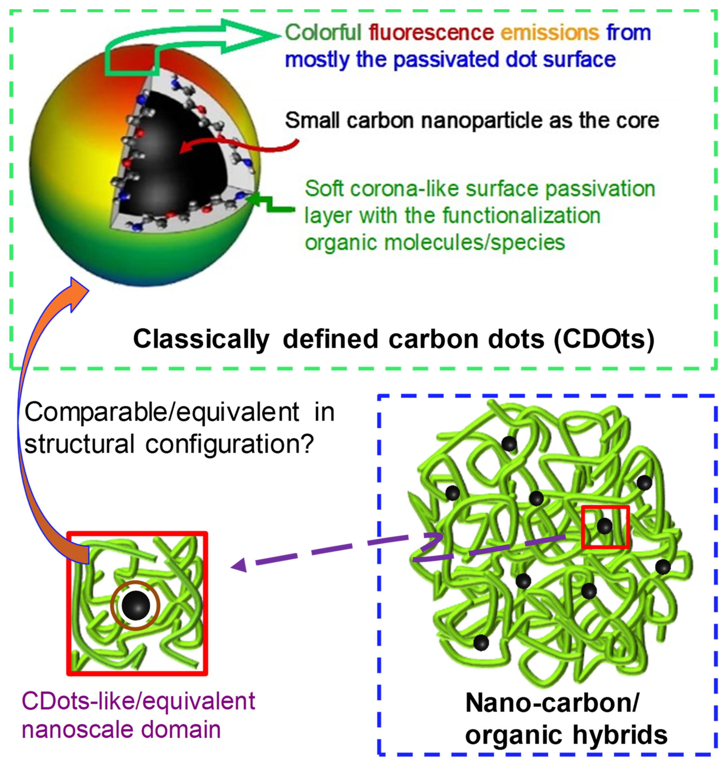
Figure 1. Cartoon illustrations of the classically defined carbon dots (CDots) prepared by the organic functionalization of preexisting small carbon nanoparticles (CNPs) versus the “nanocarbon/organic hybrids” (“hybrids”) produced by the thermal carbonization processing of organic precursors, showing the overall sample structural/morphological differences and also some CDot-like/equivalent nanoscale domains in the hybrids.
Micro 05 00014 g001
Figure 2. Optical absorption spectra of the low-C (solid), mid-C (short dash), and high-C (long dash) samples in ethanol solutions, with that of PEI-CDots (dash-dot) for comparison.
Micro 05 00014 g002
Figure 3. The TGA results of (A) neat citric acid (dash-dot-dot) and neat PEI (dash), (B) the low-C sample, (C) the mid-C sample, and (D) the high-C sample. The sharp drops at 800 °C correspond to the exposure of the samples to air.
Micro 05 00014 g003
Figure 4. Results for the high-C sample from TEM imaging ((A), with the inset showing the particle size distribution curve derived from the statistical size analysis of multiple images); SEM imaging ((B) for the sample with an ultra-thin metal coating); and AFM imaging (C).
Micro 05 00014 g004
Figure 5. Absorption (ABS) and excitation-wavelength-dependent fluorescence (FLSC) spectra (displayed from left to right in different colors, corresponding to excitations ranging from 400 nm to 520 nm in 20 nm increments) of the low-C (lower), mid-C (middle), and high-C (upper) samples in ethanol solutions.
Micro 05 00014 g005
Figure 6. The visible-light-activated antibacterial activity of the high-C and low-C samples against the drug-resistant L. monocytogenes (A) and laboratory model bacterium B. subtilis (B), as expressed by the viable cell numbers in the treated samples compared to those in the control.
Micro 05 00014 g006
Disclaimer/Publisher’s Note: The statements, opinions and data contained in all publications are solely those of the individual author(s) and contributor(s) and not of MDPI and/or the editor(s). MDPI and/or the editor(s) disclaim responsibility for any injury to people or property resulting from any ideas, methods, instructions or products referred to in the content.