Article

Green Synthesis and Characterization of Silver Nanoparticles from Minthostachys acris Schmidt Lebuhn (Muña) and Its Evaluation as a Bactericidal Agent Against Escherichia coli and Staphylococus aureus

by
Fabián Ccahuana Ayma
1,2,†,
Ana María Osorio Anaya
1,*,†,
Gabrielle Caroline Peiter
2,†,
Silvia Jaerger
2,*,† and
Ricardo Schneider
2
1
Materials for Technological Innovation Research Group (GI MATINTEC-FQIQ), National University of San Marcos (UNMSM), Lima 15081, Peru
2
Group of Polymers and Nanostructures, Universidade Tecnológica Federal do Paraná—UTFPR, Toledo 85902-490, PR, Brazil
*
Authors to whom correspondence should be addressed.
These authors contributed equally to this work.
Micro 2024, 4(4), 706-720; https://doi.org/10.3390/micro4040043
Submission received: 25 September 2024 / Revised: 9 November 2024 / Accepted: 11 November 2024 / Published: 20 November 2024

Abstract

:
The search for new synthesis methodologies based on the principles of green chemistry has led to various studies for the production of silver nanoparticles (AgNPs) using extracts from different parts of plants. Based on this, the present study aims to carry out green synthesis (biosynthesis), characterization, and antibacterial evaluation of reduced and stabilized silver nanoparticles (AgNPs) with aqueous extracts of Minthostachys acris in a simple, ecological, and environmentally safe manner. The extraction process of the organic components is performed using two methods: immersion and the agitation of the leaves of Minthostachys acris Schmidt Lebuhn (Muña) at 0.1% for different times (0.5, 1, 3, 6, and 10 min). Compounds such as hydroxycinnamic acid derivatives, quinic, caffeic, rosmarinic acids, and flavonols present in the Muña extract facilitate the formation of AgNPs; this compounds act as a coating and stabilizing agent. The bioactive components from natural resources facilitate the formation of AgNPs, partially or completely replacing the contaminating and toxic elements present in chemical reagents. The biosynthesis is carried out at room temperature for pH 7 and 8. The synthesized AgNPs are characterized by UV-visible spectroscopy to identify the surface plasmon resonance (SPR) band, which shows an absorption peak around 419 nm and 423 nm for pH 7 and p.H 8, respectively, and Fourier-transform infrared spectroscopy (FTIR) to identify the possible biomolecules responsible for bioreduction and stabilization, with a peak at 1634 cm−1. Dynamic light scattering (DLS) shows the hydrodynamic size of the colloidal nanoparticles between 11 and 200 nm, and scanning electron microscopy (SEM) reveals monodisperse AgNPs of different morphologies, mostly nanospheres, while Laser-Induced Breakdown Spectroscopy (LIBS) demonstrates the presence of Ag in the colloidal solution. The evaluation of the bactericidal activity of the AgNPs using the disk diffusion method against Escherichia coli (E. coli) and Staphylococus aureus (S.aureus) shows that the synthesized AgNPs have effective antibacterial activity against E. coli for the extracts obtained at 6 min for both the immersion and agitation methods, respectively. The significance of this work lies in the use of bioactive components from plants to obtain AgNPs in a simple, rapid, and economical way, with potential applications in biomedical fields.

1. Introduction

Materials at nanoscale, as nanoparticles (NPs), due to their dimensions show unique properties for use in catalysis [1,2,3], photonics, optical [4], bactericidal, surface-enhanced Raman scattering (SERS) [5], and photothermal therapy [6,7]. The proprieties mainly depend on the size, morphology, stability, chemical composition, and method of obtaining them. Nanoparticles can be obtained using physical, chemical, biological [8], and hybrid methods [6]. Although the synthesis was widely developed using chemical and physical approach methods, [9,10] make use of toxic chemicals, which are costly and potentially harmful to biological systems and the environment [11].
Green synthesis (biosynthesis) is an environmentally friendly process [12] for obtaining metallic NPs based on the reduction in metals, employing active biological compounds with a reducing capacity. In this process, the phytochemical components are used as a reducing agent and also as a stabilizer, which in turn results in toxic chemicals being substituted, making biosynthesis an alternative method that is applied in a more straightforward, efficient, non-toxic, less expensive, ecological [11,12,13,14], and environmentally safe way [15].
Biosynthesis has been reported to employ microorganisms such as fungi [16], bacteria [17], and yeasts [13,18], also making use of multiple plant materials such as the root extracts [19], barks [20], flowers [21], fruits [14,22,23,24,25], pericarp [26], peels [27], and leaves [8,28,29,30,31,32] of different plants.
The genus Minthostachys is characterized by its presence in various ecological zones of the Andean region, encompassing countries such as Venezuela, Bolivia, Colombia, Ecuador, Peru, and parts of Argentina, including 17 species [33]. Among them, Minthostachys acris (Figure 1) is a typical aromatic wild shrub of Southern Peru, where it is known as Muña. It features conical, serrated leaves and small, zygomorphic white flowers located at the top of the plants. Traditionally, it has been used by local communities for its medicinal properties, such as infusions for dizziness, treatment of colic, and protection against pests in stored tubers [33]. Studies on the chemical composition of the essential oil of M. acris using gas chromatography revealed the presence of between 36 and 60 different volatile organic components, with the main ones being the monoterpenes [33] pulegone, trans-cis-menthone, and thymol [34,35] characterized by the presence of carboxyl groups. In previous research involving the analysis of Minthostachys by High-Performance Liquid Chromatography (HPLC), a significant presence of several phenolic compounds was identified. In particular, hydroxycinnamic acid derivatives [36], quinic acids, caffeic acid and rosmarinic acid were found in substantial amounts, while flavonoids and other compounds were identified in smaller proportions [37,38]. Additionally, these components can vary depending on the plant’s location, soil moisture composition, and the collection period.
Numerous metals can be used to obtain nanoparticles (NPs) through biosynthesis with plant extracts, and this occurs due to the chemical interaction of the bioactive compounds in the plants, allowing for the formation of nanostructures. This process is much more environmentally friendly because the extract replaces the reducing and stabilizing agents that are commonly high-cost solutions and potentially toxic at elevated concentrations.
In this study, we propose two extraction methods, immersion and agitation, to obtain an aqueous extract of Minthostachys acris at room temperature over different periods. These extracts were used in the green synthesis of AgNPs at pH 7 and 8. Additionally, the effect of extraction time on the antibacterial capacity of the AgNPs obtained at pH 7 against Gram-negative and Gram-positive bacteria was evaluated. Several previous studies on the green synthesis of nanomaterials have been conducted using high temperatures and extended reaction times [39,40]. However, there is currently a growing demand for nanometric materials that are both functional and stable [41]. This research proposes an innovative, simple, and rapid process that does not require high temperatures, with an ecological focus that minimizes the use of chemical substances in producing AgNPs. Therefore, the proposed approach overcomes several drawbacks of synthesizing biocidal materials using plant extracts. The materials are characterized by UV-visible spectroscopy (UV-vis) to identify the surface plasmon resonance (SPR), Fourier transform infrared spectroscopy (FTIR) to identify functional groups, scanning electron microscopy (SEM) to observe the morphology and distribution of the obtained AgNPs, dynamic light scattering (DLS) to measure the hydrodynamic size of the AgNPs, and Laser-Induced Breakdown Spectroscopy (LIBS). Finally, the antibacterial action against bacterial strains of Escherichia coli (ATCC: 25922) and Staphylococus aureus (ATCC: 6538) is evaluated [42].

2. Materials and Methods

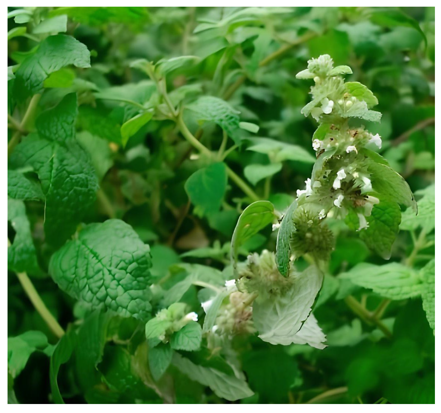
2.1. Plant Collection and Characterization

The collection process of the plant species was by simple random sampling, visually evaluating the characteristics of the plants at a height between 8 and 10 cm from the ground with a sickle in March 2021 in the district of Pomacanchi, Acomayo, Department of Cusco, Peru. The taxonomic identification was determined in the Museum of Natural History of the Universidad Nacional Mayor de San Marcos classified as ORDER: LAMIALES, FAMILY: LAMIACEAE, GENUS: Minthostachys and SPECIES: Minthostachys acris Schmidt Lebuhn, commonly known as Muña.

2.2. Plant Extract Preparation

The extraction of organic components can be performed using various solvents, including ethanol [43] and aqueous extraction of the powdered material [40,44]. Based on this, the aqueous extraction was carried out using two processes: the immersion method and the stirring method. The previously pulverized sample was sieved through meshes in the range of 150–200 mesh. In the first process, five samples of approximately 0.1 g were weighed on an analytical balance, added to new filtering tea bags, and placed in previously washed and dried 150 mL plastic containers. Then, 100 mL of distilled water was added, and the mixture was allowed to stand for different times of 0.5, 1, 3, 6, and 10 min, after which the filtering bags were extracted. In the second process, 0.1 g of the pulverized sample was added to five 150 mL Erlenmeyer flasks, and taken to the shaker with the addition of 100 mL of distilled water at 114 RPM. The extraction was carried out for different stirring times: 0.5, 1, 3, 6, and 10 min, respectively. Then, it was filtered with Whatman N°. 1 paper into 125 mL flasks and stored at a temperature of 5 °C for further study.

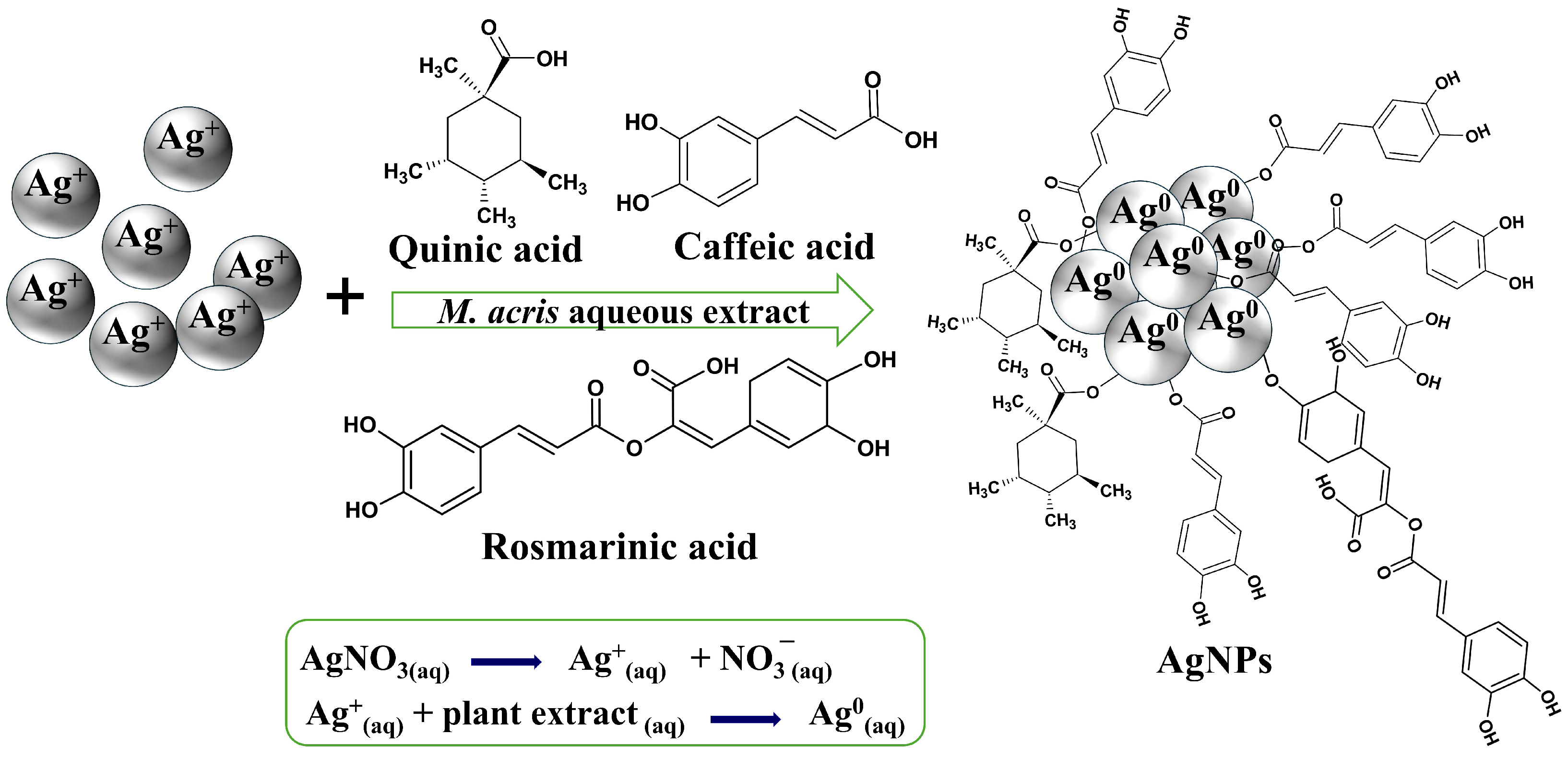
2.3. Green Synthesis of Silver Nanoparticles

The green synthesis of AgNPs was carried out in 10 mL vials by adding 9 mL of AgNO3 (1 mM) to 1 mL of the previously obtained extracts. Instantly, a color change of the solution from yellowish to light brown was observed, indicating the formation of AgNPs, which were stored for further characterization and application.

2.4. Characterization of Green Synthesized Silver Nanoparticles

UV-vis analysis was performed with a PG INSTRUMENTS model T80+ spectrophotometer (Lutterworth, UK) in the range of 250–600 nm using a quartz cuvette with 1 cm off pathlength after 48 h of AgNPs synthesis (Section 2.3). The FTIR spectrum was acquired on a Perkin-Elmer Spectrum Two FT-IR Spectrometer (Waltham, MA, USA), with a wavelength range of 4000–500 cm−1, with an operating range of 5–45 °C, and a LiTaO3 detector. The hydrodynamic diameter distribution of the colloidal AgNPs was determined on a Nicomp 380 Nanoparticle Size Analyzer (Entegris, Billerica, MA, USA) after 72 h. The morphology of the biosynthesized AgNPs was determined by scanning electron microscope (SEM) in a EVO 10Z6 microscopy from Zeiss (Oberkochen, Germany). Additionally, the elementary composition was evaluated by Laser-Induced Breakdown Spectroscopy (LIBS) using a J200 Tandem spectrometer (Applied Spectra, West Sacramento, CA, USA) operating with a 266 nm laser (25 mJ and full width at half maximum (FWHM) < 6 ns) and equipped with a six-channel CCD detector supporting the extract and AgNPs on a paper filter. The measurements were performed using a laser power operating at 50%, gate delay of 0.5 μs, in line mode (velocity and acceleration set at 1 mm/s and 20 mm/s, respectively, totaling 270 shots), and a spot size of 35 μ m.

2.5. Antimicrobial Activity of Silver Nanoparticles

The antibacterial activity of the extract from M. acris leaves and the AgNPs obtained with the aqueous extract was evaluated against the bacterial strains Escherichia coli (ATCC:25922) and Staphylococcus aureus (ATCC:6538) using the standard disk diffusion method [45]. The bacterial strains were maintained on Petri dishes containing Müeller–Hinton agar (MH) (KASVI®) and incubated in a incubator (Tecnal, Piracicaba, Brazil) at 35 °C for 24 h. For inoculum standardization, five isolated colonies of each bacterium were selected and transferred using a loop to 10 mL of MH broth. The cultures were incubated at 35 °C with agitation at 120 rpm for 12 h in a shaker. The inoculum was adjusted to a turbidity standard of 0.5 on the McFarland scale, and the concentration was confirmed using a spectrophotometer (FENTO) at 625 nm, with absorbance values between 0.08 and 0.10, corresponding to 1.0 × 10 8 CFU/mL. Once the absorbance was adjusted, 100 μ L of the inoculum was spread using a swab on Petri dishes containing MH agar. Subsequently, 6 mm cellulose discs impregnated with 20 μ L of the extracts were placed on the agar surface. The plates were incubated at 35 °C for 24 h, after which the inhibition zones were measured using ImageJ software (Version 1.54). Each assay was performed in triplicate, and the final diameter was calculated as the mean of the three measurements, reported in millimeters.

3. Results and Discussion

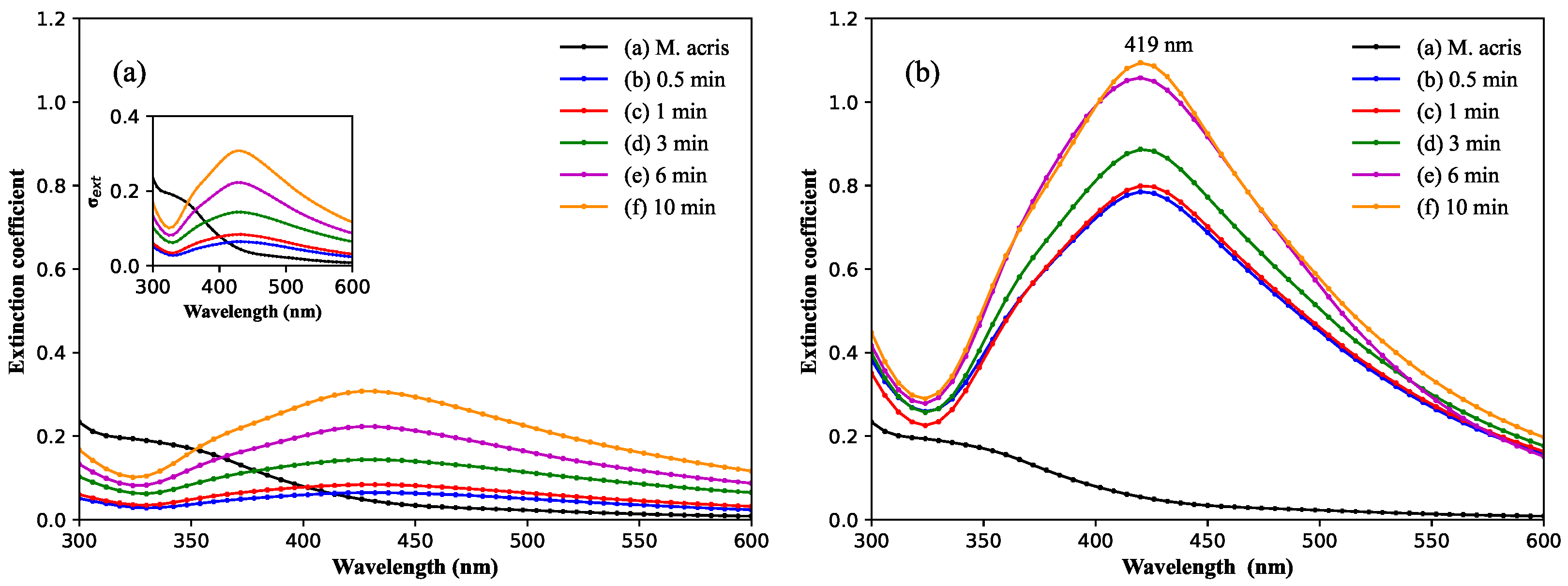
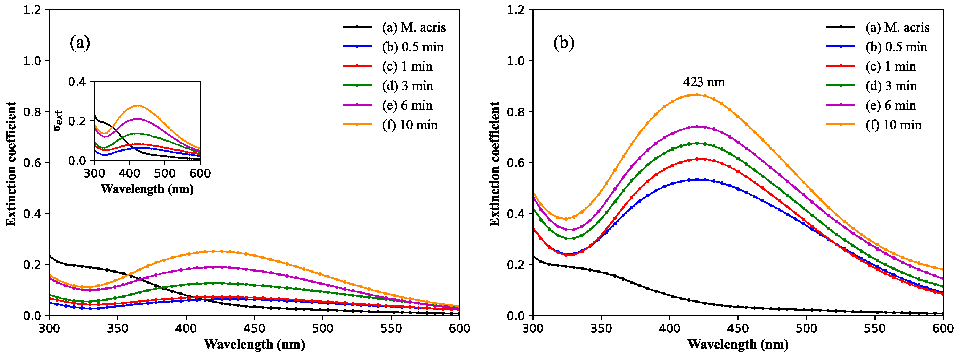
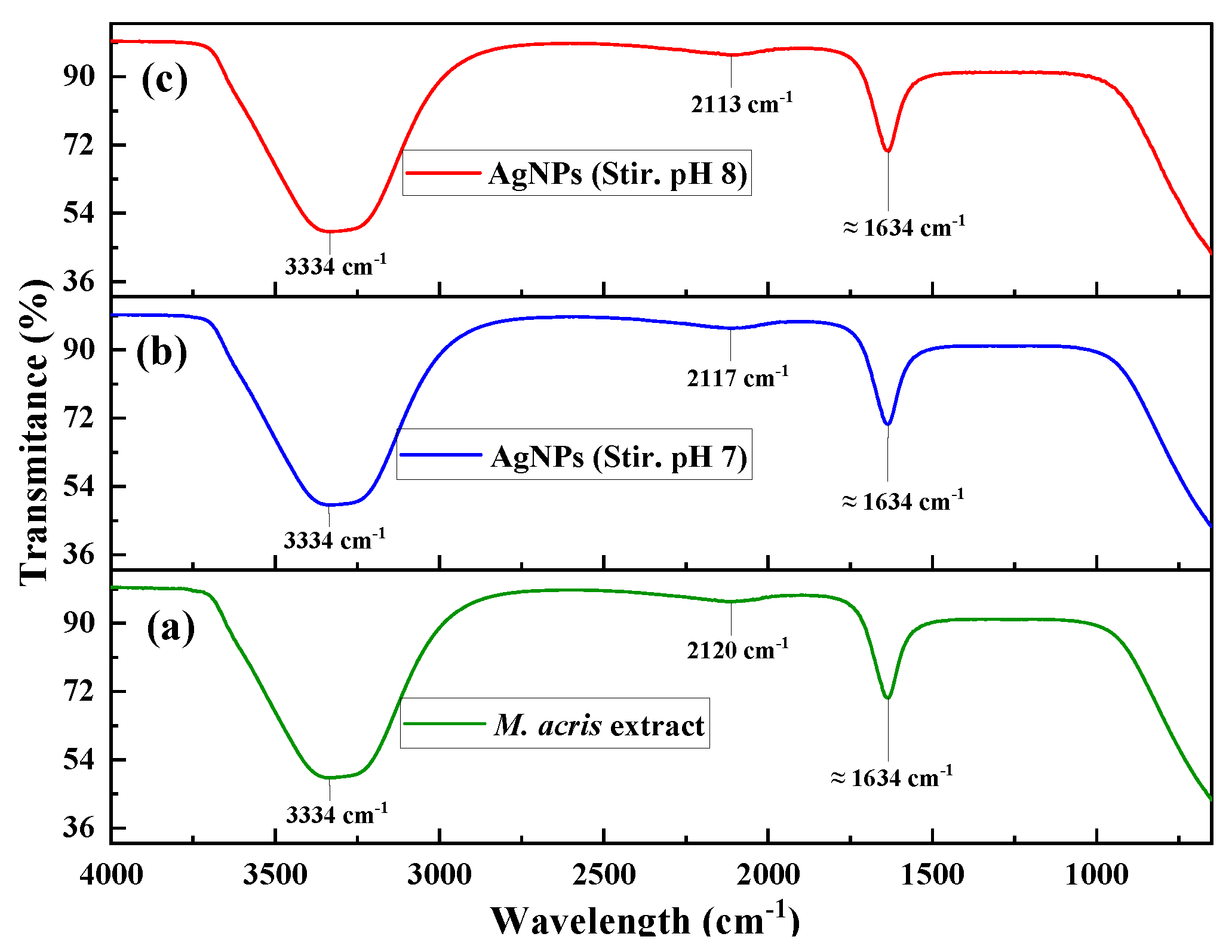
3.1. Spectroscopy Characterization

Plant-derived biomolecules can exhibit variable behavior to changes in pH, which in turn can influence the formation of AgNPs [46]. Figure 2 shows the UV-visible spectra of silver nanoparticles at pH 7. The band centered at ≈419 nm is noticed when the synthesis of nanoparticles is performed using the immersion method (Figure 2a) and can be associated to the surface plasmon resonance of spherical-like AgNPs [47]. Nevertheless, using the stirring method, a rise in the band intensity at ≈423 nm was noticed with time (Figure 2b). Figure 3 shows the AgNPs synthesis at pH 8. The increase in pH reduced the band intensity of SPR at 10 min for the immersion and stirring approach, Figure 3a and Figure 3b, respectively, when compared with the SPR band intensity obtained at pH 7 (Figure 2). Aryan et al. demonstrated the effect of pH on the wavelength during the formation of AgNPs by green synthesis using Kalanchoe pinnata extract, showing that an increase in pH causes a bathochromic shift due to an effect of the change in the charge of organic molecules [48], and the same effect was observed in the SPR band from 419 nm (Figure 2) to 423 nm (Figure 3).
The formation of AgNPs by biosynthesis is a result of the action of the reducing agents present in the extracts of plants [49]. The reduction in metallic ions is related to the presence of proteins/amine groups, sugars and/or terpenoids in the plant extract. The green synthesis of AgNPs primarily relies on phytochemicals such as flavonoids, alkaloids, glycosides, terpenoids, phenols, carbohydrates, proteins, enzymes, and coenzymes [48]. Specifically, Minthostachys extract contains quinic acids, caffeic acid, and rosmarinic acid, along with a small amount of flavonoids [36,37,38]. These compounds act as a coating and stabilizing agent, donating electrons to reduce Ag+ ions to metallic silver (Ag0) as illustrated in the schematic diagram (Figure 4). Several key factors, including the ratio of plant extract and metal ions, reaction time, temperature, and pH, significantly influence the yield, quality, and final properties of the synthesized AgNPs. These parameters determine the nanoparticles’ origin, efficiency, and characteristics as end products [50]. The amount of reducing agents will increase with extraction time, which results in more zero valence ions/NPs formation, which can be monitored by intensity increase in the SPR band [30,51]. In this sense, the extraction by stirring (Figure 2b and Figure 3b) results in more reducing agents in the solution extract, as does the increase in the extract time, when compared with the immersion approach (Figure 2a and Figure 3a). Considering these parameters, the formation of AgNPs is favored by the stirring approach and conducted at pH = 7, where the SPR intensity band reaches ≈ 1.1 (Figure 2b), while at pH = 8, it reaches ≈ 0.87 (Figure 3b). In this case, the SPR band intensity is proportional to the number of nanoparticles in the solution with an identical size and geometry [52].
Figure 5 and Figure 6 show the FTIR analyses for immersion and stirring, respectively. The infrared technique enables to determine the functional groups in the extracts and the presence on the nanoparticle surface. For all spectra, it can be noticed that there is a broad band 2960–3670 due to the stretching of hydroxyl groups, while the peak centered at ≈1634 cm−1 is associated with C=O stretching [27].
Thus, considering the high SPR band intensity, the neutral pH, and the identical profile noticed in infrared spectroscopy analyses, only the synthesis by stirring and pH = 7 was considered for further characterization and further application as the antimicrobial nanomaterial.

3.2. Physical Characterizations: DLS and SEM Analyses

The measurement of the hydrodynamic size of the AgNPs by dynamic laser scattering (DLS) showed the formation of nanoparticles of different sizes (Figure 7). The AgNPs obtained by the immersion approach resulted basically in three particle size distributions centered at 11.2 nm, 55.0 nm, and 167.9 nm (Figure 7a), while by stirring, we noticed two particle size distributions at 11.2 nm and 72.1 nm (Figure 7b). Considering the more intense distributions, the stirring approach showed the AgNPs with a particle size distribution with a lower diameter.
The morphological structure was determined by scanning electron microscopy (SEM); for the AgNPs obtained by the immersion method at pH 7 for a time of 10 min, we observed the formation of spherical-like AgNPs, Figure 8a,b, while for the agitation method, the formation of anisotropic nanoparticles was noticed. As observed in Figure 7, we observed the formation of nanoparticles of different sizes, which can be attributed to the M. acris single and assembled structures, such as quinic acids, caffeic acid, and rosmarinic acid with nucleophilic characteristics.
The elemental analysis (Figure 9) revealed the presence of silver (Ag) in the paper to which a colloidal solution of silver nanoparticles (AgNPs) was added, evidenced by peaks around 327.8 nm and 338.3 nm, characteristics according to the NIST Database [53]. In contrast, no presence of Ag was detected in the paper to which the aqueous extract was incorporated. Additionally, the presence of sodium (Na) due to the two characteristics peaks at 588.9 nm and 589.5 nm was identified, possibly as part of the filter/paper composition and extract, along with hydrogen (H) at 653 nm and oxygen (O) at 776 nm, attributed to the effect of water molecules present in the atmosphere, while (*) indicates the laser line (266 nm).
Table 1 shows the results of the morphological structure of AgNP compounds prepared with different leaf extract, solvents, and temperatures. In general, the results show that the morphology was spherical mainly for the AgNPs, with the exception of the NPs prepared with Olea europaea L., where the NPs obtained asymmetrical morphology.

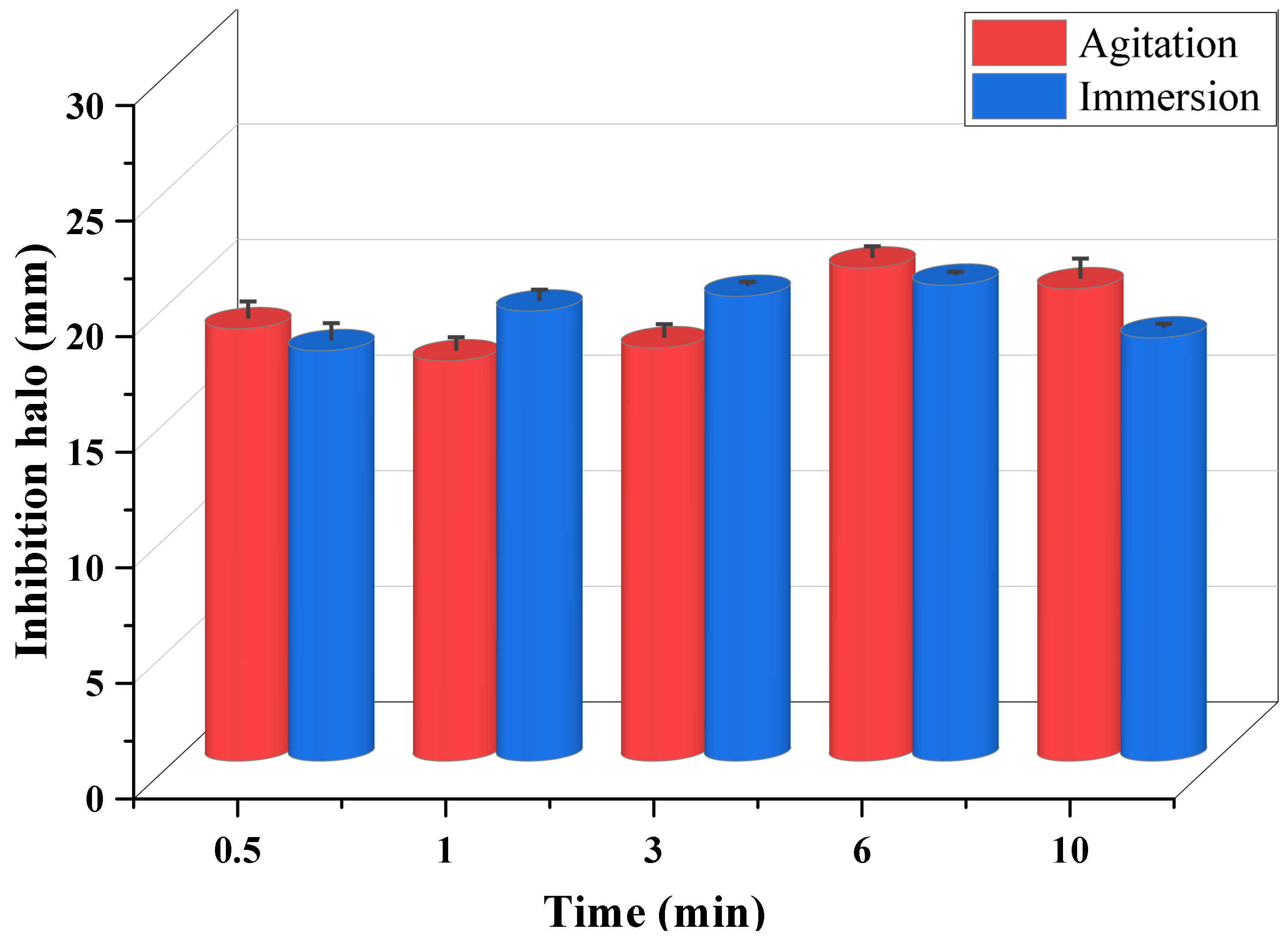
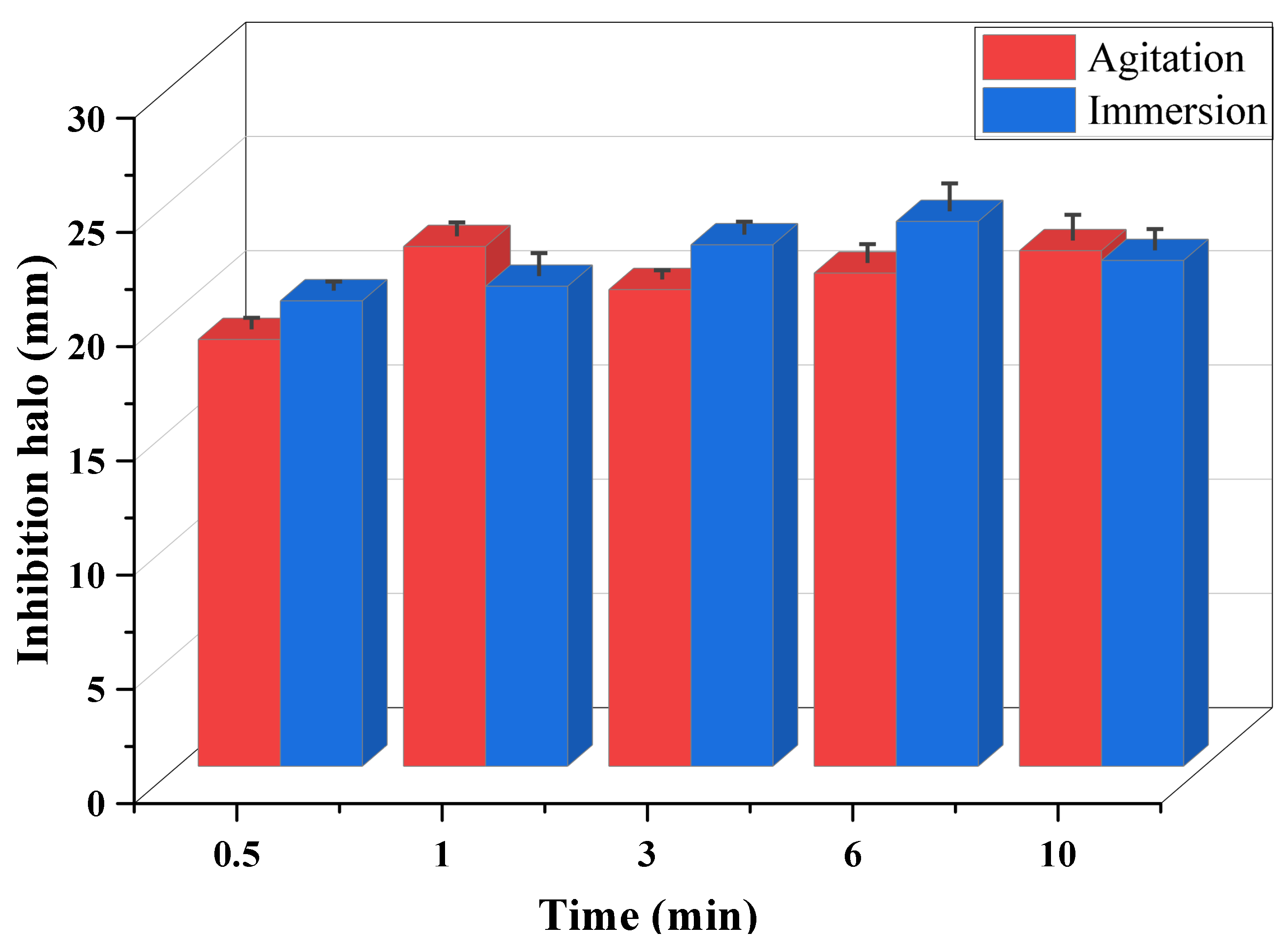
3.3. Antimicrobial Activity of Silver Nanoparticles

Table 2 and Table 3 present the results of the statistical analysis following the ONE-WAY ANOVA model and Tukey’s test (significance of 5%), comparing the measurements of the inhibition zones obtained for the different samples (immersion and agitation) at the different times 0.5, 1, 3, 6 and 10 min. In order to verify the adequate precision of each time, the coefficients of variation were calculated from the measurements of the inhibition zones for the agitation and immersion samples. For the agitation and immersion samples, the inhibition zones for Escherichia coli 100% of the measurements presented a coefficient of variation lower than 5%. For the inhibition zones for Staphylococus aureus 100% of the measurements of the immersion samples presented a coefficient of variation lower than 5%. However, the inhibition zones for Staphylococus aureus only 40% of the measurements of the agitation samples presented a coefficient of variation lower than 5%. The results indicate that there was significant precision between the measurements of the inhibition zones for most measurements.
It was observed that there was no statistically significant difference between the inhibition zones corresponding to the samples shaken at times of 0.5, 1 and 3 min for Escherichia coli (p < 0.05). However, there was a statistically significant difference for times of 6 and 10 min for Escherichia coli (p < 0.05). The inhibition zones presented for times of 0.5 and 10 min were statistically similar, as well as for times of 6 and 10 min and 0.5, 6 and 10 min for Escherichia coli (p < 0.05) for shaken samples.
For the immersion samples, the inhibition zones for Escherichia coli were statistically significant for all times studied. On the other hand, the inhibition halos presented for the times of 0.5 and 10 min were statistically similar, as well as the times 1, 3, 6 and 10 min (p < 0.05). The values were also statistically similar for 3, 6 and 10 min (p < 0.05).
For the agitation samples, the inhibition zones for Staphylococus aureus were similar and statistically significant for all times (p > 0.05). However, for the immersion samples, the inhibition zones for Staphylococus aureus were different and statistically significant for all times studied. However, the inhibition zones presented for the times of 0.5 and 10 min were statistically similar, as well as 1, 3, 6 and 10 min (p < 0.05). The values were also statistically similar for 3, 6 and 10 min (p < 0.05).
It was observed that there was no statistically significant difference between the inhibition zones corresponding to the agitation samples at times of 0.5, 1 and 3 min Escherichia coli (p < 0.05). However, there was a statistically significant difference for the times of 6 and 10 min for Escherichia coli (p < 0.05). The inhibition zones presented for the times 0.5 and 10 min were statistically similar, as well as for the times of 6 and 10 min and 0.5, 6 and 10 min Escherichia coli (p < 0.05) for the agitation sample.
For the immersion samples, the inhibition zones for Escherichia coli were statistically significantly different for all the times studied. On the other hand, the inhibition zones presented for the times of 0.5 and 10 min were statistically similar, as well as 1, 3, 6 and 10 min (p < 0.05). The values were also statistically similar for 3, 6 and 10 min (p < 0.05).
Table 4 shows the comparative results of the antibacterial effect of AgNPs obtained with leaf extracts against Escherichia coli and Staphylococcus aureus compared with the inhibition halos. Chouhan e Gueleria (2020) [44] prepared AgNPs by green synthesis using Cannabis sativa leaf extract; the authors investigated its characterization, antibacterial, anti-yeast and a-amylase inhibitory activity. The authors investigated different concentrations (5, 10, 15, and 20 μ L) of AgNPs as an inhibitory agent. The better inhibition halos for E. coli and S. aureaus obtained in this study were 28 and 23.56 mm, respectively, using 20 μ L of AgNPs. Recently, Kumari et al. (2023) [40], using Anoectochilus elatus leaf extract, green synthesized AgNPs and evaluated the antioxidant, anti-inflammatory, antidiabetic, and antimicrobial activities. In addition, AgNPs demonstrated the most significant zone of inhibition using different concentrations of AgNPs (25, 50, 75 and 100 μ L). The inhibition zone using 25 μ L of AgNPs was 15 ± 1.20 and 13 ± 1.04 mm E. coli and S. aureaus, respectively. Sharifi-Rad et al. (2021) [43] performed the phytofabrication of silver nanoparticles (AgNPs) with pharmaceutical capabilities using Otostegia persica (Burm.) Boiss. leaf extract. The above-mentioned AgNPs showed higher antioxidant activity as compared with the O. persica leaf extract. They also exhibited significant antibacterial activity against both Gram-positive (Staphylococcus aureus) and Gram-negative (Escherichia coli) bacteria, using 50, 100, 150, 200, 250, and 300 μ L of AgNPS. The inhibition halos were 15 ± 0.3 and 16 ± 0.2 mm for S. aureaus and E. coli, respectively, using 50 μ L of AgNPs. Zargar et al. (2011) [64] investigated green synthesis and the antibacterial effect of AgNPs using Vitex negundo L. The authors showed in this study that one of the most important applications of AgNPs is their use as an antibacterial agent. The AgNPs showed antimicrobial activity against Gram-negative and Gram-positive bacteria, obtaining the values 12.0 ± 0.7 and 11.0 ± 0.3 for E. coli and S. aureaus, respectively. Jayaprakash et al. (2017) [65] obtained green-synthesized AgNPs using Tamarind fruit extract for the antibacterial studies. The AgNPs showed good results regarding antibacterial activities, and the inhibition halos for E. coli and S. aureaus were 15 and 16 mm, respectively. It was found that the inhibition zone of AgNPs was slightly better than AgNO3 against the bacterial strains and the respective inhibition zone. The results present in Table 4 show that the AgNP prepared by M. acris demonstrated better antibacterial activity against Escherichia coli and Staphylococus aureus compared to the AgNPs prepared with another leaf extract. These results demonstrate that the biosynthesis method has proven to be a simple, efficient, non-toxic, less expensive, ecological, and antibacterial method in the synthesis of AgNPs. Among these two processes, the optimal time for obtaining extracts in both methods is likely 6 min, as shorter or longer times result in lower or higher concentrations of the metabolites. It is also important to note that the aqueous extract of M. acris was used as a control, which demonstrated no effect, indicating that the formed AgNPs were responsible for the antibacterial effect. Figure 10 and Figure 11 illustrates the antibacterial effect of AgNPs obtained through green synthesis with M. acris extracts and how time influences their antibacterial study, clearly affecting the extraction of the metabolites that enable the formation of AgNPs.

4. Conclusions

The extracts from the leaves of M. acris demonstrated the reducing and stabilizing capacity of AgNPs; due its bioactive compounds such as hydroxycinnamic acid derivatives, quinic, caffeic, rosmarinic acids, this compound acts as reducing and stabilizing agents in the formation of silver nanoparticules. According to the results obtained, predominantly spherical nanoparticles were produced, which were determined through SEM. The DLS study showed the formation of AgNPs of different sizes, while UV-Vis spectroscopy demonstrated that surface plasmon resonance forms a band around 419 and 423 nm for pH 7 and pH 8, respectively, characteristic of spherical AgNPs. FTIR analysis revealed the presence of carboxyl functional groups (C=O) belonging to secondary metabolites, possibly monoterpenes, that act as reducing and stabilizing agents. Additionally, LIBS analysis demonstrated the presence of Ag in the mixture with the extract, while the pure extract did not show its presence.
The antibacterial capacity of the AgNPs obtained through green synthesis against E. coli strains demonstrated the potential of nanometric materials. The AgNPs prepared by the M. acris extract prepared by the immersion and agitation achieved inhibition halos around 21 mm at the time of 6 min. Finally, with this study, AgNPs, as a compound with antibacterial activity, were prepared in a green, innovative, simple, and rapid process that does not require high temperatures, with an ecological focus that minimizes the use of chemical substances during production.

Author Contributions

F.C.A.: Investigation, Methodology, Writing—original draft. A.M.O.A.: Conceptualization, Investigation, Methodology. G.C.P.: Investigation, Methodology, Formal analysis. S.J.: Supervision, Software, Writing—review and editing, Visualization. R.S.: Supervision, Conceptualization, Funding, Formal analysis, Visualization. All authors have read and agreed to the published version of the manuscript.

Funding

This research has received funding from project code: C21072001 VRI-UNMSM.

Data Availability Statement

The original contributions presented in this study are included in the article.

Acknowledgments

The authors would like to thank the Nanotechnology and Technological Innovation Laboratory (LabNanoInTec-FQIQ-UNMSM) and Multi-user Laboratory of the Analytical Center of UTFPR-Campus Toledo for providing the types of equipment and instrumentation to researchers.

Conflicts of Interest

The authors declare no conflicts of interest.

References

  1. Lenz, G.F.; Bini, R.A.; Bueno, T.P.; de Oliveira, R.J.; Felix, J.F.; Schneider, R. Self-supported copper (Cu) and Cu-based nanoparticle growth by bottom-up process onto borophosphate glasses. J. Mater. Sci. 2017, 52, 6635–6646. [Google Scholar] [CrossRef]
  2. Lenz, G.F.; Schneider, R.; Rossi de Aguiar, K.M.F.; Bini, R.A.; Chaker, J.A.; Hammer, P.; Botteselle, G.V.; Felix, J.F.; Schneider, R. Self-supported nickel nanoparticles on germanophosphate glasses: Synthesis and applications in catalysis. RSC Adv. 2019, 9, 17157–17164. [Google Scholar] [CrossRef] [PubMed]
  3. Locatelli, P.P.; Gurtat, M.; Lenz, G.F.; Marroquin, J.F.R.; Felix, J.F.; Schneider, R.; Borba, C.E. Simple borophosphate glasses for on-demand growth of self-supported copper nanoparticles in the reduction of 4-nitrophenol. J. Hazard. Mater. 2021, 416, 125801. [Google Scholar] [CrossRef] [PubMed]
  4. Vilchis-Nestor, A.R.; Sánchez-Mendieta, V.; Camacho-López, M.A.; Gómez-Espinosa, R.M.; Camacho-López, M.A.; Arenas-Alatorre, J.A. Solventless synthesis and optical properties of Au and Ag nanoparticles using Camellia sinensis extract. Mater. Lett. 2008, 62, 3103–3105. [Google Scholar] [CrossRef]
  5. Schneider, R.; Felix, J.F.; Moura, L.G.; Morais, P.C. One step fabrication of glass–silver@core–shell fibers: Silver-doped phosphate glasses as precursors of SERS substrates. J. Mater. Chem. C 2014, 2, 9021–9027. [Google Scholar] [CrossRef]
  6. Li, X.; Xu, H.; Chen, Z.S.; Chen, G. Biosynthesis of Nanoparticles by Microorganisms and Their Applications. J. Nanomater. 2011, 2011, 1–16. [Google Scholar] [CrossRef]
  7. Lu, X.; Rycenga, M.; Skrabalak, S.E.; Wiley, B.; Xia, Y. Chemical Synthesis of Novel Plasmonic Nanoparticles. Annu. Rev. Phys. Chem. 2009, 60, 167–192. [Google Scholar] [CrossRef]
  8. Dubey, S.P.; Lahtinen, M.; Sillanpää, M. Green synthesis and characterizations of silver and gold nanoparticles using leaf extract of Rosa rugosa. Colloids Surf. Physicochem. Eng. Asp. 2010, 364, 34–41. [Google Scholar] [CrossRef]
  9. Carmona, E.R.; Benito, N.; Plaza, T.; Recio-Sánchez, G. Green synthesis of silver nanoparticles by using leaf extracts from the endemic Buddleja globosa hope. Green Chem. Lett. Rev. 2017, 10, 250–256. [Google Scholar] [CrossRef]
  10. Gómez-Garzón, M. Nanomateriales, Nanopartículas y Síntesis verde. Rev. Repert. Med. Cir. 2018, 27. [Google Scholar] [CrossRef]
  11. Njagi, E.C.; Huang, H.; Stafford, L.; Genuino, H.; Galindo, H.M.; Collins, J.B.; Hoag, G.E.; Suib, S.L. Biosynthesis of Iron and Silver Nanoparticles at Room Temperature Using Aqueous Sorghum Bran Extracts. Langmuir 2010, 27, 264–271. [Google Scholar] [CrossRef] [PubMed]
  12. Bhattacharya, D.; Gupta, R.K. Nanotechnology and Potential of Microorganisms. Crit. Rev. Biotechnol. 2005, 25, 199–204. [Google Scholar] [CrossRef] [PubMed]
  13. Santos, A.; Troncoso, C.; Lamilla, C.; Llanquinao, V.; Pavez, M.; Barrientos, L. Nanopartículas Sintetizadas por Bacterias Antárticas y sus Posibles Mecanismos de Síntesis. Int. J. Morphol. 2017, 35, 26–33. [Google Scholar] [CrossRef]
  14. Kumar, B.; Smita, K.; Cumbal, L.; Debut, A. Green synthesis of silver nanoparticles using Andean blackberry fruit extract. Saudi J. Biol. Sci. 2017, 24, 45–50. [Google Scholar] [CrossRef] [PubMed]
  15. Makarov, V.; Love, A.; Sinitsyna, O.; Makarova, S.; Yaminsky, I.; Taliansky, M.; Kalinina, N. “Green” nanotechnologies: Synthesis of metal nanoparticles using plants. Acta Naturae 2014, 6, 35–44. [Google Scholar] [CrossRef]
  16. Mukherjee, P.; Ahmad, A.; Mandal, D.; Senapati, S.; Sainkar, S.R.; Khan, M.I.; Parishcha, R.; Ajaykumar, P.V.; Alam, M.; Kumar, R.; et al. Fungus-Mediated Synthesis of Silver Nanoparticles and Their Immobilization in the Mycelial Matrix: A Novel Biological Approach to Nanoparticle Synthesis. Nano Lett. 2001, 1, 515–519. [Google Scholar] [CrossRef]
  17. Balasooriya, E.R.; Jayasinghe, C.D.; Jayawardena, U.A.; Ruwanthika, R.W.D.; de Silva, R.M.; Udagama, P.V. Honey Mediated Green Synthesis of Nanoparticles: New Era of Safe Nanotechnology. J. Nanomater. 2017, 2017, 5919836. [Google Scholar] [CrossRef]
  18. Love, A.J.; Makarov, V.; Yaminsky, I.; Kalinina, N.O.; Taliansky, M.E. The use of tobacco mosaic virus and cowpea mosaic virus for the production of novel metal nanomaterials. Virology 2014, 449, 133–139. [Google Scholar] [CrossRef]
  19. Benakashani, F.; Allafchian, A.; Jalali, S.A.H. Green synthesis, characterization and antibacterial activity of silver nanoparticles from root extract of weed. Green Chem. Lett. Rev. 2017, 10, 324–330. [Google Scholar] [CrossRef]
  20. Avoseh, O.N.; Oyedeji, O.O.; Aremu, O.; Nkeh-Chungag, B.N.; Songca, S.P.; Oyedeji, A.O.; Mohan, S.; Oluwafemi, O.S. Biosynthesis of silver nanoparticles from Acacia mearnsii De Wild stem bark and its antinociceptive properties. Green Chem. Lett. Rev. 2017, 10, 59–68. [Google Scholar] [CrossRef]
  21. Azarbani, F.; Shiravand, S. Green synthesis of silver nanoparticles by Ferulago macrocarpa flowers extract and their antibacterial, antifungal and toxic effects. Green Chem. Lett. Rev. 2020, 13, 41–49. [Google Scholar] [CrossRef]
  22. Dubey, S.P.; Lahtinen, M.; Sillanpää, M. Tansy fruit mediated greener synthesis of silver and gold nanoparticles. Process Biochem. 2010, 45, 1065–1071. [Google Scholar] [CrossRef]
  23. Nadagouda, M.N.; Iyanna, N.; Lalley, J.; Han, C.; Dionysiou, D.D.; Varma, R.S. Synthesis of Silver and Gold Nanoparticles Using Antioxidants from Blackberry, Blueberry, Pomegranate, and Turmeric Extracts. ACS Sustain. Chem. Eng. 2014, 2, 1717–1723. [Google Scholar] [CrossRef]
  24. Soshnikova, V.; Kim, Y.J.; Singh, P.; Huo, Y.; Markus, J.; Ahn, S.; Castro-Aceituno, V.; Kang, J.; Chokkalingam, M.; Mathiyalagan, R.; et al. Cardamom fruits as a green resource for facile synthesis of gold and silver nanoparticles and their biological applications. Artif. Cells Nanomed. Biotechnol. 2017, 46, 108–117. [Google Scholar] [CrossRef] [PubMed]
  25. Chokkalingam, M.; Singh, P.; Huo, Y.; Soshnikova, V.; Ahn, S.; Kang, J.; Mathiyalagan, R.; Kim, Y.J.; Yang, D.C. Facile synthesis of Au and Ag nanoparticles using fruit extract of Lycium chinense and their anticancer activity. J. Drug Deliv. Sci. Technol. 2019, 49, 308–315. [Google Scholar] [CrossRef]
  26. Huong, V.T.L.; Nguyen, N.T. Green synthesis, characterization and antibacterial activity of silver nanoparticles using Sapindus mukorossi fruit pericarp extract. Mater. Today Proc. 2021, 42, 88–93. [Google Scholar] [CrossRef]
  27. Niluxsshun, M.C.D.; Masilamani, K.; Mathiventhan, U. Green Synthesis of Silver Nanoparticles from the Extracts of Fruit Peel of Citrus tangerina, Citrus sinensis, and Citrus limon for Antibacterial Activities. Bioinorg. Chem. Appl. 2021, 2021, 6695734. [Google Scholar] [CrossRef]
  28. Shankar, S.; Ahmad, A.; Sastry, M. Geranium Leaf Assisted Biosynthesis of Silver Nanoparticles. Biotechnol. Prog. 2003, 19, 1627–1631. [Google Scholar] [CrossRef]
  29. Jha, A.K.; Prasad, K.; Kumar, V.; Prasad, K. Biosynthesis of silver nanoparticles using Eclipta leaf. Biotechnol. Prog. 2009, 25, 1476–1479. [Google Scholar] [CrossRef]
  30. Song, J.Y.; Kim, B.S. Rapid biological synthesis of silver nanoparticles using plant leaf extracts. Bioprocess Biosyst. Eng. 2008, 32, 79–84. [Google Scholar] [CrossRef]
  31. Begum, N.A.; Mondal, S.; Basu, S.; Laskar, R.A.; Mandal, D. Biogenic synthesis of Au and Ag nanoparticles using aqueous solutions of Black Tea leaf extracts. Colloids Surf. B Biointerfaces 2009, 71, 113–118. [Google Scholar] [CrossRef] [PubMed]
  32. Dwivedi, A.D.; Gopal, K. Biosynthesis of silver and gold nanoparticles using Chenopodium album leaf extract. Colloids Surf. A Physicochem. Eng. Asp. 2010, 369, 27–33. [Google Scholar] [CrossRef]
  33. Schmidt-Lebuhn, A. Ethnobotany, biochemistry and pharmacology of Minthostachys (Lamiaceae). J. Ethnopharmacol. 2008, 118, 343–353. [Google Scholar] [CrossRef] [PubMed]
  34. Svendsen, A.B.; Scheffer, J.J.C.; Looman, A. Composition of the volatile oil of Minthostachys glabrescens epl. Flavour Fragr. J. 1987, 2, 45–46. [Google Scholar] [CrossRef]
  35. Pino, J.A.; Dueñas-Mendoza, M.M.; Solís-Quispe, L. Chemical Composition of the Essential Oil from Minthostachys acris Schmidt-Leb. Grown in the Peruvian Andes. Nat. Prod. Commun. 2019, 14, 1934578X1985749. [Google Scholar] [CrossRef]
  36. Yapuchura Mamani, R. Estudio de los Componentes Antioxidantes de las Hojas de Muña (Minthostachys mollis (Kunth) Griseb.) E inca muña (Clinopodium bolivianum (benth.) Kuntze). Master’s Thesis, Universidad Nacional Agraria La Molina, Lima, Peru, 2010. [Google Scholar]
  37. Serrano, C.A.; Villena, G.K.; Rodríguez, E.F. Phytochemical profile and rosmarinic acid purification from two Peruvian Lepechinia Willd. species (Salviinae, Mentheae, Lamiaceae). Sci. Rep. 2021, 11, 7260. [Google Scholar] [CrossRef]
  38. Serrano, C.A.; Villena, G.K.; Rodríguez, E.F.; Calsino, B.; Ludeña, M.A.; Ccana-Ccapatinta, G.V. Phytochemical analysis for ten Peruvian Mentheae (Lamiaceae) by liquid chromatography associated with high resolution mass spectrometry. Sci. Rep. 2023, 13, 10714. [Google Scholar] [CrossRef]
  39. Althubiti, A.A.; Alsudir, S.A.; Alfahad, A.J.; Alshehri, A.A.; Bakr, A.A.; Alamer, A.A.; Alrasheed, R.H.; Tawfik, E.A. Green Synthesis of Silver Nanoparticles Using Jacobaea maritima and the Evaluation of Their Antibacterial and Anticancer Activities. Int. J. Mol. Sci. 2023, 24, 16512. [Google Scholar] [CrossRef]
  40. Venkataesan Kumari, B.; Mani, R.; Asokan, B.R.; Balakrishnan, K.; Ramasamy, A.; Parthasarathi, R.; Kandasamy, C.; Govindaraj, R.; Vijayakumar, N.; Vijayakumar, S. Green Synthesised Silver Nanoparticles Using Anoectochilus elatus Leaf Extract: Characterisation and Evaluation of Antioxidant, Anti-Inflammatory, Antidiabetic, and Antimicrobial Activities. J. Compos. Sci. 2023, 7, 453. [Google Scholar] [CrossRef]
  41. Hasim, H.; Rao, P.V.; Sekhar, A.C.; Muthuraju, S.; Asari, M.A.; Sirajudeen, K.N.S. Green synthesis and characterization of silver nanoparticles using Tualang honey and evaluation of their antioxidant activities. Adv. Nat. Sci. Nanosci. Nanotechnol. 2020, 11, 025010. [Google Scholar] [CrossRef]
  42. Cavassin, E.D.; de Figueiredo, L.F.P.; Otoch, J.P.; Seckler, M.M.; de Oliveira, R.A.; Franco, F.F.; Marangoni, V.S.; Zucolotto, V.; Levin, A.S.S.; Costa, S.F. Comparison of methods to detect the in vitro activity of silver nanoparticles (AgNP) against multidrug resistant bacteria. J. Nanobiotechnol. 2015, 13, 64. [Google Scholar] [CrossRef] [PubMed]
  43. Sharifi-Rad, M.; Pohl, P.; Epifano, F. Phytofabrication of Silver Nanoparticles (AgNPs) with Pharmaceutical Capabilities Using Otostegia persica (Burm.) Boiss. Leaf Extract. Nanomaterials 2021, 11, 1045. [Google Scholar] [CrossRef] [PubMed]
  44. Chouhan, S.; Guleria, S. Green synthesis of AgNPs using Cannabis sativa leaf extract: Characterization, antibacterial, anti-yeast and α-amylase inhibitory activity. Mater. Sci. Energy Technol. 2020, 3, 536–544. [Google Scholar] [CrossRef]
  45. Bauer, A.W.; Kirby, W.M.M.; Sherris, J.C.; Turck, M. Antibiotic Susceptibility Testing by a Standardized Single Disk Method. Am. J. Clin. Pathol. 1966, 45, 493–496. [Google Scholar] [CrossRef] [PubMed]
  46. Fernando, I.; Zhou, Y. Impact of pH on the stability, dissolution and aggregation kinetics of silver nanoparticles. Chemosphere 2019, 216, 297–305. [Google Scholar] [CrossRef]
  47. Pham, X.N.; Nguyen, H.T.; Pham, N.T. Green Synthesis and Antibacterial Activity of HAp@Ag Nanocomposite Using Centella asiatica (L.) Urban Extract and Eggshell. Int. J. Biomater. 2020, 2020, 8841221. [Google Scholar] [CrossRef]
  48. Aryan; Ruby; Mehata, M.S. Green synthesis of silver nanoparticles using Kalanchoe pinnata leaves (life plant) and their antibacterial and photocatalytic activities. Chem. Phys. Lett. 2021, 778, 138760. [Google Scholar] [CrossRef]
  49. Keat, C.L.; Aziz, A.; Eid, A.M.; Elmarzugi, N.A. Biosynthesis of nanoparticles and silver nanoparticles. Bioresour. Bioprocess. 2015, 2, 47. [Google Scholar] [CrossRef]
  50. Habeeb Rahuman, H.B.; Dhandapani, R.; Narayanan, S.; Palanivel, V.; Paramasivam, R.; Subbarayalu, R.; Thangavelu, S.; Muthupandian, S. Medicinal plants mediated the green synthesis of silver nanoparticles and their biomedical applications. IET Nanobiotechnology 2022, 16, 115–144. [Google Scholar] [CrossRef]
  51. Shankar, S.; Rai, A.; Ahmad, A.; Sastry, M. Rapid synthesis of Au, Ag, and bimetallic Au core–Ag shell nanoparticles using Neem (Azadirachta indica) leaf broth. J. Colloid Interface Sci. 2004, 275, 496–502. [Google Scholar] [CrossRef]
  52. Kelly, K.L.; Coronado, E.; Zhao, L.L.; Schatz, G.C. The Optical Properties of Metal Nanoparticles:The Influence of Size, Shape, and Dielectric Environment. J. Phys. Chem. B 2002, 107, 668–677. [Google Scholar] [CrossRef]
  53. Kramida, A.; Ralchenko, Y. NIST, Atomic Spectra Database, NIST Standard Reference Database. Available online: https://physics.nist.gov/asd (accessed on 10 November 2024).
  54. Sangameshwaran, V.; Ragu Prasath, A.; Selvam, K.; Obaidullah, A.J.; Kumar Yadav, K.; Sudhakar, C. Green synthesis of silver nanoparticles using Senna alexandrina leaf extracts and their antibacterial, larvicidal, nanotoxicity and photocatalytic activity. Inorg. Chim. Acta 2024, 571, 122220. [Google Scholar] [CrossRef]
  55. Revathi, S.; Sutikno, S.; Hasan, A.F.; Altemimi, A.B.; ALKaisy, Q.H.; Phillips, A.J.; Hesarinejad, M.A.; Abedelmaksoud, T.G. Green synthesis and characterization of silver nanoparticles (AgNP) using Acacia nilotica plant extract and their anti-bacterial activity. Food Chem. Adv. 2024, 4, 100680. [Google Scholar] [CrossRef]
  56. George, N.; Joy, J.; Mathew, B.; Koshy, E.P. Green mediated synthesis of silver nanoparticle using Euphorbia mculate leaf extract and their catalytic reduction and antibacterial properties. Mater. Today Proc. 2024. [Google Scholar] [CrossRef]
  57. Singh, R.; Singh, R.; Parihar, P.; Mani, J.V. Green synthesis of silver nanoparticles using Solanum sisymbriifolium leaf extract: Characterization and evaluation of antioxidant, antibacterial and photocatalytic degradation activities. Process Biochem. 2024, 143, 337–352. [Google Scholar] [CrossRef]
  58. Alotaibi, N.F.; ALqarni, L.S.; Alghamdi, S.Q.; Al-Ghamdi, S.N.; Amna, T.; Alzahrani, S.S.; Moustafa, S.M.; Hasanin, T.H.; Nassar, A.M. Green Synthesis of Uncoated and Olive Leaf Extract-Coated Silver Nanoparticles: Sunlight Photocatalytic, Antiparasitic, and Antifungal Activities. Int. J. Mol. Sci. 2024, 25, 3082. [Google Scholar] [CrossRef]
  59. Sultan, M.; Siddique, M.; Khan, R.; Fallatah, A.M.; Fatima, N.; Shahzadi, I.; Waheed, U.; Bilal, M.; Ali, A.; Abbasi, A.M. Ligustrum lucidum Leaf Extract-Assisted Green Synthesis of Silver Nanoparticles and Nano-Adsorbents Having Potential in Ultrasound-Assisted Adsorptive Removal of Methylene Blue Dye from Wastewater and Antimicrobial Activity. Materials 2022, 15, 1637. [Google Scholar] [CrossRef]
  60. Sahu, K.; Kurrey, R.; Pillai, A.K. Green synthesis of silver nanoparticles from Manilkara zapota leaf extract for the detection of aminoglycoside antibiotics and other applications. RSC Adv. 2024, 14, 23240–23256. [Google Scholar] [CrossRef]
  61. Chinnathambi, A.; Alharbi, S.A.; Joshi, D.; V, S.; Jhanani, G.; On-uma, R.; Jutamas, K.; Anupong, W. Synthesis of AgNPs from leaf extract of Naringi crenulata and evaluation of its antibacterial activity against multidrug resistant bacteria. Environ. Res. 2023, 216, 114455. [Google Scholar] [CrossRef]
  62. Bhusal, M.; Pathak, I.; Bhadel, A.; Shrestha, D.K.; Sharma, K.R. Synthesis of silver nanoparticles assisted by aqueous root and leaf extracts of Rhus chinensis Mill and its antibacterial activity. Heliyon 2024, 10, e33603. [Google Scholar] [CrossRef]
  63. Islam, M.J.; Khatun, N.; Bhuiyan, R.H.; Sultana, S.; Ali Shaikh, M.A.; Amin Bitu, M.N.; Chowdhury, F.; Islam, S. Psidium guajava leaf extract mediated green synthesis of silver nanoparticles and its application in antibacterial coatings. RSC Adv. 2023, 13, 19164–19172. [Google Scholar] [CrossRef] [PubMed]
  64. Zargar, M.; Hamid, A.A.; Bakar, F.A.; Shamsudin, M.N.; Shameli, K.; Jahanshiri, F.; Farahani, F. Green Synthesis and Antibacterial Effect of Silver Nanoparticles Using Vitex negundo L. Molecules 2011, 16, 6667–6676. [Google Scholar] [CrossRef] [PubMed]
  65. Jayaprakash, N.; Vijaya, J.J.; Kaviyarasu, K.; Kombaiah, K.; Kennedy, L.J.; Ramalingam, R.J.; Munusamy, M.A.; Al-Lohedan, H.A. Green synthesis of Ag nanoparticles using Tamarind fruit extract for the antibacterial studies. J. Photochem. Photobiol. B Biol. 2017, 169, 178–185. [Google Scholar] [CrossRef] [PubMed]
Figure 1. Minthostachys acris Schmidt Lebuhn.
Micro 04 00043 g001
Figure 2. UV-vis spectra of obtained AgNPs at pH 7 (a) by the immersion method and (b) by the stirring method. The time is refereed to the extraction time.
Micro 04 00043 g002
Figure 3. UV-vis spectra of AgNPs at pH 8 (a) by the immersion method and (b) by the stirring method.
Micro 04 00043 g003
Figure 4. Proposed synthesis of AgNPs using M. acris extract with silver nitrate (AgNO3) solution.
Micro 04 00043 g004
Figure 5. FT−IR Spectra by the immersion method of (a) M. acris extract, (b) AgNPs at pH 7, and (c) at pH 8.
Micro 04 00043 g005
Figure 6. FT−IR Spectra by the stirring method of (a) M. acris extract, (b) AgNPs at pH 7, and (c) at pH 8.
Micro 04 00043 g006
Figure 7. Dynamic laser scattering for AgNPs obtained at pH 7 and 10 min.
Micro 04 00043 g007
Figure 8. Morphological study of silver nanoparticles by scanning electron microscopy of AgNPs synthesized at pH 7 (a) by the immersion method for 10 min and (b) by the stirring method for 10 min.
Micro 04 00043 g008
Figure 9. LIBS of aqueous solution and AgNPs supported in filter paper.
Micro 04 00043 g009
Figure 10. Quantitative evaluation of antibacterial activity of AgNPs against Escherichia coli.
Micro 04 00043 g010
Figure 11. Quantitative evaluation of antibacterial activity of AgNPs against Staphylococus aureus.
Micro 04 00043 g011
Table 1. Green synthesis conditions of AgNPs with leaf extract.
Leaf ExtractExtractAgNPsRef.
Solvent Temperature (°C)
Minthostcahys acrisdistilled waterroomlike-sphericalThis work
Cannabis sativadistilled water60 °Cspherical[44]
Senna alexandrinadistilled water90 °Cspherical[54]
Acacia niloticadistilled water60 °Cspherical[55]
Euphorbia Maculatedeionized water100 °Cspherical[56]
Solanum sisymbriifoliumdistilled water50–60 °Cspherical[57]
Olea europaea L. distilled water100 °Casymmetrical[58]
Jacobaea maritimadistilled water100 °Cspherical[39]
Anoectochilus elatusdistilled water100 °Cspherical[40]
Ligustrum lucidummethanol25 ± 5 °Cspherical[59]
Manilkara zapotaMilli-Q water80 °Cspherical[60]
Naringi crenulatamethanol64 °Cspherical[61]
Rhus chinensisdeionized water60 °Cspherical[62]
Psidium guajavadeionized water90 °Cspherical[63]
Table 2. Antibacterial effect of AgNPs against Escherichia coli.
Time (min)AgitationHalo (mm)ImmersionHalo (mm)
0Micro 04 00043 i001-Micro 04 00043 i002-
0.5Micro 04 00043 i00318.705 ± 0.72
CV = 3.850
a c
Micro 04 00043 i00417.753 ± 0.73
CV = 4.120
a e
1Micro 04 00043 i00517.313 ± 0.56
CV = 3.251
a
Micro 04 00043 i00619.472 ± 0.46
CV = 2.374
b c d e
3Micro 04 00043 i00717.878 ± 0.56
CV = 3.145
a
Micro 04 00043 i00820.107 ± 0.16
CV = 0.814
c b d e
6Micro 04 00043 i00921.33 ± 0.48
CV = 2.250
b c
Micro 04 00043 i01020.587 ± 0.12
CV = 0.606
d c e
10Micro 04 00043 i01120.432 ± 0.84
CV = 4.147
a b c
Micro 04 00043 i01218.317 ± 0.15
CV = 0.833
e a
Distinct letters show significant differences between times according to the Tukey test ( α = 0.05); CV means the coefficient of variation.
Table 3. Antibacterial effect of AgNPs against Staphylococus aureus.
Time (min)AgitationHalo (mm)ImmersionHalo (mm)
0Micro 04 00043 i013-Micro 04 00043 i014-
0.5Micro 04 00043 i01518.683 ± 0.48
CV = 2.584
a
Micro 04 00043 i01620.375 ± 0.37
CV = 3.251
a e
1Micro 04 00043 i01722.753 ± 0.58
CV = 2.5582
a
Micro 04 00043 i01821.011 ± 0.89
CV = 3.374
b c d e
3Micro 04 00043 i01920.866 ± 0.37
CV = 13.507
a
Micro 04 00043 i02022.826 ± 0.54
CV = 2.250
c d e
6Micro 04 00043 i02121.586 ± 0.80
CV = 11.585
a
Micro 04 00043 i02223.85 ± 0.87
CV = 0.606
d b c e
10Micro 04 00043 i02322.57 ± 1.20
CV = 18.310
a
Micro 04 00043 i02422.14 ± 0.90
CV = 3.145
e a
Distinct letters show significant differences between times according to the Tukey test ( α = 0.05); CV means the coefficient of variation.
Table 4. Comparative table of the antibacterial effect of AgNPs obtained with leaf extracts against Escherichia coli and Staphylococcus aureus.
Leaf ExtractAgNPsE. coliS. aureusReference
( μ L) Halo (mm)
Minthostachys acrisspherical-like (20)21.33 ± 0.4821.58 ± 0.80This work
Minthostachys acrisspherical-like (20)20.58 ± 0.1223.85 ± 0.87This work
Cannabis sativaspherical (20)2812[44]
Acacia niloticaspherical (100)2520[55]
Anoectochilus elatusspherical (25)15 ± 1.2013 ± 1.04[40]
Otostegia persicaspherical (50)16 ± 0.215 ± 0.3[43]
Otostegia persicaspherical (100)17 ± 0.116 ± 0.1[43]
Vitex negundo L.spherical (20)12.0 ± 0.711.0 ± 0.3[64]
Disclaimer/Publisher’s Note: The statements, opinions and data contained in all publications are solely those of the individual author(s) and contributor(s) and not of MDPI and/or the editor(s). MDPI and/or the editor(s) disclaim responsibility for any injury to people or property resulting from any ideas, methods, instructions or products referred to in the content.

