Article

Geochemistry and Microbiology of Atacamite-Paratacamite Biofilms Floating on Underground Brine and Petroleum Pools in the White Pine Copper Mine, Michigan (USA)

by
Eleanora I. Robbins
1,*,
Mark R. Stanton
2,† and
Cheryl D. Young
3
1
Department of Geological Sciences, San Diego State University, San Diego, CA 92182, USA
2
Independent Researcher, 12597 West Maryland Place, Lakewood, CO 80228, USA
3
College of Arts and Sciences, Heritage University, Toppenish, WA 98948, USA
*
Author to whom correspondence should be addressed.
Retired.
Micro 2023, 3(3), 728-738; https://doi.org/10.3390/micro3030051
Submission received: 14 January 2023 / Revised: 31 July 2023 / Accepted: 7 August 2023 / Published: 28 August 2023

Abstract

:
At depth in an abandoned tunnel of the White Pine Copper Mine, green films of the Cu-OH-Cl minerals atacamite and paratacamite were found on standing pools of brine. Some pools were also coated with a thin layer of petroleum. Green films of atacamite were composed of individual blebs that averaged 20 μm in diameter and enclosed mixed colonies of Gram-negative, short rod-shaped, and sheathed filamentous bacteria. Carbon δ13C values in the atacamite–paratacamite mixtures reflect the isotopic values of bacteria and minor amounts of petroleum mixed with the minerals. Heterotrophic bacteria are interpreted to be using petroleum as a carbon source and may be catalyzing the precipitation of the copper hydroxy chloride minerals or acting as a template.

1. Introduction

At a depth of 686 m in an abandoned tunnel in the southwest portion of the former White Pine Copper Mine (Figure 1), green films formed on standing pools of water, some of which were also coated with a thin layer of petroleum residue (Figure 2A,B). The mine, then owned by the Copper Range Company, was one of the largest underground copper mines in the United States [1]. We collected the films [2] before the mine closed in 1995 and subsequently flooded.
The host rocks are part of a 1.1 Ga Proterozoic rift sequence that includes the overlying fluvial Freda Sandstone, the black Nonesuch Shale lakebeds, and the underlying Copper Harbor Conglomerate (Figure 1) [4,5]. Copper source minerals are chalcocite in the Nonesuch Shale and native copper in the Freda Sandstone [6,7]. Minor amounts of covellite, bornite, and pyrite are also present [8]. Most of these sulfide minerals are bound in micrometer-sized framboids enmeshed in the organic residues in the black shale [9,10,11].
Petroleum is associated with the Nonesuch Shale and was evident in many places in the mine. The petroleum contains unusual compounds similar to those in the source kerogen of this black shale [12,13].
Standing water in the mine is a brine that was studied by Kelly et al. [14], who noted that northern Michigan is essentially underlain by a vast brine pool below 300 m. The salinity of the mine brine is about eight times that of seawater (200 g/L or more). Given such salinities, any organisms living in the subsurface will be adapted to high salinities or perhaps have the ability to form protective sheaths.
Copper affects living organisms throughout the food chain, including the microbial population. Copper is an essential trace element for all living organisms, but it is also toxic, so many organisms have developed a variety of mechanisms to remove any in excess [15]. Biocorrosion by the formation of copper biofilms on copper pipes drives research into the microbial connection [16,17,18]. To control deleterious organisms such as bacteria and fungi in water, copper sulfate has been found to be among the most efficient compounds [19]. It also eliminates or reduces the presence of algae [20]. Barranguet et al. [21] found that copper has both direct and indirect effects of both increasing and reducing the microbial biofilm community.
The stability relations of some cupric hydroxy minerals have been studied in detail [22,23]. Given the chemistry of the mine, as analyzed first by Crisman [24], these stabilities are useful in understanding the mineral dynamics of the floating films. Both atacamite and paratacamite are secondary copper minerals reported as forming crusts in the oxidized zones of copper deposits associated with arid conditions and brines [25,26,27]. Synthetic paratacamite was found to form where copper chloride concentrations are smaller than those required for atacamite formation [28].
Isotopic fractionation of stable carbon (δ13C) has been studied in sedimentary copper mineralization [29,30], including the mines of the Keweenaw Peninsula that includes the White Pine Mine [31]. Carbonate carbon has been the focus of those studies. Dissolved inorganic carbon in subsurface waters generally ranges from −5 to −25, whereas stable carbon in atmospheric CO2 is −7 [32].
Surprisingly, bacteria and fungi were seen in flexible atacamite films using SEM. The presence of microbes in the atacamite films prompted an initial survey for general microbial types (heterotrophs, autotrophs). Thus, non-specific microbial analysis, mineralogy, water chemistry, stable carbon isotopes, and thermodynamics were used to understand the formation and implications of the green films floating on the brine pools in the White Pine Mine.
Microbiology studies typically assess copper mine drainage and tailings [33,34]. Phylogentic analyses of bacteria have been performed within mines in India [35] and Chile [36]. Airborne fungi have been isolated from a copper mine in Poland [37], and fungi have been studied for their ability to uptake copper [38]. Copper tolerance has been analyzed in bacteria [39].

2. Materials and Methods

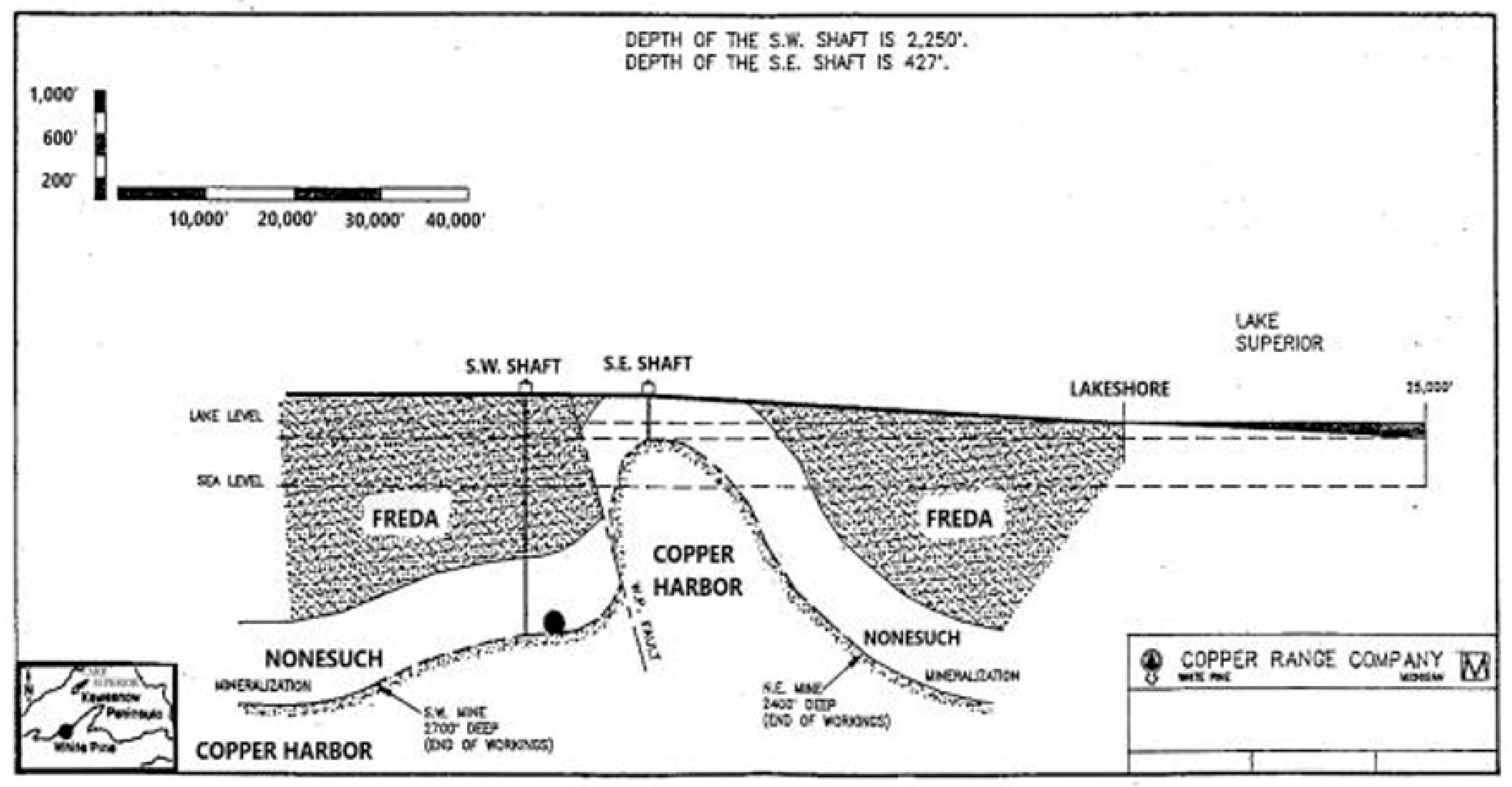
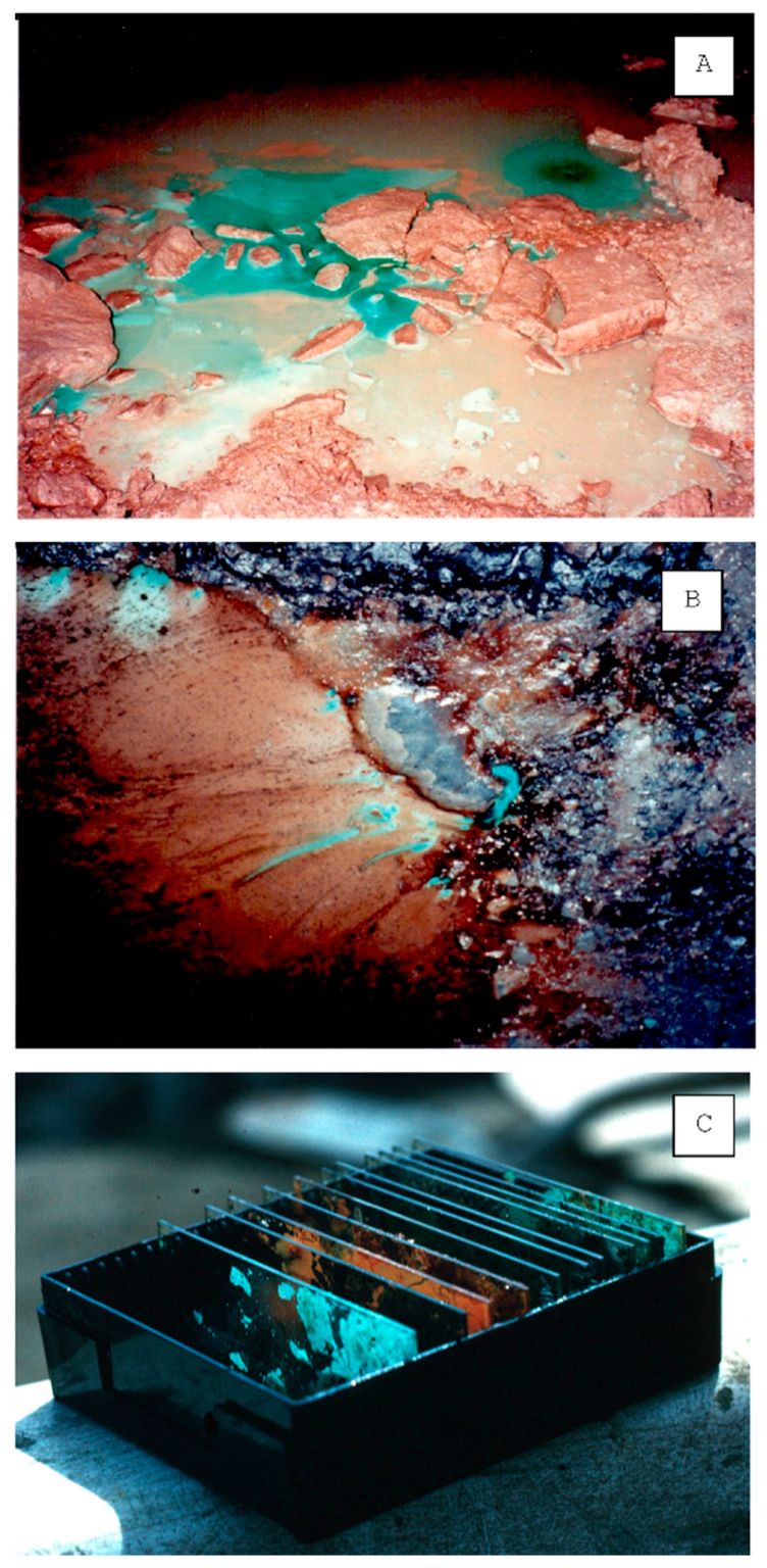
2.1. Geological Setting and Biofilm Sampling

Mining at the White Pine Mine ceased in 1992 in Area 91, which remained accessible for visits and scientific research until the mine flooded. The rocks exposed there are part of the Nonesuch Shale and contain approximately one weight % Cu, primarily in the form of chalcocite (Cu2S). The environment was humid, and newly-formed green copper minerals appeared on bedding planes thought to be due to oxidation by the ambient air. Petroleum and water dripped from cracks around roof bolts. Floating green films mixed with brown petroleum films and red iron oxide films in the vicinity of rusting iron rebar made a colorful mix of modern-day mineral authigenesis. Ventilation served as a source of oxygen. No smell of H2S was present in any standing mine pools.
Two types of samples were collected for mineral and microbial analyses. (1) For solids, green films floating on water pools in the mine were lifted off by slipping glass microscope slides underneath them (Figure 2C). (2) Other glass slides were left in pools for six weeks to allow mineral and bacterial growth, and then retrieved for microbial analysis.

2.2. Analytical Methods

2.2.1. X-ray Powder Diffraction and Energy Dispersive X-ray Spectroscopy
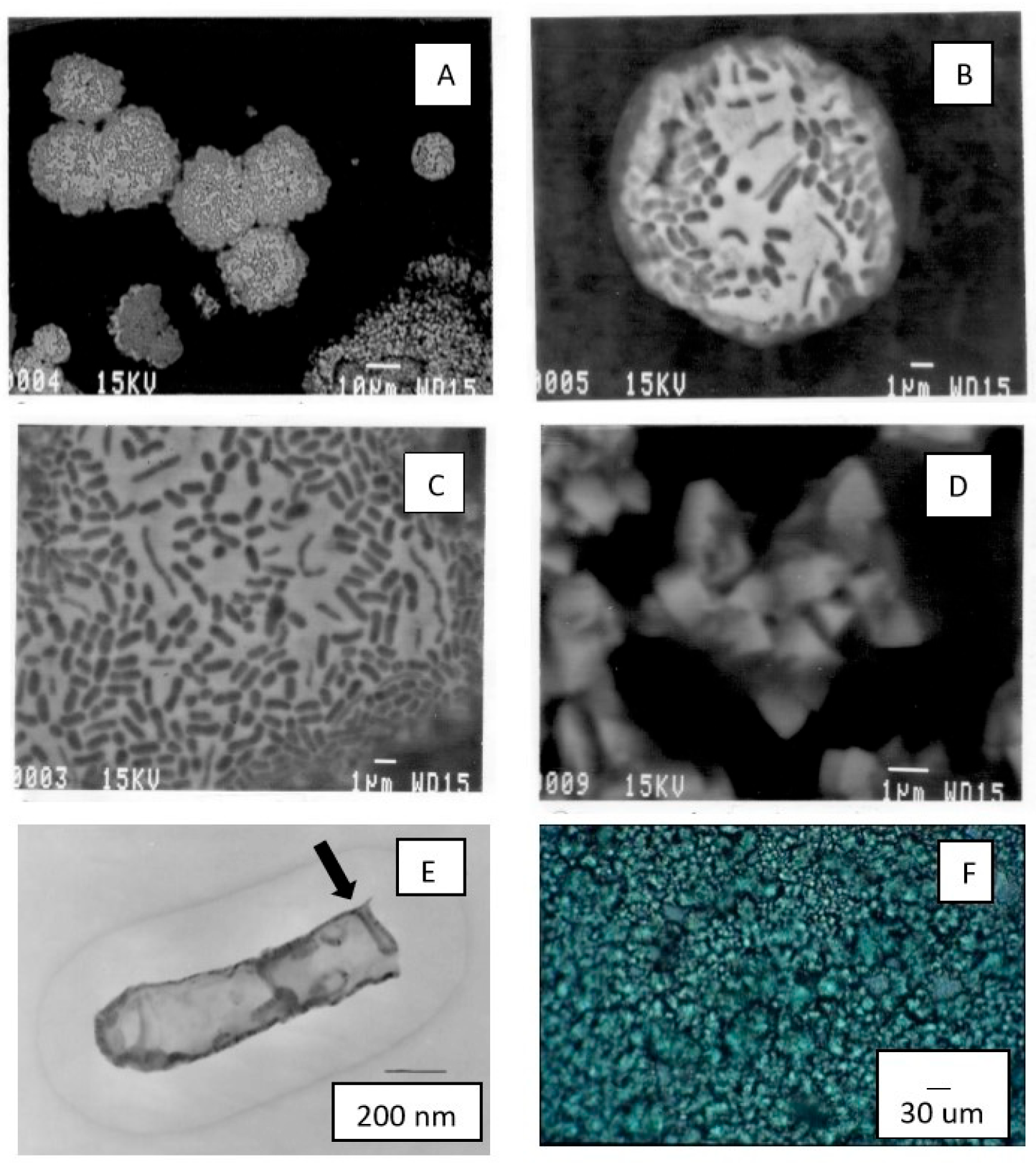
X-ray powder diffraction (XRD) was used to analyze the mineral phases on the microscope slides using a Guinier-Haegg focusing X-ray powder camera (Incentive Research and Development AB) equipped with a sealed X-ray tube with a Cu anode. The light green film and dark-green crystalline material were powdered and then analyzed in an aqueous vehicle. The produced patterns were traced through the International Centre for Diffraction Data Samples (ICDD) files. Energy dispersive X-ray spectroscopy (EDS) was performed with a Princeton Gamma Tech instrument attached to the scanning electron microscope (below).

2.2.2. Microscopy

Samples were analyzed using transmitted light microscopy (Leitz Ortholux), scanning electron microscopy (SEM) (JEOL JSM-840) with an attached Princeton Gamma Tech EDS, and transmission electron microscopy (TEM) (Philips EM-400T with an attached LINK-Analytical eXL system for EDS using a Cu target). Standard dehydration and fixing techniques employing nadic methyl anhydride, dodecenylsuccinic anhydride, and tri-dimethylaminoethyl phenol were used to embed and then slice microbial surfaces for TEM analysis [40].

2.2.3. Microbiology

Conventional microbiology techniques were used because this experiment was performed before genomics became the standard identification method. Genomics, of course, can provide unequivocal identifications, such as the anaerobes identified in a zone of sulfide mineralization at a copper deposit in Spain [41] and Germany [42].
A suspension of green film solids was prepared and used to inoculate a series of dilutions into the commercially available nutrient broth (Becton Dickinson, BD) to isolate any viable bacteria that might be present. Reagent grade CuCl2 was used as needed to adjust the final Cu(tot) concentrations in the tubes to 10, 1.0, 0.1, and 0.01 mM/L to examine the tolerance of bacteria to different Cu concentrations. Nutrient agar (BD) pour plates were prepared from similar dilution series to attempt to isolate any other bacteria and fungi; colonies that formed were noted for their growth characteristics. Isolates were tested for utilization of energy and/or carbon sources using minimal salt media, NO3 broth, nutrient broth, SO4 broth selective for sulfate-reducing bacteria, and Fe media (pH < 2.0) selective for thiobacilli.

2.2.4. Water Chemistry

Mine pool water was collected, filtered, and analyzed using standard ASTM (American Society for Testing and Materials) methods for inorganic constituents in water by the Colorado State University, Soil, Water and Plant Testing Lab. The pH and ORP measurements were made in situ in the laboratory (Horiba U-20 multiparameter meter), measuring oxidation reduction potential (ORP) as the difference between a Pt and a Ag/AgCl reference electrode. The activity of the Cl- ion was determined electrochemically by dipping an exposed Ag/AgCl electrode and a reference Ag/AgCl/KCl (4N) electrode into the brine sample in the laboratory at 25.5 °C.

2.2.5. Isotope Geochemistry

Stable carbon isotopes in the green films associated with petroleum and petroleum not associated with green films were analyzed using a Finnigan Delta Plus V mass spectrometer coupled to a Carlo Erba NC2500 elemental analyzer on a scale defined by USGS-24 (graphite, δ13C = −15.9 ‰). The oxidation column in the elemental analyzer was packed with chromium oxide and silvered cobaltous oxide, and the reduction column was packed with copper and copper oxide. Results are reported as per mil (‰) relative to Vienna Pee Dee Belemnite (PDB) [43].
Samples included green films associated with petroleum, petroleum not associated with green films, two sets of calcite veins that formed contemporaneously with copper mineralization and one that formed slightly after ore deposition, and organic matter from the Nonesuch Shale (bitumen, kerogen, and petroleum). Data included one sample of oxidized petroleum one sample of iron oxide/hydroxide “slime”, and three samples of copper salt “slime”.

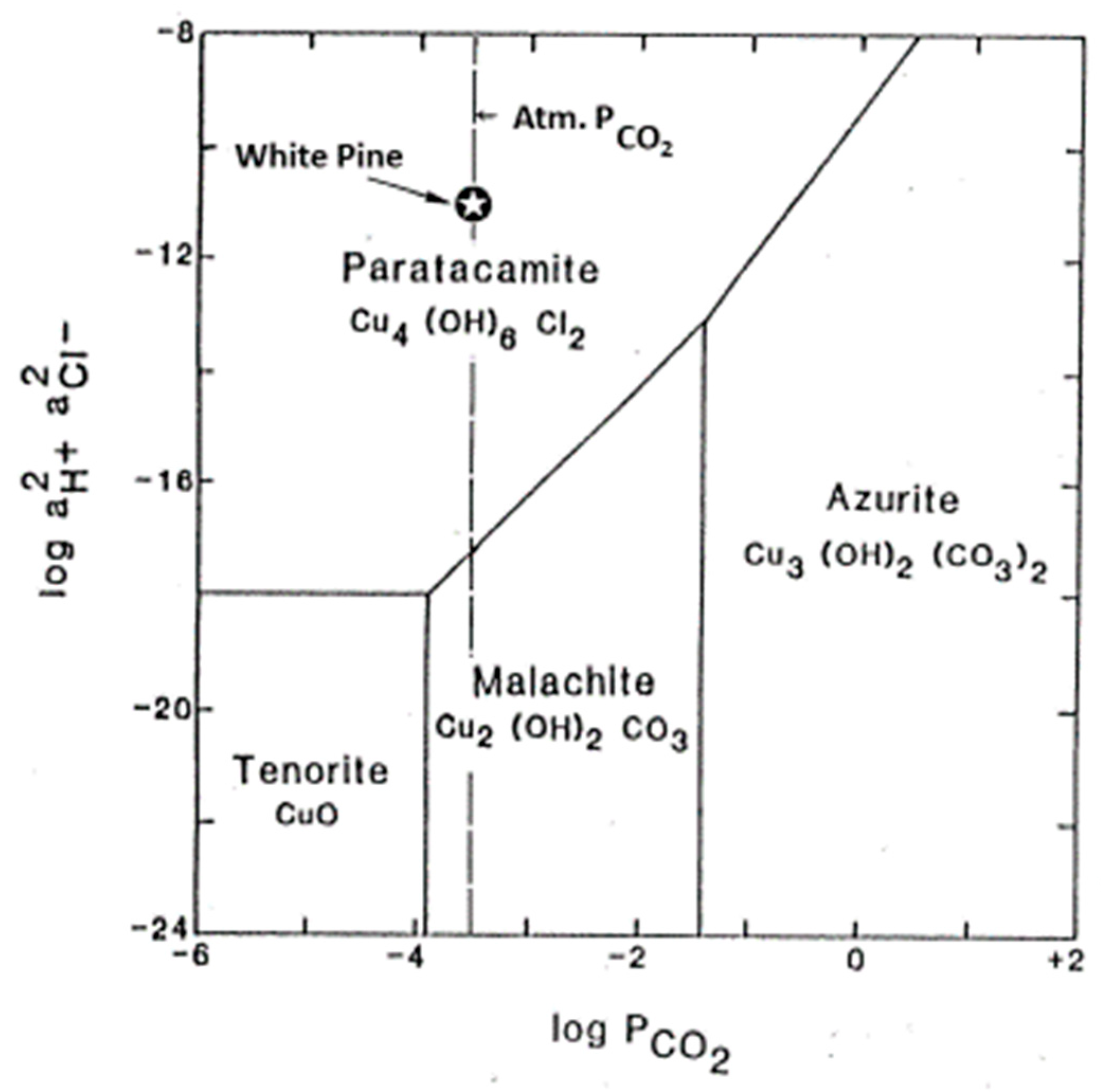
2.2.6. Thermodynamic Analysis

Data have not been published for Cu-bearing saline phases, which necessitated in situ analysis and mathematical manipulation to understand the dynamics of mineral formation. Because of large uncertainties inherent to the thermochemistry of highly concentrated aqueous saline solutions in general, it was important to determine ORP and pH using Ag/AgCl electrodes, as detailed in ‘water chemistry’ above.

3. Results

3.1. Mineralogy

X-ray powder diffraction indicated that the dark-green film was paratacamite, while the light-green film was a mixture in nearly equal proportions of atacamite and paratacamite. These are both dimorphs of Cu2(OH)3Cl [23]. Walter-Levy and Goreaud [44] showed that these dimorphs have closely similar free energies and commonly precipitate together from saline water solutions such as those found in the Atacama Desert of Chile. Halite was also present as micrometer-size cubes on the microscope slides.
EDS analysis of a green film showed peaks of Cl-Si-Ca-C-Na-Cu, in descending order (Figure 3). The silicon peak can be ascribed to the underlying glass microscope slide.

3.2. Water Chemistry

The standing water in the mine was a Ca-Na-Cl brine that contains as much as 7 mg/L total Cu and 132 mg/L SO4 (Table 1). In the water sample carried from the mine and analyzed immediately in the laboratory, pH was 6.0 ± 0.2 and ORP was >+488 mV. The temperature of the air and mine water at that time was 17 °C.

3.3. Microscopy

Under the light microscope, the light green atacamite films were seen to be agglomerations of circular blebs (Figure 4A,F) filled with bacteria (Figure 4B,C). SEM examination of these films showed that individual atacamite blebs enclosed a mixture of short rod-shaped bacteria and narrow rod-shaped, filament-forming cells encased in thick sheath material (Figure 4C). The blebs give the distinct impression of being individual colonies. The paratacamite appears to be more crystalline and takes the form of octahedrons (Figure 4D). Cocci and short rods colonized the petroleum film. Fungal hyphae attached to individual mineral blebs.
Glass slides left in the standing mine water for six weeks (Figure 4C) were colonized primarily by rod-shaped bacteria. Fungal hyphae and spores and clear filaments of rod-shaped bacteria were also present.
Slices of bacterial cell walls and membranes analyzed by TEM showed that the walls were electron dense. This suggests that the minerals precipitated along the cell membranes. Copper but not chloride was attached to the membranes. The membranes were microcrystalline and invaginated in several different places (Figure 4E, arrow). Typically, bacteria living in environments having toxic substances can wall off such substances with invaginated membrane vesicles [45].

3.4. Microbiology

Conventionally cultured bacteria from the blebs grew in pellicles at the air–liquid interface in liquid media; their growth indicates an aerobic, heterotrophic mode of respiration. Growth was observed in nitrate broth but not in sulfate broth; this difference indicates facultative but not obligate anaerobic nutrition, meaning that they could grow under either oxic or anoxic conditions. Iron broth lacking an organic carbon source (selective for thiobacilli) showed no microbial growth, thus indicating that the Gram-negative organisms were not Fe-chemoautotrophs. On solid media, two different colony types grew: (1) a dark yellow, clear, lustrous colony, and (2) a white, milky, non-lustrous colony. Although specific identification was not made, these characteristics, coupled with the rosette patterns in the blebs, are reminiscent of Leucothrix and filamentous bacteria.
No growth was observed after several weeks in minimal media having CuCl2 concentrations of 0.01, 0.1, 1.0, and 10 mM/L. Therefore, the bacteria were not deriving energy from Cu electrons, although some Cu was probably incorporated into necessary enzymes. In nutrient media, growth was observed in 1 mM/L but not 10 mM/L Cu. This establishes the upper limit of Cu tolerance in nutrient media. In contrast, a fungus grew in all Cu concentrations.
The thickness of the floating surficial films made it impossible to count the number of individual colonies; therefore, slides that were immersed in water for six weeks were used for quantification. Treating the blebs in the green films as individual colonies, statistics were generated on colony size, number of bacteria per colony, and the number of colonies per cm2. Individual colonies averaged 20 μm in diameter. Per colony, the mean number of rods was 184 and filaments was 10. A slide from which the green film was vigorously washed had a mean number of single colonies of 105/cm2. A slide from which the green film was not washed contained a mean number of single colonies of 1900/cm2.

3.5. Isotope Geochemistry

The δ13C values helped to determine the source of bacterial carbon. Several carbon sources were present, some abiotic and others biotic. Calcite veins were present in the Nonesuch, along with CO2 in the groundwater. Kerogen, petroleum, and CO2 from ventilation were also available. Carbonates from the mine have relatively positive values [46,47]; in contrast, the Nonesuch kerogen and petroleum range from −30 to −35 (Figure 5).
δ13C PDB values for carbon in the atacamite-paratacamite mixtures ranged from −28.8 to −24.9‰ and, presumably, reflect the isotopic values of bacteria and minor amounts of petroleum mixed with the minerals (Figure 5). These values are 1.5 to 7.5‰ greater than published values for pristine petroleum from the mine [46]. This is significant because the bacteria in the minerals are enriching the 13C relative to the petroleum.

3.6. Thermodynamics

The activity of Cl- was 2.39 molal. The pH measured with a glass electrode at the same time was 5.92. The ORP was +488 mV, indicating that the Cu was in the Cu2+ cupric state. This being the case, the bacteria cannot be getting any energy from the oxidation of Cu. At this pH, the minerals are outside of the stability fields of native copper and copper sulfides, and indeed, sulfate concentration in the freshwater sample was negligible.
The lack of a redox sulfur cycle (SO4 ←→ S2−) is attributed to several factors. Water in the mine moves slowly, thus limiting reactant availability, sulfide minerals make up a very small percentage of the rocks, and the dominant sulfide mineral (chalcocite) does not contain much S.
Given the salinity, the paratacamite group of minerals was at saturation. The measured Cl ion times H+ ion activities plot well within the paratacamite field [22] (Figure 6).

4. Discussion

Field and laboratory evidence suggests that bacteria may play a role in the precipitation of these basic copper chloride minerals discovered as floating films at depth in the White Pine Copper Mine. Bacteria may be catalyzing the precipitation of the paratacamite group minerals or acting as templates. The cell walls were electron dense and were the site of Cu ions, suggesting that they played an active role in Cu precipitation. The membranes were invaginated, which is a typical occurrence when toxic compounds are present in the environment.
In the laboratory, bacteria did not grow in Cu concentrations greater than 1 mM/L. This is equivalent to 64 mg/L of Cu, which is more Cu than in the mine pool water. The salinity of the mine pools may offset Cu toxicity, but this factor was not controlled and obviously should be tested in the future. The fungal colony was the most nutritionally diverse among the cultured consortium. The fungal isolate may play a role in the degradation of the petroleum residue and, therefore, provide a metabolizable form of carbon or other growth factors to the bacteria.
It is also possible to speculate on the relationship between individual paratacamite octahedrons and bacteria. The octahedrons were similar in size to the short rods, suggesting that rods may be entombed in the individual octahedrons.
The enrichment of 13C in the bacteria relative to the petroleum is consistent with an aerobic, heterotrophic mode of respiration. CO2 would be an expected metabolic waste product, and this compound could shift the pH into the paratacamite group range. Because the bacteria in the minerals are enriching the 13C relative to the petroleum, this suggests that they were actively using petroleum as their carbon source.
Atacamite is a frequent secondary copper mineral in the supergene zone of copper deposits associated with arid conditions and brines [27]. But the presence of external carbon sources is rarely reported in copper deposits [41]. It would be interesting to know if atacamite precipitation by heterotrophic bacteria is a universal phenomenon or just a result of special circumstances of a copper deposit being associated with petroleum or other metabolizable sources of carbon.

Author Contributions

Conceptualization, E.I.R.; investigations, E.I.R., M.R.S. and C.D.Y.; methodology, E.I.R., M.R.S. and C.D.Y.; writing, E.I.R.; writing—review and editing, M.R.S. and C.D.Y. All authors have read and agreed to the published version of the manuscript.

Funding

The research was funded while in employment with the U.S. Geological Survey (E.I.R., M.R.S.) and Clark Atlanta University (C.D.Y.).

Data Availability Statement

The data presented in this study are available on request from the corresponding author.

Acknowledgments

We would like to thank, Jochen Tilk, and M.B. Sanders, who was then at the Copper Range Company. Various aspects of this research were performed by colleagues at the USGS—mineralogy by Howard Evans, isotope geochemistry by Bob Seal, thermodynamics by Moto Sato, and EDS by Roger Congdon. We also thank Todd Schaef, Matilde Urrutia, and Yupha Vatcharapijarn for helping with laboratory experiments, Doug Owen and Pam Winsky for field assistance, David Crisman and Harvey Belkin for raw data, and Chris McKay for IT aid.

Conflicts of Interest

The authors declare no conflict of interest.

References

  1. Phelps, R.W. Copper Range, Last All-Underground Copper Mine in the United States is a Game Survivor--Now with a More Assured Future. Eng. Min. J. 1990, 191, C47–C57. [Google Scholar]
  2. Robbins, E.I.; Stanton, M.R.; Tilk, J.E.; Congdon, R.D.; Evans, H.T., Jr.; Gullett, C.D.; Sanders, M.B.; Sato, M.; Schaef, H.T.; Seal, R.R. Association of Microbes with Authigenic Copper Chloride Mineral Films (Atacamite and Paratacamite) and Petroleum Residues at Depth in the White Pine Copper Mine, Michigan. In Proceedings of the Waterloo ’94, Geol. Assoc. Canada and Mineral. Assoc. Canada, Waterloo, ON, Canada, Abs. with Program. 13–18 May 1994; p. A94. [Google Scholar]
  3. Copper Range Company. Cross Section of the White Pine Mine; Copper Range Company: Houghton, MI, USA, 1993. [Google Scholar]
  4. Daniels, P.A., Jr. Upper Precambrian Sedimentary Rocks; Oronto Group, Michigan-Wisconsin. In Geology and Tectonics of the Lake Superior Basin; Wold, R.J., Hinze, W.J., Eds.; The Geological Society of America, Inc.: Boulder, CO, USA, 1982; Chapter 7C; Volume 156, pp. 107–133. [Google Scholar] [CrossRef]
  5. White, W.S.; Wright, J.C. The White Pine Copper Deposit, Ontonagon County, Michigan. Econ. Geol. 1954, 49, 675–716. [Google Scholar] [CrossRef]
  6. Mauk, J.L.; Hieshima, G.B. Organic Matter and Copper Mineralization at White Pine, Michigan. Chem. Geol. 1992, 99, 189–211. [Google Scholar] [CrossRef]
  7. White, W.S.; Wright, J.C. Sulfide Mineral Zoning in the Basal Nonesuch Shale, Northern Michigan. Econ. Geol. 1966, 61, 1171–1190. [Google Scholar] [CrossRef]
  8. Brown, A.C. Mineral Assemblages in the White Pine Copper Deposit, Northern Michigan (USA). In Freiberger Forschungshefte, Reihe C, Geowissenschafte, Mineralogie, und Geochemie; Akademie-Verlag: Freiberg, Germany, 1973; Volume 293, pp. 65–83. [Google Scholar]
  9. Alyanak, N.; Vogel, T.A. Framboidal Chalcocite from White Pine, Michigan. Econ. Geol. 1974, 69, 697–703. [Google Scholar] [CrossRef]
  10. Ho, E.S.; Mauk, J.L. Relationship between Organic Matter and Copper Mineralization in the Proterozoic Nonesuch Formation, Northern Michigan. Ore Geol. Rev. 1996, 11, 71–87. [Google Scholar] [CrossRef]
  11. Robbins, E.I. Accumulation of Fossil Fuels and Metallic Minerals in Active and Ancient Rifts. Tectonophysics 1983, 94, 633–658. [Google Scholar] [CrossRef]
  12. Eglinton, G.; Scott, P.M.; Belsky, T.; Burlingame, A.L.; Calvin, M. Hydrocarbons of Biological Origin from a One-Billion-Year-Old Sediment. Science 1964, 145, 263–264. [Google Scholar] [CrossRef]
  13. Eglinton, G.; Scott, P.M.; Belsky, T.; Burlingame, A.L.; Richter, W.; Calvin, M. Occurrence of isoprenoid alkanes in a precambrian sediment. In Advances in Organic Geochemistry; Hobson, G.D., Louis, M.C., Eds.; Pergamon Press: Oxford, UK, 1964; pp. 41–74. [Google Scholar] [CrossRef]
  14. Kelly, W.C.; Rye, R.O.; Livnat, A. Saline Minewaters of the Keweenaw Peninsula, Northern Michigan: Their Nature, Origin, and Relation to Similar Deep Waters in Precambrian Crystalline Rocks of the Canadian Shield. Am. J. Sci. 1986, 286, 281–308. [Google Scholar] [CrossRef]
  15. Andrei, A.; Öztürk, Y.; Khalfaoui-Hassani, B.; Rauch, J.; Marckmann, D.; Trasnea, P.-J.; Daldal, F.; Koch, H.-G. Cu Homeostasis in Bacteria: The Ins and Outs. Membranes 2020, 10, 242. [Google Scholar] [CrossRef]
  16. Beale, D.J.; Morrison, P.D.; Key, C.; Palombo, E.A. Metabolic Profiling of Biofilm Bacteria Known to Cause Microbial Influenced Corrosion. Water Sci. Technol. 2014, 69, 1–8. [Google Scholar] [CrossRef] [PubMed]
  17. Vargas, I.T.; Alsina, M.A.; Pavissich, J.P.; Jeria, G.A.; Pasten, P.A.; Walczak, M.; Pizarro, G.E. Multi-Technique Approach to Assess the Effects of Microbial Biofilms Involved in Copper Plumbing Corrosion. Bioelectrochemistry 2014, 97, 15–22. [Google Scholar] [CrossRef]
  18. Narenkumar, J.; Devanesan, S.; AlSalhi, M.S.; Kokilaramani, S.; Ting, Y.-P.; Rahman, P.K.S.M.; Rajasekar, A. Biofilm Formation on Copper and its Control by Inhibitor/Biocide in Cooling Water Environment. Saudi J. Biol. Sci. 2021, 28, 7588–7594. [Google Scholar] [CrossRef] [PubMed]
  19. Pschedt, J.W. Copper-Based Bactericides and Fungicides, Pacific Northwest Pest Management Handbook, Oregon State University. Available online: https://pnwhandbooks.org/plantdisease/pesticide-articles/copper-based-bactericides-fungicides (accessed on 28 July 2023).
  20. Wisconsin Department of Natural Resources. Copper Compounds Chemical Fact Sheet, DNR PUB-WT-968. 2012; 2p. Available online: https://dnr.wi.gov/lakes/plants/factsheets/CopperFactsheet.pdf (accessed on 28 July 2023).
  21. Barranguet, C.; van den Ende, P.F.; Rutgers, J.; Breure, A.M.; Greijdanus, M.; Sinke, J.S.; Admiraal, W. Copper-Induced Modifications of the Trophic Relations in Riverine Algal-Bacterial Biofilms. Environ. Toxicol. Chem. 2003, 22, 1340–1349. [Google Scholar] [PubMed]
  22. Woods, T.L.; Garrels, R.M. Phase Relations of Some Cupric Hydroxyl Minerals. Econ. Geol. 1986, 81, 1989–2007. [Google Scholar] [CrossRef]
  23. Pollard, A.M.; Thomas, R.G.; Williams, P.A. Synthesis and Stabilities of the Basic Copper (II) Chlorides Atacamite, Paratacamite and Botallackite. Mineral. Mag. 1989, 53, 557–563. [Google Scholar] [CrossRef]
  24. Crisman, D.P. Groundwater Geochemistry in the Portage Lake Volcanics, Hancock, Michigan and Implications for Native Copper Stability. Master’s Thesis, Michigan Technical University, Houghton, MI, USA, 1982. [Google Scholar]
  25. Mindat, Atacamite, Paratacamite. Available online: https://www.mindat.org/min (accessed on 28 July 2023).
  26. Palacios, C.; Rouxel, O.; Reich, M.; Cameron, E.M.; Meybourne, M.L. Pleistocene Recycling of Copper in a Porphyry System, Atacama Desert, Chile: Cu Isotope Evidence. Mineral. Deposita 2011, 46, 1–7. [Google Scholar] [CrossRef]
  27. Reich, M.; Palacios, C.; Parada, M.A.; Fehn, U.; Cameron, E.M.; Leybourne, M.L.; Zuniga, A. Atacamite Formation by Deep Saline Waters in Copper Deposits from the Atacama Desert, Chile: Evidence from Fluid Inclusions, Groundwater Geochemistry, TEM, and 36Cl Data. Miner. Deposita 2008, 43, 663. [Google Scholar] [CrossRef]
  28. Shaerkey, J.B.; Lewin, S.Z. Conditions Governing the Formation of Atacamite and Paratacamite. Am. Mineral. 1971, 56, 179–192. [Google Scholar]
  29. Calagari, A.A. Stable Isotope (S, O, H and C) Studies of the Phyllic and Potassic-Phyllic Alteration Zones of the Porphyry Copper Deposit at Sungun, East Azarbaidjan, Iran. J. Asian Earth Sci. 2003, 21, 767–780. [Google Scholar] [CrossRef]
  30. Kojima, S.; Astudillo, J.; Rojo, J.; Tristá, D.; Hayashi, K. Ore Mineralogy, Fluid Inclusion, and Stable Isotopic Characteristics of Stratiform Copper Deposits in the Coastal Cordillera of Northern Chile. Miner. Depos. 2003, 38, 208–216. [Google Scholar] [CrossRef]
  31. Bodden, T.J.; Bornhorst, T.J.; Bégué, F.; Deering, C. Sources of Hydrothermal Fluids Inferred from Oxygen and Carbon Isotope Composition of Calcite, Keweenaw Peninsula Native Copper District, Michigan, USA. Minerals 2022, 12, 474. [Google Scholar] [CrossRef]
  32. Kendall, C.; Sklash, M.G.; Bullen, T.D. Isotope Tracers in Water and Solute Sources in Catchments. In Solute Modelling in Catchments Systems; Trudgill, S.T., Ed.; John Wiley & Sons: New York, NY, USA, 1995; pp. 261–303. [Google Scholar]
  33. Enders, M.S.; Knickerbocker, C.; Titley, S.R.; Southam, G. The Role of Bacteria in the Supergene Environment of the Morenci Porphyry Copper Deposit, Greenlee County, Arizona. Econ. Geol. 2006, 101, 59–70. [Google Scholar] [CrossRef]
  34. Liu, J.; Hua, Z.-S.; Chen, L.-X.; Kuang, J.-L.; Li, S.-J.; Shu, W.-S.; Huang, L.-N. Correlating Microbial Diversity Patterns with Geochemistry in an Extreme and Heterogeneous Environment of Mine Tailings. Appl. Environ. Microbiol. 2014, 80, 3677–3686. [Google Scholar] [CrossRef] [PubMed]
  35. Banerjee, P.C.; Ray, M.K.; Koch, C.; Bhattacharyya, S.; Shivaji, S.; Stackebrandt, E. Molecular Characterization of Two Acidophilic Heterotrophic Bacteria Isolated from a Copper Mine of India, System. Appl. Microbiol. 1996, 19, 78–82. [Google Scholar] [CrossRef]
  36. Galleguillos, P.A.; Hallberg, K.B.; Johnson, D.B. Microbial Diversity and Genetic Response to Stress Conditions of Extremophilic Bacteria Isolated from the Escondida Copper Mine. Adv. Mater. Res. 2009, 71–73, 55–58. [Google Scholar] [CrossRef]
  37. Pusz, W.; Kita, W.; Weber, R. Microhabitat Influences the Occurrence of Airborne Fungi in Copper Mine in Poland. J. Cave Karst Stud. 2014, 76, 14–19. [Google Scholar] [CrossRef]
  38. Cervantes, C.; Gutierrez-Corona, F. Copper Resistance Mechanisms in Bacteria and Fungi. FEMS Microbiol. Rev. 1994, 14, 121–138. [Google Scholar] [CrossRef]
  39. Ladomersky, E.; Petris, M.J. Copper Tolerance and Virulence in Bacteria. Metallomics 2016, 7, 957–964. [Google Scholar] [CrossRef]
  40. Glauert, A.M.; Lewis, P.R. Biological Specimen Preparation for Transmission Electron Microscopy. In Practical Methods in Electron Microscopy; Glauert, A.M., Ed.; Portland Press: London, UK, 1998; Volume 17. [Google Scholar] [CrossRef]
  41. Tornos, F.; Oggerin, M.; de los Rios, A.; Rodriguez, N.; Amils, R.; Sanz, J.L.; Rojas, P.; Velasco, F.; Escobar, J.M.; Gomez, C.; et al. Do Microbes Control the Formation of Giant Copper Deposits? Geology 2019, 47, 143–146. [Google Scholar] [CrossRef]
  42. Arif, S.; Nacke, H.; Schliekmann, E.; Reimer, A.; Arp, G.; Hoppert, M. Composition and Niche-Specific Characteristics of Microbial Consortia Colonizing Marsberg Copper Mine in the Rhenish Massif. Biogeosciences 2022, 19, 4883–4902. [Google Scholar] [CrossRef]
  43. Kendall, C.; Caldwell, E.A. Fundamentals of isotope geochemistry. In Isotope Tracers in Catchment Hydrology; Kendall, C., McDonnell, J.J., Eds.; Elsevier Science: Amsterdam, The Netherlands; New York, NY, USA, 1998; Chapter 2; pp. 51–86. [Google Scholar]
  44. Walter-Levy, L.; Goreaud, M. Sur la Formation des Chlorures Basiques Cuivriques en Solution Aqueuse de 25° à 200 °C; Bulletin de la Societe Chimique de France: Paris, France, 1969; pp. 2623–2634. [Google Scholar]
  45. Nagakubo, T.; Nomura, N.; Toyofuku, M. Cracking Open Bacterial Membrane Vesicles. Front. Microbiol. 2020, 10, 3026. [Google Scholar] [CrossRef]
  46. Hieshima, G.B. Organic and Isotopic Geochemical Study of the Middle Proterozoic Nonesuch Formation, North American Midcontinent Rift. Ph.D. Thesis, Indiana University, Bloomington, IN, USA, 1992. [Google Scholar]
  47. Mauk, J.L. Geologic and Geochemical Investigations of the White Pine Sediment Hosted Stratiform Copper Deposit, Ontonagon County, Michigan. Ph.D. Thesis, University of Michigan, Ann Arbor, MI, USA, 1993. [Google Scholar]
  48. Cameron, E.M.; Leybourne, M.L.; Palacios, C. Atacamite in the Oxide Zone of Copper Deposits in Northern Chile: Involvement of Deep Formation Waters? Miner. Depos. 2007, 42, 205–218. [Google Scholar] [CrossRef]
Figure 1. Historical map of the White Pine Mine (Reprinted with written permission from Copper Range Company, 1993 [3]. Sampling locality in mine (black dot). Location of mine (lower left inset).
Micro 03 00051 g001
Figure 2. Floating green films and petroleum on mine pools. (A) Green film floating on pool (pink color is mine mud). (B) Petroleum and green film floating on pool. (A,B) scale: (photographs of floor from a standing position) (C). Mineralized films (<50 µm thick) lifted by microscope slides slipped under floating films.
Micro 03 00051 g002
Figure 3. EDS trace of a green film lifted onto a microscope slide from a floating film on a mine pool.
Micro 03 00051 g003
Figure 4. Photomicrographs of bacteria and minerals in green films from the mine pools. (A) Blebs in green atacamite film, SEM. (B) Close-up of bleb in green atacamite film, SEM. (C) Close-up of short rods and sheathed filaments in green atacamite film bleb, SEM. (D) Octahedral crystals in dark green paratacamite, SEM. (E) Invagination (arrow) in membrane of sliced bacterial cell, TEM. (F) Atacamite growing on slide immersed in pool for one month, transmitted light.
Micro 03 00051 g004
Figure 5. Carbon isotopic data from White Pine Mine samples (data ranges from [23,46]).
Micro 03 00051 g005
Figure 6. Stability field for White Pine Mine sample compared to cupric hydroxy minerals (stability field from [22,48]. Adapted with permission from Ref. [22], 1986, T.L. Woods and R.M. Garrels.
Micro 03 00051 g006
Table 1. Mine water chemistry (mg/L) (nd = not determined).
CrismanThis StudyThis Study 1
(1982) [24](10/93 raw water)(8/94 collected; 12/95 analyzed)
Ca91,78069,19013,264
Na17,64016,4002156
Mg1373940256
Sr1261nd125
K234nd200
Alndnd2.6
Cdndnd0.122
Cund70.35
Fend71.59
Mn40nd6.97
Nindnd0.33
Pbndnd1.31
Znndnd2.34
Cl156,500105,15241,000
Br1520nd0.1
HCO315nd21.8
SO4nd0.49132
NO3ndnd946
TDS270,363nd58,117
pHnd6.0 + 0.25.9
T(C)17°17°nd
Eh (mV) ORP (Pt)nd+488.6nd
Eh (mV) Ag/AgClnd199.6nd
Eh (mV) Cund64.6nd
1 Analysis performed by Colorado State University, Soil, Water and Plant Testing Lab.
Disclaimer/Publisher’s Note: The statements, opinions and data contained in all publications are solely those of the individual author(s) and contributor(s) and not of MDPI and/or the editor(s). MDPI and/or the editor(s) disclaim responsibility for any injury to people or property resulting from any ideas, methods, instructions or products referred to in the content.

