Article

Lipase Production and Characterization from Serratia liquefaciens Isolated from Petroleum-Contaminated Soil

by
Abayomi Baruwa
and
Kugenthiren Permaul
*
Department of Biotechnology and Food Science, Durban University of Technology, Durban 4001, South Africa
*
Author to whom correspondence should be addressed.
Appl. Microbiol. 2026, 6(8), 87; https://doi.org/10.3390/applmicrobiol6080087
Submission received: 14 July 2026 / Revised: 20 July 2026 / Accepted: 25 July 2026 / Published: 31 July 2026

Abstract

Lipases are important enzymes in the esterase family that hydrolyze ester bonds in triglycerides, producing simpler molecules. This property makes them valuable in biotechnology and environmental cleanup. In this study, lipase-producing bacteria were isolated and characterized from petroleum-contaminated soil to establish a cost-effective platform for enzyme production and bioremediation. Among the recovered isolates, Serratia liquefaciens AB1 exhibited the highest lipolytic activity and was therefore selected for further investigation. The influence of various inducer oils and agro-industrial residues on enzyme production was systematically assessed. In addition, fermentation parameters were optimized through the evaluation of different carbon and nitrogen sources to enhance lipase yield. Waste frying oil was identified as the most effective inducer, while glucose and yeast extract supported optimal enzyme production. The enzyme lipase AB1 was fully purified using CM-Sephadex C-50 chromatography, Sephadex G-100 and further characterized by SDS-PAGE, kinetic studies, and stability assays. Purification of the enzyme resulted in a specific activity of 610.92 U/mg, corresponding to a 9.42-fold increase in purity with an overall recovery of 76%. The enzyme exhibited an apparent molecular mass of approximately 64 kDa. It demonstrated optimal catalytic activity at 60 °C and pH 8 and retained substantial stability at this temperature for up to 120 min. Kinetic analysis revealed a low Km value of 30 µM, indicating strong substrate affinity, along with a Vmax of 23.89 U/mL, reflecting a high catalytic efficiency under the tested conditions. Enzyme activity was enhanced by Ca2+, Na+, and Ba2+, but inhibited by Mn2+ and Hg2+. These findings demonstrate the favorable biochemical properties of the purified lipase and provide a basis for future investigations into its potential application as a biocatalyst for bioremediation.

1. Introduction

Lipases are versatile biocatalysts with broad applications in biotechnology because they catalyze diverse reactions, including hydrolysis, aminolysis, interesterification, peroxidation, and alcoholysis [1]. These enzymes are widely applied in food processing, detergent formulation, biodiesel production, leather treatment, and pharmaceutical manufacturing [2]. As members of the esterase family, lipases catalyze the hydrolysis of ester bonds and primarily act on triglycerides, the major constituents of fats and oils [1].
Lipolytic enzymes provide environmentally sustainable solutions for lipid waste management and contribute to renewable energy development. Their use as green biocatalysts in biodiesel synthesis from renewable waste feedstocks offers an eco-friendly alternative to fossil-based processes [2]. Their unique biochemical and physicochemical properties further increase their industrial relevance. Lipases are widely distributed in plants, animals, and microorganisms, but microbial lipases are considered the most commercially valuable because of their high catalytic efficiency, broad operational stability, and ease of large-scale production [3]. Bacterial lipases are among the most widely studied microbial enzymes because of their high catalytic activity, broad substrate specificity, and remarkable stability over a wide range of temperatures and pH conditions. These properties enable them to withstand harsh industrial processing conditions, making them valuable for various biotechnological applications. For industrial use, lipases must be purified and thoroughly characterized after production [4]. Purification removes unwanted contaminants, increases enzyme purity, and improves the accuracy of biochemical analyses. It also facilitates the investigation of the enzyme’s structural, conformational, and kinetic properties. Comprehensive characterization is essential because the biochemical properties of lipases vary depending on their microbial source. Therefore, the effects of temperature, pH, metal ions, inhibitors, and organic solvents on enzyme activity and stability should be evaluated to determine the enzyme’s optimal operating conditions and robustness [5].
In addition, determining the kinetic parameters, including the Michaelis constant (Km) and maximum reaction velocity (Vmax), using appropriate substrates provides valuable information on substrate affinity and catalytic efficiency. These characteristics are critical for assessing the suitability of lipase produced by Serratia liquefaciens AB1 isolated from petroleum-contaminated soil for potential industrial applications. In 2023, the global microbial lipase market was valued at approximately USD 591 million and is projected to grow at a compound annual growth rate of 6.7% between 2024 and 2032 [1]. Their stability across broad pH and temperature ranges makes them particularly attractive for industrial applications [6]. In addition, agro-industrial residues and food waste can serve as economical substrates and inducers for microbial lipase production, thereby lowering production costs while reducing environmental pollution [7]. Ref. [8] used soybean frying oil to produce lipases from Yarrowia lipolytica, while [9] utilized various plant-based oils as inducers to produce a lipase from Aeromonas media for the biodegradation of lubricating oil waste.
Among lipase-producing bacteria, Serratia liquefaciens has attracted increasing attention because of its metabolic adaptability and ability to thrive under diverse environmental conditions, including low-temperature habitats [8]. Serratia liquefaciens occurs naturally in soil, water, plants, and food but is also associated with nosocomial infections in humans [9]. Although the species is commonly associated with dairy spoilage and occasional opportunistic infections [7], it also produces robust extracellular lipases with promising industrial properties, including thermostability, psychrotolerance, and catalytic activity in non-aqueous systems [9]. These characteristics make S. liquefaciens lipases attractive candidates for applications in food preservation, biodiesel synthesis, and environmental bioremediation [8]. Biodiesel can be produced through microemulsification, pyrolysis, or transesterification, with transesterification being the most widely used industrial process. Although chemical transesterification using strong acid or alkaline catalysts is the conventional method, enzymatic transesterification catalyzed by lipases has gained increasing attention because it operates under milder reaction conditions, produces fewer undesirable by-products, and offers greater environmental sustainability.
Microbial lipases have attracted considerable interest because of their diverse industrial and environmental applications. They play an important role in the bioremediation of oil-contaminated environments by degrading lipids and hydrocarbons, making them valuable for the treatment of oil spills and lipid-rich industrial wastewater. In addition, lipases are used in biosensor development, biodiesel and biolubricant production, plastic degradation, agrochemical formulation, biopolymer synthesis, wax modification, and food processing, including tea processing. One of the most important industrial applications of lipases is the production of biodiesel, also known as fatty acid methyl esters (FAMEs). Biodiesel is a renewable and environmentally friendly alternative to petroleum-based diesel that promotes the recycling of waste cooking oils, animal fats, and other lipid-rich wastes while generating glycerol as a valuable commercial by-product. Owing to the increasing demand for sustainable energy sources, the global biodiesel market is projected to experience substantial growth in the coming years [6]. Furthermore, the fatty acid methyl esters produced during biodiesel synthesis serve as important feedstocks for the manufacture of high-value industrial products, including lubricants, surfactants, plasticizers, fragrances, pheromones, and specialty chemicals. These products are synthesized through chemical processes such as amidation, hydrogenation, oxidation, isomerization, deoxygenation, ethoxylation, and metathesis [8]. Consequently, the production and characterization of lipases from Serratia liquefaciens isolated from petroleum-contaminated soil may provide enzymes with desirable catalytic properties for both environmental remediation and sustainable industrial bioprocesses. Lipases produced by extremophilic microorganisms have gained significant attention because of their ability to function under harsh environmental and industrial conditions [10]. Cold-active (psychrophilic) lipases have been isolated mainly from psychrophilic and psychrotrophic bacteria and fungi, although they have also been reported in some mesophilic microorganisms. These enzymes possess high catalytic efficiency at low temperatures, which reduces energy requirements and minimizes thermal degradation of heat-sensitive substrates during industrial processes. The unique properties of extremophilic lipases are largely attributed to structural adaptations, including differences in amino acid composition, reduced salt-bridge interactions, and modifications in the hydrophobic core. These features increase the structural flexibility of the enzyme, allowing it to maintain catalytic activity under extreme environmental conditions [11]. Although Serratia liquefaciens is generally considered a mesophilic bacterium, strains isolated from petroleum-contaminated environments may possess lipases with enhanced stability and tolerance to environmental stress due to adaptation to hydrocarbon-rich habitats. Therefore, characterizing the temperature, pH, and solvent stability of lipase produced by Serratia liquefaciens is essential for evaluating its potential use in industrial biocatalysis and environmental bioremediation [12]. These include thermophilic lipases that remain active at high temperatures (≥60 °C), psychrophilic lipases that exhibit high catalytic activity at low temperatures (0–30 °C), halophilic lipases that tolerate high salt concentrations, alkaliphilic lipases that function optimally under alkaline conditions, and acidophilic lipases that remain active at low pH. Their natural adaptation to extreme environments enables them to retain catalytic activity and stability without the need for extensive enzyme engineering, making them attractive for industrial applications [7].
In this study, isolates recovered from petroleum-contaminated soil were screened for lipolytic activity, and the best-performing strain was identified by DNA sequencing as S. liquefaciens AB1.

2. Materials and Methods

2.1. Screening for Lipolytic Activity

Petroleum-contaminated soil samples were collected aseptically from an oil depot located in Daleko, Lagos, Nigeria (Latitude: N 6°31,47; Longitude: E 3°20,17; Altitude: 48). Samples were transferred to sterile sampling containers and transported to the laboratory for immediate microbiological analysis. For bacterial isolation, 1 g of the soil sample was suspended in 9 mL of sterile distilled water and serially diluted ten-fold to 10−6 using sterile dilution blanks. Aliquots (100 µL) from appropriate dilutions were spread-plated onto nutrient agar (Neogen) and incubated aerobically at 37 °C for 24–48 h. Morphologically distinct colonies were selected and purified by repeated streaking on fresh nutrient agar plates. Pure isolates were maintained as glycerol stocks (50% v/v glycerol) at −20 °C for subsequent screening and characterization.
The purified bacterial isolates were screened for lipolytic activity using nutrient agar supplemented with phenol red and an emulsified lipid substrate, as previously described. Plates were incubated at 37 °C for 24–48 h, and isolates producing a distinct yellow halo surrounding the colony were considered presumptive lipase producers, indicating localized lipid hydrolysis.
The phenol red agar assay was employed solely as a preliminary qualitative screening method to identify candidate lipase-producing isolates. Because bacterial metabolism may alter the pH of the medium and consequently influence the color of phenol red, the qualitative plate assay was not considered definitive evidence of lipase production. Therefore, all presumptive positive isolates were subsequently evaluated using the p-nitrophenyl palmitate (pNPP) assay, a quantitative spectrophotometric method for measuring lipase activity. Only isolates exhibiting measurable enzymatic activity in the pNPP assay were selected for subsequent production, purification, and biochemical characterization.
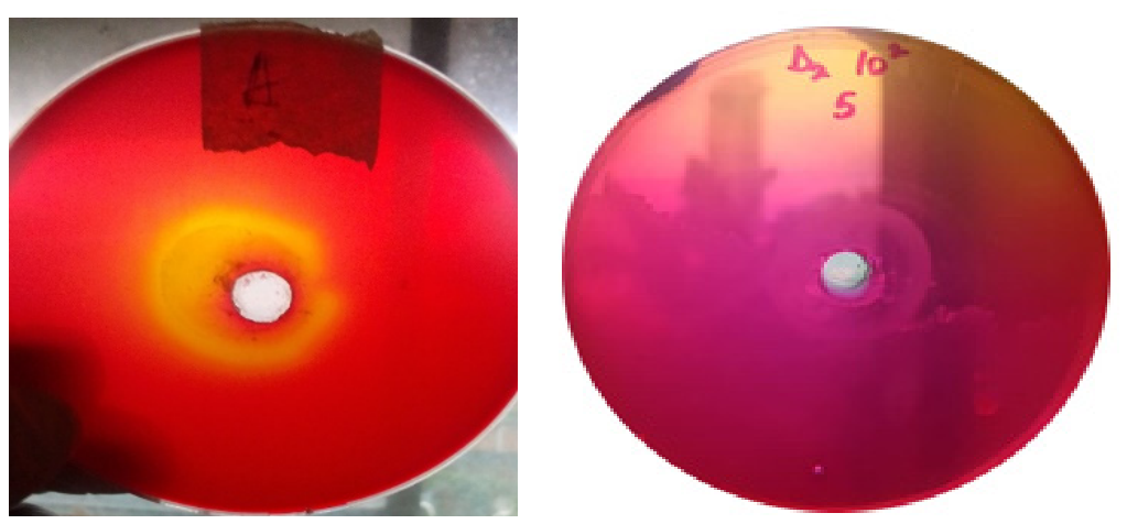
Phenol red agar plates were prepared for qualitative screening of lipolytic activity using a modified olive oil–phenol red indicator medium. The medium consisted of peptone (10 g/L), NaCl (5 g/L), CaCl2·5H2O (1 g/L), phenol red (0.1 g/L), olive oil (20 mL/L), and agar (20 g/L) dissolved in distilled water. The pH was adjusted to 7.4 using sterile 0.1 M NaOH before autoclaving at 121 °C for 15 min. After sterilization, the medium was cooled to approximately 45–50 °C, and sterile olive oil was aseptically emulsified into the molten agar with vigorous stirring to ensure uniform substrate dispersion before pouring into sterile Petri dishes. After solidification, wells measuring 5 mm in diameter were aseptically punched into the agar using a sterile cork borer. Each well was inoculated with 10 µL of an overnight bacterial culture. Plates were incubated at 37 °C for 24–48 h. Each well was inoculated with 10 µL of a standardized overnight bacterial suspension prepared by culturing individual isolates in nutrient broth at 37 °C for 18–24 h with shaking (150 rpm). The cultures were adjusted spectrophotometrically to an optical density of OD600 = 0.5, corresponding to approximately 1 × 10⁸ CFU/mL before screening. A known lipase-producing bacterial strain was included as a positive control to validate hydrolysis-zone development, while sterile nutrient broth and a non-lipolytic bacterial isolate were used as negative controls to confirm the specificity of the phenol red color change and exclude false-positive reactions. Following inoculation, the plates were incubated at 37 °C for 24–48 h, after which lipolytic activity was assessed by measuring the diameter of yellow hydrolysis zones surrounding each well [1]. Lipase production was identified by the appearance of distinct yellow zones surrounding the inoculated wells, resulting from fatty acid release during olive oil hydrolysis, which lowered the local pH and induced a phenol red colour shift from red to yellow. The diameter of the hydrolysis zone was measured as an indicator of relative lipolytic activity. All experiments were performed in triplicate [13].

2.2. Quantitative Assay for Lipase Production and Selection of the Highest-Producing Isolate

Following preliminary qualitative screening on phenol red agar plates, all isolates exhibiting visible hydrolysis zones were subjected to quantitative lipase production analysis in submerged fermentations to identify the highest-producing strain.
A loopful of each selected bacterial isolate was inoculated into 50 mL sterile nutrient broth and incubated at 37 °C for 18–24 h with shaking (150 rpm) to prepare seed cultures. Thereafter, 5% (v/v) of each inoculum standardized by spectroscopy was transferred into 100 mL Erlenmeyer flasks containing 50 mL production medium composed of glucose (8 g/L) and beef extract (3.1 g/L), supplemented with MgSO4·7H2O (2.03 mM), FeSO4 (0.36 mM), K2HPO4 (1.0 mM), and KH2PO4 (1.48 mM), and distilled water, adjusted to pH 7.0 before sterilization [2]. Fermentation was carried out at 37 °C for 48 h under agitation at 150 rpm. At the end of incubation, cultures were centrifuged at 10,000× g for 15 min at 4 °C, and the clear supernatant was collected as the crude extracellular enzyme extract for lipase assay [14].

2.3. Identification of the Highest Lipase-Producing Bacterial Isolate

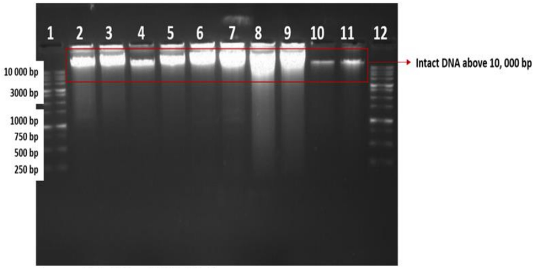
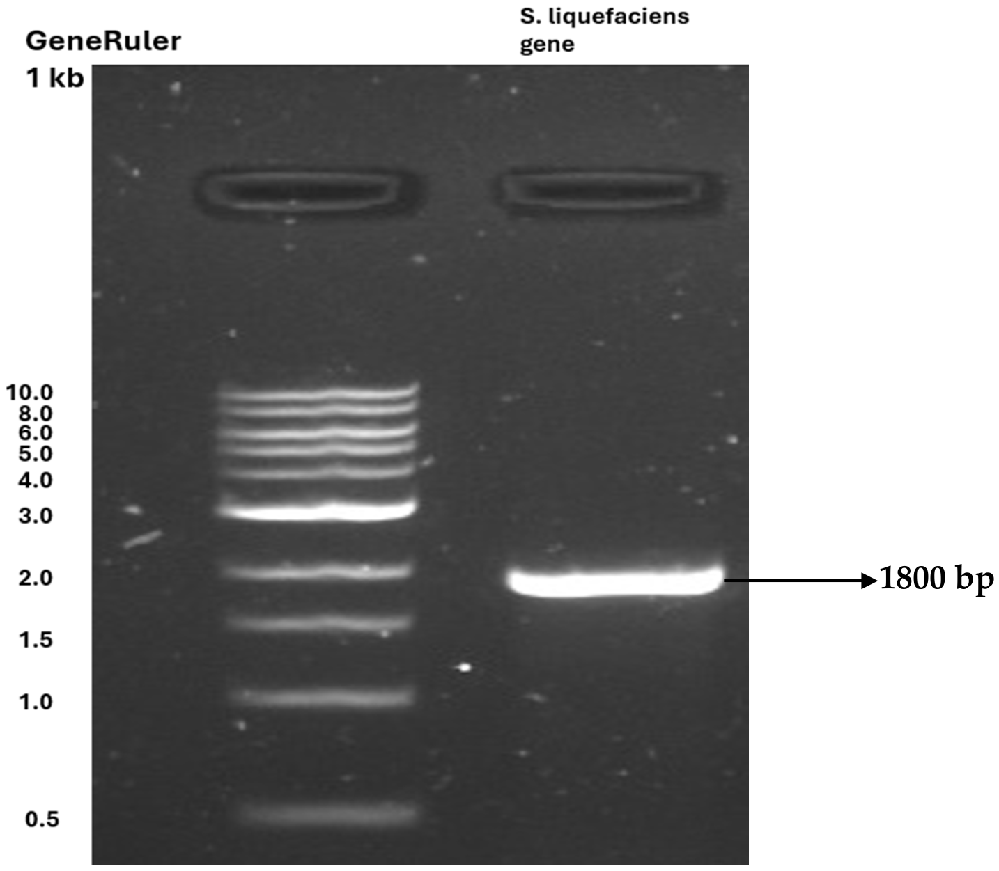
The bacterial isolate exhibiting the highest extracellular lipase activity during quantitative submerged fermentation (Section 2.2) was selected for molecular identification. Selection was based on enzymatic activity (U/mL) measured using the p-nitrophenyl palmitate (pNPP) assay, which provided a quantitative screening of extracellular lipolytic potential. The selected isolate was cultured in Luria–Bertani (LB) broth (Merck, Rahway, NJ, USA) at 37 °C for 24 h under shaking conditions to obtain sufficient biomass. Genomic DNA was extracted using the Quick-DNA Fungal/Bacterial Miniprep Kit (Zymo Research, Irvine, CA, USA) following the manufacturer’s protocol. DNA concentration and purity were assessed using a NanoDrop 2000 spectrophotometer (Thermo Fisher Scientific, Waltham, MA, USA). DNA integrity was verified by electrophoresis on a 1% agarose gel stained with ethidium bromide. A 1000 bp DNA ladder (Fermentas, Waltham, MA, USA) was used as a molecular size marker. Electrophoresis was performed at 120 V for approximately 1 h, and DNA bands were visualized under UV illumination using a Gel Doc XR system (Bio-Rad, Hercules, CA, USA). Molecular identification was performed based on amplification of the bacterial 16S rRNA gene using universal primers 27F (5′-AGAGTTTGATCMTGGCTCAG-3′) and 1492R (5′-CGGTTACCTTGTTACGACTT-3′). These primers were selected because they target highly conserved regions at the 5′ and 3′ ends of the bacterial 16S rRNA gene, enabling amplification of nearly full-length (~1.8 kb) sequences suitable for accurate taxonomic classification and phylogenetic analysis across a broad range of bacteria.
PCR amplification was carried out in a PCR thermal cycler (Techne, Minneapolis, MN, USA) under the following conditions: initial denaturation at 95 °C for 5 min, followed by 30 cycles of denaturation at 95 °C for 30 s, annealing at 58 °C for 30 s, extension at 72 °C for 1 min, and a final extension at 72 °C for 10 min. The PCR product was verified by agarose gel electrophoresis before DNA sequencing. The confirmed 16S rRNA PCR amplicon was purified and subjected to Sanger sequencing by Inqaba Biotech, South Africa. The obtained sequence was assembled and quality-checked before being compared against reference sequences in the National Center for Biotechnology Information (NCBI) database using BLASTn 2.17.0 to determine the closest taxonomic relatives based on sequence similarity [15]. Multiple sequence alignment of the obtained sequence and closely related reference sequences was performed using ClustalW implemented in MEGA 12 (v.12.1) [14]. A phylogenetic tree was constructed using the Neighbor-Joining method under the Tamura–Nei substitution model. The robustness of the phylogenetic tree was evaluated using 1000 bootstrap replicates, providing statistical support for the inferred evolutionary relationships [16].

2.4. Strategic Optimization of Conditions for Enhanced Lipase Production

Optimization of nutrient and physicochemical parameters for lipase production was performed under submerged fermentation using a one-factor-at-a-time (OFAT) approach, in which individual variables were systematically varied. At the same time, all other conditions were kept constant, enabling a clear assessment of each factor’s specific influence on enzyme yield [17]. The effects of incubation time (up to 4 days), diverse lipid inducers (waste cooking oil, olive oil, groundnut oil, palm oil, coconut oil, palm kernel oil, and orange oil), as well as varying pH (3–11) and temperature (27–60 °C), were systematically investigated to elucidate their individual influences on lipase production. In addition, the effects of different carbon sources (maltose, glucose, galactose, fructose, dextrose, lactose, and sucrose) and nitrogen sources (ammonium sulfate, ammonium nitrate, tryptone, yeast extract, peptone, and beef extract) on lipase production were systematically evaluated. Lipase production was conducted in 250 mL Erlenmeyer flasks using 1% (v/v) waste frying oil as the sole lipid inducer and primary carbon source. The basal medium was composed of glucose (8 g/L) and beef extract (3.1 g/L), supplemented with MgSO4·7H2O (2.03 mM), FeSO4 (0.36 mM), K2HPO4 (1.0 mM), and KH2PO4 (1.48 mM), providing a balanced nutritional environment to support optimal microbial growth and enzyme synthesis [18]. The medium was sterilized in an autoclave for an extended time of 30 min. It was inoculated with an actively growing 18–24 h bacterial culture and incubated at 37 °C under continuous agitation at 150 rpm for 76 h. Following incubation, the culture was centrifuged at 8000× g for 20 min at 4 °C to separate the cells from the extracellular components. The resulting cell-free supernatant was carefully collected and used for subsequent downstream analyses. Lipase production under the different cultivation conditions was evaluated based on extracellular lipase activity (U mL−1) determined from the culture supernatant. Biomass determination (optical density at 600 nm or dry cell weight) was not included as part of the experimental design; therefore, enzyme production was assessed on a volumetric basis rather than being normalized to cell biomass.

2.5. Lipase Activity Assay and Protein Estimation

The crude enzyme extract was evaluated for extracellular lipase activity using p-nitrophenyl palmitate (pNPP) as the chromogenic substrate, enabling sensitive and quantitative detection of enzymatic hydrolysis, as described by [19]. After 24 h of incubation, the cultures were centrifuged at 8000× g for 20 min to separate the cells from the medium. The resulting cell-free supernatant was carefully harvested and used as the crude source of extracellular enzymes. Lipase activity was subsequently quantified spectrophotometrically at 410 nm using p-nitrophenyl palmitate (pNPP) as the substrate. For the enzyme blank, 900 µL of substrate solution was combined with 100 µL of enzyme production medium, and the absorbance was measured under identical conditions. Lipase activity was then determined using a calculation based on the amount of p-nitrophenol released from substrate hydrolysis, providing a quantitative measure of enzymatic activity [15]. Substrate specificity of the purified SlipB lipase was evaluated using three p-nitrophenyl ester substrates of varying acyl-chain lengths: p-nitrophenyl laurate (C12), p-nitrophenyl palmitate (C16), and p-nitrophenyl stearate (C18). Each substrate was assayed independently under identical reaction conditions. Briefly, substrate solutions were prepared in isopropanol and emulsified in Tris–HCl buffer (50 mM, pH 8.0) containing 0.5% (v/v) Triton X-100 to ensure uniform dispersion. The reaction mixture consisted of appropriately diluted purified enzyme and substrate solution, followed by incubation at 37 °C for 15 min. p-Nitrophenyl palmitate (pNPP), p-nitrophenyl stearate (pNPS), and p-nitrophenyl laurate (pNPL) are synthetic chromogenic substrates commonly used in lipase assays to quantitatively evaluate enzymatic activity by measuring the release of the p-nitrophenol product upon hydrolysis. Enzymatic hydrolysis of these esters releases p-nitrophenol, which was quantified spectrophotometrically.
A p-nitrophenol standard calibration curve was constructed using concentrations ranging from 10 to 110 µM, and absorbance was recorded at 410 nm (see Figure A6) to enable accurate activity determination. Lipase activity was determined spectrophotometrically by measuring the release of p-nitrophenol at 410 nm. One unit (U) of enzyme activity was defined as the amount of enzyme required to release 1 µmol of p-nitrophenol per minute under assay conditions. Relative substrate specificity was calculated by comparing enzyme activity obtained with each substrate with activity toward p-nitrophenyl palmitate (pNPP), designated as 100% reference activity. This allowed comparative assessment of substrate-chain-length preference and catalytic selectivity. All assays were performed in triplicate using independent replicates, and results were expressed as mean ± standard deviation.
A c t i v i t y   ( U / m L ) = A s A c c m × D f ( V × T )
  • Definitions:
  • As: Absorbance of test sample at 410 nm
  • Ac: Absorbance of the control at 410 nm
  • Df: Dilution factor
  • V: Total volume of reaction
  • T: Total reaction time
  • c: Y-intercept of the p-nitrophenol standard curve
  • m: Gradient of the p-nitrophenol standard curve

2.6. Enzyme Purification

A 100 mL cell-free supernatant aliquot was subjected to protein precipitation by the gradual addition of solid ammonium sulfate to attain 70% saturation, under continuous magnetic stirring at 4 °C. The mixture was centrifuged at 8000× g for 20 min at 4 °C using a high-speed refrigerated centrifuge to separate the precipitated proteins from the supernatant. The resulting precipitate was collected for dialysis, while the protein-free supernatant was discarded. The recovered pellets were resuspended in 20 mL of 0.50 mM Tris–HCl buffer (pH 8.0) and dialysed overnight at 4 °C against three successive changes of the same buffer to ensure complete equilibration and removal of low-molecular-weight impurities. The resulting concentrated dialysate was then carefully applied to a CM-Sephadex C-50 ion-exchange column (1.5 × 20 cm) manufactured by Cytiva in Marlborough, MA, USA, pre-equilibrated with 50 mM Tris–HCl buffer (pH 8.0), to enable selective protein adsorption based on charge interactions [19]. A linear gradient of 0–1.0 M NaCl in 50 mM Tris–HCl buffer (pH 8.0) was applied to elute the bound proteins from the column. Elution was continuously monitored by measuring absorbance at 280 nm to track protein release. The collected fractions were then assayed for lipase activity using a UV–Vis spectrophotometer to identify catalytically active eluates [19]. Finally, the protein was further purified by gel filtration chromatography using a Sephadex G-100 column. Before application, the pre-swollen Sephadex G-100 resin was carefully equilibrated with 50 mM Tris–HCl buffer (pH 8.0) to ensure optimal column performance and high-resolution separation efficiency [2]. The equilibrated resin was gently packed into a column (1.5 × 65 cm), after which the enzyme preparation was carefully applied. Elution was performed, and 5 mL fractions were systematically collected. Each fraction was subsequently analysed for lipase activity and protein concentration, following the same analytical procedures used in the earlier purification stages. Each fraction was analyzed by SDS-PAGE to evaluate both the yield and the purity of the purified protein [20].

SDS-PAGE Analysis

SDS-PAGE was carried out [7] to estimate the molecular weight of the enzyme and visualise and evaluate protein purification efficiency. Before SDS-PAGE analysis, the samples (40 µg in 15 µL distilled water) were prepared by mixing them with 5 µL of sample buffer (25 mM Tris-HCl buffer, pH 8.0, 2% SDS, 2% dithiothreitol, 20% glycerol and 0.02% bromophenol blue). The samples were then heated at 100 °C for 5 min and cooled at room temperature. Samples were loaded on the gel (5% stacking gel and 10% resolving gel) and electrophoresed at 20 mA per gel at room temperature until the dye front reached the end of the gel. Coomassie Brilliant Blue R250 (0.25% dye, 10% acetic acid, 45% ethanol in distilled water) was used to stain the gels. The gels were destained (7.5% acetic acid, 5% ethanol in distilled water) and photographed with a Gel Doc XR system (Bio-Rad).

2.7. Characterization of Lipase

2.7.1. Determination of Temperature and pH Optima and Assessment of Enzyme Stability

The purified Lipase AB1 was systematically characterized to evaluate its catalytic performance across a range of temperature and pH conditions. Optimal temperature was determined by assaying lipase activity using the p-nitrophenyl palmitate (pNPP) substrate across a temperature gradient of 10–90 °C at pH 8.0, with each reaction standardized to a 15-min incubation period [19]. The optimum pH for lipase activity was determined across a broad pH range of 3 to 10 at 60 °C. Briefly, 900 µL of p-nitrophenyl palmitate (pNPP) substrate was prepared in buffer systems spanning the selected pH range and pre-incubated at 60 °C for 15 min to ensure thermal equilibration. Thereafter, 100 µL of enzyme solution was added to initiate the reaction, which was allowed to proceed for a further 15 min under the same conditions. Enzyme activity was subsequently quantified based on product formation. For each pH condition, a corresponding reaction mixture containing buffer in place of the enzyme served as the blank to account for background absorbance [19]. The experiments were carried out in triplicate.

2.7.2. Influence of Organic Solvents on Lipase Activity

The influence of organic solvents on lipase activity was systematically investigated using methanol, ethanol, n-butanol, ethyl acetate, isopropanol, dichloromethane, hexane, and toluene. Each solvent was incorporated into the reaction system at final concentrations of 10% and 50% (v/v) to assess concentration-dependent effects on enzymatic performance. Enzyme activity measured in the absence of any organic solvent was defined as 100% and used as the control reference for all comparative analyses [21].

2.7.3. Influence of Metal Ions on Lipase Activity

The effect of metal ions on lipase activity was examined using the chloride salts of sodium, potassium, barium, calcium, magnesium, manganese, and silver. Each metal ion was introduced into the reaction system to achieve final concentrations of 5 mM, 10 mM, and 20 mM, and the resulting enzymatic activity was subsequently quantified under each condition. Enzyme activity measured in the absence of metal ions was designated as 100% and used as the baseline reference for all comparative evaluations [22].

2.7.4. Determination of Kinetic Parameters

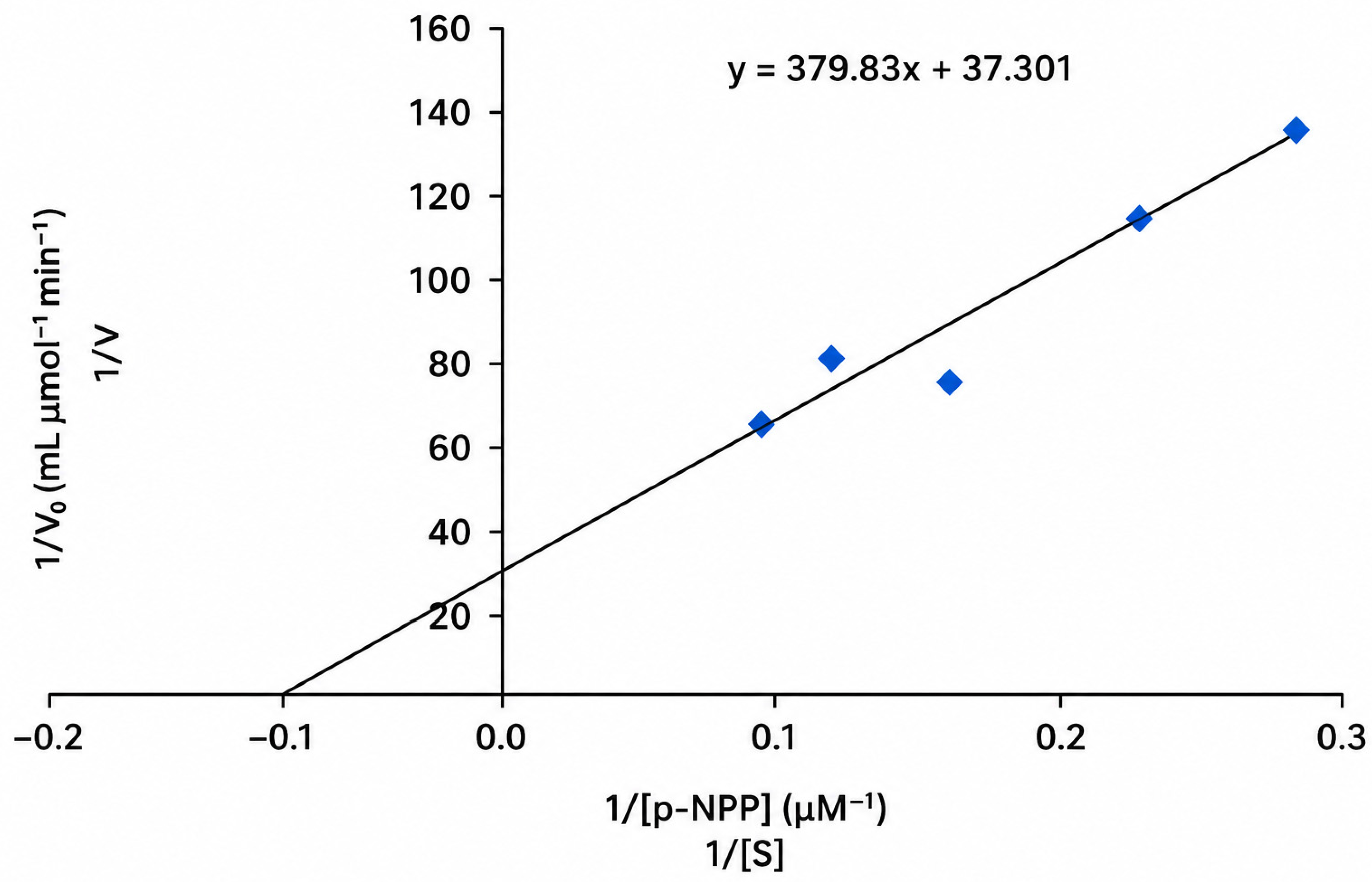
The apparent kinetic parameters (Km and Vmax) of lipase for pNPP were determined by varying the concentration of pNPP between 0.005 and 0.07 mM in 50 mM Tris-HCl buffer, pH 8.0, and measuring the initial reaction velocities (µmol/mL/min) at a fixed volume of the enzyme. First, a 0.3 mM stock solution of the substrate (pNPP) was prepared by dissolving 0.001 g of pNPP in 1 mL of isopropanol and adding it to a solution containing 0.01 g of gum arabic, 50 µL of Triton X-100, and 9 mL of Tris-HCl buffer, pH 8.0. Each reaction mixture for initial velocity determination contained an appropriate volume of the pNPP (taken from the 0.3 mM stock solution) in 50 mM Tris-HCl buffer, pH 8.0, and 300 µL of the enzyme. The reaction rate was determined for each concentration of pNPP, and the data were plotted according to [23,24,25].

2.7.5. Statistical Analysis

Statistical analyses were performed using one-way analysis of variance (ANOVA) in OriginPro 2025 and Microsoft Excel to evaluate differences among experimental groups. All data are presented as mean ± standard deviation (n = 3), ensuring reproducibility and reliability of the results. Statistical significance was defined at p < 0.05. Where applicable, distinct superscript letters (a, b, c, d, e) were used to denote significant differences between means, as determined by one-way ANOVA followed by Tukey’s honestly significant difference (HSD) post hoc test for multiple comparisons. Error bars were used to represent the standard deviation of three independent replicates. Statistical significance was calculated using replicate values; technical repeats were averaged before analysis (p < 0.05).

3. Results

3.1. Screening and Identification of the Lipolytic Bacterial Isolate

In this study, 47 bacterial isolates from petroleum-contaminated soil were screened, and 10 exhibited clear zones on phenol red agar (see Table A1 and Figure A1), indicating lipase production. Table 1 shows lipase activities from both cultures. The isolate demonstrating the highest lipase activity was designated as S. liquefaciens AB1. Based on 16S rRNA gene sequence analysis and BLAST comparison, the isolate was assigned to the Serratia liquefaciens complex, as members of this complex exhibit highly conserved 16S rRNA gene sequences that cannot be reliably resolved to the species level (see Figure A2 and Figure A3). Consequently, definitive identification as S. liquefaciens would require higher-resolution genomic approaches, such as average nucleotide identity (ANI), multilocus sequence typing (MLST), or whole-genome sequencing.

3.2. The Effect of Cultivation Conditions on Lipase Production in S. liquefaciens AB1

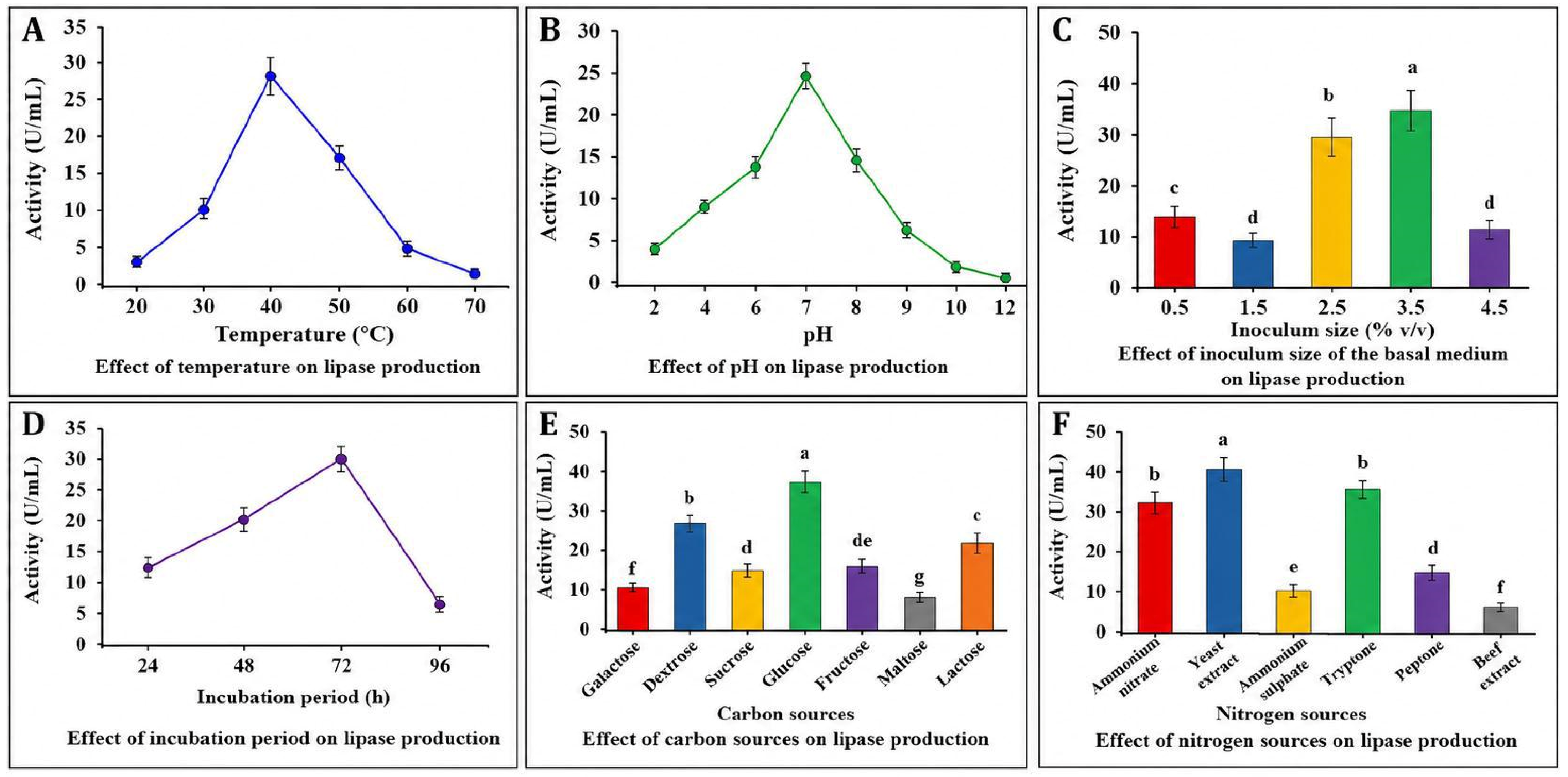
The growth and lipase production of S. liquefaciens AB1 were systematically characterized under various conditions. Lipase activity was first detecfiguret 24 h of cultivation, exhibiting a marked increase by 48 h. Maximum enzyme production (40.7 U/mL) was achieved at 72 h, after which activity gradually declined (Figure 1).
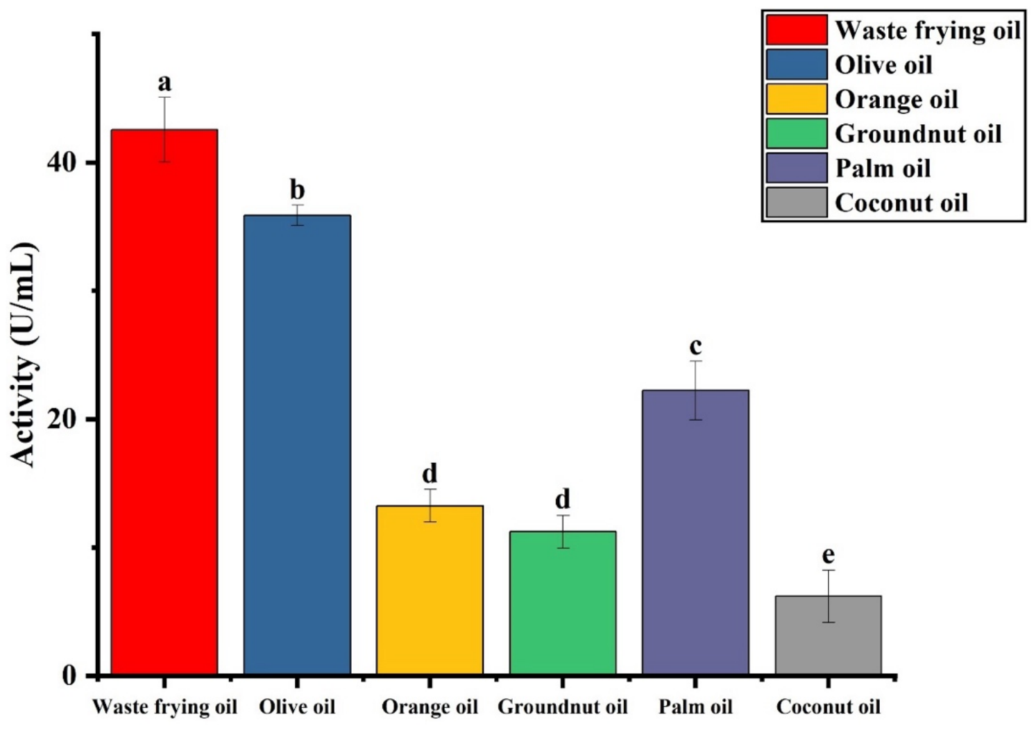
Among the vegetable oils assessed as inducers of lipase production, waste cooking oil (WCO) emerged as the most potent enhancer of enzyme synthesis, while coconut oil exhibited the weakest induction potential (Figure 2). The enzyme displayed optimal catalytic performance at pH 7.0, achieving 25.4 U/mL, with activity sharply declining under highly alkaline conditions, reaching a minimum of 2.8 U/mL at pH 11.0. Optimization of the cultivation conditions demonstrated that the highest lipase production by the isolate occurred at 37 °C (30.7 U/mL), indicating that this temperature was optimal for enzyme biosynthesis under the fermentation conditions employed. This observation should not be interpreted as the catalytic optimum of the enzyme itself, which was determined separately using the purified enzyme preparation. In contrast, enzymatic activity diminished drastically at elevated temperatures, dropping to a residual 0.3 U/mL at 62 °C, indicating pronounced thermal sensitivity. Evaluation of carbon source utilization demonstrated that glucose most effectively supported lipase production, yielding the highest activity of 35.8 U/mL. This was followed by fructose (12.6 U/mL) and galactose (10.1 U/mL), while maltose proved least favorable, supporting only 5.9 U/mL. Collectively, these results highlight the enzyme’s clear dependence on specific nutritional and physicochemical conditions for optimal expression and activity. For nitrogen sources, yeast extract produced the highest enzyme activity (40.7 U/mL), followed by ammonium nitrate (32.5 U/mL), whereas ammonium sulfate supported the lowest activity (2.7 U/mL). The optimal inoculum size was 2.5 mL, resulting in 40.7 U/mL lipase activity, while the largest inoculum tested (3.5 mL) yielded the lowest activity (5.4 U/mL) (Figure 1). This study underscores how growth conditions, the choice of carbon and nitrogen sources, and inoculum size collectively shape the lipase production profile of S. liquefaciens AB1.

3.3. Purification of Serratia liquefaciens Lipase

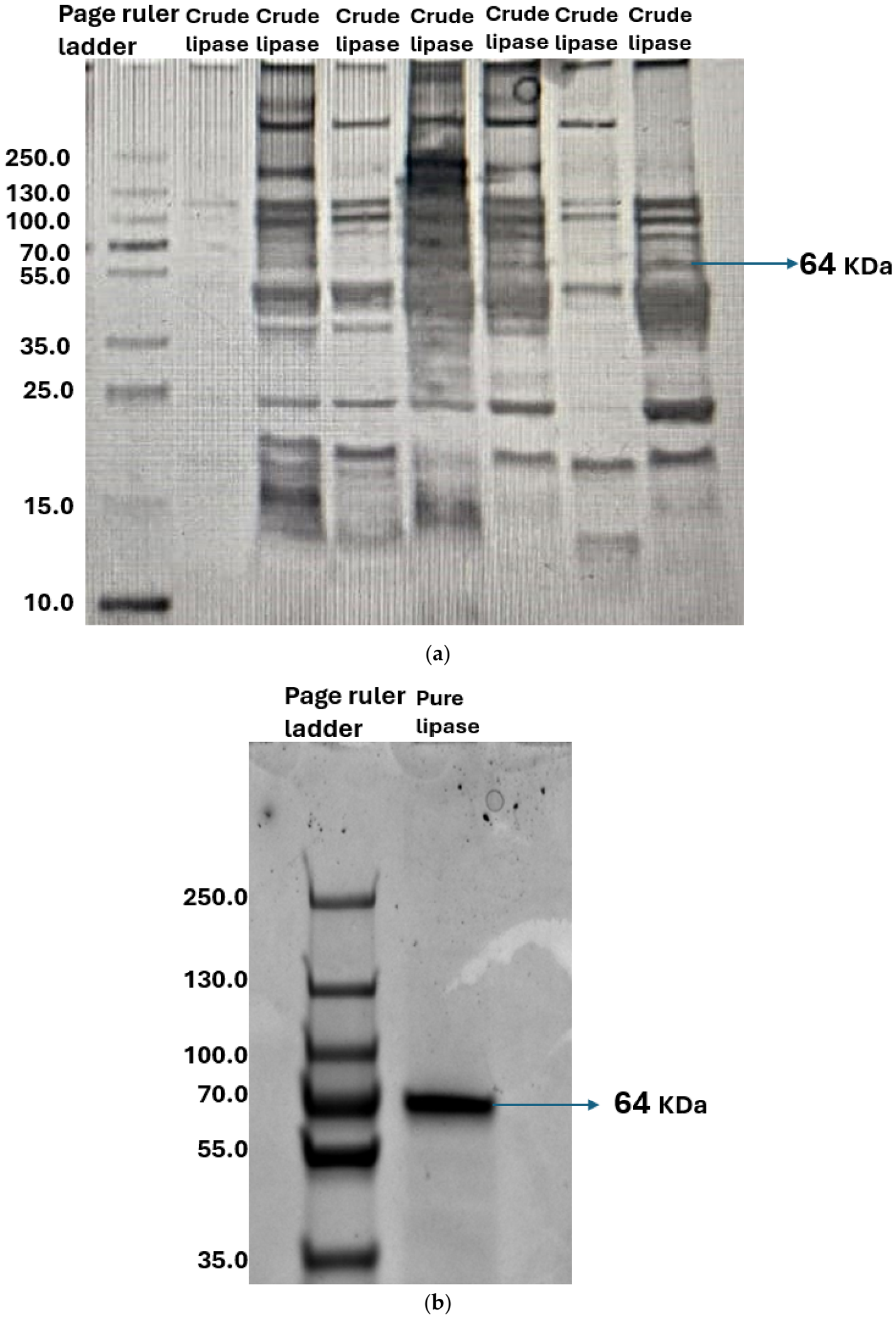
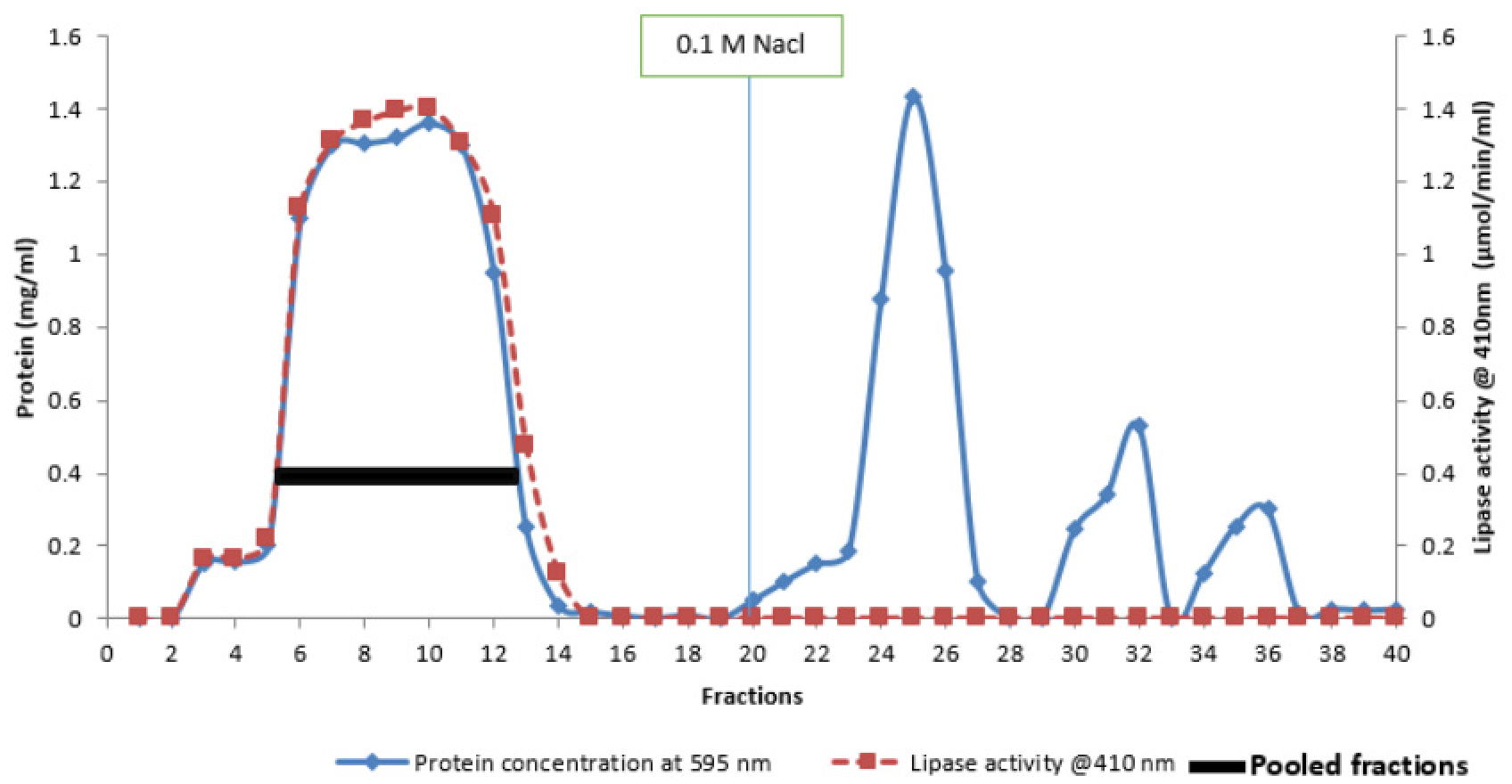
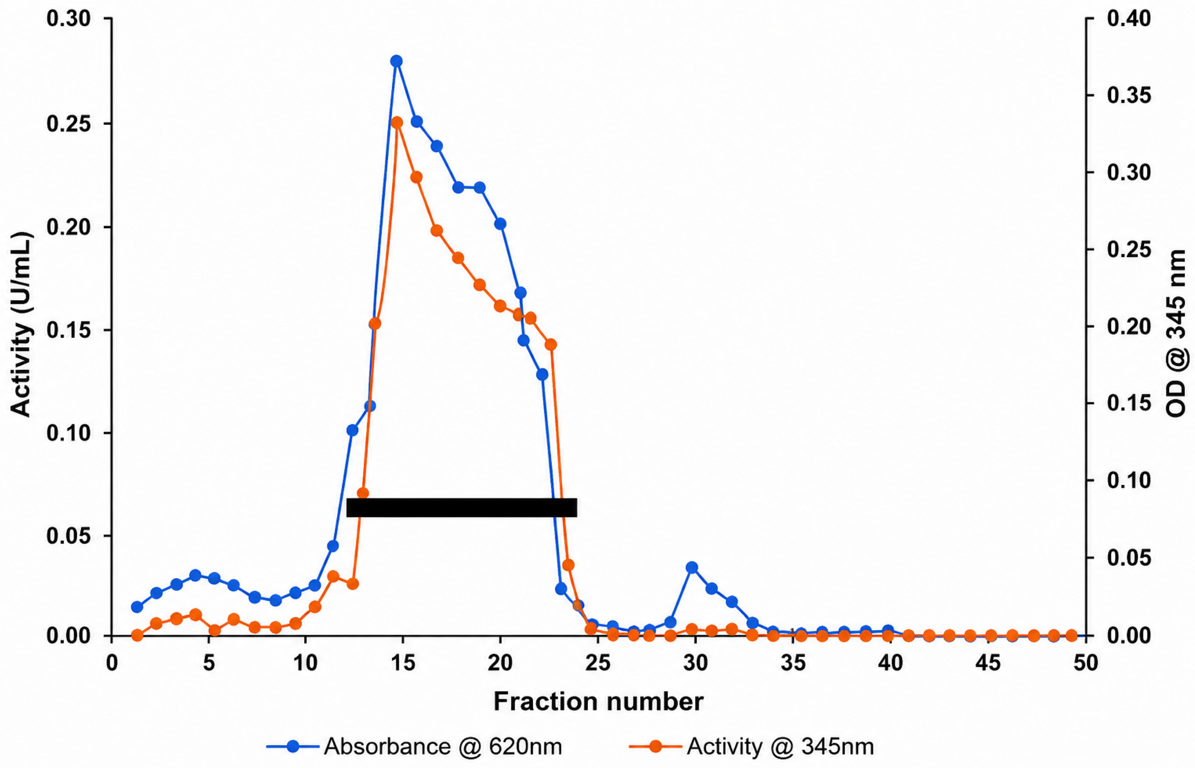
Ion-exchange chromatography resulted in a single (see Figure A4), well-defined lipase activity peak, indicating effective purification of the enzyme. This step achieved a 6.3-fold increase in purity with a 19.26% recovery, and the active fractions were carefully pooled for subsequent refinement. Further purification by gel filtration chromatography also yielded a single, distinct activity peak (see Figure A5), confirming the successful isolation of the target enzyme. This step yielded a final protein concentration of 15.27 mg/mL and a total enzymatic activity of 9328.8 U/mL in 5 mL. The purified lipase AB1 demonstrated an enhanced specific activity of 610.92 U/mg, corresponding to a 9.42-fold purification with an overall recovery of 76%. Together, these results highlight the efficiency and selectivity of the purification strategy employed for S. liquefaciens lipase AB1. A detailed overview of the purification workflow and associated metrics is presented in Table 2. The purified lipase analysis by Electrophoretogram are shown in Figure 3a,b.

3.4. Biochemical Characterization of Lipase AB1

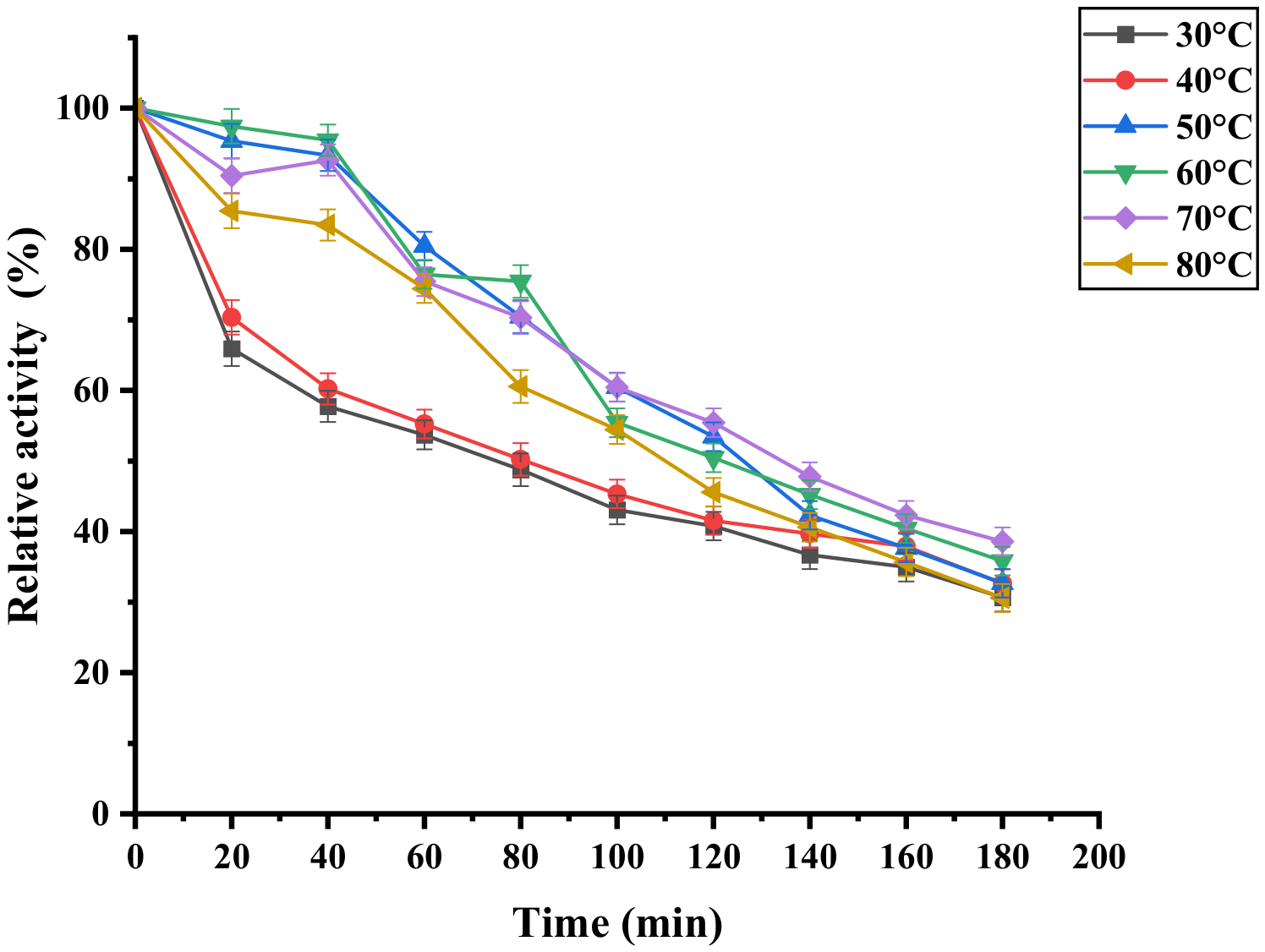
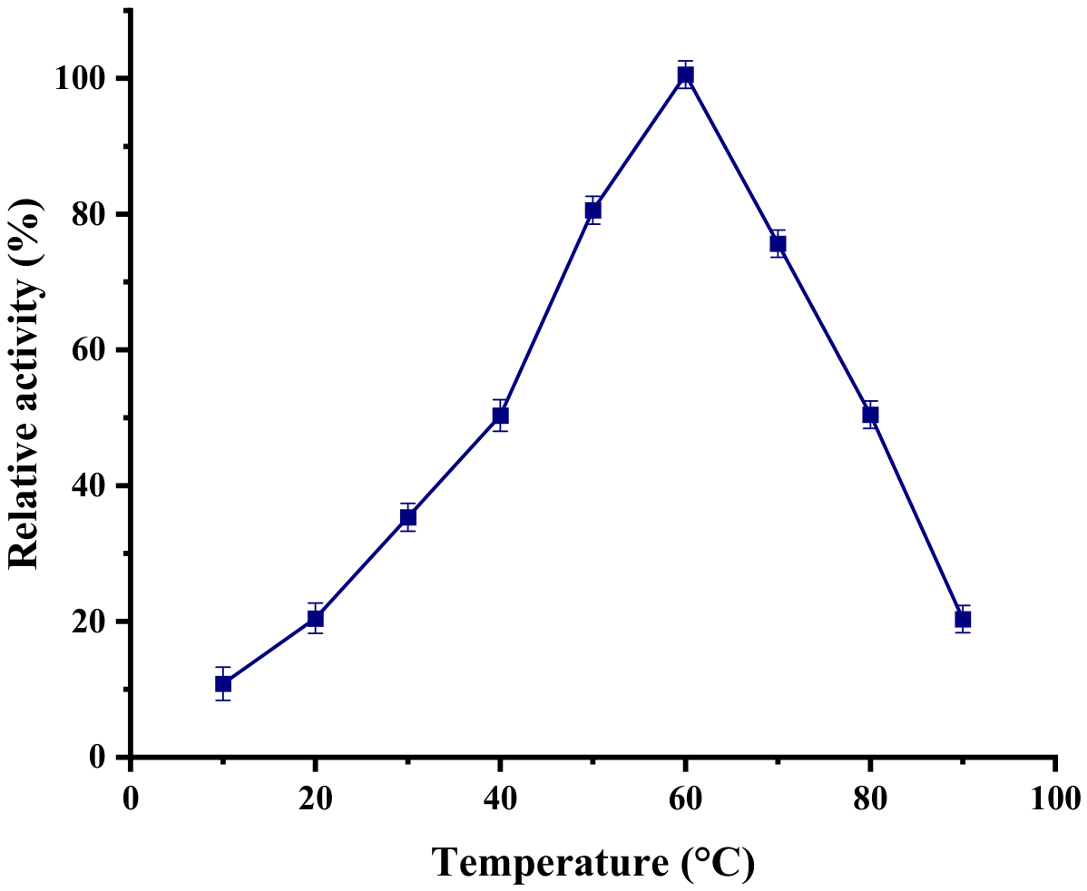
The lipase from S. liquefaciens AB1 (LipAB1) exhibited an optimal catalytic temperature of 60 °C, while retaining appreciable activity across a broad range of 40 °C to 80 °C. The effect of temperature on the catalytic activity of the purified lipase was determined by performing the enzyme assay at temperatures ranging from 20 to 80 °C under otherwise identical reaction conditions (Figure 4). The enzyme exhibited activity over the entire temperature range tested, with catalytic activity increasing progressively from 20 to 60 °C and reaching a maximum at 60 °C. Beyond this temperature, enzyme activity declined, indicating reduced catalytic efficiency at elevated temperatures. The optimum temperature of 60 °C, therefore, represents the temperature at which the enzyme displayed its maximum instantaneous catalytic activity during the assay and should not be interpreted as an indication of prolonged thermal stability. The thermal stability of the purified lipase was evaluated by pre-incubating the enzyme at different temperatures (30–80 °C) for up to 180 min in the absence of substrate, after which the residual activity was determined under the standard assay conditions (Figure 5). Unlike the temperature optimum experiment, which measures catalytic activity directly at different assay temperatures, this experiment assesses the enzyme’s ability to retain activity following prolonged exposure to elevated temperatures. The purified lipase exhibited a gradual decline in residual activity with increasing incubation time at all temperatures tested. Activity retention was higher during the initial stages of incubation at 50–70 °C than at 30 and 40 °C, whereas prolonged incubation resulted in a progressive loss of activity across all temperatures. After 180 min, residual activity ranged from approximately 30–38%, indicating partial thermal inactivation following extended heat exposure. These findings demonstrate that although the enzyme exhibits maximum catalytic activity at 60 °C during short-term assays, its activity decreases progressively upon prolonged incubation at this and other temperatures, reflecting the distinction between optimum catalytic conditions and thermal stability.
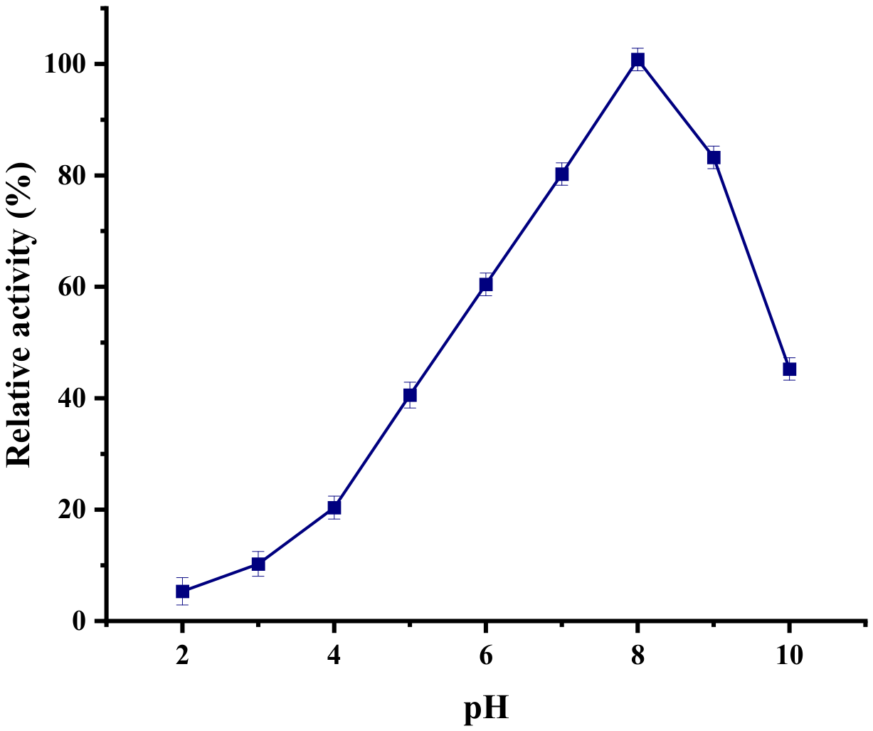
S. liquefaciens lipase AB1 (LipAB1) showed optimal activity at pH 8, functioning well within pH 6.0–7.0 as shown in Figure 6. The effect of pH on the catalytic activity of the purified lipase was determined by performing the standard enzyme assay in buffers ranging from pH 2 to pH 10 under otherwise identical reaction conditions (Figure 6). The enzyme exhibited activity over a broad pH range, with maximum catalytic activity observed at pH 8.0. Enzyme activity decreased progressively at pH values above and below the optimum. The optimum pH therefore represents the condition at which the enzyme exhibited the highest catalytic efficiency during the assay and does not necessarily reflect its stability following prolonged exposure to different pH conditions.
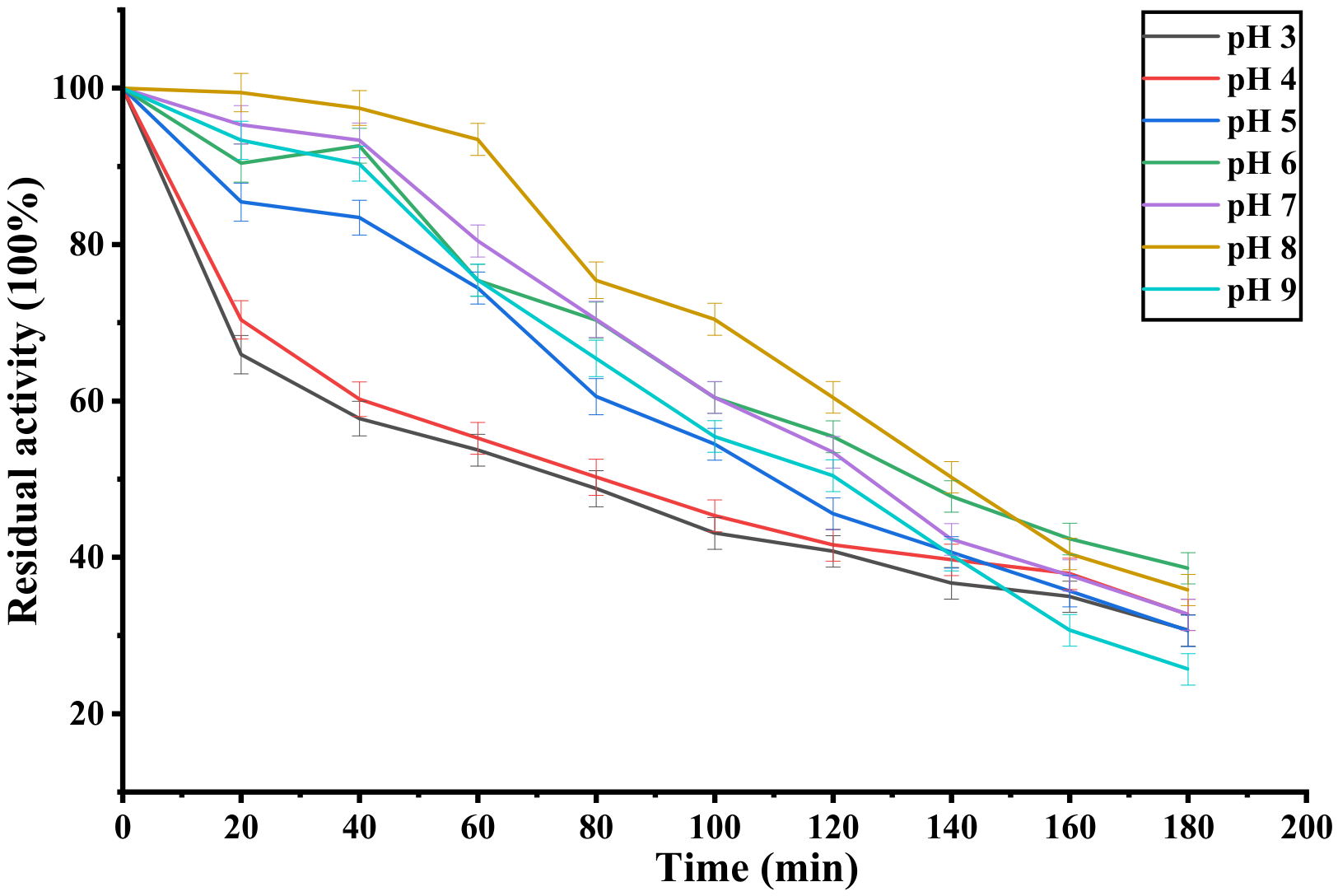
The pH stability of the purified lipase was evaluated by pre-incubating the enzyme in buffers of varying pH for the specified period, after which the residual activity was determined under the standard assay conditions (Figure 7). Unlike the pH optimum experiment, this assay measures the enzyme’s ability to retain catalytic activity following prolonged exposure to different pH environments. The enzyme retained substantial activity over a broad pH range but exhibited a gradual decline in residual activity following prolonged incubation under both acidic and alkaline conditions. These findings indicate that although the enzyme displays maximum catalytic activity at pH 8.0 during short-term assays, prolonged exposure to different pH conditions results in partial loss of activity, demonstrating that optimum catalytic pH and pH stability represent distinct biochemical properties.

3.4.1. Substrate Specificity

The substrate specificity analysis revealed that LipAB1 exhibits a marked preference for p-nitrophenyl palmitate (pNPP), while displaying minimal activity toward p-nitrophenyl laurate (pNPL), as summarized in Table 3. Comparative evaluation of enzyme activity across the tested p-nitrophenyl (pNP) ester substrates (pNPP, pNPL, and pNPS) demonstrated that the enzyme achieved its highest relative activity (91%) with pNPP. This pronounced affinity for pNPP, a long-chain fatty acid ester, underscores the enzyme’s substrate selectivity and strongly supports its classification as a true lipase. Such enzymes are characteristically adept at catalyzing the hydrolysis of long-chain triglycerides, highlighting the catalytic efficiency and functional relevance of LipAB1 in lipid transformation processes. Activity was lower when pNPL was used, with a relative activity of 77%, demonstrating moderate affinity for medium-chain substrates. The lowest activity was observed with pNPS, a short-chain ester, where the enzyme showed only 42% activity. The statistical letters a, b, and c indicate the outcome of a post hoc multiple comparison test (commonly Tukey’s HSD) performed after a one-way ANOVA to determine whether the relative activities differ significantly among the three substrates.

3.4.2. Influence of Organic Solvents on Lipase Activity

In this study, the effect of different organic solvents on the lipolytic activity of LipAB1 was investigated (Table 4), which showed varying stabilities across the hydrophobic and hydrophilic solvents tested. The highest activity was observed in isopropanol, which is a hydrophilic solvent compared to the most hydrophobic solvent used (hexane). The activity of lipase was enhanced by 10% isopropanol, reaching 140% relative activity. Lipase also retained more than 70% residual activity in 10% methanol and 10% ethanol. The enzyme lost over 70% of its activity in the presence of 10% n-butanol and hexane.

3.4.3. Influence of Salt Ions on Lipase Catalytic Activity

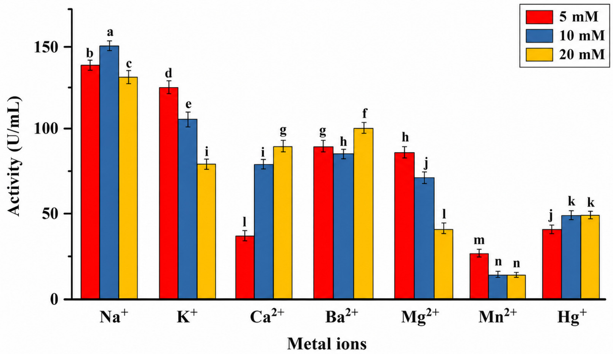
The effects of chloride salts of selected monovalent (Na+, K+) and divalent (Ca2+, Ba2+, Mg2+, Mn2+, Hg2+) metal ions on the catalytic performance of lipase AB1 were systematically investigated. The enzyme responded differentially depending on both ion type and concentration, revealing a clear pattern of activation and inhibition. Among all tested ions, Ca2+ emerged as the most potent activator, consistently enhancing lipase activity across all concentrations, with maximal stimulation observed at 10 mM (Figure 8). Similarly, K+ promoted enzyme activity at lower concentrations (5 and 10 mM); however, at 20 mM, it exerted an inhibitory effect, reducing activity by approximately 25%. Na+ and Ba2+ also acted as effective activators over the tested range (5–20 mM), with Na+ at 10 mM producing the most pronounced enhancement, elevating relative activity to nearly 150%. In contrast, Mn2+ and Hg+ displayed strong inhibitory effects even at the lowest concentrations (5 and 10 mM), reflecting their disruptive interaction with the enzyme’s catalytic architecture. Mg2+ showed a concentration-dependent decline in activity, with noticeable inhibition at 20 mM, while Mn2+ completely abolished enzymatic activity at this concentration.

3.4.4. Lipase Kinetic Parameters

The kinetic parameters of LipAB1 were calculated from the Lineweaver–Burk double-reciprocal plot generated from varying concentrations of p-nitrophenyl palmitate. The linear regression analysis showed a strong goodness-of-fit (R2 = 0.98xx), indicating that the Michaelis–Menten model accurately describes the catalytic behaviour of LipAB1 under the assay conditions employed. The high correlation coefficient validates the reliability of the calculated Km and Vmax values and confirms consistent substrate–enzyme interaction across the tested concentration range. A strong linear fit further suggests minimal experimental deviation and supports the interpretation that LipAB1 follows classical saturation kinetics typical of true lipases.

4. Discussion

Bacterial lipases are among the most industrially relevant biocatalysts due to their catalytic versatility, operational stability, and adaptability to diverse reaction environments [2]. In the present study, S. liquefaciens AB1 isolated from petroleum-contaminated soil exhibited pronounced lipolytic activity, supporting the premise that hydrocarbon-rich environments selectively enrich for microorganisms with enhanced lipid-degrading capabilities [1]. Such ecological niches provide continuous exposure to complex hydrophobic substrates, thereby promoting the evolution and expression of robust lipases with biotechnological relevance [1].
The isolation and screening of lipase-producing bacteria from petroleum-contaminated soil underscore the ecological significance of hydrocarbon-rich environments as reservoirs of metabolically specialized microorganisms. The strong lipolytic activity observed for S. liquefaciens AB1 confirms that continuous exposure to lipidic substrates enhances the selection of strains with efficient hydrolytic systems. Although phenol red agar is widely used for the preliminary screening of lipase-producing microorganisms, changes in medium pH resulting from bacterial metabolism may contribute to color changes independently of lipid hydrolysis. To minimize the likelihood of false-positive identification, qualitative screening was used only to identify candidate isolates, and lipase production was subsequently confirmed by quantitative p-nitrophenyl palmitate (pNPP) assays before further characterization. This aligns with the concept that environmental pressure in contaminated niches drives the evolution of enzymes with improved catalytic adaptability and industrial relevance. A study by [21] isolated Serratia sp. TAN 611, a lipase-producing bacterium, was isolated from an environmentally contaminated site in China. The production kinetics (Figure 1 and Figure 2) revealed a peak in lipase activity at 72 h, followed by a gradual decline. This trend is characteristic of growth-associated enzyme synthesis, where maximal production coincides with the late exponential phase. The subsequent reduction in activity is likely due to nutrient depletion, accumulation of inhibitory by-products, or proteolytic degradation. There were variations in extracellular lipase activity under different carbon-source supplementation conditions. The highest measured activity was observed at 37 °C and pH 7.0 for 72 h with an enzyme activity of 40 U/mL, indicating that this condition supported greater detectable lipase activity in the culture supernatant. However, because biomass accumulation and carbon-source utilization were not quantified, these differences cannot be unequivocally attributed to enhanced enzyme expression at the cellular level and may partly reflect differences in bacterial growth or substrate metabolism. The effectiveness of waste frying oil as an inducer further highlights its dual role as a carbon source and a regulatory signal for lipase expression, supporting its application in cost-effective fermentation strategies. Notably, waste frying oil serves as an economical and sustainable lipid substrate for enzyme production, thereby enhancing process viability while aligning with circular economy principles that promote waste valorization and resource recycling [23]. The observed variation in lipase production in response to supplementary carbon sources should be interpreted cautiously, since waste frying oil itself constitutes a complex lipid substrate capable of serving as both a carbon source and an inducer of lipase synthesis. Thus, the effects of added carbohydrates may reflect interactions between substrate induction and metabolic regulation rather than simple carbon-source preference. Since a waste frying oil WFO-only control was not included in the present study, the relative contribution of supplementary carbon sources versus basal lipid utilization could not be fully resolved. Future work should include carbon-free supplementation controls and molecular analyses to clarify these regulatory interactions.
The effect of cultivation conditions on lipase production in S. liquefaciens AB1 (Figure 1) resulted in a 9.42-fold increase in enzyme production, demonstrating the strong influence of medium composition on metabolic output. The combined use of beef extract, glucose, and waste cooking oil provided a balanced nutrient matrix that enhanced microbial growth and enzyme secretion. These findings reinforce the feasibility of utilizing waste cooking oil to improve process efficiency while reducing production costs in large-scale applications. Fermentation optimization, particularly with suitable carbon and nitrogen sources, significantly enhances yield [2]. The present study evaluated lipase production based on volumetric enzyme activity (U mL−1), which is commonly used for the preliminary optimization of microbial enzyme production. However, biomass accumulation was not determined under the different cultivation conditions. Consequently, the observed increases in lipase activity cannot be unequivocally attributed to enhanced enzyme induction alone, as differences in cell growth may also have contributed to the measured enzyme yields. Determination of optical density or dry cell weight would enable calculation of biomass-normalized specific productivity and provide a clearer distinction between increased microbial growth and true induction of lipase biosynthesis. This represents a limitation of the present study and should be addressed in future investigations to further optimize enzyme production and improve the understanding of regulatory responses to different cultivation conditions. Although the enzyme showed the highest hydrolytic activity toward the long-chain substrate p-nitrophenyl palmitate, the present study did not include comparative assays using emulsified and non-emulsified triglyceride substrates to evaluate interfacial activation, a characteristic feature of true lipases. Therefore, while the biochemical properties observed are consistent with those of microbial lipases, additional studies employing natural triglyceride substrates and interfacial activation assays are required.
The lipase (LipAB1) from S. liquefaciens AB1 was successfully purified using a conventional stepwise purification strategy involving ammonium sulfate precipitation, ion-exchange chromatography, and gel filtration. Although the overall purification factor (9.42-fold) appears modest compared to recombinant affinity-tag systems [24], this outcome is consistent with native enzyme purification workflows where no engineered affinity handles are present and multiple host-derived proteins and isoforms may co-elute. The progressive increase in specific activity from 68.32 to 610.92 U/mg confirms effective enrichment of the target lipase throughout the purification scheme. The observed 75% recovery further indicates that the enzyme remained relatively stable during processing, which is important for downstream biochemical characterization. Higher purification yields could potentially be achieved in future work through recombinant expression systems incorporating affinity tags or by integrating additional polishing steps such as hydrophobic interaction chromatography. Nevertheless, the current purification approach is sufficient to support the functional characterization presented in this study. Similarly, P. aeruginosa BUP2 lipase showed a 36-fold increase in purity and a 20% yield upon purification with ammonium sulfate and Sephadex G-100. S. marcescens VT 1 produced a psychrophilic lipase, stable in organic solvents and at high temperatures, which was successfully purified using 60% ammonium sulfate [25]. These findings confirm that combining precipitation with chromatography significantly improves enzyme purification.
To study the molecular properties of proteins, scientists determine their molecular weight and subunits [26]. The molecular weight of proteins is commonly determined by SDS-PAGE, a robust analytical technique that resolves proteins primarily by size. Although separation is largely governed by molecular mass, the migration pattern may be subtly influenced by residual effects of protein charge and conformational characteristics [27]. SDS-PAGE analysis revealed a molecular mass of approximately 64 kDa for the purified lipase (Figure 3b), placing it within the upper range of bacterial lipases. This relatively higher molecular weight may reflect structural complexity, such as additional domains or regulatory regions, which could influence substrate binding and catalytic efficiency. The variation in molecular mass compared to other microbial lipases highlights the structural diversity within this enzyme class. Ref. [23] reported that the purified lipase from B. coagulans BTS-3 is a monomeric enzyme with an estimated molecular mass of approximately 31 kDa. Consistently, multiple studies have shown that lipases derived from Bacillus species generally fall within a molecular weight range of 30–90 kDa, highlighting the structural diversity yet conserved functional framework of this enzyme family [18]. Studies have shown that lipases from B. coagulans ZJU318, B. thermoleovorans ID-1, B. cereus C71, and Bacillus sp. J33 exhibit distinct molecular masses of approximately 32, 34, 42, and 45 kDa, respectively. This variation underscores the pronounced structural diversity within Bacillus-derived lipases, reflecting their evolutionary adaptability and functional versatility [18].
The effects of temperature and pH (Figure 4, Figure 5, Figure 6 and Figure 7) showed optimal activity at 60 °C and pH 8.0, with stability across a moderately alkaline range. The purified lipase (LipAB1) exhibited maximal catalytic activity at 60 °C, indicating efficient substrate turnover at elevated temperatures. However, prolonged incubation above 50 °C resulted in a gradual loss of residual activity, suggesting moderate thermotolerance rather than exceptional thermostability. This behavior differs from that of highly thermostable industrial lipases such as those from Thermomyces lanuginosus [18], which retain high catalytic activity after extended exposure to elevated temperatures. Nevertheless, the observed thermal tolerance remains sufficient for potential applications requiring transient exposure to moderately elevated processing temperatures. This profile is consistent with many bacterial lipases and indicates suitability for processes operating under mild to moderately alkaline conditions. The broad pH tolerance suggests that the enzyme maintains structural integrity and catalytic functionality despite changes in protonation states, contributing to its operational robustness. Enzyme activity and stability are highly dependent on temperature and pH [18]. This thermostability is comparable to that of lipases from Bacillus and Pseudomonas species, which typically function best between 30 °C and 55 °C [28]. For instance, B. subtilis 168 lipase remains fully stable at 40 °C, while recombinant versions peak at 43 °C but decline above 45 °C. In contrast, cold-adapted lipases, such as those from S. marcescens VT1, operate best at 10–30 °C but lose stability at higher temperatures [29]. S. liquefaciens lipase (LipAB1) demonstrates strong thermostability suitable for industrial use at moderate temperatures. The lipase from S. liquefaciens displayed peak catalytic activity at pH 8.0, while maintaining considerable enzymatic activity across a moderately alkaline pH range (6.0–9.0). This broad pH tolerance highlights the enzyme’s robustness and adaptability under near-physiological to alkaline conditions. This pH-dependent behavior likely reflects alterations in the protonation states of key amino acid residues, which can modulate the enzyme’s three-dimensional conformation and its affinity for the substrate [30]. Although S. liquefaciens grows best under slightly acidic conditions, its lipase aligns with the alkaline optima commonly reported for bacterial lipases, such as those from S. marcescens (pH 7–8), B. subtilis, Psychrobacter spp., and lipase ZC12 (pH 8–9), which also maintain stability between pH 6.0 and 10.0. This alkaline preference enhances its applicability in industrial processes requiring high-pH environments, such as detergents and bioremediation [1].
The type of substrate significantly influences enzyme activity, as enzymes are most effective with specific substrates [31]. Table 3 reveals substrate specificity on lipase activity. The purified enzyme exhibited the highest activity toward the long-chain substrate p-nitrophenyl palmitate (pNPP), indicating a preference for long-chain fatty acid esters. Although this substrate preference is consistent with the catalytic characteristics reported for many bacterial lipases, hydrolysis of synthetic p-nitrophenyl esters alone is insufficient to unequivocally distinguish lipases from esterases. Therefore, definitive classification would require complementary assays using natural emulsified triglyceride substrates such as tributyrin, olive oil, or triolein. This substrate preference aligns with findings in B. megaterium, which also showed the highest activity with pNPP [18]. While pNPP generally enhances enzyme activity more than pNPL, some lipases display lower or no activity with these substrates [32].
Solvent stability and metal ion effects (Figure 8 and Table 4) further highlight the enzyme’s adaptability. The retention of activity in organic solvents indicates structural resilience, a key requirement for non-aqueous biocatalysis. The observed effects of metal ions on LipAB2 activity suggest that enzyme catalysis is strongly influenced by ion-specific interactions with amino acid residues located within or proximal to the catalytic domain. Metal ions can modulate lipase activity through several mechanisms, including stabilization of enzyme conformation, electrostatic shielding of charged residues, alteration of substrate accessibility, or direct interaction with catalytically important side chains. The distinct responses observed for different ions indicate that LipAB1 possesses metal-responsive structural regions that influence catalytic efficiency. The enhanced lipase activity observed in the presence of Na+, K+, Ca2+, and Ba2+ suggests that these ions promote structural stabilization through electrostatic interactions with acidic residues, thereby improving active-site integrity, substrate binding, and catalytic turnover [22]. Calcium, in particular, likely stabilizes flexible loop regions near the catalytic site, facilitating proper residue orientation and interfacial activation, as commonly reported for bacterial lipases [31]. In contrast, the strong inhibition by Hg+ likely results from its interaction with thiol-containing residues, causing conformational distortion and disruption of catalytic alignment. The inhibitory effects of Mn2+ and Mg2+ may arise from nonspecific binding to acidic side chains, inducing structural constraints or competing with beneficial stabilizing ions such as Ca2+. The metal-ion response profile indicates that LipAB1 activity depends on a finely balanced structural framework in which stabilizing cations enhance catalytic efficiency, whereas inhibitory ions disrupt active-site geometry. These findings support the canonical α/β-hydrolase fold of bacterial lipases and provide insight into metal-responsive structural determinants relevant for future enzyme engineering and industrial optimization [31].
In organic synthesis, enzymes must retain both catalytic activity and structural stability when exposed to organic solvents [32]. Organic solvents are widely used in industrial processes involving lipases, including the production of fatty acid esters and biodiesel [33]. The stability of an enzyme in these solvents depends on both the enzyme and the solvent properties. Choosing the right solvent is important for efficient enzyme activity [34]. Enzyme stability is strongly governed by the nature of the surrounding solvent environment, particularly its polarity. Hydrophobic (water-repelling) and hydrophilic (water-attracting) solvents differentially influence protein conformation, thereby affecting structural integrity, flexibility, and overall catalytic stability [2]. According to Table 4 on the Influence of Organic Solvents on Lipase Catalytic Activity, the purified lipase exhibited differential tolerance to the tested organic solvents, with enzyme activity generally decreasing as solvent concentration increased from 10% to 50% (v/v). At 10% (v/v), isopropanol significantly enhanced lipase activity to 140.89 ± 0.21%, indicating a strong activating effect. Methanol (80.75 ± 0.61%) and ethanol (61.47 ± 0.56%) retained relatively high residual activities, whereas toluene (50.63 ± 0.51%), ethyl acetate (40.95 ± 0.60%), dichloromethane (35.76 ± 0.64%), n-butanol (21.53 ± 1.32%), and hexane (10.54 ± 0.50%) caused varying degrees of inhibition. At 50% (v/v), all solvents markedly reduced enzyme activity, with methanol retaining the highest residual activity (30.66 ± 0.55%), while hexane exhibited the strongest inhibitory effect (5.65 ± 0.53%). These findings indicate that the lipase possesses moderate tolerance to polar organic solvents at low concentrations but is substantially inhibited at higher solvent concentrations, likely due to solvent-induced disruption of enzyme conformation and the essential hydration layer required for catalytic activity. The activation observed in 10% isopropanol suggests potential suitability of the enzyme for biocatalytic applications involving low concentrations of polar organic solvents.
Metal ions play a pivotal role in modulating enzyme activity, functioning as either activators or inhibitors depending on the system. Beyond catalytic modulation, they can stabilize or perturb protein conformation and, in some cases, participate directly in redox processes by acting as electron donors or acceptors, thereby influencing overall enzymatic function and efficiency [31]. The influence of metal ions on enzymes is highly dependent on their specific type and concentration; certain ions can enhance catalytic activity and structural stability, whereas others may inhibit function or even cause enzyme inactivation [14,35]. To overcome the negative effects of metal ions, immobilization is a useful strategy. Immobilization allows the enzyme to be reused multiple times, which is beneficial for industrial applications like biodiesel (FAME) production [12]. Heavy metals can interact with enzymes’ sulfhydryl groups and inhibit catalytic activity if an essential cysteine residue is involved [12]. This observation is consistent with the reduced lipase activity detected in the presence of Hg2+ in the present study. However, the fact that inhibition plateaued at approximately 50% across all tested concentrations suggests that the cysteine residue potentially involved in heavy metal interaction is not critical for the enzyme’s core catalytic function, indicating a degree of structural or functional resilience within the active site architecture [36]. The enhancement of lipase activity by Ca2+ is primarily due to its ability to form insoluble calcium salts with fatty acids during hydrolysis, which prevents product inhibition. Ref. [34] reported that calcium ions (Ca2+) had the greatest stimulatory effect on S. marcescens VT 1, allowing the enzyme to retain 96% of its activity. On the other hand, mercury ions (Hg2+) had the strongest inhibitory effect, reducing enzyme activity by 88% [8]. Earlier investigations on Serratia lipases have revealed that Ca2+ ions frequently serve as activators, markedly boosting the enzyme’s catalytic efficiency by stabilizing its active conformation [37].
Kinetic characterization (Figure 9) demonstrated clear substrate-dependent catalytic behaviour of LipAB1, with the enzyme exhibiting a marked preference for p-nitrophenyl palmitate. This substrate selectivity indicates a strong affinity for long-chain fatty acid esters, a feature commonly associated with lipases involved in the hydrolysis of hydrophobic substrates, such as petroleum hydrocarbons. Such specificity is particularly advantageous for applications in hydrocarbon bioremediation and industrial lipid transformation processes. The observed variation in kinetic parameters across substrates further highlights the importance of substrate selection when assessing catalytic efficiency and predicting industrial suitability. The kinetic properties of lipases, particularly the Michaelis constant (Km) and maximum reaction velocity (Vmax), are strongly influenced by enzyme origin, structural conformation, and substrate physicochemical characteristics, all of which govern enzyme–substrate interactions under specific assay conditions. In the present study, LipAB1 displayed kinetic behaviour consistent with efficient long-chain ester hydrolysis, supporting its classification as a true lipase rather than a nonspecific esterase [25]. A limitation of the present kinetic characterization is that catalytic constants such as kcat and kcat/Km could not be determined because the enzyme was only partially purified, preventing precise molar quantification of active enzyme concentration. Consequently, kinetic observations are restricted to apparent activity parameters measured using the chromogenic surrogate substrate p-nitrophenyl palmitate. Comparison with previously reported microbial lipases further emphasizes the distinctive catalytic characteristics of lipase AB1 [7] reported a lipase from B. licheniformis with a Km of 29 mM and Vmax of 0.64 μM/min under standard assay conditions. However, when p-nitrophenyl palmitate was used as a substrate, the Km decreased substantially to 4.345 mM. At the same time, Vmax increased to 38.46 μM/min, reflecting enhanced substrate affinity and catalytic efficiency toward long-chain esters [7]. Similarly, [10] described a lipase from Trichoderma viride with a Km of 1.14 mM and Vmax of 0.056 μM/min, indicative of relatively high substrate affinity but lower catalytic turnover. Compared with these reports, LipAB1 exhibited a catalytic profile suggestive of efficient substrate recognition coupled with strong turnover capacity for hydrophobic long-chain substrates. More importantly, its kinetic behaviour compares favourably with previously described Serratia lipases, which have generally been reported as extracellular enzymes with moderate catalytic efficiency and limited substrate-specific kinetic characterization. Earlier studies of S. liquefaciens lipases primarily focused on production optimization and broad activity profiling rather than detailed kinetic evaluation against defined synthetic esters. Consequently, direct kinetic comparisons within the genus remain limited. Nevertheless, the strong preference of LipAB1 for p-nitrophenyl palmitate distinguishes it from several previously characterized Serratia lipases, which often display broader substrate promiscuity with reduced discrimination toward long-chain esters. The stability profile of LipAB1 further strengthens its industrial relevance. Unlike several previously reported Serratia lipases that exhibit activity loss at elevated temperatures or under extended incubation, LipAB1 retained substantial catalytic activity across a broad operational range, indicating enhanced structural resilience. This behaviour may reflect sequence-level adaptations within the lipase AB1 isoform that confer improved conformational stability. Such stability is particularly advantageous for industrial processes requiring prolonged reaction times, fluctuating thermal conditions, or operation in hydrocarbon-rich environments. Comparable studies on S. liquefaciens lipases have reported thermostable activity following pasteurization treatments; however, the sustained catalytic performance observed for LipAB1 suggests greater robustness than many previously described members of the genus. These findings demonstrate that LipAB1 combines strong substrate specificity for long-chain esters with a favourable kinetic and stability profile that compares well with previously reported Serratia lipases and other microbial lipases. These distinguishing characteristics support its potential as a novel biocatalyst for applications in petroleum hydrocarbon degradation, biodiesel synthesis, and other industrial processes requiring stable and efficient lipid hydrolysis.
The industrial relevance of LipAB1 extends beyond its catalytic activity, as its biochemical characteristics suggest several advantages over many commercially available lipases currently used in biotechnological applications. Commercial lipases, such as those derived from Candida antarctica, T. lanuginosus, and Rhizomucor miehei, are widely valued for their catalytic efficiency and stability; however, their industrial deployment is often constrained by high production costs, dependence on refined fermentation substrates, and reduced catalytic performance in complex environmental matrices such as petroleum-contaminated soils and hydrocarbon-rich waste streams. In contrast, LipAB1 was produced using low-cost agro-industrial food waste as both the principal carbon source and inducer, demonstrating a more sustainable and economically attractive production strategy. This substantially reduces production costs while simultaneously contributing to waste valorisation and circular bioeconomy objectives.
A further distinguishing feature of LipAB1 is its strong preference for long-chain fatty acid esters, particularly p-nitrophenyl palmitate, indicating a catalytic architecture well suited to the degradation of hydrophobic, lipid-rich substrates. This characteristic is especially relevant for environmental remediation, where petroleum hydrocarbons and industrial oily wastes often present limited aqueous solubility and require enzymes capable of maintaining catalytic efficiency at hydrophobic interfaces. Many commercial lipases are optimized primarily for controlled industrial esterification or transesterification reactions under relatively purified process conditions, whereas LipAB1 appears naturally adapted for activity in heterogeneous and contaminated environments due to its origin from petroleum-polluted soil. This ecological adaptation likely contributes to enhanced substrate accessibility and catalytic persistence under environmentally challenging conditions.
The enzyme’s operational stability further supports its superiority for practical deployment. Commercial lipases frequently require immobilization, formulation additives, or tightly controlled reaction conditions to maintain long-term catalytic performance. LipAB1 retained substantial activity across a broad operational range without requiring such stabilization strategies, suggesting intrinsic structural robustness. This resilience reduces downstream processing requirements and operational complexity, which could lower implementation costs in large-scale applications such as wastewater treatment, biodiesel production, and industrial effluent remediation.
Additionally, recombinant expression of S. liquefaciens lipases in E. coli is generally straightforward and cost-effective compared with fungal expression systems often required for commercial lipases. This offers a practical advantage for scalable manufacturing, enabling rapid production optimization and potential genetic engineering for further enhancement of catalytic performance. From an environmental perspective, LipAB1 production aligns strongly with green biotechnology principles. Its production from renewable waste feedstocks minimizes resource consumption, while its capacity for efficient hydrolysis of hydrophobic substrates supports environmentally benign remediation processes that reduce reliance on harsh chemical treatments. These combined attributes position LipAB1 not merely as another microbial lipase but as a potentially superior alternative to several existing commercial biocatalysts for applications requiring low-cost production and environmental resilience. These features provide strong justification for the industrial and environmental superiority of LipAB1 and support its development as a next-generation biocatalyst for sustainable biotechnology applications.
The integration of production and characterization (Figure 1, Figure 2, Figure 3, Figure 4, Figure 5, Figure 6, Figure 7, Figure 8 and Figure 9 and Table 1, Table 2, Table 3 and Table 4) establishes S. liquefaciens lipase (LipAB1) as a robust and industrially relevant biocatalyst. Its favorable biochemical properties position it as a promising candidate for environmental and biotechnological applications. Future studies will focus on structural elucidation and molecular engineering to further optimize its catalytic performance in environmental remediation, especially in oil-contaminated soils.


5. Conclusions

This study reports the isolation of the lipolytic Serratia liquefaciens AB1 strain from petroleum-contaminated soil and the production, purification, and biochemical characterization of its extracellular lipolytic enzyme. The isolate demonstrated efficient lipase production using waste frying oil as an inexpensive and sustainable inducer, highlighting the potential of agro-industrial waste for cost-effective enzyme production and supporting circular bioresource utilization. The purified enzyme exhibited a specific activity of 610.92 U mg−1, a recovery yield of 75%, and a 9.42-fold purification, indicating an efficient purification strategy. Biochemical characterization revealed a preference for the long-chain substrate p-nitrophenyl palmitate (pNPP) and demonstrated favorable catalytic properties over a broad range of temperatures and pH values. The enzyme also retained substantial activity in the presence of several organic solvents, particularly isopropanol, suggesting potential utility in non-aqueous biocatalytic processes.
The findings presented in this study are limited to the production and biochemical characterization of the purified enzyme under the experimental conditions employed. Although the observed substrate preference and catalytic properties are consistent with those reported for microbial lipolytic enzymes, comparative assays using emulsified and non-emulsified substrates to evaluate interfacial activation were not performed. Similarly, biomass-normalized productivity and additional confirmatory control experiments were not included in the experimental design. Therefore, definitive conclusions regarding enzyme classification, induction mechanisms, or its effectiveness as a biocatalyst for bioremediation cannot be drawn from the present data. The manuscript has been revised accordingly to ensure that all interpretations remain directly supported by the experimental evidence.
Finally, although S. liquefaciens AB1 represents a promising source of industrially relevant lipolytic enzymes, its occasional classification as an opportunistic pathogen warrants careful consideration for large-scale applications. Consequently, heterologous expression of the enzyme in well-established, non-pathogenic production hosts such as Escherichia coli, Bacillus subtilis, or Komagataella phaffii may provide a safer and more scalable platform for future industrial exploitation while facilitating regulatory compliance and downstream processing.

Author Contributions

All authors contributed to the manuscript. A.B. wrote the article; K.P. supervised the project. All authors have read and agreed to the published version of the manuscript.

Funding

This work is based on research supported in part by the National Research Foundation of South Africa (Grant Number: RCHDI250116297586) and by the Durban University of Technology scholarship scheme (Grant Number: 22290753).

Institutional Review Board Statement

Not applicable.

Informed Consent Statement

All authors agreed and consented to the publication of the information provided in this manuscript.

Data Availability Statement

The data reported in this study are accessible from the corresponding author upon request.

Conflicts of Interest

The authors declare no conflicts of interest and report no financial or personal relationships that could have influenced this work.

Appendix A

Table A1. Colonial and Cellular Morphologies of Some Selected Isolates.
S/nIsolate CodeColourElevationMarginShapeGram ReactionCell ShapeCell ArrangementEndospore
1ACreamRaisedUndulateIrregularPositiveRodChainsPresent; Central
2BCreamConvexEntirePunctiformPositiveRodChainsPresent
3CCreamConvexEntirePunctiformPositiveCocciClustersAbsent
4DCreamRaisedUndulateIrregularPositiveRodChains in clustersAbsent
5EYellowConvexEntirePunctiformPositiveCocciClustersAbsent
6FCreamConvexEntireCircularPositiveLong rodsChainsPresent; Central
7GCreamConvexEntireCircularPositiveRodClustersPresent
8HCreamConvexEntirePunctiformPositiveRodClustersPresent
Figure A1. Lipolytic activities of phenol red-olive oil agar plates.
Applmicrobiol 06 00087 g0a1
Figure A2. Agarose Gel of Eluted DNA.
Applmicrobiol 06 00087 g0a2
Figure A3. Agarose Gel of the lipase gene.
Applmicrobiol 06 00087 g0a3
Figure A4. Elution profile of lipase on CM Sephadex C-50 ion-exchange chromatography.
Applmicrobiol 06 00087 g0a4
Figure A5. Gel-filtration chromatography on Sephadex G-100 of lipase. The Black bar represents the pure enzyme pooled fraction.
Applmicrobiol 06 00087 g0a5
Figure A6. Paranitrophenol standard curve.
Applmicrobiol 06 00087 g0a6

References

  1. Nadaf, P.; Ghagane, S.; Nadaf, R.D.; Timmappa, S.C. Purification and characterization of lipase from Bacillus subtilis kubt4 for biodiesel production. Discov. Appl. Sci. 2025, 7, 116. [Google Scholar] [CrossRef]
  2. Ferreira, A.N.; Silva, T.P.; Felix, C.R.; Lopes, J.L.; Dos Santos, C.W.V.; Dos Santos, D.; Landell, M.F.; Gomes, F.S.; Pereira, H.J.V. Use of waste frying oil and coconut pulp for the production, isolation, and characterization of a new lipase from Moesziomyces aphidis. Protein Expr. Purif. 2025, 225, 106584. [Google Scholar] [CrossRef]
  3. Abdulkareem, R.S.; Al-Ezee, A.M.M.; Musafer, H.K. New Bioremediation by Lipase Purified from Ralstonia mannitolilytica for Petroleum Hydrocarbons. Samarra J. Pure Appl. Sci. 2024, 6, 148–164. [Google Scholar] [CrossRef]
  4. Abro, A.A.; Qureshi, A.S.; Naqvi, M.; Khushk, I.; Jatt, A.N.; Ali, C.H.; Makhdoom, F.; Shafaq, U. Lipase Production from alkalophilic-thermophilic-Ionic liquid tolerant Bacillus cereus using agricultural residues for its applications in biodiesel and detergents. Ind. Crops Prod. 2024, 220, 119208. [Google Scholar] [CrossRef]
  5. Dapuliga, C.C.; Claussen, M.; Schmidt, S. A novel Serratia marcescens bacteriophage from South Africa—A potential tool to tackle produce-associated antibiotic-resistant Serratia marcescens. Food Control 2026, 179, 111534. [Google Scholar] [CrossRef]
  6. Kowsalya, R.; Saravanan, K.; Selvam, K.; Senthilkumar, B.; Senbagam, D. Enhanced lipase production and characterization from Aeromonas media VBC8: Applications in biodegradation of lubricating oil waste. Biocatal. Agric. Biotechnol. 2024, 62, 103423. [Google Scholar] [CrossRef]
  7. Khazaal Kadhim Almansoori, A.; Reddy, N.S.; Abdulfattah, M.; Ismail, S.S.; Abdul Rahim, R. Characterization of a novel subfamily 1.4 lipase from Bacillus licheniformis IBRL-CHS2: Cloning and expression optimization. PLoS ONE 2024, 19, e0314556. [Google Scholar] [CrossRef] [PubMed]
  8. Vivek, K.; Sandhia, G.S.; Subramaniyan, S. Purification and characterization of a psychrophilic lipase from Serratia marcescens VT 1 and its application in methyl ester synthesis. Bioresour. Technol. Rep. 2023, 22, 101443. [Google Scholar] [CrossRef]
  9. Salgado, C.A.; Baglinière, F.; Vanetti, M.C.D. Spoilage potential of a heat-stable lipase produced by Serratia liquefaciens isolated from cold raw milk. LWT 2020, 126, 109289. [Google Scholar] [CrossRef]
  10. Deborah Aanuoluwa, S. Thermophilic lipases in industrial applications: Stutzerimonas stutzeri as a Case Study in Nigeria and the US. GSC Biol. Pharm. Sci. 2024, 27, 060–069. [Google Scholar] [CrossRef]
  11. Chandratre, S.J.; Pande, S.R.; Chaudhari, D.S.; Upadhye, K.R. Screening of Lipase-Producing Microbes for Oil Bioremediation: An Eco-Friendly Approach. Int. J. Sci. Res. Sci. Technol. 2025, 12, 912–921. [Google Scholar] [CrossRef]
  12. Iswareya Lakshimi, V.; Kavitha, M. Response Surface Optimization of Solvent-Tolerant Cold-Active Lipase Production by Pseudomonas sp. VITCLP4. Catal. Lett. 2024, 155, 21. [Google Scholar] [CrossRef]
  13. Nande, M.u.Y. Isolation and characterization of lipase producing bacteria in hydrocarbon contaminated sites in Kano Metropolis. Dutse J. Pure Appl. Sci. 2024, 10, 410–421. [Google Scholar] [CrossRef]
  14. Al-Bedak, O.A.M.; Ramadan, A.; El-Sheikh, H.H.; Shehata, R.M. Production of alkaline lipase by Aspergillus terreus AUMC 15762 for laundry application. AMB Express 2025, 15, 64. [Google Scholar] [CrossRef] [PubMed]
  15. Zhang, H.; Yang, Y.; Qi, H.; Liu, J.; Jia, X. Enhanced Degradation of Petroleum and Chlorinated Hydrocarbons by a Dual-Bacteria System. Toxics 2026, 14, 119. [Google Scholar] [CrossRef] [PubMed]
  16. Ananda, A.; Brigiyanti, L.N.; Widhiastuty, M.P.; Haryati, T.; Suharti; Ma’rUf, I.F. Akhmaloka Characterization and molecular dynamics simulation of Lk2 lipase expressed in Pichia pastoris. Mol. Biol. Rep. 2025, 52, 342. [Google Scholar] [CrossRef] [PubMed]
  17. Krishnankutty, V. Optimization of lipase production by response surface methodology from Serratia marcescens VT 1 isolated from oil contaminated soil. Biologia 2024, 79, 1471–1486. [Google Scholar] [CrossRef]
  18. Issa, H.K.; Abou-Dobara, M.I.; El-Sayed, A.; El-Bana, M.I. Optimization, purification and characterization of extracellular lipase produced by Serratia marcescens EGHK-19. Egypt. J. Bot. 2024, 64, 107–119. [Google Scholar] [CrossRef]
  19. Kaur, D.; Gupta, S. Enhanced recombinant lipase production in Pseudomonas aeruginosa SDK-6: Medium optimization using OFAT and RSM with purification and stability studies. Folia Microbiol. 2025, 70, 1359–1371. [Google Scholar] [CrossRef] [PubMed]
  20. Jo, E.; Kim, J.; Lee, A.; Moon, K.; Cha, J. Identification and characterization of a novel thermostable gdsl-type lipase from geobacillus thermocatenulatus. J. Microbiol. Biotechnol. 2021, 31, 483–491. [Google Scholar] [CrossRef] [PubMed]
  21. Maria, T.C.; Maldaner Pereira, P.A.; Pepe, E.S.G.; Lemos, E.G.M. Biochemical study and digestion profile of olive oil by LipBK: Revealing the potential applications of a new acid/broad thermal range true lipase. Int. J. Biol. Macromol. 2025, 297, 139892. [Google Scholar] [CrossRef] [PubMed]
  22. Iswareya Lakshimi, V.; Kavitha, M. Cold-active lipase from Psychrobacter alimentarius ILMKVIT and its application in selective enrichment of omega-3 polyunsaturated fatty acids in flax seed oil. Bioprocess Biosyst. Eng. 2025, 48, 461–481. [Google Scholar] [CrossRef] [PubMed]
  23. Abdelaziz, A.A.; Abo-Kamar, A.M.; Elkotb, E.S.; Al-Madboly, L.A. Microbial lipases: Advances in production, purification, biochemical characterization, and multifaceted applications in industry and medicine. Microb. Cell Fact. 2025, 24, 40. [Google Scholar] [CrossRef] [PubMed]
  24. Pérez-Jiménez, S.L.; Aranda-Valdés, F.J.; Quintanilla-Villanueva, G.E.; Luna-Moreno, D.; Rodríguez-Delgado, J.M.; Arvizu-De León, I.C.; Gómez-Loredo, A.; Blanco-Gámez, E.A.; Villarreal-Chiu, J.F.; Rodríguez-Delgado, M.M. Production of Prodigiosin by Serratia marcescens 11E Using Cheese Whey-Based Medium: Optimizing Sustainable Pigment Production and Waste Valorization. Colorants 2025, 4, 33. [Google Scholar] [CrossRef]
  25. Abubakar, A.; Abioye, O.P.; Aransiola, S.A.; Maddela, N.R.; Prasad, R. Crude oil biodegradation potential of lipase produced by Bacillus subtilis and Pseudomonas aeruginosa isolated from hydrocarbon contaminated soil. Environ. Chem. Ecotoxicol. 2024, 6, 26–32. [Google Scholar] [CrossRef]
  26. Abdulkadir, M.; Garga, M.A.; Bello, I.; Abdurrahman, S.A.; Nagogo, H.S.; Daniyan, A.M.; Umar, A.A. Isolation and Molecular Characterization of Some Fungi Species in Soil Contaminated with Spent Engine Oil in Mechanic Garages at Kaduna Metropolis, Kaduna State, Nigeria. J. Appl. Sci. Environ. Manag. 2025, 29, 467–475. [Google Scholar] [CrossRef]
  27. Bashari, M.; Ahmed, H.; Mustafa, A.; Riaz, A.; Wang, J.; Saddick, S.; Omar, A.; Afifi, M.; Al-Farga, A.; AlJumaiah, L.; et al. Fabrication and Characterization of Dextranase Nano-Entrapped Enzymes in Polymeric Particles Using a Novel Ultrasonication–Microwave Approach. Catalysts 2023, 13, 125. [Google Scholar] [CrossRef]
  28. Oh, J.; Shin, N.; Lim, G.; Han, Y.; Joo, J.C.; Jeon, W.Y.; Ahn, J.; Kim, H.T.; Bhatia, S.K.; Yang, Y.H. Enhanced production of extracellular triacylglycerol lipase for bioplastic degradation by replacing signal peptide. J. Biotechnol. 2025, 403, 93–102. [Google Scholar] [CrossRef] [PubMed]
  29. Najafi, M.; Shahbazzadeh, D.; Yaghmaie, P.; Mirzahoseini, H. Biochemical characterization and activity profiling of recombinant phospholipase A2 from Hemiscorpius lepturus expressed in E. coli with in vivo antibody response. Sci. Rep. 2025, 15, 14609. [Google Scholar] [CrossRef] [PubMed]
  30. Noxhaka, M.; Nnolim, N.E.; Mpaka, L.; Nwodo, U.U. Biocatalytic Potential of a Raoultella terrigena-Derived Lipolytic Enzyme for High-Performance Detergents. Fermentation 2025, 11, 225. [Google Scholar] [CrossRef]
  31. Ng, C.L.; Lim, T.S.; Choong, Y.S. The Role of Calcium Ions in Restoring Lipase Activity of Recombinant Human Lipoprotein Lipase Expressed in Bacteria. Mol. Biotechnol. 2025, 68, 1271–1279. [Google Scholar] [CrossRef] [PubMed]
  32. He, Y.; Huang, A.; Liu, Y. A New Phospholipase D-Producing Bacillus cereus: Taxonomy, Mutagenesis, Fermentation Optimization and Enzyme Characterization. Appl. Biochem. Biotechnol. 2025, 197, 5042–5062. [Google Scholar] [CrossRef] [PubMed]
  33. Kidanemariam, A.; Cho, S. Recent Advances in Metal-Organic Framework-Based Nanozymes for Intelligent Microbial Biosensing: A Comprehensive Review of Biomedical and Environmental Applications. Biosensors 2025, 15, 437. [Google Scholar] [CrossRef] [PubMed]
  34. Alzahrani, A.A.; Krayem, N.; Alonazi, M.; Al-Ghamdi, J.M.; Horchani, H.; Ben Bacha, A. Versatile biocatalyst: Lipase from Streptomyces gobitricini for ester synthesis and detergent innovation. Front. Bioeng. Biotechnol. 2025, 13, 1589087. [Google Scholar] [CrossRef] [PubMed]
  35. Gocheva, Y.; Krumova, E.; Lazarkevich, I.; Eneva, R.; Engibarov, S. Enhancing Sialidase Production from the Oerskovia paurometabola O129 Strain by the Optimization of Fermentation Parameters and the Addition of Stimulative Compounds. Appl. Microbiol. 2025, 5, 50. [Google Scholar] [CrossRef]
  36. Khuong, L.D.; Nguyen, V.M.; Do, R.T.; Doan, H.T.T.; Nguyen, D.; Van Vu, S.; Nguyen, H.T.; Kamei, I.; Tri, C.L. Isolation, Screening, Identification of Cellulolytic Bacteria and Optimization of Factors Affecting FPUase Synthesis for Bagasse Hydrolysis. Curr. Microbiol. 2025, 82, 330. [Google Scholar] [CrossRef] [PubMed]
  37. Singh, B.; Jana, A.K. Utilization of local agro-residue substrates for fungal lipase production via solid-state fermentation, optimization, and direct immobilization of crude enzyme. Biofuels Bioprod. Biorefining 2025, 19, 789–805. [Google Scholar] [CrossRef]
Figure 1. Optimization of lipase production by S. liquefaciens AB1 in submerged fermentation. (A) Effect of temperature (20–70 °C). (B) Effect of pH (2–12). (C) Effect of inoculum size (0.5–4.5% v/v). (D) Effect of incubation period (24–96 h). (E) Effect of carbon sources (1% w/v). (F) Effect of nitrogen sources (0.5% w/v). Values represent mean ± SD of three independent experiments. Different lowercase letters indicate significant differences (p ˂ 0.05) by Tukey’s HSD test. Legend: Red = 0.5% (v/v); Blue = 1.5% (v/v); Yellow = 2.5% (v/v); Green = 3.5% (v/v); Purple = 4.5% (v/v); Gray = 1.5% (v/v); Orange = 4.5% (v/v).
Applmicrobiol 06 00087 g001
Figure 2. Effect of different lipid inducers on lipase AB1 production by Serratia liquefaciens AB1 under submerged fermentation conditions.
Applmicrobiol 06 00087 g002
Figure 3. (a) Electrophoretogram of crude lipase AB1. (b) Electrophoretogram of pure lipase AB1.
Applmicrobiol 06 00087 g003
Figure 4. Effects of assay temperature on the catalytic activity of the purified lipase from Serratia liquefaciens AB1. Enzyme activity was determined by performing the standard lipase assay at temperatures ranging from 20 to 80 °C under otherwise identical reaction conditions. Activities are expressed relative to the maximum activity, which was defined as 100%. Values represent the mean ± standard deviation of three independent experiments.
Applmicrobiol 06 00087 g004
Figure 5. Thermal stability of the purified lipase from Serratia liquefaciens AB1. The purified enzyme was pre-incubated at 30, 40, 50, 60, 70, and 80 °C in the absence of substrate for the indicated time intervals (0–180 min). Following pre-incubation, residual enzyme activity was determined under the standard assay conditions and expressed relative to the initial activity (100%). Values represent the mean ± standard deviation of three independent experiments.
Applmicrobiol 06 00087 g005
Figure 6. Effects of pH on the catalytic activity of the purified lipase from Serratia liquefaciens AB1. Enzyme activity was determined by performing the standard lipase assay in buffers of different pH values under otherwise identical reaction conditions. Activities are expressed relative to the maximum activity, which was defined as 100%. Data represent the mean ± standard deviation of three independent experiments.
Applmicrobiol 06 00087 g006
Figure 7. pH stability of the purified lipase from Serratia liquefaciens AB1. The purified enzyme was pre-incubated in buffers of different pH values for the specified incubation period in the absence of substrate. Residual enzyme activity was subsequently determined under the standard assay conditions and expressed relative to the initial activity (100%). Data represent the mean ± standard deviation of three independent experiments.
Applmicrobiol 06 00087 g007
Figure 8. The effect of different metal ions (Na+, K+, Ca2+, Ba2+, Mg2+, Mn2+, and Hg+) at varying concentrations (5, 10, and 20 mM) on lipase activity, highlighting their stimulatory and inhibitory effects on enzyme activity. Bars with different lowercase letters (a–n) indicate statistically significant differences among treatments (p < 0.05), whereas bars sharing the same lowercase letter are not significantly different according to Tukey’s honestly significant difference (HSD) test following one-way ANOVA.
Applmicrobiol 06 00087 g008
Figure 9. Lineweaver–Burk double-reciprocal plot for the determination of kinetic parameters of LipAB1 using varying concentrations of p-nitrophenyl palmitate. Reciprocal substrate concentration (1/[S]) was plotted against reciprocal reaction velocity (1/V0) to determine the Michaelis constant (Km) and maximum reaction velocity (Vmax) from the slope and intercept of the regression line. Data points represent mean values obtained from three independent biological replicates. The linear regression equation was y = 379.83x + 37.301, with a goodness-of-fit coefficient of R2 = [0.98xx], confirming the accuracy of the kinetic model and supporting the reliability of the calculated catalytic parameters.
Applmicrobiol 06 00087 g009
Table 1. Lipase activity of the ten lipase-producing isolates.
Isolate CodeLipase Activity (U/mL)
AB14.60 ± 0.27
CK22.59 ± 1.22
DY31.69 ± 0.35
BC41.51 ± 0.16
FD51.22 ± 0.66
GD61.21 ± 0.65
KV70.97 ± 0.11
HN80.85 ± 0.21
IP90.79 ± 0.18
JX100.35 ± 0.15
Table 2. Purification table for Serratia liquefaciens lipase AB1.
Volume
(mL)
Total Activity
(U/mL)
Total Protein
(mg/mL)
Specific Activity (U/mg)Yield (%)Purification
Fold
Crude4055,210 ± 0.45852 ± 0.8164.8 ± 0.74100.001.00
60% Ammonium sulphate precipitate2019,376 ± 0.29283.6 ± 0.4068.32 ± 0.5135.091.05
Ion exchange on CM-Sephadex C-501010,637.4 ± 0.6926.05 ± 0.55408.34 ± 0.8419.266.3
Sephadex G-1005.09328.8 ± 0.3615.27 ± 0.47610.92 ± 0.7316.899.42
Table 3. Substrate specificity on lipase activity.
SubstratesRelative Activity (%)
pNPP91.21 ± 0.31 a
pNPL76.57 ± 0.62 b
pNPS41.83 ± 0.79 c
Table 4. Influence of Organic Solvents on Lipase Catalytic Activity.
SolventRelative Activity (%)
10%50%
Methanol80.75 ± 0.61 b30.66 ± 0.55 g
Ethanol61.47 ± 0.56 c20.79 ± 0.70 h
n-Butanol21.53 ± 1.32 h10.69 ± 0.53 k
Ethylacetate40.95 ± 0.60 e14.61 ± 0.48 j
Isopropanol140.89 ± 0.21 a15.92 ± 0.14 j
Toluene50.63 ± 0.51 d18.71 ± 0.54 i
Dichloromethane35.76 ± 0.64 f8.64 ± 0.50 l
Hexane10.54 ± 0.50 k5.65 ± 0.53 m
The statistical letters indicate the outcome of a post hoc multiple-comparison test performed after a one-way ANOVA to determine whether the relative activities differ significantly among the organic chemicals. Different lowercase superscript letters (a–m) within the table indicate statistically significant differences among the mean values (p < 0.05), whereas values sharing the same letter are not significantly different (p > 0.05). Data are presented as mean ± standard deviation (SD) of three independent experiments.
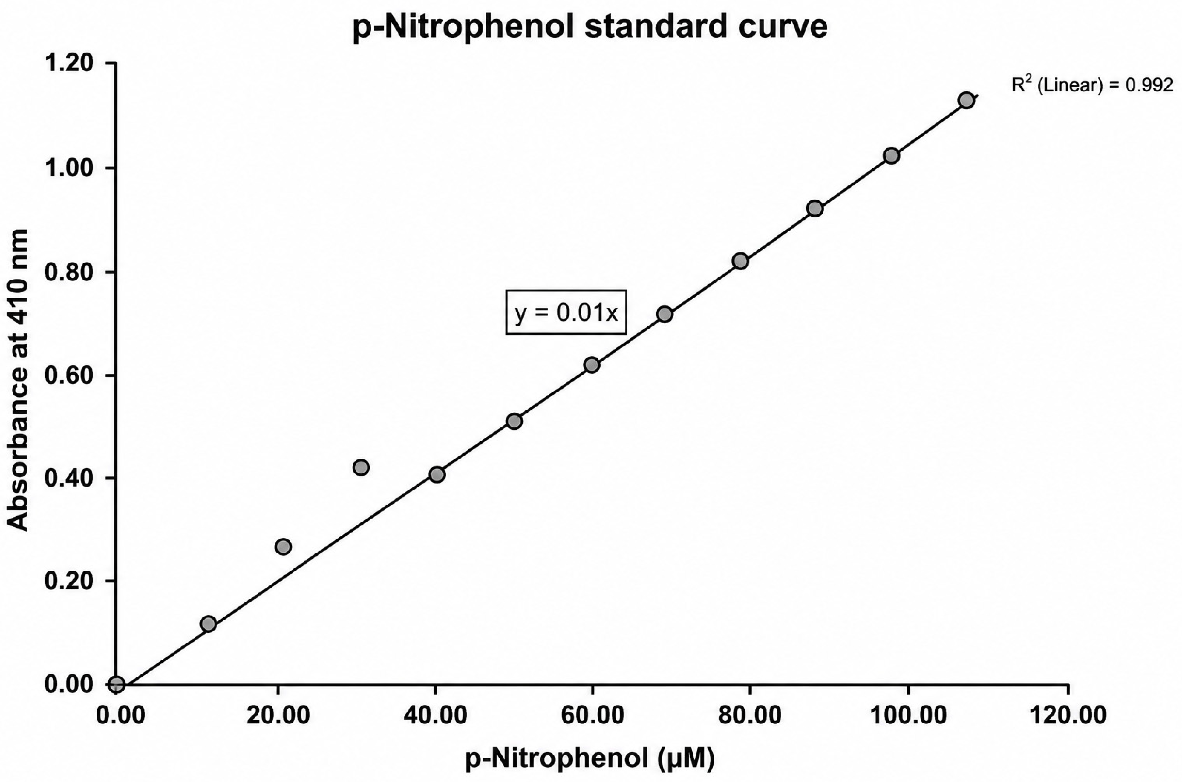
Disclaimer/Publisher’s Note: The statements, opinions and data contained in all publications are solely those of the individual author(s) and contributor(s) and not of MDPI and/or the editor(s). MDPI and/or the editor(s) disclaim responsibility for any injury to people or property resulting from any ideas, methods, instructions or products referred to in the content.