Article

Lactic Acid Bacteria-Mediated Synthesis of Selenium Nanoparticles: A Smart Strategy Against Multidrug-Resistant Pathogens

1
Department of Microbiology, Faculty of Science, Ain Shams University, Cairo 11566, Egypt
2
Department of Botany and Microbiology, Faculty of Science, Al-Azhar University, Cairo 11751, Egypt
3
Department of Bioscience and Biotechnology, Faculty of Agriculture, Graduate School, Kyushu University, Fukuoka 819-0395, Japan
*
Authors to whom correspondence should be addressed.
Appl. Microbiol. 2025, 5(4), 121; https://doi.org/10.3390/applmicrobiol5040121
Submission received: 30 September 2025 / Revised: 21 October 2025 / Accepted: 21 October 2025 / Published: 29 October 2025

Abstract

This study reports the biosynthesis of selenium nanoparticles (Se-NPs) using four newly isolated strains of lactic acid bacteria, molecularly identified as Lactiplantibacillus pentosus, Lactiplantibacillus plantarum, Lactiplantibacillus plantarum, and Lactobacillus acidophilus. The synthesized Se-NPs were characterized using Transmission Electron Microscopy (TEM), Energy Dispersive X-ray Spectroscopy (EDX), Fourier Transform Infrared Spectroscopy (FTIR), and UV-Vis Spectroscopy, and zeta potential analysis. The result revealed that their size ranged from 16 nm to 90 nm with favorable stability and purity. The Se-NPs exhibited significant antimicrobial and antibiofilm activities against certain Gram-positive, Gram-negative bacteria, and Candida albicans, particularly those produced by isolate S4, which showed the lowest MIC values and highest biofilm inhibition. Furthermore, MTT assays revealed selective cytotoxicity against the A549 cancerous lung cell line, with minimal toxicity toward normal Wi38 cells. These findings suggest that biosynthesized Se-NPs are a promising, biocompatible candidate for combating antibiotic-resistant pathogens and biofilm-associated infections.

1. Introduction

Nanofabrication involves manipulating elements at the nanoscale to improve human health and make evolutionary advances in medicine. This is done through cost effective and more rapidly functioning biological components. It involves precise control or modification of matter at the nanometer scale, spanning roughly 1 to 100 nm. When dimensions shrink below 100 nm, significant property transformations often take place. The distinctive physicochemical characteristics of nanomaterials enable their use in commercial applications and innovative solutions that advance societal progress. Many current medical problems have been solved through incorporating nanoscale components into full-scale implementations, including the delivery of drugs [1].
Selenium is a vital micronutrient required by the human body in small amounts. With potent antioxidant properties, selenium exists as selenoproteins in animal systems, serving as a crucial cofactor for essential enzymatic processes [2]. Furthermore, Selenium has extensive pharmaceutical and medical applications owing to its anticancer properties, role in muscle function, and beneficial impact on thyroid metabolism [3]. Se-NPs have garnered significant attention due to their superior bioavailability, high biocompatibility, potent antioxidant properties, and minimal toxicity. Accordingly, a growing number of recent efforts and studies indicate a higher antitumor activity of Se-NPs relative to its organic and inorganic counterparts [4]. A broad range of applications and the low toxicity of selenium nanoparticles have recently attracted much interest [5].
Several physical and chemical methods have been designed to fabricate Se-NPs based on the chemical characteristics of the compounds. The residual of these chemicals limits the applications of the formed Se-NPs in the medicinal area. The development of nano biohybrids using enzymes as natural inducers provides an environmentally friendly and highly efficient strategy for the synthesis of metal nanoparticles, overcoming the harsh conditions usually required in conventional methods [6,7]. To synthesize Se-NPs using biological methods, plants, algae, fungi, bacteria, and actinomycetes are preferred. These methods are safe, clean, cheap, and easy to scale up [8]. Micro-organisms utilize detoxification processes to convert selenite into nano selenium, making them promising biological factories for the synthesis of Se-NPs [9]. Selenium-tolerant bacteria initially transform selenium into a harmless organic compound within their cells. However, when their selenium tolerance threshold is surpassed, they start generating nano-scale elemental selenium particles (Se0), either inside or outside the cells [10].
Among microbial producers of Se-NPs, lactic acid bacteria (LAB) are the preferred choice due to their general safety. To achieve scalable fabrication of Se-NPs, it is necessary to consider the physiological properties of the bacteria used as a bio-transformer of inorganic forms of selenium [11]. LAB are widespread gram-positive non-spore forming cocci, coccobacilli or rods [12]. They are aero-tolerant, microaerophilic, or facultative anaerobes without catalase and a respiratory chain. Their optimum temperature is between 30 °C and 40 °C [13]. They can be found in any environment containing carbohydrates such as fermented foods, plants, the mucosal surfaces of humans, terrestrial and marine animals [12]. LAB belongs to the phylum Firmicutes, class Bacilli and order Lactobacillales [13]. LAB are extremely important micro-organisms for humans, not only because they play an important role in the food and feed industry, but also because they are a natural part of the gastrointestinal tract of healthy individuals. LAB play an imperative role in the industry for the synthesis of chemicals, pharmaceuticals, or other useful products [12,13].
Several chronic infections are caused by pathogenic micro-organisms grouped together into spatially organized communities known as biofilms [14]. Additionally, the dense cellular arrangement in biofilms promotes horizontal gene transfer, enhancing the spread of antibiotic resistance [15]. The most frequent genera of micro-organisms forming biofilms and reported in infections are Staphylococcus spp., Escherichia spp., and Candida spp. in implants, heart valves, catheters, medical devices, and prostheses [16]. In an effort to combat the MDR crisis, this study hypothesized that Se-NPs biosynthesis within lactic acid bacteria may have biomedical applications against pathogenic bacteria and offer an alternative to antibiotics, especially when combined with traditional antibiotics. Therefore, we investigated the biosynthesis of Se-NP inside four lactic acid bacteria, characterized them using the available techniques including: TEM, FTIR, EDX, ZETA and UV–visible spectroscopy as well as tested the antimicrobial, drug combination, and antibiofilm activities of the produced Se-NPs against a range of biofilm producer pathogenic, Gram negative bacteria like E.coli, K. pneumoniae, P. aeruginosa and A. baumannii, and Gram positive bacteria like MRSA, S. agalactiae, Enterococci, in addition to C. albicans. Furthermore, we assessed Se-NPs cytotoxicity against normal lung cells Wi38 and cancerous lung cells A549. The novelty of this research lies in its integration of a “trifecta” of activities into one platform. This includes antimicrobial and anti-biofilm action against both floating and resilient bacterial communities, as well as the most compelling result: selective cytotoxicity against a cancer cell line without harming healthy ones.

2. Materials and Methods

Sodium hydrogen selenite NaHSeO3 that used in this study was purchased from (LANXCESS AG, Cologne, German). Culture media including MRS, nutrient agar, blood agar media, MacConkey agar, and Sabaroud’s dextrose agar and other chemicals used were purchased from (HiMedia, Mumbai, Indian).

2.1. Bacterial Isolates and Growth Conditions

2.1.1. Isolation of Lactic Acid Bacteria (LAB) from Raw Milk

Ten milliliters of each raw cow milk sample were homogenized with a peptone-saline solution (1% peptone, 0.9% NaCl) [17]. Serial dilutions were prepared from each diluent. One milliliter of these dilutions was plated on MRS agar media and maintained at 37 °C anaerobically using a gas pack jar (BBL GasPak jar150, vented model, Franklin Lakes, NJ, USA) for 48 to 72 h. After incubation colonies were purified by streaking again to fresh MRS agar plates, followed by macroscopic examination of colonies morphology and microscopic examination by gram stain and catalase test [18]. According to primary screening by inoculating fresh overnight isolates on MRS broth supplemented with NaHSeO3. The four isolates with excellent growth and strong red color were selected for the biogenic production of Se-nanoparticles and molecular identification by 16S rRNA was carried out using the universal Forward primer: 5′-AGA GTT TGATCC TGG CTC AG-3′16SrRNA and Reverse primer: 5′-GGT TAC CTTGTT ACGACTT-3′. The PCR product was sequenced on an ABI 3730xl DNA sequencer by GATC Biotech ( Cologne, Germany Company) using forward and reverse primers.

2.1.2. Pathogenic Isolates

The pathogens used to study bacterial growth suppression and biofilm prevention capabilities of Se-NPs were highly biofilm producing strains and identified by Biomerieux VITEK® 2 Compact system (Craponne, France). A variety of pathogenic bacteria and fungi cultures were collected from the microbiology lab of Dar El-Fouad hospital. For the purification of bacteria, various enrichment and selective media were used, including blood agar media, MacConkey agar media, and Sabaroud’s dextrose agar. The bacterial cultures were incubated (FISHER Incubator 200 series model 255 D, Waltham, MA, USA) at 37 °C for 24 h. And then investigated by Gram stain for selection of appropriate isolates to be identified by Biomerieux VITEK® 2 Compact system.

2.2. Production and Recovery of Purified Nano Selenium

Reduction of NaHSeO3 inside four different isolated lactic acid bacteria by addition of 100 mg/mL of NaHSeO3 (Citizen Scale model CY 204, Edison, NJ, USA) to MRS broth media and inoculation with 1 mL of fresh cultures of each lactic acid bacteria and incubation for 3 days in a shaking incubator at 120 rpm (model: VS-37S series, Daejeon-Si, South Korea) at 37 °C. The media changed to a reddish color to indicate the production of elemental selenium then the samples were centrifuged (model: IEC clinical centrifuge, Thermo Fisher Scientific, Waltham, MA, USA) and the pellets were collected. HCL 37% was added to the product to digest bacterial cell wall and placed in room temperature for five days then centrifugation (8000 rpm for 10–15 min) was made to get rid of the acid. After that, the extracts were washed with distilled water until neutralization. Finally, to liberate the Se-NPs from the digested cellular debris, the pellet was subjected to sonication using an ultrasonic (FISHER Sonic dismembrator model: 300, Pittsburgh, PA, USA) was performed for 10 min [19].

2.3. Characterization of Se-NPs

The light absorption behavior of the synthesized selenium nanoparticles was assessed via UV-Visible spectral measurements using a JASCO V730(JASCO Corporation, Tokyo, Japan) double beam spectrophotometer. Spectral data was collected between 200 and 900 nm using a 5-nm wavelength step size. Also, the Fourier transform infrared spectrometer (FTIR) spectra were recorded in the range from 400 to 4000 cm−1 using JASCO UK FT/IR-4600(Tokyo, Japan) Fourier transform infrared spectrometer (FT-IR) with S/N Ratio 25,000:1. The exact morphology and particle size of Se-NPs were performed by a Transition Electron microscope (TEM model JEOL JEM-2100 - Akishima, Tokyo, Japan). The polydispersity of the population of nanoparticles was expressed in PDI by calculating the average radius and standard deviation of the synthesized nanoparticles. PDI was calculated as p = σ/RAvg where p = dispersity, σ = standard deviation of a radius of a batch of nanoparticles, and RAvg = average radius of nanoparticles [20]. The topographical study was carried out using high resolution scanning electron microscopy (SEM Quanta FEG 250 with field emission gun, FEI Company, Eindhoven, Netherlands) with energy dispersive electron spectroscopy (EDX) [21]. The zeta potential measurement was analyzed via dynamic light scattering (DLS) of the prepared samples, using Nicomp™ 380 ZLS size analyzer, Billerica, MA, USA. Laser light scattering was used where the zeta potential was measured at 18°.

2.4. Antimicrobial Activity of Se-NPs and MIC Determination

To determine the antimicrobial and MIC for Se-NPs, a microdilution assay was performed. The selected identified pathogens were inoculated in nutrient broth media and incubated for 24 h at 37 °C. Briefly, a two-fold serial dilution [20 − 0.07 mg/mL] of each Se-NPs was prepared in 96-well polystyrene microtiter plates [each well contain 200 µL]. The pathogens were inoculated in each well at 0.5 McFarland standard (1.5 × 10⁸ CFU) and incubated for 24 h at 37 °C. After incubation, plates were read by an ELISA reader (Statfax, Palm City, FL, USA). The MIC of Se-NPs was determined as the lowest concentration at which Se-NPs prevent the multiplication of the pathogen [22].

2.5. Biofilm Inhibition Assay

All pathogens tested for MIC assay were subjected for detection of biofilm formation and they were all able to form biofilm in vitro except for Proteus mirabilis, so it was neglected from this study. The different produced Se-NPs were assayed for their potential to suppress biofilm formation against high-biofilm producing strains from isolated pathogens. Briefly, overnight cultures of the selected pathogens were prepared in nutrient broth media supplemented with glucose 1% and distributed in a sterile microtiter plate (200 µL in each well) containing different concentrations of Se-NPs (MIC, 1/2MIC, 1/4MIC, 1/8MIC and 1/16MIC). In a negative control, only broth media was distributed without nanoparticles or bacteria. Bacterial suspension without the tested particles was used as positive control. The plates were incubated at 37 °C for 48 h. After incubation, the excess of the media was discarded and washed three times by phosphate buffered saline (PBS) and then dried by inverting the plates at room temperature for 30 min., then fixed by 95% methanol for 10 min. Then, alcohol was discarded and all plates were stained with 0.1% crystal violet for 20 min. After that, plates were gently washed with distilled water and let dry. The biofilm was examined by adding 30% glacial acetic acid to each well. Then the optical densities (OD) were measured by an ELISA reader at 492 nm [23].
The percentage of biofilm degradation was calculated as follows:
% of biofilm degradation = (Control absorbance − Sample absorbance)/Control absorbance × 100

2.6. Cytotoxicity of Biogenic Se-NPs

Se-NPs cytotoxicity was evaluated by MTT assay on normal WI-38 and cancerous A549 cell lines using a 96 well tissue culture plate. Both cell lines were purchased from VACSERA (Cairo, Egypt). Plates were inoculated with 1 × 10⁵ cells/ml (100 µL/well) and incubated at 37°C for 24 h to develop a complete monolayer sheet. Once a confluent sheet of cells had been formed, growth medium was decanted from 96-well micro-titer plates. The monolayer of cells was washed twice with wash medium. The following concentrations (200, 100, 50, 25, 12.5, and 6.25 µg/mL) of each tested sample were made in RPMI medium with 2% serum (maintenance medium). 0.1 mL of each dilution was tested in different wells leaving 3 wells as a control, receiving only maintenance medium. Plates were incubated at 37°C and examined. Cells were inspected for outward signs of toxic effects, such as rounding, shrinkage, cell granulation, or partial or total loss of the monolayer. A 5-mg/mL MTT solution in PBS was made (Bio Basic, Toronto, Canada). Twenty microliters of MTT solution were added to each well. To fully blend the MTT with the media, place on a shaking table and spin at 150 rpm for five minutes. To enable formazan production, cells were treated with MTT for 1–5 h at 37 °C with 5% CO2 after treatment. The media was then carefully aspirated, and any residual liquid was removed by blotting the plate on paper towels. The resulting formazan crystals were dissolved in 200 µL of dimethyl sulfoxide (DMSO). The optical density (OD) was measured at 560 nm, with background subtraction at 620 nm. Since the OD at 560 nm is directly proportional to the number of viable cells, cytotoxicity was quantified by calculating the half-maximal inhibitory concentration (IC50) [24].

2.7. Drug Combination Assay

Using 96-well plates along with the twofold drug microdilution in broth checkboard assessment, drugs combination experiment was conducted to determine Fractional Inhibitory Concentration (FIC) index among the Se-NPs produced by isolates S2, S3 and levofloxacin antibiotic versus multidrug-resistant P. aeruginosa and E. faecalis clinical isolates [25]. The plates had been incubated for 24 h at 37 °C in a triplicate experiment. Following that, 30 µL of resazurin dye (0.02% w/v) was added to every well of the growing medium and incubated again for a total of six hours in order to look for bacterial growth. A change in color from blue to red revealed the presence of bacteria. Afterwards, employing the subsequent formula to determine the fractional inhibitory concentration (FIC) index, assess the data: (Se-NPs by S2 or S3 combined/Se-NPs by S2 or S3 alone) + (levofloxacin combined/levofloxacin alone) = FIC index. 1 < FIC index ≤ 2 indicates additives, FIC index > 2 indicates antagonism, FIC index ≤ 1 indicates synergism.

2.8. Statistical Analysis

All of the results are the averages of three separate studies, each of which was conducted in three duplicates. To ascertain the significance between groups, the data was examined using a one-way ANOVA model of analysis of variance (ANOVA) (α = 0.05). Tukey’s test was used for multiple comparisons when pairwise comparisons revealed a significant difference.

3. Results & Discussion

3.1. Isolation of Lactic Acid Bacteria

From the four raw milk samples, four isolates were picked up and examined morphologically. Colonies were convex in shape with a creamy color. Based on microscopic examination and biochemical tests, the bacteria were Gram-positive, catalase-negative. The amplified sequences of 16S rRNA genes obtained from the isolates were aligned with published sequences on NCBI website and revealed similarity to the bacteria shown in (Table 1).

3.2. Identification of the Pathogenic Isolates

As a result of identification with the (VITEK compact 2 system), a variety of bacterial and fungal strains were identified in Table 2. According to their antibiotic susceptibility test, multidrug resistant strains of bacteria including five Gram negative strains, and three Gram positive strains were selected to test the antimicrobial activity of Se-NPs along with Candida.

3.3. Production and Recovery of Purified Nano Selenium

After the incubation of different LAB in MRS media with sodium hydrogen selenite in a shaking incubator for three days, LAB samples (S1, S2, S3, and S4) could reduce selenium ions to elemental selenium nanoparticles. The reduction of Se+ ions in metal nanoparticles was visualized by tracing the color change in the solution.

3.4. Characterization of Se-NPs

3.4.1. Transmission Electron Microscopy (TEM)

Based on TEM images of the produced Se-NPs (Figure 1), the particles synthesized via isolates S1 and S2 ranged in size from 40.5–95.02 nm with a PDI value of 0.31, while the particles synthesized via isolates S3 size average was 68.8 nm with a PDI value of 0.57 and S4 average size was 56.7 nm with a PDI value of 0.5. The significant differences in the average size and size distribution (polydispersity index, PDI) of the Se-NPs synthesized by the four lactic acid bacteria (LAB) isolates (S1, S2, S3, S4) can be attributed to the strain-specific biochemical machinery involved in the reduction of selenite (SeO32−) to elemental selenium (Se0). In bacterial biosynthesis, nanoparticles are formed through an enzymatic process where microbial metabolites act as reducing and capping agents. The variation in these metabolites across different strains directly influences the kinetics of nucleation and growth, ultimately determining the nanoparticle’s characteristics [26]. Kaur et al., 2018 [27], synthesized Se-NPs by Lactobacillus acidophilus ranged in sizes of 11–23 nm when characterized by TEM. While Alam et al., 2020 [28], used Lactobacillus acidophilus and sodium selenite solution under optimum conditions to obtain spherical Se-NPs of 2–15 nm and the size of Se-NPs attained by culture of Lactobacillus paracasei HMN1 ranged from 3 to 50 nm [29].

3.4.2. EDEX

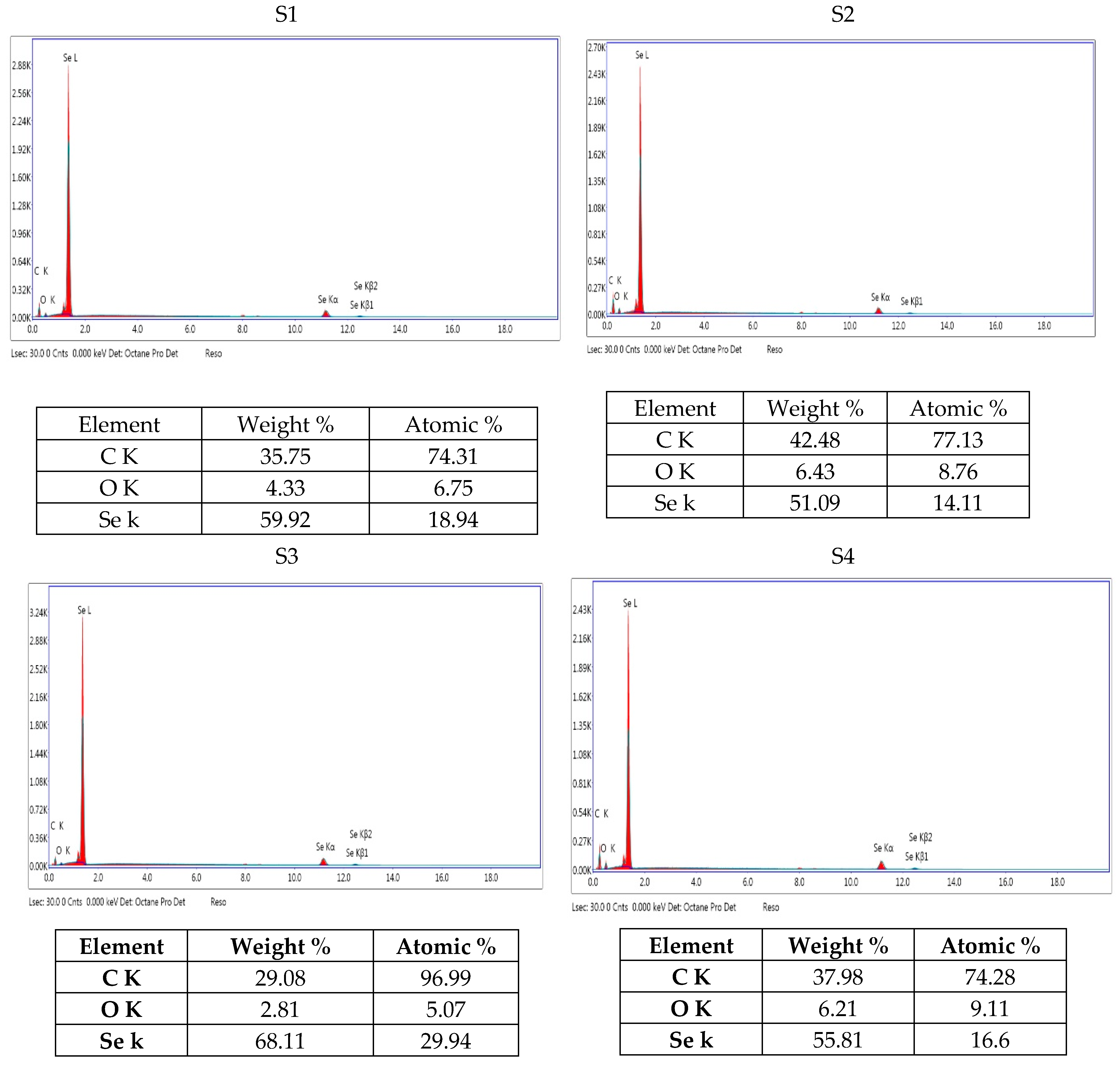
Figure 2 depicts a semi-quantitative representation of the constituent composition in the inspection field in weight percent and atomic percent units. The weight % of elemental constituents of the four separate Se-NPs revealed that the main elements contained within the analyzed samples are carbon (C), oxygen (O), and selenium (Se) atoms, with Se being the most abundant. The concentrations of nano-selenium were highest in S3 (68.11%) and lowest in S2 (51.01%). Xu et al., 2018, discovered that Se-NPs produced by Lactobacillus casei 393 and analyzed by energy dispersive X-ray spectroscopy (EDX) were made up of carbon (C), nitrogen (N), oxygen (O) selenium (Se), and phosphorus (P) elements [30].

3.4.3. UV Results

The spectrum of Se-NPs represented in Figure 3 was set from 200 nm to 900 nm. The peaks indicate that there are uniformly distributed particles, and most particles are nanosized. Observing no other peak in the spectrum indicates that Se-NPs have successfully formed. El-deeb et al., 2023, observed that according to the surface plasmon resonance, UV-Vis absorption spectra of biogenic selenium nanoparticle formation is indicated by a change in the absorbance around 582 nm [31]. On the other hand, Alam et al., 2020, recorded that Se-NPs synthesized by L. acidophilus surface have plasmon resonance at 385 nm as determined by UV-Vis spectroscopy [28].

3.4.4. Fourier Transform Infrared Spectroscopy Analysis (FTIR)

The presence of functional groups involved in Se-NPs bioreduction is confirmed by FTIR spectroscopy. Structural and compositional information about the Se-NPs provided by (FT-IR) are shown in Figure 4. S1 shows different peak positions at 3409, 2032, 1454 and 970 cm−1 (6,10,13,17). The peak at 3409 cm−1 corresponding to OH stretching. A peak at 2032 cm−1 assigned to the isothiocyanate group found in the extract. The broad spectrum and intensity at 1454 cm−1 assigned to the C-H bend or methyl group. The peak at 970 cm−1 belongs to the alkene group. FTIR results in S2 show major peak positions at 3394, 1942, 1321 and 802 cm−1 (1,6,10,14). The peak at 3394 cm−1 corresponds to O-H stretching of the phenol group present in the extract. A peak at 1942 cm−1 assigned to C=C=C stretching vibrations of the allene group. The peak at 1321 cm−1 corresponding to C-N stretching or amine group. And the peak at 802 cm−1 is associated with C=C bending or alkene group. FTIR results of S3 show peak positions at 2929, 2537,1544, 607 cm−1 (8,10,16,23). The peak at 2929 cm−1 corresponds to C-H stretching or alkane group. While the peak at 2537 cm−1 assigned to O-H stretching or carboxylic group. And the peak at 1544 cm−1 corresponding to N-O stretching vibrations of the nitro group. FTIR results of Se-NPs produced by S4 show different peak positions at 2979, 1619, 1384, 798 cm−1 (4,8,10,12). The peak at 2979 cm-1 corresponding to C-H stretching or alkane group. The signal at 1619 cm−1, on the other hand, was assigned to the C=C stretching or ketone group. The alkane group has a peak at 1384 cm−1. The 798 cm−1 peak was ascribed to C=C bending or the alkene group. The overall FTIR spectral fingerprint pattern was discovered to match the contour of the Se-NPs synthesized using a green synthesis technique. Alam et al., 2020, discovered that FTIR results show that proteins in the bacterial extract are primarily responsible for the synthesis of LA-Se-NPs [28], whereas El-Saadony et al., 2021, demonstrated that FTIR analysis offered a clear image of the active groups involved in the stability of L. paracasei-Se-NPs [29].

3.5. Zeta Potential Measurements

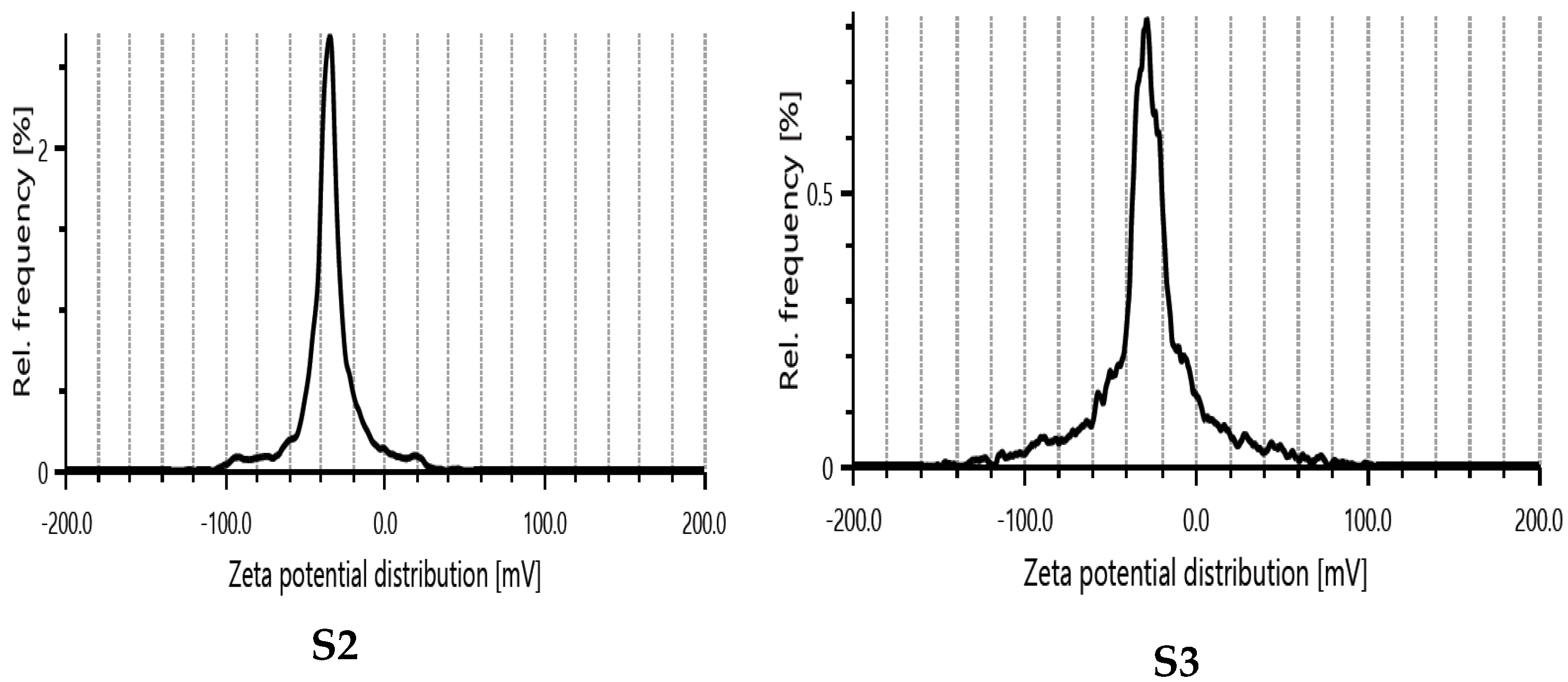
Zeta potential measurements included Electrophoretic Mobility (μm * cm/Vs.), and average zeta potential value (mV). Table 3 and Figure 5 shows the zeta potential measurements for S2 and S3 nanoparticles. The Se-NPs observed zeta potential of −27.6 and −34.8 mV for S2 and S3 respectively which represent the excellent zeta potential value compromised as stable colloidal materials. Results demonstrate that the Se-NPs are a stable system and have unique stability properties.

3.6. Antimicrobial Activity and MIC of Se-NPs

Based on the microdilution method, the produced selenium nanoparticles exhibited strong inhibitory activity against some human pathogens. MICs were also determined for the four produced Se-NPs. Table 4 illustrate that Se-NPs synthesized by S1 exhibited MIC 8.12 mg/mL against E. coli, P. aeruginosa, P. mirabilis, and E. faecalis while the MIC against MRSA and S. agalactiae was 4.06 mg/mL. The lowest MIC against A. baumannii was 1.015 mg/mL and the highest MIC was 16.25 mg/mL against C. albicans and K. pneumonia. Results revealed that Se-NPs synthesized by S2 exhibited MIC 1.07 mg/mL against E. coli, P. aeruginosa, S. agalactiae and C. albicans and the MIC against K. pneumonia was 3.15 mg/mL where the lowest was 0.62 mg/mL against P. mirabilis and A. baumanii and the highest MIC was 25 mg/mL against MRSA and E. faecalis. Se-NPs synthesized by S3 showed the highest MIC 25 mg/mL for MRSA while, the lowest MIC for K. pneumonia and A. baumanii was 0.562 mg/mL. The Se-NPs produced by S4 showed the lowest MIC of 0.625 mg/mL against E. coli, S. agalactiae and A. baumannii while the highest MIC was 10 mg/mL against C. albicans. Alam et al., 2020, reported that the MIC of L. acidophilus Se-NPs against drug-sensitive strains was in the range of 1–10 μg/mL, whereas in the case of drug-resistant bacteria MIC was found to be 6.5 μg/mL for K. pneumonia and 4 μg/mL P. aeruginosa respectively [28]. The antimicrobial activity of the Se-NPs shows activity against all the tested bacteria and the lowest MIC and MBC of Se-NPs was 8 μg/200 μL against P. aeruginosa (NCIM 5031) [32]. Similarly, a majority of the bacteria tested were destroyed within 12 h when treated with 500 mg/L Se-NPs, with Gram-negative bacteria showing a significantly better mortality rate [33]. The Se-NPs were shown to have multi-modal mechanisms of action that depended on their size, including depleting internal ATP, inducing ROS production (it triggers oxidation of proteins, lipids and DNA damage) and disrupting membrane potential. Results of leakage tests illustrated that there were proteins and polysaccharides outside the cells treated with Se-NPs. It was indicated that the leakages of proteins and polysaccharides were caused by permeability changes in membranes and cell walls disruption. Also the change in reactive oxygen species (ROS) intensity indicated that oxidative damage may play a significant role in the antibacterial processes [33,34]. The gradual release of toxic selenium ions is what causes the antibacterial action. Despite being present, the released selenium ions’ contribution is regarded as secondary because of the nanoparticulate form’s greater activity [35]. Previous studies have reported that biogenic selenium nanoparticles, as well as selenium/chitosan nano-incorporates, exert their antimicrobial activity mainly through the inhibition of essential bacterial metabolic enzymes, thereby disrupting vital cellular functions [36].

3.7. Biofilm Inhibition

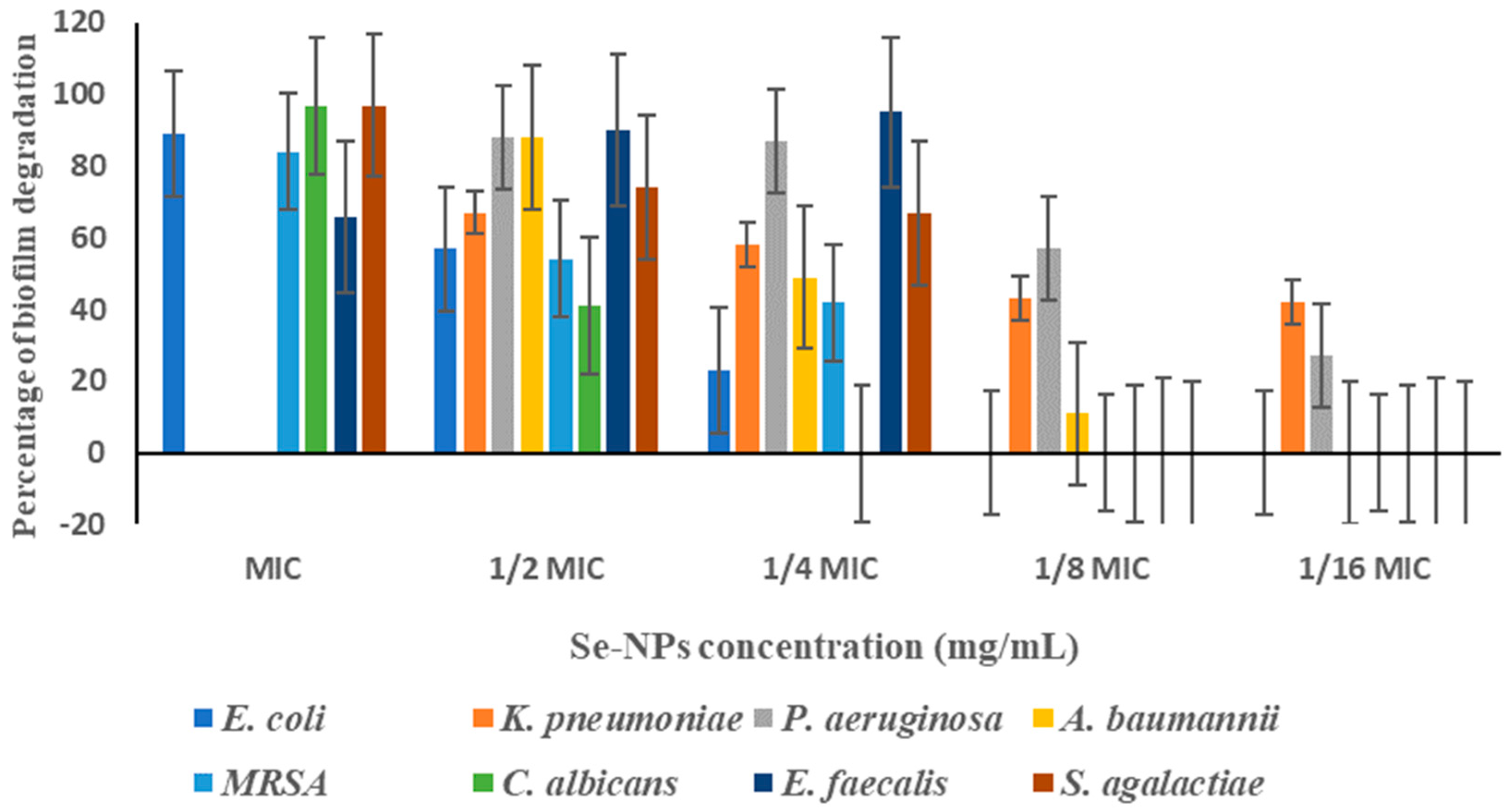
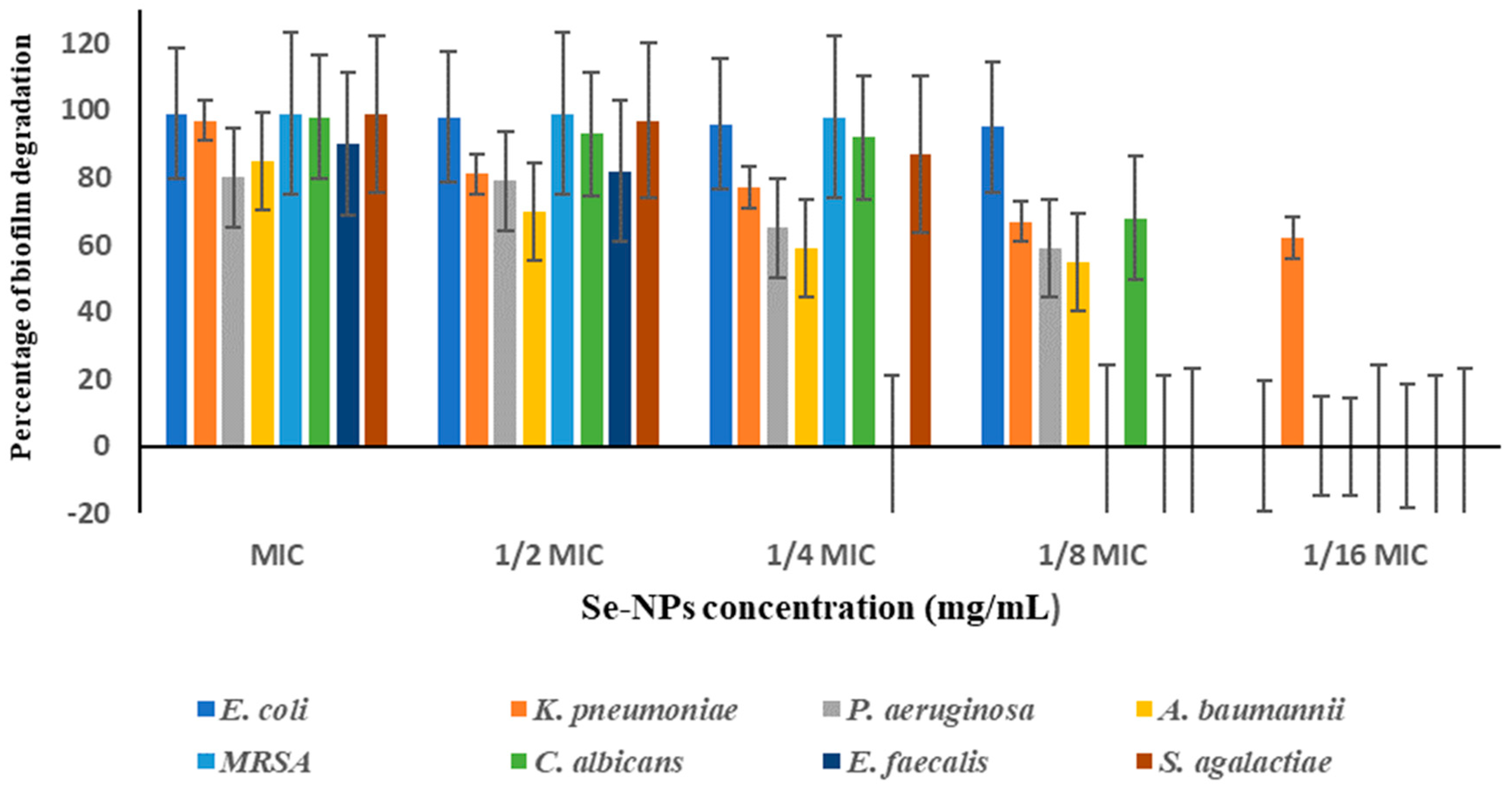
The selenium nanoparticles prepared by the four bacterial isolates were evaluated for sub-inhibitory concentrations (1/2 MIC, 1/4 MIC, 1/8 MIC, and 1/16 MIC) against high biofilm producing strains of identified pathogens. Figure 6, Figure 7, Figure 8 and Figure 9 show the antibiofilm activity of Se-NPs against different pathogenic bacteria and fungi. Higher concentrations of selenium nanoparticles correlate with enhanced biofilm degradation. In contrast control of each pathogen, growth and biofilm formation were normal in absence of Se-NPs. S1 nanoparticles were effective by ¼ MIC against all tested pathogens with biofilm degradation up to 93% in case of Streptococcus agalactiae. S2 nanoparticles were more effective at combating Gram positive than Gram negative bacteria. S3 nanoparticles were very potent in biofilm degradation of E. coli with very low concentrations 1/8 MIC by 67%. S4 nanoparticles were the most potent against all the tested Gram positive, negative bacteria and C. albicans with very low concentration. Degradation of preformed biofilms by LAB Se-NPs was studied by Alam et al., 2020 [28], against Gram-negative and Gram-positive bacteria and these nanoparticles are more effective against E. coli, S. aureus and P. aeruginosa than B. subtilis and K. pneumonia. As the concentration of selenium nanoparticles increases biofilm degradation increases and the results of Kaur et al., 2018, revealed that Se-NPs synthesized by Lactobacillus acidophilus was effective against biofilm forming micro-organisms like E. coli, Staphylococcus aureus, Pseudomonas aeruginosa, Klebsiella and C. albicans [27]. Results of atomic force spectroscopy elucidated the disintegrating effect of Se-NPs against Gram-negative (P. aeruginosa ATCC 9027) and Gram-positive (S. aureus ATCC 25923) pathogens. The disintegrating effect of Se-NPs was caused by the destruction of the biofilm producing bacterial cell by rupturing the cell membrane [37]. Moreover, Cremonini et al., 2016, concluded that the lowest concentrations of biogenic Se-NPs (50 and 100 μg/mL) inhibited biofilm synthesis by P. aeruginosa strains CFC20, CFC21, and CFCA by 70–90%, indicating that these strains are particularly sensitive to these Se-NPs [38]. Conversely, clinical strains CFCB, and INT, as well as reference strains, were significantly less sensitive to Se-NPs, showing significant inhibition of biofilm synthesis (at least 70%) only at Se-NP concentrations of 250 mg/mL. Finally, a study by Salem et al., 2022, showed that biogenic Se-NPs exhibited the largest inhibition of biofilm formation toward MDR K. pneumonia, by 62% and 92% at 0.25 and 0.5 MIC, respectively [39].

3.8. Drug Combination Assay

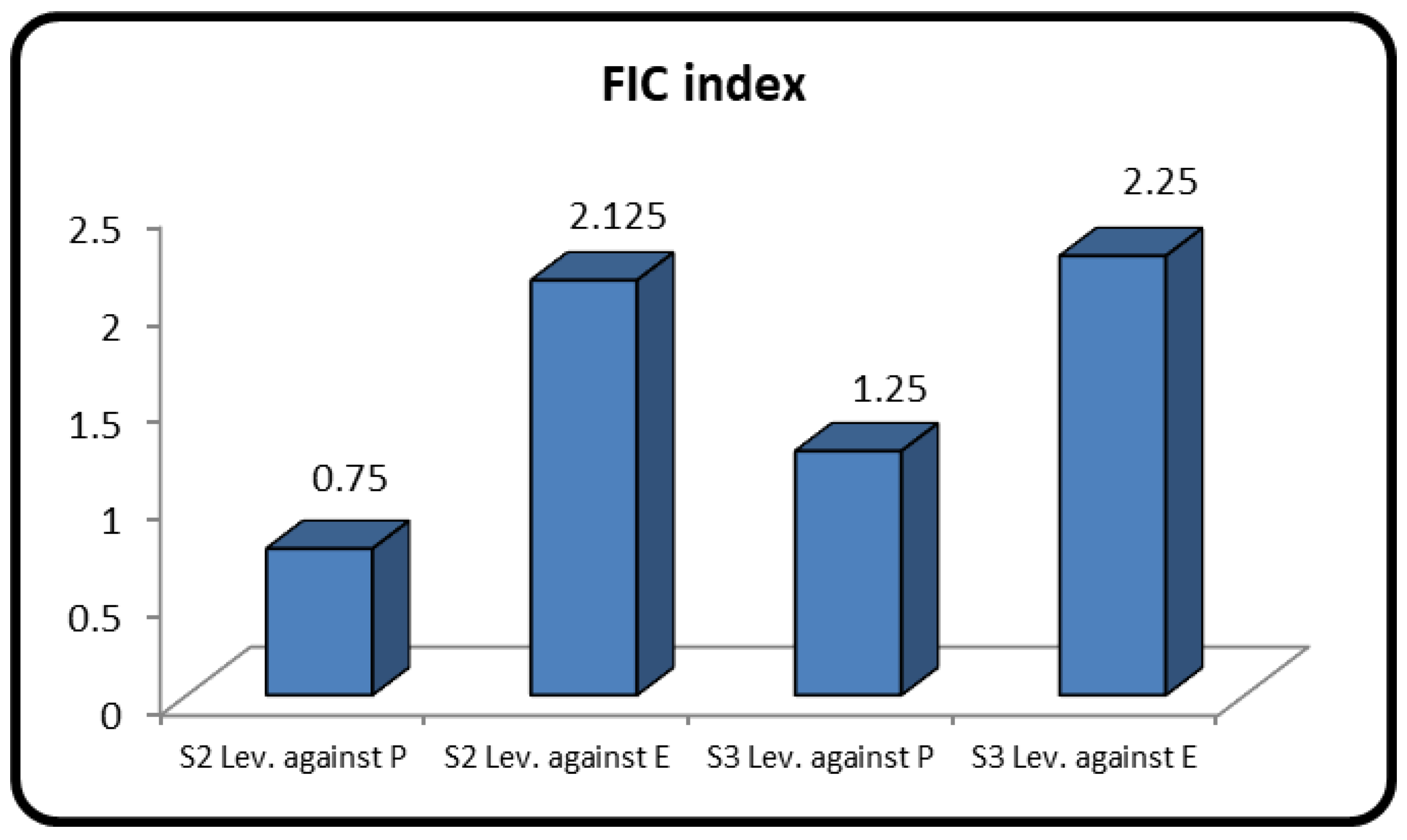
Research has shown that combination therapy is a potentially effective treatment for severe microbial infections. Studies demonstrate that combining therapeutically antibacterial medications can boost their bactericidal efficacy, lessen, or suppress undesirable responses, and even defeat multiple drug resistance through different modes of action. Therefore, in order to combat multidrug-resistant P. aeruginosa and E. faecalis clinical isolates, the Se-NPs produced by S2 and S3 isolates were used in combination with the broad-spectrum antibiotic Levofloxacin in the present study. After determining the fractional inhibitory concentration index (FIC), the findings showed that there was synergistic effect (FIC ≤ 0.5) between Se-NPs produced by S2 with Levofloxacin against P. aeruginosa in contrast there was an antagonistic effect between them when applied against E. faecalis. Conversely, S3 Produced Se-NPs with Levofloxacin was antagonistic against E. faecalis and additive against P. aeruginosa (Table 5 and Figure 10 and Figure 11). The synergistic effect displayed that these Se-NPs were unable to outcompete these antibiotics in terms of binding to the intended pathogen.

3.9. Cytotoxicity of Biogenic Se-NPs

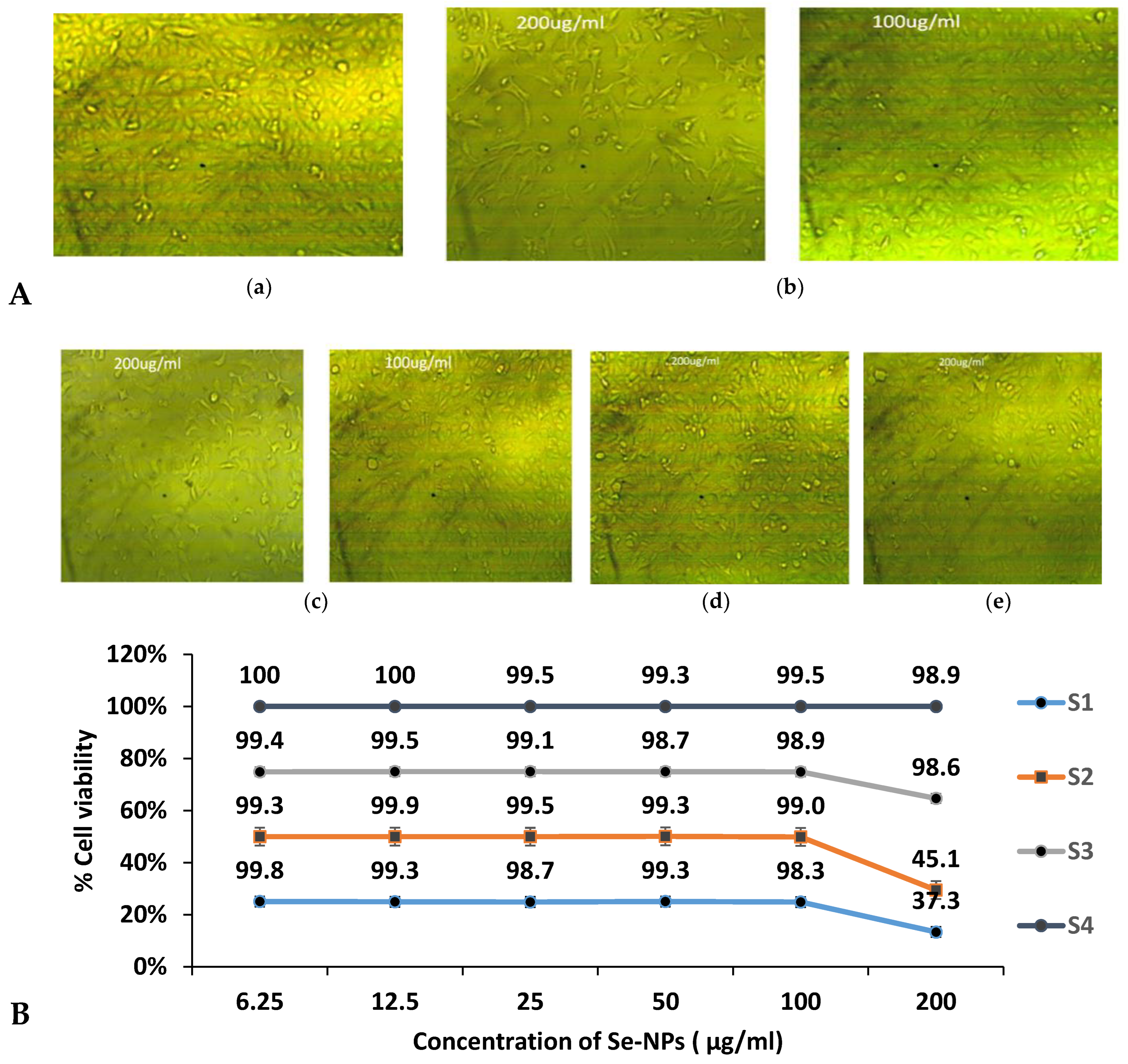
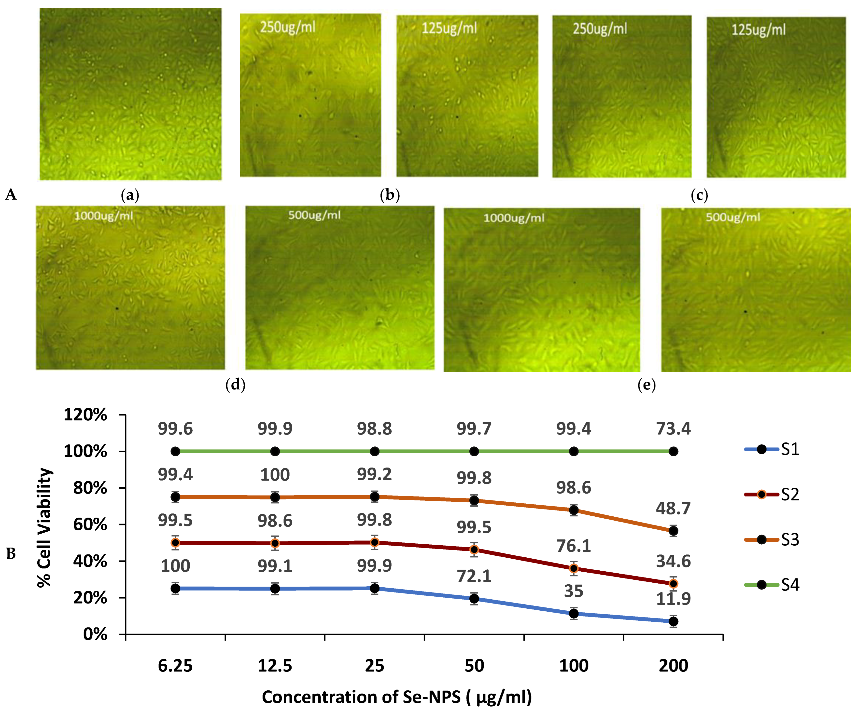
The effect of Se-NPs produced by isolates (S1, S2, S3, and S4) on Wi38 cells viability was shown in (Figure 12). A total of 200 μg/mL of Se-NPs produced by S1 and S2 exhibited cytotoxicity of 62% and 55% with IC50 179 and 190, respectively. While lower concentrations of both nanoparticles showed very slight loss of cell viability. On the other hand, all tested concentrations of nanoparticles produced by isolates S3 and S4 did not harm Wi38 cells’ viability. Similarly, Alam et al., 2020, synthesized selenium nanoparticles extracellularly using the probiotic bacteria Lactobacillus acidophilus and found that the synthesized nanoparticles were non-disruptive to human HEK-293 normal cell lines as shown by MTT assay [28]. As shown (Figure 13) 200 μg/mL of Se-NPs produced by isolate S1 showed the highest toxicity 88% toward A549 cells with IC50 80. Followed by Se-NPs produced by isolate S2 with toxicity 65% and IC50 163. Also, Se-NPs of S3 isolate showed 51% toxicity toward cells and IC50 205. Se-NPs of S4 isolate exhibited the lowest cytotoxicity, 26% and 335 IC50. MTT assay of nanoparticles produced by different isolates results showed cyto-compatibility toward normal lung cell line Wi38 and exhibited variable degrees of toxicity toward cancerous lung cell line A549. The results demonstrated a selective cytotoxic effect: all Se-NPs exhibited high cytocompatibility with the normal Wi38 cell line, while showing significant and variable toxicity toward the cancerous A549 cells [39]. Se-NPs (50–80 nm particle size) produced anaerobically by Bacillus licheniformis ATCC 10716 have been shown to have a chemo preventive effect on lung cancer [40]. Moreover, Se/Chitosan nanoconjugate is easily absorbed by tumor cell lines and has very low cytotoxicity on WI-38 with an IC50 of 153.3 μg/mL [41]. Moreover, all the biosynthesized Se-NPs were non-toxic towards mammalian cells up to 25 μg/mL [42]. Se-NPs demonstrated synergic properties as well as protective properties against the side effects of chemotherapy drugs [43]. The physiochemical properties of nanoparticles, such as size, structure, shape, aggregation state, surface charge, chemistry, dose, and substance type, are closely connected to their cytotoxicity [44]. Three replicates were done, and all resulted values are the averages of three independent experiments. Data were analyzed using a one-way ANOVA model of analysis of variance (ANOVA) (α = 0.05) to determine significance between groups.

4. Conclusions

Four LAB have been isolated from different milk samples and screened for their ability to reduce sodium hydrogen selenite inside their cells to nano-selenium. TEM images of the four produced Se-NPs showed that their size ranged from 30 to 90 nm. The EDX analysis also revealed a range of 51% to 68% for Se concentrations in the four preparations. Moreover, the four different Se-NPs had potential antimicrobial and antibiofilm activity against clinical isolates including both Gram positive and negative bacteria (MICs ranging from 0.5 to 25 mg/mL). The relatively high MIC values for some isolates (up to 25 mg/mL) may pose a challenge for practical therapeutic dosing and formulation. Additionally, they were cytotoxic to cancerous lung cells A549 but showed cytocompatibility with normal lung cells Wi38. Based on these promising findings, biogenic Se-NPs produced inside lactic acid bacteria can be considered non-toxic, and environmentally friendly. Future research should elucidate the exact pathways by which these Se-NPs exert their antimicrobial, anti-biofilm, and selective cytotoxic effects. This includes studying their interaction with bacterial cell membranes, biofilm matrices, and specific apoptotic pathways in cancer cells. As a result of their low toxicity and potential biological activity, biosynthesized Se-NPs could be used as nanomedicine-based therapies or in combination with other therapies as innovative agents to treat human infections, but further preclinical studies are still needed. This should include comprehensive in vivo toxicology studies in animal models to confirm the systemic safety suggested by the in vitro cytocompatibility data. Finally, Pharmacokinetic studies (absorption, distribution, metabolism, excretion) are also crucial. The efficacy and selectivity need to be validated across a broader panel of cancer and normal cell lines to confirm the generalizability of this effect.

Author Contributions

Conceptualization, M.A.-E., T.Z. and H.Y.; Methodology, N.F., M.A.-E. and H.G.; Validation, N.F.; Investigation, H.G.; Writing – original draft, N.F.; Writing—review & editing, M.A.-E., T.Z. and H.Y.; Supervision, T.Z. and H.Y.; Project administration, T.Z. and H.Y. All authors have read and agreed to the published version of the manuscript.

Funding

This research received no external funding.

Institutional Review Board Statement

Not applicable.

Data Availability Statement

The original contributions presented in this study are included in the article. Further inquiries can be directed to the corresponding authors.

Acknowledgments

Mohamed Abu-Elghait gratefully acknowledge the ministry of Higher education and scientific research of Egypt for providing a post-doctoral fellowship in Japan.

Conflicts of Interest

The authors declare no conflict of interest.

Abbreviations

The following abbreviations are used in this manuscript:
CFUColony forming unit
DLSDynamic light scattering
EDXEnergy Dispersive X-ray Spectroscopy
ELISAEnzyme-linked immunosorbent assay
FICFractional Inhibitory Concentration
FTIRFourier Transform Infrared Spectroscopy
IC50Half-maximal inhibitory concentration
LABLactic acid bacteria
MICMinimum inhibitory concentration
MRSde Man, Rogosa, and Sharpe
MRSAMethicillin-resistant Staphylococcus aureus
MTT3-(4,5-Dimethylthiazol-2-yl)-2,5-diphenyltetrazolium bromide
ODOptical density
PDIPolydispersity index
ROSReactive oxygen species
Se-NPsSelenium nanoparticles
TEMTransmission Electron Microscopy
UV-visUV-Visible absorption spectra
16s rRNA6S ribosomal ribonucleic acid

References

  1. Bhushan, B. Introduction to nanotechnology. In Springer Handbook of Nanotechnology; Springer: Berlin/Heidelberg, Germany, 2017; pp. 1–19. [Google Scholar] [CrossRef]
  2. Dawood, M.A.; Basuini, M.F.E.; Yilmaz, S.; Abdel-Latif, H.M.; Kari, Z.A.; Abdul Razab, M.K.A.; Ahmed, H.A.; Alagawany, M.; Gewaily, M.S. Selenium nanoparticles as a natural antioxidant and metabolic regulator in aquaculture: A review. Antioxidants 2021, 10, 1364. [Google Scholar] [CrossRef]
  3. McClements, J.; McClements, D.J. Standardization of nanoparticle characterization: Methods for testing properties, stability; functionality of edible nanoparticles. Crit. Rev. Food Sci. Nutr. 2016, 56, 1334–1362. [Google Scholar] [CrossRef]
  4. Fardsadegh, B.; Vaghari, H.; Mohammad-Jafari, R.; Najian, Y.; Jafarizadeh-Malmiri, H. Biosynthesis, characterization and antimicrobial activities assessment of fabricated selenium nanoparticles using Pelargonium zonale leaf extract. Green Process. Synth. 2019, 8, 191–198. [Google Scholar] [CrossRef]
  5. Indhira, D.; Aruna, A.; Manikandan, K.; Albeshr, M.F.; Alrefaei, A.F.; Vinayagam, R.; Kathirvel, A.; Priyan, S.R.; Kumar, G.S.; Srinivasan, R. Antimicrobial and Photocatalytic Activities of Selenium Nanoparticles Synthesized from Elaeagnus indica Leaf Extract. Processes 2023, 11, 1107. [Google Scholar] [CrossRef]
  6. Fairweather-Tait, S.J.; Bao, Y.; Broadley, M.R.; Collings, R.; Ford, D.; Hesketh, J.E.; Hurst, R. Selenium in human health and disease. Antioxid. Redox Signal. 2011, 14, 1337–1383. [Google Scholar] [CrossRef]
  7. Palomo, J.M. Nanobiohybrids: A new concept for metal nanoparticles synthesis. Chem. Commun. 2019, 55, 9583–9589. [Google Scholar] [CrossRef] [PubMed]
  8. Salem, S.S.; Fouda, A. Green synthesis of metallic nanoparticles and their prospective biotechnological applications: An overview. Biol. Trace Elem. Res. 2021, 199, 344–370. [Google Scholar] [CrossRef] [PubMed]
  9. Husen, A.; Siddiqi, K.S. Phytosynthesis of nanoparticles: Concept, controversy and application. Nanoscale Res. Lett. 2014, 9, 229. [Google Scholar] [CrossRef]
  10. Ullah, A.; Yin, X.; Wang, F.; Xu, B.; Mirani, Z.A.; Xu, B.; Chan, M.W.H.; Ali, A.; Usman, M.; Ali, N. Biosynthesis of selenium nanoparticles (via Bacillus subtilis BSN313); their isolation, characterization; bioactivities. Molecules 2021, 26, 5559. [Google Scholar] [CrossRef] [PubMed]
  11. Stabnikova, O.; Khonkiv, M.; Kovshar, I. Biosynthesis of selenium nanoparticles by lactic acid bacteria and areas of their possible applications. World J. Microbiol. Biotechnol. 2023, 39, 230. [Google Scholar] [CrossRef]
  12. Khan, I.; Saeed, K.; Khan, I. Nanoparticles: Properties, applications and toxicities. Arab. J. Chem. 2019, 12, 908–931. [Google Scholar] [CrossRef]
  13. Biscola, V.; Albuquerque, M.A.C.; Nunes, T.P.; Vieira, A.D.S.; Franco, D.G.M. Lactic Acid Bacteria: A Functional Approach, 1st ed.; Albuquerque, M.A.C., LeBlanc, A.M., LeBlanc, J.G., Bedani, R., Eds.; CRC Press (Taylor and Francis Group): Boca Raton, FL, USA, 2020; p. 292. [Google Scholar]
  14. Sahli, C.; Moya, S.E.; Lomas, J.S.; Gravier-Pelletier, C.; Briandet, R.; Hémadi, M. Recent advances in nanotechnology for eradicating bacterial biofilm. Theranostics 2022, 12, 2383–2405. [Google Scholar] [CrossRef]
  15. Ferreres, G.; Ivanova, K.; Ivanov, I.; Tzanov, T. Nanomaterials and Coatings for Managing Antibiotic-Resistant Biofilms. Antibiotics 2023, 12, 310. [Google Scholar] [CrossRef]
  16. Mejía-Manzano, L.A.; Vázquez-Villegas, P.; Prado-Cervantes, L.V.; Franco-Gómez, K.X.; Carbajal-Ocaña, S.; Sotelo-Cortés, D.L.; Atehortúa-Benítez, V.; Delgado-Rodríguez, M.; Membrillo-Hernández, J. Advances in Material Modification with Smart Functional Polymers for Combating Biofilms in Biomedical Applications. Polymers 2023, 15, 3021. [Google Scholar] [CrossRef]
  17. Delavenne, E.; Mounier, J.; Déniel, F.; Barbier, G.; Le Blay, G. Biodiversity of antifungal lactic acid bacteria isolated from raw milk samples from cow, ewe and goat over one-year period. Int. J. Food Microbiol. 2012, 155, 185–190. [Google Scholar] [CrossRef] [PubMed]
  18. Davati, N.; Yazdi, F.T.; Zibaee, S.; Shahidi, F.; Edalatian, M.R. Study of lactic acid bacteria community from raw milk of Iranian one humped camel and evaluation of their probiotic properties. Jundishapur J. Microbiol. 2015, 8, e16750. [Google Scholar] [CrossRef] [PubMed]
  19. Eszenyi, P.; Sztrik, A.; Babka, B.; Prokisch, J. Elemental, nano-sized (100–500 nm) selenium production by probiotic lactic acid bacteria. Int. J. Biosci. Biochem. Bioinform. 2011, 1, 148. [Google Scholar] [CrossRef]
  20. Rudrappa, M.; Rudayni, H.A.; Assiri, R.A.; Bepari, A.; Basavarajappa, D.S.; Nagaraja, S.K.; Chakraborty, B.; Swamy, P.S.; Agadi, S.N.; Niazi, S.K. Plumeria alba-Mediated Green Synthesis of Silver Nanoparticles Exhibits Antimicrobial Effect and Anti-Oncogenic Activity against Glioblastoma U118 MG Cancer Cell Line. Nanomaterials 2022, 12, 493. [Google Scholar] [CrossRef]
  21. Salem, S.S.; Fouda, M.M.; Fouda, A.; Awad, M.A.; Al-Olayan, E.M.; Allam, A.A.; Shaheen, T.I. Antibacterial, cytotoxicity and larvicidal activity of green synthesized selenium nanoparticles using Penicillium corylophilum. J. Clust. Sci. 2021, 32, 351–361. [Google Scholar] [CrossRef]
  22. Filipović, N.; Ušjak, D.; Milenković, M.T.; Zheng, K.; Liverani, L.; Boccaccini, A.R.; Stevanović, M.M. Comparative study of the antimicrobial activity of selenium nanoparticles with different surface chemistry and structure. Front. Bioeng. Biotechnol. 2021, 8, 624621. [Google Scholar] [CrossRef]
  23. Goel, S.; Mishra, P. Thymoquinone inhibits biofilm formation and has selective antibacterial activity due to ROS generation. Appl. Microbiol. Biotechnol. 2018, 102, 1955–1967. [Google Scholar] [CrossRef] [PubMed]
  24. Soflaei, S.; Dalimi, A.; Abdoli, A.; Kamali, M.; Nasiri, V.; Shakibaie, M.; Tat, M. Anti-leishmanial activities of selenium nanoparticles and selenium dioxide on Leishmania infantum. Comp. Clin. Pathol. 2014, 23, 15–20. [Google Scholar] [CrossRef]
  25. Almaaytah, A.; Farajallah, A.; Abualhaijaa, A.; Al-Balas, Q. A3, a scorpion venom derived peptide analogue with potent antimicrobial and potential antibiofilm activity against clinical isolates of multi-drug resistant gram positive bacteria. Molecules 2018, 23, E1603. [Google Scholar] [CrossRef] [PubMed]
  26. Zinicovscaia, I. Use of Bacteria and Microalgae in Synthesis of Nanoparticles. Chem. J. Mold. 2021, 7, 32–38. [Google Scholar] [CrossRef] [PubMed]
  27. Kaur, R.; Kaudal, T.; Sharma, A. Probiotic mediated synthesis of selenium nanoparticles: Characterization and biofilm scavenging analysis. Res. J. Life Sci. Bioinform. Pharm. Chem. Sci. 2018, 4, 291. [Google Scholar] [CrossRef]
  28. Alam, H.; Khatoon, N.; Khan, M.A.; Husain, S.A.; Saravanan, M.; Sardar, M. Synthesis of selenium nanoparticles using probiotic bacteria Lactobacillus acidophilus and their enhanced antimicrobial activity against resistant bacteria. J. Clust. Sci. 2020, 31, 1003–1011. [Google Scholar] [CrossRef]
  29. El-Saadony, M.T.; Saad, A.M.; Taha, T.F.; Najjar, A.A.; Zabermawi, N.M.; Nader, M.M.; AbuQamar, S.F.; El-Tarabily, K.A.; Salama, A. Selenium nanoparticles from Lactobacillus paracasei HM1 capable of antagonizing animal pathogenic fungi as a new source from human breast milk. Saudi J. Biol. Sci. 2021, 28, 6782–6794. [Google Scholar] [CrossRef]
  30. Xu, C.; Guo, Y.; Qiao, L.; Ma, L.; Cheng, Y.; Roman, A. Biogenic synthesis of novel functionalized selenium nanoparticles by Lactobacillus casei ATCC 393 and its protective effects on intestinal barrier dysfunction caused by enterotoxigenic Escherichia coli K88. Front. Microbiol. 2018, 9, 1129. [Google Scholar] [CrossRef]
  31. El-deeb, B.A.; Asem, E.; Mohammed, K. Biosynthesis and Optimization of Selenium Nanoparticles Using Streptomyces sp. Sohag J. Sci. 2023, 8, 1–6. [Google Scholar] [CrossRef]
  32. Bharathi, S.; Kumaran, S.; Suresh, G.; Ramesh, M.; Thangamani, V.; Pugazhvendan, S.; Sathiyamurthy, K. Extracellular synthesis of nanoselenium from fresh water bacteria Bacillus sp.; its validation of antibacterial and cytotoxic potential. Biocatal. Agric. Biotechnol. 2020, 27, 101655. [Google Scholar] [CrossRef]
  33. Zhang, H.; Li, Z.; Dai, C.; Wang, P.; Fan, S.; Yu, B.; Qu, Y. Antibacterial properties and mechanism of selenium nanoparticles synthesized by Providencia sp. DCX. Environ. Res. 2021, 194, 110630. [Google Scholar] [CrossRef]
  34. Wang, H.; Zhang, J.; Yu, H. Elemental selenium at nano size possesses lower toxicity without compromising the fundamental effect on selenoenzymes: Comparison with selenomethionine in mice. Free Radic. Biol. Med. 2007, 42, 1524–1533. [Google Scholar] [CrossRef]
  35. Blinova, A.; Blinov, A.; Kravtsov, A.; Nagdalian, A.; Rekhman, Z.; Gvozdenko, A.; Kolodkin, M.; Filippov, D.; Askerova, A.; Golik, A.; et al. Synthesis, characterization and potential antimicrobial activity of selenium nanoparticles stabilized with cetyltrimethylammonium chloride. Nanomaterials 2023, 13, 3128. [Google Scholar] [CrossRef] [PubMed]
  36. Hamed, A.A.; Hawwa, M.T.; Baraka, D.M.; El-Shora, H.M.; El-Sayyad, G.S.; Al-Hazmi, N.E.; Hassan, M.G. Understanding antimicrobial activity of biogenic selenium nanoparticles and selenium/chitosan nano-incorporates via studying their inhibition activity against key metabolic enzymes. J. Trace Elem. Med. Biol. 2022, 72, 126981. [Google Scholar] [CrossRef]
  37. Ullah, A.; Mirani, Z.A.; Binbin, S.; Wang, F.; Chan, M.W.H.; Aslam, S.; Yonghong, L.; Hasan, N.; Naveed, M.; Hussain, S.; et al. An elucidative study of the anti-biofilm effect of selenium nanoparticles (SeNPs) on selected biofilm producing pathogenic bacteria: A disintegrating effect of SeNPs on bacteria. Process Biochem. 2023, 126, 98–107. [Google Scholar] [CrossRef]
  38. Cremonini, E.; Zonaro, E.; Donini, M.; Lampis, S.; Boaretti, M.; Dusi, S.; Melotti, P.; Lleo, M.M.; Vallini, G. Biogenic selenium nanoparticles: Characterization, antimicrobial activity and effects on human dendritic cells and fibroblasts. Microb. Biotechnol. 2016, 9, 758–771. [Google Scholar] [CrossRef]
  39. Salem, S.S.; Badawy, M.S.E.M.; Al-Askar, A.A.; Arishi, A.A.; Elkady, F.M.; Hashem, A.H. Green Biosynthesis of Selenium Nanoparticles Using Orange Peel Waste: Characterization, Antibacterial and Antibiofilm Activities against Multidrug-Resistant Bacteria. Life 2022, 12, 893. [Google Scholar] [CrossRef] [PubMed]
  40. Wu, H.; Zhu, H.; Li, X.; Liu, Z.; Zheng, W.; Chen, T.; Yu, B.; Wong, K.-H. Induction of apoptosis and cell cycle arrest in A549 human lung adenocarcinoma cells by surface-capping selenium nanoparticles: An effect enhanced by polysaccharide-protein complexes from Polyporus rhinocerus. J. Agric. Food Chem. 2013, 61, 9859–9866. [Google Scholar] [CrossRef]
  41. Ullah, A.; Mu, J.; Wang, F.; Chan, M.W.H.; Yin, X.; Liao, Y.; Mirani, Z.A.; Sebt-e-Hassan, S.; Aslam, S.; Naveed, M. Biogenic Selenium Nanoparticles and Their Anticancer Effects Pertaining to Probiotic Bacteria—A Review. Antioxidants 2022, 11, 1916. [Google Scholar] [CrossRef] [PubMed]
  42. Hassan, M.G.; Hawwa, M.T.; Baraka, D.M. Biogenic selenium nanoparticles and selenium/chitosan-Nanoconjugate biosynthesized by Streptomyces parvulus MAR4 with antimicrobial and anticancer potential. BMC Microbiol. 2024, 24, 21. [Google Scholar] [CrossRef]
  43. Ferro, C.; Florindo, H.F.; Santos, H.A. Selenium Nanoparticles for Biomedical Applications: From Development and Characterization to Therapeutics. Adv. Health Mater. 2021, 10, 2100598. [Google Scholar] [CrossRef] [PubMed]
  44. Abbasi, R.; Shineh, G.; Mobaraki, M. Structural parameters of nanoparticles affecting their toxicity for biomedical applications: A review. J. Nanoparticle Res. 2023, 25, 43. [Google Scholar] [CrossRef] [PubMed]
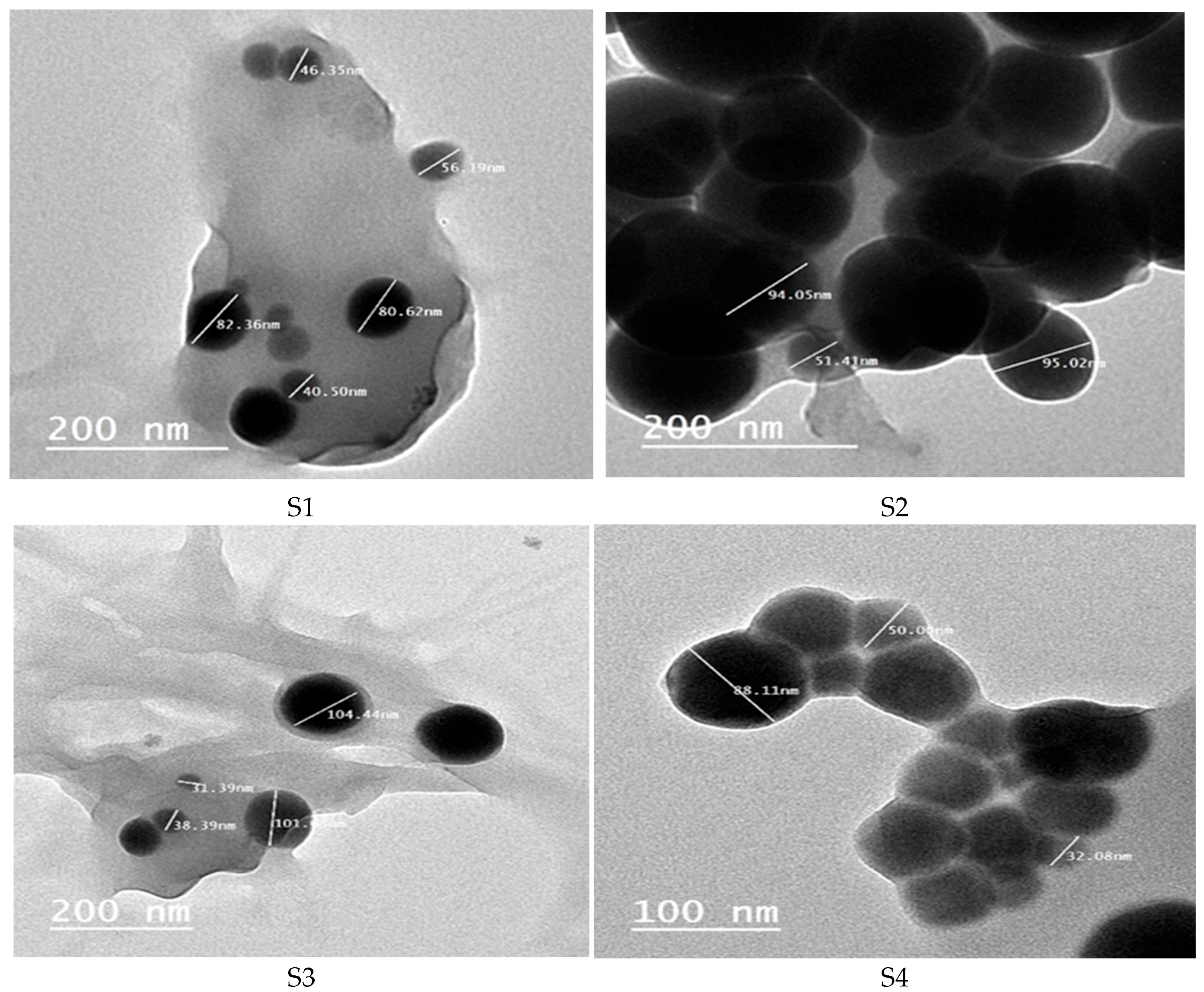
Figure 1. TEM images for Se-NPs produced by isolates S1, S2, S3, and S4.
Applmicrobiol 05 00121 g001
Figure 2. Energy Dispersive X-Ray Analysis of the four Se-NPs samples (S1–S4).
Applmicrobiol 05 00121 g002
Figure 3. UV-vis spectra of Se-NPs produced by S1, S2, S3, and S4 isolates.
Applmicrobiol 05 00121 g003
Figure 4. Fourier Transform Infrared (FTIR) spectroscopy analysis of Se-NPs produced by the four isolates (S1–S4).
Applmicrobiol 05 00121 g004aApplmicrobiol 05 00121 g004b
Figure 5. Zeta potential measurements of prepared Se-NPs produced by the isolates (S2, S3).
Applmicrobiol 05 00121 g005
Figure 6. Anti-biofilm activity of Se-NPs produced by sample S1 at sub-inhibitory concentrations.
Applmicrobiol 05 00121 g006
Figure 7. Anti-biofilm activity of Se-NPs produced by sample S2 at sub-inhibitory concentrations.
Applmicrobiol 05 00121 g007
Figure 8. Anti-biofilm activity of Se-NPs produced by sample S3 at sub-inhibitory concentrations.
Applmicrobiol 05 00121 g008
Figure 9. Anti-biofilm activity of Se-NPs produced by sample S4 at sub-inhibitory concentrations.
Applmicrobiol 05 00121 g009
Figure 10. Drug combination test between: (A) Levofloxacin (Lev.) and Se-NPs (S2), (B) Levofloxacin (Lev.) and Se-NPs (S3) against P. aurginosa (P) and E. faecalis (E).
Applmicrobiol 05 00121 g010
Figure 11. Fractional inhibitory index of the drug combination between S2, S3 Se-NPs and Levofloxacin against P. aurginosa and E. faecalis.
Applmicrobiol 05 00121 g011
Figure 12. Cytotoxicity of the four different Se-NPs on Wi38 cells. (A) Microphotograph of Wi-38 cell under microscopic observation in comparison to untreated cells (a) control cells, (b) cells treated with S1, (c) cells treated with S2, (d) cells treated with S3, and (e) cells treated with S4. (B) Graph illustrates the relation between cell viability and concentration of nanoparticles produced by different sources.
Applmicrobiol 05 00121 g012
Figure 13. Cytotoxicity of the four different Se-NPs on A549 cells. (A) Microphotograph of Wi-38 cell under microscopic observation in comparison to untreated cells (a) control cells, (b) cells treated with S1, (c) cells treated with S2, (d) cells treated with S3 and (e) cells treated with S4. (B) Graph illustrates the relation between cell viability and concentration of nanoparticles produced by different sources.
Applmicrobiol 05 00121 g013
Table 1. Isolate names based on 16S rRNA identification.
Isolate CodeIdentificationSimilarity %
S1 Lactiplantibacillus pentosus 96.87
S2 Lactiplantibacillus plantarum 97.51
S3 Lactiplantibacillus plantarum 98.41
S4 Lactobacillus acidophilus 95.36
Table 2. Pathogens names based on VITEK identification.
Gram Negative IsolatesGram Positive IsolatesFungi
Escherichia coliMRSACandida albicans
Klebsiella pneumoniaeEnterococcus faecalis
Acinetobcter baumanniiStreptococcus agalactiae
Pseudomonas aeruginosa
Proteus mirabilis
Table 3. Zeta potential measurements of prepared Se-NPs.
SampleElectrophoretic Mobility μm * cm/VsAv. Zeta Potential, mVTransmittance %Conductivity mS/cm
S2−2.1542−34.80.20.123
S3−2.7123−27.60.00.057
Table 4. MIC (mg/mL) of Se-NPs prepared by lactic acid bacteria isolates against pathogens.
Human PathogenS1
MIC
(mg/mL)
S2
MIC
(mg/mL)
S3
MIC
(mg/mL)
S4
MIC
(mg/mL)
Gram negative bacteria
Escherichia coli8.12 ± 1.71.07 ± 3.51.125 ± 1.80.625 ± 1.03
Klebsiella pneumoniae16.25 ± 1.73.15 ± 3.518 ± 1.81.25 ± 1.03
Pseudomonas aeruginosa8.12 ± 1.71.07 ± 3.59 ± 1.85 ± 1.03
Proteus mirabilis8.12 ± 1.70.62 ± 3.54.5 ± 1.81.25 ± 1.03
Acinetobacter baumannii1.015 ± 1.70.62 ± 3.50.562 ± 1.80.625 ± 1.03
Gram positive bacteria
MRSA4.06 ± 1.725 ± 3.59 ± 1.81.25 ± 1.03
Streptococcus agalactiae4.06 ± 1.71.07 ± 3.52.25 ± 1.80.625 ± 1.03
Eenterococcus faecalis8.12 ± 1.725 ± 3.59 ± 1.82.5 ± 1.03
Fungus
Candida albicans16.25 ± 1.71.07 ± 3.52.25 ± 1.810 ± 1.03
Results were expressed as mean ± standard error of the mean (M ± SEM).
Table 5. Summary of fractional inhibitory index results.
Test ItemFIC IndexEffect
S2 Se-NPs + Lev. against P0.75Synergism
S2 Se-NPs + Lev. against E2.125Antagonism
S3 Se-NPs + Lev. against P1.25Addition
S3 Se-NPs + Lev. against E2.25Antagonism
Disclaimer/Publisher’s Note: The statements, opinions and data contained in all publications are solely those of the individual author(s) and contributor(s) and not of MDPI and/or the editor(s). MDPI and/or the editor(s) disclaim responsibility for any injury to people or property resulting from any ideas, methods, instructions or products referred to in the content.