Article

Composition and Occurrence of Airborne Fungi in Two Urbanized Areas of the City of Sofia, Bulgaria

by
Margarita Ivanova
1,
Galina Stoyancheva
2,
Vladislava Dishliyska
2,
Jeny Miteva-Staleva
2,
Radoslav Abrashev
2,
Boryana Spasova
2,
Yana Gocheva
2,
Lyudmila Yovchevska
2,
Galina Satchanska
1,
Maria Angelova
2 and
Ekaterina Krumova
2,*
1
Department of Natural Sciences, New Bulgarian University, Montevideo Str. 21, 1618 Sofia, Bulgaria
2
The Stephan Angeloff Institute of Microbiology, Bulgarian Academy of Sciences, Academician G. Bonchev Str. 26, 1113 Sofia, Bulgaria
*
Author to whom correspondence should be addressed.
Appl. Microbiol. 2025, 5(3), 96; https://doi.org/10.3390/applmicrobiol5030096
Submission received: 7 August 2025 / Revised: 2 September 2025 / Accepted: 9 September 2025 / Published: 11 September 2025

Abstract

Air pollution remains one of the most urgent global challenges, affecting both public health and environmental integrity, with its severity escalating in parallel with industrialization and urban expansion. Defined as the presence of harmful substances in the atmosphere, air pollution poses risks to human health and disrupts the development of plant and animal life. Urban areas, particularly large cities, frequently exhibit pollutant concentrations that exceed safety thresholds established by the World Health Organization (WHO). This study presents a comprehensive analysis of airborne fungal microbiota in two distinct districts of Sofia, Bulgaria: the highly urbanized city center (Orlov Most) and a less urbanized southwestern area (New Bulgarian University, Ovcha Kupel). Weekly fluctuations in mold spore abundance were monitored, revealing elevated contamination levels on Fridays, likely due to intensified vehicular traffic preceding weekends and public holidays. Taxonomic identification of dominant mold species was conducted using both classical and molecular genetic methods. The isolated fungal strains predominantly belonged to the phylum Ascomycota (80%), with Talaromyces and Alternaria emerging as the most prevalent genera. Additionally, antifungal susceptibility testing indicated that most isolates were sensitive to commonly used antifungal agents, although resistance was observed in two strains of Talaromyces wortmannii. These findings underscore the significance of fungal bioaerosols in urban air quality assessments and highlight the need for targeted monitoring and mitigation strategies.

1. Introduction

Air pollution poses a significant challenge for residents of major urban centers, where road traffic and industrial activities substantially degrade air quality. The dispersion of pollutants is primarily influenced by natural factors—such as atmospheric stability, wind conditions, and temperature inversions—as well as anthropogenic sources, including emissions from transportation, industry, and residential heating [1,2]. Recent studies indicate that over 96% of the global urban population is exposed to unhealthy levels of fine particulate matter (PM2.5) and nitrogen dioxide (NO2) [3,4]. Global and regional dynamics—such as international trade and the relocation of industrial activities to developing countries—further affect urban air quality [5]. By 2050, approximately 68% of the world’s population is expected to reside in urban areas, which is likely to exacerbate emissions. Although cities have the potential to play a pivotal role in mitigating pollution, 40% of countries lack ground-based air quality monitoring systems. Access to such systems is particularly limited in Africa, where only 6% of children are covered, compared to 72% in Europe and North America [6].
The World Health Organization identifies six major air pollutants: particulate matter, ground-level ozone, carbon monoxide, sulfur oxides, nitrogen oxides, and lead [7]. In addition to these, biological agents—collectively referred to as bioaerosols—are emerging as a growing concern and constitute the focus of this study. Bioaerosols are airborne particles of biological origin, including bacteria, viruses, fungi, pollen, allergens, and mycotoxins [8,9]. They originate from both natural processes and human activities [10,11]. Particle sizes range from 0.003 μm (viruses) to 20 μm (bacteria and pollen), enabling many of them to penetrate deeply into the respiratory system [8]. Their composition and distribution are influenced by particle characteristics (e.g., size, shape, density) and environmental conditions such as temperature, humidity, airflow, and anthropogenic activity [12,13]. Warm and humid conditions promote microbial growth and aerosol formation [14]. Bioaerosol concentrations also vary seasonally and with light exposure—for instance, higher bacterial concentrations have been observed during summer and autumn in cities such as Beijing, Washington, and Moscow [8,15]. Fungal bioaerosols exhibit similar seasonal patterns [16]. Over the past two decades, scientific interest in bioaerosols has increased dramatically, with the number of studies rising more than 100-fold [17].
Soil is the primary natural source of bioaerosols. Its rich organic content sustains diverse microbial communities [18], which are dispersed into the atmosphere by wind or precipitation. Studies in cities such as Madrid have shown that most airborne pathogenic bacteria and fungi originate from soil [19,20]. Raindrops can further facilitate the release of microbes from soil surfaces. Aquatic environments—including rivers, lakes, and seas—also contain various biological particles [21,22], contributing to natural bioaerosol emissions, along with vegetation and animals [23,24,25].
Human activity is also a significant contributor to bioaerosol emissions. The human body and everyday behaviors release biological particles, particularly in indoor environments, where they can persist for extended periods and pose health risks [26]. Major anthropogenic sources include farms, landfills, and industrial processing facilities, which emit microbial toxins and metabolites [27,28,29].
Given the critical role of bioaerosols in human health, their monitoring is essential for assessing the total number of viable microorganisms. Microbiological analysis of air is conducted using both culture-dependent and culture-independent techniques. The use of culture-dependent methods is limited, as only a small fraction of microorganisms (approximately 1%) can be cultivated under laboratory conditions [30,31,32]. The influence of certain abiotic factors further restricts these approaches, a limitation that can be overcome through the application of metagenomic analysis. Molecular techniques offer greater accuracy and provide insights into both culturable and non-culturable microorganisms [33].
Assessing fungal spores in bioaerosols is essential for understanding respiratory health in densely populated areas. Climatic factors—such as temperature, humidity, precipitation, wind speed, and direction—strongly influence the concentrations of fungal bioaerosols [34,35]. Peak spore levels are typically observed in spring, followed by summer, winter, and autumn. Diurnal variations and the presence of fog also affect fungal diversity, with greater species richness recorded during nighttime hours [36]. Approximately 8–10% of the identified fungal species are pathogenic.
Fungal species isolated from the atmosphere predominantly belong to the phyla Ascomycota and Basidiomycota. Numerous studies have shown that the most widely distributed airborne spores are the asexual spores of saprophytic fungi from the genus Cladosporium, followed by Penicillium and Aspergillus [37,38,39,40]. Pathogenic representatives frequently detected in bioaerosols include Aspergillus sp., Fusarium sp., Scedosporium sp., and Mucor sp. [41,42,43]. In Taipei, Taiwan, ascospores (Ascomycota) dominate urban areas, with Aspergillus and Penicillium being prevalent, while basidiospores (Basidiomycota) are more common in less trafficked zones [33].
In Sofia, fungal isolates from the air are primarily affiliated with Ascomycota [44]. Seasonal patterns reveal higher concentrations of fungal bioaerosols in summer compared to spring, with recorded levels of 987.0 CFU/m3 in summer versus 228.0 CFU/m3 in spring [44]. Following a metagenomic analysis, Angelova and Iliev [33] reported Cladosporium as the most dominant genus, accounting for 64% of all fungal sequences in air samples collected from central Sofia. Fifteen percent of sequences belonged to Alternaria, 2% to Pseudopithomyces, and 2% to Aureobasidium, and 11% remained unclassified. In addition to Ascomycota, representatives of Basidiomycota (1.94%) and Chytridiomycota (0.04%) were also identified. The presence of all three phyla has likewise been confirmed in the air of Seoul, South Korea [45], where Cladosporium again emerged as the dominant genus. The predominance of Ascomycota and Cladosporium has been consistently reported in other metagenomic studies [46]. In Xi’an, China, Li et al. identified Aspergillus, Aureobasidium, Penicillium, and Paecilomyces as the dominant genera in urban air samples [47].
The majority of airborne mycoflora consists of spores that can remain suspended for varying durations and travel distances, ranging from mere centimeters to hundreds of kilometers—even across continents—facilitated by raindrops and air currents [48]. These spores contribute to air pollution, alter environmental biotic factors, and impact human health. Their diversity and concentration depend on environmental conditions, human activity, and substrate availability.
Fungal bioaerosols can cause infectious diseases, allergic reactions, and toxic effects on living organisms at local, regional, and global scales [49]. Numerous fungal pathogens affecting plants, animals, and humans are transmitted via airborne droplets and are capable of long-distance dispersal, thereby facilitating the transcontinental spread of disease. It is known that over 100 fungal species identified in bioaerosols are responsible for serious infections in animals and humans [50]. For instance, Aspergillus flavus produces aflatoxin, a potent carcinogen linked to liver and lung cancer. Inhalation of fungal spores and hyphal fragments may lead to aspergillosis, hypersensitivity pneumonitis, asthma, and systemic toxicosis. A. fumigatus accounts for approximately 90% of invasive aspergillosis cases, while A. niger is associated with invasive pulmonary diseases.
Airborne fungi from genera such as Alternaria, Penicillium, Aspergillus, and Cladosporium are strongly associated with respiratory allergies, asthma exacerbations, and severe respiratory complications, which in some cases may result in respiratory failure or death [51,52].

Status of the Problem in Sofia

Sofia, the capital of Bulgaria, is situated in a mountainous valley—a geographical feature that contributes to frequent occurrences of fog and smog. Despite the surrounding mountain ranges, there are days with minimal air circulation. Urbanization exacerbates the issue: high-rise buildings are densely concentrated, green spaces are diminishing, and the expanding built environment disrupts the natural air corridor known as the “Vladaysko current,” which no longer delivers fresh air to the city center.
While existing studies in Sofia primarily focus on abiotic air pollutants, biotic pollution—particularly outdoor bioaerosol concentrations—remains largely underexplored. The city’s unique ecological characteristics contribute to the accumulation of pollutants and underscore the need for targeted scientific analyses. The rapid population growth and intense urbanization of the capital further emphasize the necessity for systematic monitoring of bioaerosol components.
Iliev et al. [44] reported that the highest concentrations of fungal aerosols were recorded in June and July. In springtime, bioaerosol concentrations exhibited a notable association with a broader range of environmental variables, including ambient temperature, solar intensity, and the presence of accompanying pollutants. Despite the importance of such research, studies to date have been limited and fragmented.
The aim of this study was to compare airborne mycobiota in highly urbanized versus less urbanized zones of Sofia. The specific objectives of the research were
  • To assess the weekly dynamics of fungal spore distribution at the two sampling sites;
  • To determine the taxonomic affiliation of the isolated fungi and identify the dominant fungal genera and strains;
  • To test the sensitivity of the isolates to commercially available antifungal agents.
Such investigations would enrich the scientific understanding of this issue and could serve as a foundation for establishing a monitoring system to track air quality and its impact on human health.

2. Materials and Methods

2.1. Research Focus and Sampling Locations

This study investigates bioaerosol contamination by filamentous fungi in the atmospheric air over two selected locations in Sofia, Bulgaria. The first site is situated in the highly urbanized central area of the city (Orlov Most), while the second is located in the western part of Sofia, in the vicinity of New Bulgarian University (NBU) (Figure 1). The specific meteorological conditions in the city of Sofia during the sampling period are presented in Table 1.

2.2. Sampling Methods

Two complementary sampling techniques were employed to collect airborne fungal spores.

2.2.1. Koch Sedimentation Method

This passive sampling method relies on the gravitational settling of airborne microorganisms onto a solid nutrient medium. At each sampling site, Petri dishes with a diameter of 9 cm containing the culture media Yeast Extract Glucose Chloramphenicol Agar (YGC) and Gause Medium (G1) (Hi-Media, Thane, India) were exposed to ambient air for 20 min at a height of approximately 150 cm above ground level. This method allows for the qualitative and quantitative assessment of fungal contamination in the air.

2.2.2. Andersen Cascade Impactor

The Andersen impactor is a widely used active air sampling device designed to capture bioaerosol particles based on their aerodynamic diameter. It consists of six stages, each equipped with a Petri dish containing agarized culture medium. As air is drawn through the impactor, particles are separated by size and deposited directly onto the medium. Following collection, the samples are incubated and monitored for microbial growth.
The exposed plates were incubated at 28 °C for 7 days. The samples were taken between 15:00 and 17:30 each day. All experiments were performed in three repetitions, i.e., three Petri dishes per sample.

2.3. Culture Media and Incubation Conditions

To isolate filamentous fungi, two types of culture media were used:
  • Yeast Extract Glucose Chloramphenicol Agar (YGC): Yeast extract 5.0, D (+)-Glucose 20.0, Chloramphenicol 0.1, Agar 14.9, final pH 6.6 ± 0.2 at 25 °C;
  • Gause Medium (G1): Soluble starch 20.0 g, KNO3 1.0 g, NaCl 0.5 g, MgSO4 × 7H2O 0.5 g, K2HPO4 0.5 g, FeSO4 × 7H2O 10.0 mg, Agar 15.0 g, distilled water 1.0 L, Adjusted pH 7.4.
Petri dishes containing YGC and G1 media were incubated at 25 °C for 72 to 96 h. After the incubation period, the number of germinated fungal colonies was counted to assess bioaerosol concentration. The isolated fungal strains were subsequently subcultured and maintained on Potato Dextrose Agar (PDA) for further analysis.

2.4. Isolation of Pure Cultures

Germinated single fungal colonies were transferred to Petri dishes containing PDA to obtain pure cultures. These cultures were subsequently maintained on slanted PDA in test tubes and stored at 4 °C for further analysis.

2.5. Identification of Fungal Isolates

2.5.1. Classical Identification Methods

Fungal isolates were identified to the genus level based on macromorphological and micromorphological characteristics, following the guidelines outlined in the Compendium of Soil Fungi (1980) [53]. The nutrient media used for cultivation included Beer Agar (BA), Sabouraud Agar, and PDA. Cultures were incubated at 28 °C for 7 days.

2.5.2. Molecular Genetic Identification

DNA Isolation and PCR Amplification
Chromosomal DNA was isolated from 100 mg of 48 h mycelia with the GeneMATRIX Plant & Fungi DNA Purification Kit (EURx Ltd., Gdańsk, Poland) according to the manufacturer’s instructions.
PCR was performed with a BioRad iCycler Thermal Cycler (Bio-Rad Laboratories, Hercules, CA, USA) using PCR master mix (GenetBio, Daejeon, Republic of Korea). PCR products were visualized with 1% agarose gel electrophoresis and purified with Gene JET PCR Purification Kit (Thermo Fisher Scientific Inc., Waltham, MA, USA). PCR was used to amplify a DNA phylogenetic marker (ITSs 1 and 2 of the rRNA gene cluster, including the 5.8S rRNA). We used the following primers: ITS1 (5′-TCCGTAGGTGAACCTGCGG-3′) and ITS4 (5′-TCCTCCGCTTATTGATATGC-3′).
The complete sequence of internal transcribed spacers 1 and 2 (ITS1 and ITS2), including the sequence of the gene encoding 5.8 S rRNA, is a primary DNA barcode locus for identification of fungi [54,55]. Primer synthesis and sequencing were performed at Macrogen Europe (Amsterdam, The Netherlands).
Bioinformatics programs and analyses. Molecular identification (DNA barcoding) is based on the similarity of DNA fragments. The nucleotide sequences were processed and analyzed with the Chromas and CAP3 programs. The BLAST application at NCBI (National Center for Biotechnology Information, Bethesda, MD, USA) was used to compare the sequences with sequences available in the GenBank database (http://www.ncbi.nlm.nih.gov/BLAST, 11 June 2025). All data were also compared with Index Fungorum (https://www.indexfungorum.org/names/names.asp, 11 June 2025) and Mycobank (https://www.mycobank.org/, 11 June 2025). The sequences of the identified strains have been deposited in GenBank.

2.6. Estimation of Airborne Fungal Diversity

The fungal diversity in the studied locations was evaluated through diversity parameters, including the Shannon–Wiener index (H′), Simpson’s index (D), and Sorensen index [56,57].

2.7. Sensitivity of Dominant Fungal Species to Antifungal Agents

The antifungal susceptibility of dominant fungal strains was assessed using the microdilution method in liquid nutrient medium. Nystatin and amphotericin B were employed as antifungal agents. Fungal spores were cultivated in 96-well plates containing potato dextrose broth, with resazurin added as a growth indicator.
The antifungal agents were tested at concentrations of 50 μg/well, 25 μg/well, 12 μg/well, and 6 μg/well. Growth inhibition was monitored over a period ranging from 24 h to 7 days. Negative controls consisted of the medium with spores but without antifungal agents. Absorbance was measured using an ELISA reader at 600 nm. The percentage of inhibition was calculated relative to the initial measurement at 0 h, which was considered 100%.

2.8. Determination of the Minimum Inhibitory Concentration of the Two Tested Antifungal Preparations, Nystatin and Amphotericin, Against the Identified Fungal Species

The minimum inhibitory concentration was defined as the minimum concentration of the antifungal preparation that inhibits the visible growth of the tested fungal strain (resazurin microdilution method).

2.9. Statistical Evaluation

The results obtained in this investigation were evaluated through at least three repeated experiments using three parallel runs, and the reported values represent the means. The error bars indicate the standard deviation (SD) of the mean of triplicate experiments. The data were analyzed using one-way analysis of variance (ANOVA), followed by Tukey’s test. For the statistical processing of the data, the version of the ANOVA software built into the Origin program (OriginPro 2019b, 64-bit) was used.

3. Results

3.1. Quantitative Analysis

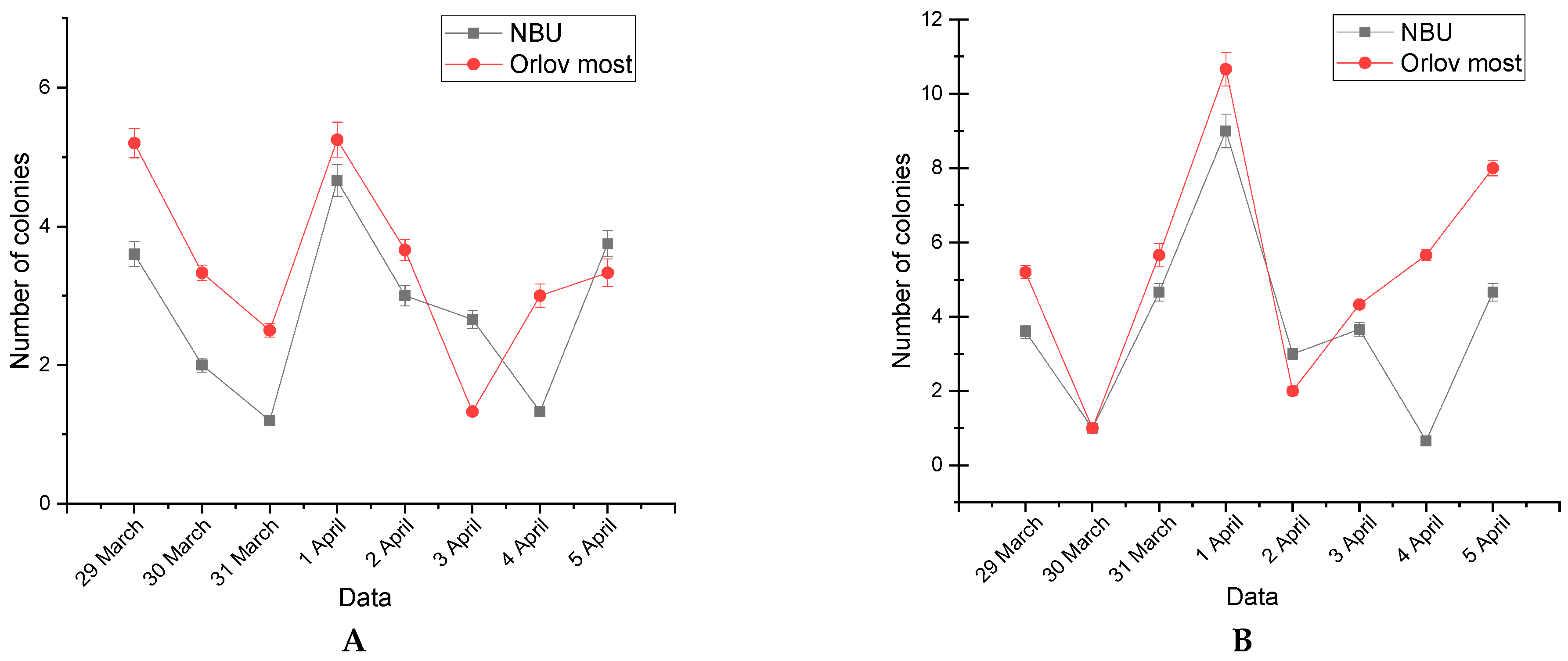
The number of fungal colonies grown on Petri dishes exposed to ambient air at the two sampling locations is presented in Figure 2. Two types of nutrient media were used for the isolation of filamentous fungi: Gause Medium (G1) (Figure 2A) and Yeast Extract Glucose Chloramphenicol Agar (YGC) (Figure 2B).

3.2. Weekly Dynamics of Airborne Fungal Spores

The weekly dynamics of airborne fungal spore contamination in the two studied areas were assessed through daily sampling. The results are illustrated in Figure 2C,D.
During the initial sampling on Tuesday, 29 March, elevated levels of fungal spores were detected on both nutrient media (G1 and YGC). Over the following two days (Wednesday and Thursday), a noticeable decline in spore concentrations was observed. On Friday, fungal contamination levels increased again, probably due to intensified vehicular traffic associated with the upcoming weekend and public holidays. During the weekend (Saturday and Sunday), the number of isolated mold colonies decreased, followed by a renewed increase on Monday and Tuesday, suggesting a cyclical pattern influenced by human activity and urban dynamics.
Although microorganisms are ubiquitous in the atmosphere, their abundance and distribution are shaped by environmental conditions and geographic location. The initial working hypothesis posited that the distribution of filamentous fungi would differ between the two sampling sites. However, the quantitative analysis revealed that the overall number of airborne spores was relatively similar in both locations (Figure 2), indicating that urban fungal bioaerosol levels may be governed more by temporal factors than by spatial ones.
One possible explanation is that the weekly fluctuations in fungal spore concentrations are largely attributable to anthropogenic influences, such as traffic density, human movement, and urban activity patterns.

3.3. Identification of Fungal Isolates

3.3.1. Classical Taxonomic Identification

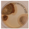
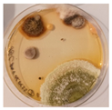
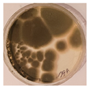
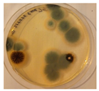
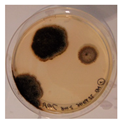
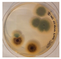
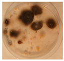
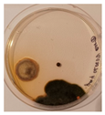
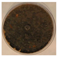
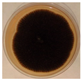
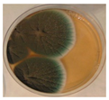
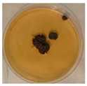
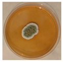
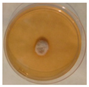
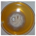
A total of 126 air samples were collected, from which 142 pure fungal cultures were successfully isolated (Table 2). Identification of the isolates was performed using classical taxonomic methods, based on the morphological characteristics of filamentous fungi.
Based on morphological similarities among the isolates, the total number was reduced to 24 representative strains, which were identified to the genus level. Table 3 presents selected macroscopic images of representative genera identified in the study.
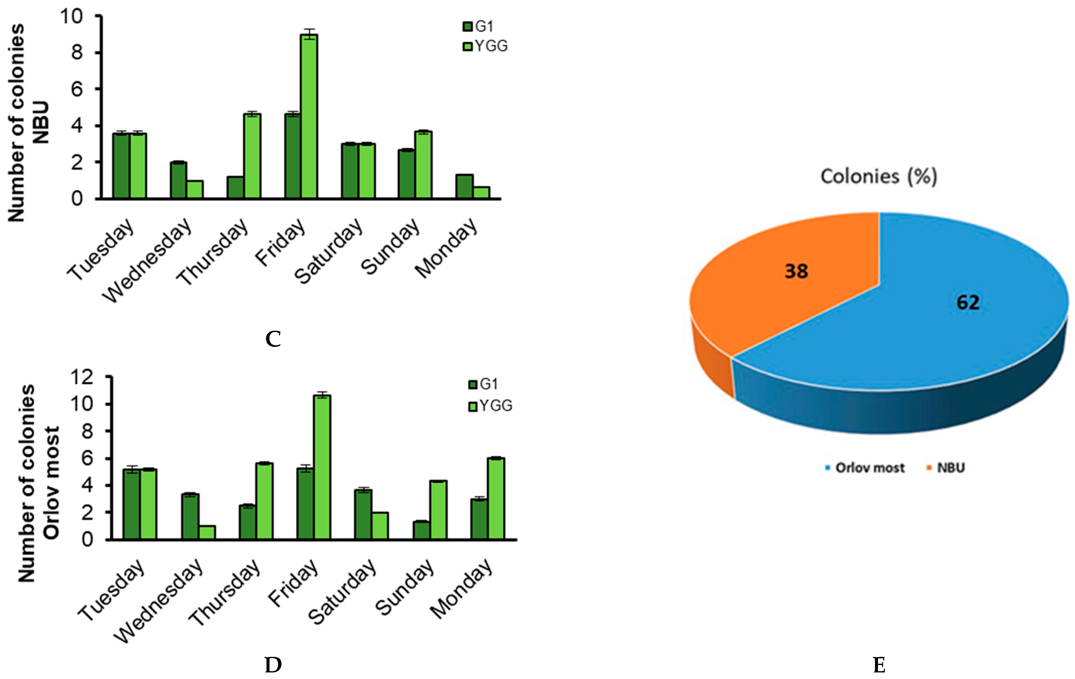
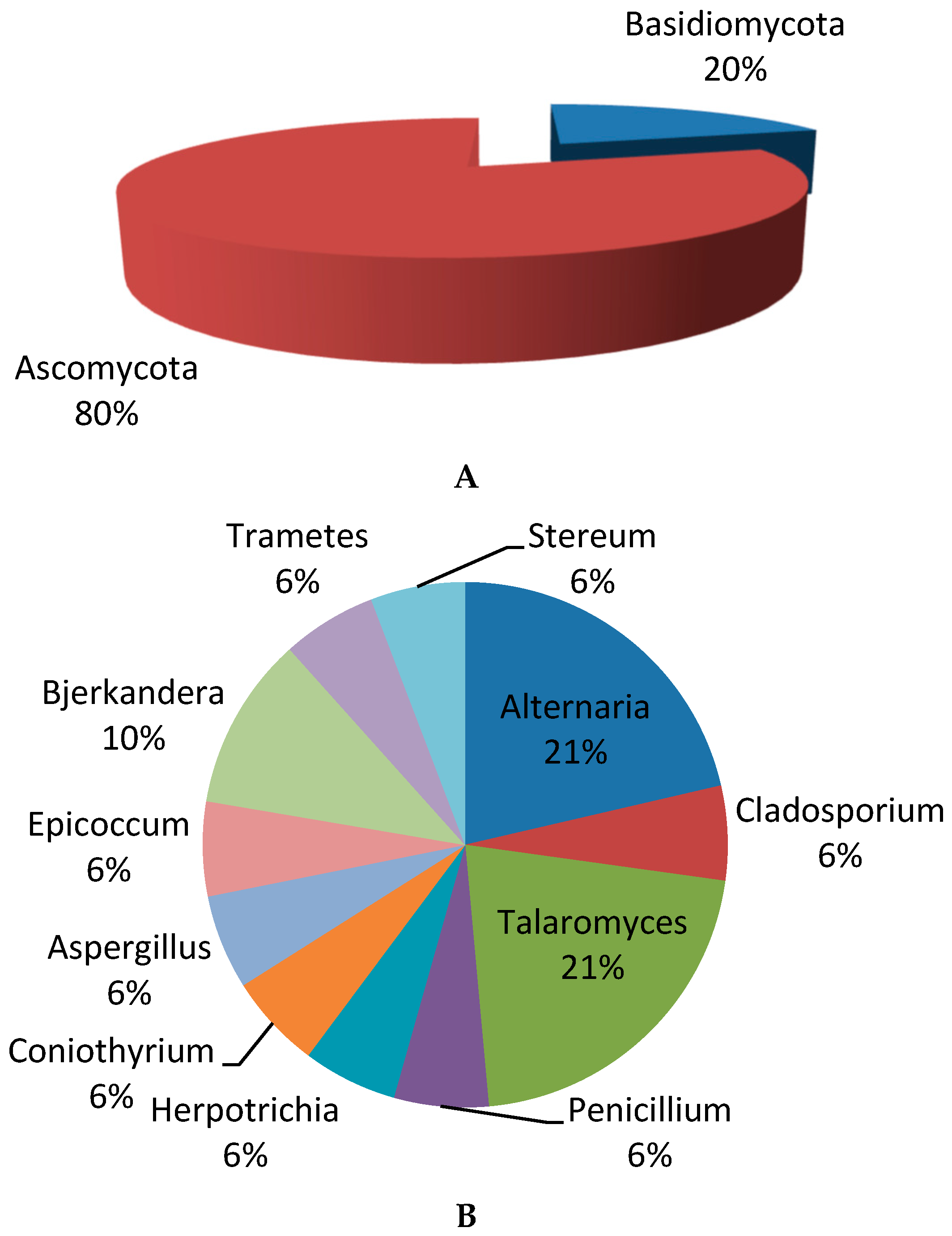
The analysis of the identified isolates established that 80% of the isolated fungi belong to the Ascomycota division and 20% to Basidiomycota (Figure 3A). Representatives of the genera Alternaria (21%) and Talaromyces (21%) dominate (Figure 3B). In addition to the previously described isolates, representatives of the genera Penicillium, Alternaria, Herpotrichia, Stereum, Trametes, Bjerkandera, Talaromyces, and Epicoccum were identified.
Species from these genera are well-documented as common allergens and are frequently associated with respiratory conditions in humans [51,58,59].

3.3.2. Molecular Genetic Identification

To achieve species-level identification of the fungal isolates, molecular genetic methods were employed. These techniques are based on the conservation of the small ribosomal RNA subunit, which serves as a reliable taxonomic marker.
Polymerase Chain Reaction (PCR) analysis was used to amplify target DNA sequences from filamentous fungi using specific primers homologous to conserved regions of the fungal genome. This approach offers high sensitivity and rapid diagnostic capability, surpassing classical microbiological techniques in precision and efficiency [59]. It applies to a broad spectrum of fungal genera isolated from diverse environmental sources. For precise taxonomic classification, genomic DNA was extracted and purified from the isolates. PCR amplification targeted the internal transcribed spacer (ITS) region of ribosomal DNA using universal primers (see Section 2). These primers were designed based on reference rDNA sequences from various fungal species [60], optimized for high annealing temperatures to minimize non-specific amplification. The resulting sequences were analyzed and compared with published ITS sequences in the NCBI GenBank database. Sequence alignment and similarity analysis confirmed species-level identification by matching the obtained sequences with those of reference strains. The results of this comparative analysis are presented in Table 4.
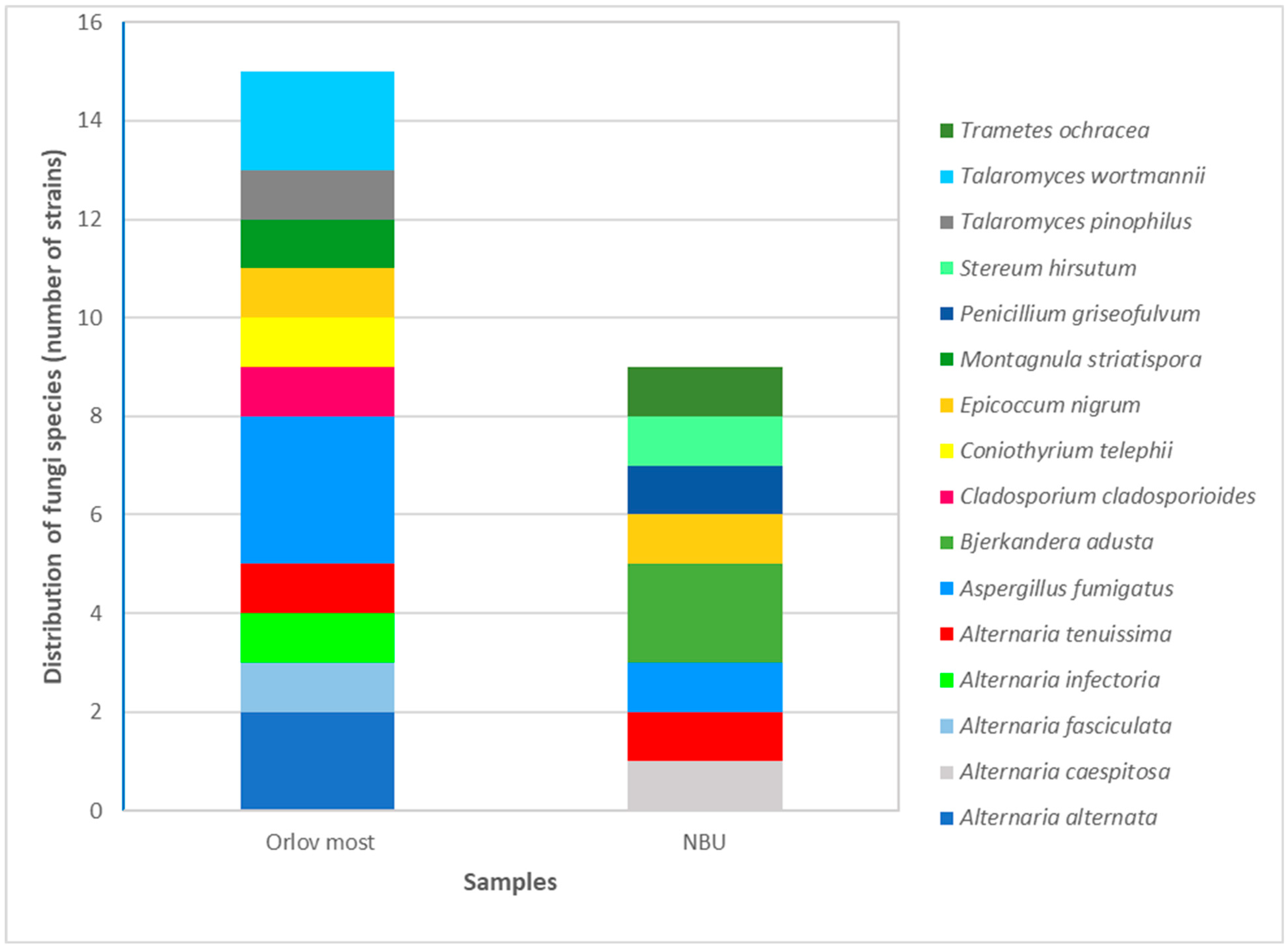
The taxonomic diversity of fungal isolates collected from the two sites studied was assessed. As shown in Figure 4, a higher number of genera and species were identified in samples from Orlov Most compared to those from the vicinity of NBU.
Table 5 provides additional information on the diversity of fungi in the studied locations, represented by Shannon, Simpson, and Sørensen indices.
Orlov Most has a higher species diversity and a more balanced ecosystem according to the Shannon index, while the Simpson index shows lower species dominance at NBU. The Sorensen index reports low similarity between the two locations—only 25% species coincidence, with Orlov Most showing higher fungal diversity.
This increased diversity is likely due to the higher traffic density and greater pedestrian activity in the central urban area of Orlov Most, which may contribute to elevated concentrations of airborne fungal spores and a broader representation of species.

3.4. Sensitivity of Isolated Fungal Strains to the Antifungal Agents Nystatin and Amphotericin

Given that many of the identified fungal species are known to cause various human diseases, the sensitivity of selected isolates to two antifungal agents—nystatin and amphotericin B—was examined. The results are presented in Table 6 and Table 7.
The analysis showed that isolates from the genus Alternaria were sensitive to both antifungal agents, with a more pronounced response to nystatin. Three out of four Alternaria strains exhibited complete growth inhibition for up to seven days when treated with nystatin at a concentration of 50 μg/well. All studied Alternaria strains also demonstrated sensitivity to amphotericin B, with growth suppression observed for up to 96 h after the onset of cultivation.
These findings suggest that nystatin may be more effective than amphotericin B against Alternaria strains under the tested conditions. Further studies could explore the mechanisms of resistance and the broader applicability of these agents to other genera identified in the study. The Cladosporium strain exhibited high sensitivity to both nystatin and amphotericin B, with complete growth inhibition observed for up to 7 days following the start of cultivation.
Among the four identified representatives of the genus Talaromyces and the single Penicillium strain, sensitivity to nystatin was generally higher. Notably, Talaromyces wortmannii 83 and Talaromyces pinophilus 82 exhibited sustained growth inhibition up to day 7 in the presence of nystatin. In contrast, T. wortmannii 83 showed no sensitivity to amphotericin B, while T. wortmannii 322 did not respond to nystatin (see Table 6 and Table 7). The remaining isolates from these genera demonstrated sensitivity to both antifungal agents at higher concentrations, with growth inhibition observed for up to 96 h.
These findings underscore a critical point: strains belonging to the same species may exhibit variable sensitivity to antifungal agents, highlighting the importance of strain-level analysis in antifungal susceptibility testing.
All other fungal species examined demonstrated sensitivity to both antifungal agents. Among them, Stereum hirsutum 921 and Epicoccum nigrum 621 exhibited growth inhibition lasting up to seven days. Coniothyrium telephii 92 showed growth suppression for up to 72 h following antifungal treatment. Herpotrichia striatispora 62 displayed greater sensitivity to amphotericin B, with growth inhibition observed for 72 h with nystatin and 96 h with amphotericin. Aspergillus fumigatus 84 also showed enhanced sensitivity to amphotericin B, with sustained growth inhibition for 96 h across all tested concentrations.
Isolates belonging to the phylum Basidiomycota exhibited pronounced sensitivity to both antifungal agents. As shown in Table 6 and Table 7, all tested concentrations of nystatin and amphotericin B affected fungal growth, with the highest concentrations resulting in complete inhibition by day 7.
The minimum inhibitory concentrations (MICs) of nystatin and amphotericin B against the identified fungal species were determined and are presented in Table 8.
Based on the results of this experimental series, it can be concluded that the filamentous fungi isolated from the atmospheric air over the two studied locations in Sofia exhibit sensitivity to the antifungal agents nystatin and amphotericin B. Notable exceptions include Talaromyces wortmannii 83, which demonstrated resistance to all tested concentrations of amphotericin B, and Talaromyces wortmannii 322, which showed no sensitivity to nystatin. These findings highlight the importance of strain-specific susceptibility testing when assessing the potential health risks associated with airborne fungal species.

4. Discussion

Air pollution remains a major threat to both human health and environmental integrity. While considerable attention has been devoted to the adverse effects of fine particulate matter (PM), significantly less focus has been placed on the potential hazards associated with the increased presence of microorganisms in the ground-level air layer.
Few studies have examined the natural background levels of culturable microorganisms in relation to ambient particulate matter. However, such assessments are crucial, as the composition of both bioaerosols and PM varies widely across geographic regions. Factors such as topography, meteorological conditions, and urban activity can significantly influence bioaerosol content.
Previous research has documented background concentrations of culturable fungal spores and particulate matter in diverse regions [61]. One study found that light cloud cover and light wind positively correlated with fungal spore concentrations, while relative humidity was negatively associated with total fungal abundance [60]. In a recent investigation, Grigorova-Pesheva and Kadinov (2024) [62] examined differences in airborne microbial communities between urban “hot spots” and green areas at varying altitudes. Their findings revealed that both the composition and abundance of airborne bacteria, fungi, and actinomycetes varied depending on location and altitude. Hot spots exhibited higher total microbial counts, and certain microbial groups showed altitude-dependent distribution patterns. The study suggests a strong correlation between vehicular traffic intensity and elevated levels of airborne microorganisms [62].
Our results support these observations, demonstrating weekly dynamics in fungal spore concentrations (see Figure 2). Notably, elevated levels of fungal contamination were recorded on Fridays, which may be attributed to increased traffic and urban activity preceding weekends and public holidays.
The two sampling locations selected for this study are situated in distinct areas of Sofia: one in the central urban core (Orlov Most) and the other in the western part of the city near New Bulgarian University. The initial working hypothesis posited that the degree of bioaerosol contamination would differ between the two sites due to variations in urban density and human activity. Interestingly, the quantitative analysis of airborne fungal spores revealed comparable concentrations in both locations (see Figure 2). This finding likely reflects the high level of urbanization across both areas, suggesting that anthropogenic factors—such as traffic, construction, and population density—play a dominant role in shaping bioaerosol levels. These factors also appear to influence the weekly cyclical patterns of biocontamination observed in the study. Supporting this interpretation, Hai et al. (2019) [63] reported significantly higher concentrations of bacterial bioaerosols during weekdays compared to weekends, highlighting the impact of human activity on airborne microbial dynamics [63]. Our findings similarly confirm the influence of anthropogenic pressure on bioaerosol contamination in urban air.
Fungal spores are widely dispersed in the atmosphere, earning them the designation “airborne fungi” [64]. Their concentration in different environments varies depending on multiple factors, including the availability of fungal substrates, meteorological conditions, and human activities [65,66,67,68]. Airborne fungi are increasingly recognized as contributors to air pollution and have been implicated in adverse health effects in humans, animals, and plants [69,70,71,72].
In the context of urban air contamination, it is essential to determine the species composition of airborne fungi and assess their potential health impacts. A study conducted at three locations in Sofia—Sofia Tech Park, Orlov Most, and Dragan Tsankov Blvd—identified filamentous fungi from the genera Aspergillus, Penicillium, Cladosporium, Botrytis, and Symmetrospora in bioaerosol samples collected above these areas [73]. In their research on the microbiota of the air in Sofia, they mainly focused on bacterial isolates. Information on fungi is scarcer. Our study focused on airborne fungi and the potential danger of these fungi to human health.
Published data in the scientific literature consistently report that fungal spores from the genera Alternaria, Aspergillus, Penicillium, and Cladosporium are among the most frequently isolated in similar bioaerosol studies [74,75,76,77,78,79,80]. These genera have been identified as dominant airborne fungi in urban environments across various geographic regions, including Doha, Qatar [66]; Istanbul, Turkey [81]; Tulsa, USA [82]; and Ho Chi Minh City, Vietnam [63]. Several international studies have characterized dominant airborne fungal genera across various regions: In Tianjin, China, the most prevalent genera include Alternaria (35%), Cladosporium (18%), Penicillium (5.6%), Talaromyces (3.9%), Didymella (3%), and Aspergillus (2.8%). The most frequently identified species were A. alternata (24.7%), C. cladosporioides (11%), A. tenuissima (5.3%), P. oxalicum (4.53%), and T. funiculosus (2.66%) [73]. In Styria, Austria, the dominant genera include Cladosporium, Penicillium, and Aspergillus, with A. fumigatus being particularly prevalent [83]. In Monterrey, Mexico, high concentrations of Cladosporium, Aspergillus, Fusarium, and Penicillium were detected in downtown areas [84].
These findings reinforce the global relevance of airborne fungal contamination and highlight the need for continued monitoring and species-level identification in urban environments.
In Ahvaz, Iran, Cladosporium and Alternaria dominate the outdoor air, while Cladosporium, Aspergillus, and Penicillium are prevalent in indoor environments [85]. In our study, representatives of both Ascomycota and Basidiomycota phyla were isolated, including the genera Alternaria, Talaromyces, Penicillium, Aspergillus, Cladosporium, Epicoccum, Bjerkandera, Trametes, Stereum, Coniothyrium, and Herpotrichia. Notably, Alternaria and Talaromyces were the most frequently detected (see Figure 3).
The fungal diversity and dominance are presented in Figure 4. It clearly shows a larger number of fungal strains in the Orlov Most location compared to the NBU location. As is known, biodiversity can be described by different indices: species richness (the number of species) and evenness (how abundant each species is). Our investigation included the Shannon and Simpson indices (Table 5). Orlov Most has a higher Shannon index, which means greater diversity and a more even distribution of species; i.e., it has a more diverse and balanced ecosystem than the other location studied. The Simpson index shows that NBU has a lower dominance and a more even distribution of species. The slight discrepancy in the two results comes from the fact that the Shannon index is more sensitive to rare species, and the Simpson index to dominant ones. The Sorensen index shows the similarities in communities (the commonality between communities in terms of species). Based on our results the calculated Sorensen index comparing the biodiversity of fungi in two samples shows that the fungal communities in the two locations do not have much similarity. The diversity of fungal species is higher in the Orlov Most location, with only 25% of the species overlapping between the two locations (Table 5).
Many of the filamentous fungi identified in this study are recognized as potential human pathogens. These organisms can colonize the human body and affect the respiratory system through the release of toxins, proteases, enzymes, and volatile organic compounds (VOCs). Consequently, molds may elicit a stronger immunological response than common allergens such as pollen. The immune response to fungal allergens involves Type I to Type IV hypersensitivity reactions, which can manifest as a wide range of clinical conditions, including allergic rhinitis; asthma; allergic bronchopulmonary mycosis (ABPM); and atopic dermatitis (AD).
Fungal allergies are particularly challenging to diagnose and treat due to the diversity and antigenic variability of fungal species. Reported prevalence rates range from 6% to 24% in the general population, reaching 44% among atopic individuals and up to 80% in asthmatics [60]. Prominent fungal genera implicated in allergic rhinitis include Alternaria, Aspergillus, Cladosporium, Curvularia, and Penicillium. Due to their small size, fungal spores can penetrate deeply into the lungs, inducing chronic inflammation. Genera such as Alternaria spp., Aspergillus spp., Cladosporium spp., Helminthosporium, Epicoccum, Aureobasidium, and Penicillium spp. are frequently associated with allergic asthma [86].
In the context of atopic dermatitis, Malassezia furfur has been linked to disease pathogenesis [87], while Saccharomyces cerevisiae has shown strong correlations with positive skin prick test (SPT) reactions [88].
In all samples collected from Sofia—specifically from Sofia Tech Park, Orlov Most, and Dragan Tsankov Blvd.—Penicillium and Aspergillus were consistently present. Sampling was conducted using the Hygitest 106 (Maimex) device and the Koch sedimentation method. A LIDAR beam was directed toward Tsarigradsko Shose Blvd. to monitor particulate dispersion [73].
The growing concern surrounding airborne fungal pathogens is largely driven by the increasing number of immunocompromised individuals. In addition to causing invasive infections, fungal spores can trigger allergic reactions and toxigenic syndromes. Many fungi produce secondary metabolites, such as mycotoxins, which are harmful upon inhalation or contact. Exposure to high concentrations of VOCs may result in fatigue, headaches, and irritation of the eyes, nose, and throat [89]. Fungal genera such as Aspergillus, Candida, Penicillium, Cladosporium, and Rhizomucor are well-documented opportunistic pathogens capable of causing life-threatening infections, particularly in immunosuppressed individuals [90,91].
In Bulgaria, Alternaria alternata and other fungal species have frequently been implicated in respiratory allergies. Component-resolved allergy diagnostics using Alt a 1, the major allergen of A. alternata, have provided compelling evidence of species-specific sensitization in affected individuals [92].
Given the pathogenic potential of many fungal genera identified in our study, we proceeded to investigate their susceptibility to two commonly used antifungal agents—nystatin and amphotericin B. Our results demonstrated that the majority of fungal isolates were sensitive to the conventional antifungal agents nystatin and amphotericin B. The only exceptions were two strains of Talaromyces wortmannii, which exhibited resistance to both agents under the tested conditions.
T. wortmannii has previously been isolated from soil samples and plant hosts such as Aloe vera and Tripterygium wilfordii [93]. This species is known to produce a wide array of bioactive secondary metabolites with antimicrobial and anticancer properties, including Cyclic peptides: Talaromins A and B; Polyketides: Deacetylisowortmin; Quinones: Biemodin, Skyrin; and Macrolides: Vermiculine and Wortmannilactones A–H. Extracts from T. wortmannii cultures have shown cytotoxic activity against the cancer cell lines HepG2 (liver), MCF7 (breast), and A549 (lung). Tests on the normal cell line WI-38 indicated acceptable selectivity indices, suggesting potential therapeutic applications. In addition to known compounds such as stigmasterol, stigmasterol glucoside, thymine, and uracil, a novel phytoceramide—talaroceramide—has also been identified among its metabolites [93]. Finally, recent reports by Chinese researchers have implicated T. wortmannii as a causative agent of subcutaneous infections [94], further highlighting the clinical relevance of this species.
The current study has several limitations. The study focuses on only two sites in the Sofia district and specific periods, which may not represent broader regional or seasonal variations in fungal populations. Future research should incorporate year-round monitoring across multiple locations to better capture seasonal dynamics and environmental influences on fungal diversity and abundance. Such efforts will enhance the accuracy and generalizability of findings related to airborne fungal communities.

5. Conclusions

This study highlights the diversity and abundance of airborne fungal genera in urban environments, with genera such as Alternaria, Talaromyces, Aspergillus, Penicillium, and Cladosporium consistently detected in outdoor air samples.
A clear weekly dynamic in airborne fungal contamination was observed across the two locations in Sofia, influenced by anthropogenic factors such as traffic density and urban activity. The quantitative analysis revealed differences in the distribution of filamentous fungal spores between the two regions, despite their similar urban profiles.
Several of the identified genera include species with allergenic or pathogenic potential, posing health risks, particularly to immunocompromised individuals. The observed resistance of Talaromyces wortmannii strains to conventional antifungal agents emphasizes the need for ongoing surveillance and susceptibility testing.
Moreover, the bioactive metabolites produced by these fungi—some with promising antimicrobial and anticancer properties—highlight their dual role as both environmental health threats and potential biomedical resources.
As a primary study on airborne mycodiversity in different parts of Sofia, the obtained results present information that can impact risk assessment and future air quality monitoring related to human health.

Author Contributions

Conceptualization E.K. and G.S. (Galina Stoyancheva); methodology E.K. and G.S. (Galina Stoyancheva); software, E.K. and G.S. (Galina Stoyancheva); validation, M.I., V.D., and G.S. (Galina Satchanska); investigation, M.I., G.S. (Galina Stoyancheva), V.D., J.M.-S., G.S. (Galina Satchanska), R.A., and B.S.; resources, Y.G. and L.Y.; writing—original draft preparation, E.K. and G.S. (Galina Stoyancheva); writing—review and editing, M.A.; supervision, E.K. and G.S. (Galina Stoyancheva). All authors have read and agreed to the published version of the manuscript.

Funding

This research received no external funding.

Institutional Review Board Statement

Not applicable.

Informed Consent Statement

Not applicable.

Data Availability Statement

Data are contained within the article.

Conflicts of Interest

The authors declare no conflicts of interest.

References

  1. Kelishadi, R.; Poursafa, P. Air pollution and non-respiratory health hazards for children. Arch. Med. Sci. 2010, 6, 483–495. [Google Scholar] [CrossRef]
  2. Pietras, B.G. The origin of dust particles in atmospheric air in Krakow (Poland) (atmospheric background). Land 2022, 11, 155. [Google Scholar] [CrossRef]
  3. Krzyzanowski, M.; Apte, J.S.; Bonjour, S.P.; Brauer, M.; Cohen, A.J.; Prüss-Ustun, A.M. Air pollution in the mega-cities. Curr. Environ. Health Rep. 2014, 1, 185–191. [Google Scholar] [CrossRef]
  4. States of Global Air. Air Pollution and Health in Cities. 2022. Available online: https://www.stateofglobalair.org/resources/health-in-cities (accessed on 22 September 2022).
  5. Marlier, M.E.; Jina, A.S.; Kinney, P.L.; DeFries, R.S. Extreme air pollution in global megacities. Curr. Clim. Change Rep. 2016, 2, 15–27. [Google Scholar] [CrossRef]
  6. Health Effects Institute. Air Quality and Health in Cities: A State of Global Air Report 2022; Health Effects Institute: Boston, MA, USA, 2022. [Google Scholar]
  7. US EPA (US Environmental Protection Agency). Particulate Matter (PM) Basics. 2018. Available online: https://www.epa.gov/pm-pollution/particulate-matter-pm-basics (accessed on 22 September 2018).
  8. Ghosh, B.; Lal, H.; Srivastava, A. Review of bioaerosols in indoor environment with special reference to sampling, analysis and control mechanisms. Environ. Int. 2015, 85, 254–272. [Google Scholar] [CrossRef]
  9. Franchitti, E.; Caredda, C.; Anedda, E.; Traversi, D. Urban aerobiome and effects on human health: A systematic review and missing evidence. Atmosphere 2022, 13, 1148. [Google Scholar] [CrossRef]
  10. Szylak-Szydlowski, M.; Kulig, A.; Miaśkiewicz-Peska, E. Seasonal changes in the concentrations of airborne bacteria emitted from a large wastewater treatment plant. Int. Biodeterior. Biodegrad. 2016, 115, 11–16. [Google Scholar] [CrossRef]
  11. Emetere, M.E.; Oloke, O.C.; Afolalu, S.A.; Joshua, O. Short review on the atmospheric bioaerosols. IOP Conf. Ser. Earth Environ. Sci. 2021, 665, 012054. [Google Scholar] [CrossRef]
  12. Pillai, S.D.; Ricke, S.C. Bioaerosols from municipal and animal wastes: Background and contemporary issues. Can. J. Microbiol. 2002, 48, 681–696. [Google Scholar] [CrossRef]
  13. Pasanen, P.; Pasanen, A.L.; Janunen, M. Water condensation promotes fungal growth in ventilation ducts. Indoor Air 2000, 3, 106–112. [Google Scholar] [CrossRef]
  14. Jones, A.M.; Harrison, R.M. The effects of meteorological factors on atmospheric bioaerosol concentrations—A review. Sci. Total Environ. 2004, 326, 151–180. [Google Scholar] [CrossRef] [PubMed]
  15. Fang, Z.G.; Ouyang, Z.Y.; Zheng, H.; Wang, X.K.; Hu, L.F. Culturable airborne bacteria in outdoor environments in Beijing, China. Microb. Ecol. 2007, 54, 487–496. [Google Scholar] [CrossRef] [PubMed]
  16. Jones, B.L.; Cookson, J.T. Natural atmospheric microbial conditions in a typical suburban area. Appl. Environ. Microbiol. 1983, 45, 919–934. [Google Scholar] [CrossRef] [PubMed]
  17. Xie, W.; Li, Y.; Bai, W.; Hou, J.; Ma, T.; Zeng, X.; Zhang, L.; An, T. The source and transport of bioaerosols in the air: A review. Front. Environ. Sci. Eng. 2021, 15, 44. [Google Scholar] [CrossRef]
  18. Veresoglou, S.D.; Rillig, M.C. Challenging cherished ideas in mycorrhizal ecology: The Baylis postulate. New Phytol. 2014, 204, 1–3. [Google Scholar] [CrossRef]
  19. Núñez, A.; Amo De Paz, G.; Rastrojo, A.; Ferencova, Z.; Gutiérrez-Bustillo, A.M.; Alcamí, A.; Moreno, D.A.; Guantes, R. Temporal patterns of variability for prokaryotic and eukaryotic diversity in the urban air of Madrid (Spain). Atmos. Environ. 2019, 217, 116972. [Google Scholar] [CrossRef]
  20. Qi, Y.Z.; Li, Y.P.; Xie, W.W.; Lu, R.; Mu, F.F.; Bai, W.Y.; Du, S.L. Temporal-spatial variations of fungal composition in PM2.5 and source tracking of airborne fungi in mountainous and urban regions. Sci. Total Environ. 2020, 708, 135027. [Google Scholar] [CrossRef]
  21. Hwang, S.H.; Cho, J.H. Evaluation of airborne fungi and the effects of a platform screen door and station depth in 25 underground subway stations in Seoul, South Korea. Air Qual. Atmos. Health 2016, 9, 561–568. [Google Scholar] [CrossRef]
  22. Liu, H.; Hu, Z.; Zhou, M.; Hu, J.; Yao, X.; Zhang, H.; Li, Z.; Lou, L.; Xi, C.; Qian, H.; et al. The distribution variance of airborne microorganisms in urban and rural environments. Environ. Pollut. 2019, 247, 898–906. [Google Scholar] [CrossRef]
  23. Gopalakrishnan, S.; Arigela, R.; Gupta, S.K.; Raghunathan, R. Dynamic response of passive release of fungal spores from exposure to air. J. Aerosol Sci. 2019, 133, 37–48. [Google Scholar] [CrossRef]
  24. Hagerman, A.D.; South, D.D.; Sondgerath, T.C.; Patyk, K.A.; Sanson, R.L.; Schumacher, R.S.; Delgado, A.H.; Magzamen, S. Temporal and geographic distribution of weather conditions favorable to airborne spread of foot-and-mouth disease in the coterminous United States. Prev. Vet. Med. 2018, 161, 41–49. [Google Scholar] [CrossRef] [PubMed]
  25. Bui, V.N.; Nguyen, T.T.; Hung, N.V.; Bui, A.N.; McCallion, K.A.; Lee, H.S.; Than, S.T.; Coleman, K.K.; Gray, G.C. Bioaerosol sampling to detect avian influenza virus in Hanoi’s largest live poultry market. Clin. Infect. Dis. 2019, 68, 972–975. [Google Scholar] [CrossRef]
  26. Xie, W.; Fan, C.; Qi, J.; Li, H.; Dong, L.; Hu, W.; Kojima, T.; Zhang, D. Decrease of bioaerosols in westerlies from Chinese coast to the northwestern Pacific: Case data comparisons. Sci. Total Environ. 2023, 864, 161040. [Google Scholar] [CrossRef]
  27. Dai, P.; Shen, D.; Tang, Q.; Huang, K.; Li, C. PM2.5 from a broiler breeding production system: The characteristics and microbial community analysis. Environ. Pollut. 2020, 256, 113368. [Google Scholar] [CrossRef] [PubMed]
  28. Xu, G.; Han, Y.; Li, L.; Liu, J. Characterization and source analysis of indoor/outdoor culturable airborne bacteria in a municipal wastewater treatment plant. J. Environ. Sci. China 2018, 74, 71–78. [Google Scholar] [CrossRef]
  29. Górny, R.L. Microbial aerosols: Sources, properties, health effects, exposure assessment—A review. KONA Powder Part. J. 2020, 37, 64–84. [Google Scholar] [CrossRef]
  30. Sahu, A.; Grimberg, S.J.; Holsen, T.M. A static water surface sampler to measure bioaerosol deposition and characterize microbial community diversity. J. Aerosol Sci. 2005, 36, 639–650. [Google Scholar] [CrossRef]
  31. Després, V.; Huffman, J.A.; Burrows, S.M.; Hoose, C.; Safatov, A.; Buryak, G.; Fröhlich-Nowoisky, J.; Elbert, W.; Andreae, M.O.; Pöschl, U.; et al. Primary biological aerosol particles in the atmosphere: A review. Tellus B Chem. Phys. Meteorol. 2012, 64, 15598. [Google Scholar] [CrossRef]
  32. Wei, J.; Li, F.; Yang, J.; Liu, X.; Cho, W.C. MicroRNAs as regulators of airborne pollution-induced lung inflammation and carcinogenesis. Arch. Toxicol. 2015, 89, 677–685. [Google Scholar] [CrossRef]
  33. Angelova, B.; Iliev, M. Metagenomic analysis of microbial diversity in the urban air of Sofia (Bulgaria) during spring and winter. Proc. Bulg. Acad. Sci. 2024, 77, 1449–1457. [Google Scholar] [CrossRef]
  34. Kumar, P.; Goel, A.K. Prevalence of methicillin resistant Staphylococcal bioaerosols in and around residential houses in an urban area in Central India. J. Pathog. 2016, 2016, 7163615. [Google Scholar] [CrossRef] [PubMed]
  35. Zeng, X.; Kong, S.; Zheng, S.; Cheng, Y.; Wu, F.; Niu, Z.; Yan, Q.; Wu, J.; Zheng, H.; Zheng, M.; et al. Variation of airborne DNA mass ratio and fungal diversity in fine particles with day–night difference during an entire winter haze evolution process of Central China. Sci. Total Environ. 2019, 694, 133802. [Google Scholar] [CrossRef]
  36. Fatahinia, M.; Zarei-Mahmoudabadi, A.; Shokri, H.; Ghaymi, H. Monitoring of Mycoflora in outdoor air of different localities of Ahvaz, Iran. J. Mycol. Med. 2018, 28, 87–93. [Google Scholar] [CrossRef] [PubMed]
  37. Li, D.W.; Kendrick, B. A year-round study on functional relationships of airborne fungi with meteorological factors. Int. J. Biometeorol. 1995, 39, 74–80. [Google Scholar] [CrossRef] [PubMed]
  38. Celenk, S.; Bicakci, A.; Erkan, P.; Aybeke, M. Cladosporium Link ex Fr. and Alternaria Nees ex Fr. spores in the atmosphere of Edirne. J. Biol. Environ. Sci. 2007, 1, 3. [Google Scholar]
  39. Grinn-Gofron, A. Cladosporium spores in the air of Szczecin. Acta Agrobot. 2007, 60, 2. [Google Scholar] [CrossRef]
  40. Ianovici, N.; Tudorica, D. Aeromycoflora in outdoor environment of Timisoara City (Romania). Not. Sci. Biol. 2009, 1, 21–28. [Google Scholar] [CrossRef]
  41. Morrissey, C.O. Diagnosis and management of invasive fungal infections due to non-Aspergillus moulds. J. Antimicrob. Chemother. 2025, 80 (Suppl. S1), i17–i39. [Google Scholar] [CrossRef]
  42. Lonc, E.; Plewa, K. Comparison of indoor and outdoor bioaerosols in poultry farming. In Advanced Topics in Environmental Health and Air Pollution Case Studies; InTech: Rijeka, Croatia, 2011; pp. 339–351. [Google Scholar]
  43. Jung, J.H.; Lee, J.E.; Lee, C.H.; Kim, S.S.; Lee, B.U. Treatment of fungal bioaerosols by a high-temperature, short-time process in a continuous-flow system. Appl. Environ. Microbiol. 2009, 75, 2742–2749. [Google Scholar] [CrossRef]
  44. Iliev, M.; Ilieva, R.; Nedkov, I.; Pelkin, A.; Terziyska, V. Seasonal variability of ambient bioaerosol concentrations over densely populated central area in Sofia, Bulgaria: Focus on spring and summer. Proc. Bulg. Acad. Sci. 2025, 78, 331–339. [Google Scholar] [CrossRef]
  45. Oh, S.Y.; Fong, J.J.; Park, M.S.; Chang, L.; Lim, Y.W. Identifying airborne fungi in Seoul, Korea using metagenomics. J. Microbiol. 2014, 52, 465–472. [Google Scholar] [CrossRef]
  46. Calderón-Ezquerro, M.C.; Serrano-Silva, N.; Brunner-Mendoza, C. Metagenomic characterisation of bioaerosols during the dry season in Mexico City. Aerobiologia 2020, 36, 493–505. [Google Scholar] [CrossRef]
  47. Li, Y.; Fu, H.; Wang, W.; Liu, J.; Meng, Q.; Wang, W. Characteristics of bacterial and fungal aerosols during the autumn haze days in Xi’an, China. Atmos. Environ. 2015, 122, 439–447. [Google Scholar] [CrossRef]
  48. Wessels, J.G.H. Fungal hydrophobins: Proteins that function at an interface. Trends Plant Sci. 1996, 1, 9–15. [Google Scholar] [CrossRef]
  49. Simon-Nobbe, B.; Denk, U.; Poll, V.; Rid, R.; Breitenbach, M. The spectrum of fungal allergy. Int. Arch. Allergy Immunol. 2008, 145, 58–86. [Google Scholar] [CrossRef] [PubMed]
  50. Martinez-Bracero, M.; Markey, E.; Clancy, J.H.; McGillicuddy, E.J.; Sewell, G.; O’Connor, D.J. Airborne fungal spore review, new advances and automatisation. Atmosphere 2022, 13, 308. [Google Scholar] [CrossRef]
  51. Abel-Fernández, E.; Martínez, M.J.; Galán, T.; Pineda, F. Going over fungal allergy: Alternaria alternata and its allergens. J. Fungi 2023, 9, 582. [Google Scholar] [CrossRef]
  52. Pandit, T.; Singh, S.; Singh, A.B. Prevalence of culturable and nonculturable airborne fungi in a grain store in Delhi. Aerobiologia 1995, 11, 177–182. [Google Scholar] [CrossRef]
  53. Gams, W.; Anderson, T.-H. Compendium of Soil Fungi; Academic Press: Cambridge, MA, USA, 1980. [Google Scholar]
  54. White, T.J.; Bruns, T.; Lee, S.; Taylor, J. Amplification and Direct Sequencing of Fungal Ribosomal RNA Genes for Phylogenetics. In PCR Protocols; Innis, M.A., Gelfand, D.H., Sninsky, J.J., White, T.J., Eds.; Academic Press: San Diego, CA, USA, 1990; pp. 315–322. [Google Scholar] [CrossRef]
  55. Schoch, C.L.; Seifert, K.A.; Huhndorf, S.; Robert, V.; Spouge, J.L.; Levesque, C.A.; Chen, W.; Fungal Barcoding Consortium. Nuclear Ribosomal Internal Transcribed Spacer (ITS) Region as a Universal DNA Barcode Marker for Fungi. Proc. Natl. Acad. Sci. USA 2012, 109, 6241–6246. [Google Scholar] [CrossRef]
  56. Magurran, A.E. Measuring Biological Diversity; Blackwell Publishing: Oxford, UK, 2004. [Google Scholar]
  57. Krebs, C.J. Ecological Methodology, 2nd ed.; Benjamin/Cummings: Menlo Park, CA, USA, 1999. [Google Scholar]
  58. Adhikari, A.; Sen, M.M.; Gupta-Bhattacharya, S.; Chanda, S. Airborne viable, non-viable, and allergenic fungi in a rural agricultural area of India: A 2-year study at five outdoor sampling stations. Sci. Total Environ. 2004, 326, 123–141. [Google Scholar] [CrossRef]
  59. Nikolov, G.; Kandova, Y.; Hristova, R.; Nedyalkov, M.; Petrunov, B. The role of fungal allergens in respiratory diseases in Bulgaria. In Allergy, Asthma & Immunology: From Basic Science to Clinical Application, Proceedings of the V World Asthma and COPD Forum, New York, NY, USA, 21–24 April 2012; WIPO: Geneva, Switzerland, 2012; pp. 47–54. Available online: https://www.researchgate.net/profile/Georgi-Nikolov-7/publication/236031853_The_role_of_fungal_allergensin_respiratory_diseases_in_Bulgaria/links/55d4108a08ae7fb244f59250/The-role-of-fungal-allergensin-respiratory-diseases-in-Bulgaria.pdf (accessed on 11 June 2025).
  60. Jaeger, E.E.; Carroll, N.M.; Choudhury, S.; Dunlop, A.A.; Towler, H.M.; Matheson, M.M.; Adamson, P.; Okhravi, N.; Lightman, S. Rapid detection and identification of Candida, Aspergillus, and Fusarium species in ocular samples using nested PCR. J. Clin. Microbiol. 2000, 38, 2902–2908. [Google Scholar] [CrossRef]
  61. Haas, D.; Ilieva, M.; Fritz, T.; Galler, H.; Habib, J.; Kriso, A.; Kropsch, M.; Ofner-Kopeinig, P.; Reinthaler, F.F.; Strasser, A.; et al. Background concentrations of airborne, culturable fungi and dust particles in urban, rural and mountain regions. Sci. Total Environ. 2023, 892, 164700. [Google Scholar] [CrossRef]
  62. Grigorova-Pesheva, B.; Kadinov, G. Airborne microorganisms in hot point and green areas at different height of ground air. Int. Multidiscip. Sci. GeoConf. SGEM 2024, 24, 367–374. [Google Scholar]
  63. Hai, V.D.; Hoang, S.M.T.; Hung, N.T.Q.; Ky, N.M.; Gwi-Nam, B.; Ki-hong, P.; Chang, S.W.; Bach, Q.V.; Nhu-Trang, T.T.; Nguyen, D.D. Characteristics of airborne bacteria and fungi in the atmosphere in Ho Chi Minh city, Vietnam—A case study over three years. Int. Biodeterior. Biodegrad. 2019, 145, 104819. [Google Scholar] [CrossRef]
  64. Cvetnić, Z.; Pepeljnjak, S. Distribution and mycotoxin-producing ability of some fungal isolates from the air. Atmos. Environ. 1997, 31, 491–495. [Google Scholar] [CrossRef]
  65. Miller, J.D. Fungi as contaminants in indoor air. Atmos. Environ. Part A 1992, 26, 2163–2172. [Google Scholar] [CrossRef]
  66. Al-Subai, A.A. Air-borne fungi at Doha, Qatar. Aerobiologia 2002, 18, 175–183. [Google Scholar] [CrossRef]
  67. Sorenson, W.G. Fungal spores: Hazardous to health? Environ. Health Perspect. 1999, 107, 469–472. [Google Scholar] [CrossRef]
  68. Kurup, V.P.; Shen, H.-D.; Banerjee, B. Respiratory fungal allergy. Microbes Infect. 2000, 2, 1101–1110. [Google Scholar] [CrossRef]
  69. Bush, R.K.; Portnoy, J.M. The role and abatement of fungal allergens in allergic diseases. J. Allergy Clin. Immunol. 2001, 107, S430–S440. [Google Scholar] [CrossRef]
  70. Vagelas, I.; Gougoulias, N.; Nedesca, E.-D.; Liviu, G. Bread contamination with fungus. Carpathian J. Food Sci. Technol. 2011, 3, 1–6. [Google Scholar]
  71. Hargreaves, M.; Parappukkaran, S.; Morawska, L.; Hitchins, J.; He, C.; Gilbert, D. A pilot investigation into association between indoor airborne fungal and non-biological particle concentrations in residential houses in Brisbane, Australia. Sci. Total Environ. 2003, 312, 89–101. [Google Scholar] [CrossRef]
  72. Nageen, Y.; Wang, X.; Pecoraro, L. Seasonal variation of airborne fungal diversity and community structure in urban outdoor environments in Tianjin, China. Front. Microbiol. 2023, 13, 1043224. [Google Scholar] [CrossRef]
  73. Angelova, B.; Ilieva, R.; Iliev, M.; Nedkov, I.; Stoyanov, D.; Groudeva, V. Investigation of microbiota in atmospheric aerosols during lidar monitoring of highly urbanized area in Sofia city. Sci. Sess. Fac. Biol. 2020, 105, 24–31. [Google Scholar]
  74. Singh, A.; Ganguli, M.; Singh, A.B. Fungal spores are an important component of library air. Aerobiologia 1995, 11, 231–237. [Google Scholar] [CrossRef]
  75. Rosas, I.; Calderón, C.; Martínez, L.; Ulloa, M.; Lacey, J. Indoor and outdoor airborne fungal propagule concentrations in Mexico City. Aerobiologia 1997, 13, 23–30. [Google Scholar] [CrossRef]
  76. Waisel, Y.; Ganor, E.; Glikman, M.; Epstein, V.; Brenner, S. Airborne fungal spores in the coastal plain of Israel: A preliminary survey. Aerobiologia 1997, 13, 281–287. [Google Scholar] [CrossRef]
  77. Picco, A.M.; Rodolfi, M. Airborne fungi as biocontaminants at two Milan underground stations. Int. Biodeterior. Biodegrad. 2000, 45, 43–47. [Google Scholar] [CrossRef]
  78. Huang, C.Y.; Lee, C.C.; Li, F.C.; Ma, Y.P.; Su, H.J. The seasonal distribution of bioaerosols in municipal landfill sites: A 3-yr study. Atmos. Environ. 2002, 36, 4385–4395. [Google Scholar] [CrossRef]
  79. Shelton, B.; Kirkland, K.H.; Flanders, W.D.; Morris, G.K. Profiles of airborne fungi in buildings and outdoor environments in the United States. Appl. Environ. Microbiol. 2002, 68, 1743–1753. [Google Scholar] [CrossRef] [PubMed]
  80. Kuder, E.M. Seasonal variations in the occurrence of culturable airborne fungi in outdoor and indoor air in Cracow. Int. Biodeterior. Biodegrad. 2003, 52, 203–205. [Google Scholar] [CrossRef]
  81. Colakoglu, G. Airborne fungal spores at the Belgrad forest near the city of Istanbul (Turkey) in the year 2001 and their relation to allergic diseases. J. Basic Microbiol. 2003, 43, 376–384. [Google Scholar] [CrossRef]
  82. Troutt, C.; Levetin, E. Correlation of spring spore concentrations and meteorological conditions in Tulsa, Oklahoma. Int. J. Biometeorol. 2001, 45, 64–74. [Google Scholar] [CrossRef]
  83. Denning, D.W.; O’Driscoll, B.R.; Hogaboam, C.; Bowyer, P.; Niven, R.M. The link between fungi and severe asthma: A summary of the evidence. Eur. Respir. J. 2006, 27, 615–626. [Google Scholar] [CrossRef] [PubMed]
  84. Fernández-Gracia, M.D.; Elizondo-Zertuche, M.; Orué, N.; Treviño-Rangel, R.D.J.; Rodríguez-Sánchez, I.P.; Adame-Rodríguez, J.M.; Zapata-Morín, P.A.; Robledo-Leal, E. Culturable airborne fungi in downtown Monterrey (Mexico) and their correlation with air pollution over a 12-month period. Atmosphere 2023, 14, 983. [Google Scholar] [CrossRef]
  85. Baboli, Z.; Hayati, R.; Mosavion, K.; Goudarzi, M.; Sadeghi-Nejad, B.; Ghanbari, F.; Maleki, H.; Yazdani, M.; Davoudi, G.H.; Goudarzi, G. An evaluation of fungal contamination and its relationship with PM levels in public transportation systems. Environ. Res. 2024, 252, 118901. [Google Scholar] [CrossRef]
  86. Chen, Z.; He, Y.; Bu, H. Association between sensitization to common fungi and severe asthma. Front. Public Health 2025, 13, 1582643. [Google Scholar] [CrossRef] [PubMed]
  87. Trautmann, A.; Wuthrich, B.; Blaser, K.; Scheynius, A.; Crameri, R. IgE-mediated and T cell-mediated autoimmunity against manganese superoxide dismutase in atopic dermatitis. J. Allergy Clin. Immunol. 2005, 115, 1068–1075. [Google Scholar]
  88. Kortekangas-Savolainen, O.; Lammintausta, K.; Kalimo, K. Skin prick test reactions to brewer’s yeast (Saccharomyces cerevisiae) in adult atopic dermatitis patients. Allergy 1993, 48, 147–150. [Google Scholar] [CrossRef] [PubMed]
  89. Jalili, D.; Dehghani, M.; Fadaei, A.; Alimohammadi, M. Assessment of airborne bacterial and fungal communities in Shahrekord hospitals. J. Environ. Public Health 2021, 2021, 8864051. [Google Scholar] [CrossRef]
  90. Hardin, B.; Kelman, B.; Saxon, A. Adverse human health effects associated with molds in the indoor environment. J. Occup. Environ. Med. 2003, 45, 470–478. [Google Scholar] [CrossRef]
  91. Vonberg, R.; Gastmeier, P. Nosocomial aspergillosis in outbreak settings. J. Hosp. Infect. 2006, 63, 246–254. [Google Scholar] [CrossRef]
  92. Hristova, D.; Kandova, Y.; Nikolov, G.; Petrunov, B. Sensitization to fungal allergens in patients with respiratory allergy–accuracy in diagnostic process. J. Microbiol. Epidemiol. Immunobiol. 2020, 97, 119–124. [Google Scholar] [CrossRef]
  93. Bar, F.M.A.; Galala, A.A.; El-Sokkary, M.M.; Khalil, A.T.; Sallam, A. In vitro cytotoxicity and secondary metabolites of Talaromyces wortmannii isolated from Arundo donax L.: Identification of a new phytoceramide. Sci. Afr. 2025, 27, e02600. [Google Scholar] [CrossRef]
  94. Wang, Y.; Dong, F.; Bao, F.; Chen, S.; Tian, H. Cutaneous mycosis caused by Talaromyces wortmannii: A novel fungal pathogen. J. Dtsch. Dermatol. Ges. 2024, 22, 1145–1147. [Google Scholar] [CrossRef] [PubMed]
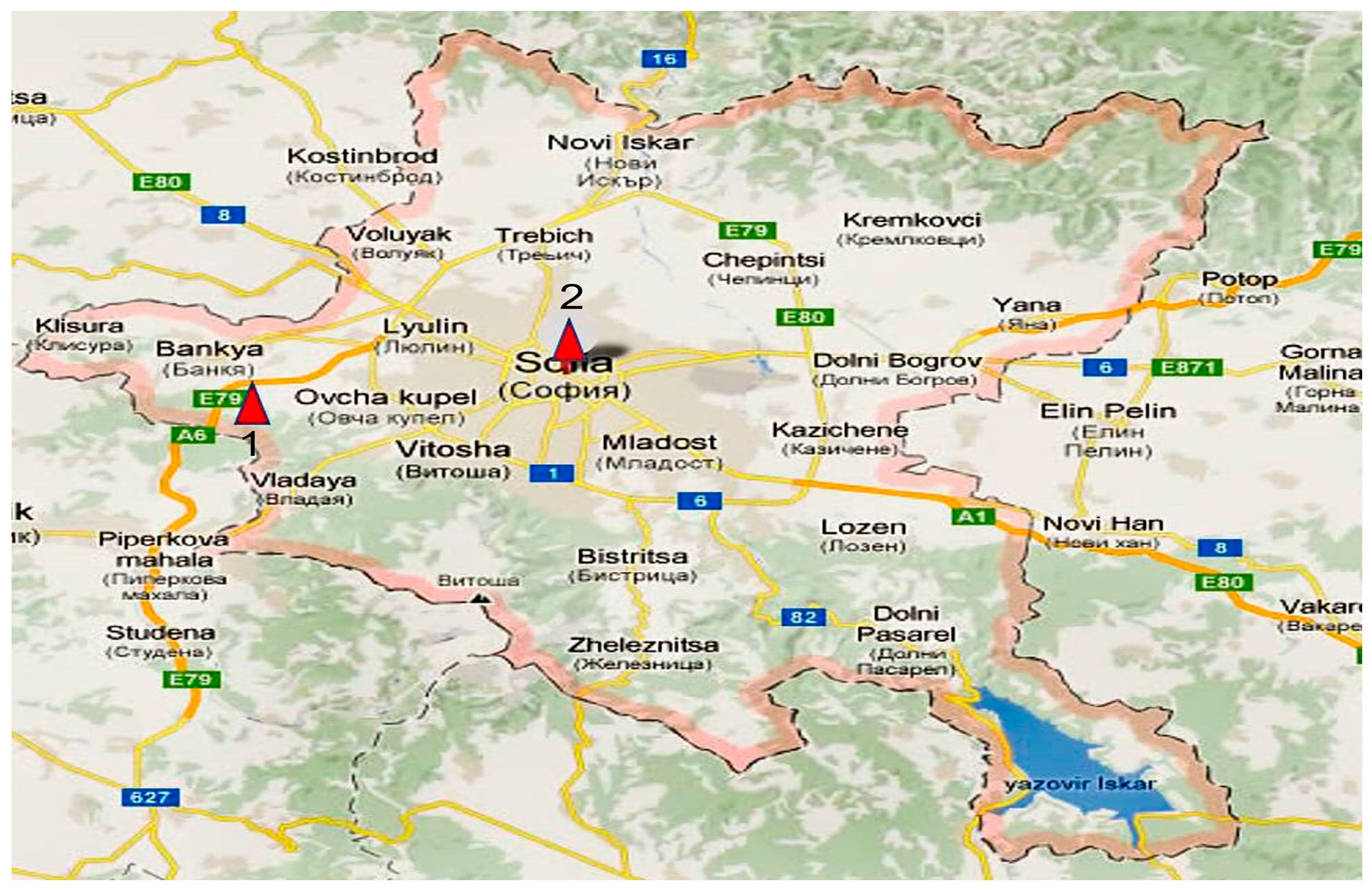
Figure 1. Map of Sofia with the sampling points labeled as follows: 1—New Bulgarian University (NBU); 2—Orlov Most. The map of Sofia was taken from BGmaps.com.
Applmicrobiol 05 00096 g001
Figure 2. Quantitative analysis of the mycobiota in the two studied locations (A)—on medium G1, (B)—on medium YGC, (C)—around NBU, and (D)—on Orlov Most. (E)—The ratio between isolated colonies in the two locations. The difference between isolated colonies in the two locations is statistically significant (p < 0.05).
Applmicrobiol 05 00096 g002aApplmicrobiol 05 00096 g002b
Figure 3. Distribution of fungal isolates in both locations (A)—at the phylum level, (B)—at the genus level.
Applmicrobiol 05 00096 g003
Figure 4. Taxonomic diversity of isolates from the studied locations.
Applmicrobiol 05 00096 g004
Table 1. Specific meteorological conditions in the city of Sofia during the sampling period.
DataTemperature [°C]Wind Speed [m/s]Amount of Precipitation
[mm]
Recorded Phenomena
AverageMin.Wind Speed [m/s]Av.MaxGusts of Wind
29.03.202210.22.018.52.58-0-
30.03.202211.10.019.02.47-0-
31.03.202215.210.619.0410180Rain/Sleet
01.04.202213.98.021.93.78190Rain/Sleet, Thunderstorm
02.04.202210.86.017.24.2122213.2Rain/Sleet, Thunderstorm
03.04.20225.82.010.24.58-2.5Rain/Sleet
04.04.20225.40.010.02.86-1Rain/Sleet
05.04.20227.90.016.01.33-0-
Table 2. Pictures of some of the air samples collected.
Orlov Most
Applmicrobiol 05 00096 i001Applmicrobiol 05 00096 i002Applmicrobiol 05 00096 i003Applmicrobiol 05 00096 i004Applmicrobiol 05 00096 i005
NBU
Applmicrobiol 05 00096 i006Applmicrobiol 05 00096 i007Applmicrobiol 05 00096 i008Applmicrobiol 05 00096 i009Applmicrobiol 05 00096 i010
Table 3. Macroscopic pictures of the strains identified by classical taxonomy to the genus level.
AspergillusAspergillusAspergillusEpicoccum
Applmicrobiol 05 00096 i011Applmicrobiol 05 00096 i012Applmicrobiol 05 00096 i013Applmicrobiol 05 00096 i014
PenicilliumHerpotrichiaStereumTrametes
Applmicrobiol 05 00096 i015Applmicrobiol 05 00096 i016Applmicrobiol 05 00096 i017Applmicrobiol 05 00096 i018
AlternariaCladosporiumTalaromycesBjerkandera
Applmicrobiol 05 00096 i019Applmicrobiol 05 00096 i020Applmicrobiol 05 00096 i021Applmicrobiol 05 00096 i022
Table 4. Molecular identification of the isolates.
Strain №Identified asGenBank Accession NumberSimilarity
to the Reference Strains, %
Location
1.6A1Alternaria alternataPV77152599.80%,
Alternaria alternata
CBS 102,600
Orlov Most
2.6A2Herpotrichia striatisporaPV77152697%,
Herpotrichia striatispora
CBS 385.65
Orlov Most
3.6A9Alternaria angustiovoideaPV77152799.62%
Alternaria angustiovoidea
CBS 195.86
Orlov Most
4.7A1Aspergillus fumigatusPV77152899.23%
Aspergillus fumigatus
ATCC 1022
Orlov Most
5.7A2Alternaria alternataPV77152999.00%
Alternaria alternata
CBS 102,595
Orlov Most
6.8A2Talaromyces pinophilusPV77153099.0%
Talaromyces pinophilus
CBS 631.66
Orlov Most
7.8A3Talaromyces wortmanniiPV77153199.42%
Talaromyces wortmannii
CBS 316.63
Orlov Most
8.8A4Aspergillus fumigatusPV77153299.46%
Aspergillus fumigatus
ATCC 1022
Orlov Most
9.9A1Alternaria tenuissimaPV771533100%
Alternaria tenuissima
CBS 117.44
Orlov Most
10.9A2Coniothyrium telephiiPV77153499.60%
Coniothyrium telephii
CBS 188.71
Orlov Most
11.18A2Alternaria infectoriaPV77153599.44%
Alternaria infectoria
CBS 210.86
Orlov Most
12.23A2Cladosporium cladosporioidesPV771536100%
Cladosporium pseudocladosporioides
CBS 126,356
Orlov Most
13.23A3Epicoccum nigrumPV77153799.59%
Epicoccum nigrum
CBS 236.59
Orlov Most
14.27A1Aspergillus fumigatusPV77153899.0%
Aspergillus fumigatus
ATCC 1022
Orlov Most
15.32A1Aspergillus fumigatusPV77153999.62%
Aspergillus fumigatus
ATCC 90,906
NBU
16.32A2Talaromyces wortmanniiPV77154099.46%
Talaromyces wortmannii
CBS 316.63
Orlov Most
17.62A1Epicoccum nigrumPV771541100%
Epicoccum nigrum
CBS 173.73
NBU
18.76A2Alternaria tenuissimaPV77154299.22%
Alternaria tenuissima
CBS 117.44
NBU
19.77A1Penicillium griseofulvumPV77154399.63%
Penicillium griseofulvum
CBS 185.27
NBU
20.83A1Alternaria caespitosaPV77154499.46%
Alternaria caespitosa
CBS 177.80
NBU
21.90A1Bjerkandera adustaPV77154599.83%
Bjerkandera adusta
CBS 371.52
NBU
22.92A1Stereum hirsutumPV77154699.65%
Stereum hirsutum
CBS 930.70
NBU
23.117A2Trametes ochraceaPV77154799.82%
Trametes ochracea
CBS 287.33
NBU
24.126Bjerkandera adustaPV77154899.00%
Bjerkandera adusta
CBS 371.52
NBU
Table 5. Indices of diversity, dominance, and similarity of fungal strains in both locations investigated.
BiodiversityShannon IndexSimpson IndexSørensen Index
Orlov Most233868870.25
NBU20427518
Table 6. Fungal strains’ sensitivity to nystatin (0.36 mg/mL).
Fungal Species Nystatin—% of Growth Inhibition
24 h48 h72 h96 h7 Days
AscomycotaAlternaria alternata 6194.64 ± 1.7391.12 ± 1.5288.68 ± 1.1984.18 ± 1.6579.56 ± 2.1
Alternaria tenuissima 9198.99 ± 1.5892.97 ± 1.5589.07 ± 1.4987.72 ± 2.1184.78 ± 2.06
Alternaria infectoria 182100 ± 0.5100 ± 0.593.74 ± 2.0386.1 ± 1.9875 ± 1.97
Alternaria sp. 6396.89 ± 1.0496.46 ± 1.7092.12 ± 2.0287.69 ± 1.7685.57 ± 1.32
Cladosporium cladosporioides 23′96.64 ± 1.1993.69 ± 2.5688.52 ± 2.2288 ± 1.9880.66 ± 3.02
Talaromyces wortmannii 8397.17 ± 2.0396.59 ± 1.9896.48 ± 1.7990.78 ± 2.4583.68 ± 2.89
Talaromyces pinophilus 8294.39 ± 1.9992.53 ± 2.0487.16 ± 1.4585.57 ± 1.9382.83 ± 2.01
Talaromyces pinophilus 82′96.09 ± 1.2395 ± 1.7890.07 ± 3.06
Talaromyces wortmannii 322
Penicillium griseofulvum 77192.19 ± 2.6788.17 ± 2.0182.9 ± 1.8882.79 ± 2.0179.71 ± 3.34
Stereum hirsutum 92199.22 ± 1.5686.95 ± 2.0586.81 ± 1.9783.49 ± 2.4573.11 ± 2.76
Herpotrichia striatispora 6281.05 ± 3.7878.12 ± 2.7878.02 ± 3.02
Coniothyrium telephii 92100 ± 0.6100 ± 0.392.64 ±0.4
Aspergillus fumigates 8488.43 ± 2.6587.56 ± 1.8886.12 ± 2.57
Epicoccum nigrum 62191.89 ± 3.5691.9 ± 3.0191.96 ± 1.9690.86 ± 1.4290.51 ± 1.89
BasidiomycotaBjerkandera adusta 12687.28 ± 3.1287.48 ± 2.6685.75 ±2.0886.22 ± 1.3584.37 ± 1.87
Trametes ochracea 1172100 ± 0.72100 ± 0.5571.22 ± 2.3568.34 ± 1.79
Bjerkandera adusta 90193.63 ± 1.3391.59 ± 2.2191.67 ± 1.9988.06 ± 0.8988.79 ± 1.56
Legend95–100%
90–94.99%
80–89.99%
60–79.99%
1, 2: These are working numbers. It means isolate 1 or 2 of the sample.
Table 7. Sensitivity of isolated strains to amphotericin (0.36 mg/mL).
Fungal SpeciesAmphotericin—% of Growth Inhibition
24 h48 h72 h96 h7 Days
AscomycotaAlternaria alternata 6189.21 ± 2.3487.41 ± 1.9984.45 ± 3.1282.09 ± 1.2380.72 ± 3.21
Alternaria tenuissima 9190.18 ± 3.2186.35 ± 2.0685.95 ± 2.2283.81 ± 1.63
Alternaria infectoria 182100 ± 0.3100 ± 0.590.97 ± 2.5789.88 ± 0.94
Alternaria sp. 6389.72 ± 4.3387.66 ± 2.7888.11 ± 1.5091.87 ± 1.50
Cladosporium cladosporioides 23′90.38 ± 2.8788.53 ± 1.6888.63 ± 1.7787.24 ± 2.1685.57 ± 3.01
Talaromyces wortmannii 83
Talaromyces pinophilus 8293.54 ± 3.0192.47 ± 1.5689.21 ± 2.1286.32 ± 1.29
Talaromyces pinophilus 82′91.88 ± 2.7988 ± 1.9987.84 ± 1.4587.23 ± 2.76
Talaromyces wortmannii 32291.46 ± 1.6687.32 ± 2.0384.67 ± 2.3684.85 ± 1.44
Penicillium griseofulvum 77192.14 ± 1.9886.29 ± 2.4280.17 ± 1.4580.52 ± 2.52
Stereum hirsutum 92196.63 ± 2.0494.03 ± 1.6993.98 ± 1.7291.71 ± 3.4490.03 ± 1.59
Herpotrichia striatispora 6293.09 ± 2.2689.59 ± 2.1788.59 ± 1.9477.27 ± 3.94
Coniothyrium telephii 9279.84 ± 4.9878 ± 1.8877.36 ± 2.46
Aspergillus fumigates 8490.48 ± 1.3390.31 ± 1.7690.22 ± 2.3489.16 ± 2.45
Epicoccum nigrum 62194.11 ± 1.7993.87 ± 2.0392.75 ± 1.2378.72 ± 4.02
BasidiomycotaBjerkandera adusta 12687.99 ± 2.6587.59 ± 1.9586.35 ± 2.5183.77 ± 3.2181.31 ± 2.35
Trametes ochracea 117289.93 ± 1.5789.88 ± 1.8389.64 ± 1.6389.59 ± 2.3389.53 ± 2.62
Bjerkandera adusta 90187.9 ± 2.1786.38 ± 1.5478.56 ± 2.0576.13 ± 1.8962.7 ± 2.25
Legend95–100%
90–94.99%
80–89.99%
60–79.99%
1, 2, 4: These are working numbers. It means isolate 1 or 2 or 4 of the sample.
Table 8. Minimum inhibitory concentration of the two tested antifungal preparations, nystatin and amphotericin, against the identified fungal species.
Fungal StrainMIC mg/mL
NystatinAmphotericin
Alternaria alternata 610.180.042
Herpotrichia striatispora 620.0420.18
Alternaria sp. 630.180.18
Talaromyces sp. 820.0420.085
Talaromyces pinophilus 820.180.18
Talaromyces wortmannii 830.18resistant
Aspergillus fumigates 840.180.18
Alternaria tenuissima 910.0420.042
Coniothyrium telephii 920.360.36
Alternaria infectoria 1820.360.36
Cladosporium cladosporioides 23′0.0850.18
Talaromyces wortmannii 322resistant0.18
Epicoccum nigrum 6210.0420.085
Penicillium griseofulvum 7710.360.18
Bjerkandera adusta 9010.360.18
Stereum hirsutum 9210.0420.36
Trametes ochracea 11720.0420.18
Bjerkandera adusta 1260.0420.042
1, 2, 3: These are working numbers. It means isolate 1 or 2 or 3 of the sample.
Disclaimer/Publisher’s Note: The statements, opinions and data contained in all publications are solely those of the individual author(s) and contributor(s) and not of MDPI and/or the editor(s). MDPI and/or the editor(s) disclaim responsibility for any injury to people or property resulting from any ideas, methods, instructions or products referred to in the content.