Article

Ruminal Planktonic, Weakly, and Tightly Feed-Adhered Bacterial Community as Affected by Two Trichoderma reesei Enzyme Preparations Fed to Lactating Cattle

by
Marjorie A. Killerby
1,2,
Juan J. Romero
1,3,*,
Zhengxin Ma
4 and
Adegbola T. Adesogan
3,*
1
Animal and Veterinary Sciences, School of Food & Agriculture, University of Maine, Orono, ME 04469, USA
2
Animal and Dairy Science, University of Wisconsin-Madison, Madison, WI 53706, USA
3
Global Food Systems and Department of Animal Sciences, Institute of Food and Agricultural Sciences, University of Florida, Gainesville, FL 32603, USA
4
Molecular and Biomedical Sciences, University of Maine, Orono, ME 04469, USA
*
Authors to whom correspondence should be addressed.
Appl. Microbiol. 2025, 5(3), 93; https://doi.org/10.3390/applmicrobiol5030093
Submission received: 29 July 2025 / Revised: 27 August 2025 / Accepted: 29 August 2025 / Published: 3 September 2025

Abstract

This study evaluates the effects of two Trichoderma reesei exogenous fibrolytic enzyme (EFE) preparations on the taxonomic profile, diversity, relative abundance, and population shifts of three ruminal bacteria fractions of lactating cows: free-floating (LIQ), weakly (AS), and tightly (SOL) feed-adhered. Three lactating cows were fed three EFE treatments in a 3 × 3 Latin square design: one control (CON) without enzymes, a cellulase/xylanase mix (MIX), and a high-xylanase treatment (XYL). Rumen contents were collected, and bacteria were extracted from the three ruminal content fractions for next-generation sequencing analysis. Alpha diversity was higher in XYL compared to CON. However, no EFE effect was observed on beta diversity. The relative abundance (RA) of the family Prevotellaceae increased, while that of Ruminococcaceae and Rikenellaceae decreased in XYL compared to MIX and CON. The bacterial community structure (beta diversity) of LIQ was differentiated from that of SOL and AS (p = 0.03), but no effects of fraction were observed on alpha diversity. Lachnospiraceae RA was greater in SOL, followed by AS, and lower in LIQ (p < 0.001), while Spirochaetaceae RA was greater in SOL and AS compared to LIQ (p = 0.003). The effects of EFE supplementation on rumen bacterial RA were independent of the ruminal content fraction.

1. Introduction

The reticulorumen contains a highly diverse microbial community that is essential for converting fibrous plant-based substrates into high-quality products (e.g., milk, meat), among other essential functions for the animal. The microbial fermentation of feed nutrients in the rumen results in the production of volatile fatty acids (VFAs), microbial protein, and vitamins that are used to cover the requirements of the ruminant animal [1,2]. Fibrolytic rumen microbes are key because ruminant animals depend on their activity to break down plant vegetative tissues [3], especially in forages of low quality, such as warm-season grasses (e.g., bermudagrass, Cynodon dactylon).
Certain conditions in the rumen may negatively affect the microbiome and its capacity to break down fiber, such as a low pH and a high ruminal rate of passage [4,5]. These conditions often occur in dairy cows in early lactation due to the high-concentrate diets they receive to satisfy their high energy requirements [6]. In tropical and subtropical regions, the low quality and high lignification of warm-season forages can further constrain the availability of nutrients to the rumen microbes [7,8]. The use of exogenous fibrolytic enzymes (EFE) in ruminant diets to improve fiber digestibility and productive performance has been studied extensively with variable but mostly positive results [9,10]. Notably, Arriola et al. [11] observed that supplementation with a Trichoderma reesei EFE preparation (Dyadic International; Jupiter, FL) increased the total tract digestibility of dry matter (DM), crude protein (CP), and neutral detergent fiber (NDF). The benefits of EFE supplementation were observed in both low-concentrate (67:33 forage–concentrate ratio) and high-concentrate (52:48) diets in that study. Additionally, EFE improved feed efficiency in low-concentrate diets [11].
Furthermore, out of a broad selection of EFE preparations from various manufacturers, Romero et al. [12] identified promising candidates to improve the in vitro ruminal NDF digestibility (NDFD) of bermudagrass. Afterwards, these preparations were assessed for dose [13] and cofactor dose effects [14]. Two of these preparations were evaluated in a lactation study with a diet that included corn silage, bermudagrass silage, and alfalfa–orchardgrass hay. Romero et al. [15] found that a high-xylanase EFE derived from T. reesei (Xylanase Plus, Dyadic International; Jupiter, FL) was the most effective at increasing intake and milk yield during peak lactation. However, the effects of EFE preparations on the ruminal microbiome have not been extensively studied, especially across ruminal content fractions. Based on their distribution and association with feed particles, ruminal bacteria can be categorized into three groups or fractions: freely floating (planktonic), weakly adhered, and tightly adhered to feed particles [1]. When incubating barley straw and alfalfa hay in vitro with known ruminal fibrolytic bacteria (Ruminococcus flavefaciens and Fibrobacter succinogenes), Wang et al. [16] observed that the addition of EFEs increased the attachment of these bacteria to the feed particles and increased NDF digestibility. However, the effects of EFE supplementation on the ruminal microbiome of lactating cows across the three ruminal fractions in vivo have not been evaluated to date. Thus, this study aims to evaluate the effects of two T. reesei enzyme preparations on the taxonomic profile, diversity, relative abundance, and population shifts of planktonic, weakly, and tightly feed-adhered ruminal bacteria of lactating cows. Mid-lactation cows are used in this study to avoid the confounding effects of the disrupted rumen conditions often observed during early lactation (i.e., low pH, high passage rates, etc.). Given that EFE aid in the degradation of fiber particles into soluble compounds, we hypothesize that the supplementation of these enzymes would increase the abundance of planktonic non-cellulolytic bacteria, which feed on the products of enzymatic degradation.

2. Materials and Methods

2.1. Animals and Treatments

This study is the microbiome complement of Romero et al. [15], which evaluated the effects of two T. reesei EFE preparations on the digestion kinetics and performance of lactating cattle. Therefore, details on site, animal management, diets, and enzymatic activity determination can be found in that publication. The protocol for this study was approved by the University of Florida Institute of Food and Agricultural Sciences Animal Research Committee.
Briefly, three ruminally cannulated Holstein cows in mid-lactation (159 ± 47 days in milk) were randomly assigned to one of the three treatments (TRT): (1) a control feed with no enzymes (CON), (2) a high-xylanase additive (Xylanase Plus; Dyadic International, Jupiter, FL, USA; XYL), and (3) a 75:25 (vol/vol) mixture of Cellulase Plus and Xylanase Plus EFE (Dyadic International, Jupiter, FL; MIX). The treatments were sprayed on the total mixed ration immediately before feeding at a rate of 3.4 mL/kg of DM for MIX and 1 mL/kg of DM for XYL [15]. The total mixed ration was composed of 45% concentrate, 35% corn silage, 10% bermudagrass silage, 5% alfalfa–orchardgrass hay, and 5% whole cottonseed on a DM basis. The chemical composition included 16.5% crude protein, 32.9% NDF, 17.4% acid detergent fiber, and 39.3 non-fibrous carbohydrates, on a DM basis [15]. The application rate of XYL is based on the dose that effectively optimized the NDF digestibility of bermudagrass hay in vitro in previous studies (2× the manufacturer-recommended dose) [12,13]. Meanwhile, the application rate of MIX is based on the manufacturer-recommended dose used by Arriola et al. [11], who observed improved DM, crude protein, and NDF digestibility in lactating cows fed a total mixed ration treated with this EFE preparation. Both EFE additives (MIX and XYL) were sourced from non-recombinant T. reesei. The enzymatic activities and protein composition of the additives were determined as outlined in Romero et al. [15] and are described in Table 1.
This experiment was performed as a single 3 × 3 Latin square with 23 d periods. Each period had 18 d for adaptation to the diets followed by 3 d for measuring the in situ ruminal digestibility of the total mixed ration, 1 d to rest the rumen, and 1 d for measuring indices of ruminal fermentation [15] and obtaining rumen content samples for sequencing. Rumen contents (liquid and solid mixture) were aseptically sampled from each cow through the cannula on the last day of each period in the Latin square from multiple points in the ventral, dorsal, and caudal sacs 3 h after morning feeding. The rumen content samples were immediately processed to extract the bacterial pellets from three different ruminal content fractions (FRC) according to the protocol outlined by Larue et al. [17], resulting in a total of 27 samples (experimental units) at the end of this study.
Briefly, the isolation of bacteria from the three rumen content fractions was as follows: (1) Planktonic (free-floating) bacteria (LIQ), which are found in the ruminal liquid, were extracted by filtering the ruminal contents through two layers of sterile cheesecloth and squeezing by hand. The resulting filtrate was centrifuged at 10,000× g for 20 min at room temperature to isolate the planktonic bacterial cells. (2) To obtain the weakly solid-associated bacteria (AS), 50 g of the squeezed solids were mixed with 150 mL of phosphate-buffered saline, shaken, and centrifuged at 350× g for 15 min at room temperature to sediment plant particles. The microbial cells were harvested from the supernatant by centrifuging it at 10,000× g for 20 min at room temperature. Finally, (3) the tightly solid-associated bacteria (SOL) were obtained by re-suspending the ruminal contents in 25 mL of a 0.15% (vol/vol) Tween-80 solution in an anaerobically prepared diluent and placing them on ice for 2.5 h. The mixture was then centrifuged at 500× g for 15 min at room temperature to separate plant particles, followed by centrifugation of the supernatant at 10,000× g for 20 min at 4 °C to extract the microbial pellet. Finally, the microbial pellets were stored at −80 °C until further analysis.
Additionally, the measurements of rumen fermentation and digestion kinetics reported by Romero et al. [15] in these same cows and periods were used in this study to evaluate numerical relationships with the microbial communities. These variables included the following: in situ ruminal lag time, washout DM, potentially digestible DM, rate of digestion, and indigestible DM of the total mixed ration; rumen fluid pH; ammonia-N; acetic, propionic, isobutyric, butyric, isovaleric, and valeric acids; and total VFA concentrations. Details of the methods used to measure these variables are described by Romero et al. [15]. Briefly, dried and ground samples (5 g) of the total mixed ration were treated with equivalent doses of the three EFE treatments (CON, MIX, or XYL) and incubated in Ankom R1020 in situ bags (Ankom Technology, Macedon, NY, USA) inside the rumen of the cows receiving the respective EFE-treated ration. The bags were incubated for 0, 4, 8, 16, 24, 48, and 72 h, removed simultaneously, and washed with cold water to remove adherent feed particles and bacteria. The washed bags were dried (at 60 °C for 48 h) and weighed, and the resulting data was used to estimate DM degradation kinetics with the model outlined by Mertens [18]. Six samples of ruminal fluid (200 mL) per cow were collected by aspiration through the rumen cannula on d 23 of each period. The first sample was collected before feeding (at 0850 h) and subsequent samples every other hour. The rumen fluid was immediately filtered through 2 layers of cheesecloth and analyzed for pH using an Accumet XL25 pH meter (Fisher Scientific, Pittsburgh, PA, USA). Then, rumen fluid samples (13 mL) were acidified with 130 μL of 9.0 M H2SO4 and frozen at −40 °C for further analysis. Thawed samples were centrifuged at 8000× g for 20 min at 4 °C, and the supernatant was analyzed for VFA using a Merck Hitachi Elite LaChrome HPLC system (L2400, Hitachi, Tokyo, Japan) and a Bio-Rad Aminex HPX-87H column (Bio-Rad Laboratories, Hercule, CA, USA). Ammonia-N concentration in the rumen fluid was measured with colorimetric methods [19] using a Technicon Auto Analyzer (Technicon, Tarrytown, NY, USA).

2.2. DNA Extraction

The extraction of DNA from the bacterial pellets was achieved using the PowerLyzer-PowerSoil DNA isolation kit (MO BIO Labs Inc.; Carlsbad, CA, USA) following the manufacturer-recommended procedure. Extracted DNA was analyzed using the Illumina (San Diego, CA, USA) MiSeq platform for 2 × 300 base pair-end reads (600 sequencing cycles). The amplification of the V1–V3 regions of the bacterial 16S rRNA gene was achieved using the barcoded primer pair forward F27 (AGAGTTTGATCCTGGCTCAG) and reverse R519 (GTNTTACNGCGGCKGCTG) [20]. PCRs were performed in 30 cycles using the HotStarTaq Plus Master Mix Kit (Qiagen; Germantown, MD, USA) under the following conditions: 94 °C for 3 min, followed by 28 cycles of 94 °C for 30 s, 53 °C for 40 s, and 72 °C for 1 min, after which a final elongation step at 72 °C for 5 min was performed. After amplification, PCR products were checked in 2% agarose gel to determine the success of amplification and the relative intensity of bands. Multiple aliquots were pooled together per sample (an aliquot for each of the 27 samples) in equal proportions based on their molecular weight and DNA concentrations. Pooled samples were purified using calibrated AMPure XP beads. Then, the pooled and purified PCR product was used to prepare the DNA library by following the Illumina TruSeq DNA library preparation protocol (Illumina; San Diego, CA, USA). Sequencing was performed at MR DNA (www.mrdnalab.com, Shallowater, TX, USA) on MiSeq following the manufacturer’s guidelines.

2.3. Bioinformatics

Raw Illumina fastq files were demultiplexed by barcode sequence using mothur v.1.44.3 [21] and analyzed using the ‘dada2’ package in R software (v.3.9) to infer amplicon sequence variants (ASV) [22]. Both forward and reverse raw files contained a total of 1,663,322 reads each, which were then quality assessed, trimmed (left trim 10 bp, truncated length 280 bp for both F and R), filtered (maxEE = 2 and 4, for F and R, respectively), dereplicated, and consequently merged into single pair-end reads. Finally, a sequencing table was constructed, chimeras were removed (49,283 total bimeras out of 61,922 sequences), and taxonomy was assigned to the sequences down to the genus level using the Silva nr99 v.138 database for 16S. A total of 12,639 taxa were assigned to sequences of the 27 samples. Sequencing tables were used to calculate the relative abundance of the observed bacterial families and determine the 15 most abundant ones. A phylogenetic tree was built using the ‘phangorn’ package [23], and phylogenetic diversity was determined using the ‘btools’ package of R The ‘phyloseq’ package of R [24] was used to determine the alpha diversity of the resulting taxonomic profiles through the Observed ASV, Chao1 index, and Evenness index estimators. The samples were rarefied to an even depth of 10,000 sequences to ensure that the samples had enough coverage of ASV to evaluate the microbial community. A total of 558 ASV were removed in this process. Beta diversity was evaluated by developing a weighted UniFrac PCoA distance matrix, and differences were determined using a permutational multivariate analysis of variance (PERMANOVA) with the ‘vegan’ package (‘adonis’ command) in R [25]. Spearman’s correlation coefficients between the top 15 bacterial families, alpha diversity measures, and ruminal digestion kinetics reported by Romero et al. [15] were determined using the ‘corrplot’ package [26].

2.4. Statistical Analysis

Microbiota relative abundance (RA) and alpha diversity measures were analyzed as a 3 × 3 Latin square design with a 3 TRT × 3 FRC factorial design, using the following mixed effect model:
Y i j k l = μ + T i + F j + ( T F ) i j + P k + C l + ε i j k l
where Yijkl = the dependent variable; µ = general mean; Ti = fixed effect of EFE i (TRT); Fj = fixed effect of ruminal fraction (FRC); TFij = fixed effect of the interaction between TRT and FRC; Pk = fixed effect of period k; Cl = random effect of Cow l; and εijk = random experimental error. Statistical analyses were performed in SAS v.9.4 (SAS Institute, Cary, NC, USA) using the GLIMMIX procedure. The normal distribution of the data was verified with the Shapiro–Wilks test. Mean separation was performed using the PDIFF procedure of LSMEANS, and differences were considered significant when p ≤ 0.05. All experimental units were included in the analysis.

3. Results

3.1. Alpha and Beta Diversity

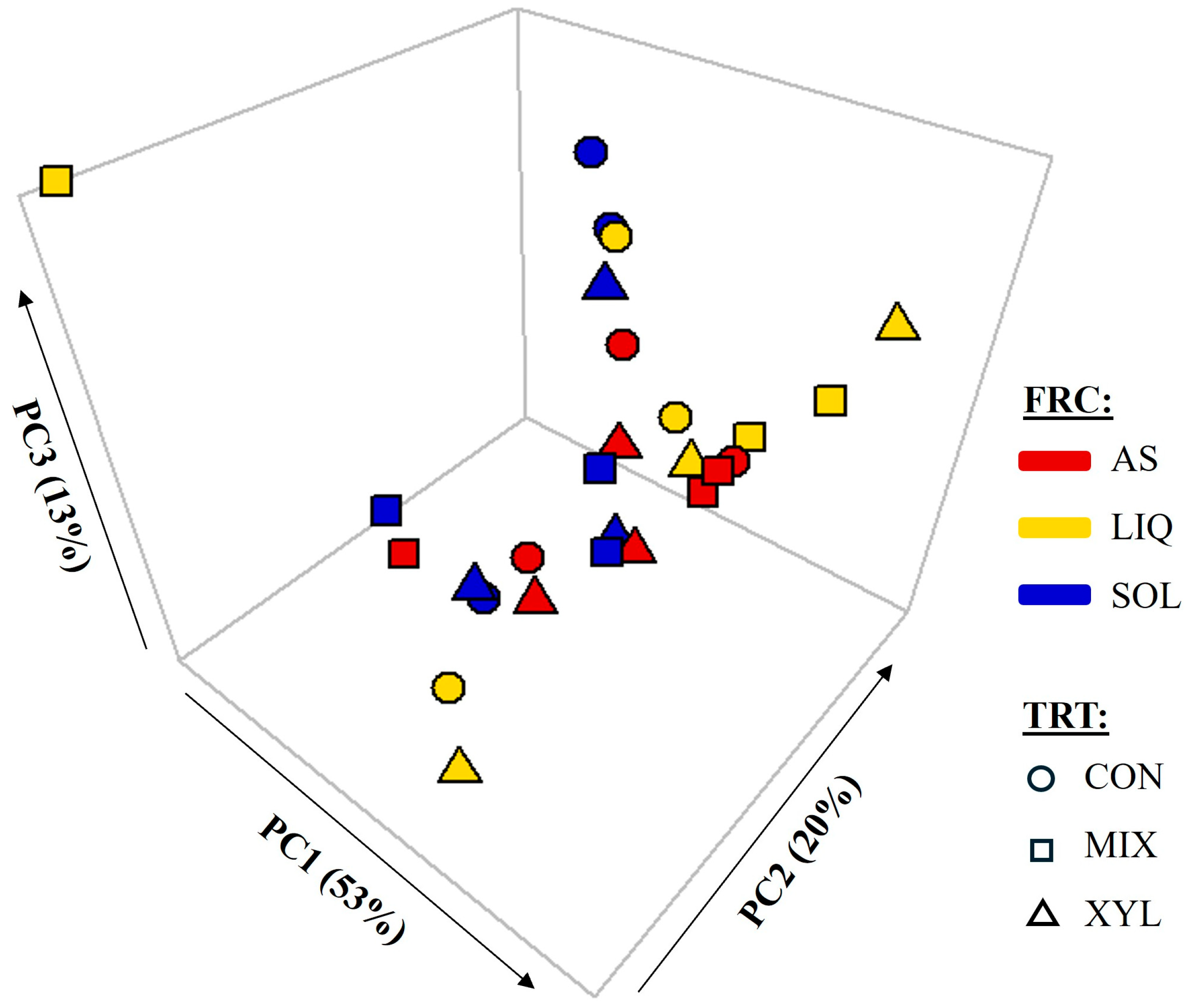
The rarefaction curves of all samples reached a saturation plateau when rarefied to an even depth of 10,000 (Figure 1), indicating that the coverage of bacterial diversity was sufficient to assess the bacterial community composition of the samples. The Observed ASV (p = 0.03) and Chao1 index (p = 0.04) were higher for XYL (800 and 856) compared to CON (690 and 750, respectively), but there were no differences in either with MIX (762 and 812, respectively; Table 2). No effects of TRT, FRC, or TRT×FRC on Simpson’s Evenness or phylogenetic diversity were observed (p > 0.05). The weighted UniFrac principal coordinate analysis plot (Figure 2) and the permutational multivariate analysis of variance (PERMANOVA) indicate a separation of the bacterial communities by FRC (p = 0.03; R2 = 15.7%), where the LIQ community structure is more widely spread than that of AS and SOL.

3.2. Relative Abundance

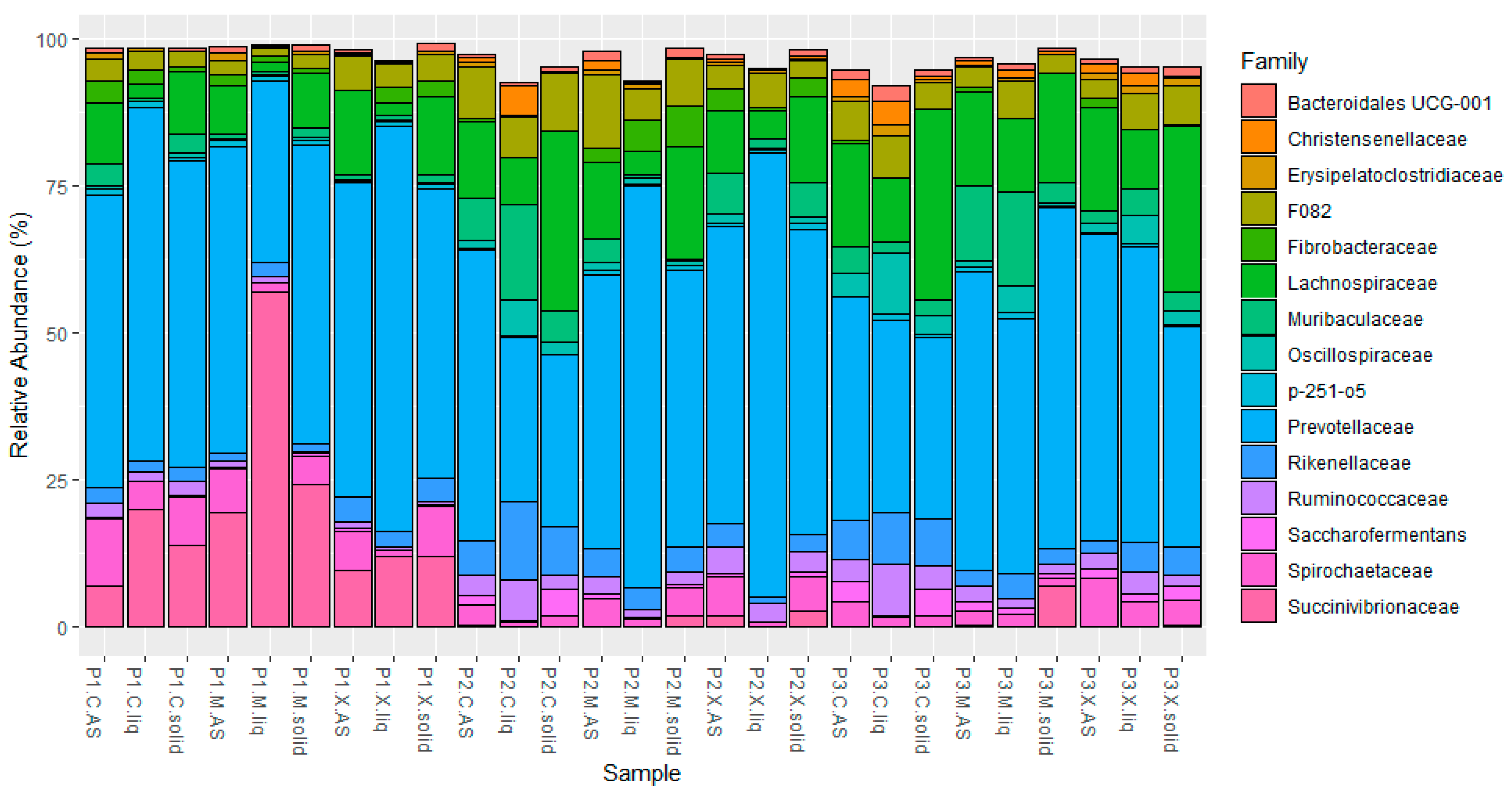
The relative abundance (RA) of the top 15 bacterial families in each experimental unit is shown in Figure 3. Members of these 15 bacterial families represented the majority (>90%) of the bacteria identified, and Prevotellaceae was the most abundant bacterial family in most samples. A notable period effect was observed for Succinivibrionaceae RA in period 1 compared to the other two periods. However, the purpose of analyzing the data in this experiment as a Latin square is to remove the variability of known “nuisance” factors from the residual error [27]. Thus, the inevitable variability attributed to period and cow across experimental units is separated from the factors of interest (TRT and FRC) when analyzed statistically. The effects of TRT and FRC on the RA of the top five phyla are shown in Table 3. No TRT × FRC interaction effects were observed on the RA of any of these phyla. The supplementation of EFE (MIX and XYL) increased the RA of the most abundant phylum, Bacteroidota ( x ¯ = 68.2; p = 0.006), but decreased the RA of Firmicutes ( x ¯ = 19.0; p < 0.001) relative to CON (60.3 ± 2.4% and 29.3 ± 2.7%, respectively). Additionally, MIX tended to increase the RA of Proteobacteria (7.7; p = 0.078) relative to XYL (4.4), but neither EFE was different from CON (4.7 ± 1.2%). Independently of TRT, Bacteroidota were found more abundantly in LIQ (72.2), followed by AS (65.2), and least in SOL (59.3 ± 2.4%; p < 0.001). Firmicutes were more abundant in SOL (27.3) relative to AS and LIQ ( x ¯ = 20.0 ± 2.7%; p = 0.003), while Spirochaetota were more abundant in SOL and AS ( x ¯ = 5.4) relative to LIQ (2.0 ± 0.7%; p = 0.003). The RA of other phyla besides the ones mentioned was <1%.
At the family level, the most abundant taxa were Prevotellaceae, Lachnospiraceae, Succinivibrionaceae, Bacteroidales F082, Rikenellaceae, Spirochaetaceae, Muribaculaceae, and Ruminococcaceae. The remaining families found had less than 5% RA each. Differences in the relative abundance of these main families across TRT and FRC are summarized in Table 4. Prevotellaceae was more abundant for XYL (54.2) compared to CON (41.0), but neither was different from MIX (49.7 ± 5.50%; p = 0.03). Conversely, both Rikenellaceae and Ruminococcaceae were more abundant for CON (6.38 and 4.00, respectively) relative to XYL and MIX ( x ¯ = 3.24 ± 1.06 and ( x ¯ = 2.00 ± 0.733%, respectively; p < 0.005). Lachnospiraceae was more abundant in SOL (19.6%) than in AS (13.3%) and least abundant in LIQ (6.27%; p < 0.001). Similarly, Spirochaetaceae was more abundant in both SOL and AS ( x ¯ = 5.38) relative to LIQ (2.03 ± 1.22%; p = 0.004). No effects of TRT or FRC were observed on Succinivibrionaceae, Bacteroidales F082, or Muribaculaceae. The main genera observed across samples were Prevotella (77.3%), Prevotellaceae UCG-003 (10.3%), and Prevotellaceae UCG-001 (8.01%) for Prevotellaceae; [Eubacterium] ruminatium group (24.2%) [28], Lachnospiraceae AC2044 group (15.1%), and Pseudobutyrivibrio (10.7%) for Lachnospiraceae; and Succinivibrionaceae UCG-002 (65.3%) and Succinivibrionaceae UCG-001 (32.6%) for Succinivibrionaceae.

3.3. Correlations

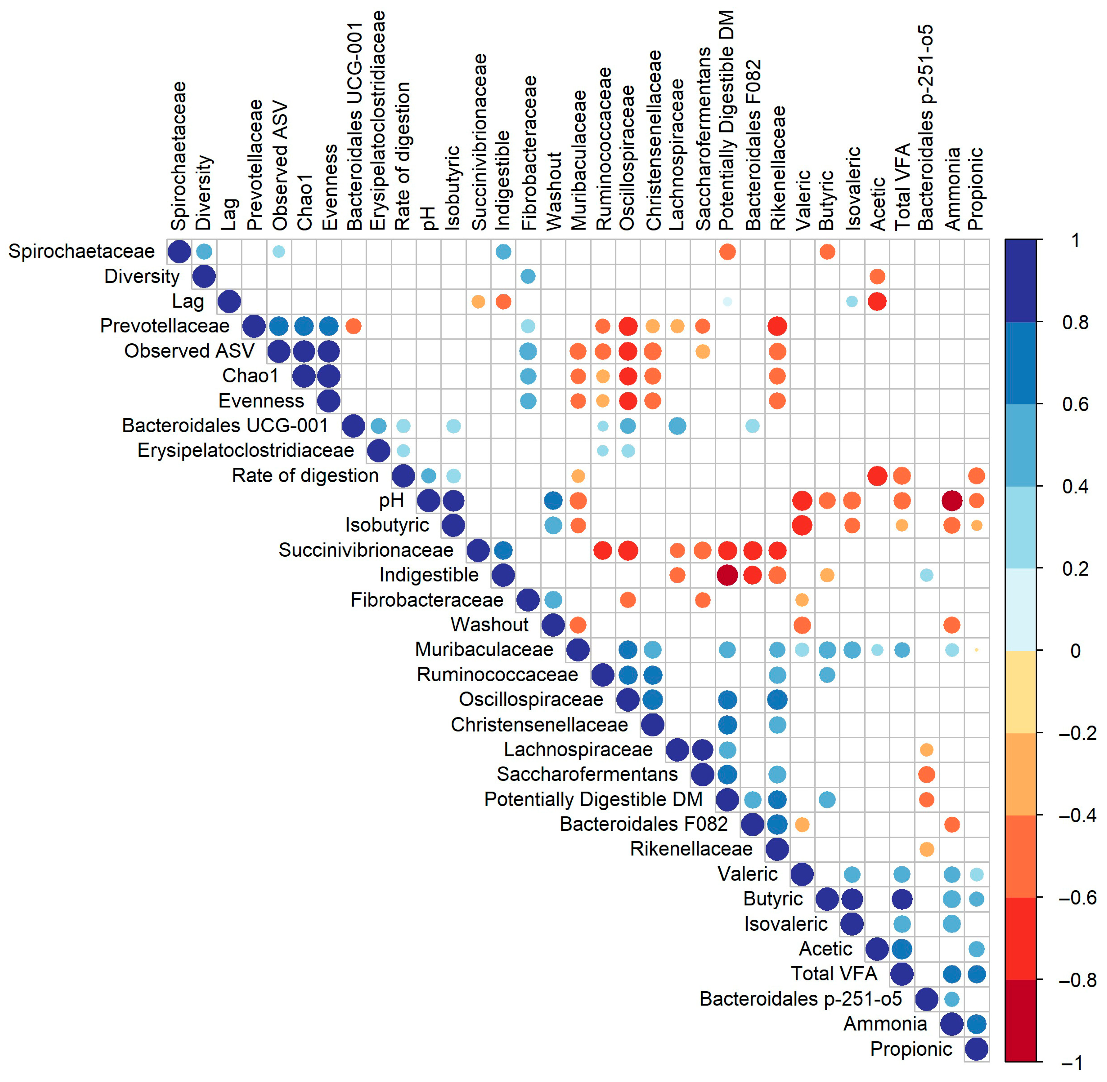
Significant Spearman correlations (ρ; p ≤ 0.05) observed between the top 15 bacterial families, alpha diversity measures, and ruminal digestion measures are depicted in Figure 4. Observed ASVs, Chao1, and Evenness measures were positively correlated (ρ = 0.71, 0.70, and 0.70, respectively) to Prevotellaceae and slightly positively correlated (ρ = 0.58, 0.49, and 0.50) to Fibrobacteraceae. Conversely, these alpha diversity measures were negatively correlated to Muribaculaceae (ρ = −0.51, −0.43, and −0.44, respectively), Ruminococcaceae (ρ = −0.49, −0.33, and −0.32), Oscillospiraceae (ρ = −0.64, −0.60, and −0.60), Lachnospiraceae (ρ = −0.21, −0.11, and −0.12), and Rikenellaceae (ρ = −0.54, −0.53, and −0.53). Phylogenetic diversity (Diversity) was only slightly positively correlated to Fibrobacteraceae (ρ = 0.42) and Spirochaetaceae (ρ = 0.45). Prevotellaceae was negatively correlated to several bacterial families, including Bacteroidales UCG-001 (ρ = −0.47), Ruminococcaceae (ρ = −0.40), Oscillospiraceae (ρ = −0.68), Saccharofermentans (ρ = −0.42), and Rikenellaceae (ρ = −0.71). Furthermore, Oscillospiraceae was positively correlated to Muribaculaceae (ρ = 0.63) and Ruminococcaceae (ρ = 0.68) and strongly positively correlated (ρ = 0.78) to Rikenellaceae and Christensenellaceae. Interestingly, Prevotellaceae was not correlated to any of the ruminal digestion measures evaluated. However, the proportion of potentially digestible DM was positively correlated to the RA of Saccharofermentans (ρ = 0.70), Lachnospiraceae (ρ = 0.54), Christensenellaceae (ρ = 0.69), Oscillospiraceae (ρ = 0.66), and Muribaculaceae (ρ = 0.53) and negatively correlated to Succinivibrionaceae (ρ = −0.68) and Spirochaetaceae (ρ = −0.48). Additionally, Muribaculaceae was the bacterial family with the most correlations to ruminal digestion measures, including positive correlations to butyric (ρ = 0.54), isovaleric (ρ = 0.56), and total VFA (ρ = 0.43) concentrations and negative correlations to rumen pH (ρ = −0.54), isobutyric acid (ρ = −0.45), and washout DM (ρ = −0.54).

4. Discussion

4.1. Spatial Distribution of Rumen Bacterial Communities

The distribution of specific bacterial taxa across different rumen content fractions is closely related to their function and substrate preference. For instance, bacterial taxa with cellulolytic activities are mainly particle-adherent bacteria, and their abundance increases with higher dietary fiber concentrations due to their need to attach to solid particles [29,30,31]. In accordance with previous literature [32,33,34], Bacteroidota represented the most abundant bacterial phylum across rumen content fractions in this study, with Prevotellaceae being its most abundant family. The second most abundant phylum was Firmicutes, with Lachnospiraceae as its most abundant family. Even though members of the main rumen bacterial taxa (e.g., Prevotellaceae, Lachnospiraceae, Ruminococcaceae, etc.) are ubiquitous in the rumen and can be found across all ruminal fractions [35], planktonic and particle-associated (both tightly and loosely) bacteria consist of distinct microbial communities [17].
The abundance and importance of Prevotellaceae in the rumen, particularly Prevotella spp., has been widely described. We did not observe differences in the RA of Prevotellaceae across FRC in this study, but this bacterial family has been described elsewhere as predominantly planktonic [35,36]. Members of Prevotellaceae are characterized by their non-cellulolytic and amylolytic activities, including the digestion of hemicellulose, pectin, and other soluble oligosaccharides and polysaccharides resulting from the breakdown of plant cell walls by the cellulolytic primary degraders [1,36,37]. In contrast, bacterial members of Lachnospiraceae have been described as primarily cellulolytic and pectinolytic, associated with strong hydrolyzing activities and the presence of multiple carbohydrate-active enzymes [32]. The predominant fibrolytic activity of members of Lachnospiraceae may explain the higher abundance of this bacterial family in SOL followed by AS and lastly in LIQ. Similarly, some studies have observed a higher abundance of Lachnospiraceae in the solid ruminal fraction compared to the loosely adhered and planktonic fractions [32,38], but others also report a high abundance of Lachnospiraceae in the liquid fraction [34,36] or similar distribution across fractions [35]. This divergence in results may be due to the wide variety of functions and enzymatic activities observed in members of Lachnospiraceae, including bacterial taxa with predominant oligosaccharide-degrading activities such as Butyrivibrio sp. [28,36]. Other cellulolytic bacterial families commonly found attached to particles include Ruminococcaceae and Fibrobacteraceae, with their main classified genera being Ruminococcus sp. and Fibrobacter sp., respectively [28,39,40]. However, in this study, no effects of FRC were observed on these latter bacterial families.

4.2. Supplementation of Fibrolytic Enzymes

The EFE preparations evaluated in this study are extracts from T. reesei with a wide array of fibrolytic enzyme activities. However, the XYL preparation had a higher protein concentration, and xylanase and endoglucanase activities, while MIX was higher in exoglucanase activity (Table 1). These enzymatic activities can be naturally found in the rumen since they are necessary for rumen microbiota to degrade plant cell walls and are consequently produced by bacteria, fungi, and to a lesser extent protozoa [41]. Xylanases are particularly important in the breakdown of plant material because they hydrolyze xylan, a major polysaccharide in the complex structure of hemicellulose, and release xylooligomers [42]. Thus, supplementing dairy cattle diets with EFE is an effective approach to boosting NDF digestibility and feed efficiency, depending on the diet characteristics [9,11]. Nevertheless, the effects of EFE on rumen bacterial communities have been inconsistent and, overall, poorly described. In this study, XYL increased the bacterial alpha diversity (Observed ASV and Chao1) compared to CON, but no effects of TRT were observed on beta diversity. As suggested by Beauchemin et al. [43], EFE act predominantly by increasing the overall enzymatic capacity of the rumen and synergistically stimulating microbial hydrolases. Therefore, it is expected that EFE supplementation would mainly affect bacterial functionality rather than shift the population structure as represented by the beta diversity. Most notably, EFE treatments increased the RA of Prevotellaceae and decreased it for Rikenellaceae and Ruminococcaceae. As mentioned before, the substrate preference of many Prevotellaceae members includes oligosaccharides and simple sugars, while members of Rikenellaceae and Ruminococcaceae are complex polysaccharide degraders. Thus, we may infer that XYL successfully increased the availability of soluble oligo- and monosaccharides by increasing the hydrolysis of fiber compounds, resulting in the respective RA changes in the bacterial taxa mentioned above. Accordingly, Romero et al. [13] observed that a variety of EFE preparations, including high-xylanase products, increased pre-ingestive fiber hydrolysis and increased the release of water-soluble carbohydrates in vitro. Notably, Romero et al. [15] did not observe TRT effects on the NDF in vivo digestibility of the total mixed ration in a companion experiment to this study that assessed the same treatments under the same conditions but with non-cannulated lactating cows in early lactation. Nevertheless, the release of oligo- and monosaccharides could have had a prebiotic effect on the rumen microbiota [44,45], leading to the increased milk yield observed with the same diets.
No effects of TRT × FRC interaction on bacterial communities were observed in this study. Namely, EFE supplementation seemingly affects bacterial RA in each fraction similarly. It is important to note that, although the Latin square design in this study helped to remove the effects of variability across cows from the statistical model, the low number of animals (n = 3) could have also limited our ability to detect interactions between factors. Few other studies evaluate the effects of EFE on bacterial population in a specific rumen content fraction. Beauchemin et al. [46] observed a decrease in liquid-associated rumen bacteria proportions in cows supplemented with exogenous cellulase/xylanase/pectinase, and Chung et al. [47] observed an increase in the RA of targeted bacteria (Ruminococcus amylophilus and Fibrobacter succinogenes) in ruminal fluid when supplemented with an EFE product containing endoglucanase and xylanase. Nevertheless, differences in experimental methods, enzyme product, diet composition, and microbial quantification methods (e.g., next generation sequencing vs. microbial culture) must be considered when comparing different study outcomes. Microbial populations in the rumen are affected by several factors including diet, feeding strategy, environment, age, breed, and other host factors [48,49], which may explain the lack of effects of different EFE preparations on the rumen microbiome across studies. For example, Liu et al. [50] observed no effects of supplementing lactating Holstein cows with a combination of fibrolytic (cellulase, xylanase, and β-glucanase) and amylolytic (amylase) enzymes on bacterial diversity and bacterial phylum RA. Similarly, other studies observed no effects of supplementing cellulase/xylanase from Trichoderma reesei to beef steers [51], cellulase from Trichoderma viride to goats [52], or xylanase from Thermomyces lanuginosus to beef calves [53] on their respective bacterial diversity or RA.

4.3. Relationships of Bacterial Relative Abundance with Nutritional Measures

There is a wide range of ruminal bacterial taxa that are specialized to degrade different types of feed nutrients, with slowly digestible fiber being the key source of energy that ruminants use to meet their requirements [2]. Thus, differences in the structure of the rumen bacterial community have been associated with differences in productive performance in ruminants. For example, positive correlations have been observed between the RA of members of Prevotella sp. and the average daily weight gain of beef calves [53], VFA production [54,55], and milk protein and fat yield [54]. Additionally, Lopes et al. [56] observed that Nellore beef steers with higher feed efficiency had a higher RA of Bacteroidetes and a lower RA of Firmicutes. The important role of Prevotellaceae bacteria in feed digestibility and productivity is supported by Wirth et al. [36], who reported that Prevotella spp. were responsible for the greatest proportion of carbohydrate (~63%), small molecule (~50%), and organic acid (~53%) metabolism in the ruminal liquid, as well as oxidation–reduction processes (~44%) and precursor metabolite generation (~42%). However, it is important to consider that it is not only the relative abundance of a taxon that determines its importance in the rumen but also its activity levels. For example, Wirth et al. [36] also described that Lachnospiraceae and Treponema had relatively low activities despite having a high relative abundance in the rumen, while Ruminococceae played an important functional role despite its low abundance.
Although correlations do not indicate causation, they can point towards possible relationships between bacterial family RA and other relevant measures such as diversity and ruminal digestion measures. In this study, Prevotellaceae was positively correlated to alpha diversity measures (Observed ASV, Chao1, and Evenness), while other bacterial families (Oscillospiraceae, Ruminococcaceae, and Rikenellaceae) were negatively correlated to alpha diversity and to Prevotellaceae. These correlations may indicate that the increased RA of Prevotellaceae is compatible with the growth of other bacterial groups and the diversification of the bacterial community in the rumen. However, bacterial families negatively correlated to diversity may discourage the growth of other bacterial species, possibly by dominating a specific microbial niche while competing for resources [57]. Additionally, we observed a positive correlation between several bacterial families (Muribaculaceae, Oscillospiraceae, Christensenellaceae, Lachnospiraceae, and Saccharofermentans) and the proportion of potentially digestible DM in the rumen, presumably because these families increase the potentially digestible fraction of the total mixed ration. However, Succinivibrionaceae and Spirochaetaceae were negatively correlated to the potentially digestible DM and to the formerly mentioned bacterial families, indicating a possible competition for a specific niche, as mentioned previously. Furthermore, despite its low RA in the rumen ( x ¯ = 4.1%), Muribaculaceae exhibited the most correlations to ruminal digestion measures of the top 15 bacterial families, possibly due to higher fermentative activities. The functional profile of Muribaculaceae, formerly known as S24-7, has not been well characterized in ruminants, but Lagkouvardos et al. [58] noted the ability of this bacterial family to degrade complex carbohydrates and utilize nitrogen in the mouse intestinal tract. In Holstein cows, Cunha et al. [59] observed that S24-7 was positively correlated to digestible fractions of DM intake and organic matter intake. Furthermore, Indugu et al. [60] observed that S24-7, Prevotella, and Succinivibrionaceae RA were positively correlated to milk yield in one of two dairy farms studied. Lastly, it is important to note that Romero et al. [15] observed milk production improvements with EFE supplementation in the lactation experiment part of this overall study, especially around peak lactation. However, when ruminally cannulated cows in mid-lactation were evaluated, no effects on ruminal VFA or in situ DM digestion kinetics were observed. The stage of lactation has a strong effect on the rumen microbiome even without changes in diet [61]. Therefore, further research on the effects of EFE supplementation across lactation stages is warranted. Although Prevotellaceae RA increased with EFE supplementation in this study, it may not have been sufficient to influence VFA production. Thus, the improvements in milk production may have been driven by the higher dry matter intake in cows supplemented with XYL [15].
In summary, this study shows that XYL affected the RA of key productive bacteria across ruminal content fractions to a greater extent than MIX, possibly due to its high xylanase content. Therefore, the use of EFE preparations with high xylanase activities is more likely to improve digestibility and milk efficiency in lactating cows. However, in this study, the main bacterial families were not correlated to digestion kinetics and rumen fermentation measures. Future research should focus on identifying factors that potentiate the effects of EFE on rumen bacteria functionality that would lead to digestibility and milk production improvements.

5. Conclusions

This study evaluated the effects of two EFE preparations from Trichoderma reesei on the planktonic, weakly, and tightly feed-adhered ruminal bacterial communities of early lactation cows. No TRT × FRC interaction effects were observed on any variable measured, indicating that the effects of TRT and FRC were independent. Namely, EFE supplementation affects bacterial populations in the three ruminal fractions similarly. The application of XYL increased bacterial richness (Observed ASV and Chao1), but no TRT effects were observed on the bacterial community structure (beta diversity). Conversely, FRC affected the bacterial community structure but had no effects on alpha diversity. Overall, XYL increased the RA of Prevotellaceae and decreased the RA of Rikenellaceae and Ruminococcaceae, while MIX had only minimal effects on bacterial RA. The higher levels of xylanase in XYL possibly contributed to the improved effects. These results suggest an effective pre-ingestion breakdown of fiber into soluble nutrients by EFE that favor the growth of non-cellulolytic bacteria like Prevotellaceae. Despite the important role of Prevotellaceae in carbohydrate metabolism and VFA production in the rumen, this study showed no correlation with select variables including rate of digestion, pH, total VFA, acetic acid, and propionic acid. Likely, the changes in these digestibility measures were not prominent enough to allow for the detection of correlations. Further characterization of the effects of EFE on the ruminal microbiome under different conditions would allow for the optimization and specialization of EFE-based products and supplementation protocols. Particularly, the response of the rumen microbiome to EFE supplementation may differ across lactation stage and rumen pH levels, which warrants further research.

Author Contributions

M.A.K.: Data curation, Formal analysis, Software, Visualization, and Writing—original draft; J.J.R.: Data curation, Investigation, Methodology, Funding acquisition, Project administration, Supervision, and Writing—review and editing; Z.M.: Investigation and Writing—review and editing; A.T.A.: Conceptualization, Funding acquisition, Project administration, Supervision, and Writing—review and editing. All authors have read and agreed to the published version of the manuscript.

Funding

This project was supported by the USDA National Institute of Food and Agriculture Hatch Project ME0-21917.

Data Availability Statement

The data presented in this study are openly available in Dryad repository at https://doi.org/10.5061/dryad.612jm64h0.

Acknowledgments

We acknowledge the contributions of Suzanne Ishaq for her guidance with the bioinformatics performed in this study.

Conflicts of Interest

The authors declare that this research was conducted in the absence of any commercial or financial relationships that could be construed as potential conflicts of interest.

Abbreviations

The following abbreviations are used in this manuscript:
ASVAmplicon sequence variant
ASLoosely particle-adhered bacterial fraction
CONUntreated control group
DMDry matter
EFEExogenous fibrolytic enzymes
FRCBacterial rumen content fraction
LIQPlanktonic or free-floating bacterial fraction
MIXCellulase/xylanase enzyme treatment
NDFNeutral detergent fiber
PDPhylogenetic diversity
RARelative abundance
SOLTightly particle-adhered bacterial fraction
TRTTreatment
XYLHigh-xylanase enzyme treatment
VFAVolatile fatty acids

References

  1. Nagaraja, T.G. Microbiology of the rumen. In Rumenology; Millen, D.D., De Beni Arrigoni, M., Lauritano Pacheco, R.D., Eds.; Springer International Publishing: Cham, Switzerland, 2016; pp. 39–62. [Google Scholar]
  2. Moran, J. (Ed.) How the rumen works. In Tropical Dairy Farming: Feeding Management for Small Holder Dairy Farmers in the Humid Tropics; Landlinks Press: Melbourne, Australia, 2005; pp. 41–49. [Google Scholar]
  3. Weimer, P.J. Degradation of Cellulose and Hemicellulose by Ruminal Microorganisms. Microorganisms 2022, 10, 2345. [Google Scholar] [CrossRef] [PubMed]
  4. Golder, H.M.; Rehberger, J.; Smith, A.H.; Block, E.; Lean, I.J. Ruminal bacterial communities differ in early-lactation dairy cows with differing risk of ruminal acidosis. Front. Microbiomes 2023, 2, 1212255. [Google Scholar] [CrossRef]
  5. Mouriño, F.; Akkarawongsa, R.; Weimer, P.J. Initial pH as a Determinant of Cellulose Digestion Rate by Mixed Ruminal Microorganisms In Vitro1. J. Dairy Sci. 2001, 84, 848–859. [Google Scholar] [CrossRef] [PubMed]
  6. Steele, M.A.; Penner, G.B.; Chaucheyras-Durand, F.; Guan, L.L. Development and physiology of the rumen and the lower gut: Targets for improving gut health. J. Dairy Sci. 2016, 99, 4955–4966. [Google Scholar] [CrossRef]
  7. Adesogan, A.T.; Arriola, K.G.; Jiang, Y.; Oyebade, A.; Paula, E.M.; Pech-Cervantes, A.A.; Romero, J.J.; Ferraretto, L.F.; Vyas, D. Symposium review: Technologies for improving fiber utilization. J. Dairy Sci. 2019, 102, 5726–5755. [Google Scholar] [CrossRef] [PubMed]
  8. Krueger, N.A.; Adesogan, A.T.; Staples, C.R.; Krueger, W.K.; Dean, D.B.; Littell, R.C. The potential to increase digestibility of tropical grasses with a fungal, ferulic acid esterase enzyme preparation. Anim. Feed. Sci. Technol. 2008, 145, 95–108. [Google Scholar] [CrossRef]
  9. Tirado-González, D.N.; Miranda-Romero, L.A.; Ruíz-Flores, A.; Medina-Cuéllar, S.E.; Ramírez-Valverde, R.; Tirado-Estrada, G. Meta-analysis: Effects of exogenous fibrolytic enzymes in ruminant diets. J. Appl. Anim. Res. 2018, 46, 771–783. [Google Scholar] [CrossRef]
  10. Beauchemin, K.; Holtshausen, L. Developments in enzyme usage in ruminants. Enzym. Farm Anim. Nutr. 2010, 2, 206–230. [Google Scholar] [CrossRef]
  11. Arriola, K.G.; Kim, S.C.; Staples, C.R.; Adesogan, A.T. Effect of fibrolytic enzyme application to low- and high-concentrate diets on the performance of lactating dairy cattle. J. Dairy Sci. 2011, 94, 832–841. [Google Scholar] [CrossRef]
  12. Romero, J.J.; Zarate, M.A.; Arriola, K.G.; Gonzalez, C.F.; Silva-Sanchez, C.; Staples, C.R.; Adesogan, A.T. Screening exogenous fibrolytic enzyme preparations for improved in vitro digestibility of bermudagrass haylage. J. Dairy Sci. 2015, 98, 2555–2567. [Google Scholar] [CrossRef]
  13. Romero, J.J.; Zarate, M.A.; Adesogan, A.T. Effect of the dose of exogenous fibrolytic enzyme preparations on preingestive fiber hydrolysis, ruminal fermentation, and in vitro digestibility of bermudagrass haylage. J. Dairy Sci. 2015, 98, 406–417. [Google Scholar] [CrossRef] [PubMed]
  14. Romero, J.J.; Ma, Z.X.; Gonzalez, C.F.; Adesogan, A.T. Effect of adding cofactors to exogenous fibrolytic enzymes on preingestive hydrolysis, in vitro digestibility, and fermentation of bermudagrass haylage. J. Dairy Sci. 2015, 98, 4659–4672. [Google Scholar] [CrossRef]
  15. Romero, J.J.; Macias, E.G.; Ma, Z.X.; Martins, R.M.; Staples, C.R.; Beauchemin, K.A.; Adesogan, A.T. Improving the performance of dairy cattle with a xylanase-rich exogenous enzyme preparation. J. Dairy Sci. 2016, 99, 3486–3496. [Google Scholar] [CrossRef]
  16. Wang, Y.; Ramirez-Bribiesca, J.E.; Yanke, L.J.; Tsang, A.; McAllister, T.A. Effect of Exogenous Fibrolytic Enzyme Application on the Microbial Attachment and Digestion of Barley Straw In vitro. Asian-Australas. J. Anim. Sci. 2012, 25, 66–74. [Google Scholar] [CrossRef]
  17. Larue, R.; Yu, Z.; Parisi, V.A.; Egan, A.R.; Morrison, M. Novel microbial diversity adherent to plant biomass in the herbivore gastrointestinal tract, as revealed by ribosomal intergenic spacer analysis and rrs gene sequencing. Environ. Microbiol. 2005, 7, 530–543. [Google Scholar] [CrossRef]
  18. Mertens, D.R. Dietary fiber components: Relationship to the rate and extent of ruminal digestion. Fed. Proc. 1977, 36, 187–192. [Google Scholar] [PubMed]
  19. Noel, R.J.; Hambleton, L.G. Collaborative study of a semiautomated method for the determination of crude protein in animal feeds. J. Assoc. Off. Anal. Chem. 1976, 59, 134–140. [Google Scholar] [CrossRef] [PubMed]
  20. Lane, D.J. 16S/23S rRNA sequencing. In Nucleic Acid Techniques in Bacterial Systematics; Stackebrandt, E., Goodfellow, M., Eds.; John Wiley & Sons Ltd.: New York, NY, USA, 1991; pp. 115–175. [Google Scholar]
  21. Schloss, P.D.; Westcott, S.L.; Ryabin, T.; Hall, J.R.; Hartmann, M.; Hollister, E.B.; Lesniewski, R.A.; Oakley, B.B.; Parks, D.H.; Robinson, C.J.; et al. Introducing mothur: Open-source, platform-independent, community-supported software for describing and comparing microbial communities. Appl. Environ. Microbiol. 2009, 75, 7537–7541. [Google Scholar] [CrossRef]
  22. Callahan, B.J.; McMurdie, P.J.; Rosen, M.J.; Han, A.W.; Johnson, A.J.; Holmes, S.P. DADA2: High-resolution sample inference from Illumina amplicon data. Nat. Methods 2016, 13, 581–583. [Google Scholar] [CrossRef]
  23. Schliep, K.P. phangorn: Phylogenetic analysis in R. Bioinformatics 2011, 27, 592–593. [Google Scholar] [CrossRef]
  24. McMurdie, P.J.; Holmes, S. phyloseq: An R Package for Reproducible Interactive Analysis and Graphics of Microbiome Census Data. PLoS ONE 2013, 8, e61217. [Google Scholar] [CrossRef] [PubMed]
  25. Oksanen, J.; Blanchet, F.G.; Friendly, M.; Kindt, R.; Simpson, G.; Solymos, P.; Stevens, M.H.; Szoecs, E.; Wagner, H. Vegan: Community Ecology Package, Version 2.5-7. Comprehensive R Archive Network. 2020. Available online: https://cran.r-project.org/web/packages/vegan/index.html (accessed on 27 August 2025).
  26. Wei, T.; Simko, V. R Package “Corrplot”: Visualization of a Correlation Matrix, Version 0.84. Comprehensive R Archive Network. 2017. Available online: https://cran.r-project.org/web/packages/corrplot/index.html (accessed on 27 August 2025).
  27. Montgomery, D.C. Randomized Blocks, Latin Squares, and Related Designs. In Design and Analysis of Experiments, 8th ed.; Montgomery, D.C., Ed.; John Wiley & Sons, Inc.: Hoboken, NJ, USA, 2013; pp. 139–181. [Google Scholar]
  28. Henderson, G.; Cox, F.; Ganesh, S.; Jonker, A.; Young, W.; Abecia, L.; Angarita, E.; Aravena, P.; Nora Arenas, G.; Ariza, C.; et al. Rumen microbial community composition varies with diet and host, but a core microbiome is found across a wide geographical range. Sci. Rep. 2015, 5, 14567. [Google Scholar] [CrossRef]
  29. Yang, W.Z.; Beauchemin, K.A.; Rode, L.M. Effect of dietary factors on distribution and chemical composition of liquid- or solid-associated bacterial populations in the rumen of dairy cows. J. Anim. Sci. 2001, 79, 2736–2746. [Google Scholar] [CrossRef] [PubMed]
  30. Biddle, A.; Stewart, L.; Blanchard, J.; Leschine, S. Untangling the Genetic Basis of Fibrolytic Specialization by Lachnospiraceae and Ruminococcaceae in Diverse Gut Communities. Diversity 2013, 5, 627–640. [Google Scholar] [CrossRef]
  31. AlZahal, O.; Li, F.; Guan, L.L.; Walker, N.D.; McBride, B.W. Factors influencing ruminal bacterial community diversity and composition and microbial fibrolytic enzyme abundance in lactating dairy cows with a focus on the role of active dry yeast. J. Dairy Sci. 2017, 100, 4377–4393. [Google Scholar] [CrossRef]
  32. Deusch, S.; Camarinha-Silva, A.; Conrad, J.; Beifuss, U.; Rodehutscord, M.; Seifert, J. A Structural and Functional Elucidation of the Rumen Microbiome Influenced by Various Diets and Microenvironments. Front. Microbiol. 2017, 8, 1605. [Google Scholar] [CrossRef]
  33. Palmonari, A.; Federiconi, A.; Formigoni, A. Animal board invited review: The effect of diet on rumen microbial composition in dairy cows. Animal 2024, 18, 101319. [Google Scholar] [CrossRef]
  34. Pinnell, L.J.; Reyes, A.A.; Wolfe, C.A.; Weinroth, M.D.; Metcalf, J.L.; Delmore, R.J.; Belk, K.E.; Morley, P.S.; Engle, T.E. Bacteroidetes and Firmicutes Drive Differing Microbial Diversity and Community Composition Among Micro-Environments in the Bovine Rumen. Front. Vet. Sci. 2022, 9, 897996. [Google Scholar] [CrossRef]
  35. Kong, Y.; Teather, R.; Forster, R. Composition, spatial distribution, and diversity of the bacterial communities in the rumen of cows fed different forages. FEMS Microbiol. Ecol. 2010, 74, 612–622. [Google Scholar] [CrossRef] [PubMed]
  36. Wirth, R.; Kádár, G.; Kakuk, B.; Maróti, G.; Bagi, Z.; Szilágyi, Á.; Rákhely, G.; Horváth, J.; Kovács, K.L. The Planktonic Core Microbiome and Core Functions in the Cattle Rumen by Next Generation Sequencing. Front. Microbiol. 2018, 9, 2285. [Google Scholar] [CrossRef]
  37. Purushe, J.; Fouts, D.E.; Morrison, M.; White, B.A.; Mackie, R.I.; Coutinho, P.M.; Henrissat, B.; Nelson, K.E.; The North American Consortium for Rumen Bacteria. Comparative Genome Analysis of Prevotella ruminicola and Prevotella bryantii: Insights into Their Environmental Niche. Microb. Ecol. 2010, 60, 721–729. [Google Scholar] [CrossRef]
  38. Ren, Q.; Si, H.; Yan, X.; Liu, C.; Ding, L.; Long, R.; Li, Z.; Qiu, Q. Bacterial communities in the solid, liquid, dorsal, and ventral epithelium fractions of yak (Bos grunniens) rumen. MicrobiologyOpen 2020, 9, e963. [Google Scholar] [CrossRef]
  39. Flint, H.J.; Bayer, E.A.; Rincon, M.T.; Lamed, R.; White, B.A. Polysaccharide utilization by gut bacteria: Potential for new insights from genomic analysis. Nat. Rev. Microbiol. 2008, 6, 121–131. [Google Scholar] [CrossRef]
  40. Michalet-Doreau, B.; Fernandez, I.; Peyron, C.; Millet, L.; Fonty, G. Fibrolytic activities and cellulolytic bacterial community structure in the solid and liquid phases of rumen contents. Reprod. Nutr. Dev. 2001, 41, 187–194. [Google Scholar] [CrossRef]
  41. Takizawa, S.; Abe, K.; Fukuda, Y.; Feng, M.; Baba, Y.; Tada, C.; Nakai, Y. Recovery of the fibrolytic microorganisms from rumen fluid by flocculation for simultaneous treatment of lignocellulosic biomass and volatile fatty acid production. J. Clean. Prod. 2020, 257, 120626. [Google Scholar] [CrossRef]
  42. Bhardwaj, N.; Kumar, B.; Verma, P. A detailed overview of xylanases: An emerging biomolecule for current and future prospective. Bioresour. Bioprocess. 2019, 6, 40. [Google Scholar] [CrossRef]
  43. Beauchemin, K.A.; Colombatto, D.; Morgavi, D.P.; Yang, W.Z.; Rode, L.M. Mode of action of exogenous cell wall degrading enzymes for ruminants. Can. J. Anim. Sci. 2004, 84, 13–22. [Google Scholar] [CrossRef]
  44. Uyeno, Y.; Shigemori, S.; Shimosato, T. Effect of Probiotics/Prebiotics on Cattle Health and Productivity. Microbes Environ. 2015, 30, 126–132. [Google Scholar] [CrossRef]
  45. Westland, A.; Martin, R.; White, R.; Martin, J.H. Mannan oligosaccharide prepartum supplementation: Effects on dairy cow colostrum quality and quantity. Animal 2017, 11, 1779–1782. [Google Scholar] [CrossRef] [PubMed]
  46. Beauchemin, K.A.; Yang, W.Z.; Rode, L.M. Effects of grain source and enzyme additive on site and extent of nutrient digestion in dairy cows. J. Dairy Sci. 1999, 82, 378–390. [Google Scholar] [CrossRef] [PubMed]
  47. Chung, Y.H.; Zhou, M.; Holtshausen, L.; Alexander, T.W.; McAllister, T.A.; Guan, L.L.; Oba, M.; Beauchemin, K.A. A fibrolytic enzyme additive for lactating Holstein cow diets: Ruminal fermentation, rumen microbial populations, and enteric methane emissions. J. Dairy Sci. 2012, 95, 1419–1427. [Google Scholar] [CrossRef]
  48. McCann, J.C.; Wickersham, T.A.; Loor, J.J. High-throughput Methods Redefine the Rumen Microbiome and Its Relationship with Nutrition and Metabolism. Bioinform. Biol. Insights 2014, 2014, 109–125. [Google Scholar] [CrossRef]
  49. Paz, H.A.; Anderson, C.L.; Muller, M.J.; Kononoff, P.J.; Fernando, S.C. Rumen Bacterial Community Composition in Holstein and Jersey Cows Is Different under Same Dietary Condition and Is Not Affected by Sampling Method. Front. Microbiol. 2016, 7, 1206. [Google Scholar] [CrossRef]
  50. Liu, Z.K.; Li, Y.; Zhao, C.C.; Liu, Z.J.; Wang, L.M.; Li, X.Y.; Pellikaan, W.F.; Yao, J.H.; Cao, Y.C. Effects of a combination of fibrolytic and amylolytic enzymes on ruminal enzyme activities, bacterial diversity, blood profile and milk production in dairy cows. Animal 2022, 16, 100595. [Google Scholar] [CrossRef]
  51. Silva, K.G.S.; Favero, I.G.; Nardi, K.T.; Kondratovich, L.B.; Hoffmann, C.A.; Hinds, J.K.; Hall, N.; Henry, D.D.; Sarturi, J.O. Effects of exogenous fibrolytic enzymes on beef cattle fed growing diets: Ruminal microbiome. J. Anim. Sci. 2020, 98, 425. [Google Scholar] [CrossRef]
  52. Li, M.; Zi, X.; Yang, H.; Ji, F.; Tang, J.; Lv, R.; Zhou, H. Effects of king grass and sugarcane top in the absence or presence of exogenous enzymes on the growth performance and rumen microbiota diversity of goats. Trop. Anim. Health Prod. 2021, 53, 106. [Google Scholar] [CrossRef] [PubMed]
  53. Lourenco, J.M.; Callaway, T.R.; Kieran, T.J.; Glenn, T.C.; McCann, J.C.; Stewart, R.L. Analysis of the Rumen Microbiota of Beef Calves Supplemented During the Suckling Phase. Front. Microbiol. 2019, 10, 1131. [Google Scholar] [CrossRef]
  54. Wu, X.; Huang, S.; Huang, J.; Peng, P.; Liu, Y.; Han, B.; Sun, D. Identification of the Potential Role of the Rumen Microbiome in Milk Protein and Fat Synthesis in Dairy Cows Using Metagenomic Sequencing. Animals 2021, 11, 1247. [Google Scholar] [CrossRef] [PubMed]
  55. Xue, M.Y.; Sun, H.Z.; Wu, X.H.; Guan, L.L.; Liu, J.X. Assessment of rumen bacteria in dairy cows with varied milk protein yield. J. Dairy Sci. 2019, 102, 5031–5041. [Google Scholar] [CrossRef]
  56. Lopes, D.R.G.; de Souza Duarte, M.; La Reau, A.J.; Chaves, I.Z.; de Oliveira Mendes, T.A.; Detmann, E.; Bento, C.B.P.; Mercadante, M.E.Z.; Bonilha, S.F.M.; Suen, G.; et al. Assessing the relationship between the rumen microbiota and feed efficiency in Nellore steers. J. Anim. Sci. Biotechnol. 2021, 12, 79. [Google Scholar] [CrossRef]
  57. Rubino, F.; Carberry, C.; Waters, S.M.; Kenny, D.; McCabe, M.S.; Creevey, C.J. Divergent functional isoforms drive niche specialisation for nutrient acquisition and use in rumen microbiome. ISME J. 2017, 11, 932–944. [Google Scholar] [CrossRef] [PubMed]
  58. Lagkouvardos, I.; Lesker, T.R.; Hitch, T.C.A.; Gálvez, E.J.C.; Smit, N.; Neuhaus, K.; Wang, J.; Baines, J.F.; Abt, B.; Stecher, B.; et al. Sequence and cultivation study of Muribaculaceae reveals novel species, host preference, and functional potential of this yet undescribed family. Microbiome 2019, 7, 28. [Google Scholar] [CrossRef]
  59. Cunha, C.S.; Veloso, C.M.; Marcondes, M.I.; Mantovani, H.C.; Tomich, T.R.; Pereira, L.G.R.; Ferreira, M.F.L.; Dill-McFarland, K.A.; Suen, G. Assessing the impact of rumen microbial communities on methane emissions and production traits in Holstein cows in a tropical climate. Syst. Appl. Microbiol. 2017, 40, 492–499. [Google Scholar] [CrossRef]
  60. Indugu, N.; Vecchiarelli, B.; Baker, L.D.; Ferguson, J.D.; Vanamala, J.K.P.; Pitta, D.W. Comparison of rumen bacterial communities in dairy herds of different production. BMC Microbiol. 2017, 17, 190. [Google Scholar] [CrossRef] [PubMed]
  61. Bainbridge, M.L.; Cersosimo, L.M.; Wright, A.D.; Kraft, J. Rumen bacterial communities shift across a lactation in Holstein, Jersey and Holstein × Jersey dairy cows and correlate to rumen function, bacterial fatty acid composition and production parameters. FEMS Microbiol. Ecol. 2016, 92, fiw059. [Google Scholar] [CrossRef] [PubMed]
Figure 1. Rarefaction curve for Observed ASV in samples from ruminal content fractions (FRC) with different EFE treatments (TRT). CON = control feed with no enzymes; MIX = 75:25 (vol/vol) mixture of Cellulase Plus and Xylanase Plus EFE (Dyadic International, Jupiter, FL); XYL = Xylanase Plus (Dyadic International, Jupiter, FL). SOL = tightly particle-associated bacteria; AS = loosely particle-associated bacteria; LIQ = planktonic (free-floating) bacteria.
Applmicrobiol 05 00093 g001
Figure 2. Weighted UniFrac PCoA plot of all 27 experimental units as affected by ruminal content fractions (FRC) with different EFE treatments (TRT). CON = control feed with no enzymes; MIX = 75:25 (vol/vol) mixture of Cellulase Plus and Xylanase Plus EFE (Dyadic International, Jupiter, FL); XYL = Xylanase Plus (Dyadic International, Jupiter, FL). SOL = tightly particle-associated bacteria; AS = loosely particle-associated bacteria; LIQ = planktonic (free-floating) bacteria. Shapes represent TRT as follows: circles for CON, squares for MIX, and triangles for XYL. Colors represent FRC as follows: red for AS, yellow for LIQ, and blue for SOL.
Applmicrobiol 05 00093 g002
Figure 3. Relative abundance of bacterial families per experimental unit (n = 27). Each experimental unit label is abbreviated as “Period. TRT initial. FRC”. C = control feed with no enzymes; M = 75:25 (vol/vol) mixture of Cellulase Plus and Xylanase Plus EFE (Dyadic International, Jupiter, FL); X = Xylanase Plus (Dyadic International, Jupiter, FL). AS = loosely particle-associated bacteria; liq = planktonic (free-floating) bacteria; solid= tightly particle-associated bacteria.
Applmicrobiol 05 00093 g003
Figure 4. Correlogram representing significant (p ≤ 0.05) Spearman correlations (−1 to 1) between the top 15 bacterial families, alpha diversity measures (Observed ASV, Chao1, Evenness, and phylogenetic diversity), and ruminal digestion kinetic measures reported by Romero et al. [15].
Applmicrobiol 05 00093 g004
Table 1. Activities of endoglucanase, exoglucanase, and xylanase (μmol of sugar released/min per mL) a, and protein concentrations (mg/mL) of exogenous fibrolytic enzyme (EFE) preparations examined. Adapted from Romero et al. [15].
EFE 1EndoglucanaseExoglucanaseXylanaseProtein
XYL27141.2126,92692.07
MIX20871.65879179.65
1 XYL = Xylanase Plus (Dyadic International, Jupiter, FL, USA); MIX = a 75:25 (vol/vol) mixture of Cellulase Plus and Xylanase Plus EFE (Dyadic International, Jupiter, FL, USA). a Measured at 39 °C and pH 6.
Table 2. Effects of TRT and FRC on alpha diversity measures of ruminal bacteria 1,2.
Treatment p-Value
CONMIXXYLMeanSEMTRTFRCTRT × FRC
Observed ASVs 46.20.0260.6290.565
   SOL685800760748
   AS730757822770
   LIQ656729819735
   Mean690 b762 ab800 a
Chao1 49.80.0420.5310.302
   SOL773864805814
   AS789799879822
   LIQ687773884782
   Mean750 b812 ab856 a
Simpson’s Evenness0.020.6510.9650.851
   SOL0.5740.5570.5770.569
   AS0.5700.5660.5820.573
   LIQ0.5530.4780.5840.574
   Mean0.5660.5690.581
Phylogenetic Diversity1.610.3800.7050.948
   SOL36.233.636.335.4
   AS36.135.737.436.4
   LIQ35.935.035.935.6
   Mean36.134.836.5
a,b Means with different lowercase letters within a row are different, p ≤ 0.05. 1 TRT = Treatment, where CON = control feed with no enzymes; MIX = 75:25 (vol/vol) mixture of Cellulase Plus and Xylanase Plus EFE (Dyadic International, Jupiter, FL); XYL = Xylanase Plus (Dyadic International, Jupiter, FL). 2 FRC = Fraction, where SOL = tightly particle-associated bacteria; AS = loosely particle-associated bacteria; LIQ = planktonic (free-floating) bacteria.
Table 3. Effects of TRT and FRC on the relative abundance (%) of the top 5 bacterial phyla 1,2.
Treatment p-Value
CONMIXXYLMeanSEMTRTFRCTRT × FRC
Bacteroidota 3.250.006<0.0010.182
   SOL54.363.060.659.3 C
   AS63.967.064.765.2 B
   LIQ62.776.277.872.2 A
   Mean60.3 b68.7 a67.7 a
Firmicutes 3.53<0.0010.0030.334
   SOL36.419.626.027.3 A
   AS25.419.522.122.3 B
   LIQ26.212.614.217.6 B
   Mean29.3 a17.2 b20.7 b
Proteobacteria 1.780.0780.2080.281
   SOL4.711.15.17.0
   AS2.56.74.04.4
   LIQ6.95.34.25.4
   Mean4.7 ab7.7 a4.4 b
Spirochaetota 1.180.2510.0030.814
   SOL4.03.76.14.6 A
   AS6.44.97.16.2 A
   LIQ2.41.72.02.0 B
   Mean4.33.45.1
Fibrobacterota 1.020.3390.9190.821
   SOL0.32.52.01.6
   AS1.61.71.71.7
   LIQ0.82.11.21.4
   Mean0.92.11.6
A–C,a,b Means with different uppercase letters within a column and lowercase letters within a row are different, p ≤ 0.05. 1 TRT = Treatment, where CON = control feed with no enzymes; MIX = a 75:25 (vol/vol) mixture of Cellulase Plus and Xylanase Plus EFE (Dyadic International, Jupiter, FL); XYL = Xylanase Plus (Dyadic International, Jupiter, FL). 2 FRC = Fraction, where SOL = tightly particle-associated bacteria; AS = loosely particle-associated bacteria; LIQ = planktonic (free-floating) bacteria.
Table 4. Effects of TRT and FRC on the relative abundance (%) of the major bacterial families 1,2.
Treatment p-Value
CONMIXXYLMeanSEMTRTFRCTRT × FRC
Prevotellaceae 6.290.0320.4560.269
   SOL37.451.946.145.1
   AS45.649.751.949.1
   LIQ40.147.564.750.8
   Mean41.0 b49.7 ab54.2 a
Lachnospiraceae 2.260.146<0.0010.359
   SOL24.515.818.619.6 A
   AS13.612.314.113.3 B
   LIQ7.086.105.636.27 C
   Mean15.111.412.8
Succinivibrionaceae 1.790.0920.2070.285
   SOL4.6611.15.046.92
   AS2.476.593.894.32
   LIQ6.705.024.095.27
   Mean4.61 ab7.55 a4.34 b
Bacteroidales F0821.380.5030.8090.842
   SOL5.624.524.835.00
   AS6.436.124.335.63
   LIQ5.804.365.325.16
   Mean5.955.004.83
Spirochaetaceae 1.180.2510.0030.814
   SOL4.003.676.144.60 A
   AS6.424.927.136.15 A
   LIQ2.401.662.032.03 B
   Mean4.273.425.10
Rikenellaceae 1.260.0030.5560.534
   SOL6.092.733.904.24
   AS5.072.953.483.83
   LIQ8.003.373.004.79
   Mean6.38 a3.02 b3.46 b
Muribaculaceae 2.670.6250.6370.853
   SOL3.741.763.493.00
   AS5.125.863.324.77
   LIQ6.145.592.264.66
   Mean5.004.413.02
Ruminococcaceae 0.690.0030.1720.179
   SOL2.901.361.952.07
   AS3.312.202.682.73
   LIQ5.731.442.403.19
   Mean4.00 a1.67 b2.34 b
A–C,a,b Means with different uppercase letters within a column and lowercase letters within a row are different, p ≤ 0.05. 1 TRT = Treatment, where CON = control feed with no enzymes; MIX = a 75:25 (vol/vol) mixture of Cellulase Plus and Xylanase Plus EFE (Dyadic International, Jupiter, FL); XYL = Xylanase Plus (Dyadic International, Jupiter, FL). 2 FRC = Fraction, where SOL = tightly particle-associated bacteria; AS = loosely particle-associated bacteria; LIQ = planktonic (free-floating) bacteria.
Disclaimer/Publisher’s Note: The statements, opinions and data contained in all publications are solely those of the individual author(s) and contributor(s) and not of MDPI and/or the editor(s). MDPI and/or the editor(s) disclaim responsibility for any injury to people or property resulting from any ideas, methods, instructions or products referred to in the content.