Article

Black Soldier Fly Gut Microbiota Resists Invasion by Bacillus subtilis 168 and Pseudomonas putida KT2440

Functional and Evolutionary Entomology, Gembloux Agro-Bio Tech, University of Liège, Passage Des Déportés 2, 5030 Gembloux, Belgium
*
Author to whom correspondence should be addressed.
Appl. Microbiol. 2025, 5(3), 82; https://doi.org/10.3390/applmicrobiol5030082
Submission received: 30 June 2025 / Revised: 14 August 2025 / Accepted: 16 August 2025 / Published: 18 August 2025

Abstract

Due to its high bioconversion efficiency and nutritional value, the black soldier fly (Hermetia illucens L. 1758) is a promising insect species for sustainable animal feed production. However, concerns remain regarding microbial safety when larvae are reared on substrates contaminated by pathogenic or spoilage bacteria. This study investigated the effects of substrate inoculation with Bacillus subtilis 168 or Pseudomonas putida KT2440 on larval performance and gut microbiota composition. Larvae reared on contaminated diets showed no significant differences in survival or development time compared to controls. However, a short-term reduction in growth was observed in the Bacillus-exposed group. qPCR analyses confirmed the temporary presence of Bacillus taxa in larval guts, while Pseudomonas taxa were effectively excluded. Amplicon sequencing of the 16S rRNA gene revealed that the contamination did not affect gut bacterial microbiota richness and composition. Instead, the bacterial communities evolved naturally with Lactobacillales-related bacteria dominating early stages and Morganellaceae taxa becoming more abundant in prepupae. Our findings demonstrate the stability and resilience of H. illucens gut bacterial microbiota, reinforcing the safety and suitability of H. illucens as a feed ingredient, even when reared under challenging microbial conditions.

1. Introduction

The black soldier fly (BSF, Hermetia illucens L. 1758; Diptera Stratiomyidae) is found throughout the world’s tropical, subtropical, and warm-temperate regions [1]. The life cycle of BSF consists of four stages: egg, larva, pupa, and adult. According to its saprophytic dietary habits, BSF larvae can feed on organic matter from plant and animal origins [2,3]. At the end of the larval stage, which lasts approximately 11–14 days, the body weight can reach about 220 mg [4]. Importantly, BSF adults will not actively enter human habitats or be attracted to food, nor can they act as vectors of human diseases, unlike the house fly, Musca domestica L. 1758 [5]. Due to these excellent characteristics, the bioconversion of organic waste by BSF larvae has attracted significant attention in recent decades.
BSF has gained attention as an efficient bioconverter of various organic wastes, including food scraps, manure, and other agricultural by-products [4,6]. The success of BSF larvae in waste processing is not only due to their biological traits, such as low substrate requirements and adaptability, but also strongly influenced by their gut microbiota. This gut microbial community plays a key role in nutrient digestion, immune function, and overall larval development, making it a crucial factor in the bioconversion process. Recent studies have identified a core gut microbiome in BSF larvae that is relatively stable across different rearing conditions and substrates. This core microbiome mainly comprises genera such as Enterococcus, Providencia, Morganella, and Lactobacillus, which are believed to support essential physiological functions and contribute to the resilience of the larval gut environment [7].
Studies have also shown that the composition of the rearing substrate significantly affects the growth and nutrient profile of BSF larvae [7,8,9,10]. For example, the conversion efficiency of digested food in the larval stage ranged from 21.5% to 49.8%, depending on the composition of the feeding substrate [7]. The substrates harbor microbial communities that likely contribute to the bioconversion processes involving BSF larvae by potentially predigesting the substrate and influencing the larval gut microbiome [11]. Like all organisms, insects live in close association with multiple microorganisms, including bacteria, archaea, fungi, protists, and viruses, either permanently or transiently. As a result, the gut microbiota is often regarded as an independent “organ” of insects [12,13,14]. The insect gut microbiota functions as an open system, permitting colonization and interaction between external microbes and resident gut microorganisms. Consequently, dietary changes can modify the composition and structure of these microbial communities [15,16]. The gut microbiota of BSF larvae reared on various organic waste streams at different facilities is highly variable during the rearing phase [17] and exhibits a diverse bacterial composition across the different midgut regions [16]. The versatility of the gut microbiota is an opportunity to use specific microorganisms to enhance the bioconversion process. For example, Deng et al. (2025) demonstrated that combining bacterial inoculation with BSF larvae synergistically enhances kitchen waste detoxification [18].
However, if the substrate used to feed the insects is contaminated with foodborne pathogens, the larvae’s gut may become a conducive environment for these microorganisms to survive and multiply, potentially creating biosafety concerns. This is particularly relevant because insects are typically consumed whole, including their gut [19]. For example, Fernandez-Cassi et al. (2018) classified a high microbial load in Acheta domesticus (L. 1758) as a moderate hazard [19]. Similarly, foodborne pathogens such as Salmonella spp. have been detected in Tenebrio molitor (L. 1758) larvae fed with contaminated wheat bran [17]. Given the feeding habits of black soldier fly larvae, the risk of contamination is notably increased [20]. Moreover, as both black soldier fly larvae and mealworms are widely used as protein sources in aquaculture and poultry feeds, contamination of their rearing substrates can facilitate the transfer of pathogenic bacteria like Salmonella and Listeria monocytogenes to the larvae, which may subsequently enter the animal feed chain [21,22]. These findings emphasize the critical need for rigorous hygiene controls in substrate management to minimize biosafety risks in insect-based feed production.
Pseudomonas Migula 1894 and Bacillus Cohn 1872 genera comprise taxa widely distributed in natural environments [23,24]. Bacillus species are Gram-positive, spore-forming bacteria that are commonly distributed in the environment, although their primary habitat is soil and rizhosphere. These bacterial microorganisms are usually found in decaying organic matter, dust, vegetables, water, and some species are part of the normal gut microbiota. Although some Bacillus species are pathogenic, Bacillus subtilis 168 is considered “Generally Recognized As Safe” (GRAS). This laboratory strain, originally derived from a soil isolate, has been extensively studied for decades and has served as a central model organism in illuminating bacterial development, sporulation, and cell biology due to its high genetic tractability and the early availability of a fully sequenced and heavily annotated genome [25]. The 2023 update of its genome annotation further refined insights into its metabolism, biofilm formation capabilities, and gene regulation dynamics, reinforcing its value in both environmental and industrial contexts [25]. Beyond its model status, B. subtilis is also considered as a beneficial bacterium applied as a probiotic for plant [24] and animals [26]. The absence of virulence factors and antibiotic production in strain 168 further supports its safety and suitability for experimental and industrial applications. Pseudomonas species are Gram-negative bacteria that are very common in fresh foods due to their association with water, soil, and vegetation. Among them, Pseudomonas putida KT2440 has become a cornerstone in synthetic biology and metabolic engineering. De Lorenzo et al. [27] trace its evolution from a soil-dwelling biodegrader to a robust “synthetic biology chassis,” a status earned thanks to its non-pathogenic nature, versatile metabolism, resilience under stress conditions, and extensive molecular toolset, including a fully sequenced and well-annotated genome. This strain is particularly valued for its ability to metabolize aromatic compounds and other pollutants, making it an attractive host for bioremediation as well as biosynthetic applications. Naturally, these two genera were found in some BSF larval and/or residual samples [17,28]. A few species of these bacteria are either spoilage, opportunistic pathogens, or potential foodborne pathogens. Some pathogenic bacteria, such as Escherichia coli, Salmonella enterica, and Staphylococcus aureus, are negatively affected by BSF larvae [29,30]. However, other pathogens can survive in the BSF gut. For example, Bacillus cereus contaminated substrate has been shown to produce contaminated BSF larvae by vegetative cells or endospores [31,32]. Consequently, since BSF larvae can be used as feed, it is crucial to identify which bacteria can coexist with BSF microbiota to prevent contamination of the food chain and to adjust sanitization processes accordingly.
In this study, we hypothesized that the core gut bacteria of BSF larvae can influence the colonization of specific bacterial microorganisms in the rearing substrate and limit the transmission of external microbes to the larval gut. We established a bacterial challenge evaluation model in the BSF feeding phase to test this hypothesis. The feeding substrate (CF) was inoculated with the Pseudomonas putida KT2440 (CFP) or Bacillus subtilis 268 (CFB) culture as bacterial models to obtain a high contamination level. Then, the survival of specific bacterial microorganisms in the CF and their transmission to and colonization of the BSF were determined. Most importantly, BSF’s biomass gain, developmental progress, and gut microbial community structure (i.e., richness and composition of bacterial communities) were assessed. With these findings, our study highlighted the necessity of having future independent research on the local and core bacterial communities in the BSF larval gut.

2. Materials and Methods

2.1. Acquisition of 6-Day-Old Larvae

The H. illucens larvae used in this study come from a stock colony established at the Functional and Evolutionary Entomology laboratory of Gembloux Agro-Bio Tech (University of Liège, Belgium). The whole life cycle of BSF, including artificial reproduction, egg incubation, pre-fattening larvae, fattening larvae, and prepupae collection, was completed in a shipping container as described by Hoc et al. (2017) in the following abiotic conditions: 27 ± 2 °C and 60 ± 5% RH [33]. Two grams of egg were collected and incubated in 30 × 15 × 5 cm non-transparent plastic boxes, covered with nets for aeration. The BSF larval population under 6 days old was maintained on a diet based on a 2:2:1 mixture of brewers‘ grains, carrot puree, and water, and was checked every 12 h until hatch. Newly emerged larvae were allowed to feed for six days in the environmental chamber before use in the experiment.

2.2. Substrate Preparation

Chicken feed and flax cake were ground to a particle size of 0.750 mm for diet formulation. Based on these two components, three different diets were produced. The control diet (i.e., CF) consisted of 40% chicken feed and 60% flax cake, supplemented with 40% saline water. Regarding the contaminated feed, the dried compounds of the control feed were rehydrated with 40% saline water containing 2.5 × 10⁷ cells/mL of bacteria. Two bacterial species were used as model contaminants: Bacillus subtilis 168 (CFB) and Pseudomonas putida KT2440 (CFP).

2.3. Bioassay

Experiments were conducted in a controlled, dark rearing room (2.45 × 2.06 × 2.72 m) with temperature and relative humidity maintained at 27 ± 1 °C and 60 ± 5%, respectively. Six batches of 100 7-day-old larvae were manually collected and inoculated into each container containing 150 g of feed (108 × 82 × 50 mm), covered with a transparent plastic lid and a square mosquito net (12 × 12 mm) in the center for ventilation. All replicates, both with and without larvae, were randomly arranged at the same height in the rearing room for 18 days, and no additional food or water was added till prepupae collection. Twenty larvae were collected on days 0 and 3, ten larvae were collected on days 7 and 11, and 10 prepupae were collected on day 18 from each container, constituting a sample, respectively. The samples of day 0 were derived from the collective initial population before the separation of larvae and the introduction of new diets. When approximately half the population had reached the prepupal stage, the batches of larvae were washed with distilled water, dried with a paper towel, fasted for 24 h, and then frozen at −20 °C until analysis.

2.4. Intrinsic Parameters

For each treatment, larval growth was determined by measuring the combined weight of 30 randomly selected larvae at each sampling day. At the end of the experiment, the number of surviving larvae and prepupae was counted. For each treatment, the biomass growth of larvae was determined by measuring the total weight of 30 randomly selected larvae on each sampling day. At the end of the experiment, the survival rate was assessed by counting the number of larvae and pupae in each repetition. Data were analyzed using R Studio (version 2023.12.1+402, Boston, MA, USA). Firstly, normality was assessed using the Shapiro–Wilk test, and homoscedasticity was checked using the Bartlett test (“bartlett.test” command, R-package “stats”, [34]). Then, one-way analysis of variance (ANOVA) was performed (“aov” command, R-package “stats”, [34]) and followed by a post hoc Tukey test (“TukeyHSD” command, R-package “stats”, [34]).

2.5. Gut Removal and DNA Extraction

Before removing the gut, we keep the larvae at −20 °C for 30 min to inactivate the larvae. After disinfecting the larval surfaces with 70% ethanol, a few millimeters of the anterior part were cut off with a sterile scalpel. The 200 mg of gut tissues were pulled out using sterile forceps and then transferred into a sterile microcentrifuge tube. Following the manufacturer’s protocol, the DNA was extracted using the QIAamp PowerFecal Pro DNA Kit (QIAGEN, Hilden, Germany) and stored at −20 °C until analysis.

2.6. q-PCR

qPCR was performed with primers specific for Bacillus subtilis, Pseudomonas putida, and total bacteria in the insect using the Bio-Rad Real-Time PCR System in a 20 μL reaction volume consisting of the following reagents: DNA template, 300 nM target primer for total bacteria or 100 nM target primer for Bacillus subtilis and Pseudomonas putida (see Supplementary Table S1), and 1× SYBR Green Mix. The amplification protocol was 95 °C for 20 s, followed by 40 cycles at 95 °C for 5 s and 60 °C for 30 s. All reactions were performed in triplicate; each run contained one negative and one positive control. The efficiency of all primers was tested using the standard curve method, as shown in Supplementary Figure S1. The CFX Maestro Software analyzed the data to obtain the crossing threshold (Ct) values.

2.7. High-Throughput Sequencing (HTS) on Bacterial Community

Sample purity and concentration of all DNA extracts were controlled using a Nanodrop spectrophotometer ND1000 V3.8.1 (Nanodrop, Wilmington, DE, USA) and Qubit 2.0 Fluorometer (Invitrogen, Carlsbad, CA, USA), respectively. We followed the Illumina MiSeq System protocol for the library preparation of the 16S ribosomal RNA gene (Illumina, San Diego, CA, USA, 2024). DNA concentrations were normalized at 5 ng/µL and stored at −20 °C. We amplified the V3-V4 region (464 bp) of the 16S rRNA gene using the primer pair S-D-Bact-0341-b-S-17 and S-D-Bact-0785-a-A-21 (see Supplementary Table S1). All PCR reactions were performed in a volume of 25 µL containing 2.5 µL of normalized sample DNA (at 5 ng/µL), 5 µL of each primer at 1 µM, and 12.5 µL of 2x KAPA Hifi HotStart Ready Mix (KAPA Biosystems, Wilmington, MA, USA). The PCR profile was set up with an initial DNA denaturation at 95 °C for 3 min, followed by 25 cycles of 95 °C for 30 s, 55 °C for 30 s, and 72 °C for 30 s, and finished with an extension part at 72 °C for 5 min. If necessary, we stored DNA amplicons at 4 °C. For quality control, 5 µL of PCR products were loaded into a 1% electrophoresis gel and visualized under UV light to verify the presence of the expected 464 bp band. The PCR products were then sent to the Institute of Biotechnology at Cornell University (Ithaca, NY, USA) for completion of the 16S library preparation and Illumina sequencing. Briefly, PCR products were purified using a PCR clean-up protocol that included the AMpure XP beads system (Beckman Coulter, Brea, CA, USA). Nextera XT index Kit (Illumina, San Diego, CA, USA) was used to attach dual indices and Illumina sequencing adapters for all samples. A second PCR was performed on sample mixtures containing 5 µL of each sample amplicon (at 3 to 5 ng/µL following manufacturer instructions), 25 µL of 2x KAPA HiFi HotStart ReadyMix, 5 µL of each Nextera XT index primer, and 10 µL of PCR-grade water, for a total volume of 50 µL. The PCR profile was set up with an initial DNA denaturation at 95 °C for 3 min, followed by eight cycles of 95 °C for 30 s, 55 °C for 30 s, and 72 °C for 30 s, and finished with an extension part at 72 °C for 5 min, followed by storage at 4 °C. A second PCR clean-up was performed using the same AMpure XP beads system as the index PCR, followed by library quantification. DNA samples were normalized to a concentration of 4 nM and were then prepared for sequencing according to the Denature and Dilute Libraries Guide of the Illumina system. Paired-end sequencing (2 × 250 nt) was processed on the MiSeq Illumina system.

2.8. Bioinformatic Analysis

In total, 72 samples were sequenced (i.e., N = 24/diet treatment or N = 18/experimental day of sampling). Raw Illumina sequencing data were deposited on the GENBANK platform with the PRJNA1295584 accession number. Paired-end demultiplexed read files were imported on QIIME2 Core 2020.11 distribution [35]. In the QIIME2 environment, DADA2 was used to trim the primers from the reads, 17 bp for the forward and 21 bp for the reverse, to filter and remove chimeric and low-quality sequences [36]. We produced amplicon sequence variants (ASVs) taxonomy and the ASVs community matrix of our samples by querying our sequences against the reference database (i.e., known taxonomic composition): SILVA release 138 at 99% similarity [37,38]. We used the naïve-Bayes classifier implemented in QIIME2 to compare our queried sequences with the SILVA database [39]. Finally, we removed singletons, chloroplast sequences, and mitochondrial contaminants from the ASVs community matrix. In our ASVs community matrix, we removed unassigned taxa and suppressed contaminant ASVs detected in the bacterial identifications of our blank sample [40]. In the R environment, we created a phyloseq object that combined the ASV taxonomy, the ASV community matrix, and the metadata [41]. The phyloseq object was then converted to a MicrobiotaProcess object for further analysis [42]. Rarefaction curves and the alpha diversity metrics (i.e., bacterial taxa richness or ASVs richness, Chao1’s estimator, and Shannon index) were estimated for diet treatments and larval development by a split number of 100 chunks using the mp_cal_rarecurve function [42]. Principal Coordinates Analysis (PCoA) using a Bray–Curtis dissimilarity matrix corrected by Hellinger’s transformation was then performed to visualize the results obtained from the multivariate analysis. Differences in microbial community composition according to the larval treatments and the larval development were tested using permutational multivariate ANOVA (PERMANOVA) with 9999 permutations under a reduced model, based on Bray–Curtis dissimilarities [42].

3. Results

3.1. Growth, Development Time, and Survival of Larvae

The development time required for H. illucens reared on different treatments, from 6-day-old larvae to the prepupal stage, ranges from 17.6 to 18.0 days (Table 1). The first prepupae were collected after 15, 15.5, and 15.5 growing days in B. subtilis 168-contaminated feed (CFB), the control (CF), and P. putida KT2440-contaminated feed (CFP), respectively. Bacterial contamination while rearing H. illucens was not found to significantly affect the development time required for larval development (F2,6 = 3.10; p = 0.69). Similarly, the survival rate between CFB, CFP, and CF was not significant (F2,6 = 0.33; p = 0.73). Overall survival rate of larvae exceeded 91% in these experiments (Table 1).
Larval development occurred over 11 days, with individuals from the CFB treatment being significantly lighter on days 7 and 11 compared to those reared on the CF and CFP diets (Figure 1). In contrast, the growth curves of larvae fed feed contaminated with P. putida KT2440 were more like those of larvae fed on CF, except for day 7, when the mean weight per larva was found to be heavier. Between days 11 and 18, larvae evolved into prepupae. B. subtilis 168 and P. putida KT2440 produce prepupal weights identical to those of CF (Figure 1).

3.2. Bacillus spp. and Pseudomonas spp. Abundance

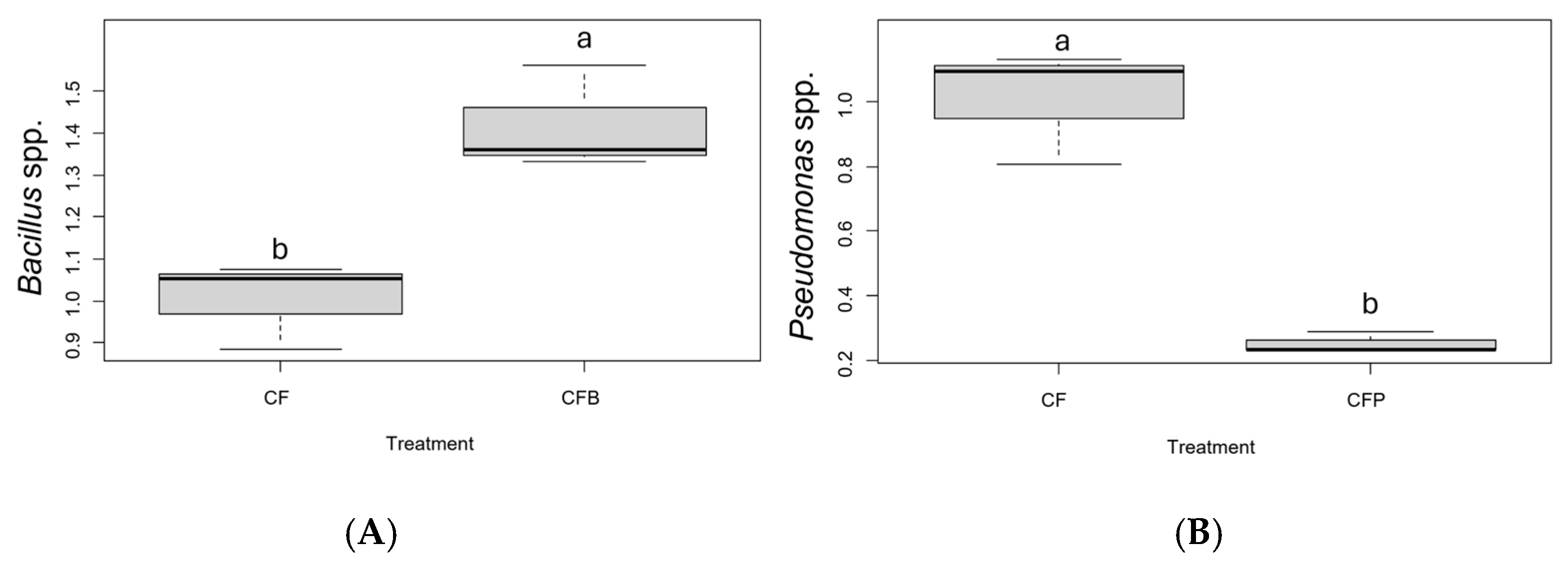
Total bacterial expression was stable across modalities at day 7 (Ct = 10.44 ± 0.33). As shown in Figure 2, larvae fed CFB exhibited a significantly higher abundance of Bacillus spp. in their gut at day 7 compared to those fed control feed (F1,10 = 104.74; p < 0.001). In contrast, larvae fed CFP showed a significantly lower abundance of Pseudomonas spp. than the control group (F1,10 = 392.14; p < 0.001).

3.3. Gut Bacterial Microbiome Diversity and Community Structure

The effective bacterial read numbers of the homogenized larval gut samples ranged from 3697 to 50,809 reads, with a mean of 24,602 ± 1207 reads per sample after filtering out low-quality reads and trimming adapters. A total of 41 distinct bacterial genera were identified within the gut microbiome of the BSF.
Rarefaction curve analysis demonstrated adequate sequencing depth across all samples, with curves approaching saturation for most treatments and developmental stages (Figure 3A,B). No significant variations in bacterial diversity were observed in BSF gut-fed control, Bacillus subtilis 168-contaminated, or Pseudomonas putida KT2440-contaminated treatments (Figure 3C). Alpha diversity metrics, including bacterial taxa richness, Chao1 estimator, and Shannon index, remained consistent across treatments, indicating that the specific bacterial contamination in the substrate had minimal impact on overall gut microbial diversity. Principal Coordinates Analysis (PCoA) using Bray–Curtis dissimilarity matrix revealed no significant effect of treatments on community composition (F = 1.48, R2 = 0.04, p = 0.14; Figure 3E), confirming that exogenous bacterial contamination did not substantially alter the overall structure of the BSF gut bacterial community. However, significant differences were observed in intestinal microbial diversity between larvae and prepupae, with the microbial composition during the larval fattening stage (days 0–11) evolving progressively as feeding time increased (Figure 3B,D). A second PERMANOVA analysis revealed a highly significant effect of larval development on community composition (F = 36.09, R2 = 0.61, p < 0.001; Figure 3F), demonstrating that developmental stage represents the primary driver of gut microbiota structure.

3.4. Dominant Bacterial Genera Composition

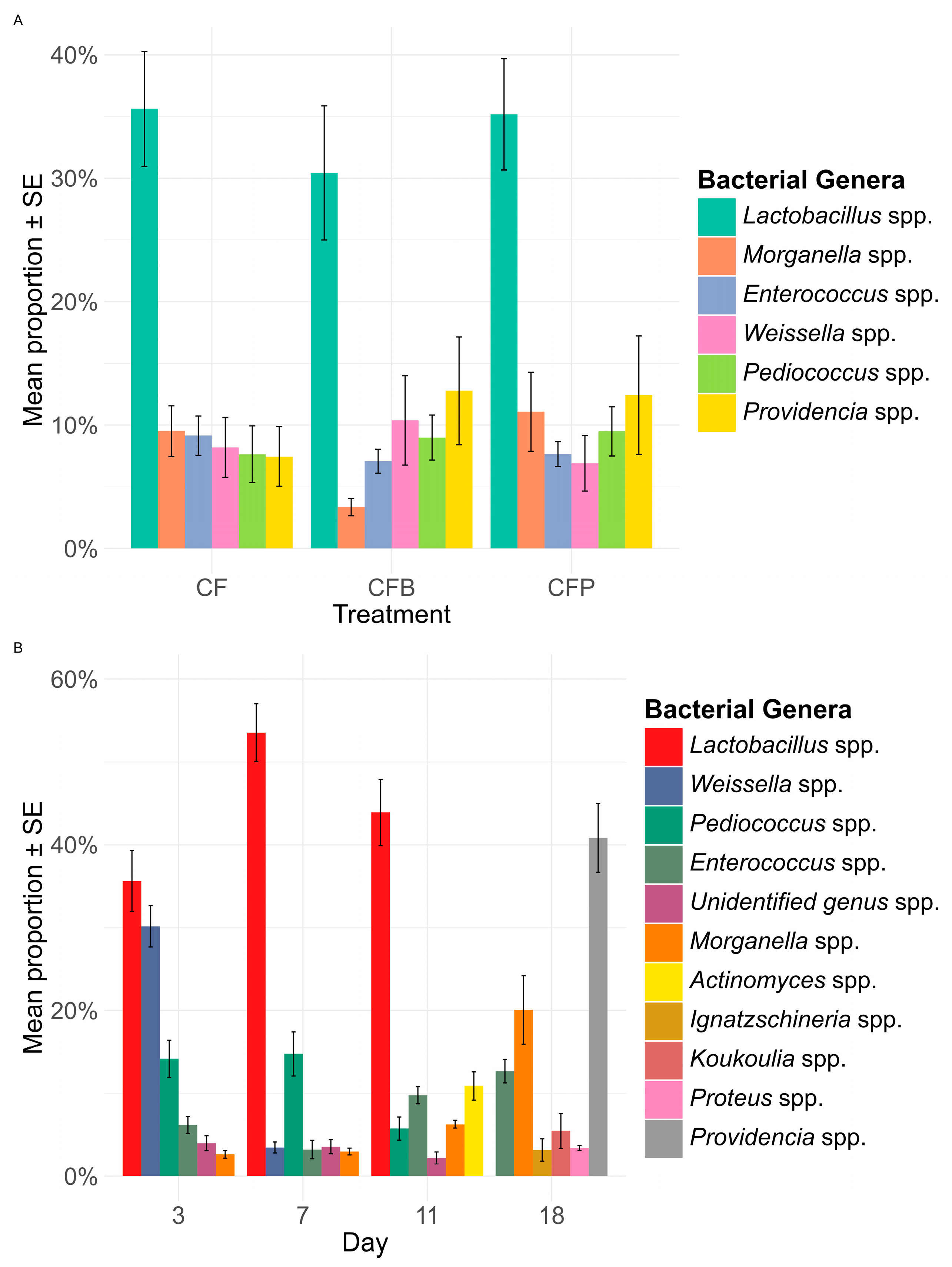
Six core bacterial genera were consistently identified across the three diets, with Lactobacillus spp. representing the dominant genus in the larval gut (Figure 4). The relative abundance of dominant genera showed treatment-specific patterns while maintaining overall community stability. Lactobacillus species comprised 35.61 ± 4.66% of the gut microbiota in control larvae, 30.43 ± 5.44% in Bacillus subtilis 168-contaminated treatment, and 35.18 ± 4.51% in Pseudomonas putida KT2440-contaminated treatment. Lactobacillus spp. showed a peak at day 7 throughout the progression of BSF larval development, while genera from Morganellaceae bacteria (i.e., Morganella spp. and Providencia spp.) dominated the bacterial proportions for the BSF prepupal stage (Figure 4B).

4. Discussion

This study aimed to investigate the effect of contamination by model contaminant strains Pseudomonas putida KT2440 and Bacillus subtilis 168 on the development of BSF larvae and to evaluate the temporal changes in their gut bacterial community structures. Six-day-old BSF larvae were fed chicken feed contaminated by either P. putida KT2440 (CFP) or B. subtilis 168 (CFB). Over 18 days, observations showed that a high contamination level of exogenous bacteria in the substrate had only a slight effect on BSF larvae growth. Specifically, at day 7, larvae fed contaminated feed were lighter than those fed the control diet. In the CFB group, this initial slowdown in growth correlates with a significantly higher abundance of Bacillus spp. in their gut compared to controls. Despite this early growth delay, larvae showed tolerance to high B. subtilis 168 abundance, demonstrated by comparable survival rates and final weights to controls. This tolerance may reflect not only the robustness of BSF larvae but also potential beneficial interactions. B. subtilis is known to produce a range of extracellular enzymes—including proteases, amylases, and lipases—that facilitate nutrient breakdown in organic substrates, potentially improving nutrient assimilation by the larvae despite initial microbial competition [43]. Such enzymatic activity may support substrate degradation and enhance feed conversion efficiency over time, contributing to the observed recovery in larval growth. Xiao et al. [44] demonstrated that inoculating BSF larvae with B. subtilis at a large scale enhanced the reduction of organic matter. Zhao et al. [45] isolated 13 strains of B. subtilis from the surface and the gut of BSF larvae and showed that these strains improved the valorization of bird manure. Similarly, Qiu et al. [46] reported that a strain of B. cereus, isolated from the BSF gut and identified as a beneficial microorganism, improved larval performance when cultivated and inoculated into the feed. Concerning the larvae fed CFP, the abundance of Pseudomonas spp. was lower after 7 days than their abundance in the gut of control larvae. This result suggests that inoculated P. putida KT2440 did not find favorable conditions for development in the decaying substrate or the larval gut. Secondly, the abundance of P. putida KT2440 at the start of the experiment may have overstimulated an immune response in BSF larvae against this microorganism, leading to a lower abundance level by day 7.
According to Vogel et al. [47], the expression of antimicrobial peptides (AMPs) was remarkably induced and expanded in H. illucens larvae by feeding diets containing high bacterial loads. Previously, these AMPs expressed in a diet-dependent mode endow larvae with inhibitory activities against Pseudomonas spp. [47]. Moreover, another key immune factor, the BsfDuox-TLR3 signaling axis, has shown targeted suppression of Pseudomonaceae in BSF larvae [48], reinforcing the hypothesis that host immune activity contributed to the decline in P. putida KT2440.
Previous studies have shown that exogenous bacteria can considerably shape the insect gut, and it has even been claimed that caterpillar intestines are settled (or invaded) by the food microbiome, without any resident gut microbiota [49]. The general view is that the composition of the intestinal microbiota of BSF depends on the source of nutrition; for example, the intestinal bacterial structure varies depending on diets [11,15,16,50,51,52]. Despite these genus-specific differences revealed by qPCR, high-throughput sequencing showed that Bacillus and Pseudomonas were not dominant members of the gut microbial community. This interpretation is supported by the PCoA analysis (Figure 3E), which shows no clear clustering by treatment, and by the similar alpha diversity indices across treatments (Figure 3C). Together, these results indicate that the presence of exogenous bacteria in the substrate did not significantly alter the overall structure of the BSF gut bacterial community.
However, several limitations should be considered. This study focused on genus-level bacterial profiles, which may mask strain-level interactions that could influence larval performance. While both qPCR and high-throughput sequencing were employed, we did not evaluate the actual viability or persistence of the inoculated strains in the substrate before ingestion. Furthermore, the experimental conditions—controlled laboratory settings and the use of a uniform chicken feed substrate—may not fully represent the complexity of waste-based or industrial rearing environments. Additionally, the limited invasion observed with the two model species could be attributed to strain-specific traits, such as the lack of virulence factors and/or antibacterials, which we did not assess. Lastly, while host immune responses were proposed as a mechanism shaping gut colonization, these responses were not directly measured. Future studies incorporating immune gene expression and functional microbial assays would help validate these hypotheses.
In this study, only the abundance of Bacillus in the larval gut showed a clear correlation with the initial inoculum, whereas the Pseudomonas community neither transferred effectively nor accumulated within the BSF intestines. Two hypotheses are given here to explain this difference. First, H. illucens appears to exert selective and interactive control over colonization by exogenous microorganisms. Multiple studies confirm that the insect gut environment generally restricts the migration and colonization of exogenous bacteria, and BSF feeding behavior can even reduce the proliferation of bacteria in the feeding substrate, such as Enterobacteriaceae [29,30,47,53]. On the other hand, there is substantial evidence that part of the microbiota in the BSF intestinal tract has been transferred from the diet. In this study, we hypothesize that Bacillus spp. did not highly proliferate in the intestines of H. illucens due to suboptimal abiotic and biotic conditions for their development. It is known that B. subtilis prefers alkaline to neutral environments, with growth occurring over a pH range of approximately 4.8 to 9.2 and an optimum near pH 6.8, although these values and optima are strain-specific [54,55]. Moreover, B. subtilis grows across a wide temperature range from about 5.5 °C to 55.7 °C, with an optimal growth temperature near 47 °C, but again, these thermal preferences can vary between strains [56]. Notably, the pH within the H. illucens larval gut varies substantially, from acidic conditions in the middle midgut to alkaline conditions in the posterior midgut, with the anterior midgut estimated to be near neutral pH, potentially suitable for B. subtilis growth [16]. B. subtilis is also considered a facultative aerobe, and its growth slows down in an anaerobic environment, such as the larval gut lumen. Finally, B. subtilis, as a Gram-positive bacterium, masters sporulation under unfavorable conditions, unlike P. putida [55]. It can be assumed that the abundance of Bacillus spp. in the larval gut is distributed between active cells and endospores.
The analysis of biotic interactions revealed that the inoculated bacteria primarily competed with Lactobacillales and Enterobacterales, indicating that the abiotic conditions of the luminal environment were more favorable to these orders. The Lactobacillales abundances were mainly represented across all treatments by the following genera: Lactobacillus spp., Enterococcus spp., Pediococcus spp., and Weissella spp. The Enterobacterales were represented by Morganella spp. and Providencia spp. The relative homogeneity of the bacterial communities across treatments confirms that the diet composition is a primary modulator, as previously described by several studies [7,50,57]. The genera listed above were previously found in the gut of H. illucens larvae [57]. The persistence and high abundance of Enterococcus spp. throughout the experiment were expected, as this genus has been identified as a prominent member of the core gut microbiota in H. illucens larvae [7,57]. Morganella spp. was also considered a core member by Wynants et al. [17]. Although this was debated by Klammsteiner et al. [7], our results support the core member status of Morganella spp., especially given its high abundance at the prepupal stage, as observed by Wynants et al. [17] and Wang et al. [58]. This genus appears to be less stable than Enterococcus spp. and may be more influenced by biotic factors, such as the presence of exogenous bacteria. Providencia spp. also reached an abundance level above 40% at the end of larval development, despite the use of a 100% plant-based feed. Comparable levels have only been reported in BSF fed with animal protein by Vecherskii et al. [57], suggesting that other nutrients could also shape this microbial profile. The high abundance of Lactobacillus spp. during larval growth was unexpected, as this genus typically accounts for only 0.05% to 5.00% of the gut microbiota [7,16,17,57]. Previous studies have shown that when BSF larvae process fruits and vegetables, the substrate may display elevated levels of Lactobacillus spp.; however, this increase does not necessarily translate to higher abundance within the larval gut [16]. Lactobacillus spp. are known to confer health benefits [59]. Therefore, some studies have tested the inoculation of Lactobacillus sp. into the feeding substrate of H. illucens, reporting beneficial effects such as improved conversion rates and enhanced nutritional content in the larvae [60]. A positive correlation was observed between Lactobacillus spp. abundance and trypsin and peptidase activities in the gut of BSF larvae, suggesting that these bacteria may support host protein digestion [61]. However, the initial inoculation of Lactobacillus spp. did not increase its abundance level as observed in our experiment. Interestingly, Wang et al. [58] observed a variable abundance of Lactobacillus spp. and Enterococcus spp. in BSF-fed food waste related to substrate particle size. Lactobacillus spp. reached a similar abundance when the particle size distribution ranged mainly from 0 mm to 2 mm. This distribution corresponds to the particle size distribution observed for our substrate (0.750 mm milled chicken feed). Particle size is a crucial factor for the digestion of H. illucens and microorganisms. Indeed, reducing particle size increases nutrient solubilization and the surface area available for microbial community colonization. Thus, particle size indirectly modifies the feed composition. It would be interesting to investigate further this parameter on different substrates, especially the control substrate used in studies on insect gut microbiota.
Finally, in addition to the abiotic and biotic conditions imposed by the digestive tract of H. illucens, the stable colonization of exogenous bacteria in BSF is constrained by its developmental mode. From the perspective of microbial colonization of bacteria, the insect gut as a microbial habitat is often unstable [11,13,14,16]. During larval development, the insect molts and the midgut continuously produce and shed the peritrophic matrix. This dynamic process disrupts or removes most of the associated bacterial populations. Only a small fraction of bacteria cross into the region adjacent to the midgut epithelium, which offers a more stable surface for colonization and supports microbial persistence [13]. In short, numerous challenges must be overcome before exogenous microbes colonize insect intestines and even achieve intergenerational vertical transmission, especially in holometabolous insects, which have distinct larval, pupal, and adult stages. The gut and other organs undergo a radical remodeling during metamorphosis, eliminating the entire larval gut and its contents. In H. illucens, this remodeling begins with the prepupal stage, and this phenomenon is perfectly highlighted in this study by the sudden abundance of the Morganellaceae genera at day 18. To conclude, the establishment of exogenous microorganisms in the H. illucens gut is not an overnight process; they must evolve to form a stable and specific association with this niche, rather than being acquired from the environment by each generation. In mass-reared insects, good rearing practices must be implemented to promote the establishment of insect lines associated with a desired exogenous microorganism selected for its beneficial traits.

5. Conclusions

Our results show that inoculating exogenous B. subtilis 168 and P. putida KT2440 into the substrate of early-stage H. illucens larvae does not disrupt the pre-established bacterial community in the larval gut. Moreover, this study highlights that Lactobacillales-related bacteria are naturally capable of colonizing the digestive system of H. illucens larvae. Among these bacteria, the genus Lactobacillus spp. is well known for its beneficial effects on health. Its abundance in fully developed larvae further enhances the potential of using these larvae as feed.

Supplementary Materials

The following supporting information can be downloaded at https://www.mdpi.com/article/10.3390/applmicrobiol5030082/s1: Figure S1: The standard curves of specific primers for detection of bacteria; Table S1: Primer pairs used during the experiment and their application [62,63,64].

Author Contributions

Conceptualization, B.L. and R.C.M.; methodology, B.L. and R.C.M.; soft–ware, J.C. and G.N.; formal analysis, J.C., G.N., and B.L.; data curation, J.C. and G.N.; writing—original draft preparation, J.C., G.N., and B.L.; writing—review and editing, J.C., G.N., and R.C.M.; supervision, F.F. and R.C.M.; funding acquisition, F.F. All authors have read and agreed to the published version of the manuscript.

Funding

This research received no external funding.

Data Availability Statement

The datasets generated and analyzed during the current study are available from the corresponding author on reasonable request.

Acknowledgments

We thank Frank Delvigne for kindly providing the bacterial strains used in this study.

Conflicts of Interest

The authors declare no conflicts of interest.

Abbreviations

The following abbreviations are used in this manuscript:
BSFBlack Soldier Fly
CFLarvae Fed Chicken Feed
CFBLarvae Fed Chicken Feed + Bacillus subtilis
CFPLarvae Fed Chicken Feed + Pseudomonas putida

References

  1. Mertenat, A.; Diener, S.; Zurbrügg, C. Black Soldier Fly Biowaste Treatment—Assessment of Global Warming Potential. Waste Manag. 2019, 84, 173–181. [Google Scholar] [CrossRef] [PubMed]
  2. Guo, H.; Jiang, C.; Zhang, Z.; Lu, W.; Wang, H. Material Flow Analysis and Life Cycle Assessment of Food Waste Bioconversion by Black Soldier Fly Larvae (Hermetia illucens L.). Sci. Total Environ. 2021, 750, 141656. [Google Scholar] [CrossRef] [PubMed]
  3. Myers, H.M.; Tomberlin, J.K.; Lambert, B.D.; Kattes, D. Development of Black Soldier Fly (Diptera: Stratiomyidae) Larvae Fed Dairy Manure. Environ. Entomol. 2008, 37, 11–15. [Google Scholar] [CrossRef]
  4. Salomone, R.; Saija, G.; Mondello, G.; Giannetto, A.; Fasulo, S.; Savastano, D. Environmental Impact of Food Waste Bioconversion by Insects: Application of Life Cycle Assessment to Process Using Hermetia illucens. J. Clean. Prod. 2017, 140, 890–905. [Google Scholar] [CrossRef]
  5. Lalander, C.; Diener, S.; Magri, M.E.; Zurbrügg, C.; Lindström, A.; Vinnerås, B. Faecal Sludge Management with the Larvae of the Black Soldier Fly (Hermetia illucens)—From a Hygiene Aspect. Sci. Total Environ. 2013, 458–460, 312–318. [Google Scholar] [CrossRef]
  6. Lalander, C.; Diener, S.; Zurbrügg, C.; Vinnerås, B. Effects of Feedstock on Larval Development and Process Efficiency in Waste Treatment with Black Soldier Fly (Hermetia illucens). J. Clean. Prod. 2019, 208, 211–219. [Google Scholar] [CrossRef]
  7. Klammsteiner, T.; Walter, A.; Bogataj, T.; Heussler, C.D.; Stres, B.; Steiner, F.M.; Schlick-Steiner, B.C.; Arthofer, W.; Insam, H. The Core Gut Microbiome of Black Soldier Fly (Hermetia illucens) Larvae Raised on Low-Bioburden Diets. Front. Microbiol. 2020, 11, 993. [Google Scholar] [CrossRef]
  8. Hoc, B.; Genva, M.; Fauconnier, M.-L.; Lognay, G.; Francis, F.; Caparros Megido, R. About Lipid Metabolism in Hermetia illucens (L. 1758): On the Origin of Fatty Acids in Prepupae. Sci. Rep. 2020, 10, 11916. [Google Scholar] [CrossRef] [PubMed]
  9. Hoc, B.; Tomson, T.; Malumba, P.; Blecker, C.; Jijakli, M.H.; Purcaro, G.; Francis, F.; Caparros Megido, R. Production of Rainbow Trout (Oncorhynchus Mykiss) Using Black Soldier Fly (Hermetia illucens) Prepupae-Based Formulations with Differentiated Fatty Acid Profiles. Sci. Total Environ. 2021, 794, 148647. [Google Scholar] [CrossRef]
  10. Lim, J.-W.; Mohd-Noor, S.-N.; Wong, C.-Y.; Lam, M.-K.; Goh, P.-S.; Beniers, J.J.A.; Oh, W.-D.; Jumbri, K.; Ghani, N.A. Palatability of Black Soldier Fly Larvae in Valorizing Mixed Waste Coconut Endosperm and Soybean Curd Residue into Larval Lipid and Protein Sources. J. Environ. Manag. 2019, 231, 129–136. [Google Scholar] [CrossRef]
  11. Carpentier, J.; Abenaim, L.; Luttenschlager, H.; Dessauvages, K.; Liu, Y.; Samoah, P.; Francis, F.; Caparros Megido, R. Microorganism Contribution to Mass-Reared Edible Insects: Opportunities and Challenges. Insects 2024, 15, 611. [Google Scholar] [CrossRef]
  12. Anderson, K.E.; Ricigliano, V.A.; Mott, B.M.; Copeland, D.C.; Floyd, A.S.; Maes, P. The Queen’s Gut Refines with Age: Longevity Phenotypes in a Social Insect Model. Microbiome 2018, 6, 108. [Google Scholar] [CrossRef]
  13. Engel, P.; Moran, N.A. The Gut Microbiota of Insects—Diversity in Structure and Function. FEMS Microbiol. Rev. 2013, 37, 699–735. [Google Scholar] [CrossRef] [PubMed]
  14. Jang, S.; Kikuchi, Y. Impact of the Insect Gut Microbiota on Ecology, Evolution, and Industry. Curr. Opin. Insect Sci. 2020, 41, 33–39. [Google Scholar] [CrossRef] [PubMed]
  15. Varotto Boccazzi, I.; Ottoboni, M.; Martin, E.; Comandatore, F.; Vallone, L.; Spranghers, T.; Eeckhout, M.; Mereghetti, V.; Pinotti, L.; Epis, S. A Survey of the Mycobiota Associated with Larvae of the Black Soldier Fly (Hermetia illucens) Reared for Feed Production. PLoS ONE 2017, 12, e0182533. [Google Scholar] [CrossRef] [PubMed]
  16. Bruno, D.; Bonelli, M.; De Filippis, F.; Di Lelio, I.; Tettamanti, G.; Casartelli, M.; Ercolini, D.; Caccia, S. The Intestinal Microbiota of Hermetia Illucens Larvae Is Affected by Diet and Shows a Diverse Composition in the Different Midgut Regions. Appl. Environ. Microbiol. 2019, 85, e01864-18. [Google Scholar] [CrossRef]
  17. Wynants, E.; Frooninckx, L.; Crauwels, S.; Verreth, C.; De Smet, J.; Sandrock, C.; Wohlfahrt, J.; Van Schelt, J.; Depraetere, S.; Lievens, B.; et al. Assessing the Microbiota of Black Soldier Fly Larvae (Hermetia illucens) Reared on Organic Waste Streams on Four Different Locations at Laboratory and Large Scale. Microb. Ecol. 2019, 77, 913–930. [Google Scholar] [CrossRef]
  18. Deng, G.; Yan, M.; Yang, S.; Ji, L.; Han, X.; Yu, X.; Zhao, K.; Zou, L. Bacillus and Lactic Acid Bacteria Inoculation to Transform Kitchen Waste Using Hermetia illucens. J. Environ. Sci. 2025, in press. [Google Scholar] [CrossRef]
  19. Fernandez-Cassi, X.; Supeanu, A.; Jansson, A.; Boqvist, S.; Vagsholm, I.; SLU, Swedish University of Agricultural Sciences, Department of Biomedical Sciences and Veterinary Public Health, Sweden. Novel Foods: A Risk Profile for the House Cricket (Acheta Domesticus). EFSA J. 2018, 16, e16082. [Google Scholar] [CrossRef]
  20. van Huis, A.; Van Itterbeeck, J.; Klunder, H.C.; Mertens, E.; Halloran, A.; Muir, G.; Vantomme, P. Edible Insects: Future Prospects for Food and Feed Security. Food Agric. Organ. United Nations 2013, 171, xiv+187. [Google Scholar]
  21. Mufungwe, J.; Namukonde, N.; Mwaanga, P.; Johnson, T.; Siamujompa, M.; Mwango, N.C.; Ngoma, J.; Hang’ombe, B.M. Critical Safety Concerns in the Production of Black Soldier Fly (Hermetia illucens) Larvae in Africa. Discov. Food 2025, 5, 74. [Google Scholar] [CrossRef]
  22. den Hil, E.F.H.; Meijer, N.P.; Rozen, K.V.; Elissen, H.; van Wikselaar, P.G.; Brust, H.; Loeke, N.A.J.M.T.; de Rijk, T.; Tienstra, M.; van de Schans, M.G.M.; et al. Safety of Black Soldier Fly (Hermetia illucens) Larvae Reared on Waste Streams of Animal and Vegetal Origin and Manure. J. Insects Food Feed 2023, 10, 771–783. [Google Scholar] [CrossRef]
  23. Gomila, M.; Peña, A.; Mulet, M.; Lalucat, J.; García-Valdés, E. Phylogenomics and Systematics in Pseudomonas. Front. Microbiol. 2015, 6, 214. [Google Scholar] [CrossRef]
  24. Shafi, J.; Tian, H.; Ji, M. Bacillus Species as Versatile Weapons for Plant Pathogens: A Review. Biotechnol. Biotechnol. Equip. 2017, 31, 446–459. [Google Scholar] [CrossRef]
  25. Bremer, E.; Calteau, A.; Danchin, A.; Harwood, C.; Helmann, J.D.; Médigue, C.; Palsson, B.O.; Sekowska, A.; Vallenet, D.; Zuniga, A.; et al. A Model Industrial Workhorse: Bacillus subtilis Strain 168 and Its Genome after a Quarter of a Century. Microb. Biotechnol. 2023, 16, 1203–1231. [Google Scholar] [CrossRef]
  26. Elshaghabee, F.M.F.; Rokana, N.; Gulhane, R.D.; Sharma, C.; Panwar, H. Bacillus As Potential Probiotics: Status, Concerns, and Future Perspectives. Front. Microbiol. 2017, 8, 1490. [Google Scholar] [CrossRef] [PubMed]
  27. de Lorenzo, V.; Pérez-Pantoja, D.; Nikel, P.I. Pseudomonas putida KT2440: The Long Journey of a Soil-Dweller to Become a Synthetic Biology Chassis. J. Bacteriol. 2024, 206, e00136-24. [Google Scholar] [CrossRef] [PubMed]
  28. Raimondi, S.; Spampinato, G.; Macavei, L.I.; Lugli, L.; Candeliere, F.; Rossi, M.; Maistrello, L.; Amaretti, A. Effect of Rearing Temperature on Growth and Microbiota Composition of Hermetia illucens. Microorganisms 2020, 8, 902. [Google Scholar] [CrossRef]
  29. Erickson, M.C.; Islam, M.; Sheppard, C.; Liao, J.; Doyle, M.P. Reduction of Escherichia coli O157:H7 and Salmonella enterica Serovar enteritidis in Chicken Manure by Larvae of the Black Soldier Fly. J. Food Prot. 2004, 67, 685–690. [Google Scholar] [CrossRef]
  30. Gorrens, E.; Van Looveren, N.; Van Moll, L.; Vandeweyer, D.; Lachi, D.; De Smet, J.; Van Campenhout, L. Staphylococcus aureus in Substrates for Black Soldier Fly Larvae (Hermetia illucens) and Its Dynamics during Rearing. Microbiol. Spectr. 2021, 9, e02183-21. [Google Scholar] [CrossRef]
  31. Moyet, M.; Morrill, H.; Espinal, D.L.; Bernard, E.; Alyokhin, A. Early Growth Patterns of Bacillus cereus on Potato Substrate in the Presence of Low Densities of Black Soldier Fly Larvae. Microorganisms 2023, 11, 1284. [Google Scholar] [CrossRef] [PubMed]
  32. van Kessel, K.; Castelijn, G.; van der Voort, M.; Meijer, N. Investigation of Bacillus cereus Growth and Sporulation during Hermetia illucens Larval Rearing. Heliyon 2024, 10, e40912. [Google Scholar] [CrossRef] [PubMed]
  33. Hoc, B.; Noël, G.; Carpentier, J.; Francis, F.; Caparros Megido, R. Optimization of Black Soldier Fly (Hermetia illucens) Artificial Reproduction. PLoS ONE 2019, 14, e0216160. [Google Scholar] [CrossRef]
  34. R Core Team. R: A Language and Environment for Statistical Computing; R Core Team: Vienna, Austria, 2024. [Google Scholar]
  35. Bolyen, E.; Rideout, J.R.; Dillon, M.R.; Bokulich, N.A.; Abnet, C.C.; Al-Ghalith, G.A.; Alexander, H.; Alm, E.J.; Arumugam, M.; Asnicar, F.; et al. Reproducible, Interactive, Scalable and Extensible Microbiome Data Science Using QIIME 2. Nat. Biotechnol. 2019, 37, 852–857. [Google Scholar] [CrossRef] [PubMed]
  36. Callahan, B.J.; McMurdie, P.J.; Rosen, M.J.; Han, A.W.; Johnson, A.J.A.; Holmes, S.P. DADA2: High-Resolution Sample Inference from Illumina Amplicon Data. Nat. Methods 2016, 13, 581–583. [Google Scholar] [CrossRef]
  37. Pruesse, E.; Quast, C.; Knittel, K.; Fuchs, B.M.; Ludwig, W.; Peplies, J.; Glöckner, F.O. SILVA: A Comprehensive Online Resource for Quality Checked and Aligned Ribosomal RNA Sequence Data Compatible with ARB. Nucleic Acids Res. 2007, 35, 7188–7196. [Google Scholar] [CrossRef]
  38. Quast, C.; Pruesse, E.; Yilmaz, P.; Gerken, J.; Schweer, T.; Yarza, P.; Peplies, J.; Glöckner, F.O. The SILVA Ribosomal RNA Gene Database Project: Improved Data Processing and Web-Based Tools. Nucleic Acids Res. 2013, 41, D590–D596. [Google Scholar] [CrossRef]
  39. Bokulich, N.A.; Dillon, M.R.; Bolyen, E.; Kaehler, B.D.; Huttley, G.A.; Caporaso, J.G. Q2-Sample-Classifier: Machine-Learning Tools for Microbiome Classification and Regression. J. Open Res. Softw. 2018, 30, 932. [Google Scholar] [CrossRef]
  40. Kirstahler, P.; Bjerrum, S.S.; Friis-Møller, A.; la Cour, M.; Aarestrup, F.M.; Westh, H.; Pamp, S.J. Genomics-Based Identification of Microorganisms in Human Ocular Body Fluid. Sci. Rep. 2018, 8, 4126. [Google Scholar] [CrossRef] [PubMed]
  41. McMurdie, P.J.; Holmes, S. Phyloseq: An R Package for Reproducible Interactive Analysis and Graphics of Microbiome Census Data. PLoS ONE 2013, 8, e61217. [Google Scholar] [CrossRef]
  42. Xu, S.; Zhan, L.; Tang, W.; Wang, Q.; Dai, Z.; Zhou, L.; Feng, T.; Chen, M.; Wu, T.; Hu, E.; et al. MicrobiotaProcess: A Comprehensive R Package for Deep Mining Microbiome. Innovation 2023, 4, 100388. [Google Scholar] [CrossRef]
  43. Su, Y.; Liu, C.; Fang, H.; Zhang, D. Bacillus subtilis: A Universal Cell Factory for Industry, Agriculture, Biomaterials and Medicine. Microb. Cell Factories 2020, 19, 173. [Google Scholar] [CrossRef]
  44. Xiao, X.; Mazza, L.; Yu, Y.; Cai, M.; Zheng, L.; Tomberlin, J.K.; Yu, J.; van Huis, A.; Yu, Z.; Fasulo, S.; et al. Efficient Co-Conversion Process of Chicken Manure into Protein Feed and Organic Fertilizer by Hermetia illucens L. (Diptera: Stratiomyidae) Larvae and Functional Bacteria. J. Environ. Manag. 2018, 217, 668–676. [Google Scholar] [CrossRef]
  45. Zhao, J.-H.; Cheng, P.; Wang, Y.; Yan, X.; Xu, Z.-M.; Peng, D.-H.; Yu, G.-H.; Shao, M.-W. Using Kin Discrimination to Construct Synthetic Microbial Communities of Bacillus subtilis Strains Impacts the Growth of Black Soldier Fly Larvae. Insect Sci. 2024, 31, 1943–1959. [Google Scholar] [CrossRef] [PubMed]
  46. Qiu, Y.; Wang, P.; Guo, Y.; Zhang, L.; Lu, J.; Ren, L. Enhancing Food Waste Reduction Efficiency and High-Value Biomass Production in Hermetia illucens Rearing through Bioaugmentation with Gut Bacterial Agent. Sci. Total Environ. 2023, 904, 166488. [Google Scholar] [CrossRef] [PubMed]
  47. Vogel, H.; Müller, A.; Heckel, D.G.; Gutzeit, H.; Vilcinskas, A. Nutritional Immunology: Diversification and Diet-Dependent Expression of Antimicrobial Peptides in the Black Soldier Fly Hermetia illucens. Dev. Comp. Immunol. 2018, 78, 141–148. [Google Scholar] [CrossRef] [PubMed]
  48. Huang, Y.; Yu, Y.; Zhan, S.; Tomberlin, J.K.; Huang, D.; Cai, M.; Zheng, L.; Yu, Z.; Zhang, J. Dual Oxidase Duox and Toll-like Receptor 3 TLR3 in the Toll Pathway Suppress Zoonotic Pathogens through Regulating the Intestinal Bacterial Community Homeostasis in Hermetia illucens L. PLoS ONE 2020, 15, e0225873. [Google Scholar] [CrossRef]
  49. Hammer, T.J.; Janzen, D.H.; Hallwachs, W.; Jaffe, S.P.; Fierer, N. Caterpillars Lack a Resident Gut Microbiome. Proc. Natl. Acad. Sci. USA 2017, 114, 9641–9646. [Google Scholar] [CrossRef]
  50. Jeon, H.; Park, S.; Choi, J.; Jeong, G.; Lee, S.-B.; Choi, Y.; Lee, S.-J. The Intestinal Bacterial Community in the Food Waste-Reducing Larvae of Hermetia illucens. Curr. Microbiol. 2011, 62, 1390–1399. [Google Scholar] [CrossRef]
  51. Jiang, C.-L.; Jin, W.-Z.; Tao, X.-H.; Zhang, Q.; Zhu, J.; Feng, S.-Y.; Xu, X.-H.; Li, H.-Y.; Wang, Z.-H.; Zhang, Z.-J. Black Soldier Fly Larvae (Hermetia illucens) Strengthen the Metabolic Function of Food Waste Biodegradation by Gut Microbiome. Microb. Biotechnol. 2019, 12, 528–543. [Google Scholar] [CrossRef]
  52. Zhang, X.; Zhang, J.; Jiang, L.; Yu, X.; Zhu, H.; Zhang, J.; Feng, Z.; Zhang, X.; Chen, G.; Zhang, Z. Black Soldier Fly (Hermetia illucens) Larvae Significantly Change the Microbial Community in Chicken Manure. Curr. Microbiol. 2021, 78, 303–315. [Google Scholar] [CrossRef]
  53. Liu, Q.; Tomberlin, J.K.; Brady, J.A.; Sanford, M.R.; Yu, Z. Black Soldier Fly (Diptera: Stratiomyidae) Larvae Reduce Escherichia Coli in Dairy Manure. Environ. Entomol. 2008, 37, 1525–1530. [Google Scholar] [CrossRef]
  54. Gauvry, E.; Mathot, A.-G.; Couvert, O.; Leguérinel, I.; Coroller, L. Effects of Temperature, pH and Water Activity on the Growth and the Sporulation Abilities of Bacillus subtilis BSB1. Int. J. Food Microbiol. 2021, 337, 108915. [Google Scholar] [CrossRef] [PubMed]
  55. Posada-Uribe, L.F.; Romero-Tabarez, M.; Villegas-Escobar, V. Effect of Medium Components and Culture Conditions in Bacillus subtilis EA-CB0575 Spore Production. Bioprocess Biosyst. Eng. 2015, 38, 1879–1888. [Google Scholar] [CrossRef] [PubMed]
  56. Belperio, S.; Cattaneo, A.; Nannoni, E.; Sardi, L.; Martelli, G.; Dabbou, S.; Meneguz, M. Assessing Substrate Utilization and Bioconversion Efficiency of Black Soldier Fly (Hermetia illucens) Larvae: Effect of Diet Composition on Growth and Development Temperature. Animals 2024, 14, 1340. [Google Scholar] [CrossRef]
  57. Vecherskii, M.V.; Kuznetsova, T.A.; Khairullin, D.R.; Chaporov, I.A.; Bastrakov, A.I.; Ushakova, N.A. Feeding Impact on the Gut Microbiome of Hermetia illucens Larvae. Appl. Biochem. Microbiol. 2025, 61, 200–210. [Google Scholar] [CrossRef]
  58. Wang, Y.; Quan, J.; Cheng, X.; Li, C.; Yuan, Z. Relationship of Black Soldier Fly Larvae (BSFL) Gut Microbiota and Bioconversion Efficiency with Properties of Substrates. Waste Manag. 2024, 180, 106–114. [Google Scholar] [CrossRef]
  59. Walter, J. Ecological Role of Lactobacilli in the Gastrointestinal Tract: Implications for Fundamental and Biomedical Research. Appl. Environ. Microbiol. 2008, 74, 4985–4996. [Google Scholar] [CrossRef]
  60. Somroo, A.A.; ur Rehman, K.; Zheng, L.; Cai, M.; Xiao, X.; Hu, S.; Mathys, A.; Gold, M.; Yu, Z.; Zhang, J. Influence of Lactobacillus Buchneri on Soybean Curd Residue Co-Conversion by Black Soldier Fly Larvae (Hermetia illucens) for Food and Feedstock Production. Waste Manag. 2019, 86, 114–122. [Google Scholar] [CrossRef]
  61. Yu, G.; Nil, C.; He, G.; Zhou, L.; Xia, Q.; Cheng, P. Isolation and identification of bacteria producing enzymes from gut and skin of black soldier fly (Hermetia illucens) larvae. Front. Microbiol. 2010, 47, 889–894. [Google Scholar]
  62. Wattiau, P.; Renard, M.-E.; Ledent, P.; Debois, V.; Blackman, G.; Agathos, S. A PCR Test to Identify Bacillus subtilis and Closely Related Species and Its Application to the Monitoring of Wastewater Biotreatment. Appl. Microbiol. Biotechnol. 2001, 56, 816–819. [Google Scholar] [CrossRef] [PubMed]
  63. Louis, P.; Flint, H.J. Development of a Semiquantitative Degenerate Real-Time PCR-Based Assay for Estimation of Numbers of Butyryl-Coenzyme A (CoA) CoA Transferase Genes in Complex Bacterial Samples. Appl. Environ. Microbiol. 2007, 73, 2009–2012. [Google Scholar] [CrossRef] [PubMed]
  64. Klindworth, A.; Pruesse, E.; Schweer, T.; Peplies, J.; Quast, C.; Horn, M.; Glöckner, F.O. Evaluation of General 16S Ribosomal RNA Gene PCR Primers for Classical and Next-Generation Sequencing-Based Diversity Studies. Nucleic Acids Res. 2013, 41, e1. [Google Scholar] [CrossRef] [PubMed]
Figure 1. Growth of Hermetia illucens larvae fed control chicken feed (CF), Bacillus subtilis 168-contaminated chicken feed (CFB), and Pseudomonas putida KT2440-contaminated chicken feed (CFP). Each point on the curves represents the mean individual weight (mg) calculated from weighing 30 larvae randomly selected from a population of 100 larvae (n = 3). Error bars indicate SEM, and asterisks indicate significant differences across modalities, as determined by ANOVA followed by Tukey’s post hoc test. On day 7, all modalities differ significantly.
Applmicrobiol 05 00082 g001
Figure 2. Bacterial abundance in the gut of Hermetia illucens larvae fed contaminated chicken feed with model contaminant strains Bacillus subtilis 168 and Pseudomonas putida KT2440 at day 7. (A) Bacillus spp. abundance. (B) Pseudomonas spp. abundance. Mean value (n = 3) ± SEM per bacterial group. Data are expressed as the normalized fold expression to control diet (i.e., CF), which is 1.000. Different letters above columns indicate significant differences (p < 0.05). Total bacteria were selected as the reference gene and were stable across treatments.
Applmicrobiol 05 00082 g002
Figure 3. (A,B) Rarefaction curves for diet treatment (A) and Hermetia illucens larval development (B). Shaded areas of both curves correspond to the 95% confidence interval. (C,D) Alpha diversity metrics comparing diet treatment (C) and BSF larval development (D) for bacterial taxa richness in ASVs, Chao1 richness estimator, and Shannon’s index. The p-value of the Kruskal–Wallis test is indicated on every boxplot. (E,F) Principal component analysis (PCoA) comparing bacterial communities of diet treatment (E) and BSF larval development (F). The dot size is scaled according to the ASVs’ richness, and the opacity is scaled according to Shannon’s index.
Applmicrobiol 05 00082 g003
Figure 4. (A,B) Composition of bacterial genera in Hermetia illucens gut microbiota across feed treatments (A) and throughout larval development (B). (A) Relative abundance of dominant bacterial genera in the gut microbiota of Hermetia illucens larvae reared on three feed treatments: Control, Bacillus subtilis 168-contaminated feed, and Pseudomonas putida KT2440-contaminated feed. (B) Temporal changes in the relative abundance of dominant bacterial genera within the Hermetia illucens gut microbiota at days 3, 7, 11, and 18 of development.
Applmicrobiol 05 00082 g004
Table 1. Development time and survival rate of Hermetia illucens larvae fed control chicken feed (CF), Bacillus subtilis 168 contaminated chicken feed (CFB), and Pseudomonas putida KT2440 contaminated chicken feed (CFP).
TreatmentsDevelopment Time
(days ± SD)
Survival Rate
(% ± SD)
CF17.8 ± 1.11 a93.00 ± 2.67 a
CFB18.3 ± 0.78 a91.33 ± 1.77 a
CFP17.8 ± 0.89 a91.67 ± 1.11 a
a Means within the same column with different letters differ significantly.
Disclaimer/Publisher’s Note: The statements, opinions and data contained in all publications are solely those of the individual author(s) and contributor(s) and not of MDPI and/or the editor(s). MDPI and/or the editor(s) disclaim responsibility for any injury to people or property resulting from any ideas, methods, instructions or products referred to in the content.