Article

Inactivation Kinetics of Escherichia coli and Staphylococcus aureus Using Ultrasound in a Model Parenteral Emulsion

by
Maricarmen Iñiguez-Moreno
1,2,*,
Montserrat Calderón-Santoyo
3,
Gabriel Ascanio
1,*,
Estefanía Brito-Bazán
1,
María Soledad Córdova-Aguilar
1,
Edmundo Brito-de la Fuente
1 and
Juan Arturo Ragazzo-Sánchez
3
1
Instituto de Ciencias Aplicadas y Tecnología, Universidad Nacional Autónoma de México, Ciudad Universitaria, Mexico City 04510, Mexico
2
Escuela de Ingeniería en Agrotecnología, Universidad Politécnica del Estado de Nayarit, Tepic 63506, Mexico
3
Laboratorio Integral de Investigación en Alimentos, Tecnológico Nacional de México/Instituto Tecnológico de Tepic, Tepic 63175, Mexico
*
Authors to whom correspondence should be addressed.
Appl. Microbiol. 2025, 5(1), 34; https://doi.org/10.3390/applmicrobiol5010034
Submission received: 12 February 2025 / Revised: 17 March 2025 / Accepted: 18 March 2025 / Published: 20 March 2025
(This article belongs to the Special Issue Applied Microbiology of Foods, 2nd Edition)

Abstract

:
Ultrasound (US) is a technology that enables microbial inactivation through cavitation-induced cell wall disruption, preserving food safety and quality. This study evaluated the impact of US parameters, including time, temperature, and surrounding media (saline solution and lipid parenteral emulsion) on Escherichia coli and Staphylococcus aureus reduction. Microbial survival was quantified via plate counting, and inactivation kinetics were modeled using GInaFiT. Microbial reductions ranged from 0.05 to 6.10 Log10 CFU/mL, with E. coli showing greater susceptibility than S. aureus. The highest reduction (6.10 Log10 CFU/mL) was observed for E. coli in the emulsion after 5 min at uncontrolled temperature, while S. aureus exhibited lower susceptibility (3.92 Log10 CFU/mL). The Weibull model provided the best fit, highlighting the non-linear nature of microbial inactivation. The US presents a promising alternative for microbial control in food and pharmaceutical applications. Future research should optimize treatment conditions, understand microbial resistance mechanisms, and integrate the US with other hurdle technologies to enhance efficiency. In addition, studies about the US’s scalability for the pharmaceutical industry could widespread its implementation in that sector.

1. Introduction

Parenteral nutrition is essential for several patient groups, yet inadvertent microbial contamination poses a severe risk, potentially leading to morbidity and even mortality. Due to their high nutritional content, some components of parenteral emulsions serve as ideal substrates for microbial growth [1]. While lipids in these formulations have demonstrated an inhibitory effect on the proliferation of certain bacteria, including Escherichia coli, Staphylococcus epidermidis, and Staphylococcus aureus [2]. The prolonged administration of emulsions at room temperature provides favorable conditions for microbial growth, increasing the risk of severe infections such as septicemia [3].
Thermal sterilization remains the gold standard in ensuring the microbiological safety of pharmaceutical products, including parenteral emulsions, due to its well-documented effectiveness in enzyme and microorganism inactivation [4]. However, the non-specific nature of heat treatments often results in undesirable alterations in emulsion stability, including the degradation of bioactive compounds, modifications in the molecular structure of polysaccharides, proteins, and lipids, as well as increased droplet size, ultimately affecting both product shelf life and functionality [5,6]. Given these limitations, techniques such as high hydrostatic pressure, pulsed electric fields, gamma irradiation, and ultrasound (US) have been explored as alternative methods to preserve pharmaceutical emulsions while minimizing the adverse effects of heat on their components [7]. Among these, the US has gained significant attention in the food and pharmaceutical industries due to its ability to facilitate emulsification, mixing, homogenization, and enzyme inactivation while maintaining product integrity [8,9,10].
The US primarily attributes microbial inactivation to cavitation phenomena, where the violent collapse of bubbles generates localized extreme temperature and high pressure, leading to cell membrane disruption and increased permeability [9,11]. Reactive oxygen species (ROS) generated during cavitation can also exacerbate external and internal cellular damage, further enhancing microbial inactivation [12]. Several studies have demonstrated the potential of the US for microbial inactivation in various applications, highlighting its effectiveness as a sterilization method. The US at 500 W, operating at 20 kHz for 10 min was used to reduce the microbial loads in cocoa honey, achieving a reduction up to undetectable levels (>4 Log CFU/mL of yeast, molds, and bacteria) and maintaining the physicochemical properties for 21 days. In contrast, in solid matrices like lettuce leaves, the thermosonication at 30 and 60 °C resulted in a reduction in S. aureus and Shigella flexneri populations by 5.1–6.9 log CFU/g and 5.5–7.4 log CFU/g, respectively [13]. Also, it has been demonstrated that ultrafine bubbles can have a synergistic effect with the US on the inactivation kinetics of E. coli in sterile phosphate-buffered saline, resulting in a disinfection rate constant (kD = 0.206 min−1), surpassing the sum of the values obtained with the US alone (kD = 0.126 min−1) and ultrafine bubbles alone (kD = 0.007 min−1) [14]. However, not all the proposed combinations exhibited a synergistic effect. For instance, the use of US in conjunction with sodium chloride (12%, w/v) did not enhance the antimicrobial efficacy compared to US alone [13]. The effects of US on food matrices have been widely explored, noting their impact on microstructure, color, nutrient retention, and reduction in microbial load. These findings highlight the potential of US as a viable alternative to conventional thermal methods, positioning it as a promising technology for ensuring food safety [15]. Its application in pharmaceutical formulations, particularly as a hurdle technology for ensuring microbial safety in parenteral emulsions, remains largely unexplored. Limited research has addressed the inactivation kinetics associated with this approach, highlighting the need for further investigation. Therefore, this study aims to (i) evaluate the efficacy of US treatments in reducing E. coli and S. aureus counts in a model parenteral emulsion, (ii) analyze the impact of temperature increment during the US processing, and (iii) model the microbial reduction kinetics using both linear and Weibull models to determine the most suitable approach for describing the inactivation behavior under the tested conditions.

2. Materials and Methods

2.1. Culture Media and Chemicals

Tryptic soy broth (TSB) and tryptic soy agar (TSA) were purchased from Becton Dickinson (Le Pont de Claix, France). Soy lecithin E322 (Cargill Texturizing Solutions, Deutschland GmbH and Co. KG, Hamburg, Germany), glycerol (98%, Golden bell, Zapopan, Mexico), soybean oil (Nutrioli®, Grupo Ragasa, Monterrey, Mexico), and sodium hydroxide (NaOH, Jalmek, San Nicolás de los Garza, Mexico).

2.2. Emulsion Preparation

Oil-in-water emulsions were prepared by homogenizing soybean oil and lecithin (oil phase) with distilled water and glycerol (aqueous phase), as described by Iñiguez-Moreno et al. [16]. Briefly, the aqueous phase was formulated by mixing 76.6 g of distilled water at 80 °C with 2.2 g of glycerol, adjusting the pH to 10.0 using 1 M NaOH, and homogenizing at 5000 rpm for 5 min. Simultaneously, the oil phase was prepared by adding 1.2 g of soy lecithin to 20 g of soybean oil at 80 °C and homogenizing under the same conditions. Subsequently, the oil phase was gradually incorporated into the aqueous phase under continuous homogenization at 10,000 rpm for 15 min. The resulting pre-emulsion was cooled to 55–60 °C, adjusted to a pH between 9.7 and 10.0, and subjected to sonication using a Digital Sonifier® Unit (model S-150D, Branson Ultrasonics Corporation, Danbury, CT, USA) with a 3.2 mm microtip at 60 kHz for 5 min. The final emulsion had a pH of 9.48 ± 0.04 and a water activity of 0.99. The emulsion was prepared for each test to ensure consistency across all experimental tests.

2.3. Bacterial Inoculation

Stationary phase cells of E. coli and S. aureus were obtained as described by Bulut et al. [17]. Pure bacterial stock cultures were preserved at −80 °C in 15% (v/v) glycerol. Before experimentation, a sample was transferred to tryptic soy broth (TSB) and incubated overnight at 37 °C. The bacteria were then streaked onto tryptic soy agar (TSA) plates and incubated under the same conditions. Three isolated colonies were selected from the plates and introduced into 200 mL emulsion in sterile 500 mL flasks to prepare the inoculum. This achieved an approximate final concentration of 109 CFU/mL. Subsequently, 15 mL of the inoculated emulsion was aseptically transferred into 30 mL sterile glass vials using a sterile syringe. All handling steps were conducted under aseptic conditions in an air-flow chamber. The process was followed to inoculate the sterile saline solution SS (NaCl 0.85%, w/v).

2.4. Ultrasound (US) Treatments

The effect of the US (24 kHz and 100 W; Model S-150D, Branson Ultrasonics Corporation) at different times (1, 3, and 5 min) and with and without temperature control in two different surrounding media (emulsion or SS) was assessed. Fifteen milliliters of E. coli or S. aureus cell suspension suspended on SS or emulsion were sonicated by submerging a 3 mm diameter microtip 10 mm below the surface and operating at 100% intensity. Both conditions were considered: without temperature control and using an ice bath at 4 °C to maintain sample temperature below 30 °C. Quantifying microbial loads before and after the treatments was carried out by the traditional counting plate technique onto soy trypticase agar (Becton Dickinson Bioxon, Le Pont de Claix, France). The Petri dishes were incubated for 48 h at 35 °C. Each treatment was carried out in triplicate and repeated once.

2.5. Effect of Temperature

To determine the effect of temperature during the sonication process, the temperature of the samples was registered at the end of the treatments. Inoculated samples (15 mL) were collocated in a cold-water bath at the temperature reached in the different US treatments (Table 1). Each treatment was carried out in triplicate and repeated once.

2.6. Synergistic Effect of US and Temperature

The synergistic effect of US treatments without temperature control was determined using Equation (1) and the data from microbial enumeration [18]:
S y n e r g i s t i c   e f f e c t = R U S W O T C ( R U S W T C + R T )
where R U S W O T C , R U S W T C , and R T are the reduction reached with US treatments without temperature control, maintaining the temperature below 30 °C, and using a water bath.
The traditional counting plate estimated bacterial survival on TSA. The Petri dishes were stored at 25 °C for 6 h, allowing the US-stressed bacteria to be repaired and replicated. Then, they were incubated at 35 °C for 48 h before enumeration [19,20].

2.7. Inactivation Kinetics Modeling

2.7.1. Linear Model

The first-order kinetic behavior is followed by the log-linear model (also known as the D-value model), which postulates that a cell population responds similarly to the treatment and that its reduction rises semi-logarithmically during the treatment. Equation (2) provides the linear model:
L o g 10 N N 0 = t D ; t   0 ;   s l o p e = 1 D
where N0 is the initial microbial load and N represents survival cell counts after each US treatment; both expressed in CFU/mL, t and D values are the treatment time (min) and the time (min) required to destroy 90% of the initial microbial population, estimated from Log10 (N/N0) vs. treatment time (min) [21].

2.7.2. Weibull Model

The add-in Geeraerd and Van Impe Inactivation Model Fitting Tool (GInaFiT) [21] was used in Microsoft Excel to assess non-log-linear microbial inactivation curves (Equation (3)):
L o g 10 N N 0 = 1 2.303   ( t α ) β  
where α and β are the time required for the first decimal reduction (min) and the fitting parameter that defines the curve shape, respectively, based on a Log10 graph, if β < 1 the Weibull distribution corresponds to a concave upward curve, instead if β > 1 the graph will have a concave downward curve, and a straight line if β = 1. α differs from the conventional D-value, which is derived from the first-order kinetic model and specifies the time for a decimal reduction independent of the heating time. The α-value represents the first decimal reduction in the initial number N0 to N0/10 [22,23]. Otherwise, to estimate the time required (td) to reduce 5 Log10 CFU/mL, Equation (4) was used:
t d = α [ ln 1 0 d ] 1 β
where d is the number of decimal reductions [24].

2.7.3. Model Evaluation

The goodness of the fit of both models was determined using the coefficient of determination (R2, Equation (5)) and root mean square error (RMSE, Equation (6)):
R 2 = 1 S y x   2 S y   2
R M S E = k = 1 z ( y k y k * ) 2 z
where S y x   2 and S y   2 are the standard deviations of estimation and sample, respectively. y and y* are the experimental and estimated data, respectively; z is the number of experimental data.

2.8. Statistical Analysis

Analysis of variance and linear models were performed using Statgraphics Centurion XVI. I software (Statpoint Technologies, Inc., Warrenton, VA, USA). The post hoc least significant difference (LSD) Fisher test (p ≤ 0.05) was used for means comparison.

3. Results and Discussion

3.1. Inactivation of the Microorganisms by the US

The inactivation of both microorganisms after 1 min of the US treatment in the two assessed media was not different from the initial counts (p > 0.05; Figure 1). Significant reductions were observed after 3 min of US treatment, especially against S. aureus in the emulsion (2.80 ± 0.20 Log10 CFU/mL). However, when controlling the temperature, US treatment does not reduce the initial count of S. aureus after this time (Figure 1b, p > 0.05). After 5 min of the US treatment in both media, at least 3.81 Log10 CFU/mL of the tested microorganisms were reduced without temperature control. The lower cell recovery was for E. coli in the emulsion, followed by SS (Figure 1a). Microbial inactivation using the US depends on several factors, including treatment conditions (media, temperature, frequency, time, etc.) and microbial characteristics [8,25]. The effect of treatments on bacterial cells increases as the time and the temperature generated by the ultrasound waves do (p ≤ 0.05), which agrees with previous researchers [9,26]. The higher survival of Gram-positive bacteria like S. aureus in comparison with E. coli (Gram-negative) was previously reported in a study that combined the US at low frequency (40 Hz) and propyl gallate (a food grade antioxidant) to reduce E. coli O157:H7 and Listeria innocua [9]. These results can be related to several factors. First, it is generally accepted that rod-shaped bacteria are more susceptible to US than spherical forms. Spherical organisms should be more resistant to crushing than rod-shaped ones, which is consistent with the idea that the lethal effect of ultrasonic waves is due to the pressure changes associated with their passage through fluids [14]. Otherwise, the US disrupts bacterial membrane/cell; hence, Gram-negative bacteria are more sensitive to this treatment than Gram-positive [9,11]. This is related to peptidoglycan content, which is higher in Gram-positive (30–70%) than in Gram-negative (<10%) bacteria, which makes them more resistant to the cavitation process [13]. When bubbles expand and compress, cavitation causes mechanical stress on the cell membrane. When the bubbles burst, a localized shock wave is created that pushes a fast-moving liquid jet toward the surface [14]. Microorganisms can withstand high pressure due to the fluidity of their membranes but cannot survive the alternating pressure generated during cavitation. The mechanical forces form pores in the outer membrane and, over time, destabilize the inner membrane, releasing the intracellular components [9,25,27]. In E. coli, the morphological damage induced by the US depends on the intensity and duration of exposure. As these parameters increase, shrinkage, deformation, and pores are observed in the cell membrane, and the formation of cell debris appears [18,27]. In addition, higher microbial inactivation was reached in the emulsion in comparison with SS (p ≤ 0.05), which is attributable to the emulsion’s oil phase, allowing it to reach a higher temperature than SS [28], favoring microbial inactivation. The higher reduction obtained was 6.10 Log10 CFU/mL for E. coli in the emulsion (Figure 1a); this is up to the minimal reduction required to consider a non-thermal treatment as effective (5 Log10 CFU/mL) [29].

3.2. Effect of Temperature

The violent collapse of bubbles during the US process increases temperature and pressure [14,30]. Tests using a water bath at the temperatures reached in each US trial were carried out to determine the effect of temperature during the US treatments. As expected, at temperatures ranging from 25 to 39 °C, the microbial populations were not reduced compared to their corresponding initial counts (p > 0.05, Table 1). Instead, significant reductions were obtained after 3 min at 55 °C in the emulsion (Table 2). Thermal treatments for microbial inactivation in several culture media have been widely studied [31]. For E. coli O157:H7, D60°C ranges from 0.8 to 1.9 min depending on the culture media conditions, which include broth, apple juice, and red meat [32]. However, S. aureus was isolated from refrigerators and cultivated in TSB, D60°C values ranged from 4.8 to 6.5 min [33]. These results show that 5 Log10 CFU/mL of E. coli or S. aureus cannot be reduced at 60 °C/5 min. Temperature can have a cumulative or synergistic effect (depending on the environmental conditions, particularly the media) on the US treatments, favoring microbial inactivation [5,34]. In this research, the temperature showed a synergistic effect that increased as time and temperature increased (Table 3). This effect has been reported in the inactivation of E. coli O157:H7 by US treatments (20 kHz applied in a pulsed mode of 2 s) in combination with thyme essential oil (10 mg/mL) [18].

3.3. Linear Model

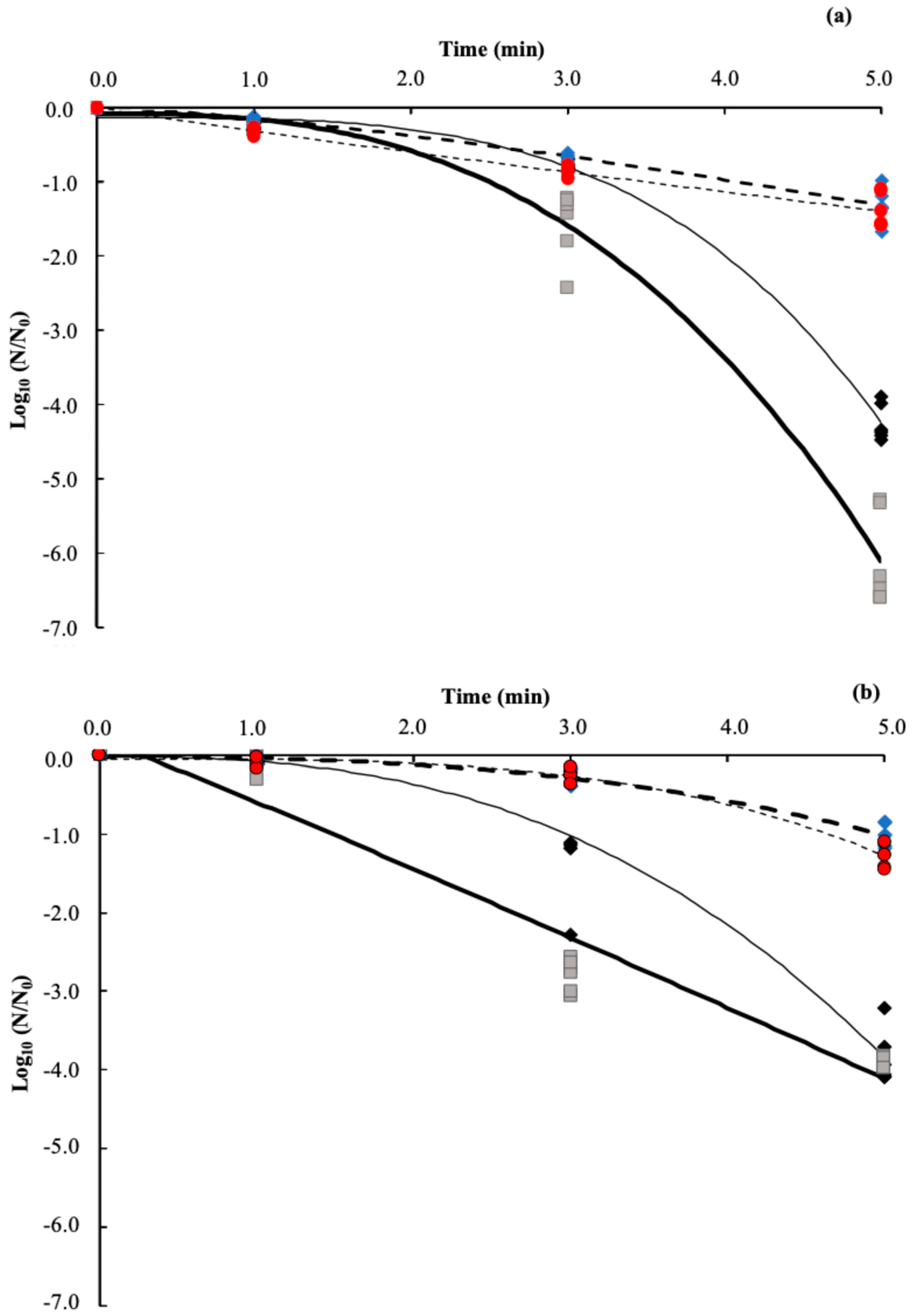
Traditionally, microbial inactivation under constant environmental conditions is estimated using a linear model if it follows first-order kinetics. Figure 2 shows the linear models obtained for E. coli and S. aureus, and Table 3 shows their fitting parameters and D-values. The goodness of fit was satisfactory for all models (R2 > 0.86 and RMSE < 0.77). E. coli D-values ranged from 1.0 to 4.04 min, and from 1.25 to 5.87 for S. aureus. D-value varies depending on the tested microorganism, media, temperature, and treatment time. The lower D-values for both microorganisms were obtained in the emulsion without temperature control during the US treatment. However, differences were observed in the D-values of both strains in any treatment maintaining the temperature below 30 °C, as was expected owing to the differences in the composition between Gram-positive and Gram-negative bacteria [9,11].

3.4. Non-Linear Weibull Model

The non-linear models obtained for E. coli and S. aureus are shown in Figure 3. The goodness-of-fit of the models was compared using the R2 and RMSE values, >0.94 and <0.99, respectively (Table 3). According to the Weibull model, the population of microorganisms can be reduced by a cumulative exponential distribution since various fractions of the population may have varying resistance to the treatment conditions [21]. The characteristic parameters α and β, necessary for the Weibull model, are detailed in Table 3. In both microorganisms, the surrounding media and using an ice bath during the US treatments affected α, as occurs in the linear model. These values were higher in the SS using an ice water bath, consistent with the data from the linear model. α-values for the US treatments carried out below 30 °C were similar to D-values obtained from the linear model (p > 0.05); this is in agreement with the increment of β-value (Table 3).
The td value represents the amount of time required to achieve numerous logarithmic reductions while considering the shape of the Weibull model’s curve [24]. The td value to diminish 5 Log10 of the microbial population determined by the Weibull model had a clear relationship with temperature on the US treatments, increasing four times when the temperature was maintained at ~27 °C. These facts are attributable to the increment of the β-value; to have similar α and D-values, the β-value must be near 1. In most cases, β was higher than 1, which is represented by concave downward inactivation curves (Figure 3). Curves’ shapes are linked to a gradual reduction rate during the first US treatment exposure and a fast inactivation rate afterward [24]. Microbial membrane follows the fluid mosaic model, which is the most thermodynamically stable configuration and establishes that the membrane can repair the pores generated by low doses of physical damage [35], resulting in shoulders in the curves (Figure 3) [24]. In addition to this, the shoulder formation in the Weibull model of S. aureus (Figure 3b) could suggest heat shock protein synthesis, which increases its thermal resistance compared to E. coli [36].
The microorganisms are inactivated when the US waves surpass the maximum capacity for membrane repair, which causes an exponential decrease in the number of survivors. [24,35]. This behavior was observed after 3 min of treatment (Figure 3). Similar behavior in α and D-values were reported by applying pulsed light for the inactivation of E. coli K-12, Clostridium sporogenes, and Geobacillus stearothermophilus, α-value increased in parallel to the increment of the separation between the flashlight and the sample [24]. It was previously reported that the use of high-power US (50 ± 5 W) to increase the microbial inhibition of E. coli, Brevundimonas diminuta, and Aspergillus niger by the supercritical state of carbon dioxide resulted in concave upward curves [37]. Combining the two treatments may affect the microbial decrease rate while avoiding the lag phase in kinetics. This could be due to a cumulative or synergistic effect, which lowers the β-value, resulting in concave upward curves [38].
In the Weibull model, the microbial inactivation rate in the initial phase has an essential effect on the computational estimation of α and td values. Instead, the D-value is based on the average inactivation rate over the treatment duration. Additionally, in the log-linear model, the degree of inactivation is a multiple of the D-value, while it cannot be calculated from td in the Weibull model (both α and β are required for such calculation) [39]. In general, in this research, the Weibull model provides a better description of the kinetic inactivation of E. coli and S. aureus in SS and emulsion under the assessed conditions (Table 3). These results agree with the combination of acidic electrolyzed water (SAEW) and fumaric acid (FA) in the US at a mild temperature against L. monocytogenes and E. coli O157:H7 on sprouts. To reduce 4 Log10 of L. monocytogenes, 12, 7, and 3 min at 23, 30, and 40 °C were required (R2 ≥ 0.9; RMSE 0.01–0.48). Whereas for E. coli, O157:H7 4 Log10 were reduced after 14, 9, and 3 min at 23, 30, and 40 °C, respectively (R2 ≥ 0.9; RMSE 0.02–0.54) [40]. The α-value should be viewed with caution and not used by itself to predict the required number of logarithmic reductions as commonly occurs with D-values [24]. Therefore, the US can be a potential alternative to heat-based sterilization, ensuring the safety of pharmaceutical products and ultimately contributing to improved patient safety by minimizing infection risks. One of its key strengths is the comprehensive evaluation of multiple parameters, including time, temperature, and surrounding media, providing valuable insights into optimizing microbial reduction. Additionally, it is important to model microbial inactivation using non-linear approaches, such as the Weibull model, which demonstrated the best fit in this study. It is crucial to make an appropriate choice of model to describe the parameters of inactivation kinetics to reduce their underestimation or overestimation. Future studies should focus on precise temperature control, a broader range of microorganisms, physicochemical changes in food matrices, and the scalability of this technology for industrial applications.

4. Conclusions

The inactivation kinetics of E. coli and S. aureus by US depended on the type of bacteria, time, temperature, and media tested. A higher reduction rate was reached for E. coli in the emulsion after 5 min of the US treatment without temperature control. This suggests that the US can be an alternative to control microbial loads in pharmaceutical products, such as parenteral emulsions. The log-linear and Weibull models demonstrated a good fit for the reduction kinetics; the non-linear model gave a slightly better description of E. coli and S. aureus reduction rates under the assessed conditions. Further studies that combine US with other treatments, such as ultraviolet light, high hydrostatic pressure, etc., aimed to increase the reduction in Gram-positive and harmful bacteria without altering the physicochemical properties of the pharmaceutical products should be carried out. Future studies should improve treatment settings, understand microbial resistance mechanisms, and integrate US with other hurdle technologies to increase efficiency. In addition, studies on the scalability of US for the pharmaceutical industry could broaden its implementation in that sector.

Author Contributions

Conceptualization, M.I.-M. and J.A.R.-S.; methodology, M.I.-M. and E.B.-B.; formal analysis, M.I.-M. and M.C.-S.; investigation, M.I.-M.; writing—original draft preparation, M.I.-M.; writing—review and editing, M.C.-S., M.S.C.-A., E.B.-d.l.F. and J.A.R.-S.; visualization, M.I.-M. and E.B.-B.; supervision, G.A., E.B.-d.l.F. and J.A.R.-S.; project administration, G.A., E.B.-d.l.F. and J.A.R.-S.; funding acquisition, G.A., E.B.-d.l.F. and J.A.R.-S. All authors have read and agreed to the published version of the manuscript.

Funding

This work was supported by Fresenius-Kabi Deutschland. Gabriel Ascanio thanks “Programa de Apoyos para la Superación del Personal Académico” PASPA-DGAPA-UNAM for the funds for the sabbatical leave.

Institutional Review Board Statement

Not applicable.

Informed Consent Statement

Not applicable.

Data Availability Statement

The raw data supporting the conclusions of this article will be made available by the authors upon request.

Conflicts of Interest

The authors declare no conflicts of interest.

References

  1. Tota, A.; Serra, A.; Raoul, P.; Gasbarrini, A.; Rinninella, E.; Mele, M.C. Lipid-Enriched Parenteral Nutrition and Bloodstream Infections in Hospitalized Patients: Is It a Real Concern? Medicina 2022, 58, 885. [Google Scholar] [CrossRef] [PubMed]
  2. Austin, P.D.; Hand, K.S.; Elia, M. Systematic Review and Meta-Analyses of the Effect of Lipid Emulsion on Microbial Growth in Parenteral Nutrition. J. Hosp. Infect. 2016, 94, 307–319. [Google Scholar] [CrossRef]
  3. Almaduri, S.; Septiani, A.M.; Pujilestari, D.; Wiyatami, M. Fungal Contamination in Lipid Emulsions of Parenteral Nutrition: A Review. J. Indones. Sehat Healthy Indones. J. 2022, 1, 1–6. [Google Scholar]
  4. Dao, H.; Lakhani, P.; Police, A.; Kallakunta, V.; Ajjarapu, S.S.; Wu, K.W.; Ponkshe, P.; Repka, M.A.; Narasimha Murthy, S. Microbial Stability of Pharmaceutical and Cosmetic Products. AAPS PharmSciTech 2018, 19, 60–78. [Google Scholar] [CrossRef] [PubMed]
  5. Nyuydze, C.; Martínez-Monteagudo, S.I. Role of Soy Lecithin on Emulsion Stability of Dairy Beverages Treated by Ultrasound. Int. J. Dairy Technol. 2021, 74, 84–94. [Google Scholar] [CrossRef]
  6. Bao, G.; Niu, J.; Li, S.; Zhang, L.; Luo, Y. Effects of Ultrasound Pretreatment on the Quality, Nutrients and Volatile Compounds of Dry-Cured Yak Meat. Ultrason. Sonochem. 2022, 82, 105864. [Google Scholar] [CrossRef]
  7. Levy, R.; Okun, Z.; Shpigelman, A. High-Pressure Homogenization: Principles and Applications beyond Microbial Inactivation. Food Eng. Rev. 2021, 13, 490–508. [Google Scholar] [CrossRef]
  8. Salleh-Mack, S.Z.; Roberts, J.S. Ultrasound Pasteurization: The Effects of Temperature, Soluble Solids, Organic Acids and PH on the Inactivation of Escherichia Coli ATCC 25922. Ultrason. Sonochem. 2007, 14, 323–329. [Google Scholar] [CrossRef]
  9. Huu, C.N.; Rai, R.; Yang, X.; Tikekar, R.V.; Nitin, N. Synergistic Inactivation of Bacteria Based on a Combination of Low Frequency, Low-Intensity Ultrasound and a Food Grade Antioxidant. Ultrason. Sonochem. 2021, 74, 105567. [Google Scholar] [CrossRef]
  10. Sharmila, G.; Muthukumaran, C.; Suriya, E.; Muppidathi Keerthana, R.; Kamatchi, M.; Kumar, N.M.; Anbarasan, T.; Jeyanthi, J. Ultrasound Aided Extraction of Yellow Pigment from Tecoma Castanifolia Floral Petals: Optimization by Response Surface Method and Evaluation of the Antioxidant Activity. Ind. Crop. Prod. 2019, 130, 467–477. [Google Scholar] [CrossRef]
  11. Costello, K.M.; Velliou, E.; Gutierrez-Merino, J.; Smet, C.; El Kadri, H.; Impe, J.F.V.; Bussemaker, M. The Effect of Ultrasound Treatment in Combination with Nisin on the Inactivation of Listeria Innocua and Escherichia Coli. Ultrason. Sonochem. 2021, 79, 105776. [Google Scholar] [CrossRef] [PubMed]
  12. Zupanc, M.; Pandur, Ž.; Stepišnik Perdih, T.; Stopar, D.; Petkovšek, M.; Dular, M. Effects of Cavitation on Different Microorganisms: The Current Understanding of the Mechanisms Taking Place Behind the Phenomenon. A Review and Proposals for Further Research. Ultrason. Sonochem. 2019, 57, 147–165. [Google Scholar] [CrossRef] [PubMed]
  13. Roohi, R.; Hashemi, S.M.B.; Zarrinpour Balaei, M.R. Synergist Effect of Thermosonication and NaCl on Inactivation of Staphylococcus Aureus and Shigella Flexneri in Lettuce: The Effect of Acoustic Field and Reaction Kinetics. Ultrason. Sonochem. 2025, 112, 107161. [Google Scholar] [CrossRef]
  14. Ninomiya, K.; Hosoi, H. Effect of Ultrafine Bubbles on Ultrasound-Induced Microbial Inactivation. Chem. Eng. Sci. 2025, 306, 121233. [Google Scholar] [CrossRef]
  15. Mustapha, A.T.; Wahia, H.; Ji, Q.; Fakayode, O.A.; Zhang, L.; Zhou, C. Multiple-Frequency Ultrasound for the Inactivation of Microorganisms on Food: A Review. J. Food Process. Eng. 2024, 47, e14587. [Google Scholar] [CrossRef]
  16. Iñiguez-Moreno, M.; Ascanio, G.; Calderón-Santoyo, M.; Brito-Bazán, E.; Brito-de la Fuente, E.; Ragazzo-Sánchez, J.A. Modeling the Inactivation of Aspergillus Niger Spores in a Model Parenteral Emulsion by High Hydrostatic Pressure and Its Effect on the Emulsion Droplet Size. High Press. Res. 2023, 43, 106–120. [Google Scholar] [CrossRef]
  17. Bulut, S.; Karatzas, K.A.G. Inactivation of Escherichia Coli K12 in Phosphate Buffer Saline and Orange Juice by High Hydrostatic Pressure Processing Combined with Freezing. LWT Food Sci. Technol. 2021, 136, 110313. [Google Scholar] [CrossRef]
  18. He, Q.; Liu, D.; Ashokkumar, M.; Ye, X.; Jin, T.Z.; Guo, M. Antibacterial Mechanism of Ultrasound against Escherichia Coli: Alterations in Membrane Microstructures and Properties. Ultrason. Sonochem. 2021, 73, 105509. [Google Scholar] [CrossRef]
  19. Huang, C.Y.; Sheen, S.; Sommers, C.; Sheen, L.Y. Modeling the Survival of Escherichia Coli O157:H7 under Hydrostatic Pressure, Process Temperature, Time and Allyl Isothiocyanate Stresses in Ground Chicken Meat. Front. Microbiol. 2018, 14, 1871. [Google Scholar] [CrossRef]
  20. Chuang, S.; Sheen, S.; Sommers, C.H.; Sheen, L.Y. Modeling the Effect of Simultaneous Use of Allyl Isothiocyanate and Cinnamaldehyde on High Hydrostatic Pressure Inactivation of Uropathogenic and Shiga Toxin-Producing Escherichia Coli in Ground Chicken. J. Sci. Food Agric. 2021, 101, 1193–1201. [Google Scholar] [CrossRef]
  21. Geeraerd, A.H.; Valdramidis, V.P.; Van Impe, J.F. GInaFiT, a Freeware Tool to Assess Non-Log-Linear Microbial Survivor Curves. Int. J. Food Microbiol. 2005, 102, 95–105. [Google Scholar] [CrossRef] [PubMed]
  22. Mafart, P.; Couvert, O.; Gaillard, S.; Leguerinel, I. On Calculating Sterility in Thermal Preservation Methods: Application of the Weibull Frequency Distribution Model. Acta Hortic. 2001, 566, 107–114. [Google Scholar] [CrossRef]
  23. van Boekel, M.A.J.S. On the Use of the Weibull Model to Describe Thermal Inactivation of Microbial Vegetative Cells. Int. J. Food Microbiol. 2002, 74, 139–159. [Google Scholar] [CrossRef] [PubMed]
  24. John, D.; Ramaswamy, H.S. Comparison of Pulsed Light Inactivation Kinetics and Modeling of Escherichia Coli (ATCC-29055), Clostridium Sporogenes (ATCC-7955) and Geobacillus Stearothermophilus (ATCC-10149). Curr. Res. Food Sci. 2020, 3, 82–91. [Google Scholar] [CrossRef] [PubMed]
  25. Kaavya, R.; Pandiselvam, R.; Abdullah, S.; Sruthi, N.U.; Jayanath, Y.; Ashokkumar, C.; Chandra Khanashyam, A.; Kothakota, A.; Ramesh, S.V. Emerging Non-Thermal Technologies for Decontamination of Salmonella in Food. Trends Food Sci. Technol. 2021, 112, 400–418. [Google Scholar] [CrossRef]
  26. Nehring, P.; Lorenzo, J.M.; Santos, S.P.; Wagner, R.; de Menezes, C.R.; dos Santos, B.A.; Barin, J.S.; Campagnol, P.C.B.; Cichoski, A.J. Effect of Ultrasound Application on the Growth of S. Xylosus Inoculated in by-Products from the Poultry Industry. Curr. Res. Food Sci. 2022, 5, 345–350. [Google Scholar] [CrossRef] [PubMed]
  27. Li, J.; Ahn, J.; Liu, D.; Chen, S.; Ye, X.; Ding, T. Evaluation of Ultrasoundinduced Damage to Escherichia Coli and Staphylococcus Aureus by Flow Cytometry and Transmission Electron Microscopy. Appl. Environ. Microbiol. 2016, 82, 1828–1837. [Google Scholar] [CrossRef]
  28. Yang, R.; Guan, J.; Sun, S.; Sablani, S.S.; Tang, J. Understanding Water Activity Change in Oil with Temperature. Curr. Res. Food Sci. 2020, 3, 158–165. [Google Scholar] [CrossRef]
  29. National Advisory Committee on Microbiological Criteria for Foods. Parameters for Determining Inoculated Pack/Challenge. J. Food Prot. 2010, 73, 140–202. [Google Scholar] [CrossRef]
  30. Iñiguez-Moreno, M.; Calderón-Santoyo, M.; Ascanio, G.; Ragazzo-Calderón, F.Z.; Parra-Saldívar, R.; Ragazzo-Sánchez, J.A. Harnessing Emerging Technologies to Obtain Biopolymer from Agro-Waste: Application into the Food Industry. Biomass Convers. Biorefinery 2023, 14, 29265–29282. [Google Scholar] [CrossRef]
  31. Titikshya, S.; Sahoo, M.; Kumar, V.; Naik, S.N. Microbial Inactivation with Heat Treatments. In Thermal Food Engineering Operations; Wiley-Scrivener: Hoboken, NJ, USA, 2022; pp. 45–74. [Google Scholar]
  32. Stringer, S.C.; George, S.M.; Peck, M.W. Thermal Inactivation of Escherichia Coli O157:H7. J. Appl. Microbiol. Symp. Suppl. 2000, 88, 79S–89S. [Google Scholar] [CrossRef]
  33. Kennedy, J.; Blair, I.S.; McDowell, D.A.; Bolton, D.J. An Investigation of the Thermal Inactivation of Staphylococcus Aureus and the Potential for Increased Thermotolerance as a Result of Chilled Storage. J. Appl. Microbiol. 2005, 99, 1229–1235. [Google Scholar] [CrossRef] [PubMed]
  34. Zhang, H.; Tikekar, R.V.; Ding, Q.; Gilbert, A.R.; Wimsatt, S.T. Inactivation of Foodborne Pathogens by the Synergistic Combinations of Food Processing Technologies and Food-Grade Compounds. Compr. Rev. Food Sci. Food Saf. 2020, 19, 2110–2138. [Google Scholar] [CrossRef] [PubMed]
  35. Nikparvar, B.; Subires, A.; Capellas, M.; Hernandez-Herrero, M.; Crauwels, P.; Riedel, C.U.; Bar, N. A Diffusion Model to Quantify Membrane Repair Process in Listeria Monocytogenes Exposed to High Pressure Processing Based on Fluorescence Microscopy Data. Front. Microbiol. 2021, 12, 598739. [Google Scholar] [CrossRef]
  36. Benjamin, K.N.; Goyal, A.; Nair, R.V.; Endy, D. Genome-Wide Transcription Response of Staphylococcus Epidermidis to Heat Shock and Medically Relevant Glucose Levels. Front. Microbiol. 2024, 15, 1408796. [Google Scholar] [CrossRef]
  37. Gomez-Gomez, A.; Brito-de la Fuente, E.; Gallegos, C.; Garcia-Perez, J.V.; Quiles, A.; Benedito, J. Microbial Inactivation by Means of Ultrasonic Assisted Supercritical CO2. Effect on Cell Ultrastructure. J. Supercrit. Fluids 2022, 179, 105407. [Google Scholar] [CrossRef]
  38. Gomez-Gomez, A.; Brito-de la Fuente, E.; Gallegos, C.; Garcia-Perez, J.V.; Benedito, J. Non-Thermal Pasteurization of Lipid Emulsions by Combined Supercritical Carbon Dioxide and High-Power Ultrasound Treatment. Ultrason. Sonochem. 2020, 67, 105138. [Google Scholar] [CrossRef]
  39. Tola, Y.B.; Ramaswamy, H.S. Combined Effects of High Pressure, Moderate Heat and PH on the Inactivation Kinetics of Bacillus Licheniformis Spores in Carrot Juice. Food Res. Int. 2014, 62, 50–58. [Google Scholar] [CrossRef]
  40. Ngnitcho, P.F.K.; Tango, C.N.; Khan, I.; Daliri, E.B.M.; Chellian, R.; Oh, D.H. The Applicability of Weibull Model for the Kinetics Inactivation of Listeria Monocytogenes and Escherichia coli O157: H7 on Soybean Sprouts Submitted to Chemical Sanitizers in Combination with Ultrasound at Mild Temperatures. LWT 2018, 91, 573–579. [Google Scholar] [CrossRef]
Figure 1. Survival cells of Escherichia coli (a) and Staphylococcus aureus (b) after ultrasound treatments. Both microorganisms were tested at 1, 3, and 5 min in a model parenteral emulsion () and sterile saline solution (SS, ) without temperature control and in the emulsion () and SS () with temperature control at 26.57 ± 1.75 °C using an ice bath. Initial counts were 9.29 ± 0.14 and 9.46 ± 0.11 Log10 CFU/mL to E. coli and S. aureus, respectively. Each bar represents the average value, and the bar errors are the standard deviation of six determinations. In each graph, different lowercase letters are significantly different according to Fisher’s LSD test at p ≤ 0.05. The detection limit was 1.0 Log10 CFU/mL.
Applmicrobiol 05 00034 g001
Figure 2. Log-linear model for the inactivation kinetics of Escherichia coli (a) and Staphylococcus aureus (b) after US treatments. Tests were carried out in a model parenteral emulsion without (, gray, ) and with temperature control (, red, ---) and saline solution (SS) without (♦, black, –) and with temperature control (, blue, ---). Symbols represent experimental data (n = 6), and the lines are estimated data. Initial counts were 9.29 ± 0.14 and 9.46 ± 0.11 Log10 CFU/mL to E. coli and S. aureus, respectively.
Applmicrobiol 05 00034 g002
Figure 3. Non-linear Weibull model for the inactivation kinetics of Escherichia coli coli (a) and Staphylococcus aureus (b) after US treatments. Tests were carried out in a model parenteral emulsion without (, gray, ) and with temperature control (, red, ---) and saline solution (SS) without (♦, black, –) and with temperature control (, blue, ---). Symbols represent experimental data (n = 6) and the lines the estimated data. Initial counts were 9.29 ± 0.14 and 9.46 ± 0.11 Log10 CFU/mL to E. coli and S. aureus, respectively.
Applmicrobiol 05 00034 g003
Table 1. The temperature reached during ultrasound treatments.
MediaTemperature ConditionTemperature (°C)
US Treatment Time (min)
135
Saline solutionWC37.5 ± 2.9 Ac55.9 ± 2.8 Ab69.0 ± 5.7 Aa
C23.8 ± 1.1 Bb27.0 ± 1.4 Ba27.8 ± 0.4 Ba
EmulsionWC37.5 ± 1.9 Ac56.3 ± 1.9 Ab75.3 ± 5.6 Aa
C25.0 ± 0.7 Bb27.8 ± 0.4 Ba28.0 ± 1.4 Ba
Values are expressed as means ± standard deviation (n = 6). WC: without an ice bath, and C: using an ice bath at 4 °C to maintain the temperature below 30 °C. Values in the same column followed by different capital letters are significantly different, and values in the same row followed by other lower letters are significantly different, according to Fisher’s LSD test at p ≤ 0.05. The initial temperature of the emulsion and saline solution was 25 °C.
Table 2. Reduction in microbial loads using a water bath and synergistic effect of temperature and ultrasound.
MicroorganismMediaTemperature (°C)Time (min)Reduction (Log10 CFU/mL)Synergistic Effect (Log10 CFU/mL)
Escherichia coliSaline solution37.5 ± 2.01--
25.0 ± 0.7--
Emulsion37.5 ± 2.0--
25.0 ± 0.7--
Staphylococcus aureusSaline solution37.5 ± 2.0--
25.0 ± 0.7--
Emulsion37.5 ± 2.0--
25.0 ± 0.7--
E. coliSaline solution55.5 ± 2.030.10 ± 0.05 d0.03 ± 0.01 f
27.0 ± 1.5--
Emulsion55.5 ± 2.00.33 ± 0.08 *bc0.21 ± 0.08 e
27.0 ± 1.5--
S. aureusSaline solution55.5 ± 2.00.57 ± 0.10 *c0.12 ± 0.04 e
27.0 ± 1.5--
Emulsion55.5 ± 2.00.86 ± 0.12 *b0.51 ± 0.13 d
27.0 ± 1.5--
E. coliSaline solution69.0 ± 2.552.28 ± 0.12 *a0.66 ± 0.19 cd
27.0 ± 1.5--
Emulsion75.5 ± 1.52.93 ± 0.18 *a1.77 ± 0.23 a
28.0 ± 1.5--
S. aureusSaline solution69.0 ± 2.52.02 ± 0.32 *a0.82 ± 0.07 c
27.0 ± 1.5--
Emulsion75.5 ± 1.51.62 ± 0.58 *a1.03 ± 0.15 b
28.0 ± 1.5--
Values are expressed as means ± standard deviation (n = 6). Initial counts were 9.29 ± 0.14 and 9.46 ± 0.11 Log10 CFU/mL to E. coli and S. aureus, respectively. *: Indicates a significant reduction in comparison to the corresponding initial count. Different letters indicate a significant difference between the values in the same column according to Fisher’s LSD test at p ≤ 0.05. The detection limit was 1.0 Log10 CFU/mL.
Table 3. Fitting parameters of linear and Weibull models for the inactivation with ultrasound treatments of Escherichia coli and Staphylococcus aureus.
MicroorganismMediaTemperature Condition Linear ModelWeibull Model
EquationD-Value (min)R2RMSEα (minβ)βtd (min)R2RMSE
Escherichia coliSaline solutionWC y = 0.6821 x 1.47 ± 0.07 f0.86700.77053.37 ± 0.09 d*3.58 ± 0.245.280.96090.9924
C y = 0.2478 x 4.04 ± 0.23 c0.96810.30604.07 ± 0.19 c1.36 ± 0.1713.290.95750.1166
EmulsionWC y = 0.9981 x 1.00 ± 0.19 g0.88130.77582.58 ± 0.17 f*2.71 ± 0.264.670.97810.3969
C y = 0.2831 x 3.53 ± 0.30 d0.98140.28043.49 ± 0.26 de0.92 ± 0.1120.070.95430.1236
Staphylococcus aureusSaline solutionWC y = 0.6409 x 1.56 ± 0.23 f 0.87110.74552.99 ± 0.25 ef*2.62 ± 0.405.530.94230.4125
C y = 0.1705 x 5.87 ± 0.13 a0.89650.35655.02 ± 0.09 a*2.56 ± 0.339.410.95750.0908
EmulsionWC y = 0.8024 x 1.25 ± 0.25 fg0.97050.58151.25 ± 0.23 g1.05 ± 0.135.790.94960.4982
C y = 0.2066 x 4.84 ± 0.09 b0.86770.42344.70 ± 0.07 b3.36 ± 0.427.590.96940.0963
Results were calculated from the mean values (n = 6). WC: without using an ice bath, C: using an ice bath, D-value: time (min) required to reduce the 90% of the microorganisms in the tested media in the linear model, R2: coefficient of determination, RMSE: root mean square error, α: the time required for first decimal reduction in the Weibull model, β: fitting parameter that defines the shape of the curve, and td: the time to reduce 5 Log10 CFU/mL, *: Indicates a significant reduction compared to the corresponding initial count. Values in the same column followed by different lower letters are significantly different according to Fisher’s LSD test at p ≤ 0.05.
Disclaimer/Publisher’s Note: The statements, opinions and data contained in all publications are solely those of the individual author(s) and contributor(s) and not of MDPI and/or the editor(s). MDPI and/or the editor(s) disclaim responsibility for any injury to people or property resulting from any ideas, methods, instructions or products referred to in the content.