Article

Gallium Resistance in Staphylococcus aureus: Polymorphisms and Morphology Impacting Growth in Metals, Antibiotics and Polyfluorinated Compounds

1
Department of Biological Sciences, Winston Salem State University, Winston-Salem, NC 27110, USA
2
Department of Mathematics, Winston Salem State University, Winston-Salem, NC 27110, USA
3
UNC Health Care Hillsborough, Hillsborough, NC 27278, USA
4
Department of Applied Sciences, North Carolina A and T State University, Greensboro, NC 27411, USA
*
Author to whom correspondence should be addressed.
Appl. Microbiol. 2025, 5(1), 32; https://doi.org/10.3390/applmicrobiol5010032
Submission received: 8 February 2025 / Revised: 14 March 2025 / Accepted: 17 March 2025 / Published: 20 March 2025

Abstract

:
Background and Objectives: The imminent threat of antibiotic resistance has spurred studies of nonconventional antimicrobial approaches. Gallium utilization is a promising and emerging approach to treating a variety of resistant bacteria using “Trojan horse” strategies to disrupt iron-dependent processes and biofilms. This study utilized experimental evolution to test the evolvability of gallium resistance in Staphylococcus aureus and resistance traits potentially correlated with metals, antibiotics and polyfluorinated compounds, as well as its genomics foundations. Methods: Whole-genome sequencing was utilized to reveal functional networks of mutations associated with gallium resistance. Additionally, scanning electron microscopy (SEM) observation was utilized to visualize distinct morphological changes on the surface of gallium-resistant populations and compare with the control populations. Results: As demonstrated by these studies, S. aureus evolved resistance to gallium after 20 days of selection. Furthermore, these populations displayed resistance traits correlated with heavy metals and polyfluorinated compounds. In contrast, the gallium-resistant populations were very sensitive to antibiotics. Whole-genome analysis revealed significant polymorphisms in the gallium (III)-resistant populations for example, polymorphisms in staphyloferrinA export MFS transporter/D ornithine citrate ligase (sfaA/sfaD), teichoic acid D Ala esterase (fmtA), DUF3169 family protein (KQ76_RS01520) and adenine phosphoribosyltransferase (KQ76_RS08360), while polymorphisms in the ABC transporter permease subunit (pstC) and acyltransferase family protein (KQ76_RS04365) were unique to the control populations. The polymorphisms directly affected the cells’ morphology. SEM images showed significant external ultrastructural changes in the gallium-selected bacterial cells compared to the control cells. Conclusions: Our study confirmed that using gallium as an antimicrobial can have significant health and environmental implications.

1. Introduction

Bacterial infections are increasingly harder and difficult to treat with antibiotics and remain a significant challenge in clinical medicine. Antibiotics provide a protective umbrella to treat bacterial infections [1]. However, bacteria can acquire resistance to one or more antibiotics [2]. Consequently, new approaches in the development of the next generation of antibiotics are exigently needed. Recently, much attention has been focused on the need for new antimicrobial agents such as heavy metals [3].
Most heavy metals are naturally occurring elements and possess antibacterial activities due to their ability to inactivate proteins and enzymes through inappropriate binding of metal-binding sites in enzymes [4,5]. Heavy metals catalyze and activate the production of reactive oxygen species (ROS), leading to oxidative stress in bacterial cells, damaging nucleic acids, proteins and lipids, which are absolutely crucial for bacterial survival [3,6]. Furthermore, heavy metals can interfere with bacterial cell walls, making cells more permeable to often-toxic oxygen compounds [7]. As bacterial resistance to existing antibiotics is on the rise, which is leading to an increase in morbidity and mortality, the discovery of metals as new alternative antimicrobial agents is paramount now more than ever. Gallium has garnered interest for its ability to inhibit bacterial growth by disrupting bacterial iron metabolism [8,9,10].
Gallium is a promising antimicrobial agent with excellent antibacterial effects, including activity against multidrug-resistant bacteria by interrupting iron homeostasis [11,12]. Gallium is a group IIIA metal with atomic number 31 and shows exceptional hallmarks compared to other metals. Gallium exhibits good biodegradability and shape deformability, low toxicity, and high electrical and thermal conductivity suitable for in vivo applications [13]. Other than its antimicrobial activity, gallium is as an excellent drug carrier for tumor treatment [14].
Gallium has similar properties to iron in that it has an atomic radius of 62 pm, an octahedral ionic radius of 0.620 Å and a tetrahedral ionic radius of 0.47 Å; due to these similarities, gallium can bond with iron-binding proteins [15]. This property allows gallium to gain access to bacterial cells interfering with iron-dependent enzymes, rendering gallium a promising “Trojan horse” [16]. Targeting bacterial iron metabolism is superior to conventional antibiotics in alleviating the development of drug resistance [17].
Gallium has shown promising antimicrobial activity against S. aureus by targeting multiple heme/iron-dependent metabolic pathways, cell walls and membrane proteins [18,19]. S. aureus resistance to gallium has never been recorded. In contrast, there is evidence of gallium resistance in Gram-negative bacteria. Graves et al. [20] reported that E. coli rapidly can develop resistance to gallium via acquisition of mutations. Thus, like other antimicrobials, at some point, S. aureus may develop resistance to gallium, making gallium ineffective in treating S. aureus infections. Alternatively, bacteria resistant to gallium may be considered relevant in terms of biodegradation of polyfluorinated compounds. Polyfluorinated compounds, also known as “forever chemicals”, are an environmentally daunting class of synthetic chemicals which, under typical environmental conditions, are not broken down, resulting in costly environmental remediation efforts [21]. The presence of polyfluorinated compounds increases the risk of cancers and health issues [22]. Scientists are endlessly researching specific microbial species that can biodegrade polyfluorinated compounds into less toxic forms [23]. Yet no studies have considered the possibility of S. aureus evolving resistance to gallium or the ability of S. aureus to acquire correlated resistance to heavy metals, antibiotics and polyfluorinated compounds.
Therefore, in this study we used Staphylococcus aureus to deduce how Gram-positive bacteria evolve resistance to gallium and evaluated the genomic and morphological changes of this adaptation. Furthermore, we investigated whether S. aureus adaptation resulted in resistance or susceptibility to increasing concentrations of metals, antibiotics and polyfluorinated compounds. To the best of our knowledge, gallium resistance in S. aureus and correlated resistance to metals, antibiotics and polyfluorinated compounds have never been studied, thus highlighting the novelty of this study.

2. Materials and Methods

2.1. Materials

Gallium (III) nitrate (60–1000 mg/L) (99.999% anhydrous; catalog number 032116.14; Thermo Scientific™, Hampton, NH, USA), iron (III) nitrate hydrate (60–1000 mg/L) (Puratronic™, 99.999%; catalog number 197810020; Thermo Scientific, Hampton, NH, USA), 96-well plates (catalog number 269787; Thermo Scientific™, Hampton, NH, USA), silver chloride (6.0–50.0 mg/L) (99.9%; catalog number 011421.14; Thermo Scientific, Hampton, NH, USA), 50 mL Erlenmeyer flasks (catalog number 4117-0125; Thermo Scientific™, Hampton, NH, USA), nutrient broth (Thermo Scientific™, Hampton, NH, USA), the GloMax®-Multi Microplate (catalog number TM297, Promega, Madison, WI, USA) and polyfluorinated compounds (60–1000 mg/L) (GenX, a synthetic chemical compound, and perfluorooctanesulfonic acid (PFOS)) were a gift from a collaborator. Chloramphenicol (6.0–125.0 mg/L) (98%; catalog number B20841.14; Thermo Fisher Scientific, Hampton, NH, USA) and tetracycline concentrations of 6.0–125.0 mg/L (catalog number AAJ6171406; Fisher Scientific, Hampton, NH, USA) were also used.

2.2. Bacterial Strains and Growth Conditions

Staphylococcus aureus (ATCC 25923) (NCBI gene ID: https://www.ncbi.nlm.nih.gov/nuccore/CP009361.1?report=fasta; accessed on 3 March 2025) strains were cultured in nutrient broth media in 50 mL Erlenmeyer flasks at 37 °C for 24 h with shaking (150 RPM) for experimental purposes.

2.3. Minimum Inhibitory Concentration (MIC) of Gallium Nitrate

The minimum inhibitory concentration (MIC) of gallium nitrate was determined by serial dilution in nutrient broth (NB). An overnight culture was diluted with 0.05 O. D650nm and transferred to 96-well plates in triplicate at concentrations ranging from 0 to 500 mg/L of gallium (III) nitrate. Bacterial growth in NB was assessed by measuring turbidity at O. D650nm nm for 0 and 24 h in the GloMax®-Multi Microplate using clear polyester 98-well plates. The O. D650nm measurement readings were mathematically subtracted from the values recorded at 24 h and for statistical analysis.

2.4. Experimental Evolution

The stock culture was propagated daily by transferring 0.1 mL of each culture into 9.9 mL of fresh NB for 7 days of regrowth prior to selection for gallium (III) nitrate. The controls were set up by transferring five different 0.1 mL samples and adding them to 9.9 mL of NB. The bacterial cultures were grown for 24 h, representing approximately 48 generations, since S. aureus is known for its quick division rate, with the ability to replicate every 30 min in ideal laboratory settings [24]. Experimental evolution by the serial transfer method was conducted in this study according to the broth microdilution method [20,25]. Staphylococcus aureus (ATCC 25923) colonies were plated from a stock solution onto NB agar, and individual colonies were isolated via serial dilution. The individual colonies were used to found 10 bacterial strains in sterile NB in 50 mL Erlenmeyer flasks. Five flasks (C1–C5) were designated as the control group, receiving only NB and the S. aureus strain, while 5 flasks (G1–G5) were designated as the treatment group and were exposed to gallium (III) over time (Figure 1). A serial transfer protocol was used, allowing cultures to grow for 24 h before transfer to new sterile media. Control group flasks received 9.9 mL of NB and 0.1 mL of the previous culture every 24 h. The treatment group was propagated using the same protocol, in addition to an increasing amount of gallium nitrate (300 mg/L) over the course of the study. The inoculated flasks were kept daily inside a 37 °C shaking incubator at 150 RPM.

2.5. Growth Assays: 24 h Growth

At 20 days of evolution, the gallium (III)-selected populations, along with the control populations, were assessed for fitness and adaptability in increasing concentrations of gallium (III) nitrate, iron (III) nitrate, Genx, perfluorooctanesulfonic acid (PFOS), chloramphenicol and tetracycline. These values were compared with the five samples of the S. aureus ancestor, which grew overnight in NB. The range used was 0–1000 mg/L for gallium (III), 0–1750 mg/L for iron (III), 0–100 mg/L for silver, and finally 0–500 nM for Genx, a synthetic chemical compound, and perfluorooctanesulfonic acid (PFOS). Bacterial growth in NB was assessed by measuring turbidity at O. D650nm nm for 0 and 24 h in a GloMax®-Multi Microplate using clear polyester 98-well plates (Figure 1). The O. D650nm measurement readings at 0 h were subtracted from the 24 h readings for statistical analysis.

2.6. Scanning Electron Microscopy (SEM) Sample Processing

Morphological characteristics of the gallium (III)-resistant bacterial, control and ancestral cells were imaged at 20 days of selection under a Carl Zeiss Auriga-BU FIB FESEM (FESEM) (Carl Zeiss, Jona, Germany) [25,26,27,28,29,30]. Briefly, the bacterial suspensions were centrifuged, and the precipitated bacteria were suspended in PBS (0.1 M, pH 7.2). The samples were then fixed within 2.5% glutaraldehyde solution (configured with PBS) overnight at 4 °C, followed by dehydration in a graded ethanol series (30%, 50%, 70%, 80%, 90% and 100%) for 15 min each. The 100% ethanol step was repeated twice before preparation for SEM.

2.7. Whole-Genome Sequencing

Following 20 days of selection, when resistance to gallium was established, isolation and sequencing of bacterial genomic DNA were performed at SeqCoast Genomics as previously described by Ewunkem et al. [28]. DNA was extracted from each population using the DNeasy 96 PowerSoil Pro QIAcube HT Kit according to the manufacturer’s instructions. Genomic libraries were produced using the Illumina DNA Prep tagmentation kit and IDT For Illumina Unique Dual Indexes according to the manufacturer’s manuals. Read demultiplexing, read trimming and run analytics were performed using DRAGEN v4.2.7, an on-board analysis software system on the NextSeq2000.

2.8. Statistical Analysis

Statistical analysis of 24 h growth in response to increasing concentrations of gallium, iron, silver, tetracycline and chloramphenicol was performed using the General Linear Model, utilizing SPSS version 23 (SPSS Inc., Armonk, NY, USA). All graphs in this paper were made via Graphpad version 8.1.

3. Results

3.1. S. aureus Resistance in Gallium (III) Nitrate

After 20 days of selection, all populations showed a reduction in growth with increasing concentrations of gallium (III) nitrate. However, the gallium (III)-selected population displayed greater growth across all concentrations of gallium (III) nitrate (Figure 2) compared to the control and ancestral populations. The gallium (III)-resistant population showed significant (p < 0.0001) growth across concentrations from 0 to 750 mg/L, with greater optical densities, compared to the ancestral and the control populations.

3.2. Phenotypic Changes in the Presence of Heavy Metals

The gallium (III)-selected, control and ancestral populations were assessed for general metal resistance to determine potential pleiotropic effects associated with gallium (III) resistance across increasing concentrations of 0–750 mg/L of iron (III) nitrate and 0–50 mg/L of silver nitrate (Figure 3A,B). The gallium (III)-selected populations showed significantly (p < 0.0001) greater growth compared to the controls and the ancestors in iron (III) (Figure 3A) and silver (Figure 3B) across concentrations from 0 to 750 mg/L and from 0 to 50 mg/L, respectively. All populations showed a reduction in growth with increasing concentrations of metals and therefore demonstrated a significant concentration interaction between iron (III), gallium and silver.

3.3. Phenotypic Changes in the Presence of Traditional Antibiotics

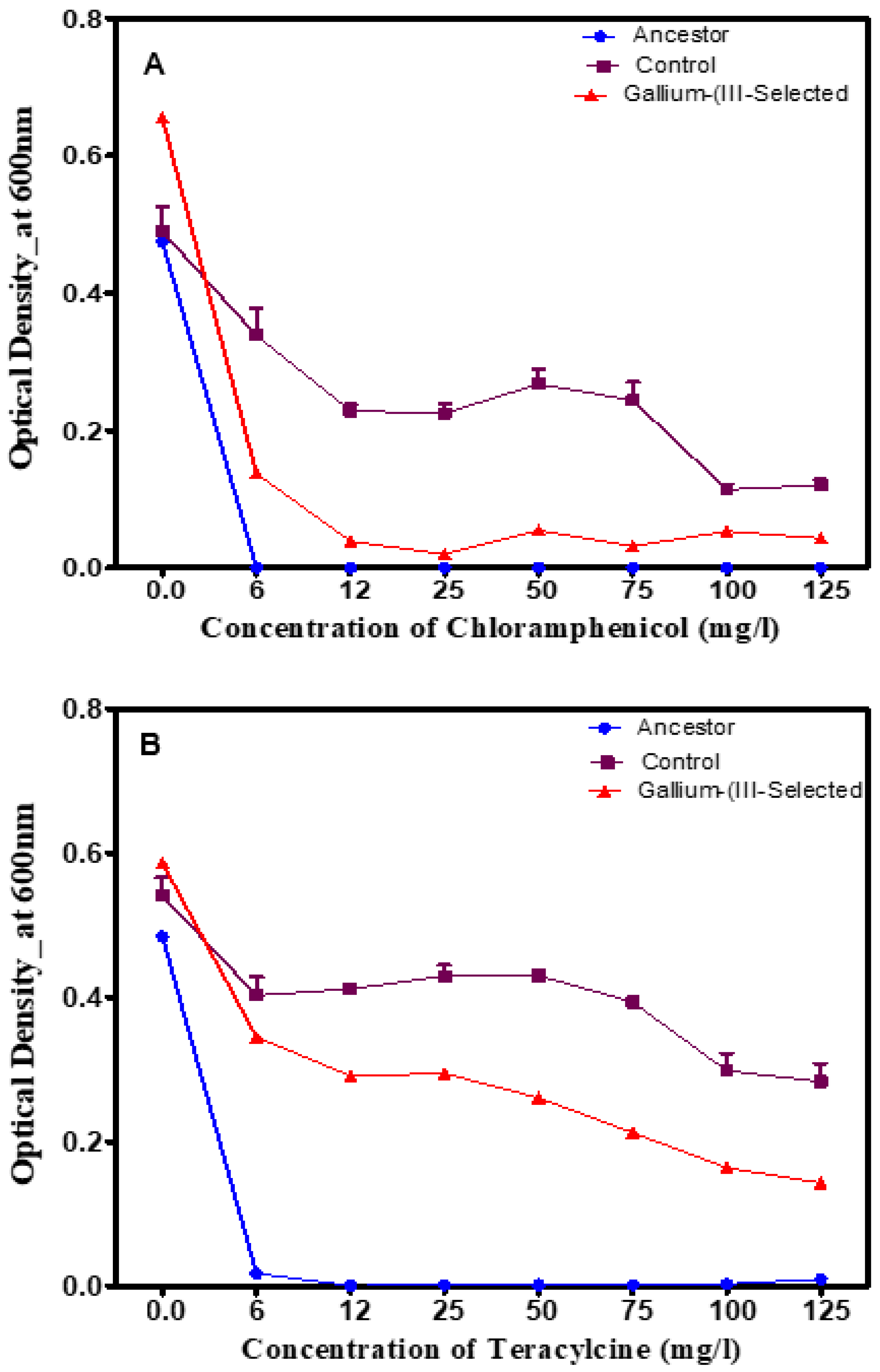
Typically, metal resistance is known to co-select for antibiotic resistance; therefore, this study assessed the growth of gallium (III)-resistant bacteria following in two traditional antibiotics (tetracycline and chloramphenicol). Figure 4 shows the 24 h growth across increasing concentrations (0–125 mg/L) of chloramphenicol (which inhibits protein synthesis) and tetracycline (which also inhibits protein synthesis) for the gallium (III)-selected versus the control and ancestral populations after 20 days of selection. The gallium (III)-selected populations showed inferior growth, which was highly statistically significant (p < 0.0001) across concentrations from 0 to 250 mg/L. The growth of all the tested populations decreased significantly (p < 0.001) in increasing concentrations of tetracycline and chloramphenicol (Figure 4).

3.4. Phenotypic Changes in the Presence of Polyfluorinated Compounds

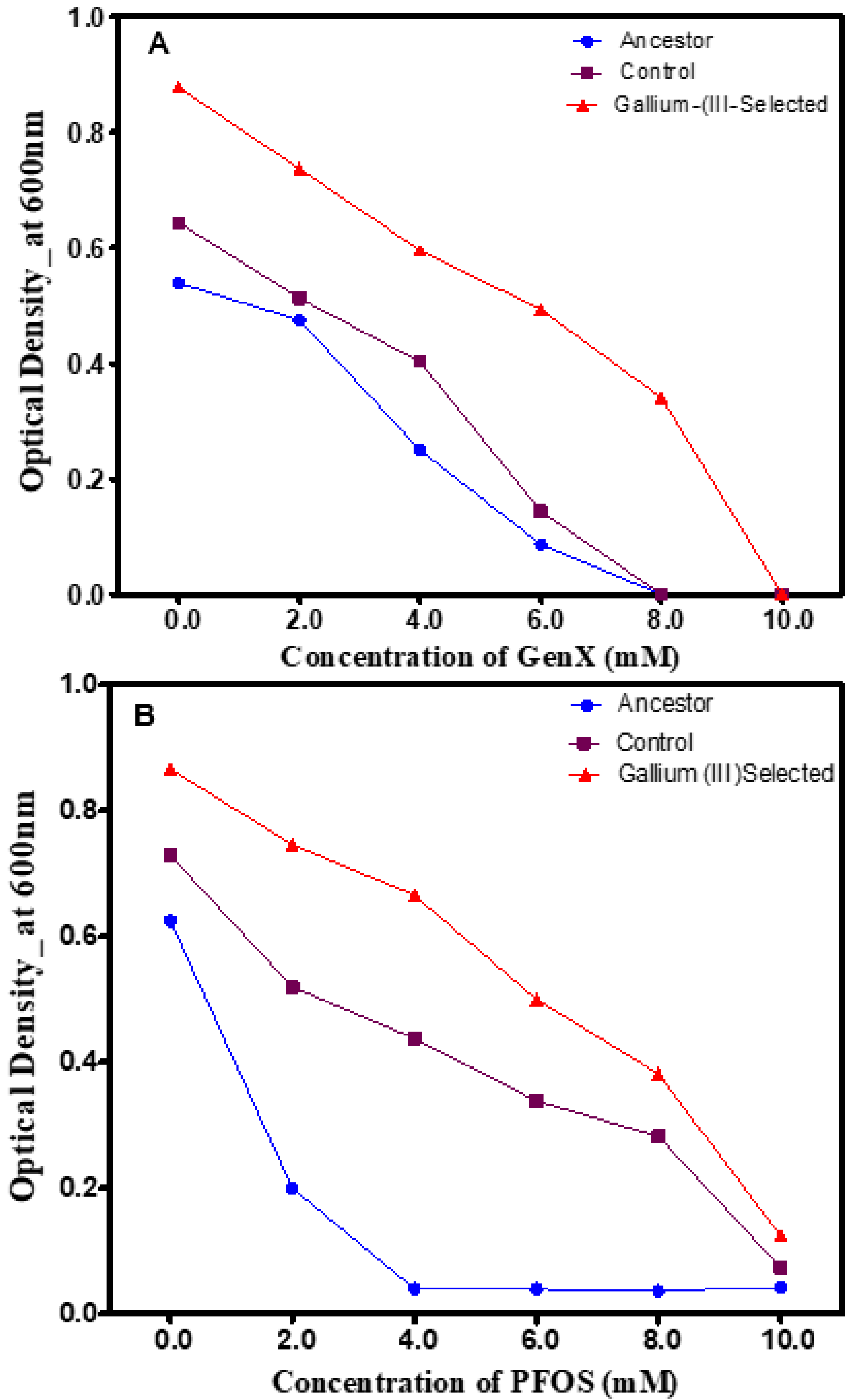
Given the challenge of biological degradation of per- and polyfluoroalkyl substances, this study evaluated the growth of gallium (III)-selected bacterial, control and ancestral populations in the presence of increasing concentrations of two polyfluoroalkyl substances (or “forever chemicals”). Figure 5 shows 24 h growth across increasing concentrations (0–1000 mg/L) of GenX, a synthetic chemical compound, and perfluorooctanesulfonic acid (PFOS) after 20 days of selection. The gallium (III)-selected populations displayed superior growth, which was highly statistically significant (p < 0.0001) across concentrations from 0 to 8 Mm of GenX and PFOS compared to the control and ancestral populations (Figure 5A,B).

3.5. Scanning Electron Microscopy (SEM)

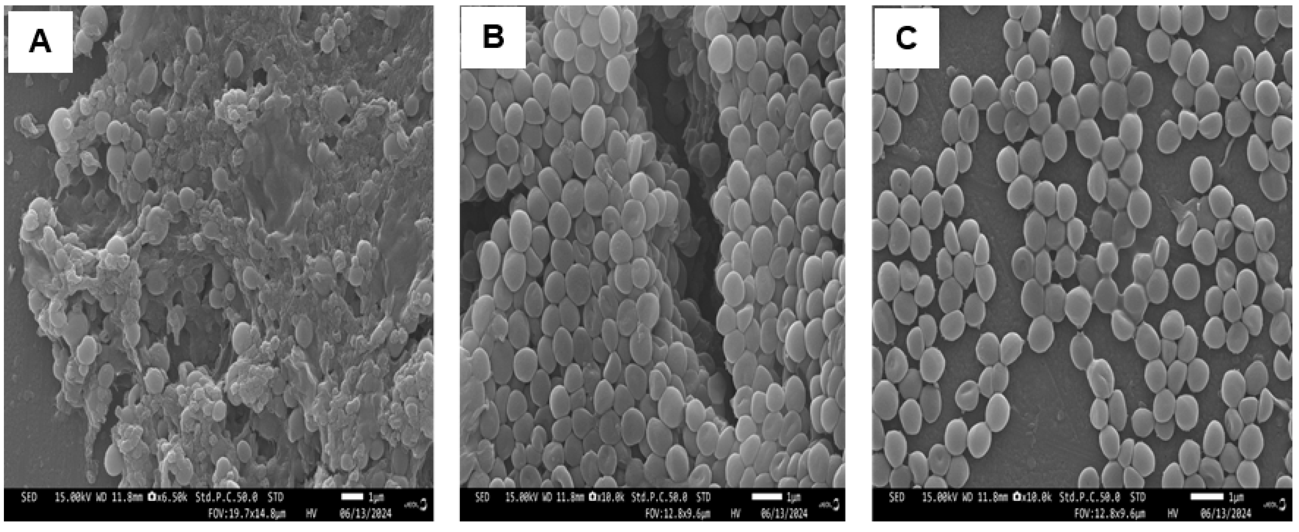
The electron micrographs obtained by SEM of the gallium (III)-selected bacterial, control and ancestral populations are shown in Figure 6. The micrographs showed that the surfaces of the control and ancestral S. aureus cells displayed characteristics typical of the surfaces of native cells, such as smooth, intact and round cocci, while the gallium (III)-selected bacteria treated with gallium (III) underwent a wide variety of cell morphological changes in response to gallium. Interestingly, some gallium-selected cells appeared oval, triangular, rod-like, as club-shaped rods and vibrio-shaped.

3.6. Genomic Results

To determine the effect of gallium selection on S. aureus genomic variations, we sequenced replicates from each selected population and defined a major polymorphism as F > 0.4, while a minor polymorphism was defined as a variant at a significantly lower frequency (F < 0.4). Table 1 shows polymorphisms in the gallium (III)-resistant populations (G1–G5) at day 20. The gallium (III)-resistant populations displayed significant polymorphisms in sfaA/sfaD, KQ76_RS01520, fmtA and KQ76_RS08360. Of the significant polymorphisms, all populations showed mutations in sfaA/sfaD, KQ76_RS01520, fmtA, KQ76_RS08360, dltB and hssR. Minor polymorphisms (F < 0.4) were observed in dltB, KQ76_RS13825, KQ76_RS13475, purS, KQ76_RS12180, graR, KQ76_RS07375, KQ76_RS09255, KQ76_RS11185, vraE, KQ76_RS02795, capA, KQ76_RS04220, KQ76_RS05175/KQ76_RS05180, KQ76_RS01815 and thrS. Descriptions of these genes are given in Table 2.
The genomic variants found in the control populations (C1–C5) are listed in Table 3. Major polymorphisms in pstC and KQ76_RS04365 were observed in all the replications (C1–C5). There were minor polymorphisms in KQ76_RS13020, PP2C family protein KQ76_RS10525, ald/KQ76_RS08720, KQ76_RS12955, pxpB/greA, KQ76_RS06325, icaR, KQ76_RS10985, mprF, rsp, mutS, KQ76_RS11280/KQ76_RS11285 M23, KQ76_RS13825, KQ76_RS12905, KQ76_RS04770, KQ76_RS13475, smpB, mnmG, KQ76_RS12190/rsp, KQ76_RS11175, gltB and KQ76_RS01360/lip2. Descriptions of these genes are given in Table 2. The control and gallium (III)-selected populations shared the following polymorphisms: hssR, KQ76_RS13020, mnmG, KQ76_RS04770, KQ76_RS12955, KQ76_RS01360/lip2, KQ76_RS10985, gltB, rsp, ylqF, KQ76_RS11280/KQ76_RS11285, smpB and KQ76_RS11175.

4. Discussion

Bacterial resistance to antibiotics is a serious threat to human health leading to more severe illnesses and increased mortality rates; this is considered an urgent public health threat by the World Health Organization (WHO) [31]. As rates of antibiotic-resistant bacterial infections soar, scientists are increasingly looking at the potential of metals as alternatives to combat these bacterial infections. Gallium is gaining attention because it targets alternative cellular processes, such as bacterial nutrition and metabolism [32]. Gallium has demonstrated bactericidal activity against Streptomyces pilosus, Pseudomonas aeruginosa, Klebsiella pneumoniae, Escherichia coli and Staphylococcus aureus [33,34,35]. Gallium exerts its bactericidal activity by interfering with enzymatic processes dependent on iron, thus making it hard for bacteria to evolve resistance to gallium [9,36,37,38,39]. A reduction in gallium uptake would reduce iron availability, which is critical for bacterial growth [40]. The fact that gallium mimics iron and interferes with multiple functions led us to hypothesize that Staphylococcus aureus may evolve resistance to gallium at high concentrations, as with successful antibiotics. In this study, we found that S. aureus evolved resistance to excess gallium (III) after 20 days, suggesting the presence of genetic adaptations, for example, efflux pumps, sequestration and enzymatic detoxification, essentially enabling them to expel or neutralize the harmful metals within their cells [41]. Previously, gallium (III) resistance acquired via transposon inactivation and mutations associated with iron metabolism has been investigated in Gram-negative bacteria [20,32]. Resistance to gallium (III) may not correlate with resistance to other metals, since the resistance pathway is largely specific to iron metabolism [42]. Other metals have different modes of action; for example, silver binds to membrane proteins, disrupting cell structure [43].
Pleiotropy is well known in the case of metals and antibiotics. The co-selection of metal and antibiotic resistance results from similarities between mechanisms, which include a reduction in cell wall permeability, substance alteration and efflux [28,44,45]. Here, gallium (III) selection showed a correlation with resistance to excess iron (III) nitrate and silver nitrate, but not tetracycline or chloramphenicol. The correlated response to iron (III) is not surprising because gallium (III) and iron (III) are chemically similar due to their similar ironic radii and charges, which enables gallium to replace iron in certain interactions [46,47]. Furthermore, all gallium (III)-resistant populations showed a mutation in staphyloferrinA export MFS transporter/D ornithine citrate ligase (sfaA/sfaD). sfaA/sfaD is known to actively pump staphyloferrin, a type of siderophore used to acquire iron from the environment, out of the bacterial cell. This process essentially exports the iron-chelating molecule to facilitate iron uptake when iron levels are low in the surrounding environment and is crucial for bacterial survival in iron-limited conditions [48]. By exporting staphyloferrin, gallium (III)-resistant S. aureus can efficiently acquire iron, which is essential for various cellular processes, including growth and replication [49]. A similar phenomenon was also reported in gallium (III)-resistant E. coli [15]. In this study, genomic analysis identified a hard selective sweep in the ferric citrate outer membrane transporter (fecA), known to mediate the transport of iron across the outer membrane, in the same manner as other siderophore transporters [50]. These results confirmed that gallium regulates iron membrane transport because it closely mimics iron in terms of its ionic radius, allowing it to be taken up by the same cellular mechanisms that transport iron. This phenomenon is often used in research and potential therapeutic applications to disrupt iron metabolism in cancer cells and bacteria. The mechanism of gallium (III) resistance to silver is not fully elucidated because selection in gallium (III) confers resistance to silver in the absence of selection of the genes cusS and ompR, which play a major role in conferring resistance [51,52]. In addition, resistance to silver may also occur through efflux of silver in cells [53]. Interestingly, in this study, genomic analysis identified polymorphisms in the magnesium transporter CorA family protein (KQ76_RS12180), NADP-dependent oxidoreductase (KQ76_RS11185) and the peptide resistance ABC transporter permease subunit (vraE), which play key roles in many metabolic pathways, including transport, efflux and detoxification [54,55,56]. In contrast, the gallium (III)-selected populations showed inferior growth to the control populations in tetracycline and chloramphenicol. The sensitivity of gallium (III)-resistant strains to tetracycline and chloramphenicol could be due to the absence of antibiotic-resistance genes. Studies have shown that bacteria which develop resistance to metals are also more likely to acquire resistance to antibiotics due to the often-shared genetic mechanisms [57]. Contrarily, mutations in genes associated with antibiotics, as in the case of acyltransferase family protein (KQ76_RS04365), were seen in all the replicates (C1–C5) of the control population. Pearson et al. [58] asserted that acyltransferase family protein (KQ76_RS04365) is essential for the biosynthesis of antibiotics and critical for regulating membrane biogenesis [58]. Additionally, the phenotypical changes of the control populations in tetracycline and chloramphenicol can also be attributed to genes associated with stress responses, cell division, detoxification and cell growth. Three genes—the phosphate ABC transporter permease subunit (pstC), PP2C family protein serine/threonine phosphatase (KQ76_RS10525) and 5 oxoprolinase subunit PxpB/transcription elongation factor (pxpB/greA)—showed major polymorphisms in the control population. The phosphate ABC transporter imports nutrients and exports toxic substances [59]. PP2C family protein serine/threonine phosphatase (KQ76_RS10525) controls many biological processes, including stress responses, development, cell division, virulence, and cell growth and death [60]. The 5 oxoprolinase subunit PxpB/transcription elongation factor (pxpB/greA) plays a crucial role in the metabolism of 5-oxoproline in bacterial cells and potentially affects their growth and survival under certain conditions [61].
Bacteria are ubiquitous and can grow in a variety of conditions, including air, water and soil. Some chemical manufacturing facilities and factories contaminate the environment by introducing polyfluoroalkyl compounds (also known as “forever compounds”) into air, water and soil. Due to their extremely slow breakdown rate, polyfluoroalkyl compounds persist in the environment, causing significant health issues in humans and animals, even at low levels of exposure [22]. Therefore, finding ways to degrade polyfluoroalkyl compounds is critical to mitigate environmental contamination and protect public health. Recent findings have shown that certain bacterial species, like Desulfovibrio aminophilus and Sporomusa sphaeroides, can break down polyfluoroalkyl compounds by cleaving key chemical bonds within the molecules, essentially allowing for the degradation of these persistent pollutants [62].
When bacteria grow in toxic waste, they can break down the waste through a process called bioremediation [63]. Gallium (III)-selected bacteria may help remove notorious polyfluoroalkyl compound chemicals. In this study, 24 h growth assays showed that the gallium (III)-selected populations displayed greater growth compared to the controls and the ancestors (gallium (III)-selected > controls > ancestor) in the polyfluoroalkyl compounds (GenX and PFBS) tested, boosting hopes that gallium-resistant bacteria might someday help remove these notoriously pervasive pollutants from the environment. The survival of these populations in GenX and PFBS could have been due to their ability to degrade Genx and PFBS by cleaving key chemical bonds within the molecules, essentially rendering them harmless. Additionally, the populations could have survived in polyfluoroalkyl compounds by evolving mechanisms to either actively pump out the toxins or modify their cell membranes to reduce permeability to the chemicals. These adaptations often involve genetic changes that allow them to sense and respond to the presence of toxic substances. For example, mutations in the response regulator transcription factor GraR/ApsR (graR) in gallium (III)-selected populations activated genes needed to survive in harsh environments [64]. Mutation in general stress protein (KQ76_RS01815) helps bacteria survive under a wide range of stressful conditions and cope with adverse conditions by activating a coordinated response, known as the “general stress response (GSR)”, which allows the bacteria to adapt to changing environments [65]. We also identified three significant polymorphisms associated with stress and detoxifications: response regulator transcription factor (GraR/ApsR), which activates genes needed to survive in harsh environments like high-temperature or low-pH conditions [64]; hypothetical protein (KQ76_RS09255), which plays a role in how bacteria adapt to their hosts [66]; and NADP dependent oxidoreductase (KQ76_RS11185), which plays a key role in many metabolic pathways and has a variety of applications, including biodegradation, detoxification and chemical synthesis [56]. It is important to note that the control populations also grew in Genx and PFBS, suggesting that they developed mechanisms to tolerate and even accumulate these toxic substances on their thick cell walls, allowing them to survive in such harsh conditions [67]. One notable observation was the presence of mutations in genes associated with efflux, detoxification and cell wall biosynthesis in these populations. This point is further elaborated on and explained in the paragraphs below.
Mutation in bacteria can alter the amino acid sequence of a protein involved in cell division, leading to changes in morphology [68]. In this study, for the first time, we examined the interaction of gallium (III) nitrate with S. aureus cells by SEM. Twenty days of selection in gallium (III) nitrate, to our surprise, resulted in variation in cell shapes, while the control and ancestral cells appeared perfectly spherical. It can be argued that long-term exposure to unfavorable conditions induces stress responses that impact cell size and shape for adaptation to the challenging environments [69]. Furthermore, stressors can perturb bacterial morphology by altering cell wall teichoic acid, binding to cross-linked peptidoglycans to interfere with cell wall maturation [70,71]. Another major factor that influences morphology is mutations in genes. Mutations can directly alter the morphology of a bacterium, suggesting that shape is important enough to merit regulation [72]. Several genomic variants involved in conferring gallium resistance were genes associated with cell walls. For example, mutation in PG:teichoic acid D alanyltransferase (dltB) (a key component of the Gram-positive bacterial cell wall) significantly impacts the cell wall’s charge and stability, influencing factors like bacterial virulence and immune response [73].
Interestingly, gallium (III) resistance seems to play a vital role in adhesion, virulence and nucleic acid synthesis, as gallium-resistant populations displayed mutations in teichoic acid D Ala esterase (fmtA), adenine phosphoribosyltransferase (KQ76_RS08360), ECF-type riboflavin transporter substrate-binding protein (KQ76_RS13825), glutathione peroxidase (KQ76_RS13475) and the phosphoribosylformylglycinamidine synthase subunit (purS). Mutation in teichoic acid D Ala esterase (fmtA) contributes to virulence by facilitating adhesion to host cells and evading immune defenses [74]. It is critical to understand the role of virulence associated with gallium (III) in vivo because it demonstrates how gallium (III) interacts with a living host, thus providing a more accurate understanding of its disease-causing capabilities. The virulence potential of gallium (III) can be tested in vivo by infecting an animal model, for example, mice, and then monitoring tissue damage and the mortality rate. Adenine phosphoribosyltransferase (KQ76_RS08360) plays a role in the recycling of nucleotides in bacteria [75]. ECF-type riboflavin transporter substrate-binding protein (KQ76_RS13825) allows the transport of essential vitamins from the surrounding medium for growth and metabolism [76,77]. Glutathione peroxidase (KQ76_RS13475) protects cells from oxidative damage and plays a role in bacterial virulence and pathogenicity [78]. The phosphoribosylformylglycinamidine synthase subunit (purS) plays a key role in the de novo purine synthesis pathway [79]. DUF3169 family protein (KQ76_RS01520) is essential for the secretion and transport of proteins [80].
The control populations displayed a total of 27 putative polymorphisms, of which 13 were shared with gallium (III)-selected populations. All the control populations carried major polymorphism (F > 0.4) sweeps in acyltransferase family protein (KQ76_RS04365), PP2C family protein serine/threonine phosphatase (KQ76_RS1052) and 5 oxoprolinase subunit PxpB/transcription elongation factor (pxpB/greA). Acyltransferase family protein (KQ76_RS04365) and PP2C family protein serine/threonine phosphatase (KQ76_RS1052) play important roles in regulating membrane biogenesis stress responses, development, cell division, virulence, and cell growth and death [58,60,61]. Three polymorphisms were detected in four of the five control populations. Polymorphisms were detected in the phosphate ABC transporter permease subunit (pstC), alpha/beta hydrolase (KQ76_RS13020) and cell elongation protein (cozEb). These genomic variants are involved in cell division, antimicrobial virulence, nutrients and export of toxic substance regulation [59,81,82]. Additionally, four mutations unique to the control populations were detected at lower frequencies (F < 0.4). These mutations included the ica operon transcriptional regulator (icaR), hypothetical protein (KQ76_RS09255), alanine dehydrogenase/universal stress protein (ald/KQ76_RS08720), ECF-type riboflavin transporter substrate-binding protein (KQ76_RS13825), bifunctional lysylphosphatidylglycerol flippase/synthetase (mprF), DNA mismatch repair protein (mutS), glutathione peroxidase (KQ76_RS13475) and the YbgA family protein/AraC family transcriptional regulator (KQ76_RS12190/rsp). Not surprisingly, these mutations are known to play roles in regulating biofilm formation, transport across membranes, replication oxidative damage and gene expression [76,78,83,84]. The control and gallium (III)-selected populations showed polymorphisms in the DNA-binding heme response regulator (hssR), alpha/beta hydrolase (KQ76_RS13020), the tRNA uridine 5 carboxymethylaminomethyl(34) synthesis enzyme (mnmG), ATP-binding protein (KQ76_RS04770), D lactate dehydrogenase (KQ76_RS12955), YjiH family protein/YSIRK domain-containing triacylglycerol lipase Lip2/Geh (KQ76_RS01360/lip2), BglG family transcription antiterminator (KQ76_RS10985), the glutamate synthase large subunit (gltB), the AraC family transcriptional regulator (rsp), ribosome biogenesis GTPase (ylqF), M23 family metallopeptidase/HAD IIB family hydrolase (KQ76_RS11280/KQ76_RS11285), SsrA-binding protein (smpB) and the BCCT family transporter (KQ76_RS11175). Polymorphisms in these genes are crucial for adaptation, antimicrobial activity, translation, import of nutrients, energy, colonization and infection, virulence, antimicrobial potential, and osmoregulation, which enable S. aureus to survive and replicate within their hosts [59,81,85,86,87,88,89,90,91,92,93,94,95,96,97]. This may explain the similarities in phenotypical changes between the control and treated populations in the presence of increasing concentrations of polyfluorinated compounds. Polymorphisms in both populations suggest the presence of genetic variations that exist naturally within S aureus [98]. Furthermore, the observed polymorphisms are not directly caused by gallium (III) and could be naturally occurring variations within S. aureus [99]. However, the genes were in different locations, potentially leading to different effects on the resulting proteins [100]. Additional experiments are needed to explore the potential functional implications of these polymorphisms. This study used Staphylococcus aureus (ATCC 25923); different strains of S. aureus could yield different results. Also, in our future studies, we intend to measure changes in gene expression and ultrastructure changes associated with gallium (III) resistance utilizing RNA sequencing and transmission electron microscopy (TEM), respectively.

5. Conclusions

With the rise in bacterial resistance to antibiotics, gallium is a promising treatment for multidrug-resistant pathogens. Gallium is a potential antimicrobial because it disrupts bacterial metabolism by mimicking iron and substituting itself into key iron-dependent enzymes [101]. In this study, we showed that gallium resistance in S. aureus occurred after 20 days of selection in gallium (III) nitrate due to specific mutational and morphological changes increasing the ability to grow in heavy metals and polyfluorinated compounds. All the replicates of gallium (III)-resistant strains showed polymorphisms in staphyloferrinA export MFS transporter/D ornithine citrate ligase (sfaA/sfaD), DUF3169 family protein (KQ76_RS01520) and teichoic acid D Ala esterase (fmtA), suggesting strong evidence of the crucial roles these genes play in conferring gallium resistance. Moreover, the genomic changes led to noticeable alterations in cell morphology. In contrast, the gallium-selected S. aureus displayed inferior growth in antibiotics compared to the control population. Our results highlight significant implications for human health, ecosystems and environmental remediation.

Author Contributions

Conceptualization, A.E., F.S., D.H., T.B., A.B., J.G., U.I., V.W., S.A.-F. and B.J.; Methodology, A.E., U.I., L.K., F.S., D.H., T.B., A.B., J.G., U.I., V.W., S.A.-F. and B.J.; Formal analysis, A.E., L.K., U.I., F.S., D.H., T.B., A.B., J.G., U.I. and V.W.; Investigation, A.E., U.I., L.K., F.S., D.H., T.B., A.B., J.G., V.W., S.A.-F. and B.J.; Writing—original draft, A.E., F.S., D.H., T.B., A.B., J.G., U.I. and V.W.; Writing—review and editing, A.E., F.S., D.H., T.B., A.B., J.G., U.I., V.W., S.A.-F. and B.J. All authors have read and agreed to the published version of the manuscript.

Funding

The Genomic Research and Data Science Center for Computation and Cloud Computing (GRADS-4C) (211512) funded this project.

Institutional Review Board Statement

Not applicable.

Informed Consent Statement

Not applicable.

Data Availability Statement

Data are contained within the article.

Acknowledgments

The authors sincerely thank the Department of Biological Sciences, Winston Salem State University, for all the logistics.

Conflicts of Interest

The authors declare no conflicts of interest.

References

  1. Danis-Wlodarczyk, K.M.; Wozniak, D.J.; Abedon, S.T. Treating bacterial infections with bacteriophage-based enzybiotics: In vitro, in vivo and clinical application. Antibiotics 2021, 10, 1497. [Google Scholar] [CrossRef]
  2. Baran, A.; Kwiatkowska, A.; Potocki, L. Antibiotics and bacterial resistance—A short story of an endless arms race. Int. J. Mol. Sci. 2023, 24, 5777. [Google Scholar] [CrossRef]
  3. Frei, A.; Verderosa, A.D.; Elliott, A.G.; Zuegg, J.; Blaskovich, M.A. Metals to combat antimicrobial resistance. Nat. Rev. Chem. 2023, 7, 202–224. [Google Scholar]
  4. Lemire, J.A.; Harrison, J.J.; Turner, R.J. Antimicrobial activity of metals: Mechanisms, molecular targets and applications. Nat. Rev. Microbiol. 2013, 11, 371–384. [Google Scholar]
  5. Turner, R.J. The good, the bad, and the ugly of metals as antimicrobials. Biometals 2024, 37, 545–559. [Google Scholar]
  6. Keren, I.; Wu, Y.; Inocencio, J.; Mulcahy, L.R.; Lewis, K. Killing by bactericidal antibiotics does not depend on reactive oxygen species. Science 2013, 339, 1213–1216. [Google Scholar]
  7. Rauf, A.; Kiran, S.A.; Raza, H.; Haroon, S.M.; Javed, M.; Jahangir, M.; Hassan, M.; Bahadur, A.; Iqbal, S.; Mahmood, S.; et al. Harnessing the power of multifunctional γ-Fe2O3@ CuO nanocomposites: Effective extraction of heavy metals and bacterial pathogens from contaminated water. Mater. Chem. Phys. 2024, 320, 129411. [Google Scholar]
  8. Li, F.; Liu, F.; Huang, K.; Yang, S. Advancement of gallium and gallium-based compounds as antimicrobial agents. Front. Bio-Eng. Biotechnol. 2022, 10, 827960. [Google Scholar]
  9. Mohammad Hanifeh, N.; Keyvani-Ghamsari, S.; Khorsandi, K.; Mahmoodi Khaledi, E. Effect of gallium nitrate on the antibacterial activity of vancomycin in methicillin-sensitive and resistant Staphylococcus aureus. Arch. Microbiol. 2024, 206, 304. [Google Scholar]
  10. Owusu, S.B.; Zaher, A.; Ahenkorah, S.; Pandya, D.N.; Wadas, T.J.; Petronek, M.S. Gallium Uncouples Iron Metabolism to Enhance Glio-blastoma Radiosensitivity. Int. J. Mol. Sci. 2024, 25, 10047. [Google Scholar] [CrossRef]
  11. Limantoro, C.; Das, T.; He, M.; Dirin, D.; Manos, J.; Kovalenko, M.V.; Chrzanowski, W. Synthesis of antimicrobial gallium nanoparticles using the hot injection method. ACS Mater. Au 2023, 3, 310–320. [Google Scholar] [CrossRef]
  12. Liu, S.; Ji, Y.; Zhu, H.; Shi, Z.; Li, M.; Yu, Q. Gallium-based metal–organic frameworks loaded with antimicrobial peptides for synergis-tic killing of drug-resistant bacteria. J. Mater. Chem. B 2023, 11, 10446–10454. [Google Scholar] [CrossRef]
  13. Shi, F.; Ma, S.; Liu, S.; Xin, R.; Chen, B.; Ye, W.; Sun, J. A novel antimicrobial strategy for bacterial infections: Gallium-based materials. Colloid Interface Sci. Commun. 2023, 56, 100735. [Google Scholar] [CrossRef]
  14. Kulkarni, S.; Pandey, A.; Mutalik, S. Liquid metal based theranostic nanoplatforms: Application in cancer therapy, imaging and biosensing. Nanomed. Nanotechnol. Biol. Med. 2020, 26, 102175. [Google Scholar] [CrossRef]
  15. Peng, X.X.; Gao, S.; Zhang, J.L. Gallium (III) complexes in cancer chemotherapy. Eur. J. Inorg. Chem. 2022, 2022, e202100953. [Google Scholar]
  16. Mjos, K.D.; Cawthray, J.F.; Polishchuk, E.; Abrams, M.J.; Orvig, C. Gallium (III) and iron (III) complexes of quinolone antimicrobials. Dalton Trans. 2016, 45, 13146–13160. [Google Scholar] [CrossRef]
  17. Claudel, M.; Schwarte, J.V.; Fromm, K.M. New antimicrobial strategies based on metal complexes. Chemistry 2020, 2, 849–899. [Google Scholar] [CrossRef]
  18. Xu, Z.; Zhao, X.; Chen, X.; Chen, Z.; Xia, Z. Antimicrobial effect of gallium nitrate against bacteria encountered in burn wound infections. RSC Adv. 2017, 7, 52266–52273. [Google Scholar] [CrossRef]
  19. Choi, S.R.; Talmon, G.A.; Hearne, K.; Woo, J.; Truong, V.L.; Britigan, B.E.; Narayanasamy, P. Combination therapy with gallium protoporphyrin and gallium nitrate exhibits enhanced antimicrobial activity in vitro and in vivo against methicillin-resistant Staphylococcus aureus. Mol. Pharm. 2023, 20, 4058–4070. [Google Scholar] [CrossRef]
  20. Graves, J.L., Jr.; Ewunkem, A.J.; Ward, J.; Staley, C.; Thomas, M.D.; Rhinehardt, K.L.; Han, J.; Harrison, S.H. Experimental evolution of gallium resistance in Escherichia coli. Evol. Med. Public Health 2019, 2019, 169–180. [Google Scholar] [CrossRef]
  21. Zhang, Z.; Sarkar, D.; Biswas, J.K.; Datta, R. Biodegradation of per-and polyfluoroalkyl substances (PFAS): A review. Bioresour. Technol. 2022, 344, 126223. [Google Scholar] [CrossRef] [PubMed]
  22. Peritore, A.F.; Gugliandolo, E.; Cuzzocrea, S.; Crupi, R.; Britti, D. Current review of increasing animal health threat of per-and polyfluoroalkyl substances (pfas): Harms, limitations, and alternatives to manage their toxicity. Int. J. Mol. Sci. 2023, 24, 11707. [Google Scholar] [CrossRef]
  23. Wackett, L.P. Nothing lasts forever: Understanding microbial biodegradation of polyfluorinated compounds and perfluorinated alkyl substances. Microb. Biotechnol. 2022, 15, 773–792. [Google Scholar] [PubMed]
  24. Pray, L. Antibiotic resistance, mutation rates and MRSA. Nat. Educ. 2008, 1, 30. [Google Scholar]
  25. Ewunkem, A.J.; Rodgers, L.; Campbell, D.; Staley, C.; Subedi, K.; Boyd, S.; Graves, J.L., Jr. Experimental evolution of magnetite nanoparticle resistance in Escherichia coli. Nanomaterials 2021, 11, 790. [Google Scholar] [CrossRef]
  26. Ewunkem, A.J.; Williams, Z.J.; Johnson, N.S.; Brittany, J.L.; Maselugbo, A.; Nowlin, K. Exploring the “Carpenter” as a substrate for green synthesis: Biosynthesis and antimicrobial potential. Gene Protein Dis. 2023, 2, 2155. [Google Scholar]
  27. Shrestha, S.; Shrestha, B.K.; Tettey-Engmann, F.; Auniq, R.B.; Subedi, K.; Ghimire, S.; Desai, S.; Bhattarai, N. Zein-coated Zn metal particles-incorporated nanofibers: A potent fibrous platform for loading and release of Zn ions for wound healing application. ACS Appl. Mater. Interfaces 2024, 16, 49197–49217. [Google Scholar]
  28. Ewunkem, A.J.; Johnson, N.; A’lyiha, F.B.; Williams, Z.J.; Tshimanga, I.; Justice, B.; Singh, D.K.; Meixner, J. Green Synthesis–Mediated Nanoparticles: Characterization, Antimicrobial Activity and Genomics Analysis. Available online: https://www.intechopen.com/online-first/1195022 (accessed on 18 March 2025).
  29. Iyer, D.; Laws, E.; LaJeunesse, D. Escherichia coli adhesion and biofilm formation on polymeric nanostructured surfaces. ACS Omega 2023, 8, 47520–47529. [Google Scholar] [CrossRef]
  30. Ebrahimi, F.; Kumari, A.; Dellinger, K. Integration of Nanoengineering with Artificial Intelligence and Machine Learning in Surface-Enhanced Raman Spectroscopy (SERS) for the Development of Advanced Biosensing Platforms. Adv. Sens. Res. 2025, 4, 2400155. [Google Scholar]
  31. Tang, K.W.; Millar, B.C.; Moore, J.E. Antimicrobial resistance (AMR). Br. J. Biomed. Sci. 2023, 80, 11387. [Google Scholar] [CrossRef]
  32. Goss, C.H.; Kaneko, Y.; Khuu, L.; Anderson, G.D.; Ravishankar, S.; Aitken, M.L.; Lechtzin, N.; Zhou, G.; Czyz, D.M.; McLean, K.; et al. Gallium disrupts bacterial iron metabolism and has therapeutic effects in mice and humans with lung infections. Sci. Transl. Med. 2018, 10, eaat7520. [Google Scholar]
  33. Frangipani, E.; Bonchi, C.; Minandri, F.; Imperi, F.; Visca, P. Pyochelin potentiates the inhibitory activity of gallium on Pseudomonas aeruginosa. Antimicrob. Agents Chemother. 2014, 58, 5572–5575. [Google Scholar] [CrossRef]
  34. Guerrini, M.; d’Agostino, S.; Grepioni, F.; Braga, D.; Lekhan, A.; Turner, R.J. Antimicrobial activity of supramolecular salts of gallium (III) and proflavine and the intriguing case of a trioxalate complex. Sci. Rep. 2022, 12, 3673. [Google Scholar]
  35. Pandey, A.; Savino, C.; Ahn, S.H.; Yang, Z.; Van Lanen, S.G.; Boros, E. Theranostic gallium siderophore ciprofloxacin conjugate with broad spectrum antibiotic potency. J. Med. Chem. 2019, 62, 9947–9960. [Google Scholar]
  36. de Assis, A.S.; Pegoraro, G.M.; Duarte, I.C.; Delforno, T.P. Gallium: A decisive “Trojan Horse” against microorganisms. Antonie Van Leeuwenhoek 2025, 118, 3. [Google Scholar]
  37. Duffin, R.N.; Kelderman, J.T.; Herdman, M.E.; Andrews, P.C. Highly selective organo-gallium hydroxamate mediated inhibition of antibiotic resistant Klebsiella pneumoniae. Dalton Trans. 2025, 54, 649–661. [Google Scholar]
  38. Poscente, V.; Di Gregorio, L.; Bernini, R.; Bevivino, A. Inhibitory Effects of Nisin and Gallium (III) Nitrate Hydrate on Planktonic and Adhered Cells and Implications for the Viable but Non-Culturable State. Microorganisms 2025, 13, 276. [Google Scholar] [CrossRef]
  39. Salazar-Alemán, D.A.; Turner, R.J. Escherichia coli growing under antimicrobial gallium nitrate stress reveals new processes of tol-erance and toxicity. Sci. Rep. 2025, 15, 1389. [Google Scholar]
  40. Abbas, M.; Hayirli, Z.; Drakesmith, H.; Andrews, S.C.; Lewis, M.C. Effects of iron deficiency and iron supplementation at the host-microbiota interface: Could a piglet model unravel complexities of the underlying mechanisms? Front. Nutr. 2022, 9, 927754. [Google Scholar]
  41. He, Z.; Shen, J.; Li, Q.; Yang, Y.; Zhang, D.; Pan, X. Bacterial metal (loid) resistance genes (MRGs) and their variation and application in environment: A review. Sci. Total Environ. 2023, 871, 162148. [Google Scholar]
  42. He, J.; Lin, X.; Zhang, D.; Hu, H.; Chen, X.; Xu, F.; Zhou, M. Wake biofilm up to enhance suicidal uptake of gallium for chronic lung infection treatment. Biomaterials 2024, 310, 122619. [Google Scholar]
  43. Mohamed, D.S.; Abd El-Baky, R.M.; Sandle, T.; Mandour, S.A.; Ahmed, E.F. Antimicrobial activity of silver-treated bacteria against other multi-drug resistant pathogens in their environment. Antibiotics 2020, 9, 181. [Google Scholar] [CrossRef]
  44. Dickinson, A.W.; Power, A.; Hansen, M.G.; Brandt, K.K.; Piliposian, G.; Appleby, P.; O’neill, P.A.; Jones, R.T.; Sierocinski, P.; Koskella, B.; et al. Heavy metal pollution and co-selection for antibiotic resistance: A microbial palaeontology approach. Environ. Int. 2019, 132, 105117. [Google Scholar] [PubMed]
  45. Yep, A.; McQuade, T.; Kirchhoff, P.; Larsen, M.; Mobley, H.L. Inhibitors of TonB function identified by a high-throughput screen for inhibitors of iron acquisition in uropathogenic Escherichia coli CFT073. mBio 2014, 5, e01089-13. [Google Scholar] [CrossRef]
  46. Kelson, A.B.; Carnevali, M.; Truong-Le, V. Gallium-based anti-infectives: Targeting microbial iron-uptake mechanisms. Curr. Opin. Pharmacol. 2013, 13, 707–716. [Google Scholar] [PubMed]
  47. Sun, W.; Qi, M.; Cheng, S.; Li, C.; Dong, B.; Wang, L. Gallium and gallium compounds: New insights into the “Trojan horse” strategy in medical applications. Mater. Des. 2023, 227, 111704. [Google Scholar] [CrossRef]
  48. Pasqua, M.; Grossi, M.; Zennaro, A.; Fanelli, G.; Micheli, G.; Barras, F.; Colonna, B.; Prosseda, G. The varied role of efflux pumps of the MFS family in the interplay of bacteria with animal and plant cells. Microorganisms 2019, 7, 285. [Google Scholar] [CrossRef]
  49. Drew, D.; North, R.A.; Nagarathinam, K.; Tanabe, M. Structures and general transport mechanisms by the major facilitator superfamily (MFS). Chem. Rev. 2021, 121, 5289–5335. [Google Scholar]
  50. Yue, W.W.; Grizot, S.; Buchanan, S.K. Structural evidence for iron-free citrate and ferric citrate binding to the TonB-dependent outer membrane transporter FecA. J. Mol. Biol. 2003, 332, 353–368. [Google Scholar] [CrossRef] [PubMed]
  51. Kedziora, A.; Wernecki, M.; Korzekwa, K.; Speruda, M.; Gerasymchuk, Y.; Lukowiak, A.; Bugla-Ptoskohska, G. Consequences of Long-Term Bacteria’s Exposure To Silver Nanoformulations with Different PhysicoChemical Properties. Int. J. Nanomed. 2020, 15, 199–214. [Google Scholar] [CrossRef] [PubMed]
  52. Li, H.; Xu, H. Mechanisms of bacterial resistance to environmental silver and antimicrobial strategies for silver: A review. Environ. Res. 2024, 248, 118313. [Google Scholar] [CrossRef] [PubMed]
  53. Randall, C.P.; Gupta, A.; Jackson, N.; Busse, D.; O’Neill, A.J. Silver resistance in Gram-negative bacteria: A dissection of endogenous and exogenous mechanisms. J. Antimicrob. Chemother. 2015, 70, 1037–1046. [Google Scholar] [CrossRef] [PubMed]
  54. Bui, D.M.; Gregan, J.; Jarosch, E.; Ragnini, A.; Schweyen, R.J. The bacterial magnesium transporter CorA can functionally substitute for its putative homologue Mrs2p in the yeast inner mitochondrial membrane. J. Biol. Chem. 1999, 274, 20438–20443. [Google Scholar] [CrossRef]
  55. Preeti Garai, P.G.; Kasturi Chandra, K.C.; Dipshikha Chakravortty, D.C. Bacterial peptide transporters: Messengers of nutrition to virulence. Virulence 2017, 8, 297–309. [Google Scholar] [CrossRef] [PubMed]
  56. Vidal, L.S.; Kelly, C.L.; Mordaka, P.M.; Heap, J.T. Review of NAD (P) H-dependent oxidoreductases: Properties, engineering and application. Biochim. Biophys. Acta (BBA)-Proteins Proteom. 2018, 1866, 327–347. [Google Scholar] [CrossRef]
  57. Murray, L.M.; Hayes, A.; Snape, J.; Kasprzyk-Hordern, B.; Gaze, W.H.; Murray, A.K. Co-selection for antibiotic resistance by environmental contaminants. Npj Antimicrob. Resist. 2024, 2, 9. [Google Scholar] [CrossRef]
  58. Pearson, C.R.; Tindall, S.N.; Herman, R.; Jenkins, H.T.; Bateman, A.; Thomas, G.H.; Potts, J.R.; Van der Woude, M.W. Acetylation of surface carbohydrates in bacterial pathogens requires coordinated action of a two-domain membrane-bound acyltransferase. mBio 2020, 11, e01364-20. [Google Scholar] [CrossRef]
  59. Davidson, A.L.; Dassa, E.; Orelle, C.; Chen, J. Structure, function, and evolution of bacterial ATP-binding cassette systems. Microbiol. Mol. Biol. Rev. 2008, 72, 317–364. [Google Scholar] [CrossRef]
  60. Bradshaw, N.; Levdikov, V.M.; Zimanyi, C.M.; Gaudet, R.; Wilkinson, A.J.; Losick, R. A widespread family of serine/threonine protein phosphatases shares a common regulatory switch with proteasomal proteases. eLife 2017, 6, e26111. [Google Scholar] [CrossRef]
  61. Niehaus, T.D.; Elbadawi-Sidhu, M.; de Crécy-Lagard, V.; Fiehn, O.; Hanson, A.D. Discovery of a widespread prokaryotic 5-oxoprolinase that was hiding in plain sight. J. Biol. Chem. 2017, 292, 16360–16367. [Google Scholar] [CrossRef]
  62. Noori, M.T.; Gupta, P.; Hellgardt, K.; Min, B. Per-and poly-fluoroalkyl substances (PFAS): An emerging environmental challenge and (microbial) bioelectrochemical treatment strategies. Curr. Opin. Environ. Sci. Health 2024, 43, 100588. [Google Scholar] [CrossRef]
  63. Ayilara, M.S.; Babalola, O.O. Bioremediation of environmental wastes: The role of microorganisms. Front. Agron. 2023, 5, 1183691. [Google Scholar]
  64. Butcher, R.J.; Tabor, J.J. Real-time detection of response regulator phosphorylation dynamics in live bacteria. Proc. Natl. Acad. Sci. USA 2022, 119, e2201204119. [Google Scholar]
  65. Lombardo, M.J.; Aponyi, I.; Rosenberg, S.M. General stress response regulator RpoS in adaptive mutation and amplification in Escherichia coli. Genetics. 2004, 166, 669–680. [Google Scholar] [PubMed]
  66. Naveed, M.; Makhdoom, S.I.; Abbas, G.; Safdari, M.; Farhadi, A.; Habtemariam, S.; Shabbir, M.A.; Jabeen, K.; Asif, M.F.; Tehreem, S. The virulent hypothetical proteins: The potential drug target involved in bacterial pathogenesis. Mini Rev. Med. Chem. 2022, 22, 2608–2623. [Google Scholar] [CrossRef]
  67. Lima e Silva, A.A.; Carvalho, M.A.; de Souza, S.A.; Dias, P.M.; Silva Filho, R.G.; Saramago, C.S.; Bento, C.A.; Hofer, E. Heavy metal tolerance (Cr, Ag and Hg) in bacteria isolated from sewage. Braz. J. Microbiol. 2012, 43, 1620–1631. [Google Scholar]
  68. van Teeseling, M.C.; de Pedro, M.A.; Cava, F. Determinants of bacterial morphology: From fundamentals to possibilities for antimicrobial targeting. Front. Microbiol. 2017, 8, 1264. [Google Scholar]
  69. Chen, J.; Zhou, H.; Huang, J.; Zhang, R.; Rao, X. Virulence alterations in Staphylococcus aureus upon treatment with the sub-inhibitory concentrations of antibiotics. J. Adv. Res. 2021, 31, 165–175. [Google Scholar]
  70. Chang, J.D.; Foster, E.E.; Thadani, A.N.; Ramirez, A.J.; Kim, S.J. Inhibition of Staphylococcus aureus cell wall biosynthesis by desleucyl-oritavancin: A quantitative peptidoglycan composition analysis by mass spectrometry. J. Bacteriol. 2017, 199, e00278-17. [Google Scholar] [CrossRef]
  71. Zhu, X.; Liu, D.; Singh, A.K.; Drolia, R.; Bai, X.; Tenguria, S.; Bhunia, A.K. Tunicamycin mediated inhibition of wall teichoic acid affects Staphylococcus aureus and Listeria monocytogenes cell morphology, biofilm formation and virulence. Front. Microbiol. 2018, 9, 1352. [Google Scholar]
  72. Young, K.D. Bacterial morphology: Why have different shapes? Curr. Opin. Microbiol. 2007, 10, 596–600. [Google Scholar] [CrossRef] [PubMed]
  73. Zhang, P.; Liu, Z. Structural insights into the transporting and catalyzing mechanism of DltB in LTA D-alanylation. Nat. Commun. 2024, 15, 3404. [Google Scholar] [CrossRef]
  74. Rahman, M.M.; Hunter, H.N.; Prova, S.; Verma, V.; Qamar, A.; Golemi-Kotra, D. The Staphylococcus aureus methicillin resistance factor FmtA is a d-amino esterase that acts on teichoic acids. mBio 2016, 7, e02070-15. [Google Scholar]
  75. Schaff, D.A. The adenine phosphoribosyltransferase (APRT) selectable marker system. Plant Sci. 1994, 101, 3–9. [Google Scholar]
  76. Karpowich, N.K.; Song, J.; Wang, D.N. An aromatic cap seals the substrate binding site in an ECF-type S subunit for riboflavin. J. Mol. Biol. 2016, 428, 3118–3130. [Google Scholar] [PubMed]
  77. Pham, V.T.; Dold, S.; Rehman, A.; Bird, J.K.; Steinert, R.E. Vitamins, the gut microbiome and gastrointestinal health in humans. Nutr. Res. 2021, 95, 35–53. [Google Scholar]
  78. Brenot, A.; King, K.Y.; Janowiak, B.; Griffith, O.; Caparon, M.G. Contribution of glutathione peroxidase to the virulence of Streptococcus pyogenes. Infect. Immun. 2004, 72, 408–413. [Google Scholar]
  79. Wang, T.Y.; Zhao, J.; Savas, A.C.; Zhang, S.; Feng, P. Viral pseudoenzymes in infection and immunity. FEBS J. 2020, 287, 4300–4309. [Google Scholar] [CrossRef]
  80. Craney, A.; Dix, M.M.; Adhikary, R.; Cravatt, B.F.; Romesberg, F.E. An alternative terminal step of the general secretory pathway in Staphylococcus aureus. mBio 2015, 6, e01178-15. [Google Scholar]
  81. Stamsås, G.A.; Myrbråten, I.S.; Straume, D.; Salehian, Z.; Veening, J.W.; Håvarstein, L.S.; Kjos, M. CozEa and CozEb play overlapping and essential roles in controlling cell division in Staphylococcus aureus. Mol. Microbiol. 2018, 109, 615–632. [Google Scholar] [CrossRef]
  82. Wullich, S.C.; Arranz San Martín, A.; Fetzner, S. An α/β-hydrolase fold subfamily comprising Pseudomonas quinolone signal-cleaving dioxygenases. Appl. Environ. Microbiol. 2020, 86, e00279-20. [Google Scholar] [CrossRef] [PubMed]
  83. Gallegos, M.T.; Schleif, R.; Bairoch, A.; Hofmann, K.; Ramos, J.L. Arac/XylS family of transcriptional regulators. Microbiol. Mol. Biol. Rev. 1997, 61, 393–410. [Google Scholar]
  84. Conlon, K.M.; Humphreys, H.; O’Gara, J.P. icaR encodes a transcriptional repressor involved in environmental regulation of ica operon expression and biofilm formation in Staphylococcus epidermidis. J. Bacteriol. 2002, 184, 4400–4408. [Google Scholar] [CrossRef]
  85. Dym, O.; Pratt, E.A.; Ho, C.; Eisenberg, D. The crystal structure of D-lactate dehydrogenase, a peripheral membrane respiratory enzyme. Proc. Natl. Acad. Sci. USA 2000, 97, 9413–9418. [Google Scholar] [CrossRef]
  86. Karzai, A.W.; Sauer, R.T. Protein factors associated with the SsrA⋅ SmpB tagging and ribosome rescue complex. Proc. Natl. Acad. Sci. USA 2001, 98, 3040–3044. [Google Scholar] [CrossRef] [PubMed]
  87. Ziegler, C.; Bremer, E.; Krämer, R. The BCCT family of carriers: From physiology to crystal structure. Mol. Microbiol. 2010, 78, 13–34. [Google Scholar] [CrossRef]
  88. Dincturk, H.B.; Cunin, R.; Akce, H. Expression and functional analysis of glutamate synthase small subunit-like proteins from archaeon Pyrococcus horikoshii. Microbiol. Res. 2011, 166, 294–303. [Google Scholar] [CrossRef] [PubMed]
  89. Gibbs, M.R.; Moon, K.M.; Chen, M.; Balakrishnan, R.; Foster, L.J.; Fredrick, K. Conserved GTPase LepA (Elongation Factor 4) functions in biogenesis of the 30S subunit of the 70S ribosome. Proc. Natl. Acad. Sci. USA 2017, 114, 980–985. [Google Scholar] [CrossRef]
  90. Chen, X.; Alonzo, F., III. Bacterial lipolysis of immune-activating ligands promotes evasion of innate defenses. Proc. Natl. Acad. Sci. USA 2019, 116, 3764–3773. [Google Scholar] [CrossRef]
  91. Nam, D.; Matsumoto, Y.; Uchida, T.; O’Brian, M.R.; Ishimori, K. Mechanistic insights into heme-mediated transcriptional regulation via a bacterial manganese-binding iron regulator, iron response regulator (Irr). J. Biol. Chem. 2020, A295, 11316–11325. [Google Scholar] [CrossRef]
  92. Kotecka, K.; Kawalek, A.; Kobylecki, K.; Bartosik, A.A. The AraC-type transcriptional regulator GliR (PA3027) activates genes of glycerolipid metabolism in Pseudomonas aeruginosa. Int. J. Mol. Sci. 2021, 22, 5066. [Google Scholar] [CrossRef]
  93. Bommisetti, P.; Young, A.; Bandarian, V. Elucidation of the substrate of tRNA-modifying enzymes MnmEG leads to in vitro reconstitution of an evolutionarily conserved uridine hypermodification. J. Biol. Chem. 2022, 298, 102548. [Google Scholar]
  94. Razew, A.; Schwarz, J.N.; Mitkowski, P.; Sabala, I.; Kaus-Drobek, M. One fold, many functions—M23 family of peptidoglycan hydrolases. Front. Microbiol. 2022, 13, 1036964. [Google Scholar]
  95. Soussan, D.; Salze, M.; Ledormand, P.; Sauvageot, N.; Boukerb, A.; Lesouhaitier, O.; Fichant, G.; Rincé, A.; Quentin, Y.; Muller, C. The NagY regulator: A member of the BglG/SacY antiterminator family conserved in Enterococcus faecalis and involved in virulence. Front. Microbiol. 2023, 13, 1070116. [Google Scholar]
  96. Chen, J.; Wu, Y.; Zhu, Y.; Zhang, L.; Xu, Y.; Liu, Y. Adaptation for Staphylococcus aureus to hosts via insertion mutation in the accessory gene regulator agrC gene: Decreased viru-lence and enhanced persistence capacity. Microbiol. Spectr. 2025, 13, e01497-24. [Google Scholar]
  97. Mulat, M.; Eshetie, T.; Getachew, F.; Tesfaye, A.; Jenber, Y.; Tilahun, B.; Damtew, B.; Berhane, N.B. The Association of MECA Gene Polymorphism and Drug Resistance Pattern of Methicillin-Resistant Staphylococcus Aureus Isolated from Keha and Shinta Rivers of Gondar Town, Northwest Ethiopia. Ethiop. J. Nat. Comput. Sci. 2025, 5, 647–660. [Google Scholar]
  98. Iannuzzi, M.C.; Maliarik, M.; Rybicki, B. Genetic polymorphisms in lung disease: Bandwagon or breakthrough? Respir. Res. 2002, 3, 7. [Google Scholar]
  99. Hanley, B. Receptors and Natural Products. In Natural and Unnatural Product Chemistry: From Molecules to Systems; Springer Nature: Cham, Switzerland, 2025; pp. 199–221. [Google Scholar]
  100. Chandrasekaran, P.; Zimmerman, O.; Paulson, M.; Sampaio, E.P.; Freeman, A.F.; Sowerwine, K.J.; Hurt, D.; Alcántara-Montiel, J.C.; Hsu, A.P.; Holland, S.M. Distinct mutations at the same positions of STAT3 cause either loss or gain of function. J. Allergy Clin. Immunol. 2016, 138, 1222–1224. [Google Scholar] [CrossRef] [PubMed]
  101. Fakher, S.; Westenberg, D. Properties and antibacterial effectiveness of metal-ion doped borate-based bioactive glasses. Future Microbiol. 2025, 15, 1–7. [Google Scholar]
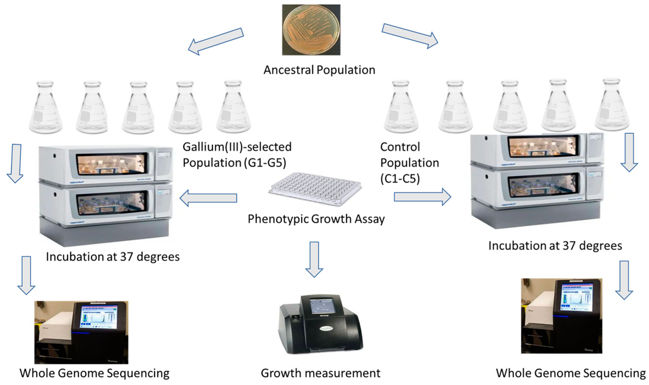
Figure 1. Experimental evolution layout.
Applmicrobiol 05 00032 g001
Figure 2. The mean of 24 h growth for S. aureus populations in increasing concentrations of gallium (III) after 30 days of evolution. Gallium (III)-selected bacteria showed significant growth compared to the control, followed by the ancestors. Gallium (III)-selected populations of S. aureus were exposed to 300 mg/L of gallium for 30 days in nutrient agar broth. Controls populations of S. aureus were grown in nutrient broth without gallium. Ancestral populations of S. aureus were grown in nutrient broth for 24 h.
Applmicrobiol 05 00032 g002
Figure 3. (A) The mean and SE of 24 h growth for populations in increasing concentrations of iron (III) up to 1000 mg/L after 20 days of evolution. Gallium (III)-selected bacteria showed significant growth compared to the control and the ancestor (B) The mean and SE of 24 h growth for populations in increasing concentrations of silver up to 50 mg/L after 20 days of evolution. The gallium (III)-selected bacteria showed significant growth compared to the controls, followed by the ancestors.
Applmicrobiol 05 00032 g003
Figure 4. (A) The mean and SE of 24 h growth for populations in increasing concentrations of chloramphenicol up to 125 mg/L after 20 days of evolution. Gallium (III)-selected bacteria showed significantly inferior growth compared to the controls and the ancestors. (B) The mean and SE of 24 h growth for populations in increasing concentrations of tetracycline up to 125 mg/L after 20 days of evolution. The growth of the gallium (III)-selected bacteria was significantly inferior compared to the controls, followed by the ancestors.
Applmicrobiol 05 00032 g004
Figure 5. (A) The mean and SE of 24 h growth for populations in increasing concentrations of GenX up to 10 nM after 20 days of evolution. Gallium (III)-selected bacteria showed significantly higher growth rates compared to the control, followed by the ancestor (B) The mean and SE of 24 h growth for populations in increasing concentrations of PFOS up to 10 nM after 20 days of evolution. The gallium (III)-selected bacteria showed significantly higher growth rates compared to the control, followed by the ancestor.
Applmicrobiol 05 00032 g005
Figure 6. Scanning electron microscopy investigations of gallium (III)-selected S. aureus. Ancestral (A) and control (B) bacterial populations were intact, smooth and spherical, while 20 days of selection in gallium (III) nitrate caused the bacterial populations to appear oval, triangular, rod-like, as club-shaped rods and vibrio-shaped (C).
Applmicrobiol 05 00032 g006
Table 1. Selective sweeps in gallium (III)-resistant populations at day 20.
GenePosition MutationG1G2G3G4G5
sfaA/sfaD2,228,365Intergenic (−35/−67)0.7670.6920.7310.7580.755
KQ76_RS01520342,330I180I* (ATT→ATC) 0.6780.620.6450.6730.631
fmtA1,017,325Q304* (CAA→TAA) 0.5830.6530.5990.6240.539
KQ76_RS083601,700,478G59S* (GGC→AGC) 0.3670.6560.6410.5410.538
dltB860,322E344K* (GAA→AAA) 0.3250.3130.370.2660.363
hssR2,389,192R188P* (CGA→CCA) 0.3270.320.3820.3280.339
rsp2,412,575G84D* (GGT→GAT) 00000.330
KQ76_RS11280/KQ76_RS112852,252,747Intergenic (−54/+157)00.29900.2810.325
KQ76_RS130202,574,726G69A* (GGC→GCC) ‡0.2740.3220.35500.318
KQ76_RS138252,746,925A172G* (GCT→GGT) 0.35200.360.2460
KQ76_RS129552,564,194A17P* (GCA→CCA) 0.2980000.281
gltB453,752Pseudogene (4457/4500 nt)00000.281
KQ76_RS134752,665,478E162V* (GAA→GTA) 00000.278
purS1,027,271A87P* (GCA→CCA) 00.270.2390.2620.272
KQ76_RS111752,233,551T39S* (ACT→AGT) 0.270000
KQ76_RS109852,190,680T500S* (ACG→TCG) 0.2550.27500.2370.262
KQ76_RS121802,408,435L213I* (TTA→ATA) 00.254000.262
graR673,306A185P* (GCA→CCA) 0.2640000.26
KQ76_RS073751,536,460Y112* (TAT→TAA) 00.275000
KQ76_RS092551,890,405S137T* (AGT→ACT) 0.2540.2660.26500
KQ76_RS111852,236,063R52P* (CGT→CCT) 0.2720000.251
KQ76_RS04770986,849K56* (AAA→TAA) 0.28100.25400
ylqF1,213,003E184D* (GAG→GAC) 0.2880.3130.2370.260
smpB810,694M1K* (ATG→AAG) †00.29000
vraE2,766,496I591I* (ATA→ATT) 0.2680000
KQ76_RS02795596,886A144V* (GCA→GTA) 000.23500
KQ76_RS01360/lip2311,944Intergenic (−69/−348)0000.2340
capA116,233V151M* (GTG→ATG) 0000.2330
KQ76_RS04220871,433T26I* (ACA→ATA) 0000.2250
KQ76_RS05175/KQ76_RS051801,066,987Intergenic (−157/+27)0.2540000
KQ76_RS01815391,361S36I* (AGT→ATT) 00.291000
mnmG2,776,116H117Q* (CAT→CAA) 00.276000
thrS1,740,864L149* (TTA→TAA) 0000.2240
Notes: After 20 days of selection, all populations were subjected to whole-genome resequencing with sequence alignments and variant calling using breseq 0.30.0. The genes/mutations and frequencies are reported. Gallium (III)-selected populations (G1–G5). * Annotation gives meaning to a given sequence and makes it much easier to view and analyze its contents. Numbers in the last five columns represent the frequencies. † Indicates a mutation or variant with a specific significance. ‡ Indicates a variant that has been flagged as potentially problematic or requiring further investigation.The underlined colors are the various mutations.
Table 2. Descriptions of genes.
pstCPhosphate ABC transporter permease subunit
KQ76_RS04365Acyltransferase family protein
hssRDNA-binding heme response regulator
KQ76_RS10525PP2C family protein–serine/threonine phosphatase
pxpB/greA5-oxoprolinase subunit PxpB/transcription elongation factor
KQ76_RS13020Alpha/beta hydrolase
icaRIca operon transcriptional regulator
KQ76_RS09255Hypothetical protein
ald/KQ76_RS08720Alanine dehydrogenase/universal stress protein
mnmGtRNA uridine-5-carboxymethylaminomethyl(34) synthesis enzyme
KQ76_RS04770ATP-binding protein
cozEbCell elongation protein
KQ76_RS12955D-lactate dehydrogenase
KQ76_RS01360/lip2YjiH family protein/YSIRK domain-containing triacylglycerol lipase Lip2/Geh
KQ76_RS10985BglG family transcription antiterminator
KQ76_RS13825ECF-type riboflavin transporter substrate-binding protein
gltBGlutamate synthase large subunit
rspAraC family transcriptional regulator
ylqFRibosome biogenesis GTPase
mprFBifunctional lysylphosphatidylglycerol flippase/synthetase
mutSDNA mismatch repair protein
KQ76_RS11280/KQ76_RS11285M23 family metallopeptidase/HAD IIB family hydrolase
KQ76_RS12905ATP-binding cassette domain-containing protein
smpBSsrA-binding protein
KQ76_RS13475Glutathione peroxidase
KQ76_RS12190/rspYbgA family protein/AraC family transcriptional regulator
KQ76_RS11175BCCT family transporter
sfaA/sfaDStaphyloferrinA export MFS transporter/D-ornithine–citrate ligase
KQ76_RS01520DUF3169 family protein
fmtATeichoic acid D-Ala esterase
KQ76_RS08360Adenine phosphoribosyltransferase
dltBPG:teichoic acid D-alanyltransferase
KQ76_RS13825ECF-type riboflavin transporter substrate-binding protein
KQ76_RS13475Glutathione peroxidase
purSPhosphoribosylformylglycinamidine synthase subunit
KQ76_RS12180Magnesium transporter CorA family protein
graRResponse regulator transcription factor GraR/ApsR
KQ76_RS07375Phage major capsid protein
KQ76_RS09255Hypothetical protein
KQ76_RS11185NADP-dependent oxidoreductase
vraEPeptide-resistance ABC transporter permease subunit
KQ76_RS02795Uracil–DNA glycosylase
capACapsular polysaccharide-type 5/8 biosynthesis protein
KQ76_RS04220FAD/NAD(P)-binding protein
KQ76_RS05175/KQ76_RS05180Nramp family divalent metal transporter/YktB family protein
KQ76_RS01815General stress protein
thrSThreonine–tRNA ligase
Note: Color coding: Polymorphisms in the control populations are shown in grey; polymorphisms in the gallium (III)-selected populations only are shown in brown; polymorphisms in both the control and gallium (III)-selected populations are shown in green.
Table 3. Selective sweeps in control populations at day 20.
GenePosition MutationC1C2C3C4C5
pstC1,384,254S178C* (AGT→TGT) 00.7280.6850.730.709
KQ76_RS04365907,688N549I* (AAT→ATT) 0.6250.660.610.680.653
KQ76_RS105252,102,321G175E* (GGA→GAA) 0.4170.4630.3990.3780.39
KQ76_RS130202,574,726G69A* (GGC→GCC) 00.3140.310.3670.468
ylqF1,213,003E184D* (GAG→GAC) 00.27900.3360
KQ76_RS130202,574,727G69R* (GGC→CGC) 0.33800.360.3160
hssR2,389,188E187Q* (GAA→CAA) 0.3490.300.410.3160
pxpB/greA1,672,825Intergenic (−76/+250)0.310.2930.390.3130.272
icaR2,727,937Q79* (CAA→TAA) 0.27800.2900
KQ76_RS105252,102,321G175E* (GGA→GAA) 0.3020000
KQ76_RS063251,302,347D54Y* (GAC→TAC) 0.293000.2960.259
cozEb1,352,269I247N* (ATT→AAT) 00.2790.2590.2930.259
mprF1,354,114G299D* (GGT→GAT) 0000.2920.253
pepF/yjbH941,253Intergenic (+205/+255)0000.2920
KQ76_RS138252,746,925A172G* (GCT→GGT) 00.27200.2900.248
KQ76_RS12190/rsp2,412,155Intergenic (+1318/170)0.2460000
KQ76_RS109852,190,680T500S* (ACG→TCG) 00.27900.2900.256
KQ76_RS092551,890,405S137T* (AGT→ACT) 00.2450.28600
KQ76_RS129552,564,194A17P* (GCA→CCA) 00.25100.2870.31
ald/KQ76_RS087201,776,393Intergenic (−57/−84)0.34900.28600.341
KQ76_RS04275/KQ76_RS04280881,138Intergenic (+150/−158)0.236000.2700.249
mnmG2,776,116H117Q* (CAT→CAA) 00.2460.27600
ald/KQ76_RS087201,776,393Intergenic (−57/−84)000.2860.2650
KQ76_RS129552,564,193M16I* (ATG→ATC) 000.2500
KQ76_RS134752,665,478E162V* (GAA→GTA) 0.2760000
mutS1,275,400K479N* (AAG→AAC) 00000.25
rsp2,412,863C180Y* (TGT→TAT) 0.2460000.251
smpB810,694M1K* (ATG→AAG) 0.2640000
KQ76_RS01360/lip2311,944Intergenic (−69/−348)000.24700
KQ76_RS109852,190,680T500S* (ACG→TCG) 000.24700
KQ76_RS04770986,858Q59K* (CAA→AAA) 00.245000.244
KQ76_RS138252,746,925A172G* (GCT→GGT) 000.24500
gltB453,752Pseudogene (4457/4500 nt)00.2380.24300
KQ76_RS129052,554,550V173L* (GTC→CTC) 00000.247
KQ76_RS111752,233,551T39S* (ACT→AGT) 0.230000
Notes: After 20 days of selection, all populations were subjected to whole-genome resequencing with sequence alignments and variant calling using breseq 0.30.0. The genes/mutations and frequencies are reported. Control populations (C1–C5). * Annotation gives meaning to a given sequence and makes it much easier to view and analyze its contents. Numbers in the last five columns represent the frequencies. The underlined colors are the various mutations.
Disclaimer/Publisher’s Note: The statements, opinions and data contained in all publications are solely those of the individual author(s) and contributor(s) and not of MDPI and/or the editor(s). MDPI and/or the editor(s) disclaim responsibility for any injury to people or property resulting from any ideas, methods, instructions or products referred to in the content.