Article

Phenotypic and Genotypic Characterization of Antimicrobial Resistance in Salmonella enterica Serovars from Colombian Pig Farms

by
Iliana C. Chamorro-Tobar
1,2,
Adriana Pulido-Villamarín
3,*,
Ana Karina Carrascal-Camacho
1,
Irina Barrientos-Anzola
1,2,
Magdalena Wiesner
4,
Ivonne Hernández-Toro
5,
Lis Alban
6,7,*,
John Elmerdahl Olsen
6,
Anders Dalsgaard
6 and
Yaovi Mahuton Gildas Hounmanou
6
1
Environmental and Industrial Biotechnology Research Group-GBAI, Research Hotbed of Food Safety, Line of Emerging Microorganisms, Department of Microbiology, Faculty of Sciences, Pontificia Universidad Javeriana, Bogotá 110231, Colombia
2
Center for Research and Technology Transfer of the Pig Sector—Ceniporcino, Asociación Porkcolombia-Fondo Nacional de la Porcicultura, Bogotá 110231, Colombia
3
Agricultural Research Unit-UNIDIA, Department of Microbiology, Faculty of Sciences, Pontificia Universidad Javeriana, Bogotá 110231, Colombia
4
Department of Biology, Faculty of Science, Universidad Antonio Nariño, Bogotá 110231, Colombia
5
Bacteriology Area, National Laboratory of Veterinary Diagnosis, Colombian Agricultural Institute-ICA, Bogotá 110231, Colombia
6
Department of Veterinary and Animal Sciences, Faculty of Health and Medical Sciences, University of Copenhagen, 1870 Frederiksberg, Denmark
7
Department for Food Safety and Veterinary Issues, Danish Agriculture and Food Council, 1613 Copenhagen, Denmark
*
Authors to whom correspondence should be addressed.
Appl. Microbiol. 2024, 4(4), 1729-1744; https://doi.org/10.3390/applmicrobiol4040116
Submission received: 22 November 2024 / Revised: 10 December 2024 / Accepted: 13 December 2024 / Published: 20 December 2024

Abstract

:
Salmonella enterica is a globally important zoonotic microorganism that affects pigs and can enter the farm through various routes. This study aimed to determine the prevalence of S. enterica in water sources and pigs at Colombian pig farms, and to characterize the antimicrobial resistance of the isolates phenotypically and genotypically. Samples were collected from 103 farms including source water (n = 104), storage tank water (n = 103), drinking water (n = 103), and individual rectal swab samples (n = 1025). The presence of Salmonella was detected/identified using MDS-3M™ agar culture medium. Isolates were serotyped, and their antibiotic susceptibility was determined by minimum inhibitory concentration (MIC). Whole genome sequencing (WGS) was performed using Illumina NovaSeq, and bioinformatics analysis focused on serovar confirmation, MLST determination, and resistance gene detection. The overall between-farm prevalence of Salmonella enterica including all types of samples was 52.4% (54/103), with 6.4% of rectal swab samples and 21.3% of water samples found to be positive. Thirty serovars were identified using WGS, with the most common being S. Typhimurium var. monophasic (1,4,[5],12:i:-) (41.2%), S. Schwarzengrund (4.2%), and S. Saintpaul (4.2%). Salmonella Typhimurium and its monophasic variant were more commonly found in rectal swabs than the remaining serotypes (relative risk = 2.9, p < 0.0001), which were commonly found in the water samples (relative risk = 5.2, p < 0.0001). High levels of phenotypic resistance were observed, particularly to amikacin (99.2%), tetracycline (59.7%), chloramphenicol (55.5%), and ampicillin (42%). All isolates carried genes conferring resistance to aminoglycosides (aac(6′)-Iaa), quinolones (qnrB19), and tetracyclines (tetA). In conclusion, S. enterica is prevalent in Colombian pig farms including the water supply, with the S. Typhimurium monophasic variant being predominant, and antimicrobial resistance is widespread.

1. Introduction

Salmonella enterica is a globally significant zoonotic pathogen, commonly associated with foodborne and waterborne diseases [1,2]. Non-typhoidal salmonellosis typically manifests as gastrointestinal symptoms, fever, and in severe cases hospitalization, and incurs significant healthcare costs. Without effective intervention, salmonellosis can place considerable strain on public health systems [3,4,5].
In animals raised for human consumption, such as pigs, S. enterica infections can lead to the contamination of meat if proper hygiene measures are not implemented during slaughter. To safeguard human health, efforts to minimize the spread of Salmonella in primary production are essential [5,6]. Additionally, subclinical infection in pigs results in reduced feed conversion [7,8], leading to decreased productivity and negative economic impacts on the pork industry.
Salmonella can enter pig farms through various sources including contaminated water, wildlife (such as rodents, insects, and birds), and human activity involving contaminated equipment like boots. Additionally, contaminated feed and the introduction of infected replacement stock are significant risk factors for Salmonella entry [9,10]. Water is particularly critical as a risk factor for Salmonella spread in pig rearing, being used in all stages of primary production including farrowing, pre-feeding, rearing, and fattening [11].
In pigs, S. enterica infections can result from both species-adapted serotypes, such as S. Choleraesuis, and non-adapted serovars, occurring at any point in the production cycle and affecting pig welfare and health. However, most infections are subclinical, making them difficult for producers to detect. Despite the absence of clinical signs, infected pigs can still serve as reservoirs for bacterial transmission across the farm, emphasizing the need for control measures to prevent its spread within pens and facilities [8,12,13].
Knowledge of the Salmonella serotypes responsible for human cases in a given country can help identify key infection sources and inform strategies for reducing and preventing outbreaks [14]. In the European Union, United States, Chile, and Colombia, the primary serotypes linked to foodborne outbreaks include S. Enteritidis, S. Typhimurium, and its monophasic variant 1,4,[5],12:i:- [7,15,16,17,18]. Transmission pathways often vary by serotype, and source attribution studies can be conducted to establish the link between infection and origin.
Historically, pulsed-field gel electrophoresis (PFGE) was used to characterize Salmonella and other bacteria. However, since 2017, PFGE is gradually being replaced by whole genome sequencing (WGS), which allows for more detailed pathogen characterization. WGS also provides accurate predictions of virulence, pathogenicity, and antimicrobial resistance [14,19].
The emergence of antimicrobial resistance in Salmonella enterica strains is a significant public health concern. Several classes of antibiotics including third-, fourth-, and fifth-generation cephalosporins and fluoroquinolones are used to treat severe human salmonellosis cases [20,21,22]. In animals, antibiotics are also employed not only for treatment, but also in some countries for metaphylaxis, prophylaxis, and as growth promoters [16,23], particularly in regions outside the EU.
This practice contributes to the development of multi-resistant Salmonella strains in the majority of countries outside the EU [14,24]. In contrast, the EU has implemented restrictions on the use of these antibiotics, reducing their consumption and the emergence of resistant strains.
The aim of this study was to determine the prevalence of Salmonella enterica in various water sources and pigs on Colombian pig farms. Additionally, we characterized the antimicrobial resistance of the isolated S. enterica strains both phenotypically and genotypically.

2. Materials and Methods

2.1. Study Design and Selection of Pig Farms

According to the 2019 census, Colombia had 6,257,189 pigs distributed across farms of various sizes. We conducted a cross-sectional study with stratified, proportional sampling based on herd type and size. Following the classification by the Instituto Colombiano Agropecuario (ICA) [25], so-called “industrial commercial farms” were defined as those with 10–99 sows and 100–599 fattening pigs, while so-called “technified” farms were classified as having ≥100 sows and ≥600 fattening pigs. When estimating the necessary sample size to assess the prevalence of infected pig farms, we assumed a between-farm prevalence of 44%, with a 95% confidence level, 10% precision, and a type I error of 5%. To account for potential data loss, samples were collected from 103 farms.

2.2. Sample Types

In each of the 103 farms, four types of samples were collected: water from the source (n = 1), storage tank (n = 1), pen drinkers (n = 1), and rectal swabs from pigs (n = 10). Standardized protocols were followed for sample collection, which included 1 L of water for each water sample [11]. Rectal swabs were individually collected from finishing pigs aged 19 to 22 weeks, just prior to slaughter, within the same pen [26].

2.3. Isolation and Identification of Salmonella spp.

2.3.1. Water

Water samples were processed according to the A.P.H.A. 9260-B protocol [27]. A total of 500 mL of water was filtered through a 0.45 µm pore size and 47 mm diameter nitrocellulose membrane (Millipore®, Darmstadt, Germany) that was then placed in a sterile 7 oz Whirl-Pak® bag containing 100 mL of 3M™ buffered peptone water and incubated at 41.5 °C ± 1 °C for 18 to 24 h. For Salmonella identification, the MDS-Neogen® Salmonella-2 Molecular Detection System (AOAC® Method Certificate # 091501) [28] (Neogen® Lansing, MI, USA) was employed following the manufacturer’s instructions. Samples detected as positive by the software were cultured on XLD agar (DIFCO™, Franklin Lakes, NJ, USA) and CHROMagar™ Salmonella Plus agar (CHROMagar Microbiology, Paris, France), then incubated at 37 °C ± 1 °C for 18 to 24 h to obtain bacterial isolates.

2.3.2. Rectal Swabs

The isolation of bacteria was performed according to ISO 6579:2017 [29]. Briefly, the swab was deposited in 10 mL buffered peptone water (DIFCO™) and incubated at 37 °C for 18–24 h. Next, 1 mL of the pre-enriched samples was transferred to Muller–Kauffmann Tetrationate Broth (Scharlau™ Barcelona, Spain) and incubated at 37 °C ±1 °C for 18 to 24 h. Following this, 100 µL of the enriched sample was added to 10 mL of Rappaport Vassiliadis Broth (Scharlau™) and incubated at 42 ± 1 °C for 24 to 48 h. Isolation of Salmonella was then performed on XLD agar (DIFCO™) and CHROMagar™ Salmonella Plus agar.

2.3.3. Serotype Determination

Serotyping was performed according to the Kauffmann–White–Le Minor scheme [30,31] through agglutination on slides using somatic antisera (O) and in tubes using flagellar antisera (H), utilizing specific antisera (DIFCO™).

2.4. Antimicrobial Susceptibility Testing

The minimum inhibitory concentration (MIC) was determined using the broth dilution method with the Sensititre™ automated system, following the manufacturer’s recommendations with microdilution plates in EUVSEC3 Broth (Thermo Scientific™, Waltham, MA, USA) for Gram-negative bacteria. The panel included 15 antimicrobials: amikacin (AMK), ampicillin (AMP), azithromycin (AZ), cefotaxime (CTX), ceftazidime (CAZ), chloramphenicol (CHL), ciprofloxacin (CIP), colistin (COL), gentamicin (GEN), meropenem (MER), nalidixic acid (NAL), sulfamethoxazole (SMX), tetracycline (TET), tigecycline (TGC), and trimethoprim (TMP) [32,33]. Plate readings were performed using Vision ™ equipment, and susceptibility was assessed with the Sensititre™ SWIN™ Software v2.4 System. Strains were classified as multi-resistant (MDR) if they exhibited resistance to at least one antimicrobial from three or more antimicrobial classes [34].

2.5. Analysis of Whole Genome Sequencing (WGS) of Salmonella enterica Isolates

The samples were processed by whole genome sequencing (WGS) at the University of Copenhagen. DNA extraction was performed using a commercial kit for Gram-negative bacteria (Qiagen®, Hilden, Germany), following the manufacturer’s instructions. Cultures of each strain were isolated on soy trypticase agar supplemented with a blood NanoDrop and used for the DNA quality measurement, while the Qubit dsDNA Assay Kit with its High Sensitivity reagent (Invitrogen®, Waltham, MA, USA) was utilized to assess the concentration of DNA. After determining the concentration, a sequencing library was prepared for Illumina sequencing using the manufacturer’s Nextera XT Kits (Illumina®, San Diego, CA, USA). Sequencing was conducted with the NovaSeq 6000 PE15 sequence [35]. Sequence reads were quality checked with fastQC, subsequently filtered with trimmomatic, and then assembled using SPAdes v.3.15.5 [36]. The genome assemblies were assessed with Quast v. 4.0 [37].
WGS analysis was carried out using bioinformatic tools including the Salmonella In Silico Typing Resource (SISTR) version 1.1.1 for serotype prediction and confirmed with [38]. Pathogenwatch (https://pathogen.watch/, accessed on 18 November 2022) was used to determine the serotype. The genomes were further analyzed for AMR genes using abricate1.0.1 against the ResFinder 4.1 database for the detection of resistance genes, and here, the tool mlst2.23.0 was used to confirm the sequence type of the sequenced genomes [39,40].
These sequences were deposited with the European Nucleotide Archive under project number PRJEB80816, also available via the NCBI.

2.6. Statistical Analyses

Data were analyzed by descriptive statistics and Chi-square tests including Fisher’s exact test where needed, to detect statistically significant differences between groups.

3. Results

3.1. Prevalence of Salmonella spp.

A total of 103 farms were included in the study, where 75 were industrial commercial and 28 technified farms. One of the farms had two entry samples taken because they had two different water sources. In total, 310 water samples and 1025 rectal swab samples were collected and processed, resulting in the detection of 132 Salmonella isolates. In total, 21.3% (66/310) of the water samples and 6.4% (66/1025) of the rectal swabs were found to be positive. The prevalence difference between the water samples and rectal swabs was statistically significant (relative risk: 3.3; p < 0.001). For each kind of water sample, 21.2% (95% CI: 13.3–29.0%) of source water (catchments), 22.3% (95% CI: 14.3–30.4%) of water from storage tanks, and 20.4% (95% CI: 12.6–28.2%) of water from drinkers were found to be positive. There were no significant differences between these three types of water samples regarding positive proportions (p = 0.94).
A farm was considered positive if at least one sample tested positive. Based upon this definition, the overall between-farm prevalence was 52.4% (54/103), with a prevalence of 49.3% for industrial commercial farms and 60.7% for technified farms. These two proportions did not differ statistically (p = 0.31). Based on the fecal swab samples, 31.1% (32/103) of farms were considered to house pigs, which were Salmonella positive.
Of the 54 farms with positive results, 12 had Salmonella detected in both water and rectal swabs, whereas 20 had Salmonella isolated only from rectal swabs, and 22 had only positive samples detected in water. On six farms, Salmonella was isolated from all three types of water samples (source water, storage tanks, and drinkers). Technified farms were associated with a higher risk of detecting positive rectal swabs than industrial farms (relative risk = 2.1, p = 0.01).

3.2. Serotypes Identified

3.2.1. Serotyping According to the Kauffmann–White–Le Minor Scheme

Serotyping of the 132 isolates revealed a diversity of 40 serovars in 54 positive farms (Supplementary Table S1). Twenty-nine different serovars were identified in water samples, while nine were detected in rectal swab isolates. S. Typhimurium var. monophasic (1,4,[5],12:i:-) was the predominant serovar in both water (15%, 10/66) and rectal swabs (69.7%, 46/66). This difference in proportions was statistically significant (relative risk: 4.4; p < 0.001).

3.2.2. Serotyping According to Whole Genome Sequencing

Thirteen strains were not sequenced because they were not viable when they arrived in Denmark. Therefore, whole genome sequencing (WGS) was performed on 119/132 strains (source water: 17; storage tank: 20; drinkers: 19; rectal swabs: 63). WGS identified 30 different serovars, with 12 detected in rectal swabs and 23 in water samples (Table 1). S. Typhimurium var. monophasic (1,4,[5],12:i:-) was the most prevalent serovar, accounting for 41.2% (49/119) of the isolates, followed by S. Typhimurium (5.9%, 7/119). Other serovars such as S. Braenderup, S. Carrau, S. Schwarzengrund, S. Soahanina, and S. Saintpaul each comprised 4.2% (5/119) (Table 1).
Multi-locus sequence typing (MLST) identified 32 sequence types (STs). ST19 was the most common (42.8%) and was found in S. Typhimurium var. monophasic and S. Typhimurium. ST226 was detected in 5.0% of the strains, corresponding to S. Carrau. ST50 and ST96 were each found in 4.2% of isolates, associated with S. Saintpaul and S. Schwarzengrund, respectively. Seven new STs were identified in serovars such as S. houtenae IV 50,z32:-, S. houtenae IV 11,z23:-, S. enterica I H:y:-, S. Braenderup, and S. Sandiego.
The agreement between Kauffmann–White–Le Minor serotyping and WGS serotyping was 63% (75/119). Supplementary Table S1 details the distribution of all serotypes by method and sample type.

3.2.3. Comparison of Salmonella Serotypes Found in Rectal Swabs and Water Samples at the Farm Level

In two technified farms, the same serovar was detected in the water samples and rectal swabs, this dealt with S. Typhimurium var. monophasic (n = 1) and S. Insangi (n = 1), and in one commercial industrial farm, S. Typhimurium var. monophasic was also detected in both the water samples and rectal swabs. The serotypes S. Soahina, S. Schwarzengrund, S. Saintpaul, S. Kedougou, S. San Diego, and S. Braenderup were detected in the water sources of six commercial industrial farms. Different serotypes were detected in twelve industrial commercial farms and eight technified farms from the rectal swab samples. Additionally, different serotypes were isolated from the water sources only in twelve industrial commercial farms and six technified farms. Only five serovars were detected in both the rectal swabs and water samples at different farms (Table 1). For the five serovars that were found present in the water and rectal swabs, there was no sequence type sharing.
Compared to all other serotypes, Salmonella Typhimurium and its monophasic variant were more commonly detected in the rectal swab samples (relative risk = 2.9, p < 0.0001), and less common in the water samples (relative risk = 0.19, p < 0.0001).

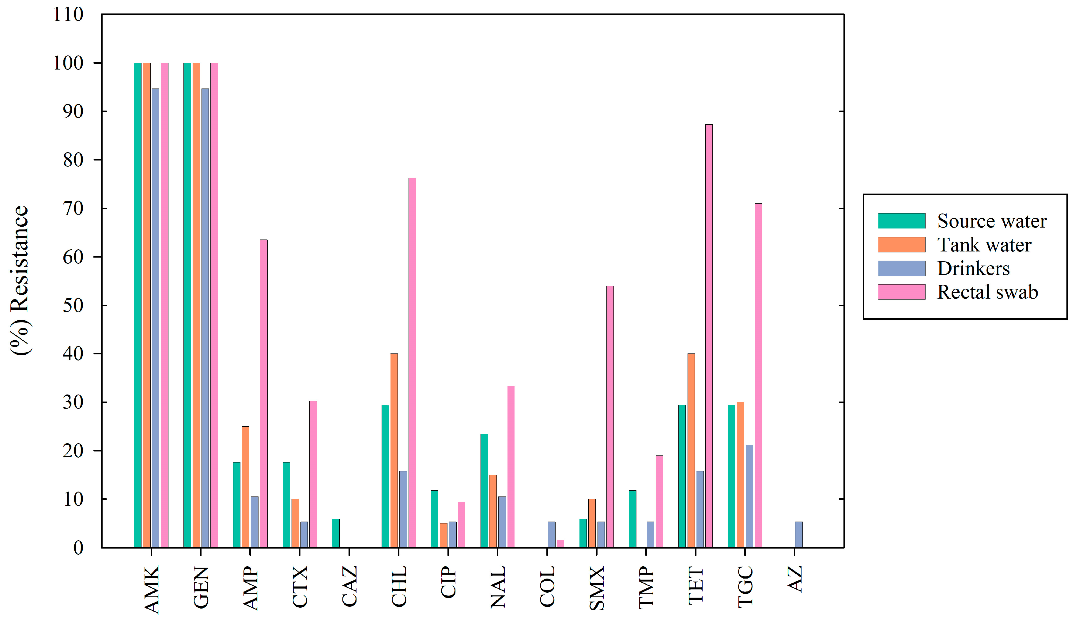
3.3. Antimicrobial Susceptibility Testing

Antimicrobial susceptibility testing was conducted on 119 Salmonella spp. isolates (the same strains sequenced by WGS). All isolates showed resistance to amikacin (100%) and a high rate of resistance to gentamicin (99.2%). Common resistances included tetracycline (59.7%), chloramphenicol (55.5%), and tigecycline (50.4%). Additional resistance was noted for ampicillin (42.0%), sulfamethoxazole (31.9%), nalidixic acid (25.2%), cefotaxime (21.0%), trimethoprim (12.6%), and ciprofloxacin (8.4%). Resistance to colistin (1.7%), azithromycin (0.8%), and ceftazidime (0.8%) was rare, while no resistance was observed to meropenem.
The proportion of resistance by antibiotic and sample type is shown in Figure 1. Statistically significant differences (p < 0.05) in the resistance patterns were found across the four sample types for seven of the fourteen antibiotics tested Rectal swabs showed the highest resistance to ampicillin (p < 0.001), chloramphenicol, ciprofloxacin, sulfamethoxazole, and tigecycline (p < 0.001). Source water had the highest resistance to ceftazidime (p = 0.003), and the storage tank water showed the highest resistance to tetracycline (p = 0.02).
Forty-three antimicrobial resistance patterns were identified, and 61% (72/119) of the isolated strains presented MDR. Table 2 shows the 10 most common resistance patterns.
All 119 isolates showed resistance to at least one family of antibiotics; however, the S. Typhimurium monophasic variant was the one with the highest proportion of resistance: AMK (100%), GEN (100%), AMP (81.6%), CHL (93.8%), TET (98%), TGC (77.5%), SMX (51%), CTX (36.7%), NAL (38.7%), TMP (16.3%), CIP (8.10%) and COL (2%) (Table S2).

3.4. Resistance Genes Identified from WGS

Through WGS, resistance genes were identified in the 119 isolates. The quinolone qnrB gene was found in 72.5% (87/119), the tetracycline tetA gene in 62.5% (75/119), and the florfenicol/chloramphenicol resistance floR gene in 47.5% (57/119) of the isolates. Hence, the genes were predominant across all isolates, as was the cryptic aminoglycoside resistance gene aac(6′)-Iaa (100%). All of the isolates also exhibited resistance to amikacin and gentamycin. In addition to aac(6′)-Iaa, several aminoglycoside resistance genes were found: (aac(3)-IId, aph(3′)-IIa, aph(3′)-Ia, aadA2, aph(3″)-Ib, aph(6)-Ic, and aph(6)-Id). The fluoroquinolone resistance mutation gyrA S83F gene was found in 11% of the qnrB19-carrying isolates; the resistance genes appeared more dispersed across these serovars, but mutations such as gyrB or parC were not observed. Beta-lactamase genes, such as blaTEM1-B and blaCMY-2, conferring resistance to third-generation cephalosporin, were found in 22 and 19 isolates out of 56 for S. Typhimurium, respectively. Moreover, the coproduction of blaTEM1-B and blaCMY-2 was found in three isolates of this serovar. Three genes of dihydrofolate reductase, dfrA12, dfrA14, and dfrA29, were found, but in low frequency. Sulfonamide genes were principally represented by the sul2 gene in 21% of the isolates. The tetracycline tetB gene was found in 20% of the isolates, and most of them were negative for tetA.
S. Typhimurium and its monophasic variant carried the highest number of resistance genes. The predominant genes included qnrB19 in 42.9% and 100%; tetA in 14.2% and 75.5%, respectively; moreover, floR was only detected in monophasic isolates (83.7%).
In isolates of the serovars Amager, Isangi, Melagridis, and Keodogou, higher numbers of resistance genes were present. Therefore, serovars Amager and Isangi shared multiple aminoglycoside and tetracycline resistance genes (aph(3″-IIa, aph(3″-Ia, aadA2, aph(3″)-Ib, aph(6)-Id, and tetM). The serovar Amager uniquely carried lincosamide genes inuF and linezolid cfr resistance. Serovar Isangi carried blaTEM-1B. Serovar Meleagridis had a class 1 integron, based on its resistance profile (qacEdelta, sul1 sul2, dfrA12, and aadA2). In the serovar Saintpaul, one isolate from rectal swabbing exhibited resistance to the widest range of antimicrobials: six aminoglycosides, three beta-lactamases, sul1, sul2, and sul3, inuF, and the inuG, floR, and cmlA1 genes.
Of the 72 isolates exhibiting tetracycline resistance, 55 carried tetA, 11 were positive for tetB, and among these, two carried both genes tetA and tetB. Among the 67 isolates that exhibited chloramphenicol resistance, 53 were positive for the floR gene. In the remaining fourteen isolates, only two carried the gene cmlA1, which also confers resistance to this antibiotic. Almost all 54 ciprofloxacin-resistant isolates were positive for qnrB19 (n = 51). The 67 isolates with gyrA mutation also carried the qnrB19 gene, however, four of these isolates were susceptible to ciprofloxacin and two were classified as intermediate susceptible. Ampicillin resistance was observed in 54 isolates, and in 42 of these isolates, 1 resistance gene for beta-lactamase was identified. In addition, of the 20 isolates positive for blaCMY-2, 18 were cefotaxime and ceftazidime resistant (Table S3).
The concordance between phenotype resistance and the presence of genes was high for aminoglycosides, tetracycline, chloramphenicol, ciprofloxacin, and ampicillin. The agreement between phenotype and genotype was low for sulfamethoxazole and trimethoprim resistance. Of the 38 resistance isolates, 21 carried one of the sul 1, sul2, or sul3 genes. Two isolates simultaneously carried the three genes, and one isolate carried sul1 and sul2. Regarding trimethoprim, only five of the 15 isolates showing this resistance contained the dfrA12 or dfrA29 genes (Table S3).

4. Discussion

This was the first study conducted at a national level in Colombia that used a representative sample size to assess the prevalence of Salmonella spp. in water samples and rectal samples collected at pig farms.
The between-farm prevalence of Salmonella spp. based on rectal swabs was 31.1% with a median of 1 (range: 1–10), indicating that most farms with positive rectal swab samples only had a low proportion of positive samples. Overall, 6.4% of the swab samples were positive, which was lower than reported by the Colombian Agriculture Institute (ICA) in 2018 (Internal report, unpublished information, based on 380 samples), where the prevalence was 28.4%. Giraldo-Cardona and collaborators reported a prevalence of 8.7% in isolates found in rectal swabs (8/231) from four Colombian regions [11]. The prevalence of Salmonella in pigs suffering clinically, with symptoms indicative of salmonellosis, in the Antioquia region of Colombia was 22.8% in a previous study [23]. In contrast, 6.4% of pigs found to excrete Salmonella in the current study were without clinical signs. Hence, the available data point to the circulation of Salmonella spp. in healthy finishing pigs, confirming the subclinical presence of the infection. In addition, in the study in Antioquia, 35% of the isolates corresponded to serovar S. Typhimurium var. monophasic [23], which was statistically lower (p < 0.001) compared to what was found in our study (69.8%). This serotype is known to be gradually replacing S. Typhimurium in swine production in many countries, probably due to specific factors of successful colonization associated with this serotype [41].
The between-herd prevalence of Salmonella in water was 33%. Moreover, 21.3% (66/310) of the water samples were found to be positive for Salmonella, and thus we found a threefold higher prevalence of Salmonella spp. in the water samples than in the rectal swabs (relative risk = 3.3; p < 0.001). This proportion of positive samples was higher than that reported in 2019 by Giraldo-Cardona and collaborators, who in a pilot study involving 21 samples found a prevalence of 14.3% (3/21) [11]. Although these prevalences differed statistically (p = 0.01), it is difficult to conclude whether the national prevalence has increased or not due to the small sample size in the study from 2019. Still, the data obtained confirm the presence of Salmonella spp. in different water sources, hereby exposing pigs to a risk of infection and highlighting the need for interventions to ensure water quality. An example of such an intervention was recently described by Roldan-Henao et al. (2023), who found an impact on production and Salmonella when disinfecting the water tubes in a finishing pig farm with organic acids [42].
The diversity of serotypes found in the water samples was higher (n = 24) than that found in the rectal swab samples (n = 12), and only five serovars occurred on both sample types, with the serovar Typhimurium var. monophasic as the most common. This finding coincides with findings by De Lucía and Ostanello (2020), who pointed out that this serovar is ubiquitous [43]. The rectal swab findings suggest that S. Typhimurium and its monophasic variant possessed the highest pig infectivity among the identified serotypes. Only a few farms showed the same Salmonella serovar in the rectal swab samples and water samples. This suggests that water might not constitute an important source of Salmonella on the pig farms. Still, it is preferable that the water is free from Salmonella.
Serotyping remains fundamental for the identification of Salmonella spp. as it provides information associated with the origin of sources and epidemiological follow-up, among others [44]. Classical serotyping requires at least 150 antisera, trained staff [30], and involves some degree of subjectivity. In contrast, WGS can be used to both identify the microorganisms and their serotypes, and provide information about antimicrobial resistance genes, plasmids, and virulence factors [45,46]. In recent years, WGS has increasingly been used as a reference method for Salmonella surveillance and for source accounts in countries such as Denmark [47], the United States [48] and the United Kingdom [49]. However, in Colombia, the traditional KWM method is still used by the official reference laboratories, although private and educational institutions are beginning to implement WGS for research purposes. The latter is contributing to the development of new knowledge in Colombia. In our study, the agreement between results of KWM and WGS was 63% (75/119). The differences in the diagnostic approaches when identifying serotypes explained above confirm the partial lack of agreement. The agreement was lower than similar results obtained by Diep et al. (2019) and Chattaway et al. (2021), who reported 97.0% [44] and 99.96% [50] agreement, respectively. Some of the serotypes identified in the present study were not easily identified since several complementary antisera were required and the serotypes were uncommon. As a result, a full identification was not achieved due to the lack of antisera. In contrast, through use of WGS, it was possible to determine the definitive serotype, since the information in the databases contained a plethora of Salmonella spp. genomes, confirmed by two different genomic analysis tools in 100% agreement.
The high diversity of serotypes found in the present study may be due to the origin of the samples, which included the collection of samples from 103 pig farms throughout all of Colombia. Moreover, the environmental conditions and the ability of Salmonella to survive in these may have added to the diversity. Although some of the serotypes found have not previously been reported in water (S. Newport, S. Typhimurium) or pigs (S. Carrau, S. Pomona), they have been recovered from other sources such as poultry (S. Give, S. Schwarzengrund), fish, lettuce, and peanuts, among others [51,52,53].
The most frequent serotype in our study was the S. Typhimurium monophasic variant (41.2%). In recent years, this serotype has appeared as an emerging serotype in pigs, where it is gradually replacing the serovar Typhimurium [6]. According to an EFSA report from 2021, it is the predominant serotype in European pigs [54], and is one of the most frequent serotypes recovered in clinical isolates from humans. In Europe, it ranks third, and in Colombia, fourth [17].
According to Campos et al. (2019) the S. Typhimurium monophasic variant is associated with multidrug resistance clones, relevant for clinical treatments in humans [16]. ST34 was present in four isolates and ST19 in forty-four isolates. These two clones are of major importance globally. The first is the ST19 type sequence with DT104/U302 phage types and the second is the ST34 phage type DT120/DT193, which has initially been found in Europe. These two clones have been isolated from pigs, pork, and numerous outbreaks in humans [55]. Likewise, the European clone frequently belonging to DT120 and DT193 has spread worldwide and is currently circulating in several regions of America, Asia, and Australia, raising concerns about resistance to commonly used antimicrobials such as cephalosporins and quinolones [16,55]. In the USA, these antibiotics are considered as the first-line treatment of severe salmonellosis in humans. This result agrees with outbreaks in Denmark, where the strains implicated came from different sources such as clinical cases and pig isolates, and where ST19 was predominant [16,47,56].
In our study, S. Typhimurium and its monophasic variant exhibited the highest number of resistances, as confirmed by both the phenotypic and genotypic analyses, and predominant resistance was observed as 100% to aminoglycosides (AMK, GEN, and aac(6′)-Iaa) and others such as chloramphenicol (floR), fluoroquinolones (qnrB19), tetracycline (tetA), ampicillin, and cephalosporine (blaCMY-2). Similar resistance patterns were also reported in isolates from the Zhejiang and Fujian Provinces in China, where strains of this serovar were linked to ST19 and ST34 and showed resistance to aminoglycosides, tetracyclines, florfenicol, quinolones, and sulfonamides [57]. The high level of resistance may be caused by a selection pressure because tetracycline and ampicillin are commonly used antibiotics to treat bacterial infections in farm animals worldwide [57]. Likewise, Qin and collaborators reported that in China, 75% of the tetracycline resistance was conferred by tetA and tetB [58].
The serotype S. Schwarzengrund was detected in 4.2% of the positive samples. Notably, three of these positive samples were recovered from the same farm across all three water types sampled (municipal aqueduct, storage tank, and drinking water). This farm’s proximity to poultry and cattle farms may explain the presence of this serotype. Currently, S. Schwarzengrund is considered as an emerging Salmonella serovar in countries such as Japan, Thailand, Denmark, the United States of America, Korea, and Brazil. In 2019, the U.S. Centers for Disease Control and Prevention—CDC—investigated an outbreak associated with this serovar, revealing that the origin had been ground turkey meat [59]. Duc et al. (2020) revealed an increased prevalence of this serotype in Japan, where it appeared in mid-2012 [60]. In Taiwan, this serovar has been found in broiler chickens [61]. Similarly, in Korea, this serotype was identified by WGS in poultry, cattle, and swine feces samples. This shows the ubiquitarian distribution of this serotype, which facilitates its dissemination in livestock production [52,60,62]. In the Midwest of the United States, Pires et al. (2014) were able to isolate this serovar from fecal samples of finishing pigs [63]. It has also been isolated in Burkina Faso in canal waters [64].
Salmonella Carrau is one of the infrequent serotypes, which was found in our study. It was identified for the first time in 1944 from the mesenteric ganglia of diseased pigs and clinical cases in humans [65]. In our study, it was isolated from water in three different Colombian regions: Meta (n = 3), Casanare (n = 1), and Santander (n = 1). In 2003, Karczmarczyk and collaborators isolated this serotype in samples of ham and sausages from the Atlántico and Bolívar regions in Colombia [66]. It may be hypothesized that this serotype is not frequently identified because it has not been implicated in reported cases of salmonellosis in humans. The strains isolated in 2003 did not show antimicrobial resistance [66], differing from the results obtained in the present study, where phenotypic resistance to aminoglycosides (AMK, GEN) and quinolones (NAL) was found (Table S2), while genotypic resistance was only present with the aac(6′)-Iaa gene of AMK, qnrB19 gene of CIP, and tet(A) gene of TET.
S. Soahanina was isolated in 4.2% of the positive water sources in the Colombian region Magdalena, and all these were ST970. So far, this serotype has not been found in other regions of Colombia. This serotype showed resistance to aminoglycosides (AMK, GEN) and expressed aac (6′)-Iaa. In a study conducted in Burkina Faso, it was isolated from one clinical sample, and this strain did not show resistance to any antibiotic tested in the study [67]. In conclusion, it remains a rare serotype.
Several studies have reported that resistance to different antibiotics is common. For example, Lopes et al., in their study in Brazil, reported that Salmonella spp. was resistant to at least one antimicrobial agent, with the most frequent resistances being to TET (54.5%), SUL (39.6%), and NAL (33.3%). Additionally, different patterns of multidrug resistance were observed in 40.4% of the samples analyzed, which included food, pens, cecal content, and pig carcasses. This value was lower than that reported in our study, where 61% (72/119) of the samples showed multidrug resistance [68]. Possebon et al. reported that the most frequent resistance in pig lymph nodes in Brazil was to streptomycin and tetracycline, followed by ampicillin, sulfonamides, and chloramphenicol [69].
Tetracycline resistance is widespread among Salmonella isolates from pigs in Colombia. Giraldo et al. reported a resistance proportion of 94.4%, underscoring the pervasive presence of resistant isolates in Colombian swine production, where tetracycline resistance is often mediated by multiple genetic determinants [11]. Still, resistance patterns differ by type of production and region, and where they are studied, as can be observed in the present study.
In the present study, isolates of S. Typhimurium showed resistance to three or more classes of antimicrobial agents and in varying combinations and can therefore be considered multidrug resistant. For the serotypes that were not S. Typhimurium or its monophasic variant, the phenotypic and genotypic resistance was diverse. The serotypes Amager, Isangui, and Meleagridis showed resistance to aminoglycosides, which coincides with the reports from samples taken from cattle, poultry, and pig species in Botswana and China in [70,71]. The absence of clinical signs in the sampled animals, coupled with the detection of multidrug-resistant (MDR) isolates in the environmental water samples, reinforces the understanding that animals may act as asymptomatic carriers of MDR pathogens. This finding highlights the potential for environmental water sources to serve as reservoirs for these resistant strains, posing a risk for wider dissemination to other animals and to humans. This underscores the importance of continuous surveillance and preventive measures to mitigate the spread of MDR pathogens in the environment and the community. This study was conducted in 26 of the 32 Departments of Colombia and can therefore be considered as representative of the entire country. However, the findings may need to be contextualized when referring to the Salmonella prevalence in other countries at the international level.

5. Conclusions

Our study showed a between-farm prevalence of Salmonella of 52.4% in Colombian pig farms. The monophasic variant of S. Typhimurium was the dominating serovar. Strains of this serovar were commonly multidrug resistant. While water supply samples at the farms were commonly contaminated with Salmonella, we did not find indications that this was a common source of infection in the pigs.

Supplementary Materials

The following supporting information can be downloaded at: https://www.mdpi.com/article/10.3390/applmicrobiol4040116/s1, Table S1. Salmonella spp. serotypes isolated from different sources (no. of isolates) by KWM and by WGS and ST. Table S2. Classification of antibiotic family by serotypes isolated from Salmonella spp. (30). Table S3. Resistance genes and antibiotic phenotypically evaluated.

Author Contributions

Conceptualization, A.D., J.E.O., A.P.-V. and A.K.C.-C.; Methodology, I.C.C.-T., A.P.-V., A.K.C.-C., I.B.-A. and I.H.-T.; Software, I.C.C.-T., I.B.-A., Y.M.G.H. and L.A.; Validation, Y.M.G.H. and M.W.; Formal analysis, I.C.C.-T., A.P.-V., A.K.C.-C., I.B.-A., L.A. and M.W.; Investigation, A.D., J.E.O., A.P.-V., A.K.C.-C., I.C.C.-T., I.B.-A. and Y.M.G.H.; Resources, A.D., J.E.O. and L.A.; Data curation, I.C.C.-T., I.B.-A., Y.M.G.H. and L.A.; Writing—original draft preparation, I.C.C.-T., A.P.-V., A.K.C.-C. and L.A.; Writing—review and editing, I.C.C.-T., A.P.-V., L.A., J.E.O. and Y.M.G.H., Visualization, L.A.; Supervision, A.D.; Project administration, A.D.; Funding acquisition, A.D. All authors have read and agreed to the published version of the manuscript.

Funding

This work was part of the project entitled “Salmonella Control in the Colombian Pig Industry” that received financial support by the Ministry of Foreign Affairs (Danida) through grant number 18-M07-KU, and the Pontificia Universidad Javeriana, SIAP-PUJ 0008825.

Data Availability Statement

The sequences have been deposited with the European Nucleotide Archive under project number PRJEB80816, also available via the NCBI.

Acknowledgments

The authors thank “DANIDA” for the financial support, the Pontificia Universidad Javeriana for the facilities, and Porkcolombia-FNP and ICA for the support with personnel for sampling and transporting the samples. We would also like to thank the farm owners for their disposition and allowing the sample collection.

Conflicts of Interest

Lis Alban works for an organization that gives advice to farmers and meat producing companies. All of the other authors declare no conflicts of interest.

References

  1. Zhang, C.-M.; Xu, L.-M.; Mou, X.; Xu, H.; Liu, J.; Miao, Y.-H.; Wang, X.C.; Li, X. Characterization and Evolution of Antibiotic Resistance of Salmonella in Municipal Wastewater Treatment Plants. J. Environ. Manag. 2019, 251, 109547. [Google Scholar] [CrossRef] [PubMed]
  2. Vico, J.P.; Lorenzutti, A.M.; Zogbi, A.P.; Aleu, G.; Sánchez, I.C.; Caffer, M.I.; Rosmini, M.R.; Mainar-Jaime, R.C. Prevalence, Associated Risk Factors, and Antimicrobial Resistance Profiles of Non-Typhoidal Salmonella in Large Scale Swine Production in Córdoba, Argentina. Res. Vet. Sci. 2020, 130, 161–169. [Google Scholar] [CrossRef]
  3. Stevens, M.P.; Humphrey, T.J.; Maskell, D.J. Molecular Insights into Farm Animal and Zoonotic Salmonella Infections. Phil. Trans. R. Soc. B 2009, 364, 2709–2723. [Google Scholar] [CrossRef]
  4. Wiedemann, A.; Virlogeux-Payant, I.; Chaussé, A.-M.; Schikora, A.; Velge, P. Interactions of Salmonella with Animals and Plants. Front. Microbiol. 2015, 5, 791. [Google Scholar] [CrossRef]
  5. OMSA. Capítulo 3.10.3. Salmonelosis. OMSA—Organización Mundial de Sanidad Animal. Available online: https://www.woah.org/fileadmin/Home/esp/Health_standards/tahm/3.10.03_SALMONELLOSIS.pdf (accessed on 2 August 2024).
  6. Bonardi, S. Salmonella in the Pork Production Chain and Its Impact on Human Health in the European Union. Epidemiol. Infect. 2017, 145, 1513–1526. [Google Scholar] [CrossRef] [PubMed]
  7. Evangelopoulou, G.; Kritas, S.; Christodoulopoulos, G.; Burriel, A.R. The Commercial Impact of Pig Salmonella spp. Infections in Border-Free Markets during an Economic Recession. Vet. World 2015, 8, 257–272. [Google Scholar] [CrossRef] [PubMed]
  8. Andres, V.M.; Davies, R.H. Biosecurity Measures to Control Salmonella and Other Infectious Agents in Pig Farms: A Review. Compr. Rev. Food Sci. Food Saf. 2015, 14, 317–335. [Google Scholar] [CrossRef]
  9. Baer, A.A.; Miller, M.J.; Dilger, A.C. Pathogens of Interest to the Pork Industry: A Review of Research on Interventions to Assure Food Safety. Compr. Rev. Food Sci. Food Saf. 2013, 12, 183–217. [Google Scholar] [CrossRef]
  10. Dickson, J.S. Salmonella in the Pork Production Chain—Hogs, Pigs, and Pork. Available online: https://swine.extension.org/salmonella-in-the-pork-production-chain/ (accessed on 2 August 2024).
  11. Giraldo-Cardona, J.P.; Gualdrón-Ramírez, D.; Chamorro-Tobar, I.; Pulido-Villamarín, A.; Santamaría-Durán, N.; Castañeda-Salazar, R.; Zambrano-Moreno, C.; Carrascal-Camacho, A.K. Salmonella spp. Prevalence, Antimicrobial Resistance and Risk Factor Determination in Colombian Swine Farms. Pesq. Vet. Bras. 2019, 39, 816–822. [Google Scholar] [CrossRef]
  12. De Busser, E.V.; De Zutter, L.; Dewulf, J.; Houf, K.; Maes, D. Salmonella Control in Live Pigs and at Slaughter. Vet. J. 2013, 196, 20–27. [Google Scholar] [CrossRef]
  13. Casanova-Higes, A.; Marín-Alcalá, C.M.; Andrés-Barranco, S.; Cebollada-Solanas, A.; Alvarez, J.; Mainar-Jaime, R.C. Weaned Piglets: Another Factor to Be Considered for the Control of Salmonella Infection in Breeding Pig Farms. Vet. Res. 2019, 50, 45. [Google Scholar] [CrossRef]
  14. Zhang, S.; Li, S.; Gu, W.; den Bakker, H.; Boxrud, D.; Taylor, A.; Roe, C.; Driebe, E.; Engelthaler, D.M.; Allard, M.; et al. Zoonotic Source Attribution of Salmonella enterica Serotype Typhimurium Using Genomic Surveillance Data, United States. Emerg. Infect. Dis. 2019, 25, 82. [Google Scholar] [CrossRef]
  15. Barreto, M.; Castillo-Ruiz, M.; Retamal, P. Salmonella enterica: A Review or the Trilogy Agent, Host and Environment and Its Importance in Chile. Rev. Chil. Infectol. 2016, 33, 547–557. [Google Scholar] [CrossRef]
  16. Campos, J.; Mourão, J.; Peixe, L.; Antunes, P. Non-Typhoidal Salmonella in the Pig Production Chain: A Comprehensive Analysis of Its Impact on Human Health. Pathogens 2019, 8, 19. [Google Scholar] [CrossRef]
  17. Cuenca-Arias, P.; Montaño, L.A.; Villarreal, J.M.; Wiesner, M. Caracterización molecular y fenotípica de aislamientos clínicos de Salmonella Typhimurium variante monofásica (1,4,[5],12:i:-) recuperados en Colombia. Biomédica 2020, 40, 722–733. [Google Scholar] [CrossRef] [PubMed]
  18. CDC. National Notifiable Diseases Surveillance System, 2019 Annual Tables of Infectious Disease. Data, 2021. Available online: https://wonder.cdc.gov/nndss/static/2019/annual/2019-table2o-H.pdf (accessed on 2 August 2024).
  19. Allard, M.W.; Bell, R.; Ferreira, C.M.; Gonzalez-Escalona, N.; Hoffmann, M.; Muruvanda, T.; Ottesen, A.; Ramachandran, P.; Reed, E.; Sharma, S.; et al. Genomics of Foodborne Pathogens for Microbial Food Safety. Curr. Opin. Biotechnol. 2018, 49, 224–229. [Google Scholar] [CrossRef] [PubMed]
  20. Nair, D.V.T.; Venkitanarayanan, K.; Kollanoor Johny, A. Antibiotic-Resistant Salmonella in the Food Supply and the Potential Role of Antibiotic Alternatives for Control. Foods 2018, 7, 167. [Google Scholar] [CrossRef]
  21. Roasto, M.; Bonardi, S.; Mäesaar, M.; Alban, L.; Gomes-Neves, E.; Vieira-Pinto, M.; Vågsholm, I.; Elias, T.; Lindegaard, L.L.; Blagojevic, B. Salmonella enterica Prevalence, Serotype Diversity, Antimicrobial Resistance and Control in the European Pork Production Chain. Trends Food Sci. Technol. 2023, 131, 210–219. [Google Scholar] [CrossRef]
  22. Resistencia a los Antimicrobianos. Available online: https://www.who.int/es/news-room/fact-sheets/detail/antimicrobial-resistance (accessed on 2 August 2024).
  23. Vidal, J.; Clavijo, V.; Castellanos, L.; Kathiresan, J.; Kumar, A.; Mehta, K.; Chaparro-Gutiérrez, J. Multidrug-Resistant Salmonella spp. in Fecal Samples of Pigs with Suspected Salmonellosis in Antioquia, Colombia, 2019–2021. Rev. Panam. Salud Publica 2023, 47, e46. [Google Scholar] [CrossRef] [PubMed]
  24. Ferrari, R.G.; Rosario, D.K.A.; Cunha-Neto, A.; Mano, S.B.; Figueiredo, E.E.S.; Conte-Junior, C.A. Worldwide Epidemiology of Salmonella Serovars in Animal-Based Foods: A Meta-Analysis. Appl. Environ. Microbiol. 2019, 85, e00591-19. [Google Scholar] [CrossRef]
  25. Instituto Colombiano Agropecuario—ICA. Available online: https://www.ica.gov.co/areas/pecuaria/servicios/epidemiologia-veterinaria/censos-2016/censo-2018 (accessed on 2 August 2024).
  26. Wilkins, W.; Rajić, A.; Waldner, C.; McFall, M.; Chow, E.; Muckle, A.; Rosengren, L. Distribution of Salmonella Serovars in Breeding, Nursery, and Grow-to-Finish Pigs, and Risk Factors for Shedding in Ten Farrow-to-Finish Swine Farms in Alberta and Saskatchewan. Can. J. Vet. Res. 2010, 74, 81–90. [Google Scholar] [PubMed]
  27. 9260 Introduction to Detecting Pathogenic Bacteria. In Standard Methods For the Examination of Water and Wastewater; American Public Health Association: Washington, DC, USA, 2017; Available online: https://www.standardmethods.org/doi/abs/10.2105/SMWW.2882.201 (accessed on 21 November 2024).
  28. 3MTM-Molecular Detection Assay 2, Salmonella. Available online: https://www.neogen.com/categories/pathogens/molecular-detection-assay-2-salmonella/?utm_medium=SocialShare (accessed on 2 August 2024).
  29. ISO 6579-1:2017; Microbiology of the Food Chain—Horizontal Method for the Detection, Enumeration and Serotyping of Salmonella Part 1: Detection of Salmonella spp. ISO: Geneva, Switzerland, 2017.
  30. Grimont, P.A.D.; Weill, F.X. Antigenic Formulae of the Salmonella Serovars, 9th ed.; WHO Collaborating Centre for Reference and Research on Salmonella: Paris, France, 2007. [Google Scholar]
  31. Hernández Toro, I.Y.; Carrascal-Camacho, A.K.; Pulido-Villamarín, A.d.P. Manual Para la Serotipificación de Cepas de Salmonella spp. Obtenidas en la Cadena Porcícola en Colombia; Editorial Pontificia Universidad Javeriana: Bogotá, Colombia, 2023. [Google Scholar]
  32. Alessiani, A.; Goffredo, E.; Mancini, M.; Occhiochiuso, G.; Faleo, S.; Didonna, A.; Fischetto, R.; Suglia, F.; De Vito, D.; Stallone, A.; et al. Evaluation of Antimicrobial Resistance in Salmonella Strains Isolated from Food, Animal and Human Samples between 2017 and 2021 in Southern Italy. Microorganisms 2022, 10, 812. [Google Scholar] [CrossRef] [PubMed]
  33. Willis, C.; Jørgensen, F.; Cawthraw, S.; Aird, H.; Lai, S.; Kesby, M.; Chattaway, M.; Lock, I.; Quill, E.; Raykova, G. A Survey of Salmonella, Escherichia coli, and Antimicrobial Resistance in Frozen, Part-Cooked, Breaded, or Battered Chicken Products on Retail Sale in the UK. J. Appl. Microbiol. 2023, 134, lxad093. [Google Scholar] [CrossRef]
  34. Magiorakos, A.-P.; Srinivasan, A.; Carey, R.B.; Carmeli, Y.; Falagas, M.E.; Giske, C.G.; Harbarth, S.; Hindler, J.F.; Kahlmeter, G.; Olsson-Liljequist, B.; et al. Multidrug-Resistant, Extensively Drug-Resistant and Pandrug-Resistant Bacteria: An International Expert Proposal for Interim Standard Definitions for Acquired Resistance. Clin. Microbiol. Inf. 2012, 18, 268–281. [Google Scholar] [CrossRef] [PubMed]
  35. Jibril, A.H.; Okeke, I.N.; Dalsgaard, A.; Kudirkiene, E.; Akinlabi, O.C.; Bello, M.B.; Olsen, J.E. Prevalence and Risk Factors of Salmonella in Commercial Poultry Farms in Nigeria. PLoS ONE 2020, 15, e0238190. [Google Scholar] [CrossRef] [PubMed]
  36. Bankevich, A.; Nurk, S.; Antipov, D.; Gurevich, A.A.; Dvorkin, M.; Kulikov, A.S.; Lesin, V.M.; Nikolenko, S.I.; Pham, S.; Prjibelski, A.D.; et al. SPAdes: A New Genome Assembly Algorithm and Its Applications to Single-Cell Sequencing. J. Comput. Biol. 2012, 19, 455–477. [Google Scholar] [CrossRef] [PubMed]
  37. Gurevich, A.; Saveliev, V.; Vyahhi, N.; Tesler, G. QUAST: Quality Assessment Tool for Genome Assemblies. Bioinformatics 2013, 29, 1072–1075. [Google Scholar] [CrossRef] [PubMed]
  38. Yoshida, C.E.; Kruczkiewicz, P.; Laing, C.R.; Lingohr, E.J.; Gannon, V.P.J.; Nash, J.H.E.; Taboada, E.N. The Salmonella In Silico Typing Resource (SISTR): An Open Web-Accessible Tool for Rapidly Typing and Subtyping Draft Salmonella Genome Assemblies. PLoS ONE 2016, 11, e0147101. [Google Scholar] [CrossRef]
  39. Zankari, E.; Allesøe, R.; Joensen, K.G.; Cavaco, L.M.; Lund, O.; Aarestrup, F.M. PointFinder: A Novel Web Tool for WGS-Based Detection of Antimicrobial Resistance Associated with Chromosomal Point Mutations in Bacterial Pathogens. J. Antimicrob. Chemother. 2017, 72, 2764–2768. [Google Scholar] [CrossRef]
  40. Ramirez-Hernandez, A.; Carrascal-Camacho, A.K.; Varón-García, A.; Brashears, M.M.; Sanchez-Plata, M.X. Genotypic Characterization of Antimicrobial Resistant Salmonella spp. Strains from Three Poultry Processing Plants in Colombia. Foods 2021, 10, 491. [Google Scholar] [CrossRef]
  41. Wiser, B.; Niebuhr, S.E.; Dickson, J.S. Impact of Interventions on the Survival of Salmonella enterica I 4,[5],1 2:I:- in Pork. J. Food Prot. 2022, 85, 27–30. [Google Scholar] [CrossRef] [PubMed]
  42. Roldan-Henao, M.; Dalsgaard, A.; Cardona-Castro, N.; Restrepo-Rivera, L.; Veloza-Angulo, L.C.; Alban, L. Pilot Study of the Productivity and Salmonella seroprevalence in Pigs Administered Organic Acids. Front. Vet. Sci. 2023, 10, 1123137. [Google Scholar] [CrossRef] [PubMed]
  43. Ostanello, F.; Lucia, A.D.D. On-Farm Risk Factors Associated with Salmonella in Pig Herds. Large Anim. Rev. 2020, 26, 133–140. [Google Scholar]
  44. Diep, B.; Barretto, C.; Portmann, A.-C.; Fournier, C.; Karczmarek, A.; Voets, G.; Li, S.; Deng, X.; Klijn, A. Salmonella Serotyping; Comparison of the Traditional Method to a Microarray-Based Method and an in Silico Platform Using Whole Genome Sequencing Data. Front. Microbiol. 2019, 10, 2554. [Google Scholar] [CrossRef] [PubMed]
  45. Zhang, S.; Yin, Y.; Jones, M.B.; Zhang, Z.; Deatherage Kaiser, B.L.; Dinsmore, B.A.; Fitzgerald, C.; Fields, P.I.; Deng, X. Salmonella Serotype Determination Utilizing High-Throughput Genome Sequencing Data. J. Clin. Microbiol. 2015, 53, 1685–1692. [Google Scholar] [CrossRef]
  46. Arnold, K.; Lim, S.; Rakler, T.; Rovira, A.; Satuchne, C.; Yechezkel, E.; Wiseman, A.; Pima, Y.; Yakunin, E.; Rokney, A.; et al. Using Genetic Markers for Detection and Subtyping of the Emerging Salmonella enterica Subspecies Enterica Serotype Muenchen. Poultry Sci. 2022, 101, 102181. [Google Scholar] [CrossRef] [PubMed]
  47. Gymoese, P.; Sørensen, G.; Litrup, E.; Olsen, J.E.; Nielsen, E.M.; Torpdahl, M. Investigation of Outbreaks of Salmonella enterica Serovar Typhimurium and Its Monophasic Variants Using Whole-Genome Sequencing, Denmark. Emerg. Infect. Dis. 2017, 23, 1631–1639. [Google Scholar] [CrossRef] [PubMed]
  48. Greening, B.; Whitham, H.K.; Aldous, W.K.; Hall, N.; Garvey, A.; Mandernach, S.; Kahn, E.B.; Nonnenmacher, P.; Snow, J.; Meltzer, M.I.; et al. Public Health Response to Multistate Salmonella Typhimurium Outbreak Associated with Prepackaged Chicken Salad, United States, 2018. Emerg. Infect. Dis. 2022, 28, 1254–1256. [Google Scholar] [CrossRef] [PubMed]
  49. Petrovska, L.; Mather, A.E.; AbuOun, M.; Branchu, P.; Harris, S.R.; Connor, T.; Hopkins, K.L.; Underwood, A.; Lettini, A.A.; Page, A.; et al. Microevolution of Monophasic Salmonella Typhimurium during Epidemic, United Kingdom, 2005–2010. Emerg. Infect. Dis. 2016, 22, 617–624. [Google Scholar] [CrossRef]
  50. Chattaway, M.A.; Langridge, G.C.; Wain, J. Salmonella Nomenclature in the Genomic Era: A Time for Change. Sci. Rep. 2021, 11, 7494. [Google Scholar] [CrossRef]
  51. Micallef, S.A.; Rosenberg Goldstein, R.E.; George, A.; Kleinfelter, L.; Boyer, M.S.; McLaughlin, C.R.; Estrin, A.; Ewing, L.; Jean-Gilles Beaubrun, J.; Hanes, D.E.; et al. Occurrence and Antibiotic Resistance of Multiple Salmonella Serotypes Recovered from Water, Sediment and Soil on Mid-Atlantic Tomato Farms. Environ. Res. 2012, 114, 31–39. [Google Scholar] [CrossRef] [PubMed]
  52. Yang, X.; Wu, Q.; Zhang, J.; Huang, J.; Chen, L.; Wu, S.; Zeng, H.; Wang, J.; Chen, M.; Wu, H.; et al. Prevalence, Bacterial Load, and Antimicrobial Resistance of Salmonella Serovars Isolated From Retail Meat and Meat Products in China. Front. Microbiol. 2019, 10, 2121. [Google Scholar] [CrossRef]
  53. Marin, C.; Chinillac, M.C.; Cerdà-Cuéllar, M.; Montoro-Dasi, L.; Sevilla-Navarro, S.; Ayats, T.; Marco-Jimenez, F.; Vega, S. Contamination of Pig Carcass with Salmonella enterica Serovar Typhimurium Monophasic Variant 1,4[5],12:I:- Originates Mainly in Live Animals. Sci. Total Environ. 2020, 703, 134609. [Google Scholar] [CrossRef] [PubMed]
  54. European Food Safety Authority; European Centre for Disease Prevention and Control. The European Union One Health 2021 Zoonoses Report. EFSA J. 2022, 20, e07666. [Google Scholar] [CrossRef]
  55. Luo, Q.; Wu, Y.; Bao, D.; Xu, L.; Chen, H.; Yue, M.; Draz, M.S.; Kong, Y.; Ruan, Z. Genomic Epidemiology of Mcr Carrying Multidrug-Resistant ST34 Salmonella enterica Serovar Typhimurium in a One Health Context: The Evolution of a Global Menace. Sci. Total Environ. 2023, 896, 165203. [Google Scholar] [CrossRef] [PubMed]
  56. Bawn, M.; Alikhan, N.-F.; Thilliez, G.; Kirkwood, M.; Wheeler, N.E.; Petrovska, L.; Dallman, T.J.; Adriaenssens, E.M.; Hall, N.; Kingsley, R.A. Evolution of Salmonella enterica Serotype Typhimurium Driven by Anthropogenic Selection and Niche Adaptation. PLOS Genetics 2020, 16, e1008850. [Google Scholar] [CrossRef] [PubMed]
  57. Tang, B.; Elbediwi, M.; Nambiar, R.B.; Yang, H.; Lin, J.; Yue, M. Genomic Characterization of Antimicrobial-Resistant Salmonella enterica in Duck, Chicken, and Pig Farms and Retail Markets in Eastern China. Microbiol. Spectr. 2022, 10, e01257-22. [Google Scholar] [CrossRef] [PubMed]
  58. Qin, X.; Yang, M.; Cai, H.; Liu, Y.; Gorris, L.; Aslam, M.Z.; Jia, K.; Sun, T.; Wang, X.; Dong, Q. Antibiotic Resistance of Salmonella Typhimurium Monophasic Variant 1,4,[5],12:I:-In China: A Systematic Review and Meta-Analysis. Antibiotics 2022, 11, 532. [Google Scholar] [CrossRef] [PubMed]
  59. 2019 Salmonella Infections Linked to Butterball Brand Ground Turkey|Outbreak of Salmonella Infections Linked to Butterball Ground Turkey|March 2019|Salmonella|CDC. Available online: https://archive.cdc.gov/#/details?url=https://www.cdc.gov/salmonella/schwarzengrund-03-19/index.html (accessed on 2 August 2024).
  60. Duc, V.M.; Shin, J.; Nagamatsu, Y.; Fuhiwara, A.; Toyofuku, H.; Obi, T.; Chuma, T. Increased Salmonella Schwarzengrund Prevalence and Antimicrobial Susceptibility of Salmonella enterica Isolated from Broiler Chickens in Kagoshima Prefecture in Japan between 2013 and 2016. J. Vet. Med. Sci. 2020, 82, 585–589. [Google Scholar] [CrossRef]
  61. Li, I.-C.; Wu, H.-H.; Chen, Z.-W.; Chou, C.-H. Prevalence of IncFIB Plasmids Found among Salmonella enterica Serovar Schwarzengrund Isolates from Animal Sources in Taiwan Using Whole-Genome Sequencing. Pathogens 2021, 10, 1024. [Google Scholar] [CrossRef] [PubMed]
  62. Gomes, V.T.M.; Moreno, L.Z.; Silva, A.P.S.; Thakur, S.; La Ragione, R.M.; Mather, A.E.; Moreno, A.M. Characterization of Salmonella enterica Contamination in Pork and Poultry Meat from São Paulo/Brazil: Serotypes, Genotypes and Antimicrobial Resistance Profiles. Pathogens 2022, 11, 358. [Google Scholar] [CrossRef] [PubMed]
  63. Pires, A.F.A.; Funk, J.A.; Bolin, C. Risk Factors Associated with Persistence of Salmonella Shedding in Finishing Pigs. Prev. Vet. Med. 2014, 116, 120–128. [Google Scholar] [CrossRef]
  64. Traoré, O.; Nyholm, O.; Siitonen, A.; Bonkoungou, I.J.O.; Traoré, A.S.; Barro, N.; Haukka, K. Prevalence and Diversity of Salmonella enterica in Water, Fish and Lettuce in Ouagadougou, Burkina Faso. BMC Microbiol. 2015, 15, 151. [Google Scholar] [CrossRef] [PubMed]
  65. Hormaeche, E.; Peluffo, C.A.; De Pereyra, V.R. A New Salmonella Type, Salmonella carrau, with Special Reference to the 1,7… Phases of the Kauffmann-White Classification. J. Bacteriol. 1944, 47, 323–326. [Google Scholar] [CrossRef]
  66. Karczmarczyk, M.; Martins, M.; McCusker, M.; Mattar, S.; Amaral, L.; Leonard, N.; Aarestrup, F.M.; Fanning, S. Characterization of Antimicrobial Resistance in Salmonella enterica Food and Animal Isolates from Colombia: Identification of a qnrB19-Mediated Quinolone Resistance Marker in Two Novel Serovars. FEMS Microbiol. Lett. 2010, 313, 10–19. [Google Scholar] [CrossRef] [PubMed]
  67. Somda, N.S.; Bonkoungou, I.J.O.; Sambe-Ba, B.; Drabo, M.S.; Wane, A.A.; Sawadogo-Lingani, H.; Savadogo, A. Diversity and Antimicrobial Drug Resistance of Non-Typhoid Salmonella Serotypes Isolated in Lettuce, Irrigation Water and Clinical Samples in Burkina Faso. J. Agric. Food Res. 2021, 5, 100167. [Google Scholar] [CrossRef]
  68. Lopes, G.V.; Pissetti, C.; da Cruz Payão Pellegrini, D.; da Silva, L.E.; Cardoso, M. Resistance Phenotypes and Genotypes of Salmonella enterica subsp. enterica Isolates from Feed, Pigs, and Carcasses in Brazil. J. Food Prot. 2015, 78, 407–413. [Google Scholar] [CrossRef]
  69. Possebon, F.S.; Tiba Casas, M.R.; Nero, L.A.; Yamatogi, R.S.; Araújo, J.P., Jr.; Pinto, J.P.D.A.N. Prevalence, Antibiotic Resistance, PFGE and MLST Characterization of Salmonella in Swine Mesenteric Lymph Nodes. Prev. Vet. Med. 2020, 179, 105024. [Google Scholar] [CrossRef] [PubMed]
  70. Mrema, N.; Mpuchane, S.; Gashe, B.A. Prevalence of Salmonella in Raw Minced Meat, Raw Fresh Sausages and Raw Burger Patties from Retail Outlets in Gaborone, Botswana. Food Control 2006, 17, 207–212. [Google Scholar] [CrossRef]
  71. Li, Y.-C.; Pan, Z.-M.; Kang, X.-L.; Geng, S.-Z.; Liu, Z.-Y.; Cai, Y.-Q.; Jiao, X.-A. Prevalence, Characteristics, and Antimicrobial Resistance Patterns of Salmonella in Retail Pork in Jiangsu Province, Eastern China. J. Food Prot. 2014, 77, 236–245. [Google Scholar] [CrossRef]
Figure 1. Proportion of phenotypic resistance of Salmonella spp. Isolates to different antimicrobials across sample types. Statistically significant differences (p < 0.05) in resistance between sample types were observed for ampicillin (AMP), ceftazidime (CAZ), chloramphenicol (CHL), ciprofloxacin (CIP), sulfamethoxazole (SMX), tetracycline (TET), and tigecycline (TGC).
Applmicrobiol 04 00116 g001
Table 1. Distribution of the different serotypes across farm type and sample type, as determined by WGS.
Number of Positive Samples Detected
SerotypeIndustrial Commercial Farms (n = 75)Technified Farms (n = 28)
Source WaterTank WaterDrinkers Rectal Swab% PositiveSource WaterTank WaterDrinkers Rectal Swab% Positive
S. Typhimurium
monophasic variant
1102532.5012196.1
S. Typhimurium21116.000025.6
S. Braenderup11103.611005.6
S. Carrau10304.801002.8
S. Saintpaul11114.800012.8
S. Schwarzengrund11204.810002.8
S. Soahanina21104.800000.0
S. Derby00033.600000.0
S. Isangi00000.011018.3
S. Newport01102.400102.8
S. Sandiego01203.600000.0
S. Anatum01023.600000.0
S. Amager00000.000025.6
S. Give00022.400000.0
S. Kedougou01102.400000.0
S. Rubislaw 10102.400000.0
S. Glostrup10001.200000.0
S. Meleagridis00011.200000.0
S. Miami10001.200000.0
S. Montevideo10001.200000.0
S. Muenster00011.200000.0
S. Nottingham00011.200000.0
S. Oranienburg01001.200000.0
S. Panama10001.200000.0
S. Poona01001.200000.0
S. Uganda00011.200000.0
S. diarizonae IIIb 60:r:z3501001.200000.0
S. enterica I H:y:-01102.400000.0
S. houtenae IV 50:z4,z32:-00000.001002.8
S. houtenae IV 11:z4,z23:-01102.400000.0
Total14151638833532536
Table 2. Description of the 10 main antimicrobial resistance patterns in 119 Salmonella serotypes detected by WGS from Colombian pigs in 2020.
No.Serotype and Number of IsolatesAntibiotic Resistance PatternNo. of IsolatesProportion of All Isolates (%)Family of Antibiotic
1S. Anatum (2)
S. Braenderup (5)
S. Carrau (4)
S. diarizonae IIIb 60:r:z35 (1)
S. enterica I H:y:- (2)
S. Give (1)
S. Miami (1)
S. Montevideo (1)
S. Newport (3)
S. Nottingham (1)
S. Panama (1)
S. Poona (1)
S. Saintpaul (3)
S. Sandiego (1)
S. Schwarzengrund (5)
S. Soahanina (4)
S. Typhimurium (1)
S. Uganda (1)
AMK-GEN383.8Aminoglycoside
2S. Typhimurium monophasic variant (7)
S. Typhimurium (1)
AMK-AMP-CTX-CHL-GEN-TET-TGC86.9Aminoglycoside, beta-lactam, amphenicol, tetracycline
3S. Typhimurium monophasic variant (4)
S. Typhimurium (1)
S. Kedougou (1)
S. Saintpaul (1)
AMK-CHL-GEN-TET-TGC76.0Aminoglycoside, amphenicol, tetracycline
4S. Typhimurium monophasic variant (7)AMK-AMP-CHL-GEN-SMX-TET-TGC76.0Aminoglycoside, beta-lactam, amphenicol, folate pathway antagonist, tetracycline
5S. Typhimurium monophasic variant (4)
S. Muenster (1)
AMK-AMP-CTX-CHL-GEN-SMX-TET-TGC-TMP54.3Aminoglycoside, beta-lactam, amphenicol, folate pathway antagonist, tetracycline
6S. Typhimurium monophasic variant (5)AMK-AMP-CHL-GEN-NAL-SMX-TET54.3Aminoglycoside, beta-lactam, amphenicol, quinolone, folate pathway antagonist, tetracycline
7S. Typhimurium monophasic variant (3)AMK-CHL-GEN-NAL-TET-TGC32.6Aminoglycoside, amphenicol, quinolone, tetracycline
8S. Typhimurium monophasic variant (3)AMK-AMP-CTX-CHL-GEN-NAL-TET-TGC32.6Aminoglycoside, beta-lactam, amphenicol, quinolone, tetracycline
9S. Amager (1)
S. Isangi (2)

S. Typhimurium monophasic variant (1)
AMK-CHL-CIP-GEN-NAL-TET-TGC43.4Aminoglycoside, amphenicol, quinolone, tetracycline
10S. Meleagridis (1)
S. Rubislaw (1)
S. Anatum (1)
AMK-GEN-SMX32.6Aminoglycoside, folate pathway antagonist
Disclaimer/Publisher’s Note: The statements, opinions and data contained in all publications are solely those of the individual author(s) and contributor(s) and not of MDPI and/or the editor(s). MDPI and/or the editor(s) disclaim responsibility for any injury to people or property resulting from any ideas, methods, instructions or products referred to in the content.

