Review

Bacterial Pigment Prodigiosin as Multifaceted Compound for Medical and Industrial Application

Institute of Fundamental Medicine and Biology, Kazan Federal University, 420008 Kazan, Russia
*
Authors to whom correspondence should be addressed.
Appl. Microbiol. 2024, 4(4), 1702-1728; https://doi.org/10.3390/applmicrobiol4040115
Submission received: 26 November 2024 / Revised: 12 December 2024 / Accepted: 13 December 2024 / Published: 17 December 2024

Abstract

Prodigiosin is a fascinating compound that has been gaining attention in the scientific community for its diverse range of potential applications. From its vibrant red color to its unique chemical properties, prodigiosin has captured the interest of researchers looking for innovative solutions in various fields. Prodigiosin, a red pigment produced by certain bacteria such as Serratia marcescens, has attracted interest from pharmaceutical researchers due to its promising potential in various therapeutic applications. A number of studies have demonstrated the potential of prodigiosin as an antimicrobial agent. With the rise of antibiotic resistance in the environment, prodigiosin is a promising solution to combat resistant strains and improve the effectiveness of existing antibiotic therapy. Moreover, different studies have shown that this natural pigment has anticancer properties by inhibiting the growth and proliferation of cancer cells. Prodigiosin exerts its anticancer effects by inducing apoptosis in cancer cells without causing significant damage to healthy cells. In this review, we will attempt to summarize the capabilities of prodigiosin and its prospects as a valuable tool in pharmaceutical research, and also review recent studies focusing on the various industries in which prodigiosin can be applied and the exciting possibilities it holds in the future.

1. Introduction

Secondary metabolites of fungi and bacteria have been studied for quite a long time in the search for safe and biocompatible alternatives to chemically synthesized drugs. Bacterial pigments, diverse and colorful compounds produced by microorganisms, are one of the promising classes of compounds with various pharmacological properties. These compounds have long attracted the attention of scientists in terms of their potential biomedical applications. These pigments, synthesized by various species of bacteria, have unique physiological properties that make them promising candidates for a wide range of biomedical research areas.
Prodigiosin (PG) belongs to a group of biologically active colored molecules that are produced during microbial metabolism. PG from S. marcescens has a linear tripyrrole skeleton (2-methyl-3-pentyl-6-methoxyprodiginine); its chemical formula is C20H25N3O. The molecular weight of the PG, determined by electrospray ionization mass spectrometry, is 323.4 g/mol with absorption maxima at 535 nm in acidic ethanol (pH 3.0) and a log p value of 4.7. PG is poorly soluble in water, but soluble in chloroform, methanol, ethanol, hexane, DMSO, acetone and acetonitrile [1,2]. The carbon source used in the bacterial growth medium appears to influence the type and length of the carbon chain, as well as the aggregation of prodigiosin molecules [2]. The structural characteristics of PG, which include a tripyrrole backbone and an alkyl chain whose length determines the type of prodigiosin, are believed to play a crucial role in its biological activity. This unique structure allows PG to interact with various biological molecules, resulting in its diverse pharmacological effects [3], such as antimicrobial, antialgal, antiprotozoal, antimalarial, antitumor, immunosuppressant and antiviral activities [4,5,6].
Mechanistic targets of PG interaction in biological systems are nucleic acids, in particular PG intercalated into the DNA helix and membrane components—proteins and lipids. The nature of the interaction depends on the presence of Cu, Zn ions and is associated with pH [7,8].
In recent years, there has been an increase in research into the practical application of PG, mainly devoted to its production, as well as its antimicrobial and anticancer potential. Despite a number of advantages of PG in the biomedical field, for example, the identified selective activity against cancer cells, its large-scale commercial production and market entry as potentially effective drugs are difficult due to several reasons. In particular, the most well-known PG-producing strain, Serratia marcescens, belonging to the Enterobacteriaceae family, causes a number of dangerous pathologies in mammals [9]. In medical practice, a very difficult problem is a hospital infection caused by this strain, which leads to sepsis, keratitis, conjunctivitis, infection of surgical wounds, pneumonia, etc. [10] and can cause serious pathologies and death in the neonatal period, especially during prolonged stay in intensive care units [11]. Moreover, the use of antiseptics, such as chlorhexidine, can lead to an increase in the virulence of this microorganism [12]. However, it should be noted that the most common cause of nosocomial infections are non-pigmented strains [13,14]. Another important aspect that hinders the industrial production of PGs is the use of rather expensive components of nutrient media for cultivating the producer strain in laboratory conditions. Agricultural wastes such as corn cobs, straw, fruit peels, and sugarcane bagasse, rich in cellulose, hemicellulose, and lignin, are considered cheap and promising substrates for the production of a high-quality product. Currently, a search is underway for substrates that are cheap and efficient in terms of PG output, taking into account, inter alia, the principles of sustainable development in the environmental field. The effectiveness of such substrates as rice straw [15] and oilseed cake [16] has been studied. Another major barrier to scaling up PG production is the procedure for extracting the pigment from cells, which requires the use of solvents, which in turn requires the use of expensive and solvent-resistant equipment.
Finally, the therapeutic use of PG requires systematic preclinical and clinical studies. PG has been involved in clinical trials as a drug for the treatment of coccidioidomycosis [17], but detailed information on this study is not available. There has been a number of clinical trials of the anticancer drug obatoclax, which is a derivative of PG. Several phase II clinical studies have been completed examining the use of obatoclax in the treatment of leukemia, lymphoma, myelofibrosis and mastocytosis [18,19]; however, it was never approved for use as a drug. Currently, there are no clinical trials being conducted, but there has been a growing interest among researchers in studying the various therapeutic and biotechnological properties of PG (Figure 1) [20,21].
The purpose of this review is to systematize the main recent trends in the use of PG as a biopharmaceutical for disease therapy, a surface treatment agent as well as raw material for different branches of biotechnology. For the first time, such aspects as the use of PG as a marker for petroleum products will be discussed.

2. Prodigiosin for Biomedical Application

2.1. Antibacterial Activity of Prodigiosin

Bacterial pigments have demonstrated powerful antimicrobial properties and the ability to fight effectively against drug-resistant bacteria. PG and PG-like pigments exhibit strong antibacterial activity against both Gram-negative and Gram-positive bacteria. In particular, the ability of PG to suppress the growth and cause lysis of wild-type Bacillus subtilis cells due to the induction of autolysins was revealed, while cells deficient in autolysin genes demonstrated significantly higher levels of survival [22]. PG also inhibited the growth of Salmonella enterica (typhimurium) [23]. Norprodigiosin, synthesized by marine Serratia sp. showed activity against Pseudomonas aeruginosa [24]. The inhibitory activity of PG has also been reported against Vibrio paraheamolyticus [25]. These studies show the promise of the natural antibiotic PG as a potential alternative to synthetic drug compounds.
A number of researchers explain the antimicrobial activity of PG by three mechanisms, such as the cleavage of bacterial deoxyribonucleic acid (DNA), cell cycle inhibition and pH modulation [26]. In addition, phototoxicity [27] and the formation of reactive oxygen species (ROS) [28] are considered among the potential mechanisms.
PG has shown strong inhibitory effects against Staphylococcus aureus, Escherichia coli, Bacillus subtilis and Pseudomonas aeruginosa. The disc diffusion method has been employed to assess the antibacterial efficacy of PG, revealing the highest zones of inhibition against Bacillus species and notable activity against Staphylococcus aureus and Enterococcus faecalis [29,30,31].
Furthermore, PG has been reported to inhibit biofilm development in Pseudomonas aeruginosa, a significant factor in chronic infections, by generating reactive oxygen species (ROS) that damage cellular components of a bacterial cell [32,33].
The antibacterial action of PG is carried out through several pathways, including the induction of autolysins in Bacillus species and disruption of the integrity of the bacterial cell membrane [22,34]. This disruption is partly due to PG’s ability to uncouple H+/Cl transporters, which modulates the pH within bacterial cells, further contributing to its antibacterial effects [35]. Additionally, the compound’s capacity to generate ROS plays a critical role in its antibacterial activity, as oxidative stress can lead to significant damage to bacterial DNA and proteins [36,37].
Moreover, PG has been explored for its potential as a natural biocide, particularly in combination with biosurfactants, enhancing its antibacterial efficacy while reducing reliance on synthetic antibiotics [38,39]. The increasing concern over antibiotic resistance underscores the importance of such natural compounds in developing alternative therapeutic strategies.
There is an assumption that the antimicrobial properties of PG are explained not only by the destruction of individual cellular targets, but, in turn, can have a pleiotropic effect on the physiology of bacteria, for example, disrupt the integrity of the outer membrane, cellular respiration as well as the synthesis of bacterial RNA and proteins [40]. Yip et al. suggested that the production of the antibacterial pigment PG by S. marcescens is most likely part of an interspecific competition strategy in the environment [41]. The authors showed that PG has higher selectivity towards Gram-positive bacteria. They suggested that mechanisms of antagonistic action might include increasing cellular permeability by PG through interaction with the peptidoglycan structure of Gram-positive bacteria, interfering with bacterial protease secretion or proteolytic activity, and inhibiting the process of biofilm formation (Figure 2).
Yan et al. demonstrated the antibiofilm activity of PG against antimicrobial-resistant Staphylococcus aureus, but the authors note that the mechanism of this process remains to be understood [42]. The work examined the antibiofilm activity of PG against 29 clinical strains of S. aureus and showed that PG exhibits promising inhibitory activity against biofilm formation and synergistic activity in combination with the antibiotic vancomycin. In addition, PG effectively reduced the metabolic activity of biofilms and suppressed the production of exopolysaccharides, which could be explained by the downregulation of genes associated with biofilm formation, such as sarA, agrA and icaA. These results suggest that PG could be used as a prophylactic coating or adjuvant against biofilms in clinical settings. In addition, the effectiveness of PG in preventing the fouling of various objects located in sea water has been shown. For example, in a study by Priya et al., PG isolated from marine isolates of S. marcescens showed inhibitory activity against marine fouling bacteria such as Gallionella sp. and Alteromonas sp. It also inhibited the adhesion of Cyanobacterium sp. on the glass surface [43]. Thus, in addition to clinical use as antibiotic, PG can be used as an antifoulant agent for coatings of pipelines, ship bottoms, piles and other structures located in seawater and this aspect of the use of PG will be discussed further.

2.2. Prodigiosin in Antibacterial Photodynamic Therapy (APDT)

In the realm of Antibacterial Photodynamic Therapy (APDT) and solar cell technology, the search for effective and sustainable photosensitizers has led researchers to explore the unique properties of PG as a tripyrolle. Synthetic dyes, such as methylene blue and toluidine blue, have been traditionally used as photosensitizers in APDT. However, the spotlight is now shifting towards natural photosensitizers like curcumin and hypericin, along with tetra-pyrrole structures such as phthalocyanines and porphyrins. These natural compounds offer a more sustainable and eco-friendly alternative for antibacterial photodynamic applications [44]. APDT combines the properties of PG as a photosensitizer with its inherent antibacterial activity, providing a multifaceted strategy to combat bacterial infections, particularly those associated with biofilms formation.
Despite some uncertainties regarding its resistance to fading when exposed to UV-vis light, PG has shown promise in APDT due to its unique chemical structure and interaction capabilities with metal ions. Additionally, recent research has highlighted PG’s potential in dye-sensitized solar cells, where it not only acts as a sensitizer but also demonstrates UV-protective properties [5,45,46].
PG’s amphiphilic nature allows it to penetrate bacterial membranes effectively, leading to cell lysis and subsequent bacterial death [47]. Furthermore, PG has been shown to inhibit critical enzymes involved in DNA replication, such as DNA gyrase and topoisomerase, which are essential for bacterial growth and survival [28]. This dual mechanism of action—disruption of cellular integrity and inhibition of vital enzymatic functions—enhances the potential of PG in APDT.
The efficacy of PG is significantly amplified when combined with light exposure. Upon irradiation, PG generates reactive oxygen species (ROS), which are highly toxic to bacterial cells. These ROS can induce oxidative stress, leading to cellular damage and death [48]. The generation of ROS during photodynamic therapy is crucial, as it allows for targeted bacterial destruction while minimizing harm to surrounding healthy tissues [49,50]. This characteristic is particularly beneficial in treating biofilm-associated infections, where traditional antibiotics often fail due to the protective nature of biofilms [51,52].

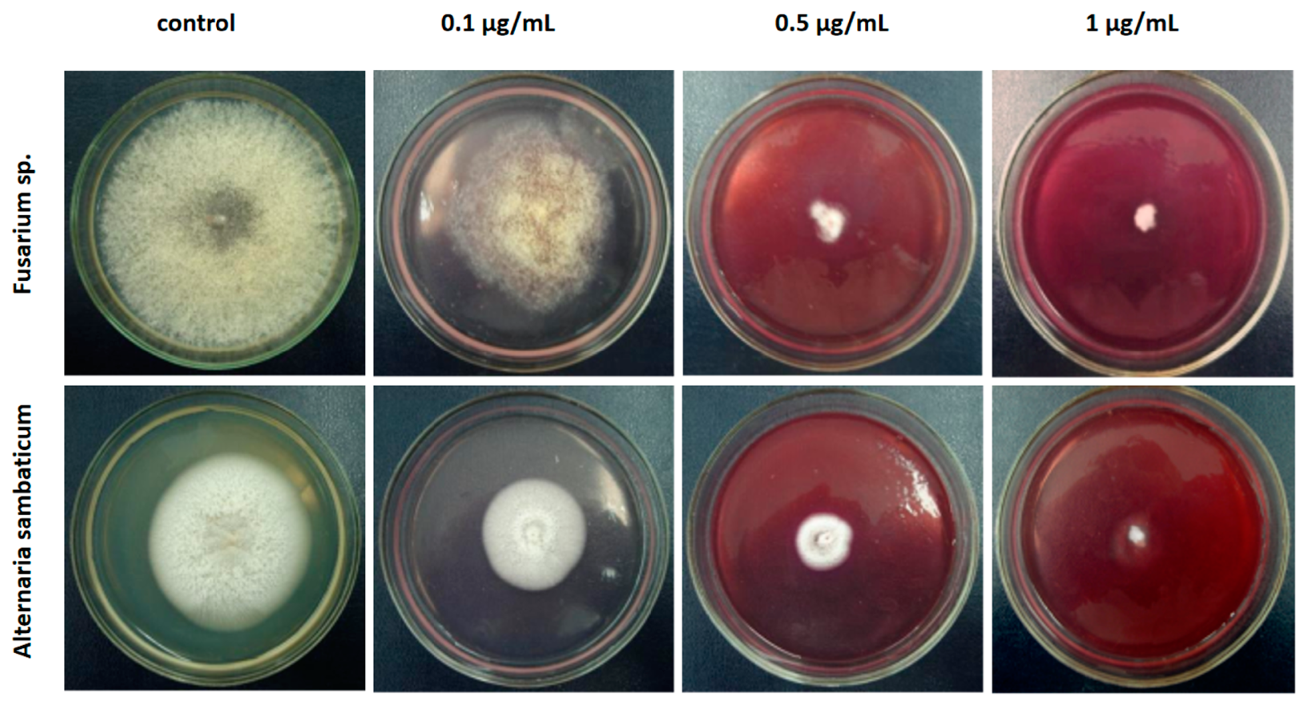
2.3. Antifungal Activity of Prodigiosin

Prodigiosin exhibits antifungal activity against a range of fungal pathogens, including dermatophytes and plant pathogenic fungi. For instance, Lapenda et al. reported that prodigiosin effectively inhibits the spores of dermatophytic fungi, such as Microsporum cookie (MIC = 2.3 μg/mL), M. ajelloi (MIC = 2.3 μg/mL) and Trichophyton longfeuseus (MIC = 8.1 lg/mL) [29]. Sumathi et al. demonstrated the effect of prodigiosin against various fungi, namely, Aspergillus niger (MTCC 281), Trichoderma viridae (MTCC 167), Penicillium chrysogenum (MTCC 160), Microsporum canis (MTCC 2820), Candida albicans (MTCC 183), Fusarium moniliforme (MTCC 156), Trichophyton rubrum (MTCC 296), Trichophyton mentagrophytes (ATCC 9533), Fusarium oxyzporum (ATCC 695) and Aspergillus flavus (ATCC 10836) [53]. Antifungal activity of the PG crude extract was determined by using the standard microdilution method CLSI M38-A. Similarly, Okamoto et al. highlighted the effectiveness of purified PG against Phytophthora capsici, Cochliobolus miyabeanus, Pythium spinosum and P. ultimum, significant plant pathogens, indicating its potential utility in agricultural applications [54]. Its strong inhibitory effect of purified PG on plant pathogenic fungi Alternaria and Fusarium we were also demonstrated previously (Figure 3).
Alijani et al. observed that PG’s antifungal effects are closely linked to its ability to disrupt cellular membranes and induce oxidative stress in fungal cells, thereby compromising their viability [55]. Woodhams et al. emphasized the potential of PG as a bioactive compound in treating fungal infections, highlighting its efficacy against Batrachochytrium dendrobatidis, a pathogen that affects amphibians [56].
Thus, the broad spectrum of antifungal activity suggests that PG could serve as a promising candidate for developing new antifungal agents without development of resistance to conventional antifungal therapies.

2.4. Antiparasitic Activity of Prodigiosin

Antimalarial action of PG was firstly documented in 60th by Castro [57] and after a break, interest in this compound as an antimalarial drug was renewed. PG and its derivatives—natural and synthetic prodiginines—were analyzed for their in vitro antimalarial activity against Plasmodium falciparum D6 with chloroquine as a reference drug [58]. Comparative evaluation of the antimalarial activity of natural PG, undecylprodiginine, metacycloprodiginine and streptorubin B revealed exceptionally potent activity with very low IC50 values (8 nM, 7.7 nM, 1.7 nM and 7.8 nM, respectively) against P. falciparum D6. Moreover, all these compounds were even slightly more active than chloroquine.
An in vitro antimalarial activity of heptyl PG similar to that of quinine was found against the chloroquine-sensitive strain P. falciparum 3D7 in the study of Lazaro et al. [59]. Low toxicity was confirmed by results showing a twenty-fold increase in the inhibitory effect of heptyl PG on malarial plasmodia compared to mouse lymphocytes, which suggests the potential of this compound in the development of a drug for the treatment of malaria. In vivo studies with single subcutaneous administration of 5 and 20 mg/kg demonstrated a significant increase in the survival of mice infected with P. berghei ANKA, but caused sclerotic lesions at the injection site. In another study, synthetic PG-like compounds exhibited antiplasmodial activity in vivo at low nanomolar concentrations against a panel of Plasmodium falciparum with a great therapeutic index [60]. The oral route of drugs administration was used in a murine model and demonstrated curative in vivo efficacy against erythrocytic P. yoelii at 25 mg/kg after 4 days. However, no overt clinical toxicity or behavioral changes were observed in any of the mice given prodiginines and tambjamins. Thus, in addition to selecting an effective compound and modifying it, it is important to select the route of administration of the drug to prevent side effects.
PG and obatoclax were studied in a comprehensive study of antiparasitic activity against unicellular and multicellular parasitic organisms causing the most common infectious diseases. These compounds demonstrated activity against Trypanosoma brucei in low concentrations (EC50 0.03 μM for PG), as well as against Schistosoma mansoni resulting in significant damage to the tegument, with numerous areas of blebbing and separation from the worm body (Figure 4) [61].
Both PG and obatoclax inhibited Giardia strains with good potency (EC50s 3.8 and 0.9 μM, respectively) and were effective in terms of the rate of suppression of the growth of Entamoeba histolytica. PG had the highest rate of suppression of Entamoeba in the study compared to other drugs, including metronidazole. The kinetics of the rate of suppression of parasitic organisms is very important in determining the effectiveness of drugs in terms of reducing the time of recovery [62]. These compounds are not structurally related to metronidazole, the standard drug for treating amebiasis [62], and are therefore effective against metronidazole-resistant strains. In addition, PG and obatoclax may be effective in treating metronidazole-resistant giardiasis [63].
PG also demonstrated the bioactivity towards plant-parasitic nematodes at their juvenile stage as well as nematode egg-hatching ability. Pigment was found effective against juvenile stages of Radopholus similis and Meloidogyne javanica at low concentrations (LC50 values, 83 and 79 μg/mL, respectively) as compared with a positive control of copper sulphate (LC50 values, 380 and 280 μg/mL, respectively) [64].
Thus, PG and obatoclax, due to their effectiveness against a broad spectrum of parasites, are consistent with the approach “one drug multiple-bug” approach that is most likely to be effective especially in developing countries where disease burden is high and resources are limited [65].

2.5. Prodigiosin as an Anticancer Drug: Mechanisms of Action and Prospects

PG exhibits a multifaceted mechanism of action, influencing both cancer cells apoptosis and immune responses [66]. Promising area for the use of PG pigment is associated with its ability to suppress the growth of cancer cells, while having a slight negative effect on healthy tissue. This selective cytotoxic activity against malignant cells makes PG an attractive candidate for the development of a pharmaceutical drug for anticancer therapy (Figure 5) [67]. The mechanism of action of the PG molecule leading to the suppression of cancer cells has not been fully studied. Currently, the main theory of the anticancer action of PG is its ability to induce apoptosis in cancer cells [68].
To date, numerous studies have been conducted on the ability of PG to suppress the growth of cancer cells in vitro and inhibit the development of tumors in vivo [6,69]. Anticancer activity of PG was reported for approximately 60 cancer cell lines [70], mediated by cell-specific mechanism, viz., p53 tumor suppressor activity (colorectal cancer stem cells) [71], inhibition of Wnt/β-catenin (breast cancer cells) and reduction of cyclin D1 levels (breast cancer cells) [6]. Selective anticancer activity was reported against melanoma [72] and liver cancer cell lines due to apoptosis induction by facilitating Cu-dependent DNA damage or alterations of membrane H+ transporters (H+-ATPase) activity and endoplasmic reticulum stress [73]. The mechanism of action includes the development of mitochondrial dysfunction and ATP depletion in the cancer cell [69]. Obatoclax, a PG-based drug, has been shown to be an antagonist of the BCL-2 family of proteins, which are overexpressed in many types of cancer [18].
One of the critical pathways through which PG exerts its effects is the modulation of the p53 signaling pathway. Research has demonstrated that PG can restore the function of mutant p53 by upregulating p73 and disrupting the interaction between mutant p53 and p73, leading to enhanced antitumor effects [71]. This restoration of p53 signaling is particularly relevant in cancer cells harboring p53 mutations, which are prevalent in various malignancies. The ability of PG to target these mutant p53 pathways distinguishes it from other agents that only disrupt p73 interactions without restoring p53 function [71].
Studies indicate that PG alters the expression of apoptosis-related genes, leading to increased apoptosis and reduced cell viability of colorectal cancer cells [74]. The PG’s ability to induce apoptosis is attributed to its pro-apoptotic properties, which have been observed across multiple cancer types, suggesting its potential as a broad-spectrum anticancer agent [75]. Moreover, PG’s immunomodulatory effects are becoming increasingly recognized. It has been reported that PG exhibits immunosuppressive activities, which may be beneficial in certain contexts, such as preventing excessive immune responses that can lead to tissue damage during cancer therapy [66]. The safety profile of PG, particularly its lack of genotoxicity in vivo and in vitro [1], further supports its potential application in cancer treatment [66].
Recent studies have also explored the synergistic effects of PG when combined with other therapeutic agents. For instance, its combination with HSP90 inhibitors has shown promise in targeting multiple oncoproteins and suppressing tumor progression in triple-negative breast cancer [76]. This combination therapy approach highlights the versatility of PG as a component of multimodal cancer treatment strategies.

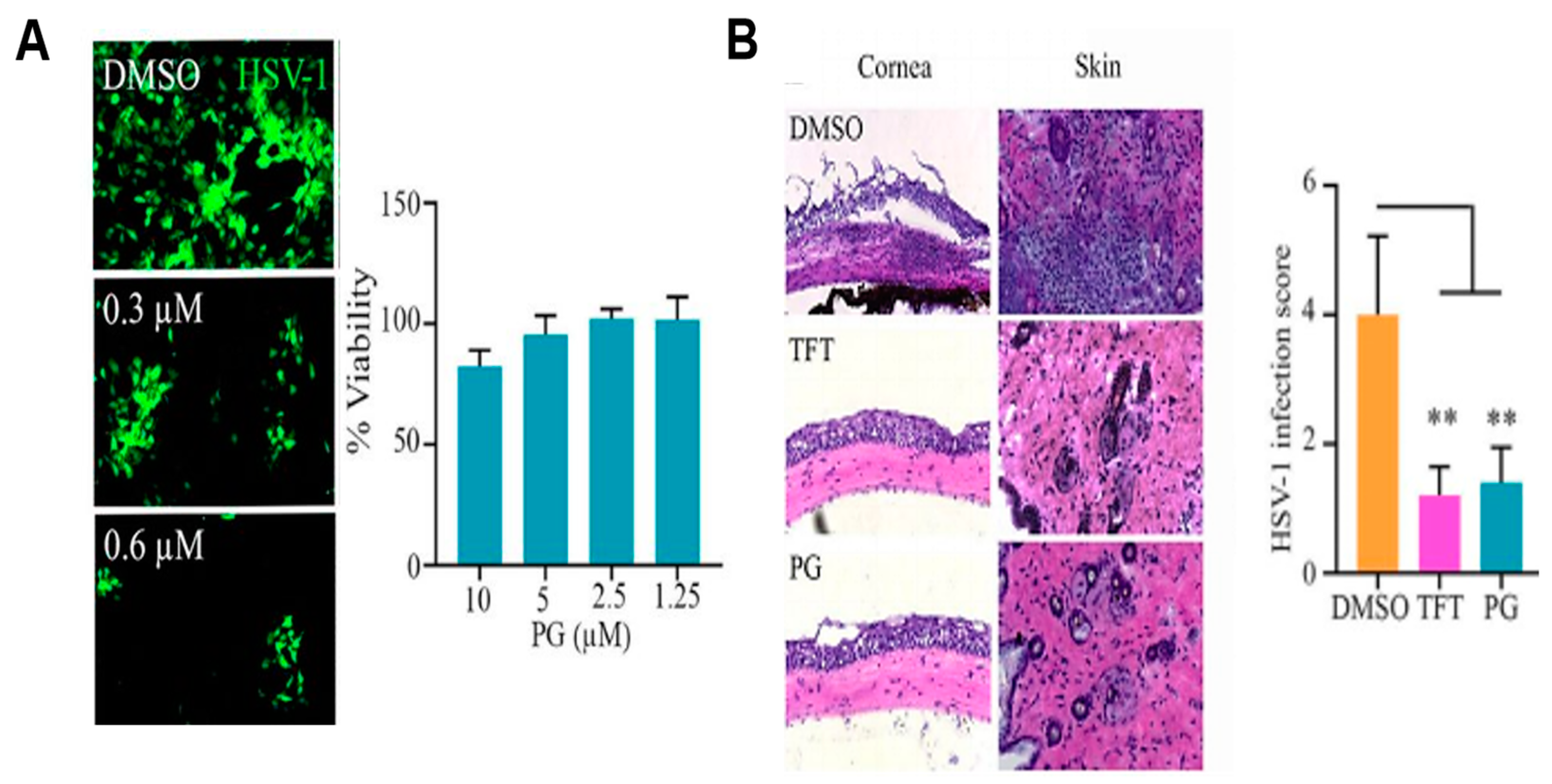
2.6. Antiviral Activity of PG

PG exhibits significant antiviral activity against various viruses, including the Bombyx mori nucleopolyhedrovirus (BmNPV) and herpes simplex virus (HSV), SARS-CoV-2. The mechanisms underlying its antiviral effects involve selective targeting of virus-infected cells, inhibition of viral gene transcription and interference with viral replication processes. In vitro studies have shown that PG can selectively kill BmNPV-infected cells, significantly inhibiting viral replication at early stages. This is achieved through the inhibition of viral gene transcription, specifically targeting early genes such as ie-1, and preventing virus-mediated membrane fusion, which is crucial for viral entry into host cells (Figure 6) [77,78].
The ability of PG to induce selective cytotoxicity in infected cells while sparing healthy cells highlights its potential as a therapeutic agent in antiviral strategies [79]. PG has been shown to exhibit inhibitory effects against Enterovirus 71, with studies indicating an IC50 value of 0.5112 μg/mL, demonstrating its potential as an antiviral agent against this pathogen [80]. PG’s ability to interfere with viral RNA synthesis has also been documented, further supporting its role as a potent antiviral agent [78]. In this study, PG exhibits high potency in inhibiting HSV viral replication and an inhibitory effect against HSV-1 ocular infection and pathogenesis in mice. By targeting cell signaling pathways such as NF-κB and Akt, PG establishes an antiviral state through inhibition of pro-survival pathways (Figure 6).
Another study demonstrated the antiviral activity of a PG derivative, obatoclax, against alphaviruses [81]. Authors postulated the mechanism of the antiviral action of obatoclax as inhibition of virus–cell fusion, mediated by changes in endosomal pH. Further antiviral studies of SARS-CoV-2 with obatoclax showed inhibition of replication in epithelial cell cultures, and the mechanism of inhibition was also associated with acidification of endosomes and disruption of the activity of furin and cathepsin [15,78].

3. Prodigiosin for Industrial Biotechnology

Industrial biotechnology is a developed branch of the industry that uses the potential of biological compounds to create various products such as chemicals, materials, food and beverages, vitamins, biofuels and bio drugs. In addition to using living organisms as producers of bioactive compounds, industrial biotechnology involves the use of bioactive compounds as additives that improve technological properties and increase the functionality of the final product. As the field of industrial biotechnology continues to expand and evolve, researchers are constantly searching for new and innovative ways to harness the power of nature for sustainable manufacturing processes. PG is a versatile compound with a wide range of applications in biotechnology. From food preservation to plastic biodegradability, soap formulations and antifouling coatings, PG has the potential to improve multiple industries.

3.1. Prodigiosin as Industrial Colorant and Antibacterial Agent for Plastic, Textile and Cellulose Materials

Traditionally, the textile industry uses synthetic dyes, many of which pose a serious threat to aquatic fauna and flora as xenobiotics and pollutants. The synthetic dyes and their effluents are recalcitrant to microbial degradation which results in accumulation with toxic and potentially carcinogenic action for the people of present and future generations [82,83]. Natural dyes are becoming increasingly popular because of their better environmental compatibility and biodegradability and low negative impact on human health unlike synthetic dyes [84].
Among natural dyes, brightly colored secondary metabolites of microorganisms are the most attractive due to their economic benefits [85]. Biotechnological production using microorganisms does not depend on geography or climate, and in total seasonal production issues, has well-developed technical support and high productivity. In addition, microbial pigments can be relatively easily isolated and purified. In this regard, microbial pigments are considered as a promising alternative not only to synthetic dyes, but also to other biopigments derived from plants or animals. As a natural colorant, PG gained more attention because of accompanying antibacterial, antifungal and antiprotozoan effects that can give additional functionalities. Along with this, PG did not demonstrate genotoxicity and mutagenicity [1]. PG has been shown to have great potential for application in various fields such as the food [86], cosmetic [87,88] and textile industries [89]. PG extracted from Serratia sp. was used for polymethyl methacrylate sheets (PMMA), rubber latex dyeing [90] and polyolefines [91]. In addition, PG has been applied as a natural colorant of paper, candles, soap and as biodegradable ink [88]. In the food industry, the pigment PG can be useful due to its antioxidant and antimicrobial properties [92].
PG was used as a thick suspension-concentrate in the technology of the polyethylene dyeing and the ultrathene in granules [91]. Due to the high colorfulness (chroma and saturation) of pigment, its content in the coloring polymer was very low (mg % mass) than in the case application to synthetic organic dyes utilized in the chemical industry. This results in its minimal influence on the technological properties of the dyeing concentrate and masterbatches. Authors noticed that the reported method for the coloration of polyolefins is more effective with minimal environmental impact than now-existing methods in application to organic dyes. Finally, this method of the coloration of polyethylene by PG allowed the consumer goods to comply with the technological standard [91].
Poly(methyl methacrylate) or poly methyl 2-methylpropenoate have been widely utilized for the construction of lenses for the exterior lights of automobiles, designing advertising signs, manufacturing aquariums, cornices, greenhouses, plastic optical fiber used for short-distance communication etc. Methanol solution of red pigment isolated from Serratia sp. BTWJ8 identified as PG (40 µg/L) was used as the stock solution for imparting color to polymethyl methacrylate sheets and different types of paper. The different color shades can be produced by varying the concentration of PG in methacrylate and rubber latex sheets [90]. The results obtained for the studies conducted with PMMA, rubber latex and pH indicator paper demonstrated probable scope for exploiting PG as a natural dye for this material. Unfortunately, the presented study did not analyze the durability of PG-pigmented compositions under the influence of solar radiation. The autofluorescence of PGs has only been rarely used as a biophysical parameter for the detection and assessment of PG despite the remarkable pH-dependent color change of the pigment [93].
The textile industry is one of the significant elements of general environmental pollution due to the use of solvents and dyes that can negatively affect biological objects. In this regard, solutions are needed to replace environmentally hazardous dyes with bio dyes and solvents with a high level of biocompatibility [94]. PG and PG-like pigments can impart color to different textile materials such as polyester [89], polyacrylonitrile, nylon 66, wool [95], silk [96] and cotton [97] fabrics to obtain a pretty deep-colored shade. Due to the inability of the PG molecule to form a hydrogen bond with the hydroxyl group of the cellulose molecule, synthetic fabrics had a higher affinity to this pigment compared to cotton fabrics as it can be observed when comparing different types of fabrics after dyeing (Figure 7) [98]. Although, cotton fabric was successfully dyed with the PGs nanomicelles in the work of Ren et al. [97] and the optimum dyeing conditions were analyzed. Authors revealed that dyed cotton possesses good rubbing, washing and perspiration color fastness. Moreover, endowing of dyed cotton with an antibacterial property against S. aureus and E. coli was demonstrated.
Therefore, fabrics can be endowed with added values when the PGs are used as dyestuff [89]. Thus, the antimicrobial activity of PG, demonstrated by a number of authors [29,99], is a prerequisite for the use of the pigment in the textile industry to create functional fabrics with a bactericidal effect. PGs also represent the great interest as fabric dyestuff due to its antileishmanial properties [100].
Antibacterial fabrics with PG were obtained and tested in many research works and some of the dyed fabrics samples exhibited sufficient antibacterial activity [96,101]. Alihosseini et al. showed antibacterial activity against S. aureus sp. and E. coli sp. within a contact time of 16 h for silk and wool fabrics dyed with PGs. The authors mentioned that PGs have characteristics similar to those of ionic and disperse dyes, which is consistent with the identified structures. Authors revealed that heating of the dyeing solution leads to the 15% reduction of its content, thus PG is not highly stable in acidic conditions at elevating temperatures [101].
Replacing methanol or ethyl alcohol with ethyl acetate or N,N-dimethyl formamide as a solvent for PG can improve its uptake by wool fiber. A number of advantages were demonstrated when using these compounds: PG has good solubility in ethyl acetate and N,N-dimethyl formamide, this solvent has a certain affinity to wool fiber, they form an adsorption layer at the wool surface plasticizing of wool fiber, thereby facilitating diffusion and improving the uptake rate of PG. As a result, the use of pigment composition with ethyl acetate or N,N-diethylformamide allows pigment and solvent to effectively disperse and does not require high temperatures. Moreover, wool fabric dyed with the above-described composition demonstrated the excellent washing rubbing fastness and bacteriostatic effect [95].
The values of temperature and pH are the most important parameters for textile dyeing with PGs [89]. The color of PG is pH-sensitive and it presents purplish red under acidic and neutral conditions, while orange-yellow under an alkaline condition [102,103] and possesses UV-resistant properties [5]. In addition, PG in different concentrations produces different color shades depending on the composition of the textile. In the case of silk and polyester fabrics dyeing, the color changed from purple to crimson as the pH increased in a dye bath. The manifestation of UV protective properties is mainly related to the structure of the pigments that bind to the fibers during dyeing.
PG application in textile dyeing is restricted by the main inherent shortages mentioned below. The first restriction lies in PG’s low solubility in water because it belongs to a group of intracellular pigments and demands organic solvent for the extraction from bacterial biomass, so textile dyeing with that is not absolutely clean and eco-friendly. Therefore, many works are aimed at developing dyeing technology with PGs without using expensive and harmful solvents and the optimum dyeing conditions were revealed.
To increase pigment extraction and more effective coloring, surfactants and nanomicelles were used [89,96]. For the isolation of PG, Tween 80 was added in the cultural medium, which was beneficial for the migration of pigments from bacterial cells. The PG wrapped into the micelles of Tween 80 colored silk in intense color and endowed it with good antibacterial properties against S. aureus [89]. In addition, dyed silk demonstrated an absence of cytotoxicity in MTT assay with L929 cells [96].
Applying natural colorants for textile dyeing usually involves disadvantages with lower fastness properties and a limited shade range of the dyed textiles over synthetic dyes [104]. Various mordants such as salts (FeSO4, CuSO4), baking soda and lemon juice can cause an intensity increase of the shade or significantly change the final color of the dyed sample, also to increase the resistance of the bacterial pigment to UV and temperature (Figure 8) [98].
PG dyeing parameters were optimized for jute fabric using tannic acid as a pre-mordant to enhance the color fastness. The antimicrobial potential PG-dyed jute fabric was tested against E. coli, S. aureus, P. aeruginosa, K. pneumoniae and E. faecalis; among these, the maximum zone of inhibition was observed for P. aeruginosa [105].
The high cost of purified PG creates the need to find alternative solutions both in the search for cheap substrates like agricultural wastes for large-scale cultivation of the producer strain and in simplifying pigment extraction [19,106]. Streptomyces strains are being considered as promising candidates for the biotechnological production of bio-pigments applicable in the textile industry.
The mechanism underlying PG’s antibacterial activity remains poorly explained [40] but it is known that it depends on the presence of functional groups and, accordingly, on the molecular conformation. For example, some bacteria and fungi were approximately 1.5-fold more sensitive to cycloprodigiosin than to PG [107].
Theoretically, the fabrics impregnated with PG can be applied in filters, dressings, antiseptic wipes, etc., where the pH-dependent color change can indicate the physiological state of the object/environment. Fabrics that harbor PG can suppress the bacteria that cause unfavorable odors. Arivizhivendhan et al. investigated the prospects for using PG as an active component of a bactericidal filter. This study was focused on the disinfection of water on a pathogenic bacteria route using a PG-impregnated cellulose column reactor (PICCR) [108]. Removal efficiency of model pathogens (antibiotic-resistant Escherichia coli and Bacillus cereus) was evaluated by the pour plate method, regrowth ability in nutrient broth and quantitative estimation of live and dead cells using fluorescent microscopy. As a result, the passing of pathogen-contaminated water through a PICCR resulted in a reduction of E. coli by 97.31% and B. cereus by 97.33%. The analysis of the residual protein and nucleic acid in the treated water using UV-visible spectroscopy confirmed the bacterial cell damage by the antibacterial activity of the PICCR. Thus, the proposed PICCR was found to be effective for the removal of Gram-positive and Gram-negative pathogens from water and this may be regarded as a viable purification technique for drinking water without the periodic addition of disinfecting chemicals into the secondary biologically treated wastewater.
Thus, the proposed PICCR was found to be effective for the removal of Gram-positive and Gram-negative pathogens from water and this may be regarded as a viable purification technique for drinking water [108]. Further, to explore the potential of prodigiosin as a water disinfectant, Arivizhivendhan et al. synthesized a reusable bioactive PG-conjugated iron-oxide-activated carbon composite ([Ac]F@Fe3O4–PG) matrix with long-term antibacterial activity with efficient inhibition of biofilm formation. It was found that the mechanism of bacterial disinfection with the [Ac]F@Fe3O4–PG matrix occurs via disruption of the surface charge of the bacterial cell membrane, thereby affecting the transport system of the cells. Antibacterial events also included the generation of reactive oxygen species which further triggered programmed bacterial cell death with apoptosis-like changes in cellular morphology [109].
It has been reported that a bacterial cellulose (BC) films, obtained from a microbial consortium of bacteria and yeast species, were functionalized with the bacterial pigment PG, produced by Serratia plymuthica and flexirubin-type pigment, from Chryseobacterium shigense, which exhibit a wide range of biological properties. The BC samples functionalized with PG presented antibacterial activity and were able to inhibit the growth of pathogenic bacteria Staphylococcus aureus and Pseudomonas aeruginosa [110].
In a novel study, similar bacterial cellulose membranes (BCMs) synthesized using Komagataeibacter intermedius were produced and loaded with PG to impart antimicrobial properties to enhance the capacity of BCMs for biomedical utilization [111].
The success of modification in PG-modified BCMs has been demonstrated by FTIR and SEM. This material has shown excellent growth inhibition and antiadhesive effect and was observed for the tested microorganisms (Gram-positive and Gram-negative and yeasts) with the inhibition rates of 82.05–96.25%. Moreover, PG-functionalized BCMs demonstrated a high level of biocompatibility towards the L929 cell line. Thus, there are a number of prerequisites for the use of PG in various industries due to its valuable technological properties; however, for mass consumption, it is necessary to search for productive bacterial or fungi strains growing on cheap nutrient media, develop the most effective methods for pigment extraction and a more detailed study of the biocompatibility of products containing pigment should be conducted.

3.2. Prodigiosin in Cosmetics

The cosmetics industry is an expeditiously emerging global business market, with its growth increasing the number of pigments used in cosmetic products. The global cosmetic pigments market size was valued at USD 700 million in 2022 and is projected to hit around USD 1532.31 billion by 2032 [112].
Synthetic colorants are currently the most popular compared to natural ones because they demonstrate excellent stability while offering a wide range of shades. A colored cosmetic substance is usually not multifunctional. Cosmetic pigments are responsible for the color and texture of cosmetic products and in some cases to cover imperfections and uneven skin tone. Some of the cosmetic pigments are toxic and even carcinogenic [113,114], which, however, does not prevent the development of microorganisms in cosmetic products rich in various nutrient substrates [115].
PG as a color additive is more functional than conventional cosmetic colorants due to their biological functional attributes. This bacterial pigment can be considered as a UV protectant [5,45,46] and, by inhibiting the development of bacteria, help reduce unpleasant odors and increase the shelf life of a cosmetic product.
Cosmetics most often contain lipids or lipophilic compounds, thus lipophility of PG makes the bacterial pigment applicable for the cosmetic industry. In cosmetology, pigment properties such as low solubility in water, changes in shades (from pink to orange and red) as well as the saturation depending on pH and fluorescence can be useful. Until now, there have been few studies devoted to the cosmetic use of prodigiosin. The stability of PG at different values of pH and NaCl concentrations confirm its high potential as a green alternative to synthetic dyes in soap coloring (Figure 9) [88].
To color the soap, the authors used a molding method with the addition of a specified amount of pigment and demonstrated excellent color stability when exposed to different salinity levels. Overall, lower toxicity level and frequency of allergic reactions in the case of using natural pigments make them more suitable as colorants by reducing exposure to harmful compounds; however, in the presented work there, the authors did not perform the analysis of either antimicrobial properties or toxic effects [88].
The bacterial pigment PG along with violacein were used as an additive to Aloe vera leaf and Cucumis sativus (cucumber) fruit extracts and commercial sunscreens to improve photoprotection properties [87]. When adding bacterial pigments, an order of magnitude increase in the values of sun protection factors of plant extracts was shown (i.e., to ~3.5), and for commercial sunscreens, it increased by 20–65% (for 4% w/w of PG). The antioxidant activities of PG was approximately 30% from those of ascorbic acid (a well-characterized, potent antioxidant).
Antimicrobial activity assay carried out in this work showed that PG was effective against S. aureus and E. coli, with IC50 values 0.68 ± 0.06 μM and 0.53 ± 0.03 μM, respectively. The presented data allow the conclusion that PG exhibited antioxidant and antimicrobial activities and were able to increase the SPF of commercial sunscreens as well as plant extracts [87].

3.3. Prodigiosin in the Food Industry

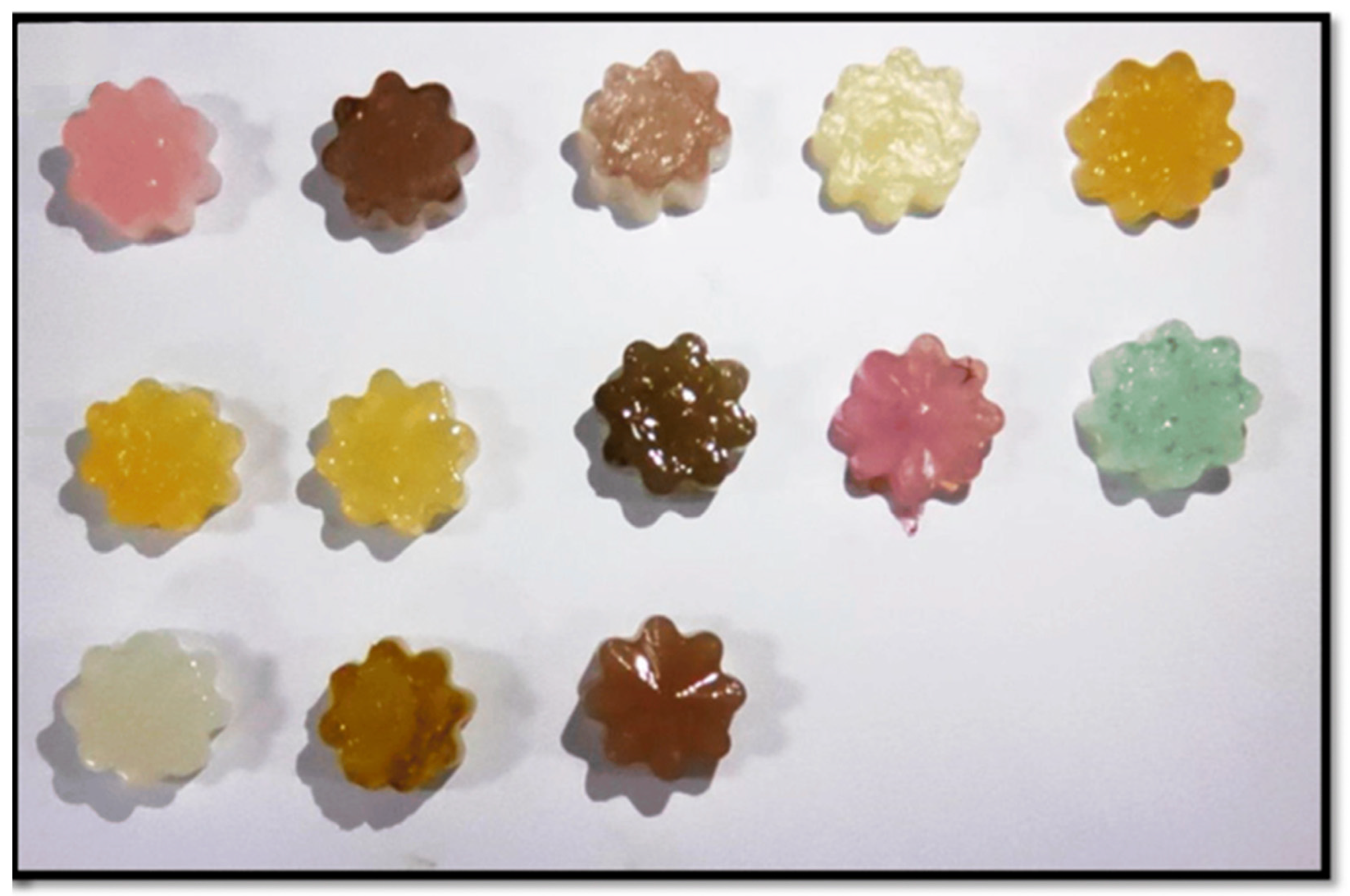
3.3.1. Prodigiosin as Food Colorant

In the food industry, Serratia strains are often considered as one of the microflora representatives that causes food spoilage. The detection of these microorganisms is often associated with the release of a pigment that binds persistently to nutrients and turns the product bright red. Growth of PG-producing microorganisms is accompanied by an inhibition of technologically valuable industrial microflora [116] and harming food production. However, some valuable properties of PG can be used in the food industry to impart a bright color to certain products and extend their shelf life due to antibacterial effectiveness.
Marine-derived bacterially produced bio-pigments (bpBPs) including PG can be utilized as food-grade pigments due to their low impact on consumer health. The high color saturation of these pigments allows them to be used in low concentrations, which further reduces the risk of their use. Pyorubrin and pyocyanin, for example, gave pleasing colors at 25 mg mLG−1 [117]. bpBPs was also suggested as an additive for ornamental fish feed-stuff to promote their growth and enhance the coloration [118]. Additionally, PG (from the marine bacterium Zooshikella sp.) has been reported to exhibit good coloring properties and a shelf life of three months [83], indicating the sustainable aspect of using marine-derived pigmented molecules as food colorants [119].
The antioxidant potential of PG in meat extract powder (MEP) as a model food material was examined by 2,2-diphenyl-1-picrylhydrazyl (DPPH) and 2,2′-azino-bis 3-ethylbenzthiazoline-6-sulfonic acid (ABTS) radical scavenging method. The results showed that the PG at the concentration of 10 mg/L completely scavenged the DPPH and ABTS radicals in MEP and inhibited growth foodborne pathogens in food stuff. This work also showed that the addition of PG can extend the shelf life of food products by almost twice that of a chemical preservative. Hence, PG can be used as a preservative due to its effective antioxidant properties and antimicrobial activity and also imparts an aesthetically pleasing color to food products [92].
Crude-concentrated PG and PG-like pigments extracted from marine bacteria were investigated as food colorants for agar jellies [86]. Agar jellies (5% agar) were prepared using various pigment extracts at a concentration of 500 μg, and after molding and cooling, the solidified jellies were stored at 4 °C for several months to see the ability of the pigment to persist over time (Figure 10) [86].
Although PG holds promise as a natural preservative and colorant in food production, it is important to consider safety aspects before the widespread adoption of this technology. Research into the toxicity and regulatory approval of prodigiosin in foods is still not as active, and more research is needed to fully evaluate its safety profile.

3.3.2. Prodigiosin as a Functional Additive for Food Packaging

Antimicrobial activity is one of the key properties of PG. This makes it an ideal candidate for use in packaging materials, as it can help extend the shelf life of food products by inhibiting the growth of harmful bacteria. The concentration of pigment in foods can be kept low to significantly inhibit the development of spoilage microorganisms, but using PG in packaging can extend the shelf life of foods without adding preservatives. The incorporation of antimicrobial compounds into food packaging biopolymers can promote a sustainable way to reduce food spoilage and can suppress the development of spoilage microorganisms and accordingly increase the shelf life of products into biodegradable packaging with incorporated bio-based antimicrobial agents [120,121].
Short-term use plastic packaging puts a strain on the environment due to low recycling rates [122] and poses a threat to aquatic life; in the form of microplastics, it penetrates all living environments, enters the food chain and ultimately poses a threat to all living organisms on earth. Natural polymers can be utilized as a replacement for plastics made from fossil fuels due to their safety and biodegradability. Such biopolymers can be used to create safe packaging that, unlike synthetic ones, does not form microplastics when decomposed by UV radiation and physical conditions [121,123,124].
As mentioned above, PG has good compatibility not only with polyesters and polyolefins, but also with natural biodegradable polymers such as cellulose and agar. PG in food packaging could theoretically play the role of colorant, antioxidant, ultraviolet protector and freshness indicator. There are only a few studies on PG as a food packaging additive. Thus, in the work of Amorim et al., PG was used for the functionalization of polymers to fabricate environmentally friendly germicidal packaging [121].
To create eco-friendly packaging, pigment was added to a biodegradable composite using two different strategies (Figure 11) [121]. The mechanical characteristics and water vapor transmission rate of the studied composite showed its suitability for fresh food packaging. PG in the outer layer of the material imparted a bright color, which is an additional advantage in terms of attracting consumer attention. Studies were also carried out on the antibacterial properties of the developed packaging, and its activity against Staphyloccus aureus and Pseudomonads aeruginosa was shown. Authors concluded that the addition of PG to the inner layer of the packaging material leads to the suppression of product spoilage associated with its own microflora, while in the outer layer PG helps prevent external microflora from entering the product.
One approach to replacing plastic with more environmentally friendly packaging options is to create paper packaging. Despite the many disadvantages of such packaging associated with the energy-intensive technological process, paper is currently the most widely used material for eco-friendly packaging. The demonstrated effectiveness of cellulose modification with PG makes it possible to create modified paper. Its use as food packaging will increase food resistance to spoilage due to the antimicrobial effects of the pigment. PG was used as dyeing staff for different types of paper. It was demonstrated that PG-dyed paper can be used as a pH indicator due to its ability to change color in acidic (pH 2.0), neutral (pH 7.0) and alkaline (pH 10.0) solutions [90]. Color change of PG may serve as an indicator of freshness or changes associated with microbiological or oxidative spoilage of foods.
A significant breakthrough was realized when PG was encapsulated with polysaccharides, resulting in heightened water solubility and an expansion of its potential applications within the food industry [19].
Thus, PG and PG-like pigments can act as dyes for food products and packaging materials, helping to preserve the freshness of food products and increase their shelf life, which is due to the pronounced antioxidant and antibacterial properties of these pigments. Incorporation of PG into food packaging materials may be more promising in terms of its antibacterial effect if loaded into nanomaterials for slow, continuous release.

3.4. Prodigiosin as Antifouling Agent

In today’s world, the issue of marine biofouling has become a significant concern for industries that operate in the maritime environment. Biofouling on ships, such as dreadnoughts, has been shown to increase the roughness of the hull, leading to higher frictional resistance. This increase in resistance ultimately results in higher fuel consumption and a need for greater environmental compliance measures. Antifoulant substances were developed to prevent and reduce biofouling of surfaces in seawater. Traditionally, heavy metal-based antifoulants have been used to combat biofouling on marine vessels. However, these antifoulants come with severe environmental complications. The most popular antifouling agent—tri-n-butyl tin—and many another widely used compositions, are currently facing a complete global prohibition due to environmental incompatibility [125]. The release of heavy metals into the marine environment can have detrimental effects on marine life and ecosystems. In response to these environmental concerns, there is a growing need for the development of “eco-friendly” antifouling agents that can effectively combat biofouling without causing harm to the environment [119].
Marine bacterial pigments have been identified as potential antifoulants. For example, polymelanin, synthesized by the marine bacterium P. lipolytica sp., has been shown to prevent metamorphosis and decrease the settlement of invertebrate larvae. This demonstrates the diverse range of marine bacterial pigments that can be harnessed for their antifouling properties [119].
Natural antifoulants, including bacterial bioactive metabolites [126], have been proposed as one of the best replacement options for toxic metal-containing antifoulants. The anti-biofilm activity of PG against Staphylococcus aureus, drug-resistant P. aeruginosa [26] associated with suppression of the production of pyocyanin and extracellular polysaccharides in vitro and the hydrophobicity of PG make it possible to create anti-fouling coatings that are resistant to leaching in seawater. PG, extracted from Serratia marcescens CMST 07, was reported to exhibit antifouling activity against marine fouling bacterial species such as Gallionella sp. and Alteromonas sp. The pigment also inhibited the adhesion of Cyanobacterium sp. on the glass surface and formation of Bacillus sp. and Pseudomonas sp. biofilms [14].

3.5. Prodigiosin as a Component of Dye-Sensitized Solar Cells

In recent years, bacterial pigments have managed to play a major role in the development of microbial fuel cells [127] and more recently in dye-sensitized solar cells (DSSCs) [128]. The chemical richness of PG, characterized by pi electrons and electron donor pyrrolic nitrogens, makes it an ideal candidate for solar cell applications. This bacterial pigment can form coordination compounds with metal ions and facilitate electron transfer processes essential for generating a short-circuit current in solar cells [46,129]. Furthermore, the ability to overproduce PG using agro-industrial residues [130] presents a cost-effective and sustainable approach to incorporating this pigment in dye-sensitized solar cells. PG is capable of interacting with metal ions to form coordination compounds. This ability allows PG to transfer electrons from its photo-excited state to the conduction band of metal oxides, generating a current. This process is similar to what occurs in many natural pigments, making PG a natural fit for use in DSSCs [131,132]. Recent research of Hernández-Velasco et al. has resulted in the optimization and overproduction of PG using agro-industrial residues. This breakthrough not only makes PG more readily available for use in DSSCs but also reduces production costs. The overproduction of PG has opened up new possibilities for its application as a sensitizer in DSSCs, paving the way for its potential commercialization. PG has a high level of photostability and competitive efficiency compared to other bacterial pigments. The authors suggest that PG may be considered as an excellent candidate of DSSC sensitizer, obtaining large volumes of pigment due to the rapid fermentation of agro-industrial waste throughout the year, which will avoid serious environmental problems [46]. Resistance to fading is another issue requiring attention in the practical use of PG.

3.6. Prodigiosin as Fuel Marker

Fuel marking is the integration of a unique identifier (marker) in trace quantities into petroleum products at depots before distribution in the market to reveal the facts of counterfeiting, adulteration and fuel smuggling. Fuel markers play a crucial role in ensuring the authenticity and integrity of various fuel products. These markers, specifically designed based on dyes, are added to fuels to provide a unique fingerprint that can be detected using spectrophotometry and chromatography. Fuel markers are necessary to combat fuel fraud, illegal blending and adulteration, which have become prevalent issues in the fuel industry. By adding specific dyes as markers to fuel products, manufacturers can track the movement of their products throughout the supply chain and easily detect any unauthorized tampering. This helps to ensure that consumers receive the high-quality fuel they expect and deserve.
Fuel markers based on dyes provide a cost-effective and efficient solution for monitoring the integrity of fuel products. By utilizing advanced analytical techniques such as spectrophotometry and chromatography, manufacturers can easily distinguish between genuine and counterfeit products. This not only safeguards consumers from potential harm but also protects the interests of legitimate fuel producers. After delivery of the fuel, the match of the marker spectrum in the sample in the field of inspection and taken after marking confirms the brand and source of origin of the product. If after the test no marker is detected or if the concentration is low, it can be sign of adulteration.
PG has shown specificity absorption in a broad range in the visible region (400–700 nm). The optical properties of PG depend on the solvent and, accordingly, the pH value of the solution (protonation/deprotonation of N atoms in pyrrole rings) [19]. In non-polar solvents, the optical spectra of PG will also have different shapes. This factor was used to create a methodology for the labeling of petroleum products [133]. The work used gasoline grades A 76, AI 92, AI 95, AI 98, diesel fuel, aviation gasoline and aviation kerosene. To identify labeled petroleum products, the optical absorption spectrum of fuel was recorded at the wavelength region λ = 350…600 nm. The optical absorption spectrum of the fuel without adding a marker served as a control. Based on the degree of intensity of the spectral lines, comparing them with the intensity of a known standard, the presence and concentration of the marker in the oil products were determined.
By analyzing the PG signature in fuel samples, researchers can quickly identify any contaminants or additives that may compromise fuel integrity. The fuel identification system with PG includes several steps. The first step is to “chemically stamp” the fuel with PG as a marker. Then, the presence of the marker and its concentration can be determined by spectrophotometric methods in the region of λ = 300…600 nm at pH below 7, in the absence of developing reagents [133].
The specificity of absorption spectra allows compounds to be distinguished from one another in a mixture. PG has a specific optical spectrum, the overall appearance of which depends on the pH and type of solvent. The spectral pattern of the pigment was elucidated at pH values of 2, 7 and 9 between 800 and 200 nm, using methanol as the blank. This factor was used to create a methodology for labeling petroleum products. PG has been proposed as a marker for identifying the adulteration of various brands of gasoline (Figure 12) and the marking method was patented. The authors of the patents propose to determine changes in the composition of the fuel by changes in the spectrum of pigment. PG can be used in both one-component and two-component (with developing reagent) safety labeling systems. Developing reagent allows the presence of a marker to be detected by changing the parameters of the material. In the case of PG, the developing factor may be alkalization/acidification of marked fuel samples [133].
PG auto fluorescence was measured at an excitation of 543 nm and an emission of 570 nm [28]. It was revealed that the optical properties of PG depend on the protonation state and not on the solvent permittivity constant [103].
Thus, PG holds great promise as a fuel marker with wide-ranging applications in tracking the consumption of fuels and monitoring of fuel quality as well as the prevention of fuel adulteration.

4. Future Perspectives and Challenges of Prodigiosin Using

By exploiting PG’s attractive properties, such as its antimicrobial and anticancer effects, as well as its vibrant color, scientists can create more sustainable and eco-friendly products that benefit both the environment and human health.
One of the most exciting prospects of PG lies in its potential in cancer treatment. Studies have shown that PG exhibits potent anticancer properties, making it a promising candidate for the development of novel cancer therapies. By targeting cancer cells specifically, PG could change the approaches to cancer treatment in the future. In addition to its potential in cancer treatment, PG also shows promise as an antimicrobial agent. Its ability to inhibit the growth of various pathogens and antioxidative activity makes PG a valuable tool in the fight against infectious diseases. With the rise of antibiotic resistance becoming a global concern, PG can become an effective solution for topical application, in particular for the treatment of infected wounds and as prospective coating.
Despite PG holding great promise for pharmaceutical development, there are a number of challenges that need to be addressed before its full potential can be realized. One of the main obstacles is the limited availability of PG from natural sources. The low yield of this pigment and the use of relatively expensive components of culture media make large-scale production difficult. However, many studies are aimed at creating a cheap nutrient medium based on agricultural waste that provides good pigment yield. In turn, advances in genetic engineering may offer innovative solutions to overcome the limitation associated with low pigment yield. Although PG has demonstrated low toxicity and genotoxicity in some studies, further studies are needed to thoroughly evaluate its safety profile. Moreover, PG in its natural form exhibits low stability, and enhancing the stability of PG is critical to ensure its efficacy and durability in pharmaceutical formulations.

5. Conclusions

In conclusion, it should be noted that PG is unique and multifaceted, which allows researchers to consider this compound as a raw material for the production of various products not only in the pharmaceutical industry, but also in food, cosmetic and many other branches of industry. First of all, PG has great promise as a valuable natural compound in the pharmaceutical field due to its antimicrobial, antiviral and antiprotozoal properties, as well as anticancer effects. One of the most exciting potential uses of PG is its antimicrobial properties. Due to its strong inhibitory effect on a wide range of bacteria and fungi, PG is a promising candidate for the development of a new class of antibiotics that do not cause the development of resistance in pathogens. By targeting bacterial cell membranes, PG offers a unique approach to combating antibiotic-resistant strains and emerging infectious diseases. PG also has potential as an anticancer agent as confirmed in many studies involving cell lines and laboratory animals. The induction of apoptosis in cancer cells upon exposure to PG demonstrates the promise of this compound as a targeted therapy for various types of cancer. In particular, its ability to selectively target cancer cells while sparing healthy cells makes it a valuable tool in the fight against cancer. In addition to its antimicrobial and anticancer properties, PG has also been studied as an anti-inflammatory agent. Beyond its medicinal and industrial applications, PG has also been explored for its potential as a sensing agent. By detecting changes in pH, temperature and other environmental factors, it can be used to develop sensitive and selective sensors for various applications from environmental monitoring and fuel marking to medical diagnostics. Looking ahead, the future prospects of PG seem broad: from its potential in drug development to its use in cosmetics and food coloring, PG’s versatility opens up a lot of possibilities.

Author Contributions

Conceptualization, E.N. and I.G.; methodology, E.N. and I.G.; writing—original draft preparation, E.N. and I.G.; writing—review and editing, E.N. and I.G.; project administration, E.N. All authors have read and agreed to the published version of the manuscript.

Funding

The reported study was funded by the Russian Science Foundation according to the research project No. 23-25-00334.

Data Availability Statement

All data generated or analyzed during this study are included in this published article.

Acknowledgments

This paper has been supported by the Kazan Federal University Strategic Academic Leadership Program (PRIORITY-2030).

Conflicts of Interest

The authors declare no conflicts of interest.

References

  1. Guryanov, I.D.; Karamova, N.S.; Yusupova, D.V.; Gnezdilov, O.I.; Koshkarova, L.A. Bacterial pigment prodigiosin and its genotoxic effect. Russ. J. Bioorg. Chem. 2013, 39, 106–111. [Google Scholar] [CrossRef]
  2. Ravindran, A.; Anishetty, S.; Pennathur, G. Molecular dynamics of the membrane interaction and localisation of prodigiosin. J. Mol. Graph. Model. 2020, 98, 107614. [Google Scholar] [CrossRef] [PubMed]
  3. Li, D.; Liu, J.; Wang, X.; Kong, D.; Du, W.; Li, H.; Hse, C.-Y.; Shupe, T.; Zhou, D.; Zhao, K. Biological Potential and Mechanism of Prodigiosin from Serratia marcescens subsp. lawsoniana in Human Choriocarcinoma and Prostate Cancer Cell Lines. Int. J. Mol. Sci. 2018, 19, 3465. [Google Scholar] [CrossRef]
  4. Williamson, N.R.; Fineran, P.C.; Gristwood, T.; Chawrai, S.R.; Leeper, F.J.; Salmond, G.P.C. Anticancer and immunosuppressive properties of bacterial prodiginines. Future Microbiol. 2007, 2, 605–618. [Google Scholar] [CrossRef]
  5. Stankovic, N.; Radulovic, V.; Petkovic, M.; Vuckovic, I.; Jadranin, M.; Vasiljevic, B.; Nikodinovic-Runic, J. Streptomyces sp. JS520 Produces Exceptionally High Quantities of Undecylprodigiosin with Antibacterial, Antioxidative, and UV-Protective Properties. Appl. Microbiol. Biotechnol. 2012, 96, 1217–1231. [Google Scholar] [CrossRef]
  6. Wang, Z.; Li, B.; Zhou, L.; Yu, S.; Su, Z.; Song, J.; Sun, Q.; Sha, O.; Wang, X.; Jiang, W.; et al. Prodigiosin inhibits Wnt/β-catenin signaling and exerts anticancer activity in breast cancer cells. Proc. Natl. Acad. Sci. USA 2016, 113, 13150–13155. [Google Scholar] [CrossRef]
  7. Montaner, B.; Castillo-Avila, W.; Martinell, M.; Ollinger, R.; Aymami, J.; Giralt, E.; Pérez-Tomás, R. DNA interaction and dual topoisomerase I and II inhibition properties of the anti-tumor drug prodigiosin. Toxicol. Sci. 2005, 85, 870–879. [Google Scholar] [CrossRef]
  8. Pantelic, L.; Skaro Bogojevic, S.; Andrejević, T.P.; Pantović, B.V.; Marković, V.R.; Ašanin, D.P.; Milanović, Ž.; Ilic-Tomic, T.; Nikodinovic-Runic, J.; Glišić, B.Đ.; et al. Copper(II) and Zinc(II) Complexes with Bacterial Prodigiosin Are Targeting Site III of Bovine Serum Albumin and Acting as DNA Minor Groove Binders. Int. J. Mol. Sci. 2024, 25, 8395. [Google Scholar] [CrossRef]
  9. Karbalaei-Heidari, H.R.; Partovifar, M.; Memarpoor-Yazdi, M. Evaluation of the Bioactive Potential of Secondary Metabolites Produced by a New Marine Micrococcus Species Isolated from the Persian Gulf. Avicenna J. Med. Biotechnol. 2020, 12, 61–65. [Google Scholar]
  10. Lim, B.; Greer, Y.; Lipkowitz, S.; Takebe, N. Novel Apoptosis-Inducing Agents for the Treatment of Cancer, a New Arsenal in the Toolbox. Cancers 2019, 11, 1087. [Google Scholar] [CrossRef]
  11. Cristina, M.L.; Sartini, M.; Spagnolo, A.M. Serratia marcescens Infections in Neonatal Intensive Care Units (NICUs). Int. J. Environ. Res. Public Health 2019, 16, 610. [Google Scholar] [CrossRef] [PubMed]
  12. Kamble, K.D.; Hiwarale, V.D. Prodigiosin production from Serratia marcescens strains obtained from farm soil. Int. J. Environ. Sci. 2012, 3, 631–638. [Google Scholar]
  13. Clayton, E.; Graevenitz, A.V. Nonpigmented Serratia marcescens. JAMA 1966, 197, 1059–1064. [Google Scholar] [CrossRef]
  14. Priya, K.; Satheesh, S.; Ashokkumar, B.; Varalakshmi, P.; Selvakumar, G.; Sivakumar, N. Antifouling Activity of Prodigiosin from Estuarine Isolate of Serratia marcescens CMST 07. In Microbiological Research in Agroecosystem Management; Velu, R., Ed.; Springer: New Delhi, India, 2013; pp. 11–21. [Google Scholar] [CrossRef]
  15. Mao, B.; Le-Trilling, V.T.K.; Wang, K.; Mennerich, D.; Hu, J.; Zhao, Z.; Zheng, J.; Deng, Y.; Katschinski, B.; Xu, S.; et al. Obatoclax inhibits SARS-CoV-2 entry by altered endosomal acidification and impaired cathepsin and furin activity in vitro. Emerg. Microbes Infect. 2022, 11, 483–497. [Google Scholar] [CrossRef]
  16. Bhagwat, A.; Padalia, U. Optimization of prodigiosin biosynthesis by Serratia marcescens using unconventional bioresources. J. Genet. Eng. Biotechnol. 2020, 18, 26. [Google Scholar] [CrossRef]
  17. Wang, F.; Luo, H.; Song, G.; Liu, C.; Wang, J.; Xu, J.; Su, X.; Ma, X. Prodigiosin found in Serratia marcescens y2 initiates phototoxicity in the cytomembrane. Electron. J. Biotechnol. 2013, 16, 7. [Google Scholar] [CrossRef]
  18. Oki, Y.; Copeland, A.; Hagemeister, F.; Fayad, L.E.; Fanale, M.; Romaguera, J.; Younes, A. Experience with obatoclax mesylate (GX15-070), a small molecule pan-Bcl-2 family antagonist in patients with relapsed or refractory classical Hodgkin lymphoma. Blood 2012, 119, 2171–2172. [Google Scholar] [CrossRef]
  19. Miglani, K.; Singh, S.; Singh, D.P.; Krishania, M. Sustainable production of prodigiosin from rice straw derived xylose by using isolated Serratia marcescens (CMS 2): Statistical optimization, characterization, encapsulation & cost analysis. Sustain. Food Technol. 2023, 1, 837–849. [Google Scholar] [CrossRef]
  20. Google Patents. Prodigiosin. Available online: https://patents.google.com/?q=(prodigiosin)&oq=prodigiosin (accessed on 23 November 2024).
  21. PubMed Databases. Prodigiosin. Available online: https://pubmed.ncbi.nlm.nih.gov/?term=prodigiosin (accessed on 23 November 2024).
  22. Danevčič, T.; Borić, V.M.; Tabor, M.; Zorec, M.; Stopar, D. Prodigiosin induces autolysins in actively grown Bacillus subtilis cells. Front. Microbiol. 2016, 7, 27. [Google Scholar] [CrossRef]
  23. Kurz, C.L.; Chauvet, S.; Andrès, E.; Aurouze, M.; Vallet, I.; Michel, G.P.; Uh, M.; Celli, J.; Filloux, A.; De Bentzmann, S.; et al. Virulence factors of the human opportunistic pathogen Serratia marcescens identified by in vivo screening. EMBO J. 2003, 22, 1451–1460. [Google Scholar] [CrossRef]
  24. Kampf, G. Biocidal agents used for disinfection can enhance antibiotic resistance in Gram-Negative species. Antibiotics 2018, 7, 110. [Google Scholar] [CrossRef] [PubMed]
  25. Jafarzade, M.; Yahya, N.A.; Shayesteh, F.; Usup, G.; Ahmad, A. Influence of Culture Conditions and Medium Composition on the Production of Antibacterial Compounds by Marine Serratia sp. WPRA3. J. Microbiol. 2013, 51, 373–379. [Google Scholar] [CrossRef] [PubMed]
  26. Ma, Z.; Xiao, H.; Li, H.; Lu, X.; Yan, J.; Nie, H.; Yin, Q. Prodigiosin as an Antibiofilm Agent against the Bacterial Biofilm-Associated Infection of Pseudomonas aeruginosa. Pathogens 2024, 13, 145. [Google Scholar] [CrossRef]
  27. Varghese, F.S.; van Woudenbergh, E.; Overheul, G.J.; Eleveld, M.J.; Kurver, L.; van Heerbeek, N.; van Laarhoven, A.; Miesen, P.; den Hartog, G.; de Jonge, M.I.; et al. Berberine and Obatoclax Inhibit SARS-CoV-2 Replication in Primary Human Nasal Epithelial Cells In Vitro. Viruses 2021, 13, 282. [Google Scholar] [CrossRef]
  28. Darshan, N.; Manonmani, H.K. Prodigiosin Inhibits Motility and Activates Bacterial Cell Death Revealing Molecular Biomarkers of Programmed Cell Death. AMB Express 2016, 6, 50. [Google Scholar] [CrossRef]
  29. Lapenda, J.C.; Silva, P.A.; Vicalvi, M.C.; Sena, K.; Nascimento, S.C. Antimicrobial Activity of Prodigiosin Isolated from Serratia marcescens UFPEDA 398. World J. Microbiol. Biotechnol. 2015, 31, 399–406. [Google Scholar] [CrossRef]
  30. Salama, A.; Toliba, A.O.; Akl, B.A. Sensory evaluation, antioxidant, antimicrobial activities and colour of strawberry nectar enriched with prodigiosin pigment produced from Serratia marcescens. Pak. J. Biotechnol. 2019, 16, 211–218. [Google Scholar] [CrossRef]
  31. Pallath, N.; Johnson, S.; Paul, E.; TA, R. Prodigiosin pigment from Serratia marcescens mbm-17 from facial acne as antimicrobial agent. Environ. Qual. Manag. 2023, 33, 275–283. [Google Scholar] [CrossRef]
  32. Kimyon, Ö.; Das, T.; Ibugo, A.I.; Kutty, S.K.; Ho, K.K.; Tebben, J.; Kumar, N.; Manefield, M. Serratia Secondary Metabolite Prodigiosin Inhibits Pseudomonas aeruginosa Biofilm Development by Producing Reactive Oxygen Species that Damage Biological Molecules. Front. Microbiol. 2016, 7, 972. [Google Scholar] [CrossRef]
  33. Sakai-Kawada, F.E.; Ip, C.G.; Hagiwara, K.A.; Awaya, J.D. Biosynthesis and Bioactivity of Prodiginine Analogs in Marine Bacteria, Pseudoalteromonas: A Mini Review. Front. Microbiol. 2019, 10, 1715. [Google Scholar] [CrossRef]
  34. Liu, X.; Wang, Z.; You, Z.; Wang, W.; Wang, Y.; Wu, W.; Peng, Y.; Zhang, S.; Yun, Y.; Zhang, J. Transcriptomic analysis of cell envelope inhibition by prodigiosin in methicillin-resistant Staphylococcus aureus. Front. Microbiol. 2024, 15, 1333526. [Google Scholar] [CrossRef] [PubMed]
  35. Herráez, R.; Mur, A.; Merlos, A.; Viñas, M.; Vinuesa, T. Using prodigiosin against some gram-positive and gram-negative bacteria and Trypanosoma cruzi. J. Venom. Anim. Toxins Incl. Trop. Dis. 2019, 25, e20190001. [Google Scholar] [CrossRef] [PubMed]
  36. Kim, H.J.; Lee, M.S.; Jeong, S.K.; Lee, S.J. Transcriptomic analysis of the antimicrobial activity of prodigiosin against Cutibacterium acnes. Sci. Rep. 2023, 13, 17412. [Google Scholar] [CrossRef] [PubMed]
  37. Nazari, T.; Ibrahim, D.; Zakaria, N.; Jalil, M. Effect of physical parameters in enhancing prodigiosin production and anti-mrsa activity of marine bacterium, Serratia marcescens ibrl usm84. Malays. J. Microbiol. 2023, 19, 1–10. [Google Scholar] [CrossRef]
  38. Hage-Hülsmann, J.; Grünberger, A.; Thies, S.; Santiago-Schübel, B.; Klein, A.S.; Pietruszka, J.; Binder, D.; Hilgers, F.; Domröse, A.; Drepper, T.; et al. Natural Biocide Cocktails: Combinatorial Antibiotic Effects of Prodigiosin and Biosurfactants. PLoS ONE 2018, 13, e0200940. [Google Scholar] [CrossRef]
  39. Boby, F.; Bhuiyan, N.H.; Saha, B.K.; Dey, S.S.; Saha, A.K.; Islam, J.; Al Bashera, M.; Moulick, S.P.; Jahan, F.; Zaman, A.U.; et al. In silico exploration of Serratia sp. brl41 genome for detecting prodigiosin biosynthetic gene cluster (bgc) and in vitro antimicrobial activity assessment of secreted prodigiosin. PLoS ONE 2023, 18, e0294054. [Google Scholar] [CrossRef]
  40. Danevčič, T.; Borić-Vezjak, M.; Zorec, M.; Stopar, D. Prodigiosin-A multifaceted Escherichia coli antimicrobial agent. PLoS ONE 2016, 11, e0162412. [Google Scholar] [CrossRef]
  41. Yip, C.H.; Mahalingam, S.; Wan, K.L.; Nathan, S. Prodigiosin inhibits bacterial growth and virulence factors as a potential physiological response to interspecies competition. PLoS ONE 2021, 16, e0253445. [Google Scholar] [CrossRef]
  42. Yan, J.; Yin, Q.; Nie, H.; Liang, J.; Liu, X.R.; Li, Y.; Xiao, H. Prodigiosin as an antibiofilm agent against multidrug-resistant Staphylococcus aureus. Biofouling 2023, 39, 444–458. [Google Scholar] [CrossRef]
  43. Pérez-Tomás, R.; Montaner, B.; Llagostera, E.; Soto-Cerrato, V. The prodigiosins, proapoptotic drugs with anticancer properties. Biochem. Pharmacol. 2003, 66, 1447–1452. [Google Scholar] [CrossRef]
  44. Ghorbani, J.; Rahban, D.; Aghamiri, S.; Teymouri, A.; Bahador, A. Photosensitizers in antibacterial photodynamic therapy: An overview. Laser Ther. 2018, 27, 293–302. [Google Scholar] [CrossRef] [PubMed]
  45. Borić, M.; Danevčič, T.; Stopar, D. Prodigiosin from Vibrio sp. DSM 14379; a new UV-protective pigment. Microb. Ecol. 2011, 62, 528–536. [Google Scholar] [CrossRef] [PubMed]
  46. Hernández-Velasco, P.; Morales-Atilano, I.; Rodríguez-Delgado, M.; Rodríguez-Delgado, J.M.; Luna-Moreno, D.; Ávalos-Alanís, F.G.; Villarreal-Chiu, J.F. Photoelectric evaluation of dye-sensitized solar cells based on prodigiosin pigment derived from Serratia marcescens 11E. Dye. Pigment. 2020, 177, 108278–108287. [Google Scholar] [CrossRef]
  47. Gohil, N.; Bhattacharjee, G.; Singh, V. Synergistic bactericidal profiling of prodigiosin extracted from Serratia marcescens in combination with antibiotics against pathogenic bacteria. Microb. Pathog. 2020, 149, 104508. [Google Scholar] [CrossRef]
  48. Liu, S.; Wang, B.; Yu, Y.; Liu, Y.; Zhuang, Z.; Zhao, Z.; Feng, G.; Qin, A.; Tang, B.Z. Cationization-enhanced type i and type ii ros generation for photodynamic treatment of drug-resistant bacteria. ACS Nano 2022, 16, 9130–9141. [Google Scholar] [CrossRef]
  49. Chiniforush, N.; Pourhajibagher, M.; Shahabi, S.; Kosarieh, E.; Bahador, A. Can antimicrobial photodynamic therapy (apdt) enhance the endodontic treatment? J. Lasers Med. Sci. 2016, 7, 76–85. [Google Scholar] [CrossRef]
  50. Zhang, J.; Zhao, S.; Zhang, S.; Zhu, H.; Zhang, Y.; Li, L.; Liu, C.; Shi, J. A nanozyme-reinforced injectable photodynamic hydrogel for combating biofilm infection. J. Mater. Chem. B 2023, 11, 10108–10120. [Google Scholar] [CrossRef]
  51. Xiu, W.; Wan, L.; Yang, K.; Li, X.; Yuwen, L.; Dong, H.; Mou, Y.; Yang, D.; Wang, L. Potentiating hypoxic microenvironment for antibiotic activation by photodynamic therapy to combat bacterial biofilm infections. Nat. Commun. 2022, 13, 3875. [Google Scholar] [CrossRef]
  52. Prażmo, E.J.; Godlewska, R.; Mielczarek, A. Effectiveness of repeated photodynamic therapy in the elimination of intracanal Enterococcus faecalis biofilm: An in vitro study. Lasers Med. Sci. 2017, 32, 655–661. [Google Scholar] [CrossRef]
  53. Sumathi, C.; Mohana, P.D.; Swarnalatha, S.; Dinesh, M.G.; Sekaran, G. Production of prodigiosin using tannery fleshing and evaluating its pharmacological effects. Sci. World J. 2014, 2014, 290327. [Google Scholar] [CrossRef]
  54. Okamoto, H.; Sato, Z.; Sato, M.; Koiso, Y.; Iwasaki, S.; Isaka, M. Identification of antibiotic red pigments of Serratia marcescens F-1-1, a biocontrol agent of damping-off of cucumber, and antimicrobial activity against other plant pathogens. Jpn. J. Phytopathol. 1998, 64, 294–298. [Google Scholar] [CrossRef]
  55. Alijani, Z.; Amini, J.; Ashengroph, M.; Bahramnejad, B. Antifungal Activity of Serratia rubidaea Mar61-01 Purified Prodigiosin Against Colletotrichum nymphaeae, the Causal Agent of Strawberry Anthracnose. J. Plant Growth Regul. 2022, 41, 585–595. [Google Scholar] [CrossRef]
  56. Woodhams, D.C.; LaBumbard, B.C.; Barnhart, K.L.; Becker, M.H.; Bletz, M.C.; Escobar, L.A.; Flechas, S.V.; Forman, M.E.; Iannetta, A.A.; Joyce, M.D.; et al. Prodigiosin, violacein, and volatile organic compounds produced by widespread cutaneous bacteria of amphibians can inhibit two Batrachochytrium fungal pathogens. Microb. Ecol. 2018, 75, 1049–1062. [Google Scholar] [CrossRef]
  57. Castro, A.J. Antimalarial activity of prodigiosin. Nature 1967, 213, 903–904. [Google Scholar] [CrossRef]
  58. Papireddy, K.; Smilkstein, M.; Kelly, J.X.; Shweta; Salem, S.M.; Alhamadsheh, M.; Haynes, S.W.; Challis, G.L.; Reynolds, K.A. Antimalarial activity of natural and synthetic prodiginines. J. Med. Chem. 2011, 54, 5296–5306. [Google Scholar] [CrossRef]
  59. Lazaro, J.E.; Nitcheu, J.; Predicala, R.Z.; Mangalindan, G.C.; Nesslany, F.; Marzin, D.; Concepcion, G.P.; Diquet, B. Heptyl prodigiosin, a bacterial metabolite, is antimalarial in vivo and non-mutagenic in vitro. J. Nat. Toxins 2002, 11, 367–377. [Google Scholar]
  60. Kancharla, P.; Li, Y.; Yeluguri, M.; Dodean, R.A.; Reynolds, K.A.; Kelly, J.X. Total Synthesis and Antimalarial Activity of 2-(p-Hydroxybenzyl)-prodigiosins, Isoheptylprodigiosin, and Geometric Isomers of Tambjamine MYP1 Isolated from Marine Bacteria. J. Med. Chem. 2021, 64, 8739–8754. [Google Scholar] [CrossRef]
  61. Ehrenkaufer, G.; Li, P.; Stebbins, E.E.; Kangussu-Marcolino, M.M.; Debnath, A.; White, C.V.; Moser, M.S.; DeRisi, J.; Gisselberg, J.; Yeh, E.; et al. Identification of anisomycin, prodigiosin and obatoclax as compounds with broad-spectrum anti-parasitic activity. PLoS Negl. Trop. Dis. 2020, 14, e0008150. [Google Scholar] [CrossRef]
  62. Leitsch, D. Drug Resistance in the Microaerophilic Parasite Giardia lamblia. Curr. Trop. Med. Rep. 2015, 2, 128–135. [Google Scholar] [CrossRef] [PubMed]
  63. Ehrenkaufer, G.M.; Suresh, S.; Solow-Cordero, D.; Singh, U. High-Throughput Screening of Entamoeba Identifies Compounds Which Target Both Life Cycle Stages and Which Are Effective Against Metronidazole Resistant Parasites. Front Cell. Infect. Microbiol. 2018, 8, 276. [Google Scholar] [CrossRef]
  64. Rahul, S.; Chandrashekhar, P.; Hemant, B.; Chandrakant, N.; Laxmikant, S.; Satish, P. Nematicidal activity of microbial pigment from Serratia marcescens. Nat. Prod. Res. 2014, 28, 1399–1404. [Google Scholar] [CrossRef] [PubMed]
  65. Morgado, P.; Manna, D.; Singh, U. Recent advances in Entamoeba biology: RNA interference, drug discovery, and gut microbiome. F1000Research 2016, 5, 2578. [Google Scholar] [CrossRef] [PubMed]
  66. Anwar, M.M.; Albanese, C.; Hamdy, N.M.; Sultan, A.S. Rise of the natural red pigment ‘prodigiosin’ as an immunomodulator in cancer. Cancer Cell. Int. 2022, 22, 419. [Google Scholar] [CrossRef]
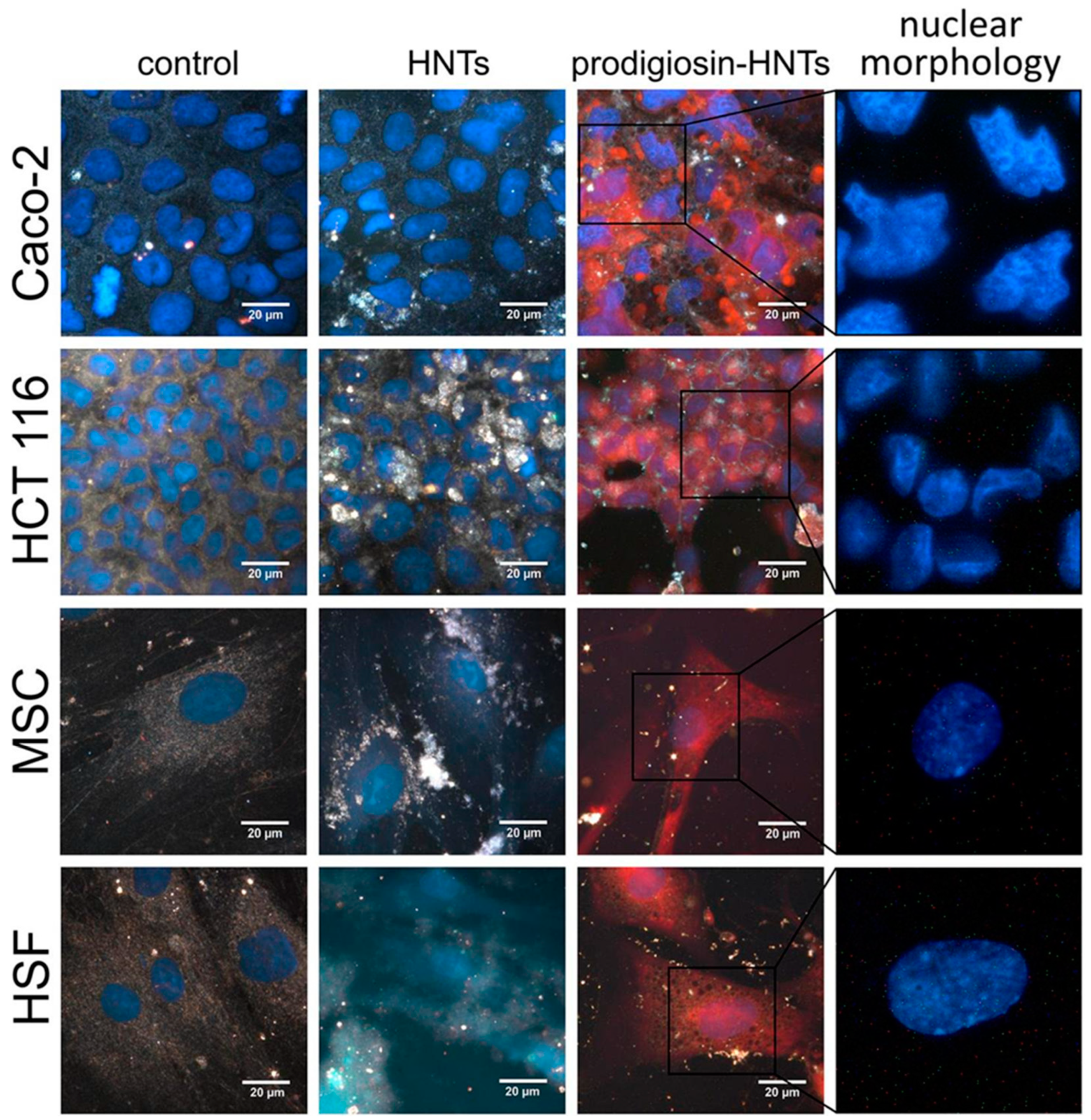
  67. Guryanov, I.; Naumenko, E.; Akhatova, F.; Lazzara, G.; Cavallaro, G.; Nigamatzyanova, L.; Fakhrullin, R. Selective Cytotoxic Activity of Prodigiosin@halloysite Nanoformulation. Front. Bioeng. Biotechnol. 2020, 8, 424. [Google Scholar] [CrossRef]
  68. Parikh, S.A.; Kantarjian, H.; Schimmer, A.; Walsh, W.; Asatiani, E.; El-Shami, K.; Winton, E.; Verstovsek, S. Phase II Study of Obatoclax Mesylate (GX15-070), a Small-Molecule BCL-2 Family Antagonist, for Patients with Myelofibrosis. Clin. Lymphoma Myeloma Leuk. 2010, 10, 285–289. [Google Scholar] [CrossRef]
  69. Francisco, R.; Pérez-Tomás, R.; Gimènez-Bonafé, P.; Soto-Cerrato, V.; Giménez-Xavier, P.; Ambrosio, S. Mechanisms of prodigiosin cytotoxicity in human neuroblastoma cell lines. Eur. J. Pharmacol. 2007, 572, 111–119. [Google Scholar] [CrossRef]
  70. Montaner, B.; Pérez-Tomás, R. The cytotoxic prodigiosin induces phosphorylation of p38-MAPK but not of SAPK/JNK. Toxicol. Lett. 2002, 129, 93–98. [Google Scholar] [CrossRef]
  71. Hong, B.; Prabhu, V.V.; Zhang, S.; van den Heuvel, A.P.; Dicker, D.T.; Kopelovich, L.; El-Deiry, W.S. Prodigiosin rescues deficient p53 signaling and antitumor effects via upregulating p73 and disrupting its interaction with mutant p53. Cancer Res. 2014, 74, 1153–1165. [Google Scholar] [CrossRef]
  72. Branco, P.C.; Pontes, C.A.; Rezende-Teixeira, P.; Amengual-Rigo, P.; Alves-Fernandes, D.K.; Maria-Engler, S.S.; da Silva, A.B.; Pessoa, O.D.L.; Jimenez, P.C.; Mollasalehi, N.; et al. Survivin modulation in the antimelanoma activity of prodiginines. Eur. J. Pharmacol. 2020, 888, 173465. [Google Scholar] [CrossRef]
  73. Wang, J.; Liu, H.; Zhu, L.; Wang, J.; Luo, X.; Liu, W.; Ma, Y. Prodigiosin from Serratia Marcescens in Cockroach Inhibits the Proliferation of Hepatocellular Carcinoma Cells through Endoplasmic Reticulum Stress-Induced Apoptosis. Molecules 2022, 27, 7281. [Google Scholar] [CrossRef]
  74. Hassankhani, R.; Sam, M.R.; Esmaeilou, M.; Ahangar, P. Prodigiosin isolated from cell wall of Serratia marcescens alters expression of apoptosis-related genes and increases apoptosis in colorectal cancer cells. Med. Oncol. 2014, 32, 366. [Google Scholar] [CrossRef] [PubMed]
  75. Lu, Y.; Liu, D.; Jiang, R.; Li, Z.; Gao, X. Prodigiosin: Unveiling the crimson wonder—A comprehensive journey from diverse bioactivity to synthesis and yield enhancement. Front. Microbiol. 2024, 15, 1412776. [Google Scholar] [CrossRef] [PubMed]
  76. Anwar, M.M.; Shalaby, M.; Embaby, A.M.; Saeed, H.; Agwa, M.M.; Hussein, A. Prodigiosin/PU-H71 as a novel potential combined therapy for triple negative breast cancer (TNBC): Preclinical insights. Sci. Rep. 2020, 10, 14706. [Google Scholar] [CrossRef] [PubMed]
  77. Zhou, W.; Zeng, C.; Liu, R.; Chen, J.; Li, R.; Wang, X.; Bai, W.; Liu, X.; Xiang, T.; Zhang, L.; et al. Antiviral activity and specific modes of action of bacterial prodigiosin against Bombyx mori nucleopolyhedrovirus in vitro. Appl. Microbiol. Biotechnol. 2015, 100, 3979–3988. [Google Scholar] [CrossRef] [PubMed]
  78. Suryawanshi, R.K.; Koujah, L.; Patil, C.D.; Ames, J.M.; Agelidis, A.; Yadavalli, T.; Patil, S.V.; Shukla, D. Bacterial Pigment Prodigiosin Demonstrates a Unique Antiherpesvirus Activity That Is Mediated through Inhibition of Prosurvival Signal Transducers. J. Virol. 2020, 94, e00251-20. [Google Scholar] [CrossRef]
  79. Sharma, N.; Shekhar, P.; Kumar, V.; Kaur, H.; Jayasena, V. Microbial pigments: Sources, current status, future challenges in cosmetics and therapeutic applications. J. Basic Microbiol. 2024, 64, 4–21. [Google Scholar] [CrossRef]
  80. Aziz, M.F.A.; Yip, C.W.; Nor, N.S.M. In silico and in vitro antiviral activity evaluation of prodigiosin from Serratia marcescens against enterovirus 71. Malays. Appl. Biol. 2022, 51, 113–128. [Google Scholar] [CrossRef]
  81. Varghese, F.S.; Rausalu, K.; Hakanen, M.; Saul, S.; Kümmerer, B.M.; Susi, P.; Merits, A.; Ahola, T. Obatoclax Inhibits Alphavirus Membrane Fusion by Neutralizing the Acidic Environment of Endocytic Compartments. Antimicrob. Agents Chemother. 2017, 61, e02227-16. [Google Scholar] [CrossRef]
  82. Singh, L. Biodegradation of synthetic dyes: A mycoremediation approach for degradation/decolourization of textile dyes and effluents. J. Appl. Biotechnol. Bioeng. 2017, 3, 430–435. [Google Scholar] [CrossRef]
  83. Berradi, M.; Hsissou, R.; Khudhair, M.; Assouag, M.; Cherkaoui, O.; El Bachiri, A.; El Harfi, A. Textile Finishing Dyes and Their Impact on Aquatic Environs. Heliyon 2019, 5, e02711. [Google Scholar] [CrossRef]
  84. Zhang, B.; Wang, L.; Luo, L.; King, M. Natural dye extracted from Chinese gall–the application of color and antibacterial activity to wool fabric. J. Clean. Prod. 2014, 80, 204–210. [Google Scholar] [CrossRef]
  85. Agarwal, H.; Bajpai, S.; Mishra, A.; Kohli, I.; Varma, A.; Fouillaud, M.; Dufossé, L.; Joshi, N.C. Bacterial Pigments and Their Multifaceted Roles in Contemporary Biotechnology and Pharmacological Applications. Microorganisms 2023, 11, 614. [Google Scholar] [CrossRef] [PubMed]
  86. Ramesh, C.; Vinithkumar, N.V.; Kirubagaran, R.; Venil, C.K.; Dufossé, L. Applications of Prodigiosin Extracted from Marine Red Pigmented Bacteria Zooshikella sp. and Actinomycete Streptomyces sp. Microorganisms 2020, 8, 556. [Google Scholar] [CrossRef] [PubMed]
  87. Suryawanshi, R.K.; Patil, C.D.; Borase, H.P.; Narkhede, C.P.; Stevenson, A.; Hallsworth, J.E.; Patil, S.V. Towards an understanding of bacterial metabolites prodigiosin and violacein and their potential for use in commercial sunscreens. Int. J. Cosmet. Sci. 2015, 37, 98–107. [Google Scholar] [CrossRef]
  88. da Silva, L.A.R.; dos Santos, R.A.; Mendonça, R.d.S.; Sá, A.V.P.; Andrade, R.F.d.S.; Rodríguez, D.M.; Campos-Takaki, G.M. Cost-effective production of stable prodigiosin by Serratia marcescens UCP 1549 and application in soap coloring. Res. Soc. Dev. 2022, 11, e9711427078. [Google Scholar] [CrossRef]
  89. Ren, Y.; Gong, J.; Fu, R.; Li, Z.; Yu, Z.; Lou, J.; Wang, F.; Zhang, J. Dyeing and functional properties of polyester fabric dyed with prodigiosins nanomicelles produced by microbial fermentation. J. Clean. Prod. 2017, 148, 375–385. [Google Scholar] [CrossRef]
  90. Krishna, J.G.; Jacob, A.; Kurian, P.; Elyas, K.K.; Chandrasekaran, M. Marine Bacterial Prodigiosin as Dye for Rubber Latex, Polymethyl Methacrylate Sheets and Paper. Afr. J. Biotechnol. 2013, 12, 2266–2269. [Google Scholar]
  91. Ryazantseva, I.; Andreyeva, I. Application of prodigiosin as a colorant for polyolefines. Adv. Biol. Chem. 2014, 4, 20–25. [Google Scholar] [CrossRef]
  92. Arivizhivendhan, K.V.; Mahesh, M.; Boopathy, R.; Swarnalatha, S.; Regina Mary, R.; Sekaran, G. Antioxidant and antimicrobial activity of bioactive prodigiosin produces from Serratia marcescens using agricultural waste as a substrate. J. Food Sci. Technol. 2018, 55, 2661–2670. [Google Scholar] [CrossRef]
  93. Tenconi, E.; Guichard, P.; Motte, P.; Matagne, A.; Rigali, S. Use of red autofluorescence for monitoring prodiginine biosynthesis. J. Microbiol. Methods 2013, 93, 138–143. [Google Scholar] [CrossRef]
  94. Mouro, C.; Gomes, A.P.; Costa, R.V.; Moghtader, F.; Gouveia, I.C. The Sustainable Bioactive Dyeing of Textiles: A Novel Strategy Using Bacterial Pigments, Natural Antibacterial Ingredients, and Deep Eutectic Solvents. Gels 2023, 9, 800. [Google Scholar] [CrossRef] [PubMed]
  95. Zhao, T.; Zhong, M. Method for Dyeing Wool Fabric by Using Bacterial Dye of Prodigiosin. CN102493228B, 7 October 2013. Available online: https://patents.google.com/patent/CN102493228B/en (accessed on 12 December 2024).
  96. Ren, Y.; Gong, J.; Fu, R.; Zhang, J.; Fang, K.; Liu, X. Antibacterial dyeing of silk with prodigiosins suspention produced by liquid fermentation. J. Clean. Prod. 2018, 201, 648–656. [Google Scholar] [CrossRef]
  97. Ren, Y.; Gong, J.; Fu, R.; Li, Z.; Li, Q.; Zhang, J.; Yu, Z.; Cheng, X. Dyeing and antibacterial properties of cotton dyed with prodigiosins nanomicelles produced by microbial fermentation. Dye. Pigment. 2017, 138, 147–153. [Google Scholar] [CrossRef]
  98. Metwally, R.A.; El Sikaily, A.; El-Sersy, N.A.; Ghozlan, H.A.; Sabry, S.A. Antimicrobial Activity of Textile Fabrics Dyed with Prodigiosin Pigment Extracted from Marine Serratia rubidaea RAM_Alex Bacteria. Egypt. J. Aquat. Res. 2021, 47, 301–305. [Google Scholar] [CrossRef]
  99. Arivuselvam, R.; Dera, A.A.; Parween Ali, S.; Alraey, Y.; Saif, A.; Hani, U.; Arumugam Ramakrishnan, S.; Azeeze, M.S.T.A.; Rajeshkumar, R.; Susil, A.; et al. Isolation, Identification, and Antibacterial Properties of Prodigiosin, a Bioactive Product Produced by a New Serratia marcescens JSSCPM1 Strain: Exploring the Biosynthetic Gene Clusters of Serratia Species for Biological Applications. Antibiotics 2023, 12, 1466. [Google Scholar] [CrossRef]
  100. Moraes, C.S.; Seabra, S.H.; Albuquerque-Cunha, J.M.; Castro, D.P.; Genta, F.A.; de Souza, W.; Brazil, R.P.; Garcia, E.S.; Azambuja, P. Prodigiosin is not a determinant factor in lysis of Leishmania (Viannia) braziliensis after interaction with Serratia marcescens D-mannose sensitive fimbriae. Exp. Parasitol. 2009, 122, 84–90. [Google Scholar] [CrossRef]
  101. Alihosseini, F.; Ju, K.S.; Lango, J.; Hammock, B.D.; Sun, G. Antibacterial colorants: Characterization of prodiginines and their applications on textile materials. Biotechnol. Prog. 2008, 24, 742–747. [Google Scholar] [CrossRef]
  102. Andreeva, I.N.; Ogorodnikova, T.I. Pigmentation of Serratia marcescens and spectral properties of prodigiosin. Mikrobiologiia 2015, 84, 28–33. [Google Scholar] [CrossRef]
  103. Drink, E.; Dugourd, P.; Dumont, E.; Aronssohn, N.; Antoine, R.; Loison, C. Optical properties of prodigiosin and obatoclax: Action spectroscopy and theoretical calculations. Phys. Chem. Chem. Phys. 2015, 17, 25946–25955. [Google Scholar] [CrossRef]
  104. Morales-Oyervides, L.; Oliveira, J.; Sousa-Gallagher, M.; Méndez-Zavala, A.; Montañez, J.C. Assessment of the Dyeing Properties of the Pigments Produced by Talaromyces spp. J. Fungi 2017, 3, 38. [Google Scholar] [CrossRef]
  105. Abirami, S.; Kumar, S.D.; Murugan, A. Dyeing of Jute Fabrics with Prodigiosin Produced from Sago Waste and their Applications. J. Pure Appl. Microbiol. 2022, 16, 147–156. [Google Scholar] [CrossRef]
  106. Venil, C.K.; Zakaria, Z.A.; Ahmad, W.A. Bacterial pigments and their applications. Process Biochem. 2013, 48, 1065–1079. [Google Scholar] [CrossRef]
  107. Lee, J.S.; Kim, Y.-S.; Park, S.; Kim, J.; Kang, S.-J.; Lee, M.-H.; Ryu, S.; Choi, J.M.; Oh, T.-K.; Yoon, J.-H. Exceptional Production of both Prodigiosin and Cycloprodigiosin as Major Metabolic Constituents by a Novel Marine Bacterium, Zooshikella rubidus S1-1. Appl. Environ. Microbiol. 2011, 77, 4967–4973. [Google Scholar] [CrossRef] [PubMed]
  108. Arivizhivendhan, K.V.; Boopathy, R.; Maharaja, P.; Regina Mary, R.; Sekaran, G. Bioactive Prodigiosin-Impregnated Cellulose Matrix for the Removal of Pathogenic Bacteria from Aqueous Solution. RSC Adv. 2015, 5, 68621–68631. [Google Scholar] [CrossRef]
  109. Arivizhivendhan, K.V.; Mahesh, M.; Murali, R.; Mary, R.R.; Thanikaivelan, P.; Sekaran, G. Prodigiosin–Iron-Oxide–Carbon Matrix for Efficient Antibiotic-Resistant Bacterial Disinfection of Contaminated Water. ACS Sustain. Chem. Eng. 2019, 7, 3164–3175. [Google Scholar] [CrossRef]
  110. Amorim, L.F.A.; Fangueiro, R.; Gouveia, I.C. Characterization of Bioactive Colored Materials Produced from Bacterial Cellulose and Bacterial Pigments. Materials 2022, 15, 2069. [Google Scholar] [CrossRef]
  111. Diken-Gür, S. Investigation of anti-adherence and antimicrobial properties of prodigiosin-functionalized bacterial cellulose membrane for biomedical applications. J. Biotechnol. 2024, 385, 58–64. [Google Scholar] [CrossRef]
  112. Bidwai, S. Cosmetic Pigments Market Size, Share, and Trends 2024 to 2034. Available online: https://www.precedenceresearch.com/cosmetic-pigments-market (accessed on 23 November 2024).
  113. Balwierz, R.; Biernat, P.; Jasińska-Balwierz, A.; Siodłak, D.; Kusakiewicz-Dawid, A.; Kurek-Górecka, A.; Olczyk, P.; Ochędzan-Siodłak, W. Potential Carcinogens in Makeup Cosmetics. Int. J. Environ. Res. Public Health 2023, 20, 4780. [Google Scholar] [CrossRef]
  114. Salles, F.J.; Paniz, F.P.; Batista, B.L.; Nardocci, A.C.; Olympio, K.P.K. Potentially Toxic Elements in Costume Cosmetics Used by Children and Adults Are Associated with Cancer Risk. Int. J. Environ. Res. Public Health 2023, 20, 531. [Google Scholar] [CrossRef]
  115. Alshehrei, F.M. Isolation and Identification of Microorganisms associated with high-quality and low-quality cosmetics from different brands in Mecca region-Saudi Arabia. Saudi J. Biol. Sci. 2023, 30, 103852. [Google Scholar] [CrossRef]
  116. Rodríguez, J.; Lobato, C.; Vázquez, L.; Mayo, B.; Flórez, A.B. Prodigiosin-Producing Serratia marcescens as the Causal Agent of a Red Colour Defect in a Blue Cheese. Foods 2023, 12, 2388. [Google Scholar] [CrossRef] [PubMed]
  117. Saha, S.; Thavasi, T.R.; Jayalakshmi, S. Phenazine Pigments from Pseudomonas aeruginosa and Their Application as Antibacterial Agent and Food Colourants. Res. J. Microbiol. 2008, 3, 122–128. [Google Scholar]
  118. Dharmaraj, S.; Ashokkumar, B.; Dhevendaran, K. Food-Grade Pigments from Streptomyces sp. Isolated from the Marine Sponge Callyspongia diffusa. Food Res. Int. 2009, 42, 487–492. [Google Scholar] [CrossRef]
  119. Nawaz, A.; Chaudhary, R.; Shah, Z.; Dufossé, L.; Fouillaud, M.; Mukhtar, H.; ul Haq, I. An Overview on Industrial and Medical Applications of Bio-Pigments Synthesized by Marine Bacteria. Microorganisms 2021, 9, 11. [Google Scholar] [CrossRef]
  120. Leong, C.R.; Daud, N.S.; Tong, W.Y.; Cheng, S.Y.; Tan, W.N.; Hamin, N.S.; Pa’ee, K.F. Gelatine Film Incorporated with Clitoria ternatea-Derived Anthocyanin Microcapsules, A Food Packaging Material Effective Against Foodborne Pathogens. Food Technol. Biotechnol. 2021, 59, 422–431. [Google Scholar] [CrossRef]
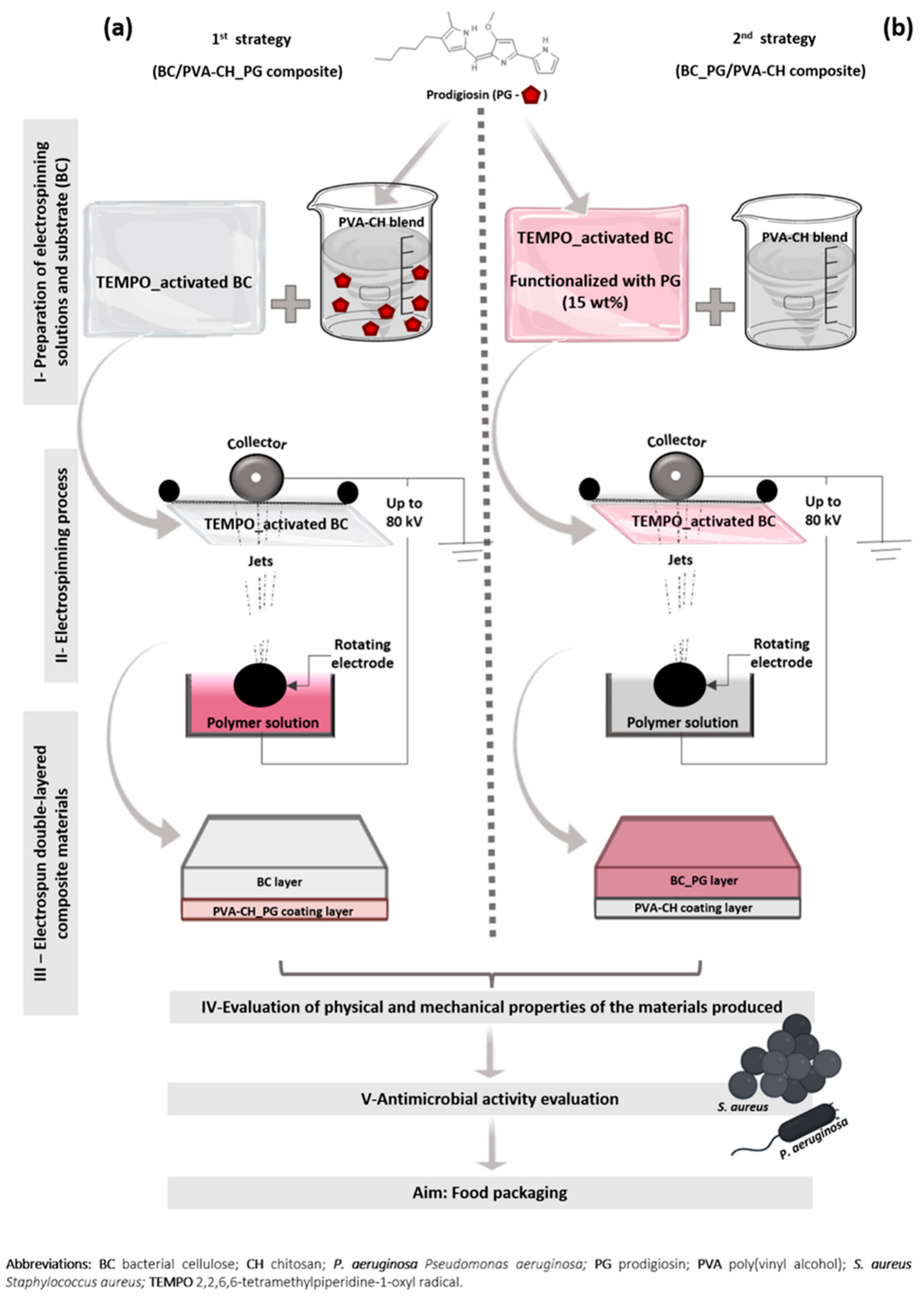
  121. Amorim, L.F.A.; Mouro, C.; Riool, M.; Gouveia, I.C. Antimicrobial Food Packaging Based on Prodigiosin-Incorporated Double-Layered Bacterial Cellulose and Chitosan Composites. Polymers 2022, 14, 315. [Google Scholar] [CrossRef]
  122. Dey, A.; Dhumal, C.V.; Sengupta, P.; Kumar, A.; Pramanik, N.K.; Alam, T. Challenges and Possible Solutions to Mitigate the Problems of Single-Use Plastics Used for Packaging Food Items: A Review. J. Food Sci. Technol. 2021, 58, 3251–3269. [Google Scholar] [CrossRef]
  123. Nemat, B.; Razzaghi, M.; Bolton, K.; Rousta, K. Design Affordance of Plastic Food Packaging for Consumer Sorting Behavior. Resour. Conserv. Recycl. 2022, 177, 105949. [Google Scholar] [CrossRef]
  124. Nakajima, T. Divorcing from Plastics for a Sustainable Future Society. In Overcoming Environmental Risks to Achieve Sustainable Development Goals. Current Topics in Environmental Health and Preventive Medicine; Nakajima, T., Nakamura, K., Nohara, K., Kondoh, A., Eds.; Springer: Singapore, 2022. [Google Scholar] [CrossRef]
  125. Parisi, C.; Sandonnini, J.; Coppola, M.R.; Madonna, A.; Abdel-Gawad, F.K.; Sivieri, E.M.; Guerriero, G. Biocide vs. Eco-Friendly Antifoulants: Role of the Antioxidative Defence and Settlement in Mytilus galloprovincialis. J. Mar. Sci. Eng. 2022, 10, 792. [Google Scholar] [CrossRef]
  126. Almeida, J.R.; Vasconcelos, V. Natural antifouling compounds: Effectiveness in preventing invertebrate settlement and adhesion. Biotechnol. Adv. 2015, 33, 343–357. [Google Scholar] [CrossRef]
  127. Lovley, D.R. Microbial fuel cells: Novel microbial physiologies and engineering approaches. Curr. Opin. Biotechnol. 2006, 17, 327–332. [Google Scholar] [CrossRef] [PubMed]
  128. Koyama, Y.; Miki, T.; Wang, X.-F.; Nagae, H. Dye-sensitized solar cells based on the principles and materials of photosynthesis: Mechanisms of suppression and enhancement of photocurrent and conversion efficiency. Int. J. Mol. Sci. 2009, 10, 4575–4622. [Google Scholar] [CrossRef] [PubMed]
  129. Tomat, E. Coordination Chemistry of Linear Tripyrroles: Promises and Perils. Comments Inorg. Chem. 2016, 36, 327–342. [Google Scholar] [CrossRef]
  130. Castillo-Zacarías, C.; Cantú-Cárdenas, M.E.; López-Chuken, U.J.; Parra-Saldívar, R.; Garza-Gonzalez, M.T.; Rostro-Alanis, M.D.J.; Villarreal-Chiu, J.F. Dissimilatory reduction of Fe(III) by a novel Serratia marcescens strain with special insight into the influence of prodigiosin. Int. Microbiol. 2019, 23, 201–214. [Google Scholar] [CrossRef]
  131. Montagni, T.; Enciso, P.; Marizcurrena, J.J.; Castro-Sowinski, S.; Fontana, C.; Davyt, D.; Cerdá, M.F. Dye Sensitized Solar Cells Based on Antarctic Hymenobacter sp. UV11 Dyes. Environ. Sustain. 2018, 1, 89–97. [Google Scholar] [CrossRef]
  132. Di Mauro, E.; Xu, R.; Soliveri, G.; Santato, C. Natural melanin pigments and their interfaces with metal ions and oxides: Emerging concepts and technologies. MRS Commun. 2017, 7, 141–151. [Google Scholar] [CrossRef]
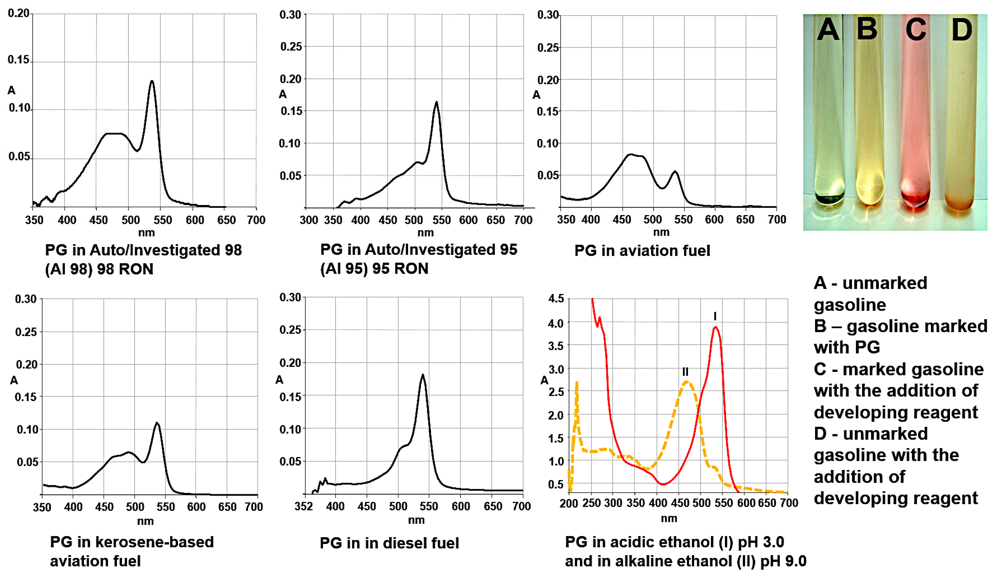
  133. Yusupova, D.V.; Gabdrakhmanova, L.A.; Kurkina, M.A.; Guryanov, I.D. Identification Method of Marked Oil. Products. Patent RU 2368645C2, 29 September 2009. Available online: https://patents.google.com/patent/RU2368645C2/en?q=(Gareishina)&oq=Gareishina#citedBy (accessed on 12 December 2024).
Figure 1. Statistical information about number of publications and patents in Google patents [20] and PubMed [21]. Numbers under bars indicate number of papers by year in PubMed database.
Applmicrobiol 04 00115 g001
Figure 2. Mechanisms of antibacterial activity of PG (A) and representative photographs of the classical disk diffusion method demonstrating the broad spectrum of antibacterial action of PG (B) Filter paper discs were impregnated with (A) 35 μg/μL chloramphenicol (positive control), (B) 95% methanol (negative control, used as solvent for solubilising prodigiosin extract), (C) 250 μg/μL crude prodigiosin (D) 500 μg/μL crude prodigiosin and (E) 99% ethanol (negative control; used as solvent for chloramphenicol); adopted from [41] under the terms of the Creative Commons Attribution License.
Applmicrobiol 04 00115 g002
Figure 3. Antifungal activity of purified prodigiosin extracted from Serratia marcescens against plant pathogenic fungi.
Applmicrobiol 04 00115 g003
Figure 4. Antiparasitic activity of prodigiosin and obatoclax. (A) Imaging of juvenile Schistosoma mansoni after 72 h treatment with DMSO control, 5 μM obatoclax or 5 μM prodigiosin. Arrows indicate changes in gross morphology as well as tegument blebbing. Position of oral sucker and anterior–posterior axis are indicated (A–P). (B) Kinetics of drug action against Entamoeba histolytica killing. Results from experiment to assess E. histolytica trophozoites. Reproduced under the terms of the Creative Commons Attribution License from [61].
Applmicrobiol 04 00115 g004
Figure 5. Hyperspectral and fluorescence images for comparison of the effect of halloysite nanotubes loaded with glycerol and prodigiosin-HNTs on malignant (Caco-2, HCT 116) and non-malignant cells (MSC, HSF). Reproduced from [67] under Creative Commons Attribution License (CC BY).
Applmicrobiol 04 00115 g005
Figure 6. Prodigiosin effects against Herpes simplex virus in vitro on keratinocytes (A) and in vivo on cornea and skin (B). Significance between the treatment and control groups was determined using the student unpaired t-test. The symbol ** indicates significant difference at the 0.01 level (p < 0.01). Adopted from [78] under permissions from American Society for Microbiology.
Applmicrobiol 04 00115 g006
Figure 7. Different fabric types dyed with prodigiosin from Serratia rubidaea RAM_Alex. Adapted from [98] under CC BY-NC-ND 4.0 Deed Attribution-NonCommercial-NoDerivs 4.0.
Applmicrobiol 04 00115 g007
Figure 8. Different color hues of prodigiosin-dyed fabrics obtained after applying different mordants. Reprinted from [98] under CC BY-NC-ND 4.0 Deed Attribution-NonCommercial-NoDerivs 4.0.
Applmicrobiol 04 00115 g008
Figure 9. Solid molded soap colored with prodigiosin (A) and relative stability of pigment under changing pH (B) and salinity (C). Adopted from [88] under Creative Commons Attribution 4.0 International License.
Applmicrobiol 04 00115 g009
Figure 10. Food colorant application as agar jellies prepared using extractions of different pigment bacterial strains. Adopted from [86] under Creative Commons Attribution 4.0 International License.
Applmicrobiol 04 00115 g010
Figure 11. Different strategies of functionalization of packaging material with prodigiosin. (a) Incorporation of prodigiosin into inner layer (PVA-CH); (b) functionalization of outer layer (BC) with prodigiosin. Reproduced from [121] under Creative Commons Attribution 4.0 International License.
Applmicrobiol 04 00115 g011
Figure 12. The optical spectra of prodigiosin in various types of fuels and view of marked gasoline.
Applmicrobiol 04 00115 g012
Disclaimer/Publisher’s Note: The statements, opinions and data contained in all publications are solely those of the individual author(s) and contributor(s) and not of MDPI and/or the editor(s). MDPI and/or the editor(s) disclaim responsibility for any injury to people or property resulting from any ideas, methods, instructions or products referred to in the content.

