Article

Escherichia coli Reporter Strains Allow for the In Vivo Evaluation of Recombinant Elongation Factor Protein (EF-P)

by
Natalia Trachtmann
1,2,
Aydar Bikmullin
2,3,
Shamil Validov
2 and
Georg A. Sprenger
1,*
1
Institute of Microbiology, University of Stuttgart, Allmandring 31, D-70569 Stuttgart, Germany
2
Federal Research Center, Kazan Scientific Center of Russian Academy of Science, ul. Lobachevskogo, 2/31, 420111 Kazan, Russia
3
Institute of Fundamental Medicine and Biology, Kazan Federal University, 18 Kremlyovskaya Street, 420008 Kazan, Russia
*
Author to whom correspondence should be addressed.
Appl. Microbiol. 2024, 4(3), 1335-1347; https://doi.org/10.3390/applmicrobiol4030092
Submission received: 2 September 2024 / Revised: 15 September 2024 / Accepted: 16 September 2024 / Published: 18 September 2024

Abstract

:
Background: Elongation factor protein (EF-P) in bacteria helps ribosomes to incorporate contiguous proline residues (xPro) into proteins. In this way, EF-P rescues ribosomes from stalling at these xPro motifs. Whereas EF-P deficiency is lethal for some species, others show reduced virulence or generally lower growth rates, such as Escherichia coli (E. coli). EF-P needs to be post-translationally modified to gain full functionality. Methods: We constructed E. coli K-12 mutant strains with deletion of the serA gene leading to an auxotrophy for L-serine. Then, we engineered a 6xPro motif in the recombinant serA gene, which was then chromosomally inserted under its native promoter. Furthermore, mutant strains which were deleted for efp and/or epmA (encoding the EF-P modification protein EpmA) were engineered. Results: Δefp, ΔepmA, and Δefp/ΔepmA double mutants showed already significantly reduced growth rates in minimal media. ΔserA derivatives of these strains were complemented by the wt serA gene but not by 6xPro-serA. ΔserA mutants with intact efp were complemented by all serA-constructs. Chromosomal expression of the recombinant efp gene from E. coli or from the pathogen, Staphylococcus aureus (S. aureus), restored growth, even without epmA expression. Conclusions: We provide a novel synthetic reporter system for in vivo evaluation of EF-P deficiency. In addition, we demonstrated that both EF-P-E. coli and EF-P-S. aureus restored the growth of a 6xPro-serA: Δefp, ΔepmA strain, which is evidence that modification of EF-P might be dispensable for rescuing of ribosomes stalled during translation of proline repeats.

1. Introduction

During protein synthesis, ribosomes become stalled on polyproline-containing sequences due to the cyclic pyrrolidine structure of proline. Polypeptides with a high proline content are thus poorly translated, especially if proline occurs in motifs consisting of two, three, or more contiguous residues [1,2,3,4]. Specific proteins can rescue ribosomes from stalling at these motifs. In Archaea and eukaryotes, this is achieved via the archaeal/eukaryotic translation initiation factor a/eIF-5A, and in bacteria, via the homologous elongation factor protein, EF-P, respectively [5]. All living organisms express either a/eIF5A or EF-P [6,7,8]. EF-P helps to incorporate contiguous proline residues into proteins and, in vitro, can even facilitate the incorporation of structurally diverse non-canonical amino acid residues, as shown recently [9]. Only one gene encodes EF-P in bacteria [5], which is a small (21 kDa) protein containing three β-barrel domains (I to III), reminiscent of the L-shape of tRNA [10,11,12]. The N-terminal domains of eIF5A and domain I of EF-P show the same fold, as do the C-terminal domain of eIF5A and domains II and III of EF-P [10,11].
For full functionality, a/eIF5A and EF-P need to be modified post-translationally [13]. These are unique modifications. In many prokaryotes, a specific lysine residue is modified at the tip of a flexible loop in the N-terminal domain of aIF5A or in domain I of EF-P. A post-translational modification occurs on a conserved lysine residue of EF-P in many bacteria and is referred to as β-lysinylation of amino acid residue Lys34 (K34). Lysinylation is only found in about 25% of analyzed bacterial species [7,13,14,15]. This modification occurs in three sequential steps, requiring the enzymes EpmA, EpmB, and EpmC [14], formerly termed YjeK, YjeA, and YfcM [5] in E. coli. Different modifications, e.g., rhamnosylation of EF-P at Arg32, have been found in Pseudomonas aeruginosa (P. aeruginosa) [13,14], and a 5-aminopentanol moiety is attached to Lys32 in the Gram-positive bacteria Bacillus subtilis [16,17] and in S. aureus [18]. Enzymes for the known EF-P lysinylation, rhamnosylation, or aminopentanolylation pathways can be identified in only about 35% of the available bacterial genomes. It is unclear as to how the remaining majority of bacteria activate their corresponding EF-Ps [19]. As pathogenic species, such as P. aeruginosa, Bacillus cereus, or S. aureus, also have their cognate EF-P modification systems, an understanding of the individual post-translational modification systems is of great interest. This could lead to targets for species-specific antimicrobial compounds, which are unlike broad-range antibiotics.
In bacteria, the effects of EF-P deficiency vary. Whereas an EF-P deficiency is lethal in Neisseria strains, Bacillus efp-mutants showed only marked defects in sporulation or swarming motility [17,20,21]. P. aeruginosa efp-mutants displayed reduced virulence and increased sensitivity towards several antibiotics [22]. For E. coli, divergent reports have been given, e.g., the temperature sensitivity of growth [23] or the slower growth of an efp-deletion mutant only [22,24]. By using transposon-mediated differential hybridization, the homologous efp gene of S. aureus was deemed to be essential for growth [25]. Recombinant expression of EF-P proteins was successfully accomplished in E. coli strains (Thermus thermophilus: [10,12], P. aeruginosa [11,14], Shewanella oneidensis [14]).
Reporter systems have been constructed with an engineered tetra-proline (4Pro) motif in GFP for the P. aeruginosa system to study the influence of the various modification systems [26]; similarly, in B. subtilis, a fluorescent reporter system has been described [17]. For E. coli, a PcadBA-dependent lacZ reporter was successfully introduced in an efp mutant strain. This allowed for complementation assays of several EF-P variants in this strain through the monitoring of β-galactosidase (LacZ) activity [14]. E. coli reporter strains used to visualize EF-P activity via GFP activity have also been described [24]. To our knowledge, no positive selection system has been given so far for E. coli efp mutants where the in vivo function of recombinant Efp protein was assessed. Here, we report a novel synthetic E. coli reporter system which allows for the in vivo functional expression of efp genes. We combined the essential serA gene for L-serine biosynthesis [27] with a 6xPro motif and showed that this leads to growth impairment of E. coli mutants carrying a chromosomal efp gene deletion. We show further that this growth defect can be complemented via the expression of either the homologous recombinant efp gene from E. coli or the recombinant gene from S. aureus (efpSa). Interestingly, this complementation also works without a cognate modification (Epm) system.

2. Materials and Methods

2.1. Bacterial Strains, Plasmids, and DNA Primers

All strains are derived from E. coli K-12, wild-type strain W3110 (lab name: LJ110 [28]). The pedigree of the strains constructed in the present study is given in Table 1, as well as the list of plasmids used and created in this work. Table 2 contains the list of DNA primers used in this study. Oligonucleotides were custom-synthesized by Biomers (Ulm, Germany).

2.2. Construction of Plasmids and Recombinant Strains

Standard methods for plasmid cloning and PCR were used unless stated otherwise [32]. Deletions and gene insertions were performed via the CRISPR-Cas method in combination with λ-red recombineering, as described [29]. Target loci in the genome of E. coli were chosen as previously described [33]. All gene modifications were verified by custom DNA sequencing (Eurofins, Ebersberg, Germany). All specific pTarget-cat-sgRNA plasmids (see Table 1) used in this work were constructed through the introduction of a 20 N specific guide sequence in pTarget-cat plasmid using the inverse PCR method. The inverse PCR method was performed using the Targ-univ-rev primer in combination with the corresponding specific primer (sgRNA-serA for pTarget-cat-sgRNA-serA; sgRNA-efp for pTarget-cat-sgRNA-efp; sgRNA-epmA for pTarget-cat-sgRNA-epmA; sgRNA-fuc for pTarget-cat-sgRNA-fuc; and sgRNA-xyl for pTarget-cat-sgRNA-xyl). After inverse PCR, the fragments were digested with DpnI, treated with T4 polynucleotide kinase (NEB GmbH, Frankfurt am Main, Germany) and religated with T4 DNA ligase (NEB GmbH, Frankfurt am Main, Germany). Selected clones after transformation in the DH5α strain [34] were verified via sequencing.
The DNA fragment containing the serA gene and its promoter region was amplified from the chromosomal DNA of the E. coli wild-type strain LJ110 using primers 3′serA-SphI and 5′PserA-serA-SphI and cloned into the SphI restriction site of the pJNNmod vector. After cloning of the plasmid using XbaI and HpaI restriction enzymes, it was treated with Klenow DNA polymerase to obtain blunt ends, and was religated. The resulting plasmid pSerA was used as a basic construct for the creation of p4Pro-serA and p6Pro-serA recombinant plasmids via inverse PCR using serA-inv-rev/serA-4Pro and serA-inv-rev/serA-6Pro primers, respectively. These recombinant plasmids were used as a template for the amplification of the fragment Ptac-serA (6Pro-serA/4Pro-serA) cassettes into the fuc-locus of chromosomal DNA. These fragments were produced with FucP-serA-int-5′/FucI-int-3′ primers.
The efp genes from E. coli and S. aureus were amplified from chromosomal DNA using efp-Ec-5’NdeI/efp-Ec-3′HindIII and efp-Sa-5′-NdeI/efp-Sa-3′-BamHI primers, respectively. After amplification, these fragments were cloned into the pJNNmod vector (or pJem2 vector) in NdeI/HindIII enzyme sites. Thus, the obtained recombinant plasmids (pJNNmod-based) were used to amplify the xylA*-Ptac-efp-Ec (efp-Sa)-xylB* fragment (primers: xylA-int-5′/xylB-int′). The fragments were integrated into the chromosome of the reporter strains using the CRISPR-Cas λ-Red method. Deletions of the serA, efp, and epmA genes from the chromosome of the E.coli cells were performed using the CRISPR-Cas method [29]. The flanking regions of HR1 and HR2 were spliced via PCR. For this, the fragments (HR1 and HR2) were amplified, cut with BamHI and BglII, respectively, then ligated with subsequent reamplification. The created fragments were used for homologous recombination with subsequent removal of the desired gene.

2.3. Growth Measurements

LB agar plates were used for strain propagation with 18 g/L of agar. Antibiotics were added to the following final concentrations: ampicillin-100 mg L−1, chloramphenicol-25 mg L−1, and kanamycin-50 mg L−1. The hydrogen-phosphate-buffered mineral salts (minimal) medium [35] with 10 g L−1 of glucose was used with supplementation of L-serine (200 mg L−1, final concentration) when appropriate. Optical density at wave light 600 nm (OD600) was followed with a UV/vis photometer (Cary50, Thermo Fisher Scientific GmbH, Dreieich, Germany) in shake flasks (250 mL with 30 mL of cell culture) on orbital shakers (Infors HT, Einsbach, Germany) at 200 rpm and a temperature of 37 °C. Growth measurements were performed in three biological replicates (independent experiments). Standard deviations are provided as error bars.

2.4. Biochemical Measurements

Total RNA was isolated from cells from the exponential growth phase using the Qiagen Kit (Qiagen, Düsseldorf, Germany). RNA was reverse transcribed using EpiScript™ Reverse Transcriptase (Biozym, Hessisch Oldendorf, Germany) and 6N random primer. Transcript measurements were conducted via RT-qPCR according to the recommendation of suppliers (Biozym, Hessisch Oldendorf, Germany) using an instrument and software from AnalyticJena (Jena, Germany). For the amplification, the Blue S’Green qPCR Kit (Biozym, Hessisch Oldendorf, Germany) was used. The RT-qPCR was performed in technical triplicate on the biological triplicate. Cycling conditions were 95 °C for 30 s, 52 °C for 10 s, and 72 °C for 20 s, along with a fluorescence measurement. The negative control (NC) was included for each primer pair. The ftsZ gene, encoding a cell division protein, was used as an internal control to normalize all data.

3. Results and Discussion

3.1. Construction of E. coli Reporter Strains Which Lack Their Own EF-P-System

We aimed to construct E. coli efp-deficient mutants with a strong, selectable phenotype. It had been shown before that efp-mutants of E. coli are viable though compromised [22,24]. Deletion of efp or epmA in a wild-type E. coli K-12 strain (LJ110) was achieved via the CRISPR-Cas method in combination with λ-red recombineering [29]. The single deletion mutants (for a list of strains and primers see Table 1 and Table 2) NT1433 (ΔepmA) and NT1435 (Δefp) showed both a significantly reduced growth rate (µ) and lower final optical density (fOD) when grown in minimal medium with the carbon source glucose (Table 3) in comparison to the wild-type strain LJ110. This is in accordance with a previous report on a BW25113 Δefp mutant [22]. A combination of the two defects (NT1437) led to a further reduction of growth rate (Table 3). These differences in growth phenotype, however, were not sufficiently strong to be used as a base for recombinant gene expression and selection.

3.2. Synthetic Reporter Strains Which Show a Growth Defect in Mineral Media

We therefore aimed for a synthetic reporter strain which allows for parallel investigations of recombinant prokaryotic EF-P expression and could thus constitute a positive selection system. To do so, we first engineered E. coli mutant strains, which are L-serine auxotrophs (NT1352). Deletion of serA gene rendered this strain strictly dependent on supplementation with L-serine when grown on minimal media (Figure 1). Transformation of the strain NT1352 (ΔserA) with recombinant plasmids containing the cloned serA gene under the control of its own promoter (PserA) or bearing the serA gene fused with 4 or 6 proline-encoding codons at the N terminus (4xPro-SerA, 6xPro-serA) completely restored cell growth on minimal medium plates (see Figure 1). Subsequently, the serA gene encoding the enzyme (3-phosphoglycerate dehydrogenase, SerA) variants with a fourfold or sixfold CCA-proline motif in the N-terminus were used for further work.
Deletion of the efp gene in the serine auxotroph NT1352 strain led to NT1367. If this strain was transformed with the empty control plasmid (pJNNmod) or a plasmid carrying a stop codon between the 6xPro encoding codons and the residual of the serA gene (4xPro-Stop-serA), it could not grow on minimal medium plates without L-serine supplementation. If transformed with plasmid-borne copies of 4xPro-serA or 6xPro-serA, only faint growth was observed on agar plates. However, the introduction of the wild-type serA gene led to the restoration of cell growth (Figure 1). The weak-growth phenotype with the 4xPro/6xPro-serA genes might be due to a metabolic burden caused by the high expression of a multi-copy plasmid vector.
To avoid problems with plasmid-borne gene expression, we therefore aimed for a single-copy gene expression. To do so, the serA gene was chromosomally integrated in the NT1352 (ΔserA) mutant strain via a CRISPR-Cas targeting method [29]. We chose the chromosomal fuc locus (Figure 2) as the integration site as this allowed for easy control of the phenotype on MacConkey agar plates with 1% L-fucose [33]. The altered serA genes are under the control of their cognate PserA promoter. Growth in minimal medium without L-serine supplementation was restored in the NT1373 strain (ΔserA fuc::PserA-6Pro-serA), albeit with a lower growth rate.
Also, PserA-4xPro-serA, PserA-6xPro-serA, and PserA-serA were introduced into the fuc-locus of strain NT1367 (Δefp ΔserA). So, the obtained strains did not show growth on minimal medium. The control strain NT1368 with an integrated wild-type copy of the serA gene, however, grew well.
Next, we introduced the ΔepmA deletion into this strain background. Strain NT1397 (Δefp ΔserA fuc::PserA-6xPro-serA ΔepmA) was constructed. This was performed with the aim of exacerbating the EF-P-dependency in the absence of L-serine. As expected, a complete growth defect in minimal medium without L-serine supplementation was observed when the efp gene was deleted, as well as in combination with ΔepmA. Deletion of epmA alone led to an intermediate growth phenotype on agar plates or in shake flask cultures (NT1433, Table 3). Interestingly the growth rates of ΔepmA and ΔefpepmA E. coli strains were reduced stronger than of the Δefp strain. It is tempting to speculate that EmpA can have additional function(s) in E. coli cells.
This strong and selectable growth phenotype was then used to study whether the defects of the NT1379 strain could be complemented by expression of heterologous efp genes such as the one from the pathogenic Gram-positive bacterium, S. aureus (efp-Sa). This would also allow us to study whether cognate posttranslational modification systems from other bacteria are required for the complementation of the growth defects in the reporter strain (grade of restoration in serine prototrophy).

3.3. Plasmid-Based Expression of Efp Genes

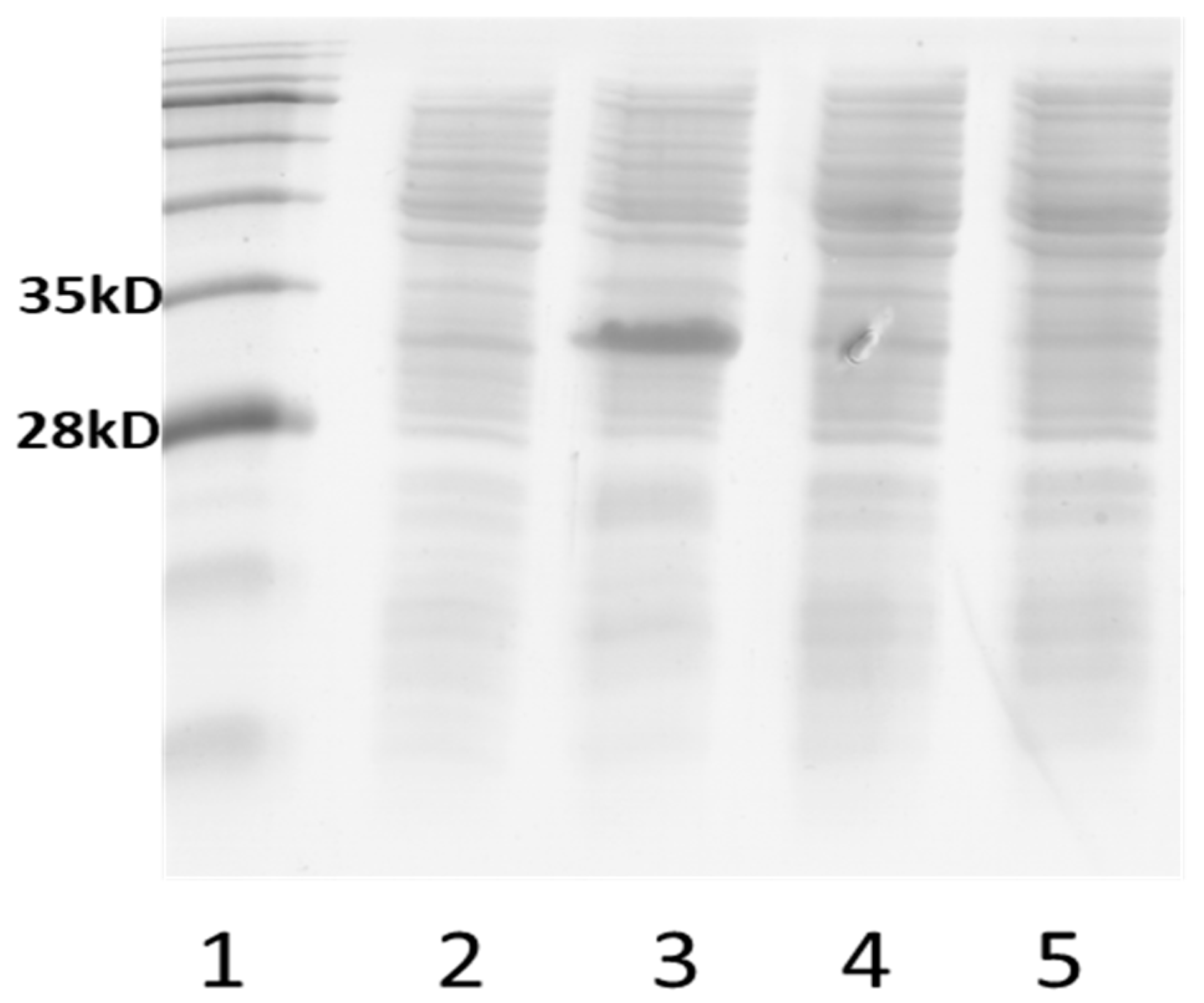
The efp genes from E. coli and S. aureus (efp-Ec and efp-Sa) were cloned into the pJNNmod vector under the control of the Ptac-promoter, as well as into the pJem2 vector (see Table 1) under the control of the rhamnose-inducible promoter PrhaA. Recombinant plasmids were introduced into the NT1379 strain to test their ability to restore growth. The resulting clones were plated on the agar plates with minimal medium supplemented with 0.4% of glucose and 0.2 mM of IPTG for pJNNmod-based variants (0.5% of rhamnose for the pJem2-based plasmid). We observed very weak growth of the NT1379 strain transformed with pJem2-efp-Ec and pJem2-efp-Sa plasmids, respectively. The strains containing the pJNNmod-based recombinant plasmids (pJNN-efp-Sa and pJNN-efp-Ec) did not show any growth on these plates. However, on the plates with pJNN-efp-Sa with prolonged incubation of plates (more than 1 week), some single colonies of the NT1379-pJNN-efp-Sa strain with restored cell growth (evo) were selected. The plasmid DNA from evolved (“evo”) clones were isolated, and the insertion was verified via sequencing. The sequencing of these plasmids, however, did not show any mutation in the efp-Sa gene. The NT1379 strain was retransformed with isolated pJNN-efp-Sa-evo1 and pJNN-efp-Sa-evo2. Although the efp gene in these pJNN-efp-Sa-evo plasmids did not contain any additional substitutions, the cells showed a serine prototrophic phenotype. To study this phenomenon further, we analyzed the expression of the efp gene from the evo-derivates in comparison with the expression of this protein located on the primary plasmid, and we analyzed the protein pattern via SDS gel electrophoresis. Expression analysis of evo clones via SDS gel electrophoresis showed a significant decrease in the amount of expressed EF-P (evo) protein in comparison to the EF-P synthesized from the primary plasmid (see Figure 3), which may be due to a lowered plasmid copy number.
The growth experiments in liquid minimal media were carried out with pJem2-based plasmids in the presence of different concentrations of rhamnose (0, 0.1, 0.2, and 1%). The pre-culture was prepared in mineral medium supplemented with 200 µg/mL of L-serine. Then, the cells were washed with mineral medium without serine and inoculated in mineral medium with 1% glucose and appropriate amount of rhamnose. Within 30 h of incubation at 37 °C, we did not observe cell growth carrying an empty vector pJem2 (Figure 4A). In the case of pJem2-efp-Ec and pJem2-efp-Sa, there was a slight increase in optical density (Figure 4B,C).

3.4. Complementation of Growth Defects by Chromosomally Integrated Recombinant Efp Genes

So far, these results have indicated that the use of plasmid-encoded genes for EF-P protein is not optimal for studying the functionality of the recombinant EF-P proteins since it was difficult to assess the effects of copy number, induction efficiency, and plasmid stability on cell growth. We therefore decided to use constructs with chromosomally integrated efp genes. E. coli strains with chromosomally integrated efp genes under the control of a Ptac promoter (IPTG inducible) were created via the CRISPR-Cas9 method [29]. This time, gene cassettes were introduced into the xyl (xylose degradation) locus of the chromosome of strain NT1379. The introduction of Ptac-efp-Ec gave strain NT1411 (efpEc), whereas insertion of Ptac-efpSa yielded strain NT1413. We also created the corresponding strains NT1415 and NT1417, respectively (see Table 1), with the deletion of the epmA gene. This was conducted to test whether the EF-P protein from S. aureus needs the modification system of E. coli (EpmA).
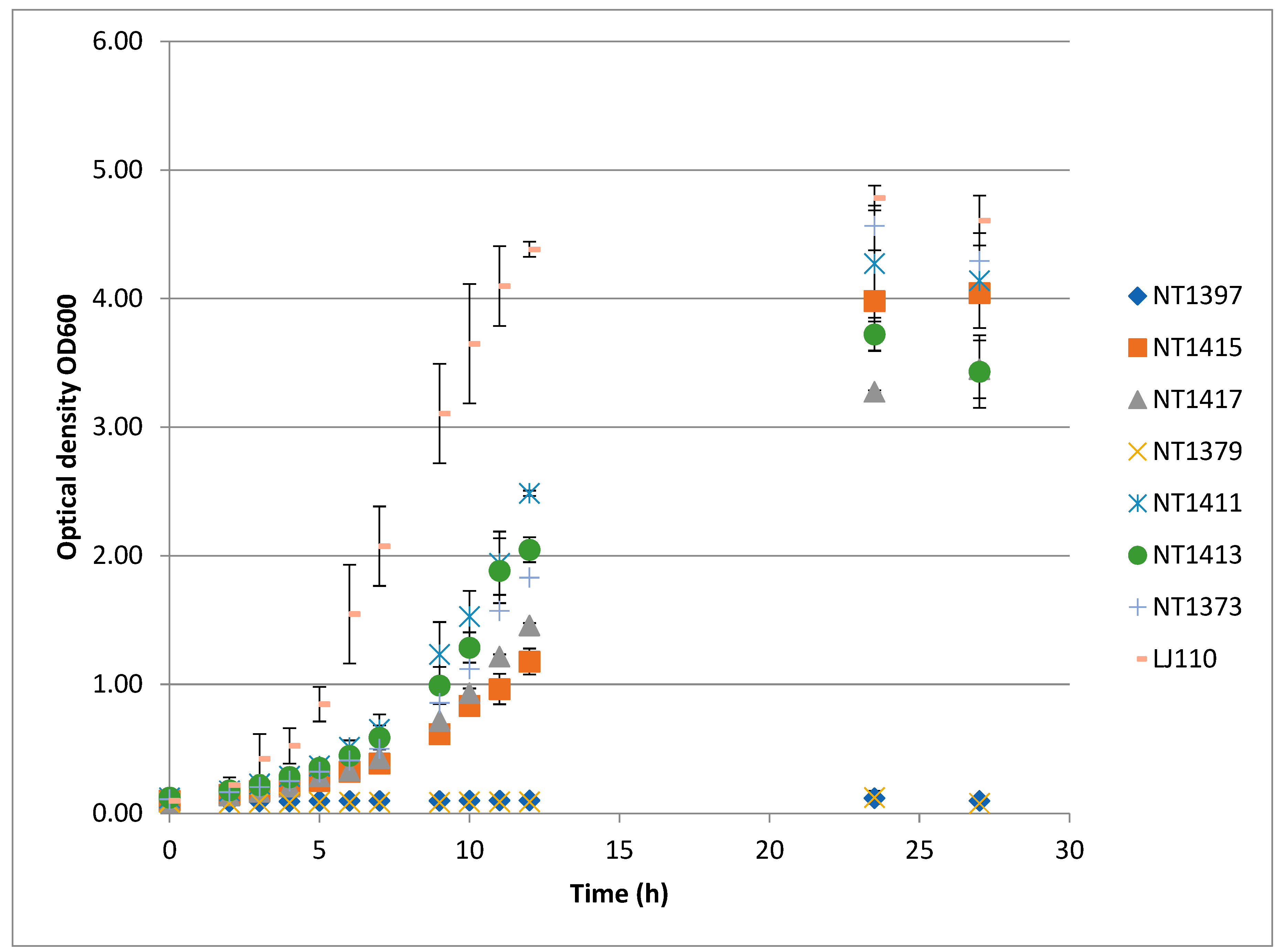
We then tested whether these single-copy genes could restore growth in our reporter gene system. Growth in shake flasks (minimal medium) was followed at 37 °C. As can be seen from Figure 5, the control strains NT1379 and NT1397 were unable to grow, whereas the wild-type strain LJ110 displayed typical growth with a final OD of ca. 4.6. The basic reporter strain NT1373 (ΔserA fuc::PserA-6xPro-serA), which is efp wild-type, apparently grew slower than LJ110, however. This may be due to the lower gene expression of the modified serA gene (see transcript analyses).
Interestingly, all strains with chromosomally inserted Ptac-efp grew and reached final OD values of >3 in 27 h of cultivation and were in the range of the control strain, NT1373. Thus, growth was restored. The strains, however (including NT1373), all showed a prolonged growth lag compared to the wild-type strain. Strain NT1411 (Ptac-efp-Ec) reached a final OD of >4. Next in rank was NT1415, with only marginally reduced growth yield. This can be interpreted as the EpmA modification system not being absolutely essential for EF-P function in E. coli.
Strain NT1413 with Ptac-efpSa grew well and reached a final OD of ca. 3.4 (see Table 4). The corresponding strain with ΔepmA (NT1417) gave the same final OD value, although its growth rate was lower. The efp gene from the S. aureus (Gram-positive) is thus able to complement the growth defect exerted by the deletion of the E. coli efp gene in our reporter system. The EpmA protein of E. coli is not necessary for the complementation.
The average values were calculated based on the data obtained from three independent experiments.

3.5. Transcript Analyses of Recombinant Strains

The serA gene was transferred to the fuc-locus with the original promoter region (PserA) in strains NT1373, NT1411, NT1413, NT1415, and NT1417. We wanted to know whether the expression of the 6xPro-serA gene was in the same range as the transcription of the wt serA gene. Therefore, a qRT-PCR analysis was performed. The transcript values were normalized to the chromosomal ftsZ gene. Differences in the expression of the serA variants became apparent in the strains with fuc-localized variants in comparison to the strains with the wild-type serA gene (see Table 5). Thus, the lack of full restoration of growth in the efp strains can be best compared with strain NT 1373 (ΔserA fuc:PserA-6Pro-serA).
The complete restoration of cell growth (to the level of NT1373 strain) after the introduction of the efp gene from S. aureus into the efp-Ec-deficient strain (NT1413) and into the isogenic strain with deleted epmA (NT1417) allows us to draw a conclusion about the functionality of the EF-P-Sa translational factor in E. coli cells. Moreover, recombinant EF-P-Sa (as well as EF-P-Ec) does not appear to require post-translational modification via the EpmA of E. coli. The gene for the EF-P protein from S. aureus was able to complement the efp deletion of the reporter strain and restored the ability of the strain to grow on minimal medium. This is in accordance with the notion that the post-translational EF-P modification is not strictly required but rather may instead play a more nuanced role in EF-P activity [8].
In conclusion, we can state that recombinant serA genes with integrated 4x or 6xPro residues can be used as excellent reporter genes with which to study EF-P-dependency in an E. coli K-12 host strain which is serA-deficient. Without a functional efp gene, growth in mineral media is not restored, highlighting the role of EF-P in rescuing ribosomes from stalling at poly-Pro residues in translation. The novel synthetic reporter system can be used successfully for the expression of the homologous efp gene from E. coli K-12. Additionally, the efp gene from the pathogenic Gram-positive bacterium S. aureus complemented the growth defect regarding serine auxotrophy. In both cases, it is advisable to use chromosomally integrated efp genes to avoid troubles pertaining to plasmid-based expression. Interestingly, the EpmA function (post-translational ß-lysinylation of EF-P) is not necessary for this complementation as the epmA mutants of E. coli did not prevent the activity of the recombinant EF-Ps.

Author Contributions

Conceptualization, N.T., S.V. and G.A.S.; methodology, N.T., S.V., A.B. and G.A.S.; software, N.T. and A.B.; validation, N.T., A.B. and G.A.S.; formal analysis, N.T., S.V. and G.A.S.; investigation, N.T. and A.B.; resources, G.A.S.; data curation, N.T. and A.B.; writing—original draft preparation, N.T. and G.A.S.; writing—review and editing, N.T., A.B., S.V. and G.A.S.; visualization, N.T.; supervision, S.V. and G.A.S.; project administration, G.A.S.; funding acquisition, G.A.S. All authors have read and agreed to the published version of the manuscript.

Funding

We thank the Deutsche Akademische Austauschdienst (German Academic Exchange Service DAAD) for a travel grant from the Evgenij Savojskij programme (No. 91704572) given to Aydar Bikmullin for his stay at the Institute of Microbiology in Stuttgart. We are grateful to the Ministerium für Forschung und Kunst of Baden-Württemberg for their continuous institutional support.

Data Availability Statement

All data presented are contained within this manuscript.

Conflicts of Interest

The authors declare no conflicts of interest. The funders had no role in the design of the study; in the collection, analyses, or interpretation of data; in the writing of the manuscript; or in the decision to publish the results.

References

  1. Ude, S.; Lassak, J.; Starosta, A.L.; Kraxenberger, T.; Wilson, D.N.; Jung, K. Translation elongation factor EF-P alleviates ribosome stalling at polyproline stretches. Science 2013, 339, 82–85. [Google Scholar] [CrossRef] [PubMed]
  2. Peil, L.; Starosta, A.L.; Lassak, J.; Atkinson, G.C.; Virumae, K.; Spitzer, M.; Tenson, T.; Jung, K.; Remme, J.; Wilson, D.N. Distinct XPPX sequence motifs induce ribosome stalling, which is rescued by the translation elongation factor EF-P. Proc. Natl. Acad. Sci. USA 2013, 110, 15265–15270. [Google Scholar] [CrossRef] [PubMed]
  3. Doerfel, L.K.; Wohlgemuth, I.; Kothe, C.; Peske, F.; Urlaub, H.; Rodnina, M.V. EF-P is essential for rapid synthesis of proteins containing consecutive proline residues. Science 2013, 339, 85–88. [Google Scholar] [CrossRef]
  4. Huter, P.; Arenz, S.; Bock, L.V.; Graf, M.; Frister, J.O.; Heuer, A.; Peil, L.; Starosta, A.L.; Wohlgemuth, I.; Peske, F.; et al. Structural basis for polyproline-mediated ribosome stalling and rescue by the translational elongation factor EF-P. Mol. Cell 2017, 68, 515–527. [Google Scholar] [CrossRef] [PubMed]
  5. Bailly, M.; de Crécy-Lagard, V. Predicting the pathway involved in post-translational modification of elongation factor P in a subset of bacterial species. Biol. Direct. 2010, 5, 3. [Google Scholar] [CrossRef] [PubMed]
  6. Rossi, D.; Kuroshu, R.; Zanelli, C.F.; Valentini, S.R. eIF5A and EF-P: Two unique translation factors are now traveling the same road. Wiley Interdiscip. Rev. RNA. 2014, 5, 209–222. [Google Scholar] [CrossRef]
  7. Rodnina, M.V. Translation in Prokaryotes. Cold Spring Harb. Perspect. Biol. 2018, 10, a032664. [Google Scholar] [CrossRef]
  8. Hummels, K.R.; Kearns, D.B. Translation elongation factor P (EF-P). FEMS Microbiol. Rev. 2020, 44, 208–218. [Google Scholar] [CrossRef]
  9. Zhang, C.; Chen, S.; Fu, X.; Dedkova, L.M.; Hecht, S.M. Enhancement of N-Methyl amino acid incorporation into proteins and peptides using modified bacterial ribosomes and elongation factor P. ACS Chem. Biol. 2024, 19, 1330–1338. [Google Scholar] [CrossRef]
  10. Hanawa-Suetsugu, K.; Sekine, S.I.; Sakai, H.; Hori-Takemoto, C.; Terada, T.; Unzai, S.; Tame, J.R.; Kuramitsu, S.; Shirouzu, M.; Yokoyama, S. Crystal structure of elongation factor P from Thermus thermophilus HB8. Proc. Natl. Acad. Sci. USA 2004, 101, 9595–9600. [Google Scholar] [CrossRef]
  11. Choi, S.; Choe, J. Crystal structure of elongation factor P from Pseudomonas aeruginosa at 1.75 Å resolution. Proteins 2011, 79, 1688–1693. [Google Scholar] [CrossRef] [PubMed]
  12. Blaha, G.; Stanley, R.E.; Steitz, T.A. Formation of the first peptide bond: The structure of EF-P bound to the 70S ribosome. Science 2009, 325, 966–970. [Google Scholar] [CrossRef] [PubMed]
  13. Rajkovic, A.; Ibba, M. Elongation factor P and the control of translation elongation. Annu. Rev. Microbiol. 2017, 71, 117–131. [Google Scholar] [CrossRef] [PubMed]
  14. Lassak, J.; Keilhauer, E.; Fürst, M.; Wuichet, K.; Gödeke, J.; Starosta, A.L.; Chen, J.-M.; Sogaard-Andersen, L.; Rohr, J.; Wilson, D.N.; et al. Arginine-rhamnosylation as new strategy to activate translation elongation factor P. Nat. Chem. Biol. 2015, 11, 266–270. [Google Scholar] [CrossRef] [PubMed]
  15. Lassak, J.; Sieber, A.; Hellwig, M. Exceptionally versatile take II: Post-translational modifications of lysine and their impact on bacterial physiology. Biol. Chem. 2022, 403, 819–858. [Google Scholar] [CrossRef]
  16. Rajkovic, A.; Hummels, K.R.; Witzky, A.; Erickson, S.; Gafken, P.R.; Whitelegge, J.P.; Faull, K.F.; Kearns, D.B.; Ibba, M. Translation control of swarming proficiency in Bacillus subtilis by 5-Amino-pentanolylated Elongation Factor P. J. Biol. Chem. 2016, 291, 10976–10985. [Google Scholar] [CrossRef]
  17. Witzky, A.; Hummels, K.R.; Tollerson, R.; Rajkovic, A.; Jones, L.A.; Kearns, D.B.; Ibba, M. EF-P posttranslational modification has variable impact on polyproline translation in Bacillus subtilis. mBio 2018, 9, e00306-1. [Google Scholar] [CrossRef]
  18. Golubev, A.; Negroni, L.; Krasnovid, F.; Validov, S.; Yusupova, G.; Yusupov, M.; Usachev, K. Posttranslational modification of Elongation Factor P from Staphylococcus aureus. FEBS Open Bio 2020, 10, 1342–1347. [Google Scholar] [CrossRef]
  19. Pinheiro, B.; Scheidler, C.M.; Kielkowski, P.; Schmid, M.; Forné, I.; Ye, S.; Reiling, N.; Takano, E.; Imhof, A.; Sieber, S.A.; et al. Structure and Function of an Elongation Factor P Subfamily in Actinobacteria. Cell Rep. 2020, 30, 4332–4342.e5. [Google Scholar] [CrossRef]
  20. Ohashi, Y.; Inaoka, T.; Kasai, K.; Ito, Y.; Okamoto, S.; Satsu, H.; Tozawa, Y.; Kawamura, F.; Ochi, K. Expression profiling of translation-associated genes in sporulating Bacillus subtilis and consequence of sporulation by gene inactivation. Biosci. Biotechnol. Biochem. 2003, 67, 2245–2253. [Google Scholar] [CrossRef]
  21. Feaga, H.A.; Hong, H.-R.; Prince, C.R.; Rankin, A.; Buskirk, A.R.; Dworkin, J. Elongation factor P is important for sporulation initiation. J. Bacteriol. 2023, 205, e00370-22. [Google Scholar] [CrossRef] [PubMed]
  22. Balibar, C.J.; Iwanowicz, D.; Dean, C.R. Elongation factor P is dispensable in Escherichia coli and Pseudomonas aeruginosa. Curr. Microbiol. 2013, 67, 293–299. [Google Scholar] [CrossRef] [PubMed]
  23. Aoki, H.; Dekany, K.; Adams, S.L.; Ganoza, M.C. The gene encoding the elongation factor P protein is essential for viability and is required for protein synthesis. J. Biol. Chem. 1997, 272, 32254–32259. [Google Scholar] [CrossRef]
  24. Krafczyk, R.; Macošek, J.; Jagtap, P.K.A.; Gast, D.; Wunder, S.; Mitra, P.; Jha, A.K.; Rohr, J.; Hoffmann-Röder, A.; Jung KHennig, J.; et al. Structural Basis for EarP-Mediated Arginine Glycosylation of Translation Elongation Factor EF-P. mBio 2017, 8, e01412-17. [Google Scholar] [CrossRef] [PubMed]
  25. Chaudhuri, R.R.; Allen, A.G.; Owen, P.J.; Shalom, G.; Stone, K.; Harrison, M.; Burgis, T.A.; Lockyer, M.; Garcia-Lara, J.; Foster, S.J.; et al. Comprehensive identification of essential Staphylococcus aureus genes using transposon-mediated differential hybridisation (TMDH). BMC Genom. 2009, 10, 291. [Google Scholar] [CrossRef]
  26. Rajkovic, A.; Erickson, S.; Witzky, A.; Branson, O.E.; Seo, J.; Gafken, P.R.; Frietas, M.A.; Whitelegge, J.P.; Fauli, K.F.; Navarre, W.; et al. Cyclic rhamnosylated Elongation Factor P establishes antibiotic resistance in Pseudomonas aeruginosa. mBio 2015, 6, e00823. [Google Scholar] [CrossRef]
  27. Stauffer, G.V. Regulation of serine, glycine and one-carbon biosynthesis. EcoSalPlus 2013, 1. [Google Scholar] [CrossRef]
  28. Zeppenfeld, T.; Larisch, C.; Lengeler, J.W.; Jahreis, K. Glucose transporter mutants of Escherichia coli K-12 with changes in substrate recognition of IICB(Glc) and induction behavior of the ptsG gene. J. Bacteriol. 2000, 182, 4443–4452. [Google Scholar] [CrossRef] [PubMed]
  29. Jiang, Y.; Chen, B.; Duan, C.; Sun, B.; Yang, J.; Yang, S. Multigene editing in the Escherichia coli genome via the CRISPR-Cas9 system. Appl. Environ. Microbiol. 2015, 81, 2506–2514. [Google Scholar] [CrossRef]
  30. Jeske, M.; Altenbuchner, J. The Escherichia coli rhamnose promoter rhaP(BAD) is in Pseudomonas putida KT2440 independent of Crp-cAMP activation. Appl. Microbiol. Biotechnol. 2010, 85, 1923–1933. [Google Scholar] [CrossRef]
  31. Rodrigues, A.L.; Trachtmann, N.; Becker, J.; Lohanatha, A.F.; Blotenberg, J.; Bolten, C.J.; Korneli, C.; de Souza Lima, A.O.; Porto, L.M.; Sprenger, G.A.; et al. Systems metabolic engineering of Escherichia coli for production of the antitumor drugs violacein and deoxyviolacein. Metab. Eng. 2013, 20, 29–41. [Google Scholar] [CrossRef] [PubMed]
  32. Sambrook, J.; Russell, D.W. (Eds.) Molecular Cloning: A Laboratory Manual, 3rd ed.; Cold Spring Harbor Laboratory Press: New York, NY, USA, 2001; ISBN 978-087969577-4. [Google Scholar]
  33. Albermann, C.; Trachtmann, N.; Sprenger, G.A. A simple and reliable method to conduct and monitor expression cassette integration into the Escherichia coli chromosome. Biotechnol. J. 2010, 5, 32–38. [Google Scholar] [CrossRef] [PubMed]
  34. Woodcock, D.M.; Crowther, P.J.; Doherty, J.; Jefferson, S.; DeCruz, E.; Noyer-Weidner, M.; Smith, S.S.; Michael, M.Z.; Graham, M.W. Quantitative evaluation of Escherichia coli host strains for tolerance to cytosine methylation in plasmid and phage recombinants. Nucleic Acids Res. 1989, 17, 3469–3478. [Google Scholar] [CrossRef] [PubMed]
  35. Tanaka, S.; Lerner, S.A.; Lin, E.C.C. Replacement of a phosphoenolpyruvate-dependent phosphotransferase by a nicotinamide adenine dinucleotide-linked dehydrogenase for the utilization of mannitol. J. Bacteriol. 1967, 93, 642–648. [Google Scholar] [CrossRef]
Figure 1. Complementation of strains NT1352 (ΔserA) (left) and NT1367 (ΔserA Δefp) (right) strains with various plasmids, containing serA (wt), 4-Pro-serA or 6Pro-serA variants. Growth on minimal medium agar supplemented with 0.4% glucose and ampicillin 100 µg/mL.
Applmicrobiol 04 00092 g001
Figure 2. Scheme of integration of various serA gene variants (PserA-serA, PserA-4xPro-serA, or PserA-6xPro-serA) in the chromosomal fuc locus of E. coli K-12.
Applmicrobiol 04 00092 g002
Figure 3. The 12% glycine SDS gel electrophoresis with cell-free extracts (cfe; 25 µg protein/lane) of different strains; lane 1. protein size ladder (Nippon Genetics, Düren, Germany ); lane 2: cfe from strain NT1379 with pJNNmod; lane 3: pJNNmod-efp-Sa; lane 4: pJNNmod-Sa-evo1; lane 5: pJNNmod-Sa-evo2 (line5). Before harvesting, cells were incubated in LB-medium containing ampicillin 100 µg/mL at 37 °C at OD600 ≈ 0.4. IPTG (final concentration of 0.2 mM) was added. After 6 h of expression, the cells were harvested via centrifugation and broken up to obtain the cfe.
Applmicrobiol 04 00092 g003
Figure 4. Growth of strain NT1379 with various pJem2-based plasmids. Shake-flask-based growth (mineral salt medium, 1% of glucose, 50 µg/L of kanamycin, various concentrations of L-rhamnose as inducer). (A) Growth of cells transformed with pJem2 vector; (B) pJem2-efp-Sa plasmid; (C) pJem2-efp-Ec.
Applmicrobiol 04 00092 g004
Figure 5. Growth of the strains in minimal medium supplemented with 1% glucose and 0.2 mM of IPTG.
Applmicrobiol 04 00092 g005
Table 1. Strains and plasmids used in this work. * gene efp from E. coli; ** gene efp from S. aureus.
Strains
NameRelevant GenotypeReference
LJ110E. coli K-12 W3110, wild-type, prototroph[28]
NT1352LJ110 ΔserAThis work
NT1367LJ110 ΔserA ΔefpThis work
NT1368LJ110 ΔserA fuc::PserA-serA ΔefpThis work
NT1373LJ110 ΔserA fuc::PserA 6Pro-serA This work
NT1379LJ110 Δefp ΔserA fuc::PserA 6Pro-serAThis work
NT1397LJ110 Δefp ΔserA fuc::PserA 6Pro-serA ΔepmA This work
NT 1411LJ110 Δefp ΔserA fuc::PserA 6Pro-serA xyl::Ptac-efp-Ec *This work
NT 1413LJ110 Δefp ΔserA fuc::PserA 6Pro-serA xyl::Ptac-efp-Sa **This work
NT 1415LJ110 Δefp ΔserA fuc::PserA 6Pro-serA ΔepmA xyl::Ptac-efp-Ec *This work
NT 1417LJ110 Δefp ΔserA fuc::PserA 6Pro-serA ΔepmA xyl::Ptac-efp-Sa **This work
NT1433LJ110 ΔepmAThis work
NT1435LJ110 ΔefpThis work
NT1437LJ110 Δefp ΔepmAThis work
Plasmids
pTarget FpMB1 aadA sgRNA[29]
pCasrepA101(Ts)kan Pcas-cas9 ParaB-λRed lacIq Plac-sgRNA-pMB1[29]
pTarget–catcatR-cassette into the BclI site of the pTargetF plasmid with disruption of Smr geneThis work
pTarget-catsg-fucpMB1 aadA sgRNA-fucThis work
pTarget-catsg-serApMB1 aadA sgRNA-serAThis work
pTarget-catsg-efppMB1 aadA sgRNA-efpThis work
pTarget-catsg-epmApMB1 aadA sgRNA-serAThis work
pJem2 rhaR rhaS rhaP BAD –T7le-eGFP , mob, kan[30]
pJNNmodColE1 Ptac,lacIq,bla[31]
pSerAColE, lacIq, PserA-serAThis work
p6Pro-serAColE1, lacIq, PserA-6Pro-serAThis work
p4Pro-serAColE1, lacIq, PserA-4Pro-serAThis work
p6Pro-stop-serAColE1, lacIq, PserA-4Pro-UAA-serAThis work
pJNN-efp-Ec *ColE1 lacIqPtac-efp (E. coli)This work
pJNN-efp-Sa **ColE1 lacIqPtac-efp **This work
pJem2-efp-Ec *rhaR rhaS Prha –efp-Ec *, mob, kan This work
pJem2-efp-Sa **rhaR rhaS Prha –efp-Sa *, mob, kan This work
Table 2. List of DNA primers. Bases in italics denote target sequences for Cas9 protein.
NameDNA Sequence
Targ-univ-revactagtattatacctaggactgagctagc
sgRNA-serAggcattctggctgaatcgctgttttagagctagaaatagcaagttaaaataaggctag
sgRNA-efpaaaccggctaccctgtctacgttttagagctagaaatagcaagttaaaataaggctag
sgRNA-epmAgagacacgtttcgttggcccgttttagagctagaaatagcaagttaaaataaggctag
sgRNA-fucgacgaccgtcaataaccggggttttagagctagaaatagcaagttaaaataaggctag
sgRNA-xylgcccaattcgctattccagcgttttagagctagaaatagcaagttaaaataaggctag
3′serA-SphIttttgcatgcttagtacagcagacgggcgcgaatg
5′PserA-serA-SphIttttgcatgcctcttcattaaatttggtgacatgtgtcacg
serA-inv-revttacccaatcctgtcttttgaaatgttgtg
serA-4Proatgccaccaccaccagcaaaggtatcgctggagaaagacaag
serA-6Proatgccaccaccaccaccaccagcaaaggtatcgctggagaaagacaag
QC-Stop-SerA-Icaggattgggtaaatgtaaccaccaccaccagc
QC-Stop-SerA-IItggtggtggtggttacatttacccaatcctgtcttttg
efp-Ec-5-NdeIttttcatatggcaacgtactatagcaacgattttc
efp-Ec-3′HindIIIttttaagcttacttcacgcgagagacgtattc
efp-Sa-3′-BamHIttttggatccttatcctcttgaaatgtagcttccatcacc
efp-Sa-5′-NdeIttttcatatgggcatttcggttaatgattttaaaacagg
L5′del-SerAcgcgtcagctggtgaaactgggcg
L3′del-SerA-BamHIttttggatccagagcaatcgacaattgcctgg
R5′del-SerA-BglIIttttagatctcccgtctgctgtactaattcccc
R3′del-SerAgggtaagggaggattgctcctccc
L5′-del-efpgcgcgatgacaaactcatcttgcg
L3′-del-efp ′-BamHIttttggatcctctaacatgattttaagaccagcacg
R5′-del-efp BglIIttttagatctatgcggttgtggtgcggcctg
R3′-del-efptgctgcgccagaaatcgcgttaccg
L5′-del-epmA-L-5tatcccacagccacgtacttcaggg
L3′-del-epmA-BamHIttttggatcctgacaagggcacgaagtctactcgc
R5′-del-epmA-BglIIttttagatctactgaattaacagcgaagaatggcgtg
R3′-del-epmAgcagctcccatttcagccatcattaagg
FucP-serA-int-5′tgctgtgctcactgttttttctttgggcggtagccaataaccttaacgacatgccccagcaggcgaaaatcctg
FucI-int-3′ggcgagagtgataaagtctgcgccaacgtggccgatggtcagaacccccagggttattgtctcatgagcg
xylA-int-5′gacgaactggtgttgggtaagcgtatggaagagcacttgcgttttgccgcctgctcaaggcgcactcccgttctgg
xylB-int-3′attaaagctgggacattgctcaggccggttaatttcgcggcccaatccagacaccagggttattgtctcatgagcg
RT-qPCR-serA 5′ caagattaagtttctgctggtagaaggcg
RT-qPCR-serA 3′tagcgaccagtttttctgcggcgttg
RT-qPCR-ftsZ-5′tgcatttgcttccgacaacg
RT-qPCR-ftsZ-3′acgtttgtccatgccgatac
Table 3. Growth rates (µ) and final optical density (fOD) in minimal medium for LJ110 (wt), NT1433 (ΔepmA), NT1435 (Δefp), and NT1437 (ΔepmA Δefp). The growth experiments in shake flasks were carried out in triplicate on minimal medium supplemented with 10 g/L glucose.
µ h−1fOD
LJ1100.58 +/− 0.017.14 +/− 0.51
NT14330.41 +/− 0.034.87 +/− 0.28
NT14350.5 +/− 0.035.27 +/− 0.16
NT1437 +/− 0.044.87 +/− 0.04
Table 4. Growth rate (µ) and final optical density (fOD) for the strains with chromosomally integrated efp gene. Cells were inoculated at a starting OD of ~0.07. The NT1411 and NT1415 strains contain the efp gene from E.coli under the control of the Ptac promoter. The NT1413 and NT1417 strains carry a chromosomal copy of the efp gene from S. aureus integrated under the control of the Ptac promoter. n.a. = not applicable, as no increase in OD could be determined.
µ h−1fOD
LJ1100.52 +/− 0.034.6 +/− 0.19
NT13730.24 +/− 0.014.29 +/− 0.42
NT1397n.a.0.09 +/−0.001
NT14150.21 +/− 0.024.04 +/− 0.05
NT14170.25 +/− 0.013.45 +/− 0.22
NT1379n.a.0.08 +/− 0.01
NT14110.26 +/− 0.0014.14 +/− 0.36
NT14130.29 +/− 0.023.43 +/− 0.28
Table 5. The relative expression ratio of the serA gene in various strains. The values were normalized with the ftsZ gene of E.coli. RNA samples were taken from cells in the exponential growth phase (for details, see Section 2).
RQ
LJ1101.76 +/− 0.13
NT13731.11 +/− 0.07
NT14111.12 +/− 0.04
NT14131.10 +/− 0.03
NT14150.92 +/− 0.03
NT14171.22 +/− 0.01
Disclaimer/Publisher’s Note: The statements, opinions and data contained in all publications are solely those of the individual author(s) and contributor(s) and not of MDPI and/or the editor(s). MDPI and/or the editor(s) disclaim responsibility for any injury to people or property resulting from any ideas, methods, instructions or products referred to in the content.