Article

Screening of Rhizobacterial Isolates from Apple Rhizosphere for Their Biocontrol and Plant Growth Promotion Activity

1
Phytopathology Unit, Department of Plant Protection, Ecole National d’Agriculture de Meknès, Km10, Rte Haj Kaddour, BP S/40, Meknes 50001, Morocco
2
Faculty of Sciences Dhar El Mehraz, Sidi Mohamed Ben Abdellah University, Fès-Atlas, B.P. 1796, Fes 30050, Morocco
3
Laboratory of Functional Ecology and Environmental Engineering, Sidi Mohamed Ben Abdellah University, P.O. Box 2202, Route d’Imouzzer, Fez 30000, Morocco
4
National Institute of Agricultural Research (INRA), CRRA, Rabat 10090, Morocco
5
Plant Pathology Laboratory, AgroBioSciences, College of Sustainable Agriculture and Environmental Sciences, Mohammed VI Polytechnic University, Lot 660, Hay Moulay Rachid, Ben Guerir 43150, Morocco
*
Author to whom correspondence should be addressed.
Appl. Microbiol. 2023, 3(3), 948-967; https://doi.org/10.3390/applmicrobiol3030065
Submission received: 6 August 2023 / Revised: 17 August 2023 / Accepted: 21 August 2023 / Published: 22 August 2023
(This article belongs to the Special Issue Applied Microbiology of Foods 2.0)

Abstract

:
Apple crops are prone to several diseases that limit their production—in particular, root rot caused by a new genus of oomycetes, mainly Phytopythium vexans. This study aims to screen antagonistic bacteria that can play an important role in the biological control of this pathogenic oomycete and to evaluate their capacity to promote plant growth. The dual culture test revealed that, out of 200 bacterial isolates, 16 have been able to inhibit the mycelial growth of P. vexans with inhibition rates greater than 50%. The selected isolates were identified based on the 16S rDNA genes: 14 bacteria belonging to the genus Bacillus, Stenotrophomonas, and the family Enterobacteriaceae. Notably, two isolates, B1 and M2-6 (identified as Bacillus velezensis), demonstrated the highest inhibition rates of 70% and 68%, respectively. These selected isolates were examined for their ability to produce different compounds related to biocontrol and plant growth promotion. Furthermore, the 16 selected isolates were evaluated for their ability to produce compounds associated with biocontrol and plant growth promotion, including hydrolytic enzymes (cellulases, proteases, and amylases), HCN (hydrogen cyanide) production, phosphate solubilization, IAA (indole-3-acetic acid) production, pectinase production, and stimulation of sorghum bicolor growth in vivo. Variations were observed among the bacterial isolates in terms of their compound production and phytostimulation capabilities. However, the secretion of proteases was consistently detected in all antagonistic isolates. The presence of genes responsible for the production of antifungal lipopeptides (bacillomycin, fengycin, and iturin) in the selected bacterial isolates was determined using polymerase chain reaction (PCR) techniques, while the absence of genes involved in surfactin biosynthesis was also confirmed through PCR studies. These isolates demonstrated inhibitory activity through the production of proteases and antifungal lipopeptides. Further research is needed to explore their potential use in biological control strategies and to improve apple crop productivity.

1. Introduction

Morocco has achieved considerable growth in apple cultivation, with substantial investment in the sector [1]. Apple trees (Malus domestica Borkh) cover a large area, estimated at 32,000 hectares, accounting for around a quarter of the country’s total rosaceous fruit area [2]. The leading apple-growing regions in Morocco comprise the Middle Atlas, Riff, Saïs, Haouz, and Moulouya, together representing over 56% of the total apple area [3]. Annual production of apple fruits in Morocco is between 560,000 and 600,000 tons, with an average yield of around 20 tons per hectare [4]. These underline the economic impact of apple trees in Morocco, contributing to national food security, rural livelihoods, and export earnings [5].
Unfortunately, Phytopythium vexans, a member of the oomycete group, has proven to be a serious pathogen responsible for apple root rot worldwide [6,7,8]. Recently during the 2000s, P. vexans was the main cause of root rot in Morocco [9,10]. This devastating disease has been responsible for serious losses in apple production [11]. P. vexans is characterized by its capacity to attack the root system, resulting in root rot, poor water, and nutrient uptake, and eventually compromising the vigor and productivity of affected apple trees [12]. Symptoms of root rot encompass stunted growth, chlorosis and yellowing of leaves, wilting, browning of roots, and the formation of brown cankers in the crown areas [9,13].
In general, chemical control measures have been broadly employed to combat plant diseases, including apple root rot [14]. Chemical fungicides, notably metalaxyl and phosphorous acid products, have been used to suppress oomycete pathogens including P. vexans and thereby control the disease [15,16,17]. These chemical measures initially showed promise in effectively reducing the incidence and severity of the disease. However, the long-term durability and negative outcomes associated with chemical control methods have raised concerns and required the exploration of alternative approaches [18].
Despite their initial effectiveness, chemical control measures have several drawbacks and limitations [19]. Firstly, the intensive and indiscriminate use of chemical fungicides can lead to the development of pesticide resistance in the target pathogen, including P. vexans. Secondly, continued exposure to fungicides exerts selective pressure on the pathogen population, favoring the survival and proliferation of resistant strains [20,21]. The emergence of fungicide-resistant strains of P. vexans considerably reduces the effectiveness of chemical control measures, making them useless in the management of root rot disease. Furthermore, chemical control measures have negative impacts on human and environmental health [22].
Recognizing the limitations and harmful effects of chemical control practices, researchers have been looking at alternative approaches to the control of P. vexans root rot disease. Biological control, with its potential for efficient disease management, while reducing negative effects on the environment and human health, involves the application of beneficial micro-organisms, such as bacteria, to suppress the growth and activity of plant pathogens [23]. These microorganisms can act through a variety of mechanisms, including the synthesis of antimicrobial compounds, competition for resources, induction of systemic resistance in plants, and modulation of the rhizosphere microbial community [24]. The use of bacteria as biological control agents has attracted increasing attention due to their different functional attributes and their potential compatibility with sustainable agricultural practices [25].
Bacteria, particularly those belonging to the genera Bacillus, Pseudomonas, and Enterobacter, have demonstrated their potential as biological control agents against various plant pathogens, including P. vexans [26,27,28,29]. These bacteria can inhibit the growth and development of phytopathogens by producing antibiotics, lytic enzymes, and siderophores that deprive the pathogen of essential resources or directly lyse its cells [30,31,32]. In addition to their antagonistic activity, some bacteria—namely, PGPR—possess plant growth-promoting properties that can improve plant vigor and productivity [33,34]. Furthermore, bacteria can promote the development of beneficial symbiotic associations, such as mycorrhizal interactions, which further contribute to plant health and resilience.
This study contributes to this growing body of research by examining the use of antagonistic bacteria to control P. vexans-induced diseases, mainly through the secretion of antibiotic substances and competition for resources. In addition, the study aims to assess the ability of selected bacterial isolates to enhance plant growth.

2. Materials and Methods

2.1. Oomycete Pathogen

The oomycete pathogen used in this study was Phytopythium vexans (MK656897). It was previously characterized by Jabiri et al. [9]. This pathogen was isolated from soil collected from an apple- and pear-growing field showing symptoms of root rot during the 2017–2018 growing season in the Fès-Meknès region of Morocco. The fungal culture was maintained on PDA medium (PDA, Merck, Darmstadt, Germany) at 4 °C for up to 6 months. For long-term use, the strain was stored in 25% glycerol at −80 °C in the phytopathology laboratory (ENA-Meknès).

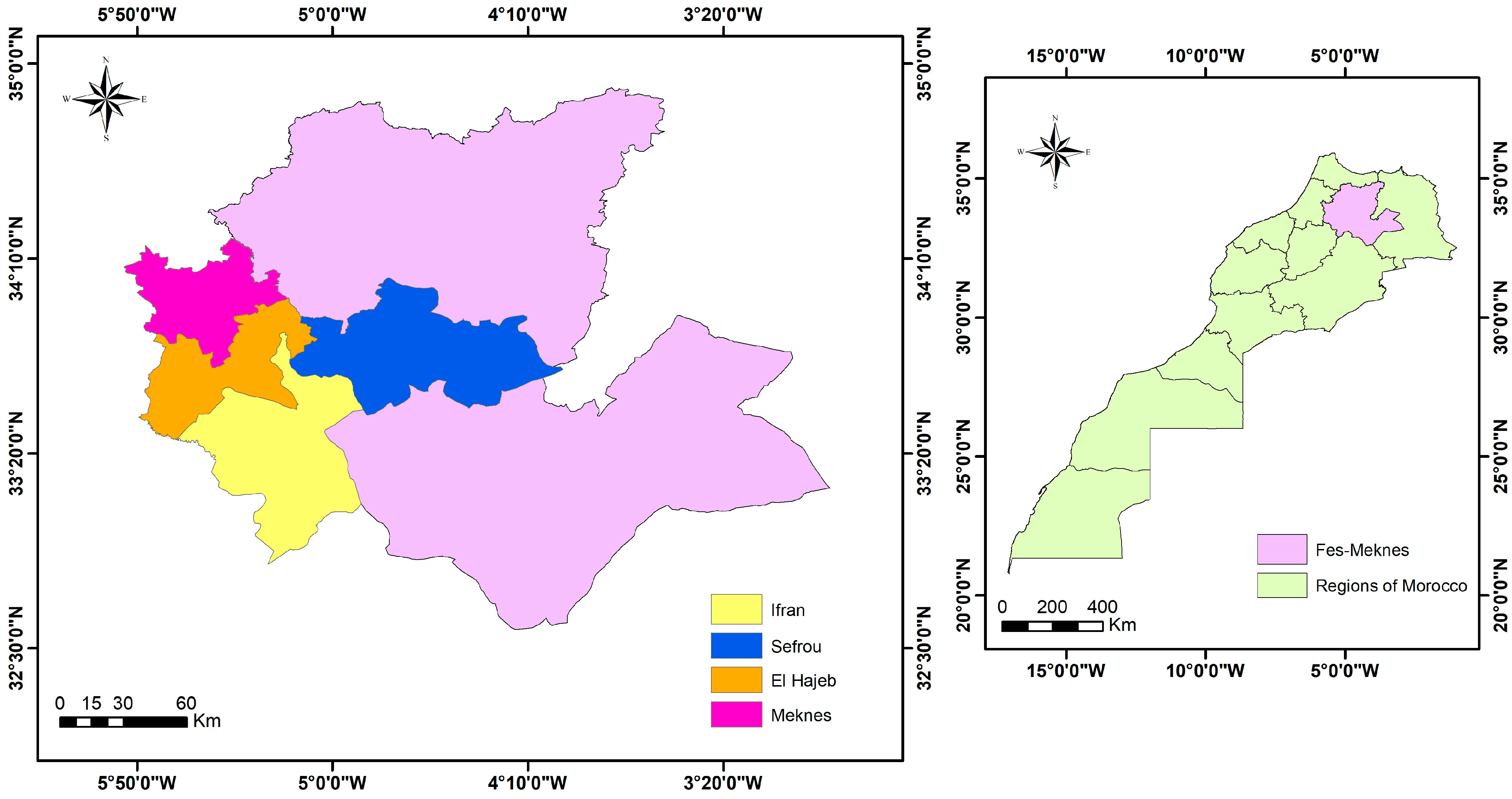
2.2. Isolation of Rhizobacteria from the Rhizospheric Soil

The microorganisms were isolated from the same soils where P. vexans was detected in the regions of Meknes, Azrou (Ifran), El Hajeb, Chelihat (Meknes), Sefrou, and Imouzzer (Sefrou) (Figure 1). The soil samples were collected close to the plants’ root systems. Next, 10 g of each soil sample was suspended in 100 mL of sterile distilled water (SDW), then stirred for 30 min to separate the particles efficiently. To obtain isolated colonies, serial dilution was carried out up to the dilution 10−4. Aliquots of 0.1 mL from suitable dilutions were spread on PDA media plates and further incubated at 25 °C for 48 h. The purification of the colonies having different macroscopic aspects (color, texture) was carried out by subculturing on a PDA medium. The purified isolates were then stored at −20 °C in LB medium with 20% glycerol until pure cultures were obtained.

2.3. Screening for Antagonistic Bacteria

In the PDA media plates, antagonism bioassay was carried out by following the double culture technique, in which the confrontation between isolated rhizospheric bacterial and fungal pathogens was observed [35]. This helped in screening the rhizospheric bacteria with antagonistic properties and substantial pathogen-limiting capacity. To establish this, four lines of each bacterial isolate were streaked with an inoculation loop, all of which were equidistant from the center. In the center of the plate, a 7 mm agar disc of a 7-day-old P. vexans culture was placed. The control was made up of a subculture of a mycelial plug of the pathogen alone in the center of the dishes containing PDA. The plates were sealed with parafilm and incubated at 28 °C for 7 days. The presence or absence of the zone of inhibition was then measured and the inhibition rate (IR) by which mycelium growth was inhibited by bacterial isolates after one week of incubation was calculated. The IR was calculated using the following formula [36]: IR (%) = (C − T)/C × 100, with IR: inhibition rate; C: diameter of the fungal colony in the control plates; and T: diameter of the fungal colony in the presence of the antagonist.

2.4. Effects of Bacterial Isolates on the Mycelial Structure of P. vexans

To reveal the potential damage caused at the mycelial level, a fragment of the mycelium was taken from the zone of inhibition and observed under a light microscope (Ceti Microscopes NLCD-307B, Chalgrove, UK) to examine the existing hyphal damage or the cytological changes, such as deformation, vacuolation, and hyphal swelling, caused by the antagonistic bacterial isolates. The microscopic observations were compared to that of the control.

2.5. Identification of Antagonistic Bacteria

All bacterial isolates with antagonistic properties against P. vexans in the in vitro bioassay were identified, and their genomic DNA was extracted using the protocol described by [37]. Partial 16S rDNA genes of the genome were used for the identification. The DNA of the antagonist isolates was amplified using universal primers, FD1: 5′AGAGTTT-GATCCTGGCT CAG 3′ and RP2: 5′ GGTTACCTTGTTACGACTT 3′ [38]. The PCRmix reaction was performed in a total volume of 25 µL, containing 5 µL of PCR buffer (5×), 1 µL (10 µM) of each primer, 0.2 µL (5 U/µL) of Bioline taq DNA polymerase (Bioline, London, UK), and 2.5 µL of template DNA; the rest of the volume was supplemented with SDW. The following cycling conditions were used: initial denaturation at 96 °C for 4 min, followed by 35 cycles of denaturation at 96 °C for 10 s, then annealing at 52 °C for 40 s and 72 °C for 2 min, and extension at 72 °C for 4 min in a Thermal Cycler. The general amplicon size was between 1500–1600 bp, depending on the bacterial isolate. PCR products were sequenced in both directions using the Sanger sequencing method. The sequences obtained were assembled using DNAMAN software (version 6.0, Lynnon Biosoft, Quebec, Canada), and compared with other bacterial DNA sequences at the National Center for Biotechnology Information (National Center for Biotechnology). Biotechnology Information (http://blast.ncbi.nlm.nih.gov/Blast.cgi (accessed on 6 July 2022)) Partial 16S rDNA sequences have been deposited in Genbank under the accession numbers shown in Table 1.

2.6. Antifungal Effect of Volatile Organic Compounds (VOCs)

The volatile metabolite tests, or the effect of volatile organic compounds (VOCs), were carried out according to the methodology described by Lahlali et al. [35] with minor edits. On a dish containing LB medium, each bacterial isolate was inoculated in three streaks. After 24 h of incubation at 25 °C, the lid of each plate was replaced by the bottom of another plate containing the PDA medium and inoculated with a fresh 7 mm mycelial plug of the pathogen. Subsequently, the two bottoms were sealed with transparent adhesive tape (Parafilm) to prevent any loss of volatile substances [39,40] and then incubated at 25 °C for 5 days. The control of the experiment was composed solely of the pathogen P. vexans. To reveal the fungal toxicity of the volatile metabolites, the growth diameter of the mycelium was measured after 7 days. The inhibition rate was calculated according to the following formula: IR = (C − T)/C × 100.

2.7. Antibiosis by Bacterial Supernatant

To assess the involvement of extracellular diffusible substances secreted by bacterial isolates in the antifungal activity, the bacterial supernatant containing the metabolites was assessed to determine the antagonistic properties [41]. An aliquot (100 µL) of each bacterial suspension (1 × 10⁸ CFU/mL) was inoculated in a nutrient broth (NB), then incubated in a rotary shaker at 30 °C for 3 days (130 rpm). The mixture was centrifuged for 25 min (5500 rpm), and the supernatant obtained from each isolate was filtered through a 0.22 µm diameter Millipore filter. The bacterial cell-free filtrate was incorporated into a PDA medium (45–50 °C) at a concentration of 10% (v/v). A 7 mm pathogen plug mycelium obtained from an actively growing culture of a 7-day-old colony was placed in the center of the plates and then incubated at 25 °C; observations were noted after 7 days of incubation. Control plates were prepared by adding a 10% concentration of liquid NB medium to PDA instead of the bacterial supernatant. The inhibition rate was calculated as described above. There were 2 independent trials with 3 replicates (n = 3).

2.8. Biochemical Characterizations

To evaluate the compounds involved in this phenomenon of observed antagonism, the presence of certain enzymatic activities was observed, namely, cellulolytic, proteolytic, and amylolytic activity.

2.8.1. Proteolytic Activity

The ability of bacterial isolates to produce proteases was determined using a solid medium containing skimmed milk. The medium was inoculated with 5 µL (1 × 10⁸ CFU/mL) of each bacterial suspension. The plates were incubated at 28 °C for 48 h. Protease activity was revealed by the development of a clear halo around the colonies [42]. The proteolytic index was then calculated as the diameter of the halo (mm) + the diameter of a colony (mm)/diameter of one colony (mm), as described by Syed-Ab-Rahman et al. [42].

2.8.2. Amylase Activity

The ability of bacterial isolates to produce amylase was assessed using a solid medium supplemented with soluble starch [43]. We proceeded as follows. First, 5 µL of each bacterial culture (1 × 10⁸ CFU/mL) was inoculated in the Petri dish and incubated at 28 °C for 72 h. To reveal starch hydrolysis, on the surface of the agar plate, 3 mL of the iodine solution was poured. After 3 min, the appearance of a clear zone around the colony indicates amylase activity. In the absence of amylase activity, the starch took on a blackish-blue color. Thus, the amylolytic index was calculated as previously described [42].

2.8.3. Cellulase Degradation

Bacterial antagonists were tested for their ability to produce cellulase using a solid medium supplemented with carboxymethylcellulose (CMC). This medium was inoculated with 5 µL (1 × 10⁸ CFU/mL) of fresh bacterial suspension (1 × 10⁸ CFU/mL). Then, the dishes were incubated at 25 °C for 3 days. Cellulase activity was detected by adding Congo red solution (1%) onto the culture plate and leaving it undisturbed for 15 min, then rinsing it with 1 M NaCl 3 times. The clear zones (halos) around the colonies showed the presence of cellulase activity. The cellulolytic index was calculated as previously described [42].

2.8.4. Pectinase Production

The ability of bacterial isolates to produce pectinases was determined using a solid medium containing pectin [44]. The isolates were spot-inoculated using the inoculum from a bacterial suspension of 1 × 10⁸ CFU/mL on a pectin-based medium. After 3 days of incubation at 25 °C, a 1% cetyltrimethylammonium bromide (CTAB) solution was poured into the dishes. Colonies showing a clear halo after 10 min incubation at room temperature were taken as pectinase producers.

2.8.5. Phosphate Solubilization

To test the ability of bacterial isolates to solubilize inorganic phosphate, Pikovskaya (PVK) medium amended with 5 g/L tricalcium phosphate (Ca3(PO4)2), as the sole source of phosphate, was used as previously described [42,45]. The medium was inoculated with 5 µL (1 × 10⁸ CFU/mL) of each bacterial suspension. The inoculated plates were incubated at 28 °C for 4 days. Bacteria capable of solubilizing phosphate were surrounded by a clear halo, which allowed calculation of the phosphate solubilization index according to the formula described above.

2.8.6. Production of Indole Acetic Acid (IAA)

The production of indole-3-acetic acid (IAA) was determined by the colorimetric method, as described by [46]. An aliquot (100 μL) of each bacterial suspension was cultured in a liquid LB medium supplemented with L-tryptophan (1 g/L) and incubated at 28 °C on a rotary shaker at 150 rpm for 4 days. The cultures were centrifuged at 5000 rpm for 20 min. Subsequently, 1 mL of the cell-free culture supernatant was mixed with 2 mL of Salkowski’s reagent (12 g of FeCl3 per liter of 7.9 M H2SO4) and color development was observed. The appearance of a pink-red color indicates IAA production, while a yellow color indicates a negative result [47,48].

2.8.7. Production of Hydrogen Cyanide (HCN)

The ability of bacterial isolates to produce hydrocyanic acid (HCN) was examined according to the protocol described by [49]. First, 100 μL of each bacterial suspension (1 × 10⁸ CFU/mL) was plated on Levure and Peptone Glucose Agar (LPGA) medium supplemented with 4.4 g of glycine per liter (4.4 g/L). Subsequently, sterile Whatman filter paper discs were saturated with picrate solution (2.5% picric acid in 12.5% anhydrous solution). Sodium carbonate (Na2CO3) solution was placed on the lid of the petri dish. Control plates were inoculated with SDW. The plates were sealed with parafilm and incubated at 28 °C for 4 days. Color change from yellow to orange, red, or reddish-brown indicated the production of volatile HCN [49].

2.8.8. Detection of Lipopeptides by the PCR Method

The total genomic DNA extracted from the selected rhizobacterial isolates was used for the detection of biosynthetic lipopeptides (bacillomycin, fengycin, iturin, and surfactin) and bacteriocin (subtilosin A) [50]. Each PCR amplification was performed in a total volume of 25 µL of PCR mix containing 5 µL of PCR buffer (5×), 1 µL of each primer (10 µM), 0.25 µL of Taq DNA polymerase (5 U/µL) (Bioline, London, UK), and 2.5 µL of genomic DNA; the rest of the volume was made up with SDW. Specific primers used for the amplification of these genes were used. The PCR reactions were carried out in the Thermal Cycler. PCR products were then evaluated on a 1.5% agarose gel stained with cyber safe (Invitrogen, Carlsbad, CA, USA) by electrophoresis and visualized with an ultraviolet illuminator and digitally recorded.

2.9. PGPR Test on Sorghum Bicolor

The PGPR capacities of the effective bacterial isolates were evaluated in vivo on sorghum bicolor plants. The protocol adopted in this experience was described by Syed Ab Rahman et al. [51], with some modifications in our experiments. Sorghum seeds were disinfected with 0.02% sodium hypochlorite for 2 min and then washed 3 times with sterile distilled water. Then, the seeds were dried for 2 h on sterile paper under the flow of the laminar hood. Seeds were immersed in 45 mL of a bacterial suspension at a concentration of 1 × 10⁸ CFU/mL (OD = 0.1 at λ = 600 nm) combined with carboxymethyl cellulose (0.4%) as a gel carrier. Control seeds were treated with carboxymethyl cellulose alone (no bacteria). All tubes were placed at room temperature and shaken (60 rpm) for 12 h. Finally, 10 seeds were sown in 20 cm long conical pots, previously disinfected with sodium hypochlorite, containing sterile soil (sterilization 3 times for 90 min at 121 °C). Sorghum seedlings were harvested 30 days after planting and the following parameters were measured: length of the aerial part (cm), length of the root (cm) and fresh weight of the root (g), and total fresh weight of the plant (g).

2.10. Statistical Analysis

All experiments were repeated twice, following a completely randomized design. Tukey’s test was conducted to determine means separation at a significance level (p ≤ 0.05) using SPSS statistical software (version 20, IBM SPSS Statistics 20, New York, NY, USA).

3. Results

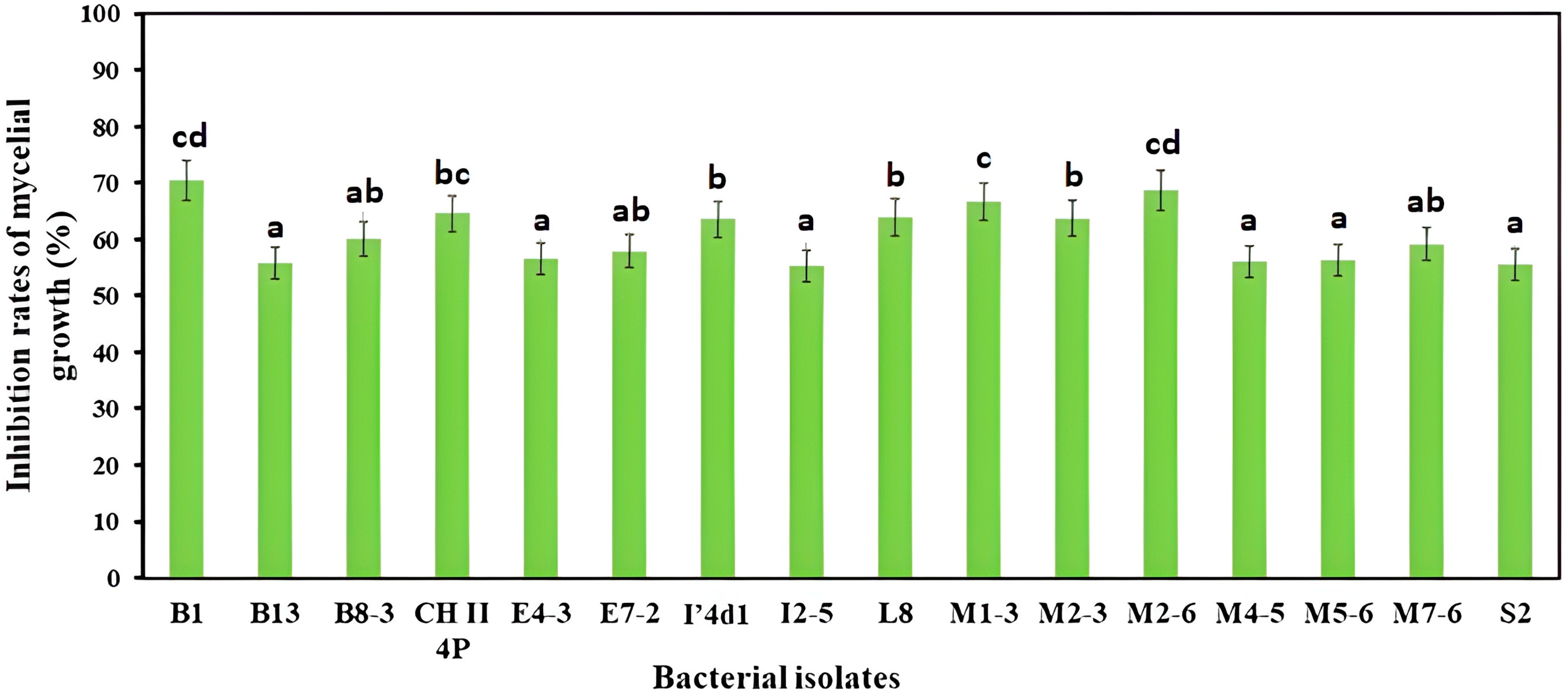
3.1. Dual Culture Bioassay

A total of 200 bacterial isolates were initially obtained from the apple trees’ rhizosphere, which was evaluated for antagonistic activity against P. vexans using the double culture test. The inhibition by each isolate was assessed 7 days after incubation (Figure 2). The results demonstrated that 16 isolates exhibited significant antifungal activity against P. vexans (>55%) (Findex = 64.4; df = 15; p ≤ 0.05). B1 and M2-6 were the most efficient bacterial isolates, with inhibition rates of mycelial growth of 70.57 and 68.72%, respectively.

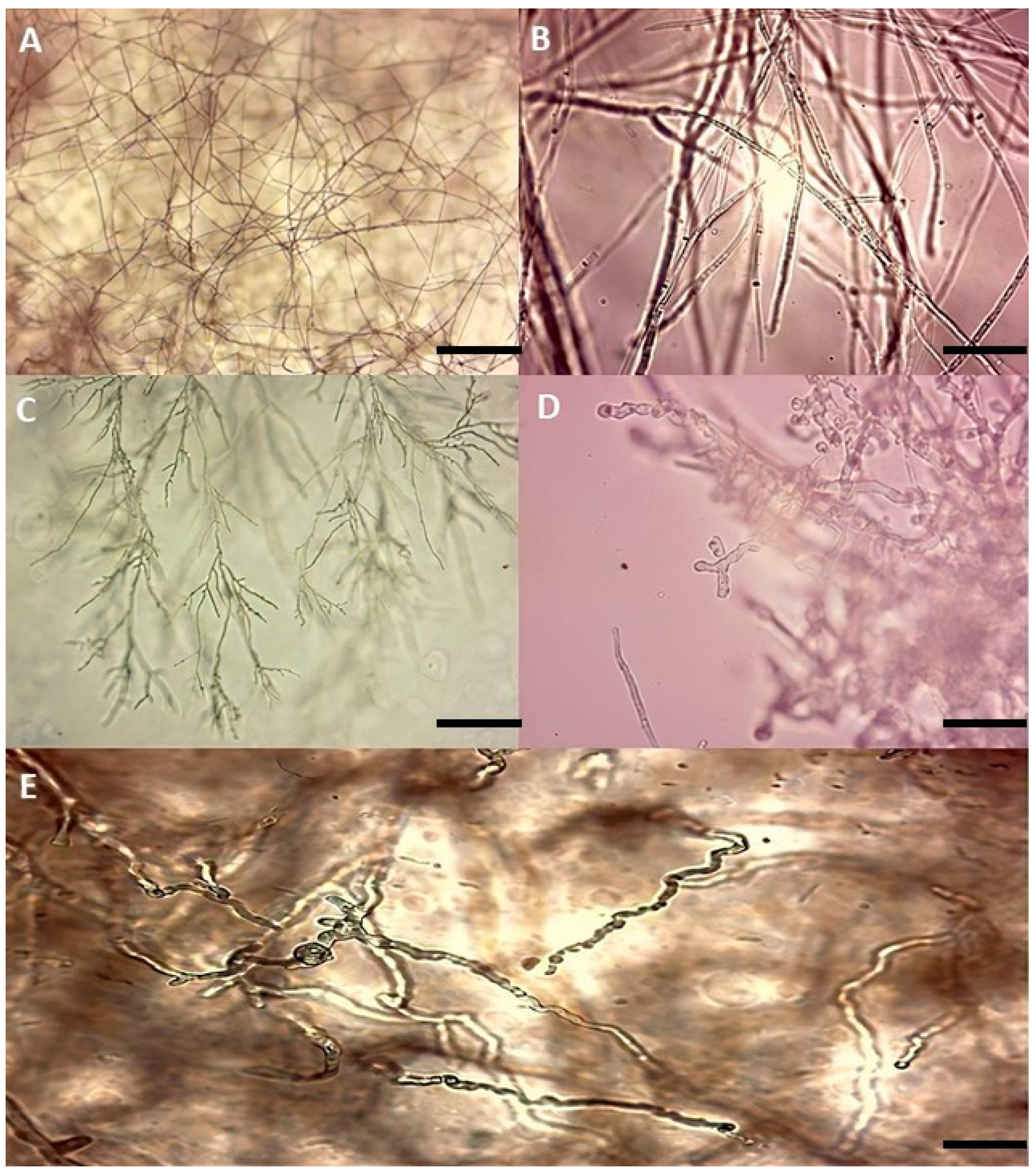
3.2. Microscopic Observation of Mycelium in the Presence of Antagonistic Isolates

Microscopic observations of P. vexans in culture with the different bacterial isolates show a visible shrinkage along with irregular and excessive branching. On the other hand, an abnormal swelling of the diameter and tips of the hyphae was also noted (Figure 3).

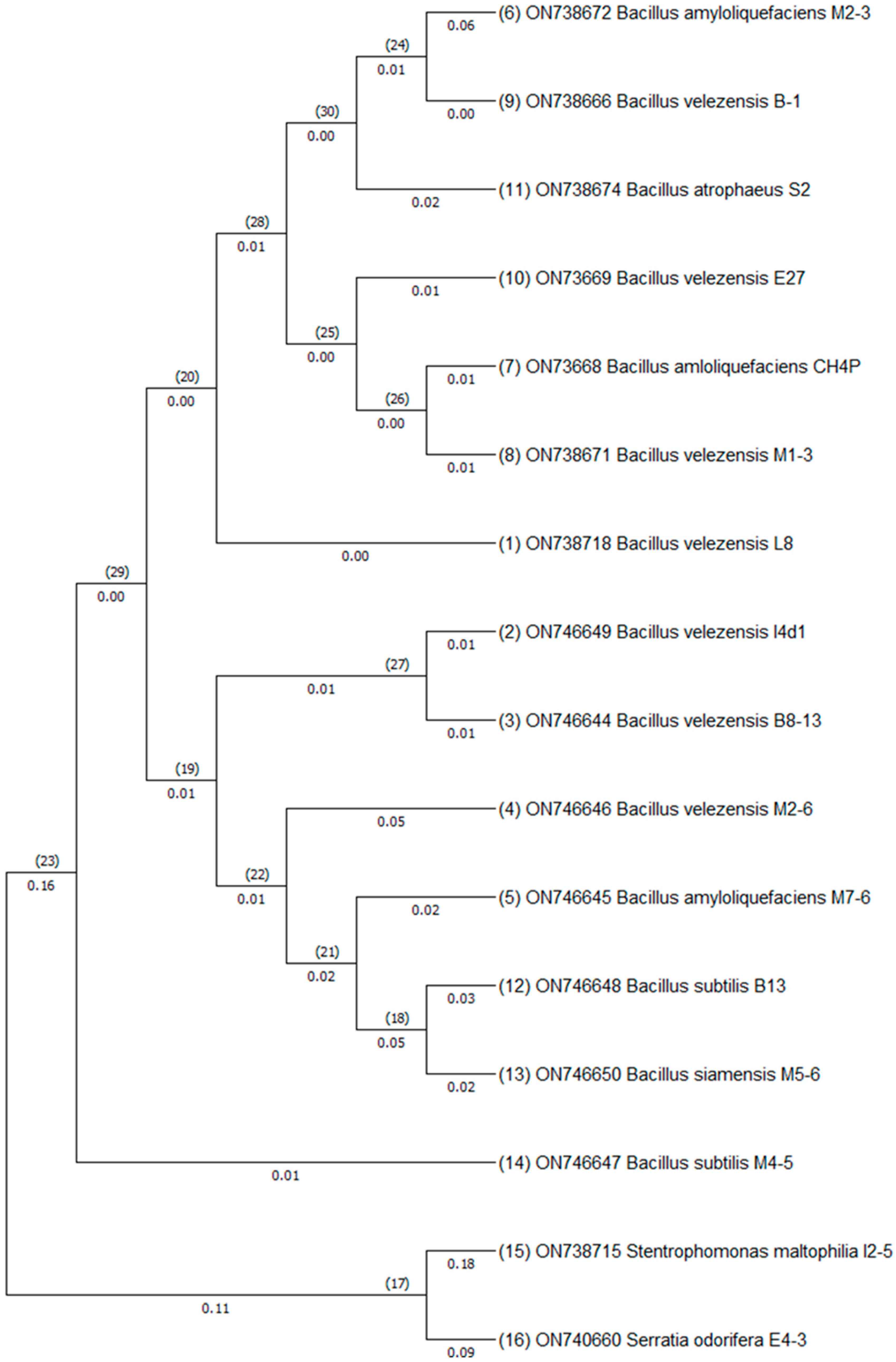
3.3. Identification of Bacterial Isolates Using 16S rDNA Amplicon Sequencing

Based on the 16S rDNA sequence and BLASTn for comparison of species identified (sequence similarity ≥ 98–99%), the 16 selected rhizobacterial isolates with an important antifungal activity against P. vexans were identified as members of the genus Bacillus (14): B. velezensis (7), B. subtilis (2), B. amyloliquefaciens (3), B. atrophaeus (1), B. siamensis (1), Stenotrophomonas maltophilia (1), and Serratia odifera (1) (Figure 4).

3.4. Volatile Organic Compound Effects

The antifungal effect of volatile organic compounds (VOCs) produced by the bacterial isolates was widely lower than those observed during the dual culture assay. Three isolates did not affect the growth of P. vexans, while the rest of the isolates had inhibition rates that did not surpass 33% (Findex = 51.1; df = 15; p ≤ 0.05) (Table 1).

3.5. Antibiosis via Bacterial Supernatant

It was observed that the bacterial cell-free supernatant inhibited mycelial growth at varying inhibition rates (Table 1). Only three isolates (E7-2, I2-5, and I’4D1) showed a rate of inhibition higher than 50%, while the other filtrates showed relatively lower inhibition rates (Findex = 56.9; df = 15; p ≤ 0.05).

3.6. Morphological Characterization of Antagonistic Isolates

Macroscopic and microscopic examination of the selected antagonistic bacterial isolates and their colonies revealed the presence of isolates with similar characteristics, while some were different (Table 2).

3.7. Biochemical Characterization

3.7.1. Lytic Enzymes and Plant-Growth Promoting Production

All the selected isolates were assessed for their potential to synthesize cell wall-degrading enzymes (Table 3). The amylolytic production findings were found to be positive for all examined rhizobacteria. Bacterial strains E4-3 (6.20), M1-3 (4.63), and E7-2 (4.34) had the highest amylolytic index (Findex = 43.2; df = 15; p ≤ 0.05). The results showed that 13 of the 16 isolates tested can degrade cellulose (Table 3). The bacterial isolate E7-2 possessed the highest cellulolytic index (1.277) (Findex = 23.8; df = 15; p ≤ 0.05). Results pointed out that, of the 16 tested bacteria, only 10 could produce pectinase (Table 3). The isolates B13, M4-5, and B8-3 had a high pectinolytic index of 3.769, 2.087, and 2.401, respectively (Findex = 35.4; df = 15; p ≤ 0.05).
It was revealed that all bacterial isolates could synthesize protease (Table 3). The bacterial isolate B8-3 showed the greatest proteolytic index (1.409) (Findex = 27.9; df = 15; p ≤ 0.05). The results showed that none of the bacterial isolates were capable of producing hydrogen cyanide. Regarding phosphate solubilization, the test showed that none of these isolates were able to make calcium triphosphate soluble; therefore, the test was negative for all isolates. The findings of IAA production were found to be positive for four out of sixteen bacterial isolates, those for which the supernatant culture changed to a red color after the addition of Salkowski’s reagent (Table 3).

3.7.2. Detection of Antifungal Lipopeptide Genes by PCR

The bamC gene was detected in nine bacterial isolates. Also, the iturin operon, containing the ituA and ituB genes responsible for iturin biosynthesis, was detected in 12 of the isolates by the apparition of a 2 kb band on the gel, whereas the fenD gene for fengycin was only detected in isolate S2. However, the surfactin sfp gene was not detected in any of the isolates (Table 4).

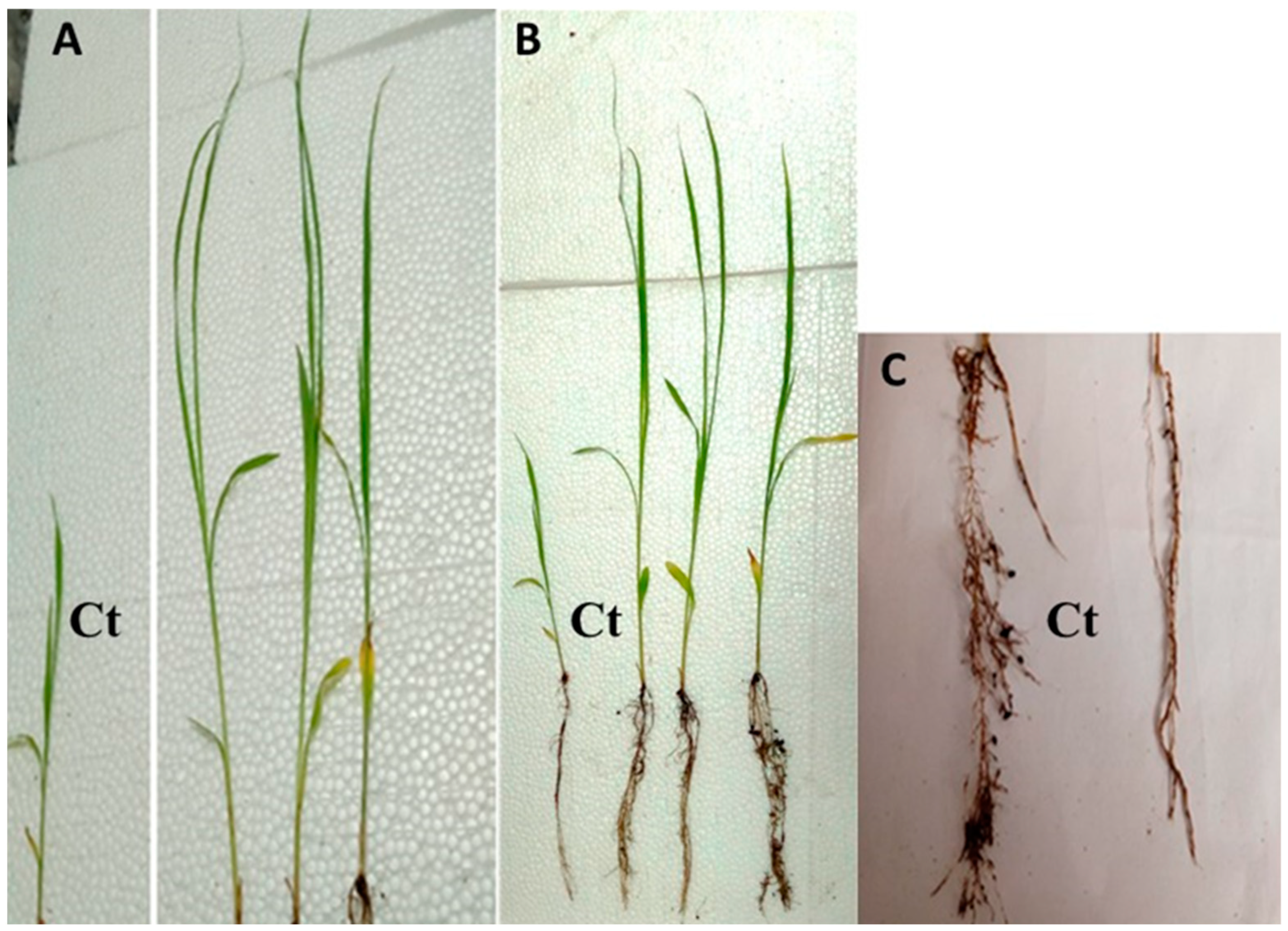
3.8. PGPR Effect of Antagonistic Bacteria In Vivo

Statistical analysis revealed that there was no significant difference in root length between the untreated control and treatment plants with PGPR bacteria (Findex = 16.8; df = 15; p > 0.05). However, there was a significant difference in branching between the control and treated plants (Findex = 29.6; df = 15; p ≤ 0.05) (Table 5). On the other hand, the bacteria had a significant effect on the lengths of the plants’ aerial parts. Isolate M1-3 had the greatest influence on S. bicolor stem length (38.97 cm), followed by isolate M4-5 (38.01 cm) (Findex = 78.4; df = 15; p ≤ 0.05) (Figure 5). Results showed that plants treated only with isolates E4-3 and M7-6 had a significant variation in fresh root weight (Findex = 30.2; df = 15; p ≤ 0.05). The obtained results also demonstrated that there was a significant change in the total fresh weight of the plant, except in the case of the isolate M1-3 (Findex = 36.5; df = 15; p ≤ 0.05) (Table 5).

4. Discussion

Soil is a valuable source of endophytic rhizobacteria that scientists and researchers are trying to harness for disease management in economically important agricultural and horticultural crops. This study aimed to find bacteria that will not only serve as an ecological solution but also bring benefits to the host plant. To achieve this, a collection of 200 bacteria was obtained from the rhizosphere of apple trees. These bacteria were selected for their antagonistic activity against P. vexans using the double culture assay. Sixteen isolates were selected based on their significant antifungal activity against P. vexans, with inhibition rates exceeding 50%. Molecular identification of the selected isolates was performed using partial 16S rDNA genes. The isolates were categorized into three families: Bacillaceae, Pseudomonadaceae, and Enterobacteriaceae. Previous studies have demonstrated the antagonistic effects of these families against various pathogens, validating their potential as biocontrol agents [52,53,54,55,56].
In a study of direct confrontation between the phytopathogens Fusarium sp., Aspergillus niger, and Alternaria sp. and the bacterial isolates Serratia odorifera and Pseudomonas fluorescens, the results revealed the ability of these bacterial species to inhibit the mycelial growth of the mentioned fungal pathogens [57]. The Bacillus genus has been extensively employed as a microbial biopesticide due to its high inhibitory effect on mycelial growth in many studies [31,58,59,60,61]. A study conducted by Liu et al. [62] demonstrated that using Bacillus amyloliquefaciens, B. velezensis, B. siamensis, B. subtilis, and B. atrophaeus as a composite microbial culture resulted in a significant reduction in the abundance of Fusarium sp. and Phytophthora cactorum. Similarly, it led to a substantial increase in the biomass of Malus hupehensis seedlings and young apple trees.
In our study, the two bacterial strains (B1, M2-6; B. velezensis) exhibited the highest inhibition percentage of the mycelial growth of P. vexans. B. velezensis is a well-known biocontrol agent with a higher ability to inhibit the growth of various phytopathogens [63,64,65,66]. In their study, Han et al. [67] demonstrated that the strain FZB24 of B. velezensis significantly inhibited the growth of Phytophthora sojae, causing root and crown rot in soybean. This was attributed to their ability to produce a variety of secondary metabolites [68]. Moreover, these bacteria have undergone structural modifications, ranging from deformation and release of the cytoplasmic content to degradation of the mycelium in certain cases observed at the microscopic examination level.
The damage observed on the hyphae was likely due to the secretion of hydrolytic and lipopeptide enzymes by B. velezensis. These findings are consistent with a biocontrol study conducted on Phythopythium sp. affecting cassava production [26]. In vivo tests conducted in the study of Ferreira et al. [26] showed that B. velezensis, capable of solubilizing phosphate and calcium and synthesizing IAA and siderophores, can promote plant growth even in the presence of the pathogenic fungus. The inhibition rate observed for B. velezensis ranged from 29.08% to 60.76%. Kanjanamaneesathian et al. [69] prepared a concentrated suspension formulation of B. velezensis and evaluated its ability to control root rot and promote growth in cultivated vegetables. The results showed that the suspension concentrates effectively controlled root rot and enhanced vegetable growth when applied by drenching the seedlings. This increase was attributed to the bacterium’s ability to produce IAA.
However, our study found that free-cell wall filtrates represented weak antifungal activity compared to dual confrontation. Similar results were observed in an antagonistic study against pathogenic fungi Stenocarpella maydis and Stenocarpella macrospora, where the bacterial filtrate was less potent [70]. Also, in the study of Legrifi et al. [31], the filtrate of all tested bacteria demonstrated less inhibitory effects against Pythium schmitthenneri than those of the dual culture assay. This implies that other inhibitory mechanisms might be involved. Moreover, it is plausible that the secretion of these inhibitory metabolites in significant quantities necessitates the presence of the pathogen.
Other than diffusible metabolites on agar, antagonism can occur through volatile compounds. The nature of volatile antifungal substances has been demonstrated in several works. For instance, trimethylamine inhibits hyphal extension and arthrospore formation in Geotrichum candidum [71], while hydrogen cyanide produced by Pseudomonas contributes to the control of tobacco root rot [72]. In addition to organic volatiles, the production of inorganic volatiles such as ammonia, as produced by Enterobacter cloacae, has been found to control damping-off caused by Pythium spp. [73]. However, our isolates demonstrated low levels of inhibition by volatile compounds, indicating that the antagonism is mainly achieved via diffusible substances. The nature of the substances responsible for this inhibition was revealed through an investigation of several enzymatic activities. Its antagonistic activity is attributed to the production of a diverse range of secondary metabolites, including antifungal lipopeptides such as surfactins, iturins, and fengycins [74,75,76]. These lipopeptides have been shown to disrupt the cell membranes of fungal pathogens, leading to their inhibition and subsequent degradation [77,78].
In addition to the three lipopeptides mentioned before, B. velezensis produces bacillomycin, a cyclic lipopeptide that exhibits strong antifungal activity against a broad range of plant pathogens [79]. Bacillomycin disrupts fungal cell membranes, leading to the leakage of cellular contents and, ultimately, the death of the pathogen [79]. It has been reported that B. velezensis strains isolated from different environments can produce varying levels of bacillomycin, suggesting a strain-specific variation in antifungal efficacy [80,81]
Moreover, the cell walls of oomycetes are primarily composed of β-1,3- and β-1,6-glucans and, especially, cellulose, which can be inducers of hydrolytic enzymes when interacting with the plant pathogen [82]. Results of our study showed that 77% of the isolates (among the 16) possessed cellulolytic activity, while all of the isolates demonstrated extracellular proteases, suggesting the involvement of cellulases and proteases in antagonism. This inhibition is more likely related to the presence of an extracellular metalloprotease and the release of a lipoprotein, which might bind to the glucan of the pathogen cell wall. These findings might explain the direct inhibition of pathogen mycelial growth by the bacterial antagonists.
Cellulases are enzymes that degrade cellulose, a major component of the fungal cell wall [83]. The ability of the isolates to produce cellulases indicates their potential to degrade fungal cell walls, thereby inhibiting pathogen growth and suppressing disease development [84,85]. Proteases produced by the isolates play a role in degrading host cell wall proteins, further impeding pathogen invasion [86]. These proteases can degrade various pathogenic factors, including cell wall-degrading enzymes secreted by pathogens, leading to a reduction in pathogen virulence and disease severity [87]. In their study, Moon et al. [88] revealed that B. velezensis strain CE 100 synthesized protease and β-1,3-glucanase, which degrade protein and the β-glucan components of phytopathogenic Phytophthora spp. cell wall, causing the mycelial growth inhibition of the pathogens. Similarly, Mota et al. [89] reported that proteolytic activity was most observed in antagonistic bacteria against a set of studied pathogens.
Regarding amylolytic activity, the majority of our isolates are capable of producing amylase. Additionally, amylases produced by the isolates can contribute to the breakdown of non-living organic matter and plant residues, enriching the soil with carbonaceous matter and promoting nutrient cycling. These enzymes play a crucial role in the decomposition of complex carbohydrates and the release of essential nutrients for plant uptake [90,91,92]. In vitro tests have demonstrated that approximately 27% of isolates are capable of producing indole-3-acetic acid (IAA), a phytohormone that stimulates root development and contributes to cell division and enlargement [93]. Studies have found that certain bacteria belonging to the native soil microbiota, known as plant growth-promoting rhizobacteria (PGPR), can produce this plant hormone [94,95].
On the other hand, the production of pectinases is another trait exhibited by PGP bacteria. Pectinases play a crucial role in preventing plant infections caused by pathogens [96]. The action of pectinases leads to the formation of pectic fragments, which act as elicitors triggering a series of signals that result in the synthesis of defense molecules, some of which have direct antimicrobial properties. This process contributes to the plant’s ability to resist infections [97]. Pectinases, along with amylases, have also been suggested to promote root colonization by bacteria, thereby playing an important role in stimulating plant growth [98]. P. fluorescence and B. subtilis are among the biocontrol and growth-promoting bacteria known for their ability to produce pectinases [96].
The in vivo PGPR test complements the findings obtained from in vitro tests. A group of bacteria exhibited enhanced growth of S. bicolor, commonly known as sorghum. Among these bacteria, S. maltophilia demonstrated the most notable effect on phytostimulation, specifically on the length of the aerial part and the total freshness of the plant. These results strongly suggest that S. maltophilia holds promise as a biocontroller and phytostimulator. However, during the in vitro test, S. maltophilia was only able to produce pectinases and not IAA. In contrast, isolates such as B. velezensis (B8-3) and B. subtilis (B13) were capable of producing both IAA and pectinases, but did not exhibit a significant phytostimulatory effect. This indicates that these isolates may be poor root colonizers and that their effects on plant growth might be mediated by other means that have not been studied in vitro, such as nitrogen fixation, ion chelation, and potassium solubilization. On the other hand, the isolate B. amyloquefaciens (M7-6) produced both IAA and pectinases and it had a significant effect on fresh root weight. These findings align with a study conducted by Dunne et al. [99] which reported that S. maltophilia isolated from the rhizosphere of field-grown sugar beets produced extracellular enzymes, including chitinase and protease, which inhibited the growth of the phytopathogenic fungus Pythium ultimate in in vitro conditions, primarily due to the production of an extracellular protease. This study also confirms the results of Sivasakthi et al. [100], that the genus Pseudomonas is recognized for its remarkable ability to colonize roots and stimulate growth.

5. Conclusions

In conclusion, the results of the in vitro dual culture bioassay, VOCs (in distance effect), and cell-free bacterial culture filtrates show that bacterial isolates have a significant effect on inhibiting the mycelial growth of P. vexans. This includes the two isolates B. velezensis B1 and M2-6. The ability of these bacteria to produce lytic enzymes and lipopeptides is critical to their efficacy as BCAs, as demonstrated herein. Further research is warranted to explore additional mechanisms contributing to their observed effects and to assess their efficacy in greenhouse and field conditions. Implementing these biocontrol strategies can offer sustainable alternatives to chemical pesticides and contribute to the overall health and productivity of apple crops.

Author Contributions

Conceptualization, S.J. and R.L.; methodology, S.J. and R.L.; software, S.J., S.-E.L. and I.L.; validation, R.L. and M.B.A.; formal analysis, R.L.; investigation, R.L.; resources, R.L.; data curation, S.J., M.B. and R.L.; writing—original draft preparation, S.J., M.B. and I.L.; writing—review and editing, S.-E.L., I.L., F.M. and R.L.; supervision, R.L. and M.B.A.; project administration, R.L.; funding acquisition, R.L. All authors have read and agreed to the published version of the manuscript.

Funding

This research was financially supported by the Phytopathology Unit, Department of Plant Protection, Ecole Nationale d’Agriculture de Meknes.

Institutional Review Board Statement

Not applicable.

Informed Consent Statement

Not applicable.

Data Availability Statement

Data are available under reasonable demand.

Conflicts of Interest

The authors declare no conflict of interest.

References

  1. Moussadek, R. Impacts de l’agriculture de Conservation Sur Les Propriétés et La Productivité Des Vertisols Du Maroc Central. Afr. Focus 2012, 25, 1–16. [Google Scholar] [CrossRef]
  2. El Jaouhari, N.; Abouabdillah, A.; Bouabid, R.; Bourioug, M.; Chaoui, M. Assessment of Sustainable de Fi Cit Irrigation in a Moroccan Apple Orchard as a Climate Change Adaptation Strategy. Sci. Total Environ. 2018, 642, 574–581. [Google Scholar] [CrossRef] [PubMed]
  3. Nabyl, B.; Hanane, L.; Houda, E.K.; Zakaria, A.; Otman, H.; Rahali, K.; Aouane, E.M. Evaluation of the Physico-Chemical Properties of Soil and Apple Leaves (Malus Domestica) in Beni Mellal-Khenifra Region, Morocco. Adv. Sci. Technol. Eng. Syst. 2020, 5, 1103–1108. [Google Scholar] [CrossRef]
  4. Moinina, A.; Lahlali, R.; Boulif, M. Important Pests, Diseases and Weather Conditions Affecting Apple Production in Morocco: Current State and Perspectives. Rev. Marocaine Sci. Agron. Vétérinaires 2019, 7, 71–87. [Google Scholar]
  5. El Yaacoubi, A.; El Jaouhari, N.; Bourioug, M.; El Youssfi, L.; Cherroud, S.; Bouabid, R.; Chaoui, M.; Abouabdillah, A. Potential Vulnerability of Moroccan Apple Orchard to Climate Change–Induced Phenological Perturbations: Effects on Yields and Fruit Quality. Int. J. Biometeorol. 2020, 64, 377–387. [Google Scholar] [CrossRef]
  6. Prencipe, S.; Savian, F.; Nari, L.; Ermacora, P.; Spadaro, D.; Martini, M. First Report of Phytopythium vexans Causing Decline Syndrome of Actinidia Deliciosa ‘Hayward’in Italy. Plant Dis. 2020, 104, 2032. [Google Scholar] [CrossRef]
  7. Thao, L.; Hien, L.; Liem, N.; Thanh, H.; Khanh, T.; Binh, V.; Trang, T.; Anh, P.; Tu, T. First Report of Phytopythium vexans Causing Root Rot Disease on Durian in Vietnam. New Dis. Rep. 2020, 41, 588–2044. [Google Scholar] [CrossRef]
  8. Boari, A.J.; Cunha, E.M.; Quadros, A.F.F.; Barreto, R.W.; Fernandes, A.F. First Report of Phytopythium Sp. Causing Storage Root Rot and Foliage Blight of Cassava in Brazil. Plant Dis. 2018, 102, 1042. [Google Scholar] [CrossRef]
  9. Jabiri, S.; Bahra, C.; Maclean, D.; Radouane, N.; Barka, E.A. Phytopythium vexans Associated with Apple and Pear Decline in the Saïss Plain of Morocco. Microorganisms 2021, 9, 1916. [Google Scholar] [CrossRef]
  10. Jabiri, S.; Lahlali, R.; Bahra, C.; Bendriss Amraoui, M.; Tahiri, A.; Amiri, S. First Report of Phytopythium vexans Associated with Dieback Disease of Apple Trees in Morocco. J. Plant Pathol. 2020, 102, 1319. [Google Scholar] [CrossRef]
  11. Zhou, C.; Pan, X.; Kong, B.; Cun, H.; Li, N.; He, Y.; Ma, J.; Zhang, Y.; Ma, Y.; Cao, K. First Report of Apple Root Rot Caused by Phytopythium vexans in China. Plant Dis. 2022, 106, 3002. [Google Scholar] [CrossRef] [PubMed]
  12. Williamson-Benavides, B.A.; Dhingra, A. Understanding Root Rot Disease in Agricultural Crops. Horticulturae 2021, 7, 33. [Google Scholar] [CrossRef]
  13. Legrifi, I.; Al Figuigui, J.; Radouane, N.; Ezrari, S.; Belabess, Z.; Tahiri, A.; Amiri, S.; Lahlali, R. First Report of Pythium Schmitthenneri on Olive Trees and in Morocco. Australas. Plant Dis. Notes 2022, 17, 3. [Google Scholar] [CrossRef]
  14. Panth, M.; Baysal-Gurel, F.; Avin, F.A.; Simmons, T. Identification and Chemical and Biological Management of Phytopythium vexans, the Causal Agent of Phytopythium Root and Crown Rot of Woody Ornamentals. Plant Dis. 2021, 105, 1091–1100. [Google Scholar] [CrossRef]
  15. Bellini, A.; Ferrocino, I.; Cucu, M.A.; Pugliese, M.; Garibaldi, A.; Gullino, M.L. A Compost Treatment Acts as a Suppressive Agent in Phytophthora Capsici–Cucurbita Pepo Pathosystem by Modifying the Rhizosphere Microbiota. Front. Plant Sci. 2020, 11, 1–13. [Google Scholar] [CrossRef]
  16. Sun, Z.B.; Yuan, X.F.; Zhang, H.; Wu, L.F.; Liang, C.; Feng, Y.J. Isolation, Screening and Identification of Antagonistic Downy Mildew Endophytic Bacteria from Cucumber. Eur. J. Plant Pathol. 2013, 137, 847–857. [Google Scholar] [CrossRef]
  17. Scott, K.; Eyre, M.; McDuffee, D.; Dorrance, A.E. The Efficacy of Ethaboxam as a Soybean Seed Treatment toward Phytophthora, Phytopythium, and Pythium in Ohio. Plant Dis. 2020, 104, 1421–1432. [Google Scholar] [CrossRef]
  18. Marin, M.V.; Seijo, T.E.; Zuchelli, E.; Peres, N.A. Detection and Characterization of Quinone Outside Inhibitor-Resistant Phytophthora Cactorum and p. Nicotianae Causing Leather Rot in Florida Strawberry. Plant Dis. 2022, 106, 1203–1208. [Google Scholar] [CrossRef]
  19. Carbú, M.; González-Rodríguez, V.E.; Garrido, C.; Husaini, A.M.; Cantoral, J.M. New Biocontrol Strategies for Strawberry Fungal Pathogens. In Strawberry: Growth, Development and Diseases; CABI: Wallingford, UK, 2016; pp. 196–211. [Google Scholar]
  20. Maridueña-Zavala, M.G.; Freire-Peñaherrera, A.; Cevallos-Cevallos, J.M.; Peralta, E.L. GC-MS Metabolite Profiling of Phytophthora Infestans Resistant to Metalaxyl. Eur. J. Plant Pathol. 2017, 149, 563–574. [Google Scholar] [CrossRef]
  21. Matić, S.; Gilardi, G.; Gisi, U.; Gullino, M.L.; Garibaldi, A. Differentiation of Pythium Spp. from Vegetable Crops with Molecular Markers and Sensitivity to Azoxystrobin and Mefenoxam. Pest Manag. Sci. 2019, 75, 356–365. [Google Scholar] [CrossRef]
  22. Martins, P.M.M.; Merfa, M.V.; Takita, M.A.; De Souza, A.A. Persistence in Phytopathogenic Bacteria: Do We Know Enough? Front. Microbiol. 2018, 9, 1099. [Google Scholar] [CrossRef] [PubMed]
  23. Pacios-Michelena, S.; Aguilar Gonzalez, C.N.; Alvarez-Perez, O.B.; Rodriguez-Herrera, R.; Chávez-González, M.; Arredondo Valdes, R.; Ascacio Valdes, J.A.; Govea Salas, M.; Ilyina, A. Application of Streptomyces Antimicrobial Compounds for the Control of Phytopathogens. Front. Sustain. Food Syst. 2021, 5, 696518. [Google Scholar] [CrossRef]
  24. Verma, M.; Mishra, J.; Arora, N.K. Plant Growth-Promoting Rhizobacteria: Diversity and Applications. In Environmental Biotechnology: For Sustainable Future; Springer: Berlin, Germany, 2019; ISBN 9789811072840. [Google Scholar]
  25. Kazan, K.; Gardiner, D.M. Targeting Pathogen Sterols: Defence and Counterdefence? PLoS Pathog. 2017, 13, e1006297. [Google Scholar] [CrossRef] [PubMed]
  26. Ferreira, S.C.; Nakasone, A.K.; do Nascimento, S.M.C.; de Oliveira, D.A.; Siqueira, A.S.; Cunha, E.F.M.; de Castro, G.L.S.; de Souza, C.R.B. Isolation and Characterization of Cassava Root Endophytic Bacteria with the Ability to Promote Plant Growth and Control the in Vitro and in Vivo Growth of Phytopythium Sp. Physiol. Mol. Plant Pathol. 2021, 116. [Google Scholar] [CrossRef]
  27. Cheffi, M.; Bouket, A.C.; Alenezi, F.N.; Luptakova, L.; Belka, M.; Vallat, A.; Rateb, M.E.; Tounsi, S.; Triki, M.A. Olea europaea L. Root Endophyte Bacillus velezensis OEE1 Counteracts Oomycete and Fungal Harmful Pathogens and Harbours a Large Repertoire of Secreted and Volatile Metabolites and Beneficial Functional Genes. Microorganisms 2019, 7, 314. [Google Scholar] [CrossRef]
  28. Panth, M.; Witcher, A.; Baysal-Gurel, F. Response of Cover Crops to Phytopythium vexans, Phytophthora nicotianae, and Rhizoctonia solani, Major Soilborne Pathogens of Woody Ornamentals. Agriculture 2021, 11, 742. [Google Scholar] [CrossRef]
  29. Savian, F.; Ginaldi, F.; Musetti, R.; Sandrin, N.; Tarquini, G.; Pagliari, L.; Firrao, G.; Martini, M.; Ermacora, P. Studies on the Aetiology of Kiwifruit Decline: Interaction between Soil-Borne Pathogens and Waterlogging. Plant Soil 2020, 456, 113–128. [Google Scholar] [CrossRef]
  30. Benfradj, N.; Tounsi, S.; Hamdi, N.B. In-Vitro Evaluation of Antagonists and Fungicides in Controlling Citrus Gummosis Caused by Phytophthora, Phytopythium and Pythium Species in Tunisia. Br. Microbiol. Res. J. 2016, 16, 1–14. [Google Scholar] [CrossRef]
  31. Legrifi, I.; Al Figuigui, J.; El Hamss, H.; Lazraq, A.; Belabess, Z.; Tahiri, A.; Amiri, S.; Barka, E.A.; Lahlali, R. Potential for Biological Control of Pythium Schmitthenneri Root Rot Disease of Olive Trees (Olea europaea L.) by Antagonistic Bacteria. Microorganisms 2022, 10, 1635. [Google Scholar] [CrossRef]
  32. Wang, S.; Ruan, C.; Yi, L.; Deng, L.; Yao, S.; Zeng, K. Biocontrol Ability and Action Mechanism of Metschnikowia Citriensis against Geotrichum Citri-Aurantii Causing Sour Rot of Postharvest Citrus Fruit. Food Microbiol. 2020, 87, 103375. [Google Scholar] [CrossRef]
  33. Tariq, M.; Noman, M.; Ahmed, T.; Hameed, A.; Manzoor, N.; Zafar, M. Antagonistic Features Displayed by Plant Growth Promoting Rhizobacteria (PGPR): A Review. J. Plant Sci. Phytopathol. 2017, 1, 38–43. [Google Scholar]
  34. El-sayed, W.S.; Akhkha, A.; El-naggar, M.Y.; Elbadry, M. In Vitro Antagonistic Activity, Plant Growth Promoting Traits and Phylogenetic Affiliation of Rhizobacteria Associated with Wild Plants Grown in Arid Soil. Front. Microbiol. 2014, 5, 651. [Google Scholar] [CrossRef] [PubMed]
  35. Lahlali, R.; Bajii, M.; Jijakli, M.H. Isolation and evaluation of bacteria and fungi as biological control agents. Commun. Agric. Appl. Biol. Sci. 2007, 72, 973–982. [Google Scholar] [PubMed]
  36. Farhaoui, A.; Adadi, A.; Tahiri, A.; El Alami, N.; Khayi, S.; Mentag, R.; Ezrari, S.; Radouane, N.; Mokrini, F.; Belabess, Z.; et al. Biocontrol Potential of Plant Growth-Promoting Rhizobacteria (PGPR) against Sclerotiorum Rolfsii Diseases on Sugar Beet (Beta vulgaris L.). Physiol. Mol. Plant Pathol. 2022, 119, 101829. [Google Scholar] [CrossRef]
  37. Llop, P.; Bonaterra, A.; Peñalver, J.; María, M.; Llop, P.; Bonaterra, A.; Pen, J. Development of a Highly Sensitive Nested-PCR Procedure Using a Single Closed Tube for Detection of Erwinia Amylovora in Asymptomatic Plant Material Development of a Highly Sensitive Nested-PCR Procedure Using a Single Closed Tube for Detection of Erwinia. Appl. Environ. Microbiol. 2000, 66, 2071–2078. [Google Scholar] [CrossRef]
  38. Weisburg, W.G.; Barns, S.M.; Pelletier, D.A.; Lane, D.J. 16S Ribosomal DNA Amplification for Phylogenetic Study. J. Bacteriol. 1991, 173, 697–703. [Google Scholar] [CrossRef]
  39. Herrera, S.D.; Grossi, C.; Zawoznik, M.; Groppa, M.D. Wheat Seeds Harbour Bacterial Endophytes with Potential as Plant Growth Promoters and Biocontrol Agents of Fusarium Graminearum. Microbiol. Res. 2016, 186, 37–43. [Google Scholar] [CrossRef]
  40. Guevara-Avendan, E.; Carrillo, J.D.; Moreno, K.; Méndez-Bravo, A.; Guerrero-Analco, J.A.; Reverchon, F. Antifungal Activity of Avocado Rhizobacteria against Fusarium euwallaceae and Graphium Spp., Associated with Euwallacea Spp. Nr. fornicatus, and Phytophthora cinnamomi. Antonie Van Leeuwenhoek 2018, 111, 563–572. [Google Scholar] [CrossRef]
  41. Li, Z.; Guo, B.; Wan, K.; Cong, M.; Huang, H.; Ge, Y. Effects of Bacteria-Free Filtrate from Bacillus megaterium Strain L2 on the Mycelium Growth and Spore Germination of Alternaria alternata. Biotechnol. Biotechnol. Equip. 2015, 29, 1062–1068. [Google Scholar] [CrossRef]
  42. Syed-Ab-Rahman, S.F.; Carvalhais, L.C.; Chua, E.; Xiao, Y.; Wass, T.J.; Schenk, P.M. Identification of Soil Bacterial Isolates Suppressing Different Phytophthora Spp. And Promoting Plant Growth. Front. Plant Sci. 2018, 9, 1502. [Google Scholar] [CrossRef]
  43. Dinesh, R.; Anandaraj, M.; Kumar, A.; Bini, Y.K.; Subila, K.P.; Aravind, R. Isolation, Characterization, and Evaluation of Multi-Trait Plant Growth Promoting Rhizobacteria for Their Growth Promoting and Disease Suppressing Effects on Ginger; Elsevier GmbH: Amsterdam, The Netherlands, 2015; Volume 173, ISBN 9104952731. [Google Scholar]
  44. Etesami, H.; Alikhani, H.A.; Mirseyed Hosseini, H. Evaluation of Halotolerant Endophytic Bacteria Isolated from the Halophyte Suaeda for Biological Control of Fungal Rice Pathogens. Arch. Phytopathol. Plant Prot. 2019, 52, 560–581. [Google Scholar] [CrossRef]
  45. Slama, H.B.; Cherif-silini, H.; Bouket, A.C.; Qader, M. Screening for Fusarium Antagonistic Bacteria from Contrasting Niches Designated the Endophyte Bacillus Halotolerans as Plant Warden Against Fusarium. Front. Microbiol. 2019, 9, 3236. [Google Scholar] [CrossRef] [PubMed]
  46. Yuttavanichakul, W.; Lawongsa, P.; Wongkaew, S.; Teaumroong, N.; Boonkerd, N.; Nomura, N.; Tittabutr, P. Improvement of Peanut Rhizobial Inoculant by Incorporation of Plant Growth Promoting Rhizobacteria (PGPR) as Biocontrol against the Seed Borne Fungus, Aspergillus niger. Biol. Control 2012, 63, 87–97. [Google Scholar] [CrossRef]
  47. Devi, A.R.; Sharma, G.D.; Majumdar, P.B.; Pandey, P. A Multispecies Consortium of Bacteria Having Plant Growth Promotion and Antifungal Activities, for the Management of Fusarium Wilt Complex Disease in Potato (Solanum tuberosum L.). Biocatal. Agric. Biotechnol. 2018, 16, 614–624. [Google Scholar] [CrossRef]
  48. Bahroun, A.; Jousset, A.; Mhamdi, R.; Mrabet, M.; Mhadhbi, H. Anti-Fungal Activity of Bacterial Endophytes Associated with Legumes against Fusarium Solani: Assessment of Fungi Soil Suppressiveness and Plant Protection Induction. Appl. Soil Ecol. 2018, 124, 131–140. [Google Scholar] [CrossRef]
  49. Lahlali, R.; Mchachti, O.; Radouane, N.; Ezrari, S.; Belabess, Z.; Khayi, S.; Mentag, R.; Tahiri, A.; Barka, E.A. The Potential of Novel Bacterial Isolates from Natural Soil for the Control of Brown Rot Disease (Monilinia fructigena) on Apple Fruits. Agronomy 2020, 10, 1814. [Google Scholar] [CrossRef]
  50. Lahlali, R.; Aksissou, W.; Lyousfi, N.; Ezrari, S.; Blenzar, A.; Tahiri, A.; Ennahli, S.; Hrustić, J.; MacLean, D.; Amiri, S. Biocontrol Activity and Putative Mechanism of Bacillus amyloliquefaciens (SF14 and SP10), Alcaligenes faecalis ACBC1, and Pantoea agglomerans ACBP1 against Brown Rot Disease of Fruit. Microb. Pathog. 2020, 139, 103914. [Google Scholar] [CrossRef]
  51. Syed Ab Rahman, S.F.; Singh, E.; Pieterse, C.M.J.; Schenk, P.M. Emerging Microbial Biocontrol Strategies for Plant Pathogens. Plant Sci. 2018, 267, 102–111. [Google Scholar] [CrossRef]
  52. Solanki, M.K.; Singh, R.K.; Srivastava, S.; Kumar, S. Isolation and Characterization of Siderophore Producing Antagonistic Rhizobacteria against Rhizoctonia Solani. J. Basic Microbiol. 2013, 54, 585–597. [Google Scholar] [CrossRef]
  53. Al-daghari, D.S.S.; Al-abri, S.A.; Al-mahmooli, I.H. Efficacy of Native Antagonistic Rhizobacteria in the Biological Control of Pythium Aphanidermatum -Induced Damping-off of Cucumber in Oman. J. Plant Pathol. 2019, 102, 305–310. [Google Scholar] [CrossRef]
  54. Fira, D.; Dimkić, I.; Berić, T.; Lozo, J.; Stanković, S. Biological Control of Plant Pathogens by Bacillus Species. J. Biotechnol. 2018, 285, 44–55. [Google Scholar] [CrossRef] [PubMed]
  55. Brígido, C.; Singh, S.; Menéndez, E.; Tavares, M.J.; Glick, B.R.; Do Rosário Félix, M.; Oliveira, S.; Carvalho, M. Diversity and Functionality of Culturable Endophytic Bacterial Communities in Chickpea Plants. Plants 2019, 8, 42. [Google Scholar] [CrossRef] [PubMed]
  56. Nair, D.N.; Padmavathy, S. Impact of Endophytic Microorganisms on Plants, Environment and Humans. Sci. World J. 2014, 2014, 250693. [Google Scholar] [CrossRef] [PubMed]
  57. Effmert, U.; Kalderás, J.; Warnke, R.; Piechulla, B. Volatile Mediated Interactions between Bacteria and Fungi in the Soil. J. Chem. Ecol. 2012, 38, 665–703. [Google Scholar] [CrossRef]
  58. Lyousfi, N.; Letrib, C.; Legrifi, I.; Blenzar, A.; El Khetabi, A.; El Hamss, H.; Belabess, Z.; Barka, E.A.; Lahlali, R. Combination of Sodium Bicarbonate (SBC) with Bacterial Antagonists for the Control of Brown Rot Disease of Fruit. J. Fungi 2022, 8, 636. [Google Scholar] [CrossRef]
  59. Aloo, B.N.; Makumba, B.A.; Mbega, E.R. The Potential of Bacilli Rhizobacteria for Sustainable Crop Production and Environmental Sustainability. Microbiol. Res. 2019, 219, 26–39. [Google Scholar] [CrossRef]
  60. Çakmakçi, R.; Dönmez, F.; Aydin, A.; Sahin, F. Greenhouse and Two Different Field Soil Conditions Growth Promotion of Plants by Plant Growth-Promoting Rhizobacteria under Greenhouse and Two Different Field Soil Conditions. Soil Biol. Biotechnol. 2006, 38, 1482–1487. [Google Scholar] [CrossRef]
  61. Shahzad, R.; Khan, A.L.; Bilal, S.; Waqas, M.; Kang, S.M.; Lee, I.J. Inoculation of Abscisic Acid-Producing Endophytic Bacteria Enhances Salinity Stress Tolerance in Oryza Sativa. Environ. Exp. Bot. 2017, 136, 68–77. [Google Scholar] [CrossRef]
  62. Liu, R.; Li, J.; Zhang, F.; Zheng, D.; Chang, Y.; Xu, L.; Huang, L. Biocontrol Activity of Bacillus velezensis D4 against Apple Valsa Canker. Biol. Control 2021, 163, 104760. [Google Scholar] [CrossRef]
  63. Huang, M.H.; Zhang, S.Q.; Di, G.L.; Xu, L.K.; Zhao, T.X.; Pan, H.Y.; Li, Y.G. Determination of a Bacillus velezensis Strain for Controlling Soybean Root Rot. Biocontrol Sci. Technol. 2017, 27, 696–701. [Google Scholar] [CrossRef]
  64. Kadiri, M.; Sevugapperumal, N.; Nallusamy, S.; Ragunathan, J.; Ganesan, M.V.; Alfarraj, S.; Ansari, M.J.; Sayyed, R.Z.; Lim, H.R.; Show, P.L. Pan-Genome Analysis and Molecular Docking Unveil the Biocontrol Potential of Bacillus velezensis VB7 against Phytophthora Infestans. Microbiol. Res. 2023, 268, 127277. [Google Scholar] [CrossRef] [PubMed]
  65. Kanjanamaneesathian, M.; Wiwattanapatapee, R.; Rotniam, W.; Wongpetkhiew, W. Spraying Hydroponic Lettuce Roots with a Suspension Concentrate Formulation of Bacillus velezensis to Suppress Root Rot Disease and Promote Plant Growth. N. Z. Plant Prot. 2014, 67, 213–219. [Google Scholar] [CrossRef]
  66. Martínez-Raudales, I.; De La Cruz-Rodríguez, Y.; Alvarado-Gutiérrez, A.; Vega-Arreguín, J.; Fraire-Mayorga, A.; Alvarado-Rodríguez, M.; Balderas-Hernández, V.; Fraire-Velázquez, S. Genome Sequence of Bacillus velezensis 2A-2B Strain: A Rhizospheric Inhabitant of Sporobolus Airoides (Torr.) Torr., with Antifungal Activity against Root Rot Causing Phytopathogens. Stand. Genom. Sci. 2017, 12, 1–10. [Google Scholar] [CrossRef] [PubMed]
  67. Han, X.; Shen, D.; Xiong, Q.; Bao, B.; Zhang, W.; Dai, T.; Zhao, Y.; Borriss, R.; Fan, B. The Plant-Beneficial Rhizobacterium Bacillus velezensis FZB42 Controls the Soybean Pathogen Phytophthora Sojae Due to Bacilysin Production. Appl. Environ. Microbiol. 2021, 87, e01601-21. [Google Scholar] [CrossRef]
  68. Ye, M.; Tang, X.; Yang, R.; Zhang, H.; Li, F.; Tao, F.; Li, F.; Wang, Z. Characteristics and Application of a Novel Species of Bacillus: Bacillus velezensis. ACS Chem. Biol. 2018, 13, 500–505. [Google Scholar] [CrossRef]
  69. Kanjanamaneesathian, M.; Wiwattanapatapee, R.; Rotniam, W.; Pengnoo, A.; Wongpetkhiew, W.; Tanmala, V. Application of a Suspension Concentrate Formulation of Bacillus velezensis to Control Root Rot of Hydroponicallygrown Vegetables. N. Z. Plant Prot. 2013, 66, 229–234. [Google Scholar] [CrossRef]
  70. Petatán-Sagahón, I.; Anducho-Reyes, M.A.; Silva-Rojas, H.V.; Arana-Cuenca, A.; Tellez-Jurado, A.; Cárdenas-Álvarez, I.O.; Mercado-Flores, Y. Isolation of Bacteria with Antifungal Activity against the Phytopathogenic Fungi Stenocarpella maydis and Stenocarpella macrospora. Int. J. Mol. Sci. 2011, 12, 5522–5537. [Google Scholar] [CrossRef]
  71. Robinson, P.M.; McKee, N.D.; Thompson, L.A.A.; Harper, D.B.; Hamilton, J.T.G. Autoinhibition of Germination and Growth in Geotrichum Candidum. Mycol. Res. 1989, 93, 214–222. [Google Scholar] [CrossRef]
  72. Ramette, A.; Moënne-Loccoz, Y.; Défago, G. Prevalence of Fluorescent Pseudomonads Producing Antifungal Phloroglucinols and/or Hydrogen Cyanide in Soils Naturally Suppressive or Conducive to Tobacco Black Root Rot. FEMS Microbiol. Ecol. 2003, 44, 35–43. [Google Scholar] [CrossRef]
  73. Bardin, S.D.; Huang, H.C.; Moyer, J.R. Control of Pythium Damping-off of Sugar Beet by Seed Treatment with Crop Straw Powders and a Biocontrol Agent. Biol. Control 2004, 29, 453–460. [Google Scholar] [CrossRef]
  74. Meena, K.R.; Tandon, T.; Sharma, A.; Kanwar, S.S. Lipopeptide Antibiotic Production by Bacillus velezensis KLP2016. J. Appl. Pharm. Sci. 2018, 8, 91–98. [Google Scholar]
  75. Vahidinasab, M.; Adiek, I.; Hosseini, B.; Akintayo, S.O.; Abrishamchi, B.; Pfannstiel, J.; Henkel, M.; Lilge, L.; Voegele, R.T.; Hausmann, R. Characterization of Bacillus velezensis UTB96, Demonstrating Improved Lipopeptide Production Compared to the Strain B. velezensis FZB42. Microorganisms 2022, 10, 2225. [Google Scholar] [CrossRef]
  76. Théatre, A.; Cano-Prieto, C.; Bartolini, M.; Laurin, Y.; Deleu, M.; Niehren, J.; Fida, T.; Gerbinet, S.; Alanjary, M.; Medema, M.H. The Surfactin-like Lipopeptides from Bacillus Spp.: Natural Biodiversity and Synthetic Biology for a Broader Application Range. Front. Bioeng. Biotechnol. 2021, 9, 623701. [Google Scholar] [CrossRef]
  77. Fazle Rabbee, M.; Baek, K.-H. Antimicrobial Activities of Lipopeptides and Polyketides of Bacillus velezensis for Agricultural Applications. Molecules 2020, 25, 4973. [Google Scholar] [CrossRef] [PubMed]
  78. Caulier, S.; Nannan, C.; Gillis, A.; Licciardi, F.; Bragard, C.; Mahillon, J. Overview of the Antimicrobial Compounds Produced by Members of the Bacillus Subtilis Group. Front. Microbiol. 2019, 10, 302. [Google Scholar] [CrossRef] [PubMed]
  79. Jin, P.; Wang, H.; Tan, Z.; Xuan, Z.; Dahar, G.Y.; Li, Q.X.; Miao, W.; Liu, W. Antifungal Mechanism of Bacillomycin D from Bacillus velezensis HN-2 against Colletotrichum Gloeosporioides Penz. Pestic. Biochem. Physiol. 2020, 163, 102–107. [Google Scholar] [CrossRef] [PubMed]
  80. Zhang, Y.; Wang, Y.; Qin, Y.; Li, P. Complete Genome Sequence of Bacillus velezensis LPL-K103, an Antifungal Cyclic Lipopeptide Bacillomycin L Producer from the Surface of Lemon. 3 Biotech 2020, 10, 1–6. [Google Scholar] [CrossRef] [PubMed]
  81. Rabbee, M.F.; Ali, M.S.; Choi, J.; Hwang, B.S.; Jeong, S.C.; Baek, K. Bacillus velezensis: A Valuable Member of Bioactive Molecules within Plant Microbiomes. Molecules 2019, 24, 1046. [Google Scholar] [CrossRef]
  82. Nasraoui, B. Pathogenic Fungi and Pseudo-Fungi of Cultivated Plants: Biology, New Systematic, Pathological Interaction; Editions Universitaires Européennes, Schaltungsdienst Lange O.H.G.: Berlin, Germany, 2006. [Google Scholar]
  83. Wilson, D.B. Three Microbial Strategies for Plant Cell Wall Degradation. Ann. N. Y. Acad. Sci. 2008, 1125, 289–297. [Google Scholar] [CrossRef] [PubMed]
  84. Han, J.-H.; Shim, H.; Shin, J.-H.; Kim, K.S. Antagonistic Activities of Bacillus Spp. Strains Isolated from Tidal Flat Sediment towards Anthracnose Pathogens Colletotrichum Acutatum and C. Gloeosporioides in South Korea. Plant Pathol. J. 2015, 31, 165. [Google Scholar] [CrossRef]
  85. Sadeghi, A.; Koobaz, P.; Azimi, H.; Karimi, E.; Akbari, A.R. Plant Growth Promotion and Suppression of Phytophthora Drechsleri Damping-off in Cucumber by Cellulase-Producing Streptomyces. Biocontrol 2017, 62, 805–819. [Google Scholar] [CrossRef]
  86. Kikot, G.E.; Hours, R.A.; Alconada, T.M. Contribution of Cell Wall Degrading Enzymes to Pathogenesis of Fusarium Graminearum: A Review. J. Basic Microbiol. 2009, 49, 231–241. [Google Scholar] [CrossRef] [PubMed]
  87. Thallinger, B.; Prasetyo, E.N.; Nyanhongo, G.S.; Guebitz, G.M. Antimicrobial Enzymes: An Emerging Strategy to Fight Microbes and Microbial Biofilms. Biotechnol. J. 2013, 8, 97–109. [Google Scholar] [CrossRef]
  88. Moon, J.; Won, S.; Ei, C.; Maung, H.; Choi, J.; Choi, S.; Ajuna, H.B.; Ahn, Y.S. Bacillus velezensis CE 100 Inhibits Root Rot Diseases (Phytophthora Spp.) and Promotes Growth of Japanese Cypress (Chamaecyparis obtusa Endlicher) Seedlings. Microorganisms 2021, 9, 821. [Google Scholar] [CrossRef]
  89. Mota, M.S.; Gomes, C.B.; Souza, I.T.; Moura, A.B. Bacterial Selection for Biological Control of Plant Disease: Criterion Determination and Validation. Braz. J. Microbiol. 2017, 48, 62–70. [Google Scholar] [CrossRef]
  90. Neeraja, C.; Anil, K.; Purushotham, P.; Suma, K.; Sarma, P.; Moerschbacher, B.M.; Podile, A.R. Biotechnological Approaches to Develop Bacterial Chitinases as a Bioshield against Fungal Diseases of Plants. Crit. Rev. Biotechnol. 2010, 30, 231–241. [Google Scholar] [CrossRef]
  91. Gajera, H.P.; Vakharia, D.N. Production of Lytic Enzymes by Trichoderma Isolates during in Vitro Antagonism with Aspergillus niger, the Causal Agent of Collar Rot of Peanut. Braz. J. Microbiol. 2012, 43, 43–52. [Google Scholar] [CrossRef]
  92. MohaMMadi, P.; Tozlu, E.; KoTan, R.; Kotan, M. Potential of Some Bacteria for Biological Control of Postharvest Citrus Green Mould Caused by Penicillium Digitatum. Plant Prot. Sci. 2017, 53, 134–143. [Google Scholar] [CrossRef]
  93. Dutta, V.; Sharma, U.; Iqbal, K.; Kumar, R.; Pathak, A.K. Impact of River Channelization and Riverfront Development on Fluvial Habitat: Evidence from Gomti River, a Tributary of Ganges, India. Environ. Sustain. 2018, 1, 167–184. [Google Scholar] [CrossRef]
  94. Kumar, P.; Dubey, R.C.; Maheshwari, D.K. Bacillus Strains Isolated from Rhizosphere Showed Plant Growth Promoting and Antagonistic Activity against Phytopathogens. Microbiol. Res. 2012, 167, 493–499. [Google Scholar] [CrossRef]
  95. Breedt, G.; Labuschagne, N.; Coutinho, T.A. Seed Treatment with Selected Plant Growth-promoting Rhizobacteria Increases Maize Yield in the Field. Ann. Appl. Biol. 2017, 171, 229–236. [Google Scholar] [CrossRef]
  96. Reetha, S.; Bhuvaneswari, G.; Thamizhiniyan, P.; Mycin, T.R. Isolation of Indole Acetic Acid (IAA) Producing Rhizobacteria of Pseudomonas Fluorescens and Bacillus Subtilis and Enhance Growth of Onion (Allim cepa L.). Int. J. Curr. Microbiol. Appl. Sci. 2014, 3, 568–574. [Google Scholar]
  97. Gerbore, J.; Benhamou, N.; Vallance, J.; Le Floch, G.; Grizard, D.; Regnault-Roger, C.; Rey, P. Biological Control of Plant Pathogens: Advantages and Limitations Seen through the Case Study of Pythium Oligandrum. Environ. Sci. Pollut. Res. 2014, 21, 4847–4860. [Google Scholar] [CrossRef]
  98. Mitrofanova, O.; Mardanova, A.; Evtugyn, V.; Bogomolnaya, L.; Sharipova, M. Effects of Bacillus Serine Proteases on the Bacterial Biofilms. Biomed Res. Int. 2017, 2017, 8525912. [Google Scholar] [CrossRef]
  99. Dunne, C.; Crowley, J.J.; Mo, Y.; Dowling, D.N.; De Bruijn, F.J.; Gara, F.O. Biological Control of Pythium Ultimum by Stenotrophomonas Maltophilia W81 Is Mediated by an Extracellular Proteolytic Activity. Microbiology 1997, 143, 3921–3931. [Google Scholar] [CrossRef]
  100. Sivasakthi, S.; Usharani, G.; Saranraj, P. Biocontrol Potentiality of Plant Growth Promoting Bacteria (PGPR)-Pseudomonas Fluorescens and Bacillus Subtilis: A Review. Afr. J. Agric. Res. 2014, 9, 1265–1277. [Google Scholar]
Figure 1. Map showing sampling area of the soil examined for screening of biological control agents against Phytopythium vexans, prepared using ArcGIS software 10.8.
Applmicrobiol 03 00065 g001
Figure 2. Inhibition rates (%) of the selected bacterial isolates showing antagonistic activity. The mean inhibition rates indicated with the same letter are not significantly different, according to the Tukey test (p ≤ 0.05).
Applmicrobiol 03 00065 g002
Figure 3. Microscopic observation of morphological changes in the mycelium of P. vexans in co-culture with selected antagonistic bacterial isolates. (A) control mycelium (×10); (B) mycelium of P. vexans in the presence of isolate M2-3 (×100); (C) mycelium of control (×40); (D) mycelium of P. vexans in the presence of isolate M2-3 (×40); (E) mycelium in the presence of isolate E4-3 (×40). Scale bar = 20 µm.
Applmicrobiol 03 00065 g003
Figure 4. Phylogenetic tree based on maximum-likelihood analysis of nucleotide sequences of 16S rDNA genes of selected antagonist bacterial isolates using a Kimura two-parameter model in MEGA X software version 10.0.5. The tree was evaluated via 1000 bootstrap replications.
Applmicrobiol 03 00065 g004
Figure 5. PGPR in vivo effect of antagonistic bacteria on the growth of S. bicolor: (A) length of aerial part of S. bicolor treated with isolate M1-3; (B) whole plant treated with isolate M1-3; (C) root branching in the presence of isolate M7-6; (Ct) control.
Applmicrobiol 03 00065 g005
Table 1. Inhibition rates (%) of bacterial VOCs and the cell-free filtrate (10%) against the fungal pathogen P. vexans after 7 days of incubation at 25 °C.
Bacterial Isolate CodeRegionSpeciesAccession
Numbers
VOCsCell-Free Filtrates
B1MeknesBacillus velezensisON73866617.92 h31.37 g
B13MeknesB. subtilisON74664820.67 j8.48 a
B8-3AzrouB. velezensisON74664419.34 i18.63 c
CH II 4PChelihat B. amyloliquefaciensON7366813.79 g24.84 d
E4-3El HajebSerratia odiferaON74066000.00 a 11.35 b
E7-2El HajebB. velezensisON7366923.17 l 52.84 o
I’4d1ImouzzerB. velezensisON7466494.59 b 51.43 m
I2-5ImouzzarStenotrophomas matipholiaON7387159.21 e 51.66 n
L8ImouzzarB. velezensisON7387185.48 d 18.72 c
M1-3MeknesB. velezensisON73867100.00 a 40.40 k
M2-3MeknesB. amyloliquefaciensON73867210.19 f 31.03 f
M2-6MeknesB. velezensisON7466464.74 c 30.51 e
M4-5MeknesB. subtilisON74664700.00 a36.43 h
M5-6MeknesB. siamensisON74665032.17 m39.21 j
M7-6MeknesB. amyloliquefaciensON74664521.36 k41.38 l
S2SefrouB. atrophaeusON7386744.74 c37.23 i
Values having the same letter are not significantly different, according to the Tukey test (p ≤ 0.05).
Table 2. Results of macroscopic and microscopic examination of selected antagonistic bacterial isolates.
IsolateColony ColorShapeSurfaceReliefOpacityConsistencyMicroscopic Shape and GroupingGram
B1yellowcircularsmooth, shinyconvextranslucentviscousCocci, diplococci, streptococci+
B13whiteRound with wavy edgematteconcave in the centertranslucentviscousBacilli, diplobacilli, streptobacilli-
B8-3yellowcircularsmooth, shinyconvextranslucentviscousCoccobacilli, in clusters, in chains-
CH II 4PwhitecircularmatteconvexopaqueviscousSporulated bacilli+
E4-3yellowcircularsmooth, shinyconvextranslucentviscousCoccobacilli-
E7-2yellowcircularsmooth, shinyconvextranslucentviscousCocci, diplococci+
I’4d1whiteround with irregular edgematteflatopaqueviscousBacilli, streptobacilli+
I2-5yellowcircularsmooth, shinyconvextranslucentviscousSporulated bacilli, streptobacilli+
L8brownirregular with wavy edgematteflatopaquegranularCoccobacilli in clusters-
M1-3whitishcircularmatteflatopaqueviscousBacilli, diplobacilli, streptobacilli-
M2-3yellowcircularsmooth, shinyconvextranslucentviscousCocci, diplococci, streptococci+
M2-6whitecircularsmooth, shinydomedopaqueviscousBacilli in chains, in clusters-
M4-5yellowcircularsmooth, shinyconvextranslucentviscousBacilli, diplobacilli-
M5-6yellowcircularsmooth, shinyconvextranslucentviscousIsolated cocci, staphylococci-
M7-6yellowcircularsmooth, shinyconvextranslucentviscousCoccobacilli in clusters-
S2yellowcircularsmooth, shinyconvextranslucentviscousIsolated coccobacilli, in chains-
(+): Bacteria Gram+; (-): Bacteria Gram-.
Table 3. The ability of 16 selected antagonist isolates to produce lytic enzymes and plant-growth promoting (PGP) traits involved in the biocontrol mechanisms.
IsolatesAICIPIPrIHCNPSIIAA
B10.66 a0.061 abcd0.000 a0.514 e
B133.09 e0.075 bcd3.769 k0.160 a
B8-31.36 bc0.145 ef2.087 i1.409 h+
CH II 4P2.83 de0.615 h0.000 a0.722 f
E4-36.20 i0.125 def0.753 d0.842 g
E7-24.34 fg1.277 i1.582 g0.251 b
I’4d15.27 h0.168 f0.952 e0.241 b
I2-53.79 f0.42 g0.546 c0.304 bc+
L83.11 e0.019 ab0.417 f0.300 bc
M1-34.63 g0.014 ab0.000 a0.271 b
M2-33.23 e0.036 abc0.467 b0.368 cd
M2-62.27 d0.066 abcd0.000 a0.294 bc
M4-51.14 abc0.000 a2.401 j0.412 d
M5-61.24 bc0.000 a0.000 a0.583 e
M7-61.61 c0.000 a1.686 h0.367 cd+
S20.85 ab0.092 cde0.000 a0.274 b+
AI: amylolytic index, CI: cellulosic index, PI: pectinolytic index, PrI: proteolytic index, HCN: hydrocyanic acid, PSI: phosphate solubilizing index, IAA: indole-3-acetic acid; (+): positive reaction; (−): negative reaction. All indices were calculated as the diameter of the halo (mm) + the diameter of a colony (mm)/diameter of a colony (mm). Values having the same letter are not significantly different, according to the Tukey test (p ≤ 0.05).
Table 4. Results of the screening for antifungal lipopeptide genes by PCR.
IsolatesBacillomycinFengycinIturinSurfactin
B1++
B13+
B8-3+
CH II 4P++
E4-3++
E7-2++
I’4d1+
I2-5+
L8++
M1-3++
M2-3++
M2-6++
M4-5+
M5-6
M7-6
S2+
(+): presence of the gene; (−): absence of the gene.
Table 5. Plant growth-promoting effect of antagonistic bacteria on S. bicolor.
IsolatesLength of Aerial PartFresh Plant WeightFresh Root Weight
B132.01 bc0.537 abcd0.199 bcd
B1333.60 def0.461 a0.172 abc
B8-333.50 de0.524 abc0.187 abc
CH II 4P30.73 ab0.511 abc0.168 ab
E4-333.23 cde0.710 ef0.317 g
E7-236.70 hi0.613 cde0.243 e
I’4d132.50 cd0.604 bcde0.242 e
I2-529.73 a0.45 a0.177 abc
L830.74 ab0.496 ab0.226 de
M1-338.97 j0.741 f0.231 de
M2-335.57 gh0.714 ef0.283 f
M2-637.63 ij0.651 ef0.203 cd
M4-538.01 ij0.646 def0.224 de
M5-634.25 efg0.610 cde0.238 e
M7-635.04 fg0.664 ef0.350 g
S234.20 efg0.486 a0.164 a
Values having the same letter are not significantly different, according to the Tukey test (p ≤ 0.05).
Disclaimer/Publisher’s Note: The statements, opinions and data contained in all publications are solely those of the individual author(s) and contributor(s) and not of MDPI and/or the editor(s). MDPI and/or the editor(s) disclaim responsibility for any injury to people or property resulting from any ideas, methods, instructions or products referred to in the content.

