Article

Probiotic Lactic Acid Bacteria for Vaginal Application. Optimization of Biomass Production and Freeze-Drying Conditions

by
Antonella Marchesi
and
María Elena Fátima Nader-Macías
*
Pharmabiotics Department, CERELA-CONICET, Chacabuco 145, San Miguel de Tucumán 4000, Argentina
*
Author to whom correspondence should be addressed.
Appl. Microbiol. 2023, 3(2), 519-535; https://doi.org/10.3390/applmicrobiol3020037
Submission received: 17 February 2023 / Revised: 3 May 2023 / Accepted: 4 May 2023 / Published: 2 June 2023

Abstract

:
Probiotic formula for the vaginal tract must contain high numbers of viable beneficial bacteria that maintain their characteristics during the production and further storage, in order to exert the claimed probiotic effect. Four probiotic strains—Lactobacillus gasseri CRL1320, Limosilactobacillus reuteri CRL1324, Ligilactobacillus salivarius CRL1328 and Lacticaseibacillus rhamnosus CRL1332—originally isolated from vagina of healthy women exhibiting beneficial properties were evaluated. Therefore, the optimization of growth parameters and dry biomass production with high viability while preserving probiotic features of strains is a great challenge. Different growth conditions in MRS medium were set up in a laboratory bioreactor; two initial pH and agitation were recorded speeds during static or controlled fermentations. Production conditions of 37 °C with controlled pH 6.5 and 5.5-MRS with 150 and 75 rpm stirring speeds were used for CRL1329 and CRL1332 strains, respectively, while static and free pH MRS for CRL1324 and CRL1328 probiotics allowed us to obtain maximal cell viability counts. However, during 12 h of fermentation, biomass yields of 19.3, 16.2 and 15.2 g/L were achieved when CRL1329, CRL1328 and CRL1324 probiotic strains were grown in static and free pH MRS. The highest biomass yield for CRL1332 strain was produced under controlled MRS-initial pH 6.5 and 75 rpm fermentation conditions. To preserve probiotic high biomass viability, freeze-drying was carried out in the presence of different cryoprotective agents. Thus, the highest viable numbers (10.9–11.8 log CFU/g) with survival rates between 91.3 and 95.6% were attained in the presence of 10% trehalose (L. reuteri and L. salivarius), lactose (L. rhammosus) and lactose + trehalose + sucrose mix (L. gasseri). When stability during post-freeze-drying storage was evaluated, probiotic strains showed a remarkably higher viability recovery when stored at 4 °C than at 25 °C for 12 and 3 months, respectively. In addition, surface characteristics of vaginal probiotics were affected to different extents during storage depending on the strain, protective agent and storage time/temperature. Critical factors for growth conditions, drying process and storage stability of probiotic lactobacilli strains were optimized in view to preserve cell high viability and surface features for the design of vaginal probiotic formula.

1. Introduction

Lactobacilli are the dominant bacteria in the healthy human vaginal tract, as evidenced through the recent Microbioma project [1]. They can be applied to restore the vaginal ecosystem and, at the same time, exert different effects, such as stimulation of the immune system and protection against pathogens. The high number of viable micro-organisms in the final formulas is one of the main challenges in the design of probiotic products aimed at producing a beneficial physiological effect in the host, improving the health of different human and animal tracts. Thus, the potential beneficial bacteria must resist the technological conditions of the applied processes for biomass production and further concentration-drying steps, surviving and maintaining the probiotic viability and properties after the storage- and shelf-life of the product.
The evaluation of technological characteristics of micro-organisms for their inclusion in the final formula is essential to complement the selection and design of products/excipients in which the vaginal lactic acid bacteria (VLB) will be combined after their industrial scaling-up [2] and further storage. These requirements are supported by a number of research reports and guidelines from different scientific organizations [3,4], which confirmed the dependence of probiotics effectiveness on the number of viable cells, which must remain between 106 and 108 CFU per formulation dose [5,6] to exert the claimed effect.
In order to obtain high biomass yield for probiotic lactobacilli in a large-scale production, optimal conditions to sustain bacterial growth must be achieved. The bacterial biomass production is frequently carried out using batch systems; laboratory bioreactors are used to look for the for best conditions [7] through adjusting or modifying different extrinsic or physicochemical factors to increase cell biomass production. Among other parameters, modifying growth factors, such as culture medium substrates, pH, temperature, and incubation time, allowed us to improve the amount of produced biomass [8,9]. Concerning optimal pH, acidification of culture media produced and accumulated using lactobacilli acid may affect cell physiology with growth reduction or interruption; pH control close to neutrality can support a higher growth rate. In addition, the growth of probiotic lactobacilli is rarely enhanced through the presence of oxygen, given the microaerophilic nature of lactic acid bacteria; thus, high agitation of the medium must be avoided [10,11].
As a further preservation of high viability and functionality of the biomass, concentration and preservation technologies to assure long-term stability must be applied. A concentration step to increase biomass cell density after fermentation is typically carried out via centrifugation or membrane filtration for water elimination to guarantee long-term storage [11,12]. However, the gold standard process applied is freeze-drying, which involves “drying” the solvent (generally water) to remove it via direct passage to gaseous state from frozen preparations (sublimation). Freeze-drying or lyophilization is carried out at very low temperatures; thus, alteration of the cells is minimal [13,14]. In addition, it is vitally important to determine the stability of the freeze-dried bacteria for their subsequent inclusion in probiotic formulas; although many factors determine the freeze-drying success of lactic acid bacteria, being the intrinsic property of the strain is of primary importance [15]. Other factors, such as water activity, oxygen level and storage temperature, can lead to a loss of cell viability during storage; therefore, the use of cryoprotectants/lyoprotectants is mandatory for cell survival. A diversity of additives was used to protect cells from freeze drying and improve stability [16,17,18,19].
Different VLB strains were previously isolated from healthy women in Tucumán, Argentina [20]. The strains showed appropriate surface and functional characteristics [21,22,23,24,25,26], such as safety [27] and resistance to osmotic stress, temperatures [28] and gastrointestinal tract conditions [29], to be used either as probiotics or combined with phytoderivatives [30]. Phase I assays were also performed to determine strains’ safety in healthy women [31]. The aim of this work was to optimize the growth conditions and biomass production of four selected VLB probiotics: Lactobacillus gasseri CRL1320, Limosilactobacillus reuteri CRL1324, Ligilactobacillus salivarius CRL1328 and Lacticaseibacillus rhamnosus CRL1332. In order to include probiotics VLB in vaginal formulations, the resistance to the freeze-drying process resulting from use of different cryoprotectant additives and probiotics strains stability during storage under different temperatures throughout 12 months was also investigated.

2. Material and Methods

2.1. Micro-Organisms and Culture Conditions

Four strains of probiotic vaginal lactic acid bacteria (VLB)—Limosilactobacillus reuteri CRL1324, Lactobacillus gasseri CRL1320, Lacticaseibacillus rhamnosus CRL1332 and Ligilactobacillus salivarius CRL1328—isolated from healthy urogenital female tract (Tucumán, Argentina) and selected because of their different and complementary properties [25,32,33,34,35] were evaluated. The taxonomic characterization of the strains defined by Zheng [36] was now used. The micro-organisms were stored in milk yeast extract (13% skim milk, 0.5% yeast extract and 1% glucose) at −20 °C, subsequently subcultured three times in MRS broth (De Man, Rogosa and Sharpe) (Biokar Diagnostics, Beauvais, France) and incubated for 12 h at 37 °C. The inoculum (2%, v/v) was performed from the third subculture (O.D560 nm = 0.9–1.0) (Spectronic 20, Bausch and Lomb, Rochester, NY, USA) corresponding to 6–8 log CFU/mL.

2.2. Optimization of Growth Conditions for VLB Biomass Production

To evaluate the influence of pH and stirring conditions on the probiotic strains, batch mode fermentation was used. A 1.5 L bioreactor (Benchtop Bioreactor INFORS HT, Switzerland) containing 1 L of MRS broth was used, with in situ sterilization (121 °C; 20 min). The first set of experiments were performed at initial pH6.5, in static conditions and with free pH drop, and samples were taken every 2 h. For pH assay, culture media were initially adjusted to 6.5 and 5.5 (mimicking vaginal pH value [37]), automatically controlled through addition of 1N NaOH during fermentation process and incubated during 12 h at 37 °C. After VLB inoculation (2%, v/v), the equipment was set up and solutions plugged to maintain stable pH, stirring conditions (150 and 75 rpm), temperature and system operation were verified. After fermentation, samples were taken aseptically from the bioreactor at 0, 2, 4, 6, 8, 10 and 12 h, and LAB growth was determined using viable cell numbers and quantified after serial dilutions and plating on MRS agar (incubated for 16 h; 37 °C). For pH measurements (free pH), a PT-10 pH meter with a penetration electrode (Sartorius AG, Göttingen, Germany) was used, and probiotic biomass (g/L) production at 12 h was determined via weighting the cellular pellet after centrifugation (Beckman Coulter Avanti J-E, Brea, CA, USA). Samples were processed via duplicate, and protocols were carried out twice. Growth in MRS broth pH 6.5 without agitation and free pH drop was used as the control. A lower stirring speed (75 rpm) was also assayed.
To determine the bacterial growth parameters, the modified Gompertz model with 4 parameters was applied, according to:
DOt = N0 + Amax × exp {−exp [(µ × e/Amax) × (λ−t) + 1]}
where ODt = OD at time t (h); N0 = initial OD value; Amax = maximal growth (log CFU/mL); μ =growth rate expressed in h−1; e = Neperian logarithm (2.718281828) and λ = length of the lag phase in h; and basis, as was previously reported [28].

2.3. Optimization of Freeze Drying Conditions

VLB grown in MRS broth after 13 h at 37 °C were washed three times, centrifuged (7000× g; 10 min at 4 °C, Beckman, Germany) and concentrated 10 times to obtain a higher number of micro-organisms (1010–1011 CFU/mL). Subsequently, each VLB was mixed with different cryoprotectants: lactose, sucrose, and trehalose (10%), as well as the combination of the three sugars at equal final concentration (10%), and re-suspended (1:1 v/v) [15]. They were then uniformly distributed in Petri dishes and stored for 48 h at −80 °C. The freeze-drying process was carried out, as previously reported [29], using vertical trays (Lyovac GT2 Freeze Dryer, Germany) for 16 h at 0.3 mbar, and VBL powders with 1% residual humidity were obtained. To evaluate the effect of the cryoprotectants, viable cells number before and after freeze-drying were determined, as described before.

2.4. Stability of Freeze-Dried VLB during Storage

2.4.1. Encapsulation and Storage at Different Temperatures

After freeze drying, VLB powders with the different protectants were uniformly distributed in No 1 hard gelatine capsules (Saporiti, Argentina). Capsules were filled using manual capsule filler (Maclen, Argentina). Later, the capsules were included in aluminium blisters and vacuum-sealed in aluminium bags (Turbovac, Argentina) for their storage at 4 and 25 °C. During different time periods (1, 3, 6, 9 and 12 months), samples were taken, and the number of viable bacteria (log CFU/g) and surface properties of the strains were determined.

2.4.2. Maintenance of VLB Probiotic Properties

Surface properties (autoaggregation and hydrophobicity) of VLB after freeze-drying and storage for periods of 3 months at 25 °C and 12 months at 4 °C, respectively, were evaluated according to the methodology previously reported [25].

2.5. Statistics

The results were expressed as the mean value ± standard deviation of the data. All the assays were performed in duplicate or triplicate. A value of p < 0.05 was considered statistically significant through applying an analysis of variance (ANOVA). The results were analysed using Minitab 17 software. The statistical analysis was carried out considering the maximal values of OD560nm up to 14 h of growth, and the log CFU/mL in each one of the growth media were assayed and selected as optimal.

3. Results

3.1. Optimization of Growth Conditions of VLB Probiotics

To optimize the growth of VLB probiotic strains, the effect of operating settings in the bioreactor were first evaluated. Since the probiotic lactobacilli were isolated from human vagina, it may be assumed that a temperature of 37 °C would be optimal for growth. Initial pH of 6.5 and 5.5, free dropped or maintained during growth, under static or stirred conditions for 12 h were assayed. Results of VLB growing in MRS broth at initial 6.5 value, free pH drop without agitation and 37 °C during 12 h incubation (control) showed final cell counts from 8.78 log CFU/mL for L. rhamnosus CRL1332 to 9.44 log CFU/mL for L. reuteri CRL1324 (Figure 1). Obtained pHs at the end of incubation time were 3.98, 3.99, 4.39 and 4.59 for CRL1328, CRL1332, CRL1320 and CRL1324, respectively.
When probiotic VLB were grown in MRS with H 5.5 controlled (during growth) and agitation (150 rpm) in the bioreactor (Figure 2A), higher viable cell counts were reached using CRL1332 and CRL1324 strains with 9.60 and 9.15 log CFU/mL, while lower final viable cell numbers (8.50 and 8.20 CFU/mL) were obtained for CRL1328 and CRL1320. With the exception of L. rhamnosus CRL1332, a lag-phase of 2 h was observed for the other probiotic strains. On the other hand, VLB probiotics growth in pH 6.5-controlledMRS and agitation (150 rpm) at 37 °C (Figure 2B) exhibited higher final viable counts compared to VLB growing in controlled pH 5.5 MRS. Indeed, final viable counts of 10.10 log CFU/mL were reached using L. gasseri CRL1320, while the other three strains showed 9.60 log CFU/mL at 12 h. A lack of lag-phase was observed for the growth of probiotic strains under these last conditions, maintaining pH 6.5 during the entire incubation time.
As a result of the previous evaluated conditions, the highest final viable count numbers were obtained for L. gasseri CRL1320 and L. rhamnosus CRL1332 when pH 6.5 was maintained/controlled in MRS broth, and 150 rpm were set as the stirring speed in the bioreactor. Due to the microaerophilic characteristic of lactic acid bacteria, excessive aeration can be detrimental for growth; therefore, a lower agitation speed (75 rpm) was assayed for CRL1320 and CRL1332 (Figure 2C), albeit maintaining pH 5.5 during growth. Results showed a dramatic reduction in the final viable numbers (8.15 log CFU/mL) for L. gasseri CRL1320 at 12 h when 75 rpm was set, while maximal viable counts (10 log CFU/mL) were maintained at a speed of 150 rpm. On the other hand, L. rhamnosus CRL1332 increased its final counts to 10 log CFU/mL when medium was stirred at 75 rpm.

3.2. Growth Parameters

Table 1 summarizes the growth parameters of VLB strains under the evaluated conditions. The growth of L. gasseri CRL1320 and L. rhamnosus CRL1332 exhibited maximal growth rate (1.62 ± 0.05 and 1.44 ± 0.01) in controlled pH 6.5-MRS at 150 rpm and controlled pH 5.5-MRS, 75 rpm, respectively. L. reuteri CRL1324, when grown in MRS (initial pH 6.5, with no pH control during growth) in static conditions, exhibited high viable cell counts, close to controlled pH 6.5-MRS at 150 rpm. However, high viable cell numbers were reached using L. rhamnosus CRL1332 and L. gasseri CRL1320 in pH 6.5 controlled (during growth)-MRS and pH 5.5-controlled and 150 rpm, respectively. The number of viable cells showed in the table were the highest obtained, albeit at different times of incubation (plots not showed). The results obtained indicate a high dependence of probiotic strain on the cultivation conditions.

3.3. Bacterial Biomass Yield

Comparison of bacterial performance required to produce cell biomass under different growth conditions is shown in Figure 3. A higher yield of bacterial biomass was obtained when L. gasseri CRL1320, L. reuteri CRL1324 and L. salivarius CRL1328 probiotic strains were grown in free and static MRS-initial pH 6.5, while L. rhamnosus CRL1332 showed maximal biomass production under controlled pH 6.5 MRS conditions and stirred at 150 rpm. However, no significant difference (p > 0.05) was found for biomass produced using L. gasseri CRL1320, L. reuteri CRL1324 and L. salivarius CRL1328 in controlled pH 6.5 MRS stirred at 150 rpm. Wet biomass yields for L. gasseri CRL1320 in free MRS was 19.03 g/L, while values of 8.21 and 6.13 g/L were obtained when it was grown in controlled pH 6.5-MRS/150 and 5.5/150 or 5.5/75 conditions, respectively. Biomass obtained for CRL1324 and CRL1328 probiotic strains also showed maximal values of 15.2 and 16.5 g/L growing in pH free-drop MRS; however, under controlled MRS pH 6.5/150 and 5.5/150 conditions, biomass production was 4.0 and 8.0 g/L, respectively. In addition, high biomass yield was found for L. rhamnosus CRL1332 when it was grown in controlled pH 5.5/150 conditions, while lower biomass yield (16.9 g/L) was recovered under free MRS cultivation; controlled pH 5.5/150 and 5.5/75 yielded 14.24 and 6.5 g/L of biomass, respectively.

3.4. Optimization of Freeze-Drying Conditions and Effect of Cryoprotectants on VLB

To maintain the high viability of VLB probiotics strains when they are administered, it is necessary to ensure the preservation of their high numbers in the obtained biomass. In this study, freeze-drying was used due to the enhanced stability reached using probiotic micro-organisms during storage via the use of cryoprotectants/lyoprotectants. Therefore, the effect of different cryoprotectants was evaluated (Figure 4).
The probiotic cells were differently affected by the protective agents assayed; L. reuteri CRL1324 and L. salivarius CRL1328 were efficiently protected when suspended in trehalose (T), showing the highest viability (11.9 and 10.8 log CFU/g) after the freeze-drying, with estimated survival rates of 95.6 and 93.9%, respectively. Using lactose (L) as a protectant, freeze-dried L. rhamnosus CRL1332 reached maximal viability (11.8 log CFU/g) with a survival recovery of 94.7%. In addition, L. gasseri CRL1320 reached maximal viability (11.6 log CFU/g) when suspended in the lyoprotectants mixture (M), reaching a survivability of 91.3%. With the exception of CRL1324 strain, sucrose (S) as protectant showed the lowest viability recovery.

3.5. Stability of Encapsulated VLB during Storage at Different Temperatures

3.5.1. Stability of VLB Stored at 25 °C

After freeze-drying, VLB probiotic strains were incorporated in hard-gelatine capsules and stored at different temperatures up to 12 months and under vacuum conditions. When maintained at 25 °C, a dramatic viability decrease was observed after 3 months, as shown in Figure 5. Therefore, and knowing that a cell concentration between 6.0 and 9.0 log CFU/g is required in probiotic formulations, the stability of the probiotic VLB was determined after different months of storage. Even when a high survival recovery was exhibited by L. reuteri CRL1324 (>10 log CFU/g) for all the assayed protectants, L. gasseri CRL1320 was able to survive in the range of 6.8–8.3 log CFU/g at 25 °C; its higher viability after 3 months was in the presence of M with 7.6 log CFU/g. In addition, L. rhamnosus CRL1332 remained viable between 7.0 and 8.4 log CFU/g after 3 months, while L. salivarious CRL1328 did not maintain the required viability to be included in probiotic formulas, with a loss of viability that was maximal when L was used as a lyoprotectant (<6.0 log CFU/g).

3.5.2. Stability of Lyophilized VLB Stored at 4 °C

Refrigeration temperatures are used to store and maintain the viability of lyophilized micro-organisms during different periods of time, with conditions being applied in research laboratories, culture collections, pharmaceutical and food product manufacturing. In this study, a decrease in VLB viability after 12 months of storage at 4 °C was observed (Figure 6). After 3 months of storage at this temperature, VLB probiotic strains remained viable, and no significant differences were observed for strains with the different lyoprotectants (Figure 6A), with this result being similar to those obtained at 25 °C (after 3 months). On the other hand, after 12 months of storage at 4 °C, L. gasseri CRL1320 and L. rhamnosus CRL1332 freeze-dried with different protectants maintained their viability around 8.0 log CFU/g (with the exception of CRL1332 when M was used), while L. reuteri CRL1324 and L. salivarius CRL1328 showed 7.0 log CFU/g under the same storage conditions (Figure 6B). As regard as lyoprotectants under evaluation, L. gasseri CRL1320 suspended in L and T, and L. rhamnosus CRL1332 suspended in S, registered the highest viability at (8.0–9.0 log CFU/g) at 12 months. However, viable cells recovered from L. salivarius CRL1328 and L. reuteri CRL1328 with different lyoprotectants showed the capacity to maintain the number required for probiotic formulations.

3.6. Maintenance of Surface Properties after VLB Freeze Drying

Surface properties (autoaggregation and hydrophobicity) of lyophilized VLB preserved during 3 and 12 months at 25 °C and 4 °C, respectively, were assessed. The results were compared with those previously obtained in order to determine whether the drying process applied modified the strains surface characteristics, which was one of the main criterium supporting their selection (Table 2). Results showed a general decrease in hydrophobicity percentage at 25 °C for all the probiotics, except for freeze-dried CRL1320 strain, which preserved high values when M was used as lyoprotectant. Storage at 4 °C lead to similar hydrophobicity percentages for L. gasseri CRL1320, L reuteri CRL1324, L. salivarius CRL1328 and L. rhamnosus CRL1332, except when S, S and M and lyoprotectant mixture were used, respectively. However, CRL1328 strain exhibited higher and similar reported hydrohobicity values when L/S and T, respectively, were used as lyoprotectants.
When the autoaggregation percentage was compared at 25 °C to values previously reported, with the exception of L. gasseri CRL1320, which increased its values, probiotic strains showed lower autoaggregative percentages. When probiotics were stored for 12 months at 4 °C, L. gasseri CRL1320 and L. reuteri CRL1324 increased self-aggregation values; a reduction in these values was observed for CRL1328 and CRL1332 probiotic strains under these conditions.

4. Discussion

As a first step in designing probiotic formulas, a high biomass yield during bacteria fermentation must be obtained; therefore, culture media and physicochemical growth conditions must be optimized [38,39]. As lactic acid bacteria are strain-dependent and fastidious micro-organisms regarding their nutrient and growth condition requirements, MRS represents a rich and suitable medium supporting optimal lactobacilli growth. Although MRS broth successfully supports adequate growth at laboratory level, it is not suitable for large-scale biomass production due to its high cost. In addition to medium formulation, LAB probiotic activity is affected by culture conditions, such as temperature, initial-controlled pH and oxygen/redox level, as well as the process used to preserve cell viability. Since the growth optimal temperature must be set for probiotic strains [11,39], 37 °C is deemed appropriate for those isolated from vagina.
Thus, in this study, VLB probiotics were cultivated in MRS as a first approach to evaluate their growth conditions, such as free dropped or controlled pH (6.5 or 5.5 during growth), static or stirring at 150 and 75 rpm during fermentation in a laboratory bioreactor. Even though other culture media, such as LAPTg, can be used for lactic acid bacteria growth [40], lactobacilli growth exhibited higher final cell numbers in MRS medium. The effects of the initial pH medium, pH maintenance during growth and temperature on probiotics growth and biomass production were investigated using MRS. It is known that not only medium formulation, but also physicochemical parameters, are critical factors in improving bacterial numbers. The results showed that both the initial and maintained pH and agitation during growth affected the number of viable cells and biomass production using the probiotic lactobacilli. Preliminary growth of L. reuteri CRL1324 and L. salivarius CRL1328 probiotic strains in MRS at 37 °C under free dropped pH and static conditions showed maximal final cell counts. In addition, L. gasseri CRL1320 exhibited highest viable cell counts when cultivated in pH 5.5-controlled-MRS stirred at 150 rpm, while L. rhamnosus CRL1332 reached maximal cell numbers and agitation at pH 5.5-controlled-MRS and 75 rpm, respectively. These results evidenced a high strain dependence on the growth conditions. It is known that pH can influence probiotics’ functionality, such as tolerance to gastric conditions, adhesion to epithelia or immunomodulatory activity. Lactic acid bacteria are relatively acid tolerant; however, without pH control, the accumulation of lactic acid may influence their physiology [11]. As the growth rate slows down when pH drops, controlling pH at values close to neutrality supports higher final counts, as shown through a probiotic L. reuteri strain isolated from swine faeces that behaves as an aciduric bacterium, growing better in a low pH medium, and yielding higher biomass without pH control [41]. Similar to our results, optimal growth conditions found for a L salivarius chicken probiotic strain were reported in MRS with a free initial pH 6.1 to 6.5 [42,43], and a spray dried L. rhamnosus strain [44]. In addition, the higher viable counts obtained for L. rhamnosus CRL1332 under controlled conditions and those produced at free pH are in correlation with the increase in one-log CFU/mL reported when this probiotic was grown under controlled pH, compared to batch fermentation in a dairy-based media without pH control. Moreover, controlled growth conditions at pH 5.5 and a higher stirring speed (150 rpm) that allowed L. gasseri CRL1320 to attain maximal cell numbers agree with the adaptation of this bacterium to aerobic environment, as was previously reported for a probiotic strain from infant faeces even though the deleterious effect of oxygen is known, most LAB can grow under aerobic conditions, and their way to utilize oxygen is through the action of flavoprotein oxidases [45].
The results showed that biomass production by probiotic lactobacilli was affected by both initial and controlled pH and agitation. Cell mass production using L. gasseri CRL1320, L. reuteri CRL1324 and L. salivarius CRL1328 was maximal when grown in free pH with MRS without agitation. In accordance, maximal biomass 4.12 g/L was produced using a probiotic L. reuteri strain growing in batch conditions in MRS double-carbon medium, with pH adjusted to 6.5 and static conditions at 37 °C [46]; nevertheless, in this study, with similar growth conditions, higher biomass for this bacterium was obtained. In agreement with our results, maximal cell biomass production using L. salivarius was also produced in MRS initial pH of 6.1 [47]. Even when L. gasseri CRL 1320 exhibited maximal viable cells under pH controlled and high speed stirring conditions, maximal biomass was attained in free pH and static MRS medium. The use of pH control does not only affect acidifying activity of the cultures, but also the biomass yield [17]. In addition, L. rhamnosus CRL 1332, which produced higher cell numbers under pH 5.5 controlled conditions stirred, also exhibited maximal biomass under controlled pH 6.5 and higher agitation speed; this finding is in agreement with the reported ability of this bacterium to uptake oxygen and increase biomass [47]. However, the differences in biomass amount produced among strains when pH-controlled batch fermentation conditions were used may be attributed to the different cell morphology developed using each strain, which depends on culture media. Nevertheless, since probiotics must be in high numbers for high viability to be administered, final viable counts are more relevant than the amount of biomass.
The success of probiotics administration to different hosts highly depends on preservation technologies to ensure high viability and functional activity. Freeze-drying is by far the most conventional process for the industrial production of dried bacterial cultures, through which water activity is reduced for the achievement of long-term storage [11]. The composition of the media used to protect cells during freeze-drying is pointed to as one of the most important issues. Thus, the use of protective additives (lyoprotectants/cryoprotectants) during the drying process is required for cell survival. The stability of probiotic bacteria during freeze-drying and storage can be enhanced via adding cryoprotectants; these agents are adsorbed on bacterial cell membranes to form a viscous layer, inhibiting the intracellular formation of ice [19]. The survival of micro-organisms during freeze drying and storage was shown to be dependent on the strain, protective agent and storage temperature. In this study, three cryoprotectants were assayed, both individually and combined. The results showed that trehalose is an efficient protectant for L reuteri CRL1324 and L. salivarius CRL1328. The most influencing factor for both strains was trehalose, which was shown to be the most effective compound for a range of lactic acid bacterial strains. Indeed, the maintenance of membrane integrity and fluidity was reported to increase the survival rates in the presence of trehalose used as a cryoprotective agent during freeze-drying [47,48]. In addition, L. rhamnosus CRL1332 was found to recover maximal viability after freeze-drying in the presence of lactose. Studies on the effect of disaccharides on survival of the probiotic L. rhamnosus GG after freeze-drying indicated a protective effectiveness of trehalose > lactose [49], while an inverse relationship was observed in this study. However, the higher viability post-freeze-drying for L. gasseri CRL1320 was found in the presence of trehalose, lactose and sucrose combined. The lower protecting efficiency of sucrose here observed could be related to the reportedly greater amount of sucrose compared to trehalosa (3:1) needed to exert the same effect during lyophilisation, as reported in [49]. Multiple compounds in a cryoprotective mixture were often found to yield synergic effects. The protection of bacterial cells using disaccharides is generally attributed to their capability to hydrate biological structures, which is referred to as a water replacement hypothesis [50].
The ability of cells to remain viable after freeze-drying during long-term storage is an important requirement for probiotic strains. In general, a higher number of viable cells in capsules containing freeze-dried bacteria are retained during storage, with this effect being strongly dependent on the temperature during storage. In general, bacteria retained a higher number of viable cells in capsules containing freeze-dried bacteria after three months of storage at 25 °C; after this period, a marked decline would be expected. The viability reduction for most of the probiotic strains (except CRL 1324) observed was higher at 4 °C during 12 months than at 25 °C during 3 months. Even for differences among strains and cryoprotectants, noted immediately after freeze-drying, the percentage of survivors was very high (>9 log CFU/g). In contrast, during storage, survival in the dried state depended on the cryoprotectant, storage temperature and strain. For all the protectants assayed, the stability of the cultures was remarkably longer when stored under refrigeration (4 °C). Similar results were reported for different lactobacilli stored at 22–23 °C and 4 °C [51,52]. Thus, freeze-drying in the presence of appropriate cryoprotectants allows the production of long shelf-life and highly concentrated dried cultures ready for administration in high numbers as probiotic cultures.
As lyophilization and storage are strain-specific, this method may interfere with the probiotics’ functionality; therefore, functional issues must be taken into consideration in product development. It is important to highlight the maintenance of surface characteristics of VLB strains, such as self-aggregation and hydrophobicity, after freeze-drying and storage. In this study, cell surface hydrophobicity related to the adhesion ability of vaginal epithelium was used as a control for freeze-drying and storage. When the surface features of probiotic cells were determined and compared with values previously reported in [28], the results indicated that their functionality was mostly retained at 4 °C, but not at 25 °C. Indeed, with the exception of L. rhamnosus CRL1322, retained hydrophobic values were found after freeze drying and storage at 4 °C when optimal cryoprotectant was used; an effect of protective disaccharides on L. rhamnosus cell membrane could be suggested as the cause of the dramatic hydrophobicity reduction. Nevertheless, protective agents were able to maintain high degrees of cell surface hydrophobicity, as was previously reported in [53]. On the contrary, cells’ autoaggregation post-freeze-drying was scarcely retained at 4 °C, while an increment at 25 °C for CRL1320 and CRL1324 was observed. Contrarily, Lactobacillus johnsonii of vaginal origin was reported to retain autoaggregative features when freeze-dried in the presence of sucrose, lactose, reconstituted skim milk and their combinations during freeze-drying, and when stored at 4 °C for up to two years [54]. Surface characteristics of VLB probiotic cells were affected to different extents during storage depending on the strain, protective agent and storage time and temperature.

5. Conclusions

Optimization of the growth conditions, drying process and storage stability of vaginal probiotic lactobacilli strains in a targeted strain-specific way were performed. Probiotic biomass exhibiting high viability, long-term stability after freeze drying and storage and functionality was obtained. Thus, these results can be successfully used as a basis for the industrial cultivation of these probiotics.

Author Contributions

Conceptualization: M.E.F.N.-M. Experimentation: A.M. and M.E.F.N.-M., Evaluation of results: A.M and M.E.F.N.-M. Plots: A.M. Draft of manuscript: A.M and M.E.F.N.-M. Final revision: M.E.F.N.-M. All authors have read and agreed to the published version of the manuscript.

Funding

This research was founding with CONICET-PIP 545 and MINCYT-ANPCYT PICT 04732 and 1187 grants.

Data Availability Statement

The data are available at the CONICET repository.

Acknowledgments

We thank Lic. Mariano Obregozo for his assistance in bioreactor use.

Conflicts of Interest

The authors declare no conflict of interest.

References

  1. Morgan, X.C.; Segata, N.; Huttenhower, C. Biodiversity and functional genomics in the human microbiome. Trends Genet. 2013, 29, 51–58. [Google Scholar] [CrossRef] [PubMed] [Green Version]
  2. Reid, G. Food and Agricultural Organization of the United Nation and the WHO. Curr. Pharm. Des. 2005, 11, 11–16. [Google Scholar] [CrossRef]
  3. FAO/WHO. Evaluation of Health and Nutritional Properties of Powder Milk and Live Lactic Acid Bacteria, Food and Agriculture Organization of the United Nations and World Health Organization Expert Consultation Report. 2001. Available online: https://scirp.org/reference/referencespapers.aspx?referenceid=1456307 (accessed on 3 March 2021).
  4. ISAPP; Gibson, G.R.; Hutkins, R.; Sanders, M.E.; Prescott, S.L.; Reimer, R.A.; Salminen, S.J.; Scott, K.; Stanton, C.; Swanson, K.S.; et al. Expert consensus document: The International Scientific Association for Probiotics and Prebiotics [ISAPP] consensus statement on the definition and scope of prebiotics. Nat. Rev. Gastroenterol. Hepatol. 2017, 14, 491–502. [Google Scholar] [CrossRef] [PubMed] [Green Version]
  5. Ding, W.K.; Shah, N.P. Effect of various encapsulating materials on the stability of probiotic bacteria. J. Food Sci. 2009, 74, 100–107. [Google Scholar] [CrossRef]
  6. Mastromarino, P.; Vitali, B.; Mosca, L. Bacterial vaginosis: A review on clinical trials with probiotics. New Microbiol. 2013, 36, 229–238. [Google Scholar] [PubMed]
  7. Maresca, D.; De Filippis, F.; Robertiello, A.; Mauriello, G. Metabolic Profiling and Cold-Starvation Stress Response of Oxygen-Tolerant Lactobacillus gasseri Strains Cultured in Batch Bioreactor. Microorganisms 2019, 7, 200. [Google Scholar] [CrossRef] [Green Version]
  8. Ren, H.; Zentek, J.; Vahjen, W. Optimization of Production Parameters for Probiotic Lactobacillus Strains as Feed Additive. Molecules 2019, 24, 3286. [Google Scholar] [CrossRef] [Green Version]
  9. Hofvendahl, K.; Hahn–Hägerdal, B. Factors affecting the fermentative lactic acid production from renewable resources. Enzyme Microb. Technol. 2000, 26, 87–107. [Google Scholar] [CrossRef]
  10. Qureshi, N.; Annous, B.A.; Ezeji, T.C.; Karcher, P.; Maddox, I.S. Biofilm reactors for industrial bioconversion processes: Employing potential of enhanced reaction rates. Microb. Cell Fact. 2005, 4, 24. [Google Scholar] [CrossRef] [Green Version]
  11. Vinderola, G.; Champagne, C.P.; Desfossès-Foucailt, E. The production of lactic acid bacteria starters and probiotics. An industrial perspective. In Lactic Acid Bacteria. Microbiological and Functional Aspects; Vinderola, G., Ouwehand, A.C., Salminen, S., von Wright, A., Eds.; CRC Press, Taylor & Francis Group: Boca Raton, FL, USA, 2019; Chapter 20; pp. 317–336. [Google Scholar]
  12. Carvalho, A.S.; Silva, J.; Ho, P.; Teixeira, P.; Malcata, F.X.; Gibbs, P. Relevant factors for the preparation of freeze-dried lactic acid bacteria. Int. Dairy J. 2004, 14, 835–847. [Google Scholar] [CrossRef]
  13. Fonseca, F.; Cenard, S.; Passot, S. Freeze-drying of lactic acid bacteria. In Cryopreservation abd Freeze-Drying Protocols. Methods in Molecular Biology (Methods and protocols); Wolkers, W., Oldemhof, H., Eds.; Springer: New York, NY, USA, 2015; Volume 1257. [Google Scholar]
  14. Gautier, J.; Passot, S.; Pénicaud, C.; Guillemin, H.; Cenard, S.; Lieben, P.; Fonseca, F. A low membrane lipid phase transition temperature is associated with a high cryotolerance of Lactobacillus delbrueckii subspecies bulgaricus CFL1. J. Dairy Sci. 2013, 96, 5591–5602. [Google Scholar] [CrossRef] [PubMed] [Green Version]
  15. Montel Mendoza, G.; Pasteris, S.E.; Otero, M.C.; Nader-Macías, M.E.F. Survival and beneficial properties of lactic acid bacteria from raniculture subjected to freeze-drying and storage. J. Appl. Microbiol. 2013, 116, 157–166. [Google Scholar] [CrossRef] [PubMed]
  16. Castro, H.P.; Teixeira, P.M.; Kirby, R. Storage of lyophilized cultures of Lactobacillus bulgaricus under different relative humidities and atmospheres. Appl. Microbiol. Biotechnol. 1995, 44, 172–176. [Google Scholar] [CrossRef]
  17. Martos, G.I.; Minahk, C.J.; Font de Valdez, G.; Morero, R. Effects of protective agents on membrane fluidity of freeze-dried Lactobacillus delbrueckii ssp. bulgaricus. Lett. Appl. Microbiol. 2007, 45, 282–288. [Google Scholar] [CrossRef]
  18. Pyar, H.; Peh, K.K. Cost effectiveness of cryoprotective agents and modified De Man Rogosa Sharpe medium on growth of Lactobacillus acidophilus. Pak. J. Biol. Sci. 2014, 17, 462–471. [Google Scholar] [CrossRef]
  19. Bagad, M.; Pande, R.; Dubey, V.; Ghosh, A.S. Survivability of freeze-dried probiotic Pediococcus pentosaceus strains GS4, GS17 and Lactobacillus gasseri (ATCC 19992) during storage with commonly used pharmaceutical excipients within a period of 120 days. Asian Pac. J. Trop. Biomed. 2017, 7, 921–929. [Google Scholar] [CrossRef]
  20. Ocaña, V.S.; Bru, E.; De Ruiz Holgado, A.A.; Nader-Macias, M.E. Surface characteristics of lactobacilli isolated from human vagina. J. Gen. Appl. Microbiol. 1999, 45, 203–212. [Google Scholar] [CrossRef]
  21. Juárez Tomás, M.S.; Zonenschain, D.; Morelli, L.; Nader-Macías, M.E. Characterization of potentially probiotic vaginal lactobacilli isolated from Argentinean women. Br. J. Biomed. Sci. 2005, 62, 170–174. [Google Scholar] [CrossRef] [PubMed]
  22. Juárez Tomás, M.S.; Saralegui Duhart, C.I.; De Gregorio, P.R.; Vera Pingitore, E.; Nader-Macías, M.E. Urogenital pathogen inhibition and compatibility between vaginal Lactobacillus strains to be considered as probiotic candidates. Eur. J. Obstet. Gynecol. Reprod Biol. 2011, 159, 399–406. [Google Scholar] [CrossRef]
  23. De Gregorio, P.R.; Juárez Tomás, M.S.; Leccese Terraf, M.C.; Nader-Macías ME, F. In vitro and in vivo effects of beneficial vaginal lactobacilli on pathogens responsible for urogenital tract infections. J. Med. Microbiol. 2014, 63, 685–696. [Google Scholar] [CrossRef]
  24. Leccese Terraf, M.C.; Juarez Tomás, M.S.; Rault, L.; Le Loir, Y.; Even, S.; Nader-Macías ME, F. Biofilms of vaginal Lactobacillus reuteri CRL 1324 and Lactobacillus rhamnosus CRL 1332: Kinetics of formation and matrix characterization. Arch. Microbiol. 2016, 198, 689–700. [Google Scholar] [CrossRef] [PubMed]
  25. Leccesse Terraf, M.L.; Mendoza, L.M.; Tomás, M.J.; Silva, C.; Nader-Macías ME, F. Phenotypic surface properties (aggregation, adhesion and biofilm formation) and presence of related genes in beneficial vaginal lactobacilli. J. Appl. Microbiol. 2014, 117, 1761–1772. [Google Scholar] [CrossRef] [PubMed]
  26. Ocaña, V.S.; Nader-Macías, M.E. Vaginal lactobacilli: Self-and co-aggregating ability. Br. J. Biomed. Sci. 2002, 59, 183–190. [Google Scholar] [CrossRef] [PubMed]
  27. De Gregorio, P.R.; Tomás MS, J.; Santos, V.; Nader-Macías ME, F. Beneficial lactobacilli: Effects on the vaginal tract in a murine experimental model. Antonie Van Leeuwenhoek 2012, 102, 569–580. [Google Scholar] [CrossRef]
  28. Silva, J.A.; Marchesi, A.; Wiese, B.; Nader-Macias, M.E.F. Screening of autochthonous vaginal beneficial lactobacilli strains by their growth at high temperatures for technological applications. Antonie Van Leeuwenhoek 2020, 113, 1393–1409. [Google Scholar] [CrossRef]
  29. Marchesi, A.; Silva, J.; Wiese, B.; Nader-Macías, M.E.F. Survival of Beneficial Vaginal Lactobacilli (VLB) to Different Gastrointestinal Tract Conditions. Curr. Pharm. Des. 2020, 26, 3608–3618. [Google Scholar] [CrossRef]
  30. Marchesi, A.; Salva, J.; Ficoseco, C.A.; Wiese, B.; Nader-Macias ME, F. Effect of phytoderivatives on the growth of homologous beneficial vaginal lactobacilli (VLB) strains and their compatibility for the design of phytobiotics for the vaginal tract health. World J. Pharm. Pharm. Sci. 2020, 9, 2278–4357. [Google Scholar]
  31. De Gregorio, P.R.; Maldonado, N.C.; Pingitore, E.V.; Terraf, M.C.; Tomás, M.S.; de Ruiz, C.S.; Nader, M.F. Intravaginal administration of gelatine capsules containing freeze-dried autochthonous lactobacilli: A double-blind, randomised clinical trial of safety. Benef. Microbes. 2020, 11, 5–17. [Google Scholar] [CrossRef] [PubMed]
  32. Ocaña, V.S.; de Ruiz Holgado, A.A.P.; Nader-Macías, M.E. Characterization of a bacteriocin-like substance produced by a vaginal Lactobacillus salivarius strain. Appl. Environ. Microbiol. 1999, 65, 5631–5635. [Google Scholar] [CrossRef] [Green Version]
  33. Juárez Tomás, M.S.; Wiese, B.; Nader-Macías, M.E. Effects of culture conditions on the growth and auto-aggregation ability of vaginal Lactobacillus johnsonii CRL1294. J. Appl. Microbiol. 2005, 99, 1383–1391. [Google Scholar] [CrossRef]
  34. Nader-Macías ME, F.; Juárez Tomás, M.S. Profiles and technological requirements of urogenital probiotics. Adv. Drug Deliv. Rev. 2015, 92, 84–104. [Google Scholar] [CrossRef] [PubMed] [Green Version]
  35. De Gregorio, P.R.; Juárez Tomás, M.S.; Nader-Macías ME, F. Immunomodulation of Lactobacillus reuteri CRL1324 on Group B Streptococcus Vaginal Colonization in a Murine Experimental Model. Am. J. Reprod. Immunol. 2016, 75, 23–35. [Google Scholar] [CrossRef] [PubMed]
  36. Zheng, J.; Wittouck, S.; Salvetti, E.; Franz, C.M.; Harris, H.M.; Mattarelli, P.; O’Toole, P.W.; Pot, B.; Vandamme, P.; Walter, J.; et al. A taxonomic note on the genus Lactobacillus: Description of 23 novel genera, emended description of the genus Lactobacillus Beijerinck 1901, and union of Lactobacillaceae and Leuconostocaceae. Int. J. Syst. Evol. Microbiol. 2020, 70, 2782–2858. [Google Scholar] [CrossRef]
  37. Geshnizgani, A.M.; Onderdonk, A.B. Defined medium simulating genital tract secretions for growth of vaginal microflora. J. Clin. Microbiol. 1992, 30, 1323–1326. [Google Scholar] [CrossRef] [PubMed] [Green Version]
  38. Aristimuño Ficoseco, C.; Mansilla, F.I.; Vignolo, G.M.; Nader-Macías, M.E.F. Optimization of Probiotic Lactobacilli Production for In-Feed Supplementation to Feedlot Cattle. Appl. Microbiol. 2023, 3, 339–357. [Google Scholar] [CrossRef]
  39. Śliżewska, K.; Chlebicz-Wójcik, A. Growth kinetics of probiotic Lactobacillus strains in the alternative, cost-efficient semi-solid fermentation medium. Biology 2020, 9, 423. [Google Scholar] [CrossRef]
  40. Juarez Tomás, M.S.; Bru, E.; Wiese, B.; de Ruiz Holgado, A.A.P.; Nader-Macías, M.E.F. Influence of pH, temperature and culture media on the growth and bacteriocin production by vaginal Lactobacillus salivarius CRL 1328. J. Appl. Microbiol. 2002, 93, 714–724. [Google Scholar] [CrossRef]
  41. Pancheniak, E.F.R.; Maziero, M.T.; Rodriguez-León, J.A.; Parada, J.L.; Spier, M.R.; Soccol, C.R. Molecular characterisation and biomass and metabolite production of Lactobacillus reuteri LPB P01–001: A potential probiotic. Braz. J. Microbiol. 2012, 43, 135–147. [Google Scholar] [CrossRef]
  42. Lim, C.M.; Rahim, R.A.; Ho, Y.W.; Arbakariya, B.A. Optimization of Growth Medium for Efficient Cultivation of Lactobacillus salivarius i 24 using Response Surface Method. Malays. J. Microbiol. 2007, 3, 41–47. [Google Scholar] [CrossRef] [Green Version]
  43. Salih, N.; Hutari, A.; Gaseem, W.; Yusoff, W.M.W. Maximization of growth and storage of locally isolated Lactobacillus salivarius subsp salivarius with high stability and functionality. In Proceedings of Southern Biomedical Engineering 2009, Miami, FL, USA, 15–17 May 2009; Springer: Berlin/Heidelberg, Germany, 2009; pp. 175–178. [Google Scholar]
  44. Lavari, L.; Ianniello, R.; Pàez, R.; Zotta, T.; Cuatrin, A.; Reihemer, J.; Parente, E.; Vinderola, G. Growth of Lactobacillus rhamnosus and the study of the effect of mildstresses on survival to spray drying. LWT Food Sci. Technol. 2015, 63, 322–330. [Google Scholar] [CrossRef]
  45. Maresca, D.; Zotta, T.; Mauriello, G. Adaptation to Aerobic Environment of Lactobacillus johnsonii/gasseri Strains. Front. Microbiol. 2018, 9, 157. [Google Scholar] [CrossRef] [PubMed]
  46. Selvamani, S.; Abd Malek, R.; Ramli, S.; Dailin, D.J.; Gupta, V.J.; Sukmawati, D.; El-Adawi, H.I.; Leng, O.M.; El Enshasy, H. Improvement of biomass production by Lactobacillus reuteri using double-carbon source cultivation strategy. AIP Conf. Proc. 2021, 2331, 050023. [Google Scholar] [CrossRef]
  47. Zotta, T.; Ricciardi, A.; Ianniello, R.G.; Parente, E.; Reale, A.; Rossi, F.; Iacumin, L.; Comi, G.; Coppola, R. Assessment of Aerobic and Respiratory Growth in the Lactobacillus casei Group. PLoS ONE 2014, 9, e99189. [Google Scholar] [CrossRef] [Green Version]
  48. Li, B.; Tian, F.; Liu, X.; Zhao, J.; Zhangm, H.; Chen, W. Effects of cryoprotectants on viability of Lactobacillus reuteri CICC6226. Appl. Microbiol. Biotechnol. 2011, 92, 609–616. [Google Scholar] [CrossRef]
  49. Miao, S.; Mills, S.; Stanton, C.; Fitzgerald, G.F.; Roos, Y.; Ross, R.P. Effect of disaccharides on survival during storage of freeze dried probiotics. Dairy Sci. Technol. 2008, 88, 19–30. [Google Scholar] [CrossRef] [Green Version]
  50. Crowe, J.H.; Crowe, L.M.; Oliver, A.E.; Tsvetkova, N.; Wolkers, W.; Tablin, F. The Trehalose Myth Revisited: Introduction to a Symposium on Stabilization of Cells in the Dry State. Cryobiology 2001, 43, 89–105. [Google Scholar] [CrossRef]
  51. Jalali, M.; Abedi, D.; Varshosaz, J.; Najjarzadeh, M.; Mirlohi, M.; Tavakoli, N. Stability evaluation of freeze-dried Lactobacillus paracasei subsp. tolerance and Lactobacillus delbrueckii subsp. bulgaricus in oral capsules. Res. Pharm. Sci. 2012, 7, 31–36. [Google Scholar]
  52. Jofré, A.; Aymerich, T.; Garriga, M. Impact of different cryoprotectants on the survival of freeze-dried Lactobacillus rhamnosus and Lactobacillus casei/paracasei during long-term storage. Benef. Microbes 2015, 6, 381–386. [Google Scholar] [CrossRef] [Green Version]
  53. Savedboworn, W.; Teawsomboonkit, K.; Surichay, S.; Riansa-ngawong, W.; Rittisak, S.; Charoen, R.; Phattayakorn, K. Impact of protectants on the storage stability of freeze-dried probiotic Lactobacillus plantarum. Food Sci. Biotechnol. 2019, 28, 795–805. [Google Scholar] [CrossRef]
  54. Juárez Tomás, M.S.; Bru, E.; Martos, G.; Nader-Macías, M.E.F. Stability of freeze-dried vaginal Lactobacillus strains in the presence of different lyoprotectors. Can. J. Microbiol. 2009, 55, 544–552. [Google Scholar] [CrossRef]
Figure 1. Growth of probiotic VLB in MRS-pH 6.5, free pH and static conditions during 12 h at 37 °C. Solid lines represent bacterial growth, while dashed lines represent pH. Limosilactobacillus reuteri CRL1324 (● circle), Ligilactobacillus salivarius CRL1328 (■ square), Lacticaseibacillus rhamnosus CRL1332 (♦ diamond) and Lactobacillus gasseri CRL1320 (▲triangle).
Applmicrobiol 03 00037 g001
Figure 2. (A). VLB growth in MRS (pH 5.5 maintained during growth) and 150 rpm. (B). VLB Growth in MRS (pH 6.5 maintained during growth) and 150 rpm. Lines represent VLB strains of Limosilactobacillus reuteri CRL1324 (■), Ligilactobacillus salivarius CRL1328 (♦), Lacticaseibacillus rhamnosus CRL1332 (●) and Lactobacillus gasseri CRL1320 (▲). (C). Growth of Lactobacillus gasseri CRL1320 and Lacticaseibacillus rhamnosus CRL1332 in MRS pH 5.5 (maintained during growth) and 75 or 150 rpm. Incubation was carried out at 37 °C during 12 h. Continue lines indicate 75 rpm, and dashed lines 150 rpm.
Applmicrobiol 03 00037 g002
Figure 3. Production of biomass using VLB at different growth conditions. Lactobacillus gasseri CRL1320 (black solid bars Applmicrobiol 03 00037 i001
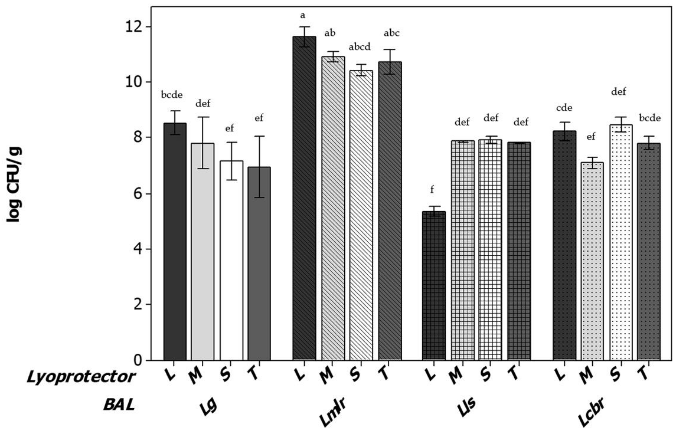
), Limosilactobacillus reuteri CRL1324 (oblique line bars Applmicrobiol 03 00037 i002), Ligilactobacillus salivarius CRL1328 (squared bars Applmicrobiol 03 00037 i003) and Lacticaseibacillus rhamnosus CRL1332 (dotted bars Applmicrobiol 03 00037 i004). Different letters indicate statistical differences between strains at different fermentation condicions (p < 0.05).
Applmicrobiol 03 00037 g003
Figure 4. Viable cell numbers of VLB before and after lyophilization with different lyoprotectants. Different letters indicate statistical differences before and after lyofilization (p < 0.05). Lactose (L), sucrose (S), trehalose (T) and cryoprotectants are mixed (M). Grey and white bars represent viable counts before and after freeze drying process, respectively. Strains are represented using each pair of grey–white bars as follows: L. gasseri CRL1320 (solid bars Applmicrobiol 03 00037 i005), L. reueri CRL1324 (oblique lines bars Applmicrobiol 03 00037 i006), L. salivarius CRL1328 (squeared bars Applmicrobiol 03 00037 i007) and L. rhamnosus CRL1332 (dotted bars Applmicrobiol 03 00037 i008).
Applmicrobiol 03 00037 g004
Figure 5. Stability of VLB stored for 3 months at 25 °C (room temperature) with different lyoprotectants. Letters indicate statistical differences between lyoprotectants used (p < 0.05). Lactose (L), sucrose (S), trehalose (T) and lyoprotectants mixture (M). Lactobacillus gasseri CRL1320 (solid bars Applmicrobiol 03 00037 i009), Limosilactobacillus reuteri CRL1324 (oblique lines bars Applmicrobiol 03 00037 i010), Ligilactobacillus alivarius CRL1328 (squeared bars Applmicrobiol 03 00037 i011) and Lacticaseibacillus rhamnosus CRL 1332 (dotted bars Applmicrobiol 03 00037 i012).
Applmicrobiol 03 00037 g005
Figure 6. Stability during storage of VLB after freeze-drying with different lyoprotectants. (A). For 3 months at 4 °C. (B) For 12 months at 4 °C. Letters indicate statistical differences between lyoprotectants of each strains (p < 0.05). Lactose (L), sucrose (S), trehalose (T) and lyoprotectants mixture (M). Lactobacillus gasseri CRL 320 (solid bars Applmicrobiol 03 00037 i013);, Limosilactobacillus reuteri CRL1324 (oblique lines bars Applmicrobiol 03 00037 i014), Ligilactobacillus salivarius CRL1328 (squeared bars Applmicrobiol 03 00037 i015) and Lacticaseibacillus rhamnosus CRL1332 (doted bars Applmicrobiol 03 00037 i016).
Applmicrobiol 03 00037 g006aApplmicrobiol 03 00037 g006b
Table 1. Growth parameters of VLB incubated under different conditions.
VLB Strain *Incubation (pH/rpm)µ (h−1)Lag Phase (h)Amax (log UFC)
Lactobacillus gasseri
(L. gasseri CRL 1320)
5.5/1500.87 ± 0.07011.09 ± 0.02
5.5/751.00± 0.01110.21 ± 0.01
6.5/1501.62 ± 0.05610.64 ± 0.09
MRS0.40 ± 0.1059.96 ± 0.03
Lacticaseibacillus rhamnosus
(L. rhamnosus CRL 1332)
5.5–1500.64 ± 0.01010.74 ± 0.04
5.5–751.44 ± 0.01210.74 ± 0.06
6.5–1500.86 ± 0.01210.75 ± 0.08
MRS0.44 ± 0.0409.78 ± 0.04
Limosilactobacillus reuteri
(L. reuteri CRL 1324)
5.5–1500.92 ± 0.0209.92 ± 0.55
6.5–1500.92 ± 0.03210.83 ± 0.03
MRS0.48 ± 0.09110.61 ± 0.01
Ligilactobacillus salivarius
(L. salivarius CRL 1328)
5.5–1500.82 ± 0.04010.47 ± 0.07
6.5–1500.93 ± 0.0188.24 ± 0.01
MRS0.75 ± 0.08110.5 ± 0.08
* The taxonomic identification of the lactic acid bacteria assayed are included in the table, according to Zheng et al. [36]. The name of the strains as originally isolated, and included in the CRL Collection strains is below, indicated in parenthesis.
Table 2. Surface properties of VLB before and after freeze-drying with different cryoprotective agents and storage at 4 and 25 °C.
BVLT °CLyoprotectant% Hydrophobicity% Autoagreggation
Pre-LyofilizationPost-LyofilizationPre-LyofilizationPost-Lyofilization
Lactobacillus gasseri
(L. gasseri CRL 1320)
4 °CLactose97.0071.4010.0050.00
Sucrose97.0010.0010.0054.16
Trehalose97.0062.5010.0055.83
Mixture97.0083.3010.0062.50
25 °CLactose97.0010.0010.0068.69
Sucrose97.0010.0010.0072.30
Trehalose97.0010.0010.0082.75
Mixture97.0061.0010.0074.13
Limosilactobacillus reuteri
(L. reuteri CRL 1324)
4 °CLactose96.0041.6023.0062.50
Sucrose96.0010.0023.0054.16
Trehalose96.0078.9023.0035.83
Mixture96.0010.0023.0039.16
25 °CLactose96.0010.0023.002.00
Sucrose96.0010.0023.002.00
Trehalose96.0010.0023.002.00
Mixture96.0010.0023.002.00
Ligilactobacillus salivarius
(L. salivarius CRL 1328)
4 °CLactose10.0023.0780.006.00
Sucrose10.0025.5080.0069.16
Trehalose10.0010.0080.0039.19
Mixture10.002.00-80.0040.00
25 °CLactose10.002.0080.002.00
Sucrose10.002.0080.002.00
Trehalose10.002.0080.002.00
Mixture10.002.0080.002.00
Lacticaseibacillus rhamnosus
(L. rhamnosus CRL 1332)
4 °CLactose42.005.0018.002.00
Sucrose42.005.0018.0010.83
Trehalose42.005.0018.002.00
Mixture42.005.0018.002.00
25 °CLactose42.002.0018.002.00
Sucrose42.005.0018.002.00
Trehalose42.005.0018.002.00
Mixture42.005.0018.002.00
Disclaimer/Publisher’s Note: The statements, opinions and data contained in all publications are solely those of the individual author(s) and contributor(s) and not of MDPI and/or the editor(s). MDPI and/or the editor(s) disclaim responsibility for any injury to people or property resulting from any ideas, methods, instructions or products referred to in the content.

