Article

Onset and Progression of Infection Based on Viral Loads in Rhesus Macaques Exposed to Zika Virus

1
Battelle, 505 King Ave., Columbus, OH 43201, USA
2
Battelle, 1425 State Route 142 (Plain City-Georgesville Road), West Jefferson, OH 43162, USA
*
Author to whom correspondence should be addressed.
Current Affiliation: Amplify-Bio, 1425 State Route 142 (Plain City-Georgesville Road), West Jefferson, OH 43162, USA.
Current Affiliation: Latham BioPharm Group, 6810 Deerpath Road, Suite 405, Elkridge, MD 21075, USA.
Appl. Microbiol. 2022, 2(3), 544-553; https://doi.org/10.3390/applmicrobiol2030042
Submission received: 18 June 2022 / Revised: 20 July 2022 / Accepted: 26 July 2022 / Published: 30 July 2022

Abstract

:
Outbreaks of Zika virus (ZIKV) have resulted in a call by global health advocates for increased surveillance and research with aggressive measures to combat ZIKV infections. There is no licensed ZIKV vaccine yet available, but a number of vaccine candidates are in development. Advancement of promising vaccine candidates to licensure may rely upon the development and use of well-characterized preclinical models developed based on the essential elements of an animal model as outlined in the U.S. FDA “Product Development Under the Animal Rule: Guidance for Industry”. Further, in the absence of adequate clinical cases to support a more traditional approval pathway based on clinical efficacy, regulatory approval could be based upon human safety data and use of a well-characterized animal model to evaluate vaccine efficacy. This report summarizes a statistical analysis that characterizes the progression of ZIKV infection in Rhesus macaques (RMs) with respect to viral load using available data on twenty-six (26) RMs from three (3) studies that were exposed to ZIKV and were not immunized with a ZIKV vaccine. Progression of infection was characterized by time to detection of viral RNA in serum (RT-qPCR) or time to positive viremia (plaque assay). Viral RNA was detected via RT-qPCR as early as day 1 post-infection and was undetectable for all animals by day 7. Viremia also was indicated by plaque assay as early as day 1 and was undetectable for all animals by day 5. Viral RNA was detected in all animals following exposure, while viremia was not observed in all animals. No significant differences in viral loads measured by either RT-qPCR or plaque assay were observed across sex, age, or study. Neither sex nor age were significant predictors of either time to detection of viral RNA or time to positive viremia following exposure to ZIKV. Progression of viral load, which is studied since infection is largely asymptomatic in both RMs and humans, is similar in RMs and humans with positive presentation ranging from 1 to 7 days post-infection and clearance by day 10. Overall, due to consistency of the model across sexes and ages and similarity to the infection profile in humans, it is concluded that the RM model of ZIKV infection is a well-characterized model for use for evaluation of ZIKV countermeasures.

1. Introduction

Zika virus (ZIKV) was first isolated in 1947 from a sentinel monkey placed in the Zika forest (Uganda) and was first reported to have been isolated from humans in 1954, though previous serology studies suggested that humans had been actively infected since at least 1952 [1,2]. Duffy et al. [3] reported the first transmission of ZIKV outside of Africa and Asia in 2009. Lanciotti et al. [4] were unable to harvest infectious virus from the patients tested during an outbreak, but RNA was detected by 1 day following onset of illness and persisted for 11 days. During a 2014 outbreak in French Polynesia affecting an estimated 28,000 patients, evidence of perinatal transmission [5] and potential transmission through blood transfusion [6] was reported. In a 2015 outbreak, concomitant with ZIKV cases across South and Central America as well as the Caribbean ZIKV infections in women were tied to microcephaly in infants [7,8]. The WHO declared the clusters of neurological disorders and microcephaly in South America a Public Health Emergency of International Concern (PHEIC) from 1 February 2016, through 18 November 2016 [9]. Since this declaration, the WHO and other global public health advocates have called for increased surveillance and research with aggressive measures to combat ZIKV infections.
There is no licensed ZIKV vaccine yet available, but a number of vaccine candidates are in development based on a variety of platform technologies. Understanding the immune responses elicited by each vaccine candidate aids in defining correlates of protection, and efficacy testing in relevant preclinical models is critical to advance the development of a ZIKV vaccine. National Institute of Allergy and Infectious Diseases (NIAID) and the Biomedical Advanced Research and Development Authority (BARDA) have supported the development of non-human primate (NHP) models of ZIKV infection, and these models have been used successfully to test several vaccine candidates. Three pathways to approval exist: ‘traditional approval’, accelerated approval, and the Food and Drug Administration (FDA) Animal Rule (AR) [10]. Marston and colleagues proposed three strategies to developing ZIKV vaccines: a traditional vaccine-development approach in which phase 1 and phase 2 studies assess safety and immunogenicity; use of human challenge studies conducted after phase 1 and 2 studies; or, reliance on the FDA AR [11]. For the purpose of this manuscript, we assume that at least some testing of ZIKV vaccines will be based on the AR as it is currently considered unethical to perform human challenge trials with ZIKV [12]. The AR requires that products be tested in well-characterized animal models that are relevant to human disease and that the animal study endpoint be clearly related to the desired benefit in humans [13]. To support use of the RM model of ZIKV for licensure of vaccines this study summarizes the analysis conducted to characterize serum viral load observed in the RM model for ZIKV, as animals did not show clinical signs of illness.

2. Materials and Methods
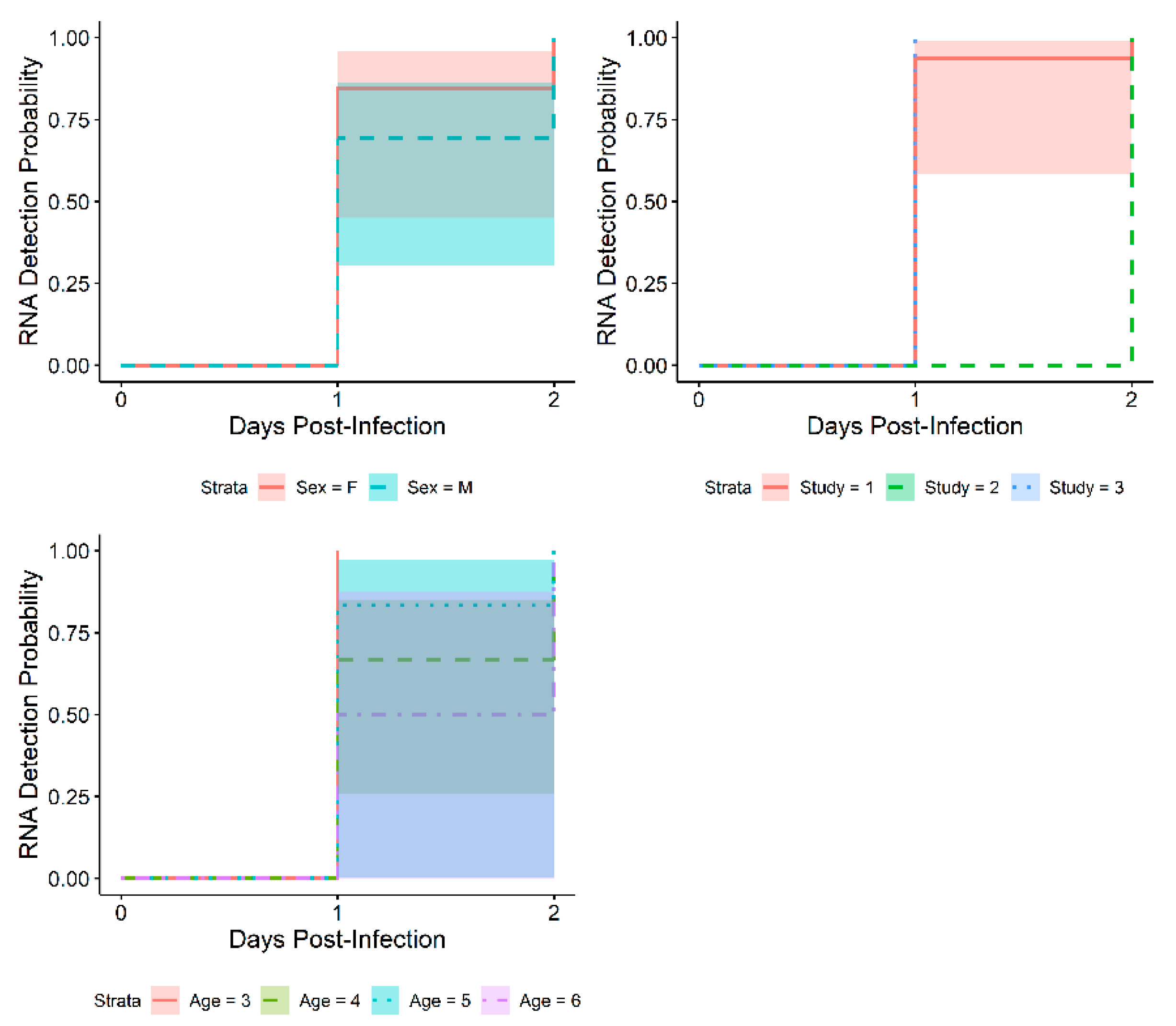
This statistical analysis utilized data from 3 studies to describe progression of infection with respect to serum viral load of ZIKV in RMs (Covance Research Products; Alice, TX). Viral RNA in serum was measured by RT-qPCR in all three studies using consistent methods according to standard operating procedure (SOP); viral load in serum was also measured by plaque assay in two studies (Study 1 and Study 2; see Table 1) using a consistent method according to SOP. All studies were performed at the Battelle Biomedical Research Center (BBRC, West Jefferson, OH, USA).
Only the data from the animals that were infected with ZIKV but not immunized with a ZIKV vaccine were utilized. These animals were treated with either nothing (Study 1), Alum (Study 2), or saline (Study 3). For Study 2, animals were treated on Study Days 0 and 28 then exposed on Study Day 56, while for Study 3 animals were treated on Study Day 1 then exposed on Study Day 2; Study 1 animals were not treated prior to exposure. The population of these animals consisted of a total of 26 RMs, 13 females and 13 males. The animals ranged in age from 3 to 6 years of age. A summary of the animal characteristics by study is presented in Table 1.
All studies presented in this manuscript were approved by the responsible Institutional Animal Care and Use Committee (IACUC), and research was conducted in compliance with the Animal Welfare Act. Experiments involving animals adhered to principles stated in the Guide for the Care and Use of Laboratory Animals from the National Research Council. Studies were performed at an institution (BBRC, West Jefferson, OH, USA) which is fully accredited by the Association for Assessment and Accreditation of Laboratory Animal Care International (AAALAC International).
All animals were infected subcutaneously with the same ZIKV stock (ZIKV strain PRVABC59 Lot # 4350-WB [14]). This exposure material originated from ATCC/BEI Resources (Lot # 64112564) (NR-50240) (Manassas, VA, USA), and underwent a single passage at the BBRC in VERO E6 cells prior to characterization for use as exposure material. The target exposure dose for all 3 studies was 1 × 10⁵ PFU, and the back-titer of challenge material determined following stock dilution varied from study to study: 8.80 × 10⁴ PFU, 4.74 × 10⁴ PFU, and 1.23 × 10⁵ PFU.
While multiple endpoints were collected for each of the 3 studies, this analysis focused only on viral load in serum. Viral RNA in serum was measured by RT-qPCR in all three studies while viremia in serum was measured by plaque assay in two studies.
The ZIKV RT-qPCR assay [14] was performed on collected samples from each study using the same method according to SOP across studies. The ZIKV RNA was isolated from serum samples using the QIAamp cador Pathogen 96 kit (currently known as Indispin Pathogen Kit; Indical Bioscience, Leipzig, Germany) on the QIAcube HT automated system per facility SOP. The ZIKV RT-qPCR was performed per facility SOP using primers and probe specific to the Pre-Membrane/Envelope (PrM-E) gene region and a synthetic RNA reference standard curve for absolute quantitation. The RT-qPCR assay was qualified for NHP serum samples; the LOD of the assay is 685 copies/mL and the LLOQ is 4138 copies/mL. For the statistical analysis, positive samples were defined as samples with results >LOD.
A standard plaque assay was performed on collected serum samples from each study using the same method according to SOP across studies. Dilutions of samples were inoculated onto VERO E6 cell monolayers in 12-well tissue culture plates. Following a 72-h incubation, cells were fixed and stained with crystal violet to observe plaques in the monolayers. The number of plaques and dilution of serum were used to calculate the concentration of infectious ZIKV in the original samples. The plaque assay was qualified for assessment of ZIKV in culture medium; the LOD is 61 PFU/mL and the LLOQ is 195 PFU/mL. For the statistical analysis, positive samples were defined as samples with results > LOD.
The primary objective of the presented statistical analysis was to characterize the progression of infection with respect to viral load in serum in ZIKV-infected RMs using unvaccinated or sham-vaccinated control animals from multiple studies. This objective was achieved using descriptive statistics and plots of viral load in serum. Group level descriptive statistics and cross-tabulations were calculated over all animals and using the following explanatory variables:
  • Animal sex;
  • Animal age;
  • Study: Animals were grouped at the study level as both control article and actual exposure dose had a 1:1 relationship with study and so were completely confounded.
Additionally, as a secondary objective, time to detection of viral RNA (RT-qPCR) or time to positive viremia (plaque assay) was examined using Kaplan–Meier curves; median time to detection of viral RNA or onset of positive viremia was estimated. Cox proportional hazards modeling [15,16,17,18,19] was used to determine factors affecting time to onset of positive viremia, if any. Demographic variables including sex and age were considered for the Cox proportional hazards model. Age was modeled as a continuous covariate. First, the Cox PH model was fit using a single variable at a time with a random effect for study entered as a frailty term in each model. Cox PH model parameters were expressed as hazard ratios (HRs), where a HR of 1 is interpreted as no difference in the relative risk (of detection) between groups; HR < 1 indicates that the comparison group has lower risk than the baseline group; and HR > 1 indicates the comparison group has higher risk than the baseline group. For continuous parameters (age), the HR is the change in hazard for a unit increase in the parameter value.
Due to the viral load measurements spanning multiple orders of magnitude, data were transformed using a base-10 logarithmic transformation.

3. Results

This paper provides graphical representations of descriptive statistics to illustrate clinically relevant changes post-Zika virus exposure over time. Tables of descriptive statistics for these endpoints have been provided as Supplementary Materials.
Viral RNA as measured by RT-qPCR with the individual data points overlaid, summarized across all animals, over time, is presented in Figure 1. Positive RT-qPCR (defined as > LOD) was observed starting on Day 1 for 20 of the 26 animals (76.9%); all animals presented positive RT-qPCR on at least one day. Geometric mean copies/mL peaked on Days 2 and 3, then started to decrease. By Day 7, viral RNA as measured by RT-qPCR was less than LOD in all animals and remained below LOD through day 28 in animals for which measurements were taken.
Figure 2 presents boxplots of viral load as measured by plaque assay with the individual data points overlaid, summarized across all animals (N = 21; Study 1 and Study 2), over time. Positive plaque assay results (defined as >LOD) were observed starting on Day 1 for 3 of the 21 animals; 6 animals never presented with viral titers above the LOD. Mean PFU/mL never exceeded LLOQ. By Day 5, viremia as measured by plaque assay was less than LOD in all animals and remained below LOD through day 28 in animals for which measurements were taken.
Minor differences were observed in viral loads in serum by sex, age, and study (See Supplemental Materials Figures S1–S3). However, no statistically significant differences were noted based on Kruskal–Wallis tests comparing peak viral loads between the groups (PCR: p = 0.7779 for sex; p = 0.8381 for age; p = 0.1652 for study; PA: p = 0.4092 for sex; p = 0.1095 for age; p = 0.2740 for study). Lack of differences by sex, age, and study supports use of the RM model for study of ZIKV infection.

3.1. Time to Onset

Kaplan–Meier plots were created to visually represent the times to detection of viral RNA with 95% confidence intervals (shaded areas in plots) for each of the explanatory variables (sex, age, and study) (Figure 3). Log-rank tests were conducted to determine if there were significant differences in time to detection of viral RNA. The log-rank test identified a statistically significant difference across studies (p < 0.0001), with Study 2 having longer median time to detection (2 days) than Study 1 or Study 3 (1 day). No differences between sexes or across ages were identified (p = 0.4 and p = 0.3, respectively).
Times to positive viremia were visually represented along with 95% confidence intervals (shaded areas in plots) for each of the explanatory variables (sex, age, and study) (Figure 4). Log-rank tests were conducted to determine if there were significant differences in time to positive viremia; no statistically significant differences were identified (p = 0.2, p = 0.2, and p = 0.5 for sex, age, and study, respectively).
Individual Cox proportional hazard models were fit to sex and age as predictor variables, with a random effect for study entered as a frailty term (gamma distributed) in each model. Table 2 summarizes the individual Cox PH models for each predictor variable fit to all available data. Neither sex nor age were statistically significant, indicating that there was no significant difference in relative risk of positive RT-qPCR or plaque assay result between sexes, and the risk did not change significantly with age.

3.2. Exploratory Analysis: Results by Weight

The data were further explored with respect to weight. Results by weight for viral load in serum as measured by both RT-qPCR and plaque assay are presented in Figure 5. Time to detection of viral RNA was similar (based on a log-rank test; p = 0.2000) for the <4.5 kg group and the ≥4.5 kg group. Mean viral RNA as measured by RT-qPCR peaked at a higher level for the <4.5 kg group compared to the ≥4.5 kg group, and this difference was statistically significant (p = 0.0102). Median time to onset of viremia was later for the ≥4.5 kg group (3 days) compared to the < 4.5 kg group (2 days), though the difference was not statistically significant (p = 0.1000). Peak mean viremia as measured by plaque assay was similar for the <4.5 kg group and the ≥4.5 kg group (p = 0.9714), though more NHPs in the <4.5 kg group presented with positive viremia (80% vs. 55%).

4. Discussion

ZIKV infection in humans is generally mild with an estimated 80% of all infections remaining asymptomatic [3]; sudden clinical onset is not common [20,21]. Duration of illness is about 1 week, and symptoms are typically self-limiting [22]. Examination of clinical signs was not included in this analysis. ZIKV infection is generally not fatal in humans [20,23]; all RMs included in the analysis survived to end of study.
Studies have suggested there may be delays between the development of viremia and onset of symptoms in humans, with low observed rates of detection of ZIKV RNA in acute phases of infection [21,24] and no correlation between viral load and specific clinical presentation [21]. In the current analysis, viral RNA was detected via RT-qPCR in all animals at least once over the course of the study; viremia measured by plaque assay did not present in all animals, suggesting this is a less sensitive model for Zika infection compared to viral RNA and adjustments to the study design would be required to account for the less than 100% positive viremia detection. Viremia in humans has been shown to persist [24] and has been reported >28 days after symptom onset [25]. However, viremia in RMs did not persist beyond 7 days in the studies included in this analysis, though this may be due to the small sample sizes. As the current analysis examined only viral load and not associated clinical signs, these relationships between viral loads and clinical presentation in humans cannot be compared to RMs. In general, outward clinical signs of disease are not reported in RMs [26], therefore the presence of virus through RT-qPCR or plaque assay provides an objective and quantitative endpoint for evaluating the efficacy of ZIKV vaccine candidates.
ZIKV RNA detection in urine has been observed up to 26 days in humans [27] and up to 17 days in RM [28]. Viral RNA can also be detected in macaque urine, saliva, cerebrospinal fluid, lymphoid tissue, and semen [28,29,30]. As only serum was considered in the current analysis, these findings cannot be compared.
Disease course and viremia in NHPs resembles that seen in infected humans with acute plasma viremia observed between 2 and 6 days post infection [28]. Duration of ZIKV RNA in both macaque and human plasma is transient, lasting about 7 to 10 days [22,28], although detection in some cases has been reported in humans up to 37 days [25] with a case report of prolonged presence of ZIKV RNA up to 70 days post infection [31]. In the three studies included in this analysis, positive serum viral loads were observed for some animals starting on Day 1, with all viral loads < LOD by Day 7. In future studies evaluating the efficacy of vaccine candidates, reduction in viremia may be used as an endpoint to evaluate the effectiveness of candidate vaccines and may provide a bridge between non-clinical and clinical data.
A consistent model for ZIKV is important for medical countermeasure development. The results from this analysis highlight consistency of the subcutaneous ZIKV model in RMs across sex, age, and studies with respect to viral RNA and viremia. Additionally, as noted, this analysis in RMs reflects serum viral load levels in humans. Viral load over time is a significant endpoint for characterizing ZIKV infection since the disease is largely asymptomatic in both humans and RMs.

5. Conclusions

The primary objective of this control animal analysis was to characterize the ZIKV viral load in RMs using unvaccinated or sham-vaccinated animal data from three studies and was achieved through descriptive analysis. Positive viral RNA was the more sensitive indicator, as viral load in serum was detected by RT-qPCR in all animals at least once over the course of the study. Viremia in serum as measured by plaque assay was positive in some animals as early as Day 1 post infection; however, positive viral load was not observed in all animals by this method. Viremia in infected humans typically has been observed between 2 and 6 days post infection. In the studies included in this analysis positive viral RNA was first detected in RMs between 1 and 2 days post infection and viremia was observed in RMs as early as 1 day post infection. Duration of ZIKV RNA in both rhesus macaque and human plasma is transient, clearing by day 7 in RM and generally by day 10 in humans. Consistency of the ZIKV model in RMs and in relation to progression of infection in humans demonstrates this to be a well-characterized model for ZIKV countermeasure development.

Supplementary Materials

The following supporting information can be downloaded at: https://www.mdpi.com/article/10.3390/applmicrobiol2030042/s1, Figure S1: Means and Confidence Intervals for Viral Load in Serum by Sex Over Time as Measured by RT-qPCR (copies/mL) and Plaque Assay (PFU/mL); Figure S2: Means and Confidence Intervals for Viral Load in Serum by Age Over Time as Measured by RT-qPCR (copies/mL) and Plaque Assay (PFU/mL); Figure S3: Means and Confidence Intervals for Viral Load in Serum by Study Over Time as Measured by RT-qPCR (copies/mL) and Plaque Assay (PFU/mL); Table S1: Descriptive Statistics for Viral Load in Serum as Measured by RT-qPCR (copies/mL), Overall; Table S2: Descriptive Statistics for Viral Load in Serum as Measured by Plaque Assay (PFU/mL), Overall.

Author Contributions

Conceptualization, C.T. and N.N.; methodology, C.T.; formal analysis, C.T. and S.D.; investigation, D.K. and D.S.; data curation, C.T. and S.D.; writing—original draft preparation, C.T., N.N. and K.M.; writing—review and editing, C.T., N.N., D.K., C.C. and D.S.; visualization, S.D.; project administration, C.C. All authors have read and agreed to the published version of the manuscript.

Funding

This study was supported by National Institute of Allergy and Infectious Disease (NIAID) under Contract HHSN272201200003I/HHSN27200026. The studies contributing to this meta-analysis were supported as follows: Biomedical Advanced Research and Development Authority (BARDA) under Contract HHSO100201700011I/HHSO10033001T (Study 1); National Institute of Allergy and Infectious Disease (NIAID) under Contract HHSN272201200003I/HHSN27200026 (Study 2); and, National Institute of Allergy and Infectious Disease (NIAID) under Contract HHSN272201200003I/HHSN27200026 (Study 3). The findings and conclusions in this report are those of the authors and do not necessarily represent the views of the National Institutes of Health (NIH).

Institutional Review Board Statement

All studies presented in this manuscript were approved by the responsible Institutional Animal Care and Use Committee (IACUC), and research was conducted in compliance with the Animal Welfare Act. Experiments involving animals adhered to principles stated in the Guide for the Care and Use of Laboratory Animals from the National Research Council. Studies were performed at an institution (BBRC, West Jefferson, Ohio) which is fully accredited by the Association for Assessment and Accreditation of Laboratory Animal Care International (AAALAC International).

Informed Consent Statement

Not applicable.

Data Availability Statement

The data presented in this study are available in Supplementary Material.

Acknowledgments

The findings and conclusions in this report are those of the authors and do not necessarily represent the views of the National Institutes of Health (NIH). The authors would like to acknowledge and thank the following individuals for support of this work: Janet Lathey, Nancy Ulbrandt, Sara Woodson, and Kaitlyn Morabito (NIAID/DMID); Carol Sabourin (Tunnell Government Services, Inc.); and, Thomas Rudge, Jr. and Gabe Meister (Battelle). Your insightful review and comments were critical in forming this work and this manuscript.

Conflicts of Interest

The authors declare no conflict of interest.

References

  1. Dick, G.W.A.; Kitchen, S.F.; Haddow, A.J. Zika virus (I). Isolations and serological specificity. Trans. R. Soc. Trop. Med. Hyg. 1952, 46, 509–520. [Google Scholar] [CrossRef]
  2. Macnamara, F.N. Zika Virus: A Report on Three Cases of Human Infection During an Epidemic of Jaundice in Nigeria. Trans. R. Soc. Trop. Med. Hyg. 1954, 48, 139–145. [Google Scholar] [CrossRef]
  3. Duffy, M.R.; Chen, T.; Hancock, W.T.; Powers, A.M.; Kool, J.L.; Lanciotti, R.S.; Pretrick, M.; Marfel, M.; Holzbauer, S.; Dubray, C.; et al. Zika virus outbreak on Yap Island, Federated States of Micronesia. N. Engl. J. Med. 2009, 360, 2536–2543. [Google Scholar] [CrossRef]
  4. Lanciotti, R.S.; Kosoy, O.L.; Laven, J.J.; Velez, J.O.; Lambert, A.J.; Johnson, A.J.; Stanfield, S.M.; Duffy, R.M. Genetic and serologic properties of Zika virus associated with an epidemic, Yap State, Micronesia, 2007. Emerg. Infect. Dis. 2008, 14, 1232–1239. [Google Scholar] [CrossRef] [PubMed]
  5. Besnard, M.; Lastere, S.; Teissier, A.; Cao-Lormeau, V.M.; Musso, D. Evidence of perinatal transmission of Zika virus, French Polynesia, December 2013 and February 2014. Eurosurveillance 2014, 19, 20751. [Google Scholar] [CrossRef] [Green Version]
  6. Musso, D.; Nhan, T.; Robin, E.; Roche, C.; Bierlaire, D.; Zisou, K.; Yan, A.S.; Cao-Lormeau, V.; Broult, J. Potential for Zika virus transmission through blood transfusion demonstrated during an outbreak in French Polynesia, November 2013 to February 2014. Eurosurveillance 2014, 19, 20761. [Google Scholar] [CrossRef] [Green Version]
  7. Schuler-Faccini, L.; Ribeiro, E.M.; Feitosa, I.M.; Horovitz, D.D.; Cavalcanti, D.P.; Pessoa, A.; Doriqui, M.J.; Neri, J.I.; Neto, J.M.; Wanderley, H.Y.; et al. Possible association between Zika virus infection and microcephaly–Brazil, 2015. MMWR Morb. Mortal. Wkly. Rep. 2016, 65, 59–62. [Google Scholar] [CrossRef]
  8. Mlakar, J.; Korva, M.; Tul, N.; Popovic, M.; Poljsak-Prijatelj, M.; Mraz, J.; Kolenc, M.; Resman, K.; Vipotnik, T.V.; Vodusek, V.F.; et al. Zika virus associated with microcephaly. N. Engl. J. Med. 2016, 374, 951–958. [Google Scholar] [CrossRef]
  9. European Centre for Disease Prevention and Control. WHO Ends Zika as a Public Health Emergency of International Concern. 2016. Available online: https://www.ecdc.europa.eu/en/news-events/who-ends-zika-public-health-emergency-international-concern (accessed on 28 June 2022).
  10. Gruber, M.F.; Krause, P.R. Regulating vaccines at the FDA: Development and licensure of Zika vaccines. Vaccines 2017, 16, 525–527. [Google Scholar] [CrossRef] [Green Version]
  11. Marston, H.D.; Lurie, N.; Borio, L.L.; Fauci, A.S. Considerations for developing a Zika virus vaccine. N. Engl. J. Med. 2016, 375, 1209–1212. [Google Scholar] [CrossRef]
  12. Shah, S.K.; Kimmelman, J.; Drapkin Lyerly, A.; Fernandez Lynch, H.; McCutchan, F.; Miller, F.G.; Palacios, R.; Pardo-Villamizar, C.; Zorrilla, C. Ethical Considerations for Zika Virus Human Challenge Trials: Report and Recommendations. Available online: https://www.niaid.nih.gov/sites/default/files/EthicsZikaHumanChallengeStudiesReport2017.pdf (accessed on 19 July 2022).
  13. U.S. Department of Health and Human Services; Food and Drug Administration; Center for Drug Evaluation and Research (CDER); Center for Biologics Evaluation and Research (CBER). Product Development under the Animal Rule: Guidance for Industry. October 2015. Available online: https://www.fda.gov/regulatory-information/search-fda-guidance-documents/product-development-under-animal-rule (accessed on 28 June 2022).
  14. Vang, L.; Morello, C.S.; Mendy, J.; Thompson, D.; Manayani, D.; Guenther, B.; Julander, J.; Sanford, D.; Jain, A.; Patel, A.; et al. Zika virus-like particle vaccine protects AG129 mice and rhesus macaques against Zika virus. PLoS Negl. Trop. Dis. 2021, 15, e0009195. [Google Scholar] [CrossRef] [PubMed]
  15. Cox, D.R. Regression Models and Life Tables (with Discussion). J. R. Stat. Soc. Ser. B 1972, 34, 187–202. [Google Scholar]
  16. Cox, D.R.; Oakes, D. Analysis of Survival Data; Chapman and Hall: London, UK, 1984. [Google Scholar]
  17. Therneau, T.M.; Grambsch, P.M. Modeling Survival Data: Extending the Cox Model; Springer: New York, NY, USA, 2000. [Google Scholar]
  18. Bradburn, M.J.; Clark, T.G.; Love, S.B.; Altman, D.G. Survival Analysis Part II: Multivariate data analysis–an introduction to concepts and methods. Br. J. Cancer 2003, 89, 431–436. [Google Scholar] [CrossRef] [PubMed]
  19. Moore, D. Applied Survival Analysis Using R, 1st ed.; Springer International: Cham, Switzerland, 2016. [Google Scholar]
  20. Musso, D.; Gubler, D.J. Zika Virus. Clin. Microbiol. Rev. 2016, 29, 487–524. [Google Scholar] [CrossRef] [Green Version]
  21. Musso, D.; Rouault, E.; Teissier, A.; Lanteri, M.C.; Zisou, K.; Broult, J.; Grange, E.; Nhan, T.-X.; Aubry, M. Molecular detection of Zika virus in blood and RNA load determination during the French Polynesian outbreak. J. Med. Virol. 2017, 89, 1505–1510. [Google Scholar] [CrossRef] [PubMed] [Green Version]
  22. Osuna, C.E.; Whitney, J.B. Nonhuman Primate Models of Zika Virus Infection, Immunity, and Therapeutic Development. J. Infect. Dis. 2017, 216 (Suppl. S10), S928–S934. [Google Scholar] [CrossRef] [PubMed] [Green Version]
  23. Gorshkov, K.; Shiryaev, S.A.; Fertel, S.; Lin, Y.-W.; Huang, C.-T.; Pinto, A.; Farhy, C.; Strongin, A.Y.; Zheng, W.; Terskikh, A.V. Zika Virus: Origins, Pathological Action, and Treatment Strategies. Front. Microbiol. 2019, 9, 3252. [Google Scholar] [CrossRef] [PubMed] [Green Version]
  24. Barzon, L.; Trevisan, M.; Sinigaglia, A.; Lavezzo, E.; Palù, G. Zika virus: From pathogenesis to disease control. FEMS Microbiol. Lett. 2016, 363, fnw202. [Google Scholar] [CrossRef] [Green Version]
  25. Mansuy, J.M.; Mengelle, C.; Pasquier, C.; Chapuy-Regaud, S.; Delobel, P.; Martin-Blondel, G.; Izopet, J. Zika Virus Infection and Prolonged Viremia in Whole-Blood Specimens. Emerg. Infect. Dis. 2017, 23, 863. [Google Scholar] [CrossRef]
  26. Newman, C.; Friedrich, T.C.; O’Connor, D.H. Macaque monkeys in Zika virus research: 1947-present. Curr. Opin. Virol. 2017, 25, 34–40. [Google Scholar] [CrossRef]
  27. Lustig, Y.; Mendelson, E.; Paran, N.; Melamed, S.; Schwartz, E. Detection of Zika virus RNA in whole blood of imported Zika virus disease cases up to 2 months after symptom onset, Israel, December 2015 to April 2016. Eurosurveillance 2016, 21, 30269. [Google Scholar] [CrossRef] [PubMed] [Green Version]
  28. Dudley, D.M.; Aliota, M.T.; Mohr, E.L.; Weiler, A.M.; Lehrer-Brey, G.; Weisgrau, K.L.; Mohns, M.S.; Breitbach, M.E.; Rasheed, M.N.; Newman, C.M.; et al. A rhesus macaque model of Asian-lineage Zika virus infection. Nat. Commun. 2016, 7, 12204. [Google Scholar] [CrossRef] [PubMed] [Green Version]
  29. Estes, J.D.; Wong, S.W.; Brenchley, J.M. Nonhuman primate models of human viral infections. Nat. Rev. Immunol. 2018, 18, 390–404. [Google Scholar] [CrossRef] [PubMed]
  30. Osuna, C.E.; Lim, S.Y.; Deleage, C.; Griffin, B.D.; Stein, D.; Schroeder, L.T.; Omange, R.; Best, K.; Luo, M.; Hraber, P.T.; et al. Zika viral dynamics and shedding in rhesus and cynomolgus macaques. Nat. Med. 2016, 22, 1448. [Google Scholar] [CrossRef] [PubMed] [Green Version]
  31. Driggers, R.W.; Ho, C.-Y.; Korhonen, E.M.; Kuivanen, S.; Jääskeläinen, A.J.; Smura, T.; Rosenberg, A.; Hill, D.A.; DeBiasi, R.L.; Vezina, G.; et al. Zika Virus Infection with Prolonged Maternal Viremia and Fetal Brain Abnormalities. N. Engl. J. Med. 2016, 374, 2142–2151. [Google Scholar] [CrossRef]
Figure 1. Boxplots of Viral Load in Serum as Measured by RT-qPCR (copies/mL) Over Time. Data are summarized across all animals as box plots (N varies by day; maximum N = 26) over time. Individual data are overlaid as black dots. Mean viral RNA (red diamond) peaked on Day 2 Post-Exposure. All animals presented positive RT-qPCR (>LOD; LOD = 685 copies/mL) on at least one day. Viral RNA was less than LOD for all animals starting on Study Day 7.
Applmicrobiol 02 00042 g001
Figure 2. Boxplots of Viral Load in Serum as Measured by Plaque Assay (PFU/mL) Over Time. Data are summarized across all animals as box plots (N varies by day; maximum N = 21) over time. Individual data are overlaid as black dots. Mean viremia (red diamond) never exceeds LLOQ. Not all animals presented with positive viremia on any day. Viremia was below LOD for all animals starting on Study Day 5.
Applmicrobiol 02 00042 g002
Figure 3. Kaplan–Meier Plots of Time to Detection of Viral RNA by Sex, Age, and Study. No differences in time to detection of viral RNA between sexes or ages; time to detection of viral RNA differed among studies, with a shorter time to detection for Study 1 and Study 3 than for Study 2.
Applmicrobiol 02 00042 g003
Figure 4. Kaplan–Meier Plots of Time to Positive Viremia by Sex, Age, and Study. No differences in time to detection of viral RNA between sexes, studies, or ages.
Applmicrobiol 02 00042 g004
Figure 5. Means and Confidence Intervals for Viral Load in Serum by Weight Over Time as Measured by RT-qPCR (copies/mL) and Plaque Assay (PFU/mL). Peak viral RNA was greater for lower weight animals; however, there was no difference in peak viremia between weight groups. Viral load as measured by both RT-qPCR and plaque assay was below LOD for all animals starting on Study Day 7.
Applmicrobiol 02 00042 g005
Table 1. Animal Characteristics by Study.
Study 1Study 2Study 3
FemaleMaleFemaleMaleFemaleMale
Age
(Years)
3110022
4532200
5230100
6010010
Table 2. Results from Individual Cox PH Models.
Effect.Baseline CategoryDetection of Viral RNAPositive Viremia
Hazard Ratio95% Confidence Interval for Hazard RatioHazard Ratio95% Confidence Interval for Hazard Ratio
SexF0.7059(0.3174, 1.5697)0.5503(0.1904, 1.5909)
Age-0.6834(0.4198, 1.1126)1.1898(0.6280, 2.2542)
Publisher’s Note: MDPI stays neutral with regard to jurisdictional claims in published maps and institutional affiliations.

