Article

A Comparison of Three Culture Media for the Detection of Rapid-Growing Nontuberculous Mycobacteria in Environmental Samples

by
Katherine E. Fisher
*,
Avneet K. Chhabra
,
Leah P. Wickenberg
and
William F. McCoy
Phigenics Research and Innovation Team, Phigenics, LLC, Reno, NV 89557, USA
*
Author to whom correspondence should be addressed.
Appl. Microbiol. 2022, 2(2), 347-356; https://doi.org/10.3390/applmicrobiol2020026
Submission received: 10 May 2022 / Revised: 25 May 2022 / Accepted: 25 May 2022 / Published: 27 May 2022

Abstract

:
Nontuberculous mycobacteria (NTM) are common in the environment and certain species can cause serious infections. Improved environmental surveillance methods are needed to combat the increased incidence of NTM disease. Recently, two methods were developed to improve NTM detection. The MYChrOme™ Culture Plate (patent-pending, Phigenics, LLC, Reno, NV, USA) is the first chromogenic medium for rapid-growing NTM detection in water samples. NTM Elite agar (Biomerieux, Marcy-l’Étoile, France), was developed for rapid-growing NTM detection in clinical samples. Fifty water samples (25 potable and 25 non-potable) with three technical replicates were analyzed by each method and Middlebrook 7H11 selective medium (7H11S) (ASTM E2563-07 method modified for water). The MYChrOme method was overall equivalent to or better than 7H11S medium and NTM Elite agar for the detection of rapid-growing NTM in potable water. All three methods detected similar amounts of NTM in non-potable water samples. The chromogenic property of MYChrOme allowed NTM colonies to be quickly identified and differentiated from other bacteria. Additional analysis is required for colony confirmation on 7H11S medium and NTM Elite agar. The use of innovative environmental NTM diagnostics, in addition to proper water management, can greatly reduce the risk of NTM disease.

Graphical Abstract

1. Introduction

Nontuberculous mycobacteria (NTM) are members of the genus Mycobacterium and vary in pathogenicity. The two groups of Mycobacterium excluded from NTM are the Mycobacterium tuberculosis complex and M. leprae complex. NTM are common in the environment, but they can be dangerous to immunosuppressed populations. Immunocompetent individuals can also acquire NTM infections. NTM lung disease cases have been increasing approximately 5% each year [1], especially in immunocompromised individuals. In 2014 there was an estimated 97,000 cases, approximately 72% were waterborne acquired, and the largest number of deaths attributed to waterborne illness (3800) was due to NTM disease [2]. NTM infections are commonly acquired through breaks in infection control and exposure to contaminated water sources, such as the terminal rinse water for endoscopy equipment [3], heater-cooler units [4], and tap water/ice [5,6] especially during construction or renovation [7]. NTM disease is also often community acquired [8,9]. An increased NTM presence has been linked to municipalities that utilize monochloramine as a secondary disinfectant [10].
There are two categories of NTM based on growth rate. Rapid-growing NTM form colonies within seven days [11] and slow-growing NTM take more than seven days to form colonies on solid media, usually ranging from two to six weeks. In a study comparing NTM disease incidence in four states in 2014, Donahue found that slow-growing and rapid-growing infections accounted for 76.4% and 19.5% of reported disease, respectively, with the remaining being unidentified species [12]. One of the most common rapid-growing species, M. abscessus, has a much higher treatment time and twice the treatment cost of M. avium, a common slow-growing NTM [13]. This is due to the increased antibiotic resistance of M. abscessus, making it difficult to treat. It is important to monitor for this pathogenic bacterium in healthcare facilities in order to be proactive about the control and the prevention of NTM disease.
NTM and M. tuberculosis detection methods have been optimized for clinical testing, and these methods are currently adapted to test water and environmental samples. These methods utilize liquid media (mycobacteria growth indicator tube (MGIT) (Becton Dickinson, Sparks, MD, USA) or solid media with a confirmation step via the acid-fast stain or a genus-specific polymerase chain reaction (PCR). Middlebrook 7H10 and Middlebrook 7H11 Selective (7H11S) are two of the most used media for NTM detection and isolation. In addition to plating, the use of antibiotics and decontamination methods are often used to reduce the background microbiota in these samples. Examples of reagents that decontaminate the samples before plating are N-acetyl-l-cysteine–sodium hydroxide (NALC + NaOH) [14] and cetylpyridinium chloride [15]. The latter has been shown to reduce the viability of certain species of NTM [16]. Decontamination methods often take more than 30 min to complete before plating. The analysis of NTM from these media is further burdened by the fact that there is little to no differentiation between NTM colonies and other bacteria.
Two new culture plate methods have recently been developed specifically for rapid-growing NTM: the MYChrOme™ Culture Plate (Phigenics, LLC, Warrenville, IL, USA) and Rapidly Growing Mycobacteria (RGM) medium [17], also known as NTM Elite agar (Biomérieux, Marcy-l’Étoile, France). MYChrOme is a patent-pending chromogenic solid culture medium that was developed for potable and non-potable water. On this medium, NTM colonies can be differentiated from background microbiota through the use of crystal violet (CV) dye. The CV turns all non-mycobacteria colonies purple/brown, while NTM colonies remain white or non-colorized. Water samples are plated with and without a decontamination reagent (MYCOn) for a more comprehensive result. This decontamination step does not require incubation, and it can be plated immediately. The MYChrOme Culture Plate is AOAC Performance TestedSM certified (No. 062101) for potable and non-potable water matrices [18].
NTM Elite agar is a patented culture medium that was developed for the detection of rapid-growing NTM in clinical specimens such as cystic fibrosis sputum samples. Due to the high background microbiota in sputum samples, NTM Elite agar includes four antimicrobial agents (colistin, fosfomycin, amphotericin, and C-390) [19], and it does not require a decontamination step. Only one study has been done to test NTM Elite agar on environmental samples to date [20]; in addition, a recent study tested NTM Elite agar on artificially contaminated water samples [21].
The aim of this study was to compare MYChrOme and NTM Elite agar to a standard method: ASTM E2563-07—originally developed for detecting NTM in metalworking fluids and modified for water, which utilizes 7H11S medium without decontamination. Potable and non-potable water were analyzed for rapid-growing NTM by each culture method. Through robust environmental testing and type strain comparisons, this study showed that the MYChrOme method was overall equivalent to or better than 7H11S agar and NTM Elite agar for detection of rapid-growing NTM in potable and non-potable water samples.

2. Materials and Methods

2.1. Type Strain Comparison

Individual cell suspensions of M. abscessus (Culture Collection University of Gothenburg (CCUG) 71636), M. chelonae (CCUG 72969), and M. fortuitum (CCUG 46694) were made to an OD600 of 0.18. Twenty microliters of each suspension were added to 200 mL of phosphate-buffered saline. Two hundred milliliters of each sample were filter concentrated using a Nalgene™ reusable filter holder (Thermo Scientific, Waltham, MA, USA) with a 0.22 µm Whatman™ Nuclepore™ track-etched polycarbonate membrane with a diameter of 47 mm (Cytvia, Chicago, IL, USA). The membrane was resuspended in 10 mL of filtrate and vortexed for 30 s. Dilutions of the filter concentrated samples were made to 10−4. One-hundred microliters of each dilution was plated in triplicate on 7H11S (Hardy Diagnostics, Santa Maria, CA, USA) and NTM Elite agar. The MYChrOme method [18] was also used to analyze the samples as follows: 100 µL was plated on the MYChrOme Culture Plate; additionally, the sample was decontaminated with MYCOn (100 mM glycine hydrochloride in 10% Sodium dodecyl sulfate, Phigenics, LLC, Warrenville, IL, USA) at approximately 1:50 (19 µL MYCOn in 981 µL of sample), and 100 µL was immediately spread plated in triplicate onto MYChrOme. All plates were incubated at 30 °C for 7 days. Colonies were counted and then divided by two to calculate CFU/mL.

2.2. Water Sample Analysis

Twenty-five potable water samples, with the majority from healthcare facilities, were collected from tap water, showers, and ice machines. The samples were collected with 100 mg/L of sodium thiosulfate to neutralize the residual oxidant, and they were shipped overnight to the Phigenics Research and Innovation Team (phiRIT Reno, NV, USA). Two hundred milliliters of potable water were filter concentrated to 10 mL using 0.22 µm, 47 mm track-etched polycarbonate membranes. Then, 100 µL was plated in triplicate on NTM Elite agar, 7H11S and MYChrOme. In addition, each sample was decontaminated per the MYChrOme method and plated in triplicate as above. All culture plates were incubated at 30 °C for 7 days.
Non-potable water from 25 cooling towers was utilized for this comparison study. One-hundred microliters of sample was directly plated in triplicate onto NTM Elite agar. For the MYChrOme method, two decontamination levels were plated on MYChrOme: the unconcentrated sample was decontaminated as above (1:50) and at 1:25 (38 µL MYCOn in 962 µL sample), then 100 µL was immediately plated in triplicate. For the 7H11S medium, 200 mL of water was concentrated as above and 100 µL was spread plated in triplicate. All culture plates were incubated at 30 °C for 7 days.

2.3. Colony Confirmation

One representative colony from each morphology for each sample per culture method was analyzed by a Mycobacterium genus level real-time PCR. Primers and probe for atpE were selected from Radomski et al., 2013 [22] and the following cycling conditions were used on a CFX96 Touch Real-Time PCR Detection System (Bio-Rad, Hercules, CA, USA): 95 °C for 3 min, 50 cycles of 95 °C for 10 s, and 60 °C for 30 s. For MYChrOme Culture Plates, the chromogenic medium allowed for only white colonies to be confirmed as NTM, all other colored colonies (usually purple) were not NTM and no further analysis was performed.

2.4. Statistical Analysis
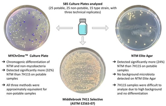
For environmental and type strain testing, triplicate colony counts were used for statistical analysis. A two-tailed t-test assuming unequal variance was used to analyze the environmental data: MYChrOme and NTM Elite agar were compared and 7H11S was compared to both novel culture methods. The agreement, Cohen’s kappa, and Pearson’s correlation coefficient between MYChrOme and NTM Elite agar environmental data were also calculated. A one-factor analysis of variance (ANOVA) was used to analyze the type strain data; p ≤ 0.05 was considered statistically significant.

3. Results

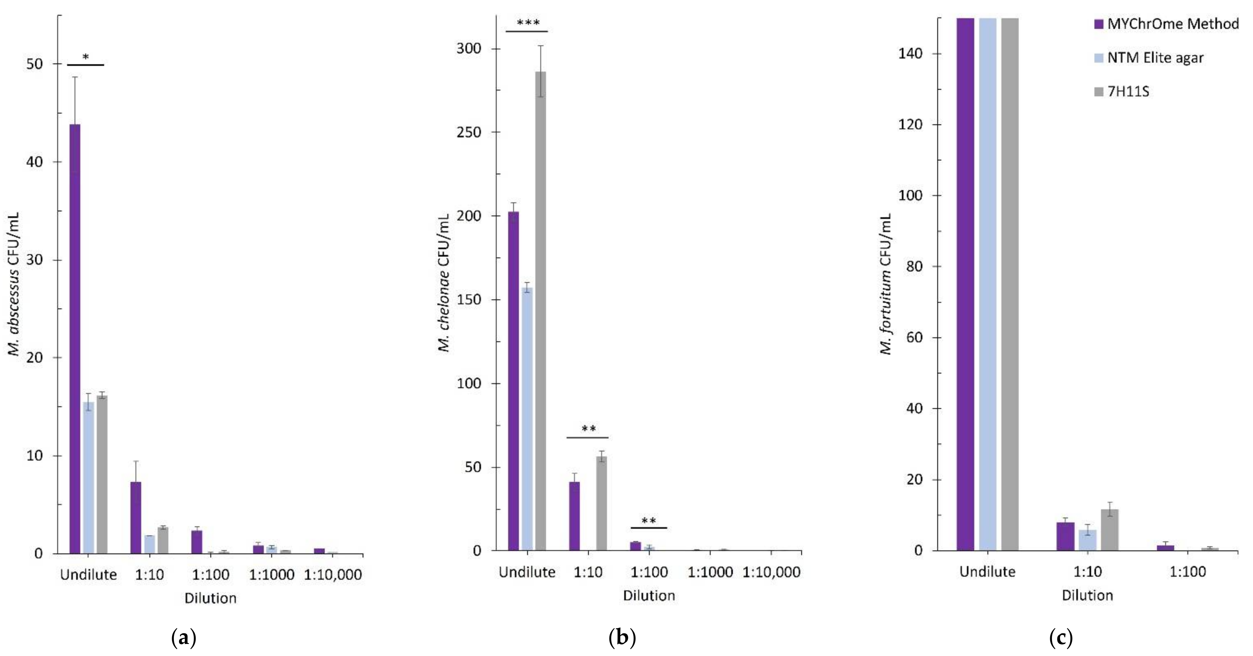
3.1. Type Strain Comparison
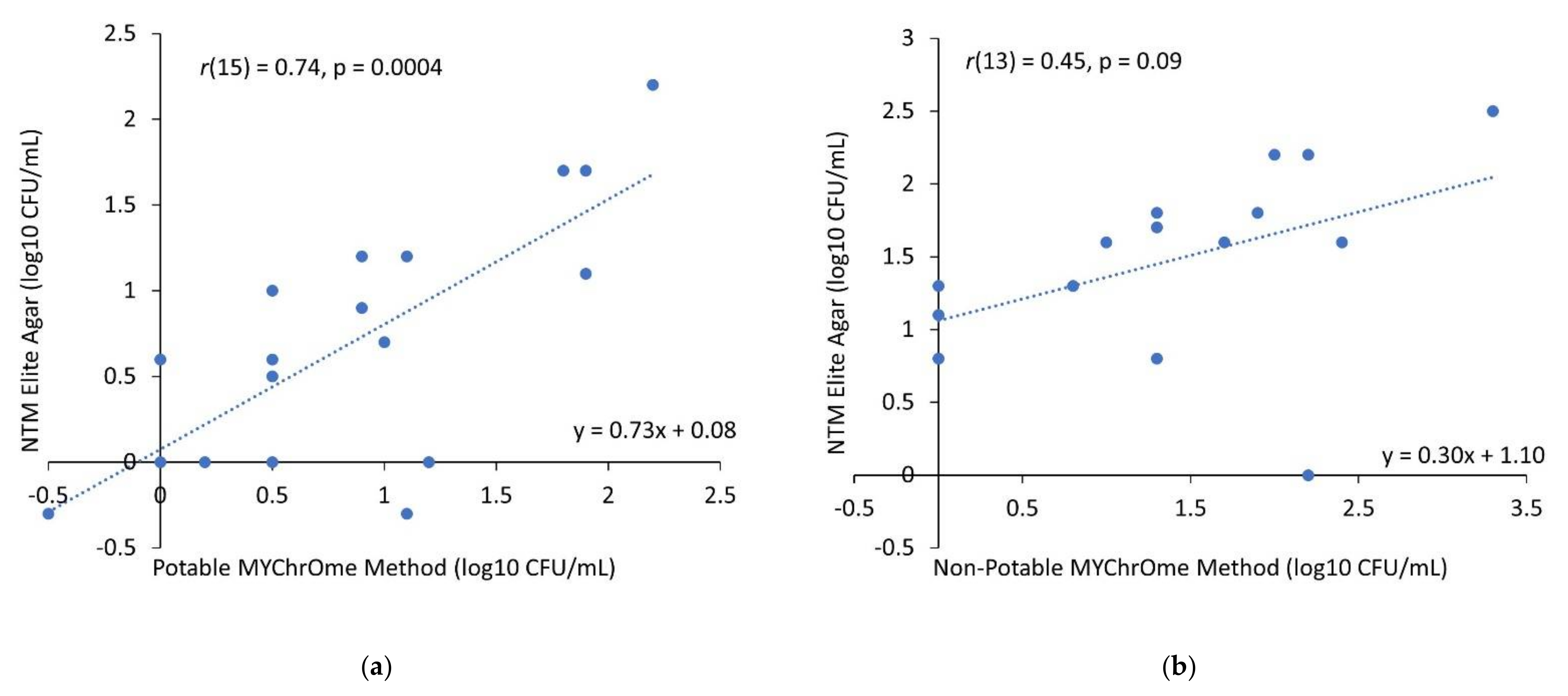
Figure 1 shows the comparison of three type strain NTM species plated on three different culture media. The dilutions with countable plates were included in the figure. For M. abscessus, the undilute (highest concentration analyzed with the MYChrOme method) was statistically higher (p = 0.024) than 7H11S and NTM Elite agar; all other dilutions were not statistically different. The M. chelonae undilute 7H11S method was significantly higher (p = 0.0002) than the other two methods. The NTM Elite result for the 1:10 dilution was significantly lower (p = 0.0001) than the other two, and the MYChrOme method 1:100 dilution was significantly higher (p = 0.004) than the NTM Elite method. The two lowest dilutions were not statistically different at a concentration of <1 CFU/mL. The M. fortuitum dilutions were not statistically different.

3.2. Potable Water

There were 18 NTM-positive potable water samples with concentrations ranging from <1 CFU/mL to too numerous to count (>300 CFU/mL). Table 1 shows the average CFU/mL for each sample per method as well as summary statistics. In comparison to each other, MYChrOme and NTM Elite colony counts were not statistically different for 80% (n = 20) of the samples. MYChrOme had significantly higher colony counts for 16% (n = 4) of the samples compared to NTM ELITE agar. Four percent (n = 1) of NTM Elite colony counts were significantly higher than MYChrOme. The p values are shown in Supplementary Table S1. There was a 96% agreement between MYChrOme and NTM Elite Agar with a Cohen’s k value of 0.905 (Table 2). Figure 2a shows a bivariate plot of log10 CFU/mL potable water results for MYChrOme and NTM Elite Agar as well as Pearson’s correlation coefficient (r). The MYChrOme method and NTM Elite agar were found to be positively correlated: r(15(degrees of freedom)) = 0.74, p = 0.0004.
In comparison to 7H11S medium, MYChrOme colony counts were not significantly different for 64% (n = 16) and significantly higher for 32% (n = 8). The remaining sample had significantly higher counts on 7H11S medium. NTM Elite agar and 7H11S medium were not statistically different for 72% (n = 18) of samples. One sample had significantly higher colony counts on 7H11S medium than the NTM Elite agar, and 24% (n = 6) of samples were significantly different in favor of NTM Elite.

3.3. Non-Potable Water

There were 22 NTM-positive cooling tower samples with concentrations ranging from <1 CFU/mL to >2000 CFU/mL (Table 1). MYChrOme and NTM Elite agar did not have statistically different colony counts for 84% (n = 21) of samples. MYChrOme and NTM Elite agar both had two samples with statistically higher colony counts than the other. Supplementary Table S2 shows the p values for all 25 samples. There was an 80% agreement between MYChrOme and NTM Elite agar for non-potable samples with a Cohen’s k value of 0.595 (Table 2). Figure 2b shows a bivariate plot of log CFU/mL non-potable water results for MYChrOme and NTM Elite Agar and Pearson’s correlation coefficient. The MYChrOme method and NTM Elite agar were found to have a moderate positive correlation: r(13) = 0.45, p = 0.09, however it was not significant.
Eighty-four percent of samples (n = 21) were not significantly different between MYChrOme and 7H11S medium. Each method had statistically more colony counts on two out of the remaining four samples. NTM Elite agar in comparison to 7H11S medium was not statistically different for 88% (n = 22) of samples, one sample (4%) was statistically significant in favor of NTM Elite agar, and 8% (n = 2) were statistically significant in favor of 7H11S.

4. Discussion

NTM disease is increasing in prevalence due to community-acquired and hospital-acquired infections, especially from contaminated tap water. This study focused on NTM detection in potable and non-potable water from hospitals and universities. NTM are extremely resistant to chemical disinfection, and they proliferate in water systems with limited nutrients [23]; therefore, it is imperative to have rapid and accurate environmental NTM testing methods to reduce the risk of NTM disease.
This study compared two novel NTM detection methods in potable and non-potable water (cooling towers) against 7H11S medium. In addition, three NTM type strains were analyzed by each method. Overall, the methods had similar NTM counts, with three significantly higher detections by the MYChrOme method and two significantly higher detections by 7H11S medium. This shows that all three methods had similar limits of detection and that using NTM Elite agar can result in reduced colony counts.
For the potable water samples, significantly more NTM were detected with the MYChrOme method and NTM Elite agar than 7H11S, 32% and 24% of the total number of samples, respectively. The reduced detection by 7H11S agar and NTM Elite agar could be due to the fact that these media contain harsh antibiotics that affect the viability of NTM. The MYChrOme culture plate does not contain any antibiotics and the simple formulation (R2A medium + CV) allows for little inhibition of NTM and the clear chromogenic differentiation of total heterotrophic aerobic bacteria (THAB). The 7H11S medium is designed to select for NTM, but many THAB are able to grow on this medium, can outcompete NTM, and look very similar to NTM colonies. Therefore, confirmation of each colony morphology is necessary, and analysis time is significantly increased. Similarly, on NTM Elite agar, every morphology must be confirmed to be NTM because this medium has an 85.6% selectivity for NTM [24]. MYChrOme eliminates this need by differentiating NTM (white colonies) through the decolorization of the CV dye by mycobacteria [25] and the pigmentation of other bacteria (purple colonies). For the purposes of this study, white colonies were confirmed by colony PCR. Figure 3 shows a comparison of the three culture plates for the analysis of one potable and one non-potable sample. In these instances, the 7H11S plates were overgrown with background microbiota and only one to three NTM colonies were detected. The potable sample plated on MYChrOme in Figure 3 was not decontaminated, and it contains white (NTM) and purple (THAB) colonies. The non-potable sample was decontaminated before plating on MYChrOme, using MYCOn with no incubation time needed, and all but one THAB colony was eliminated. The NTM Elite plates in Figure 3 show no THAB growth and NTM were enumerated after a confirmation step.
MYChrOme without MYCOn decontamination allows for the growth of some THAB alongside NTM. THAB growth can be used as a water quality indicator. Not all species of THAB can grow on MYChrOme (Gram-positive bacteria do not grow), but heavily contaminated water samples can be identified. The increased sensitivity of the MYChrOme method and the rapid MYCOn decontamination step allow for fast detection and differentiation of rapid-growing NTM and THAB.
Cooling towers are a known source of disease from pathogenic bacteria such as Legionella, therefore one section of this study was dedicated to the detection of NTM in cooling tower water. Aerosols from cooling towers are a possible source of NTM disease, however, pinpointing the source of NTM disease is difficult due to the lengthy incubation time before symptoms are present. In the book, Pathogenic Mycobacteria in Water: A Guide to Public Health Consequences, Monitoring and Management, the authors note that “potentially pathogenic mycobacteria have been isolated from cooling tower water but an epidemiological link with disease has not yet been established” [26] (pp. 174–175). Many of the cooling towers sampled in this study had large amounts of NTM detected, therefore it is essential to have robust methods for the detection of this bacteria in such complex matrices of water. MYChrOme, NTM Elite agar, and 7H11S medium performed similarly on non-potable water. In addition, the MYChrOme method allowed a fast, accurate determination of NTM colonies (no confirmation needed) as well as THAB concentration in heavily contaminated cooling towers. More robust testing to compare MYChrOme to a heterotrophic plate count method is needed to identify the correlation of purple colony counts to THAB concentration. Metal working fluid is another non-potable water source that has been linked to NTM disease [27]. The ASTM method utilized as our standard method was written specifically for this matrix. In the future, the applicability of MYChrOme and NTM Elite agar should be tested on metal working fluids. Future studies should also include a slow-growing NTM comparison and analysis of heater–cooler unit water due to the low LOD requirements (1 CFU/100 mL).
A comprehensive comparison of three solid media for NTM detection and quantification was conducted on potable and non-potable water. New media are being invented and clinical testing methods are being utilized for environmental testing. This field is moving towards preventative validation testing and away from retroactive clinical surveillance. Validation testing for NTM allows water management teams to confirm control measures are effectively reducing the NTM population. Effective water management programs can limit the risk of disease and injury due to NTM, and they should be aided by the most accurate validation test method.

5. Conclusions

The two new NTM diagnostics tested in this study improve upon 7H11S in terms of differentiation (MYChrOme), selectivity, and sensitivity for potable water samples. The MYChrOme method and NTM Elite agar had statistically higher NTM colony counts than 7H11S for 32% and 24% of potable samples, respectively. All three methods were approximately equivalent for non-potable samples. The MYChrOme method does not require confirmation of white colonies, and it shows THAB concentrations as well. For samples with high background microbiota, the simple and fast decontamination step allows for the growth and the enumeration of only NTM colonies. The MYChrOme culture plate was developed for water matrices, and it could be applied to the clinical setting. NTM Elite agar was adapted from a clinical diagnostic for use as an environmental analysis tool, with this being the third study to use this agar for water testing. Further innovation and method comparison in this growing field is needed, as well as NTM control method studies, so that water management teams can effectively identify and reduce the risk of NTM disease.

Supplementary Materials

The following supporting information can be downloaded at: https://www.mdpi.com/article/10.3390/applmicrobiol2020026/s1, Table S1: Potable sample statistical analysis.; Table S2: Non-Potable sample statistical analysis.

Author Contributions

Conceptualization, K.E.F., A.K.C. and L.P.W.; methodology, K.E.F., A.K.C. and L.P.W.; formal analysis, K.E.F. and A.K.C.; investigation, K.E.F. and A.K.C.; resources, L.P.W. and W.F.M.; data curation, K.E.F.; writing—original draft preparation, K.E.F. and A.K.C.; writing—review and editing, L.P.W. and W.F.M.; visualization, K.E.F.; supervision, L.P.W. and W.F.M.; project administration, L.P.W.; funding acquisition, W.F.M. All authors have read and agreed to the published version of the manuscript.

Funding

This research received no external funding.

Institutional Review Board Statement

Not applicable.

Informed Consent Statement

Not applicable.

Data Availability Statement

Not applicable.

Acknowledgments

The authors would like to acknowledge valuable feedback and suggestions on this manuscript from an expert in the NTM field. Additionally, special thanks to Katherine Gabrie for helping to process samples and to collect data, and for her review of this manuscript.

Conflicts of Interest

All authors were employed by Phigenics, LLC at the time of this study.

References

  1. Winthrop, K.; Marras, T.K.; Adjemian, J.; Zhang, H.; Wang, P.; Zhang, Q. Incidence and Prevalence of Nontuberculous Mycobacterial Lung Disease in a Large, U.S. Managed Care Health Plan, 2008–2015. Ann. Am. Thorac. Soc. 2020, 17, 178–185. [Google Scholar] [CrossRef] [PubMed]
  2. Collier, S.A.; Deng, L.; Adam, E.A.; Benedict, K.M.; Beshearse, E.M.; Blackstock, A.J.; Bruce, B.B.; Derado, G.; Edens, C.; Fullerton, K.E.; et al. Estimate of Burden and Direct Healthcare Cost of Infectious Waterborne Disease in the United States. Emerg. Infect. Dis. 2021, 27, 140–149. [Google Scholar] [CrossRef] [PubMed]
  3. Guimarães, T.; Chimara, E.; do Prado, G.V.B.; Ferrazoli, L.; Carvalho, N.G.F.; dos Santos Simeão, F.C.; de Souza, A.R.; Costa, C.A.R.; Viana Niero, C.; Brianesi, U.A.; et al. Pseudooutbreak of rapidly growing mycobacteria due to Mycobacterium abscessus subsp bolletii in a digestive and respiratory endoscopy unit caused by the same clone as that of a countrywide outbreak. Am. J. Infect. Control 2016, 44, e221–e226. [Google Scholar] [CrossRef] [PubMed]
  4. Sommerstein, R.; Ruegg, C.; Kohler, P.; Bloemberg, G.; Kuster, S.P.; Sax, H. Transmission of Mycobacterium chimaera from Heater-Cooler Units during Cardiac Surgery despite an Ultraclean Air Ventilation System. Emerg. Infect. Dis. 2016, 22, 1008–1013. [Google Scholar] [CrossRef] [Green Version]
  5. El Sahly, H.M.; Septimus, E.; Soini, H.; Septimus, J.; Wallace, R.J.; Pan, X.; Williams-Bouyer, N.; Musser, J.M.; Graviss, E.A. Mycobacterium simiae pseudo-outbreak resulting from a contaminated hospital water supply in Houston, Texas. Clin. Infect. Dis. 2002, 35, 802–807. [Google Scholar] [CrossRef] [Green Version]
  6. Conger, N.G.; O’Connell, R.J.; Laurel, V.L.; Olivier, K.N.; Graviss, E.A.; Williams-Bouyer, N.; Zhang, Y.; Brown-Elliott, B.A.; Wallace, R.J. Mycobacterium simiae pseudo-outbreak associated with a hospital water supply. Infect. Control Hosp. Epidemiol. 2004, 25, 1050–1055. [Google Scholar] [CrossRef]
  7. Scanlon, M.M.; Gordon, J.L.; McCoy, W.F.; Cain, M.F. Water Management for Construction: Evidence for Risk Characterization in Community and Healthcare Settings: A Systematic Review. Int. J. Environ. Res. Public Health 2020, 17, 2168. [Google Scholar] [CrossRef] [Green Version]
  8. Falkinham, J.O. Nontuberculous mycobacteria from household plumbing of patients with nontuberculous mycobacteria disease. Emerg. Infect. Dis. 2011, 17, 419–424. [Google Scholar] [CrossRef]
  9. Tzou, C.L.; Dirac, M.A.; Becker, A.L.; Beck, N.K.; Weigel, K.M.; Meschke, J.S.; Cangelosi, G.A. Association between mycobacterium avium complex pulmonary disease and mycobacteria in home water and soil a case-control study. Ann. Am. Thorac. Soc. 2020, 17, 57–62. [Google Scholar] [CrossRef]
  10. Pfaller, S.; King, D.; Mistry, J.H.; Alexander, M.; Abulikemu, G.; Pressman, J.G.; Wahman, D.G.; Donohue, M.J. Chloramine Concentrations within Distribution Systems and Their Effect on Heterotrophic Bacteria, Mycobacterial Species, and Disinfection Byproducts. Water Res. 2021, 205, 117689. [Google Scholar] [CrossRef]
  11. Brown-Elliott, B.A.; Wallace, R.J. Clinical and taxonomic status of pathogenic nonpigmented or late-pigmenting rapidly growing mycobacteria. Clin. Microbiol. Rev. 2002, 15, 716–746. [Google Scholar] [CrossRef] [PubMed] [Green Version]
  12. Donohue, M.J. Increasing nontuberculous mycobacteria reporting rates and species diversity identified in clinical laboratory reports. BMC Infect. Dis. 2018, 18, 163. [Google Scholar] [CrossRef] [PubMed] [Green Version]
  13. Strollo, S.E.; Adjemian, J.; Adjemian, M.K.; Prevots, D.R. The burden of pulmonary nontuberculous mycobacterial disease in the United States. Ann. Am. Thorac. Soc. 2015, 12, 1458–1464. [Google Scholar] [CrossRef] [PubMed]
  14. Bradner, L.; Robbe-Austerman, S.; Beitz, D.C.; Stabel, J.R. Chemical decontamination with N-acetyl-L-cysteine-sodium hydroxide improves recovery of viable Mycobacterium avium subsp. paratuberculosis organisms from cultured milk. J. Clin. Microbiol. 2013, 51, 2139–2146. [Google Scholar] [CrossRef] [PubMed] [Green Version]
  15. Bradner, L.; Robbe-Austerman, S.; Beitz, D.C.; Stabel, J.R. Optimization of hexadecylpyridinium chloride decontamination for culture of mycobacterium avium subsp. paratuberculosis from milk. J. Clin. Microbiol. 2013, 51, 1575–1577. [Google Scholar] [CrossRef] [PubMed] [Green Version]
  16. Williams, M.D.; Falkinham, J.O. Effect of cetylpyridinium chloride (CPC) on colony formation of common nontuberculous mycobacteria. Pathogens 2018, 7, 79. [Google Scholar] [CrossRef] [Green Version]
  17. Preece, C.L.; Perry, A.; Gray, B.; Kenna, D.T.; Jones, A.L.; Cummings, S.P.; Robb, A.; Thomas, M.F.; Brodlie, M.; O’Brien, C.J.; et al. A novel culture medium for isolation of rapidly-growing mycobacteria from the sputum of patients with cystic fibrosis. J. Cyst. Fibros. 2015, 15, 186–191. [Google Scholar] [CrossRef] [Green Version]
  18. Fisher, K.E.; Chhabra, A.K.; Wickenberg, L.P.; McCoy, W.F. Validation of the MYChrOmeTM Culture Plate for Detection and Differentiation of Rapid-Growing Nontuberculous Mycobacteria in potable and non-potable water: AOAC Performance Tested MethodSM 062101. J. Aoac Int. 2021, 5, 549–557. [Google Scholar] [CrossRef]
  19. Preece, C.L.; Wichelhaus, T.A.; Perry, A.; Jones, A.L.; Cummings, S.P.; Perry, J.D.; Hogardt, M. Evaluation of various culture media for detection of rapidly growing mycobacteria from patients with cystic fibrosis. J. Clin. Microbiol. 2016, 54, 1797–1803. [Google Scholar] [CrossRef] [Green Version]
  20. Alexander, K.J.; Furlong, J.L.; Id, J.L.B.; Rihs, J.D.; Stephenson, D.; Id, J.D.P.; Stout, J.E. Evaluation of a new culture medium for isolation of nontuberculous mycobacteria from environmental water samples. PLoS ONE 2021, 16, e0247166. [Google Scholar] [CrossRef]
  21. Ditommaso, S.; Giacomuzzi, M.; Memoli, G.; Garlasco, J.; Curtoni, A.; Iannaccone, M.; Zotti, C.M. Chemical susceptibility testing of non-tuberculous mycobacterium strains and other aquatic bacteria: Results of a study for the development of a more sensitive and simple method for the detection of NTM in environmental samples. J. Microbiol. Methods 2022, 193, 106405. [Google Scholar] [CrossRef] [PubMed]
  22. Radomski, N.; Roguet, A.; Lucas, F.S.; Veyrier, F.J.; Cambau, E.; Accrombessi, H.; Moilleron, R.; Behr, M.A.; Moulin, L. atpE gene as a new useful specific molecular target to quantify Mycobacterium in environmental samples. BMC Microbiol. 2013, 13, 1471–2180. [Google Scholar] [CrossRef] [PubMed]
  23. Falkinham, J.O. Surrounded by mycobacteria: Nontuberculous mycobacteria in the human environment. J. Appl. Microbiol. 2009, 107, 356–367. [Google Scholar] [CrossRef] [PubMed]
  24. Biomerieux: NTM Elite Agar. Available online: https://www.biomerieux-diagnostics.com/ntm-elite-agar (accessed on 1 March 2022).
  25. Jones, J.J.; Falkinham, J.O. Decolorization of malachite green and crystal violet by waterborne pathogenic mycobacteria. Antimicrob. Agents Chemother. 2003, 47, 2323–2326. [Google Scholar] [CrossRef] [PubMed] [Green Version]
  26. Pedley, S.; Bartram, J.; Rees, G.; Dufour, A.; Cotruvo, J.A. Pathogenic Mycobacteria in Water: A Guide to Public Health Consequences, Monitoring and Management; IWA Publishing: London, UK, 2004; pp. 174–175. ISBN 1843390590. [Google Scholar]
  27. Halstrom, S.; Price, P.; Thomson, R. Review: Environmental mycobacteria as a cause of human infection. Int. J. Mycobacteriol. 2015, 4, 81–91. [Google Scholar] [CrossRef] [PubMed] [Green Version]
Figure 1. Type strain comparison. (a) M. abscessus (Culture Collection University of Gothenburg) (CCUG) 71636), (b) M. chelonae (CCUG 72969), and (c) M. fortuitum (CCUG 46694) serial dilutions to 10−4 were analyzed with each culture method. Error bars represent plus and minus the standard error of the mean. One-factor analysis of variance (ANOVA) was used to determine significant differences: * p ≤ 0.05, ** p ≤ 0.01, *** p ≤ 0.001.
Applmicrobiol 02 00026 g001
Figure 2. Bivariate plots showing pairwise comparison of NTM Elite vs. MYChrOme NTM positive results in log CFU/mL. (a) Potable data comparison. (b) Non-potable data comparison. The equation for the linear fit is shown on the bottom right corner and the Pearson correlation coefficient, r, is shown on the top left corner with the degrees of freedom (n-2) and the p-Value.
Applmicrobiol 02 00026 g002
Figure 3. Culture plate comparison. Example culture plates of one potable and one non-potable water sample. The non-potable MYChrOme sample shown was decontaminated with 1:25 MYCOn reagent. Example purple colonies (non-mycobacteria) are circled in black on the MYChrOme plates.
Applmicrobiol 02 00026 g003
Table 1. Summary of environmental detections. Twenty-five potable and non-potable water samples were analyzed in triplicate by each plating method. The average CFU/mL from three technical replicates is shown. Summary statistics are shown at the bottom of the table.
Avg CFU a/mL PotableAvg CFU/mL Non-Potable
Sample
Number
MYChrOme MethodNTM Elite7H11SMYChrOme MethodNTM Elite 7H11S
1NDNDNDNDNDND
28.28.50.216.76.77.2
313.50.50.5166.7146.788.8
4500.0500.0500.096.7170.0181.0
5143.3157.3250.5ND6.70.7
6NDNDND3.320.0ND
77.316.525.22093.3320.0500.0
8NDNDND10.040.07.2
9NDNDNDND3.36.8
1080.354.824.8NDND0.2
113.53.7NDNDND0.3
121.24.0ND80.063.326.7
133.310.35.546.736.750.5
1414.50.20.5163.3ND5.0
15NDND0.2ND3.31.0
163.5NDND236.743.322.3
17NDNDND6.720.09.7
181.70.8ND16.753.326.5
1913.715.815.216.766.73.8
2010.55.0NDNDNDND
213.23.8NDNDND0.5
2256.749.53.3NDND0.2
23NDNDNDND13.318.5
2478.011.70.5NDND0.3
250.30.5NDNDND2.3
Positives181712131622
Mean b52.449.668.9227.263.343.6
SD c118.3122.3153.0565.784.0110.0
SEM d27.929.744.2156.921.023.4
a Colony forming unit; b Average of all positive samples in CFU/mL; c Standard deviation of the positive samples; d Standard error of the mean for the positive samples; ND = Not Detected.
Table 2. Agreement statistics between MYChrOme and NTM Elite Agar NTM detections in environmental samples.
MYChrOme MethodNTM Elite Agar
Pos aND bPosNDPA cK d
Potable18717896% 0.905
Non-Potable131216980% 0.595
a Number of positive samples; b NTM not detected; c Percent Agreement; d Cohen’s kappa.
Publisher’s Note: MDPI stays neutral with regard to jurisdictional claims in published maps and institutional affiliations.

