Article

Enhancing Human Superorganism Ecosystem Resilience by Holistically ‘Managing Our Microbes’

by
Margaret E. Coleman
1,*,
Rodney R. Dietert
2,
D. Warner North
3 and
Michele M. Stephenson
4
1
Coleman Scientific Consulting, Groton, NY 13073, USA
2
Department of Microbiology and Immunology, Cornell University, Ithaca, NY 14853, USA
3
NorthWorks, San Francisco, CA 94133, USA
4
Advancement and External Affairs, Syracuse University, Syracuse, NY 13244, USA
*
Author to whom correspondence should be addressed.
Appl. Microbiol. 2021, 1(3), 471-497; https://doi.org/10.3390/applmicrobiol1030031
Submission received: 10 August 2021 / Revised: 5 October 2021 / Accepted: 12 October 2021 / Published: 18 October 2021
(This article belongs to the Special Issue Human Microbiota Influence on Human Health Status)

Abstract

:
Microbes in the 21st century are understood as symbionts ‘completing’ the human ‘superorganism’ (Homo sapiens plus microbial partners-in-health). This paper addresses a significant paradox: despite the vast majority of our genes being microbial, the lack of routine safety testing for the microbiome has led to unintended collateral side effects from pharmaceuticals that can damage the microbiome and inhibit innate ‘colonization resistance’ against pathobionts. Examples are discussed in which a Microbiome First Medicine approach provides opportunities to ‘manage our microbes’ holistically, repair dysbiotic superorganisms, and restore health and resilience in the gut and throughout the body: namely, managing nosocomial infections for Clostridioides difficile and Staphylococcus aureus and managing the gut and neural systems (gut–brain axis) in autism spectrum disorder. We then introduce a risk analysis tool: the evidence map. This ‘mapping’ tool was recently applied by us to evaluate evidence for benefits, risks, and uncertainties pertaining to the breastmilk ecosystem. Here, we discuss the potential role of the evidence map as a risk analysis methodology to guide scientific and societal efforts to: (1) enhance ecosystem resilience, (2) ‘manage our microbes’, and (3) minimize the adverse effects of both acute and chronic diseases.

1. Background

Culture-independent methods from studies on genomic, proteomic, and metabolomic (-omics) research have dramatically advanced our knowledge of human microbiota and stimulated new paradigms of thought about our relationships with microbes and how we approach the assessment of risks and benefits. Many advances have developed or were catalyzed by long-term research consortia, including the Metagenomics of the Human Intestinal Tract (MetaHIT) project of the European Commission and China, and the Human Microbiome Project (HMP) and the Integrated HMP or iHMP of the US [1,2,3].
The -omics research characterizing the human microbiota in recent decades are weakening—perhaps destabilizing and overturning—20th-century misconceptions about human health and disease. No longer is there consensus that the healthiest humans are generally free of microbes [4,5,6]. Similarly outdated concepts focused on germaphobia and the disease triangle (host, pathogen, and environment) in public health and microbial risk analysis [7] seem to perpetuate the erroneous exclusion of our symbionts, the natural commensal and mutualistic microbiota of healthy human systems, from frameworks for safety evaluation and human health risk assessment [5,8,9]. Further, 20th-century pre-microbiome dogmas based on erroneous assumptions are barriers to holistic health and sustainable healthcare [6].
This special collection draws much needed attention to an expanding body of research that illustrates how very wrong our outdated views of Homo sapiens as an independent species were, based on misleading and invalid scientific hypotheses and assumptions from past centuries. Currently, extensive bodies of evidence provide coherent support for the theory that Homo sapiens actually function as superorganisms with our microbial partners in health, as holobionts of some eukaryotic and predominantly microbial genes that cooperate and coordinate via diverse and redundant mechanisms [4,10]. Such paradigm changes point to the need to apply formal benefit-risk methodologies to questions about how to best ‘manage our microbes’.
A grave error in the application of 20th century thinking was the exclusion of the indigenous microbiota from both safety and risk assessment frameworks [5,8]. The ‘microbiome revolution’ [11] and subsequent explosions of knowledge from decades of -omics research strongly challenges concepts from the prior century, particularly the overwhelming biological reality that dense and diverse microbial symbionts, their genes, and their metabolites provide tremendously important sources of variation in health and innate resistance to disease for human superorganisms. Expansive methods for the assessment of benefits and risks [8,12,13] are needed to assess and manage superorganism health and disease.
One 20th-century conceptualization of host–pathogen interactions that has been strengthened, not weakened, by -omics research is ‘colonization resistance’, dose- and time-dependent host protection by dense and diverse indigenous gut microbiota against enteric pathogens [7,9,14,15,16,17,18]. Mechanistic data from -omics studies not only demonstrate that the gut microbiota of healthy superorganisms can outcompete pathogens for nutrients and space (niches to colonize in the gut ecosystem), but -omics studies also identify potential mechanisms that gut microbiota are a part of which contribute to health and prevent disease throughout the body. Healthy microbiota function as ‘gatekeepers’, operationally connecting host defenses to environmental exposures through coordinated mechanisms: (i) enhancing tight junctions and the mucosal barrier; (ii) producing antimicrobial compounds active against enteropathogens; (iii) assisting in pathogen clearance from the gut lumen; (iv) stimulating cytokine release and immune functions; and (v) contributing to host tolerance of commensal and non-pathogenic microbes [15,16,19,20].
Hosts subjected to antibiotics and other pharmaceuticals suffer unintended collateral side effects on the gut microbiota, including the disruption of the composition, integrity, and function of the gut ecosystem, termed dysbiosis, with a loss of microbial diversity and richness, a loss of protective microbes, and blooms of opportunistic or frank pathogens. Pharmaceuticals not assessed for effects on the microbiota may compromise colonization resistance and greatly enhance sensitivity to enteropathogens, as demonstrated for increased sensitivity to Salmonella spanning five orders of magnitude in dysbiotic systems [7]. Recent initiatives, such as ‘Microbiome First Medicine’ [21], focus research primarily upon the human superorganism majority—the microbiome—when exploring holistic sustainable healthcare, rather than the traditional primary focus on pharmaceutics tested only on the human superorganism minority, Homo sapiens.

1.1. Risks and Benefits of Managing Gut Microbes and Colonization Resistance

Multiple factors influence health and disease in human superorganisms, as illustrated in the proposed health triangle [7], including: access to healthcare, diet and nutrition, genetics, geography, lifestyle and activity, occupation and economic status, pollution and other environmental toxicants, stress, and travel. The gut ecosystem coordinates not merely digestive functions, but also immune systems, as well as behavioral, neural, and respiratory functions. The gut epithelium is patrolled by ~106 lymphocytes per gram of gut tissue, and host epithelial and immune cells interact with approximately 100 trillion microbes [18,22]. An extensive body of evidence characterizes the dense, diverse, and stable gut microbiota that appears to vary more between individuals than over time.
A recent study [18] determined that that 91 of the top 109 microbial species were present at <1% relative abundance. Potential enteropathogens (Bacillus cytotoxicus, Campylobacter, C. difficile, Clostridium botulinum, potentially pathogenic E. coli, Klebsiella, Salmonella, Shigella, and Vibrio cholerae) were also detected at <1% abundance in healthy gut microbiota [18]. Although limited data exist for modeling the microbial ecology of the gut ecosystem to predict the likelihood of dysbiosis and illness, the conditions that favor overgrowth and blooms of potential pathogens may be associated with enteric disease.
The relative abundances of gut microbes vary considerably across studies and geographic areas. In general, longitudinal studies demonstrate higher variability in the composition and abundance of gut microbes between individuals than between time points for a given individual, suggesting that gut microbiota is stable over time. Two phyla, Bacteriodetes and Firmicutes, generally account for ~90% of gut microbes. Some studies note differences in the ratio of these two phyla between small populations of healthy and ill people. However, data for the healthy gut microbiota abundances at the phylum level reported in two recent studies [23,24], summarized below (Table 1), illustrate variability between human superorganisms and between studies. This suggests differences in the many genetic and environmental factors that influence microbiota composition, abundance, and function, as noted above.
One recent study characterized gut microbiota for 98 human superorganisms (the Human Microbiome Project and George Washington University Project), composed of more than 59 microbial genera and as many as 863 species or strains, including potential pathogens [24]. Another recent study [25] documented gut microbiota for 897 healthy human superorganisms that documented 15 genera and 31 species that may be associated with health status. Great uncertainty exists about which of the hundreds, perhaps thousands of bacterial strains, or networks or consortia of strains, in a healthy gut actually drive colonization resistance and essential cross-talk with the immune system and other systems to maintain or restore health. Due to the high variability between humans, their diets, their pharmaceuticals, and the tremendous complexity of the interacting networks of hundreds or thousands of related and unrelated microbes, simple associations or correlations may be identified in small studies, but are rarely generalizable for the prediction of human health or disease in other contexts.
The complexity of superorganism ecosystems, particularly human and mammalian gastrointestinal (GI or gut) ecosystems, poses a ‘reductionist dilemma’ for medical professionals and consumers seeking to ‘manage our microbes’. Two holistic strategies for ‘managing our microbes’ involve the use of: (1) undefined live biotherapeutic products, such as fecal microbiome transplantation (FMT) and FMT-like procedures; and (2) defined strains or consortia of strains [26]. Species richness and alpha diversity of defined live biotherapeutics appear to be drivers of colonization resistance by multiple mechanisms [27]. The limited success of single-strain products is likely due to the complexity and pleiotropism of complex microbial communities and ecosystems dependent on microbial densities or doses, diversity, community structure, and micro-environments, as well as the pharmacokinetics and pharmacodynamics of microbial interactions with other microbes and the host [26]. The current emphasis of the iHMP on multiple -omic research strands for the prediction of health and disease outcomes for defined products reflects this complexity and the dilemma of reducing complex communities to practical numbers of strains that retain effective colonization resistance activity [2,26]. Dysbiotic model systems may respond to one or a few bacterial strains, often probiotics [28], while others were restored by the genome-guided assembly of a defined consortium of 15 strains [15]. The least-reductionist strategy is administering undefined and complex microbiota from healthy donors (fecal microbiome transplants or FMTs that may include hundreds or thousands of bacterial strains) for restoring severely dysbiotic gut ecosystems. As more mechanistic data is generated by -omics studies, opportunities to ‘manage our microbes’ to strengthen or restore optimal colonization resistance [9] are likely to expand preventive and therapeutic options in microbiota-mediated medicine as alternatives to past over-reliance on pharmaceuticals now known to contribute to dysbiosis, inflammation, and recurring or chronic diseases [6].

1.2. Expanding Traditional Risk Paradigms for the Microbiota

Applications of advances in knowledge from -omics studies to microbial risk analysis (assessment, communication, and management of risk) appear to be limited by the correlative nature of much of the available evidence [13]. Microbial species may be associated with each other positively or negatively, using statistical methods to quantify correlations of the presence or abundance of microbes A and B. However, experimental designs and methodologies for microbial detection and enumeration do not at present distinguish whether the change in microbe A is a cause or an effect of the change in microbe B, or if both microbes A and B are responding to another undetermined factor in the complex ecosystems of the gut.
Assessments of the likelihood and severity of health risks and the benefits of effective interventions to ‘manage our microbes’ by altering the presence or abundance of specific microbes in complex ecosystems may be misleading without a knowledge of causality for both risks and benefits. High quality data from experimental designs that support the assessment of causality are essential for reliable, robust assessment and management that could include traditional prevention and therapeutic interventions as well as those focused on ‘managing our microbes’.
The general framework for risk assessment developed by the US National Research Council [29] was modified by Marks et al. [30] for the assessment of infectious disease from foodborne pathogens that built in flexibility for addressing host-microbial interactions. Figure 1 illustrates the types of data and analysis required by transdisciplinary risk assessment teams, including knowledge of the nature and rates of exposure to potential pathogens and microbial ecology (exposure assessment) and the likelihood that exposures at given pathogen doses will cause illness (dose–response assessment, including both threshold and more conservative non-threshold model forms) to predict risk with attendant uncertainty [30,31]. Knowledge gaps may be bridged by drawing inferences from animal studies or in vitro model systems [7,12]. However, even high-quality direct evidence, such as that from randomized double-blind clinical trials in humans, may not be generalizable to other human populations and to other environmental contexts without additional mechanistic data. Moreover, results of small studies that provide only correlative data may be insufficiently reliable and representative of highly variable populations of human superorganisms around the world, and thus poor predictors of health and disease. What the figure does not communicate is a methodology for assessing health and non-communicable diseases contributing to global epidemics of non-infectious disease.
Our work is motivated by the need to incorporate a deeper knowledge of the benefits and risks of microbiota into methodologies for the assessment and management of microbial exposures to human superorganisms. We highlight studies that identify some common mechanisms of exposure from human volunteer studies.
This paper discusses examples associated with emerging clinical evidence from -omics research largely on the gut microbiota from the recent decade, emphasizing the potential for ‘managing our microbes’ to enhance superorganism health for selected infectious and non-communicable human diseases. The first two examples focus on exposures in healthcare settings: Clostridioides difficile infection (CDI) in the gut; and Staphylococcus aureus (Staph A) associated with asthma and allergic and infectious diseases. The third focuses on autism spectrum disorder (ASD) and studies demonstrating the influence of microbiota interactions along the gut–brain axis. The fourth introduces the human breastmilk ecosystem and an approach for evaluating and communicating a complex body of evidence for benefits and risks: evidence mapping. Evidence maps provide coherent and transparent characterization of the ‘state of the science’ for benefits and risks and remaining uncertainties. Complementary work on the extensive body of evidence for the breastmilk ecosystem and benefits and risks for infants ingesting raw breastmilk and pasteurized donor milk is documented in an accepted manuscript in this same special issue in Applied Microbiology [13].
The aim of our work is to facilitate advancement of targeted -omics research for future applications using formal methods of benefit–risk analysis. Incorporating -omics research is essential, from our perspective, to minimize oversimplifications and confirmation biases that otherwise limit effective medical and societal decision making when ‘managing our microbes’ as superorganisms.

2. CDI in the Gut

Exposure to the most commonly reported nosocomial or healthcare-associated infective agent in the US and around the world is linked to Clostridioides (formerly Clostridium) difficile infections (CDI). CDI appears to be associated with dysbiosis or disturbance of the normal gut microbiota, and working knowledge of the complex of multidimensional networks of gut microbial interactions with each other, the pathogen, and host cells is limited. We are not aware of human data depicting dose–response relationships for CDI. The primary risk factors for CDI include: broad spectrum antibiotic treatments and other pharmaceuticals; hospitalization and length of hospital stay; increasing age; underlying co-morbidities; and diet [32].
The US CDC [33] reported nearly 16,000 CDI cases in 2018 with incidence rates increasing with age for both community-associated and nosocomial (healthcare-associated) CDI, as summarized in Table 2.
Yet C. difficile has long been associated with unusually high rates of asymptomatic colonization in healthy neonates and infants [32]. One hypothesis about resistance to CDI in young children is that receptors for C. difficile toxins are not expressed in children until about 24 months of age.
However, elderly patients and young children are at risk of CDI, particularly in hospital and healthcare environments, with potentially serious enteric illness and frequently recurring episodes that typical pharmaceutical interventions may not clear. Further, polymicrobial synergy with consortia of other microbes (including Enterococcus, Klebsiella, and E. coli) may be driving C. difficile persistence, blooms, and virulence, as demonstrated in surgical wound infections, otitis media, periodontal infections, and cystic fibrosis [32].

2.1. Using Commensal Bacteria for Enhancing or Restoring Colonization Resistance

Work on integrating knowledge of dysbiosis and disease severity for CDI risk using in vivo and in vitro systems for several mammalian systems has been underway for more than a decade. Metagenomic microbiota studies integrated with ecological and mathematical models identified common commensal bacteria in humans and mice [34,35] that appear to inhibit C. difficile and contribute to the restoration of colonization resistance (C. scindens, C. populeti, C. claiflavum, Akkermansia muciniphila, Barnesiella intestihominis, Blautia hansenii, Lactobacillus reuteri, Pseudoflavonifractor capillosus, and Porphyromonas catoniae). Other microbes positively associate with CDI and appear to contribute to dysbiosis (Enterococcus avlum, E. faecalis, Klebsiella oxytoca, and C. irregulare) [35]. Another laboratory has conducted pairwise and multi-species community analyses that suggest that species richness of related microbes and other commensals inhibit or exclude C. difficile in synthetic human gut communities [27]. With integrated multidisciplinary studies combining -omics, microbial ecology, and mathematical modeling, these laboratories are testing more than just correlations, but causation for colonization resistance in healthy superorganisms and CDI in dysbiotic hosts.
Further expanding knowledge about the key species which exist among thousands potentially present in the feces of healthy human superorganisms may permit the identification of smaller consortia of defined species that could replace the use of holistic but largely undefined fecal microbiota donor samples in the future to enhance efficacy and reduce the risk of unintended side effects of FMT. Although some serious and fatal adverse events have been associated with FMT for immunocompromised patients [36], no serious adverse effects were reported in the clinical trials where FMT was administered for recurrent CDI described herein. Strategies for designing defined live biotherapeutics to restore microbiota structure and function, particularly colonization resistance, are improving, and multiple defined products are working their way through clinical trials [37]. More recently, a human clinical study [38] administered FMT (~10 million bacteria from healthy donor feces) to 29 patients at risk for CDI. The results indicated that the GI systems of 12 patients were quickly colonized or engrafted, and transformed taxonomically and functionally to donor-like microbiota within 7 days. However, a recurrence of CDI occurred in 17 out of 29 patients after the first FMT treatment. Key taxa can be identified as leading to better engraftment and FMT success (Firmicutes phyla, Lachnospirciae family, Blautia, Roseburia, Anaerostipes) or FMT failure or a need for repeat interventions (Proteobacteria phyla, Enterobacteriaceae family, Escherichia/Shigella, Kelbisella, Pluralibacter). Interestingly, the detection of C. difficile was NOT predictive of the failure of FMT, and colonization resistance may not require eradication, but merely suppression of the blooms of pathogen growth. What appears essential to the recovery of colonization resistance is the dramatic and immediate restructuring of the microbial ecology of the gut of patients to more diverse, donor-like microbiota that suppress blooms of C. difficile.
Another human clinical study (case–control) in hospitalized patients characterized the gut microbiota of 28 elderly patients with an antibiotic history and CDI (average age 79 years) and 56 healthy elderly patients without diarrhea (average age 75 years) [39]. Key taxa significantly differed between CDI and healthy controls, with a decreased abundance of Bacteroides, Clostridium cluster IV, Bifidobacterium, Faecalibacterium prausnitzii, and Prevotella and increased abundance of Akkermansia muciniphila, Lactobacillus, E. coli, and Klebsiella in CDI cases.
A small longitudinal study in a Belgian nursing home [40] characterized the gut microbiota of 23 elderly residents and reported little difference between gut microbiota of asymptomatic C. difficile carriers and the one CDI case that developed during the study. Although no association was observed between microbiota diversity or richness for nursing home residents, the small size of the study may limit generalizations to other populations or facilities.

2.2. Using the Virome: Phage Therapy

A major part of the human microbiome is composed of viruses and is designated as the virome [41]. We harbor a variety of viruses at the same body sites as the bacteria, archaea, protozoans, and fungi [42,43]. Recently, a metagenomic analysis was performed on just under 190 thousand human gut DNA viruses [44]. A majority of the viruses harbored there are bacteriophages (viruses that infect bacteria) and these infect some of the most predominant bacterial species in the gut [44]. The increasing availability of information on these bacteriophages has opened up a new area of microbe-on-microbe attack against the most problematic bacterial pathobionts, their own viruses [45]. Known as phage therapy, the metagenome data has helped to direct the design of therapeutic approaches [46]. Additionally, a taxonomic approach to human phages has begun to reveal the characteristics of a healthy human phagome [47].
Phage therapy has been used combat antibiotic resistance pathobionts such as C. difficile. Characterization of the C. difficile bacteriophages has been underway [48,49]. This has provided tools for attacking C. difficile in a bacterium-specific manner [50]. In addition to direct attack against C. difficile, phages have been used to deliver CRISPR-Cas3 antimicrobials [51]. Beyond C. difficile, other antibiotic resistance bacterial pathogens such as Acinetobacter baumannii are also current targets of phage therapy [52].

2.3. C. difficile Summary

This CDI example illustrates the challenge of understanding the complexities of the gut ecosystem, the microbial strains or consortia driving colonization resistance, and the mechanisms for restoring dysbiotic ecosystems and preventing dysbiosis in healthy ecosystems. FMT has been used to restore health with some success with CDI, without serious adverse events in these studies. However, the mechanistic details of how colonization resistance can be most effectively restored in CDI patients, and CDI prevented in other hospitalized patients, remain largely unknown. Additionally, using the virome (i.e., phage therapy) to target pathobionts such as C. difficile is an important new microbiome-based approach. Applications using commensal bacteria and building microbiota communities plus the virome (phage therapy) can overcome the incomplete therapy (which results in a subsequent increase in susceptibility) provided by conventional antibiotics.

3. Staph A, Asthma, and Allergic and Infectious Diseases

Exposure in healthcare settings to Staphylococcus aureus (Staph A), a Gram-positive commensal bacterium or an opportunistic pathogen, is associated with severe disease and death around the world [53]. Staph A exposure is associated with pneumonia and other respiratory infections, surgical site, prosthetic joint, and cardiovascular infections, and surgical/hospital associated bacteremia [54]. Strategies for vaccination and antibiotic therapy are largely ineffective to date.
Staphylococcus spp., including Staph A, can be found on skin, in the GI tract, and in the airways, as well as in hospitals, healthcare settings, and other environments (air, dust, sewage, soil, surfaces, and water). Staph A can also contribute to mastitis (infection of mammary tissues) in lactating women and ruminants, including cows. Staph A foodborne illness is very rare, but Staph A can cause staphylococcal food poisoning from the ingestion of preformed, heat-stable enterotoxins [53]. Healthy people appear to possess innate resistance to Staph A food poisoning unless foods are highly contaminated (>100,000 Staph A counts per/mL or gram) [55].
Staph A is a common commensal on the skin and in the nose, but also occurs in the GI tract. It can be either carried at these body sites at various stages of development or it can appear via transient exposure (e.g., hospital visit, contaminated food). Staph A carriage/colonization at one body site can have implications for diseases arising in other regions of the body [56]. We are not aware of human data depicting dose–response relationships for Staph A.
Staph A is one of the healthcare-associated bacteria that is known to carry antibiotic resistance genes and which has multi-drug resistance to antibiotics (e.g., methicillin-resistant Staphylococcus aureus—MRSA and vancomycin-resistant Staphylococcus aureus—VRSA). Because of the problems with antibiotic resistance and the risks associated with Staph A infection, it is necessary to apply strategies to minimize or prevent Staph A carriage and to block the opportunity for infection that go beyond use of antibiotics.
One of the tools that shows promise is the optimization of colonization resistance for Staph A. Evidence exists for protection against Staph A infections by probiotics [57,58,59,60] and natural commensal Staphylococcus spp. [61]. Microbe management and the use of probiotic-derived products within the microbiota can enhance natural defenses through colonization resistance against Staph A in the respiratory system [58,59,60]. An added benefit is that the risk of carriage and infection then becomes a matter of ecological management of microbes rather than antibiotic-drug therapy. The latter can further damage the microbiota, creating dysbiosis and reducing colonization resistance. Risk of infection is only one of several pathological outcomes.

3.1. Beyond Infection to Asthma and Allergic Diseases

Staph A is not just an acute risk for potential life-threatening infections in healthcare settings. It can also be the underlying cause of certain non-communicable allergic-inflammatory diseases driven by Type 2 immune responses and IgE [62]. In fact, the linkage of Staph A to allergic rhinitis [63], atopic dermatitis [64], food allergies [65], and asthma [66,67] is one of several examples that illustrates that the boundary which exists between communicable and non-communicable diseases is soft to potentially nonexistent rather than hard, as previously believed [68].
The Staph A bacterium carries many different virulence factors as well as hemolysins and leukotoxins [69]. These aid a multi-pronged evasion of the human innate immune response against the bacterium. The specific diseases most associated with Staph A infections are: pneumonia (including necrotizing fasciitis and necrotizing pneumonia), toxic shock syndrome, infective endocarditis, scalded skin syndrome, and osteomyelitis.
Staph A produces toxins [70] that can damage membranes and kill cells, such as neutrophils, which are front line responders against the bacterial infection. Some utilize receptor-medicated processes (e.g., alpha toxin and the leukocidins) while others are non-receptor-mediated lysins (e.g., phenol-soluble modulins) [69]. Other toxins, such as the enterotoxins and secreted proteins, interfere with receptor function, both altering host response to the infection (e.g., blocking phagocytosis of the bacterium and disrupting complement pathways) and also promoting aberrant T cell-based immune responses by functioning as superantigens [69].
Additionally, Staph A-produced superantigens readily stimulate polyclonal, pro-inflammatory T cell responses [71]. One of the risks of the nasal carriage of Staph A is that it can promote epithelial barrier dysfunction and underlying T cell-driven chronic inflammation. This contributes not only to the enhanced risk of infectious disease connected to ongoing inflammation and dysbiosis (e.g., chronic sinusitis), but also the enhanced risk of allergic manifestation, such as allergic rhinitis and asthma [72,73].
The bacterium produces many different enzymes that affect immune cell stability as well as host defense/physiology. For example, staphylokinase can regulate clot formation, thereby helping to evade the body’s attempt to localize the infection [69]. The toxins and enzymes of Staph A are now known to influence not just infection but also sensitization, allergy, and asthma.

3.2. Staph A and Asthma

Overall, the process is one where the pathogen gains control of and alters the airway mucosa. An early biomarker of the changes is host production of Staph A enterotoxin-specific IgE. Immune cell balance and response is manipulated by the superantigens, such as the serine-protease-like proteins or protein A. This leads to the polyclonal Type 2 T cell responses. Epithelial cells are stimulated to release IL-33 which helps to drive innate lymphoid cells and T cells toward the Th2 skewing. Mast cell degranulation occurs with the combined result of B cell activation and eosinophil recruitment promoted by IL-5 production [74]. Once the eosinophils arrive, one of the suggested footprints that results from this Staph A-initiated process is the formation of Charcot–Leyden crystals (bipyramidal hexagonal crystals resulting from the crystallization of eosinophil-produced Galectin-10) [75,76].

3.3. Staph A and IL-36

A recent finding involves the role of an interleukin (IL-36) in promoting lung inflammation and asthma. IL-36 is one of the members of the very large IL-1 cytokine superfamily. Dysregulation in IL-36 is a hallmark that spreads across the entire allergic triad of non-communicable diseases (atopic dermatitis, allergic asthma, and allergic rhinitis), and Staph A has the capacity to produce IL-36 dysregulation [77]. The IL-36 route to the entire allergic triad has ramifications that extend beyond just the location of Staph A carriage. For example, studies suggest that epicutaneous Staph A can elicit the keratinocyte production of IL-36, which in turn elevates IgE production and Type 2 inflammation. Patrick et al. [62] found that in mice, this sequence was required for the development of lung inflammation. Importantly, Staph A’s promotion and exacerbation of allergic asthma may not require nasal carriage of the bacterium if it is colonized elsewhere [62].

3.4. Vulnerable Populations

Neonates have the highest rates of the invasive form of Staph A for any age group [58,78,79]. In the past, the go-to strategy to protect neonates, infants, and children against Staph A-associated disease had been the use of intranasal antibiotics and antiseptic measures to ‘decolonize’ Staph A from the nasal passages. However, these treatments are not single-species specific against the pathobiont and cause unintended collateral side effects on the commensal nasal microbiota. As pointed out by Khamash et al. [58], antibiotics and other pharmaceuticals are likely unsuccessful because they also kill commensals that are important in colonization resistance against pathogenic Staph A. Antibiotic-driven depopulation of nasal Staph A is likely to be a short-lived benefit since natural resistance against Staph A in the nasal microbiota niche has been eliminated as well. Khamesh et al. [58] argue for a more comprehensive microbial ecology approach to protecting the young against Staph A, which include gene- and metabolite-driven nasal microbiota management. The subsequent study by Khamesh et al. [79] demonstrated significant compositional and functional differences of the nasal microbiota of hospitalized infants compared with healthy controls. Control infants had a higher abundance of species that antagonize Staph A directly or indirectly by favoring co-colonizing commensals.
It is noteworthy that, because Staph A can be prevalent in the early neonate, persistence of this prevalence in the infant may reflect a delayed development of the nasal microbiota and poor development of nasal mucosal immunity. This would be expected to produce more frequent respiratory infections [67].
The nasal microbiota of 26 elderly patients persistently colonized by MRSA differed significantly from 26 matched non-colonized controls in diversity and evenness [80]. This study also reported that competing microbes (Streptococcus mitis and Lactobacillus gasseri) identified in controls suppressed growth of all 22 MRSA strains tested via co-culture in vitro.
As noted above for neonates, the past strategy to use broad-spectrum intranasal antibiotics to ‘decolonize’ commensal Staph A in healthcare workers has proven to actually increase the risk of transmission of the pathogen from colonized asymptomatic workers to more susceptible patients [7], a phenomenon which is also reflected in the unpublished research of Monogodin. In contrast, one study documented some effectiveness of intranasal treatments to decolonize Staph A-positive adults (not workers) in community and nursing home settings [81].
In cystic fibrosis (CF) patients, Staph A frequently colonizes the airways and produces two different problematic issues. Not only can it be infectious and potentially become life threatening, but also the bacterium can induce allergic responses specifically against a group of serine-like proteases. These proteases themselves can shift the human immune response toward an allergic/asthmatic profile by inducing Type 2 responses. Compounding the challenge, these same proteases can serve as target antigens of the allergic (Type 2) response. Nordengrün et al. [82] found that Type 2 allergic responses and high IgE produced against the serine-like proteases was seen in CF patients with accompanying elevated cytokine production of IL-4, IL-5, IL-13, and IL-6. The phenotype of T cell cytokine profiles shifted toward Th2 or Th17 in CF patients but toward Th1 in healthy controls. As a result, Staph A-specific sensitization seemed more common in CF patients than in controls.

3.5. Using Staph against Staph

Given the need to restrict Staph A carriage to prevent both infectious and non-communicable diseases, management of the microbiota is an essential part of disease prevention. Optimizing colonization resistance to inhibit pathobionts has a long history of use in animal agriculture, for example, with useful outcomes, including reduced infections or pathogen burden, improved barrier function, reduced inflammation, and even enhanced host nutrient utilization and/or growth [5,83,84,85,86]. For the inhibition of Staph A, one can look to friendly (non-pathogenic or commensal) but related bacteria as one tool in health risk reduction.
The Staphylococcus genus is comprised of at least 40 species that are coagulase negative, the majority of which are generally non-pathogenic, with several that can serve as commensals in the human microbiota [87,88]. Because the coagulase-negative species share a similar niche to the potentially pathogenic Staphylococcus aureus (Staph A), the species offer a colonization resistance opportunity to use friendly commensal Staphylococcus against Staph A [89]. Among its inhibitory activities is the ability of some Staphylococcus species to inhibit quorum sensing among Staph A bacteria, thus limiting the expression of quorum-sensing dependent genes for toxin formation [90]. Additionally, Cutibacterium acnes has been reported to inhibit Staph A biofilm formation through the production of short-chain fatty acids [91].
One of the Staphylococcus species that can disrupt Staph A biofilms is the commensal Staphylococcus epidermidis. This bacterial species can secrete small molecules (collected in cell-free conditioned media) that modulate the expression of more than 400 Staph A bacterial genes. Gene expression changes were found in approximately 30% of all Staph A genes, with approximately half of the modified gene expression being increased while the other half was depressed [92].

3.6. Other Microbes Outcompeting Staph A

Culturing methods for the nasal microbiota may underestimate the complexity of the ecosystem and confound medical monitoring of Staph A colonization. A recent longitudinal study [93] reported that Staph A DNA was present in all subjects, despite colonization status characterized as persistent, intermittent, or non-carrier. Persistent carriers were associated with higher nasal Staph A loads than intermittent carriers and non-carriers. However, colonization of the nasal ecosystem by consortia with a higher abundance of diverse Gammaproteobacter species (Klebsiella aerogenes, Citrobacter koseri, Moraxella lincolnii, and Acinetobacter spp.), as well as other Staphylococcus spp. (epidermidis, haemolyticus, and hominis) associated with non-carriage, appears to disfavor Staph A survival and growth and thus limit colonization and prevent opportunistic infection in superorganisms with healthy nasal microbiota.

3.7. Staph A Summary

Taken together, the findings of this example suggest that the community management of microbes and, more importantly, their metabolites across the airways, skin, and gut could enhance colonization resistance against Staph A, thereby reducing the risk of Staph A carriage and infection. The benefits are two-fold, in that not only would risk of infectious diseases be lowered, but also a reduced risk of Staph A-promoted ‘non-communicable’ diseases (e.g., asthma, allergic rhinitis, atopic dermatitis, and food allergies) would likely follow.
As in the CDI example, the Staph A example illustrates not only the challenge of understanding the complexities of the gut ecosystem, but a limited understanding of how the gut microbiota interacts with the immune system and the respiratory system to inhibit the colonization and expression of the virulence of Staph A. An important aspect of this example is the extent to which microbial metabolites are involved in healthy and dysbiotic human superorganisms.

4. Managing Stressors along the Gut–Brain Axis for Autism

The etiology or root cause of autism spectrum disorder (ASD) is poorly understood, but thought to include combinations of multiple genetic, epigenetic, and environmental factors. ASD is a serious neurodevelopmental disease diagnosed by impaired social communications and repetitive behaviors, but strongly affected by other co-morbidities, particularly persistent or frequent gut dysbiosis (with a range of heterogeneous symptoms, including diarrhea, constipation, bloating, abdominal pain, gastroesophageal reflux disease (GERD), and inflammatory bowel diseases) and immune dysregulation [94].
A confounding factor observed in many studies with ASD children is significantly higher antibiotic treatment in ASD versus typical children, a factor known to contribute to gut dysbiosis, but also suggesting the presence of immune impairments [94,95]. It is unclear if gut dysbiosis predisposes individuals to ASD and other neurodevelopmental disorders, or if gut dysbiosis develops as a consequence of initiation or progression of ASD [96]. Uncertainties about cause and effect contribute to the limited success of many treatments for multiple interrelated impairments in ASD.
Recent studies document expanding evidence that the gut microbiota and probiotics can modulate neural and brain development via bi-directional interactions with the immune system and the brain, likely via the indirect actions of microbial metabolites, including short-chain fatty acid (SCFA), hormones, neurotransmitters, and the regulation of pro-inflammatory and regulatory cytokines [96,97,98,99,100].

4.1. Clinical Evidence for Differences in Gut Microbiota for ASD Children

An early cohort study documented statistically significant differences between GI microbiota for 58 ASD children and 39 neurotypical children in Arizona and a strong correlation between GI symptom severity and the severity of ASD behavioral and social symptoms [95]. Although Adams and colleagues reported that ASD children had significantly SCFA levels in the gut, the study design could not distinguish the mechanisms that might contribute, including: lower saccharolytic fermentation; lower dietary intake of soluble fiber; prolonged transit time due to constipation; and/or possibly increased absorption by gut epithelial cells or chondrocytes due to increased gut permeability (leaky gut).
A more complete culture-independent characterization of the gut microbiota was generated in a subsequent case–control study that documented statistically significant differences for gut microbiota of 71 ASD children (average age 4.3 years) and 18 age- and gender-matched neurotypical controls (average age 4.6 years) in China [100]. The study reported altered gut microbiota profiles at multiple taxonomic levels (phylum, genus, and species) and identified 10 discriminatory species for ASD [100].
At the phylum level, higher Firmicutes, Proteobacteria, Actinomycetes and lower Bacteroidetes were detected in ASD children, with a lower Bacteroidetes/Firmicutes ratio reported in ASD children.
At the genus level, the gut microbiota of ASD children included: lower Escherichia, Shigella, Veillonella, Akkermansia, Providencia, Dialister, Bifidobacterium, Streptococcus, Ruminococcaceae UCG-002, Megasphaera, Eubacterium coprostanol, Citrobacter, Ruminiclostridium 5, and Ruminiclostridium 6; and higher Eisenbergiella, Klebsiella, Faecalibacterium, and Blautia.
At the species level, ten discriminatory bacterial species were associated with the gut microbiota of ASD children: Prevotella buccae, Bifidobacterium longum, Streptococcus thermophilus, Enterobacter cloacae, Klebsiella oxytoca, Eubacterium hallii, Clostridium ramosum, Erysipelotrichaceae bacterium 6_1_45, Eubacterium siraeum, and Lautropia mirabilis.

4.2. Clinical Evidence for Interventions That Restore Gut Microbiota Health for ASD Children

The studies described above do not bridge the knowledge gap for selectively ‘managing our microbes’ to prevent or treat ASD. Although the beneficial effects of probiotics (Lactobacillus acidophilus, Lactobacillus rhamnosus, and Bifidobacteria longum) in ASD children are reported [101], others view the evidence for the direct beneficial effects of probiotics and prebiotics in ASD children as limited [98]. Vuong and Hsiao [94] noted that there is little consensus on which specific microbes are associated with ASD across studies, and no defined microbial signatures predictive of ASD or healthy controls have been determined across studies. Multiple factors are likely to contribute, particularly methodological variations and the inherent heterogeneity of ASD cohorts and controls, including: ASD symptom severity; co-morbid conditions and their severity; varied genetics, lifestyle, and diet; medical and supplement history; and eating behaviors.
However, some success was documented in an initial study conducted with 18 ASD children with chronic GI symptoms (constipation or diarrhea) since infancy, without any period of normal GI health [57]. The study was an open-label trial without a control for placebo effect that included pretreatments and multiple administrations of FMT (up to 56 daily doses). Treatments appeared to successfully transform the dysbiotic gut to healthy status and improve the behavioral symptoms of ASD [57]. A follow-up study documented the long-term benefits (significant improvement of ASD symptoms and persistence and resilience of gut microbiota) two years post-treatment [96]. The small study at Arizona State University included 18 ASD children treated over 10 weeks, including 2 weeks of pre-treatment with the antibiotic vancomycin, followed by a bowel cleanse (MoviPrep), and high-dose FMT (standardized human gut microbiota) for 1–2 days and 7–8 weeks of daily maintenance doses (a total of up to 56 days of FMT) plus a stomach acid suppressant or proton pump inhibitor (PPI, Prilosec).
The follow-up study with the ASD children two years post-treatment revealed a correlation between multiple-GI symptom scoring and ASD symptom improvements, suggesting that long-term GI symptom relief provided by the treatments ‘may ameliorate behavioral severity in children with ASD, or vice versa, or that both may be similarly impacted by another factor’. The authors note that results from another GI symptom-scoring system were not significantly correlated with ASD symptom improvements.
Notably, the overall gut microbial diversity was higher two years post-treatment, suggesting some establishment or engraftment of donor microbiota into ASD recipients. Significant increases in the relative abundance of Bifidobacterium, Prevotella, and Desulfovibrio were observed after the initial study and were maintained long term in the follow-up study. The study authors state the need for a future study with a placebo-control arm to determine the proportion of treatment effects that were due to vancomycin, bowel cleanse, PPI, FMT, and the combination treatment.
After FMT, the fecal metabolomics of these 18 ASD children was compared to the metabolomes of 20 typically developing (TD) children [102]. Results on the presence and levels of a panel of 669 biochemical compounds in feces were estimated by ultra-high performance liquid chromatography-tandem mass spectroscopy. Fecal metabolite profiles for ASD children after FMT became more similar to the TD children, with an 82–88% decrease in median differences for the panel of fecal compounds, results similar but less pronounced than plasma metabolite profiles detected in the same subjects [103]. Multivariate statistical analysis distinguished ASD from TD children by a five-metabolite model of fecal metabolomics.
Despite evidence from multiple clinical trials demonstrating statistically significant differences in gut microbiota and fecal metabolomics between ASD and TD or control children within studies, a recent systematic review reported a lack of consistency of gut microbiota across studies, such that no predictive biomarkers for ASD were identified [1,2,26,103]. For example, of 11 studies reporting data on alpha diversity (species richness and diversity) for ASD and TD siblings or control children, two studies determined increases, six no significant changes, and three studies decreases. Although no global gut microbiome change for ASD and TD children was identified, some distinguishable microbial patterns (Prevotella, Firmicutes, Clostridiales clusters (including Clostridium perfringens), and Bifidobacterium species) merit further mechanistic study controlling for confounding factors (environmental, genetic, immune, and neural), in combination with other sources of -omics data (e.g., proteomics, transcriptomics, microRNAs, and exosomes [103]).

4.3. ASD Summary

This ASD example documents the health benefits (reduced gut dysbiosis and neural/behavioral effects) from undefined FMT in multiple clinical studies, with no serious adverse effects attributed to FMT in ASD children. Microbial metabolites and the microbes themselves may both contribute to health benefits. Initial understandings of the relationships and mechanisms causing health and dysbiosis for ASD children are now emerging. However, the mechanistic details of how a healthy gut microbiota can be most effectively restored in ASD children remain largely unknown, particularly regarding the defined microbial consortia. More targeted studies of causality are required, in addition to correlative evidence, to support decisions for the use of undefined FMT or defined, standardized treatments (probiotic strains or probiotic consortia) to repair dysbiotic gut microbiota and minimize the neurological symptoms of ASD children.

5. Breastmilk Ecosystem and Benefit–Risk Analysis

Exposure to potential pathogens in raw breastmilk that may cause neonatal disease is possible. However, we are unaware of any applications of the microbial risk assessment methodology illustrated in Figure 1 to breastmilk. Many studies in the past decade have characterized the dense and diverse natural microbiota of mammalian milks, including human breastmilk, and the benefits that the dense and diverse natural milk microbiota provide to developing gut, immune, respiratory, and neural systems [104]. For breastmilk, broad consensus exists that the first choice for nutrition and development of healthy gut and immune systems in infants is raw breastmilk (mother’s own milk), including low birth weight or pre-term infants at higher risk of morbidity and mortality than full term infants [105,106,107,108,109]. However, human milk banks around the world apply pasteurization policies for donor breastmilk supplied to neonatal intensive care (NICU) infants whose mothers cannot provide raw breastmilk. These policies appears inconsistent with the available evidence and the ‘state of the science’ in the 21st century as introduced in this section and more fully in a companion manuscript in this special collection [13]. Despite a lack of evidence and analysis documenting the frequency and levels of potential pathogens in raw breastmilk that may pose a theoretical risk to neonates, the neonate fed pasteurized donor milk or infant formula appears to assume measurable health risks (depressed growth, greater risk of necrotizing enterocolitis (NEC), sepsis, and mortality) and loss of benefits measured in infants fed raw breastmilk complete with its microbiota.

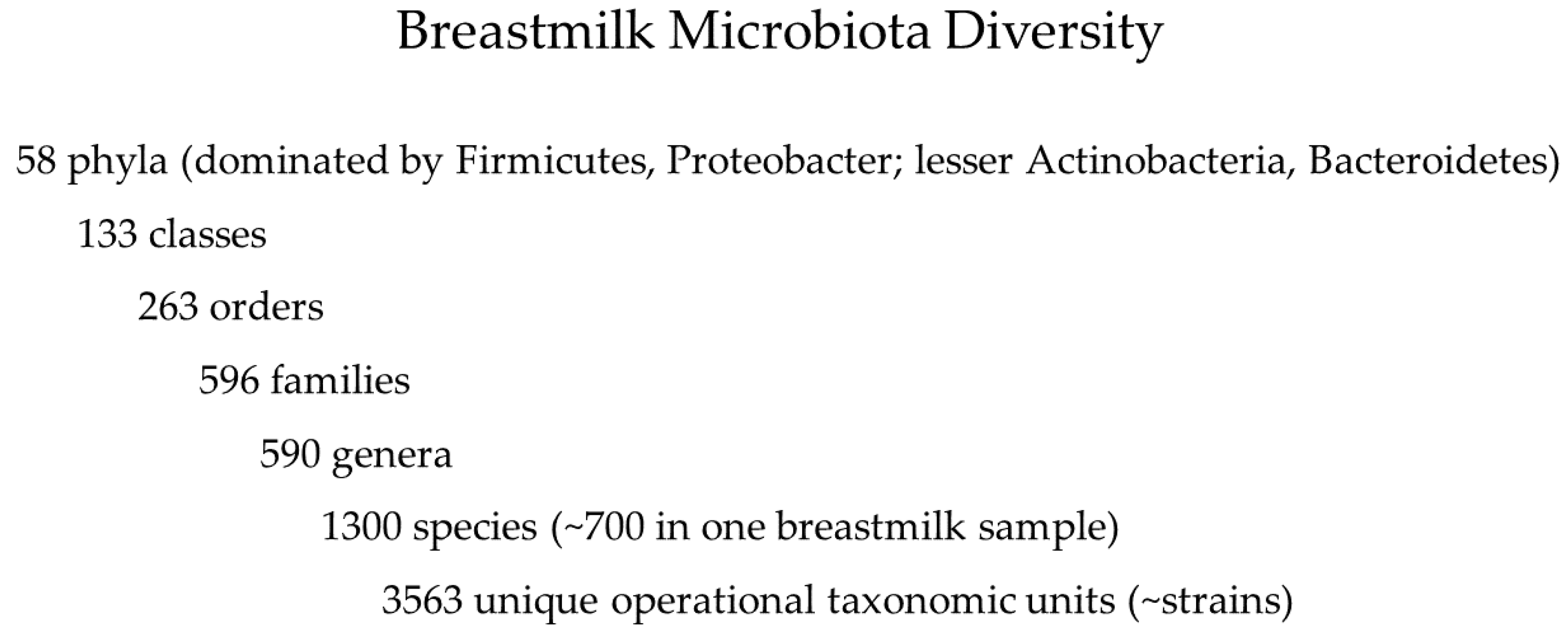
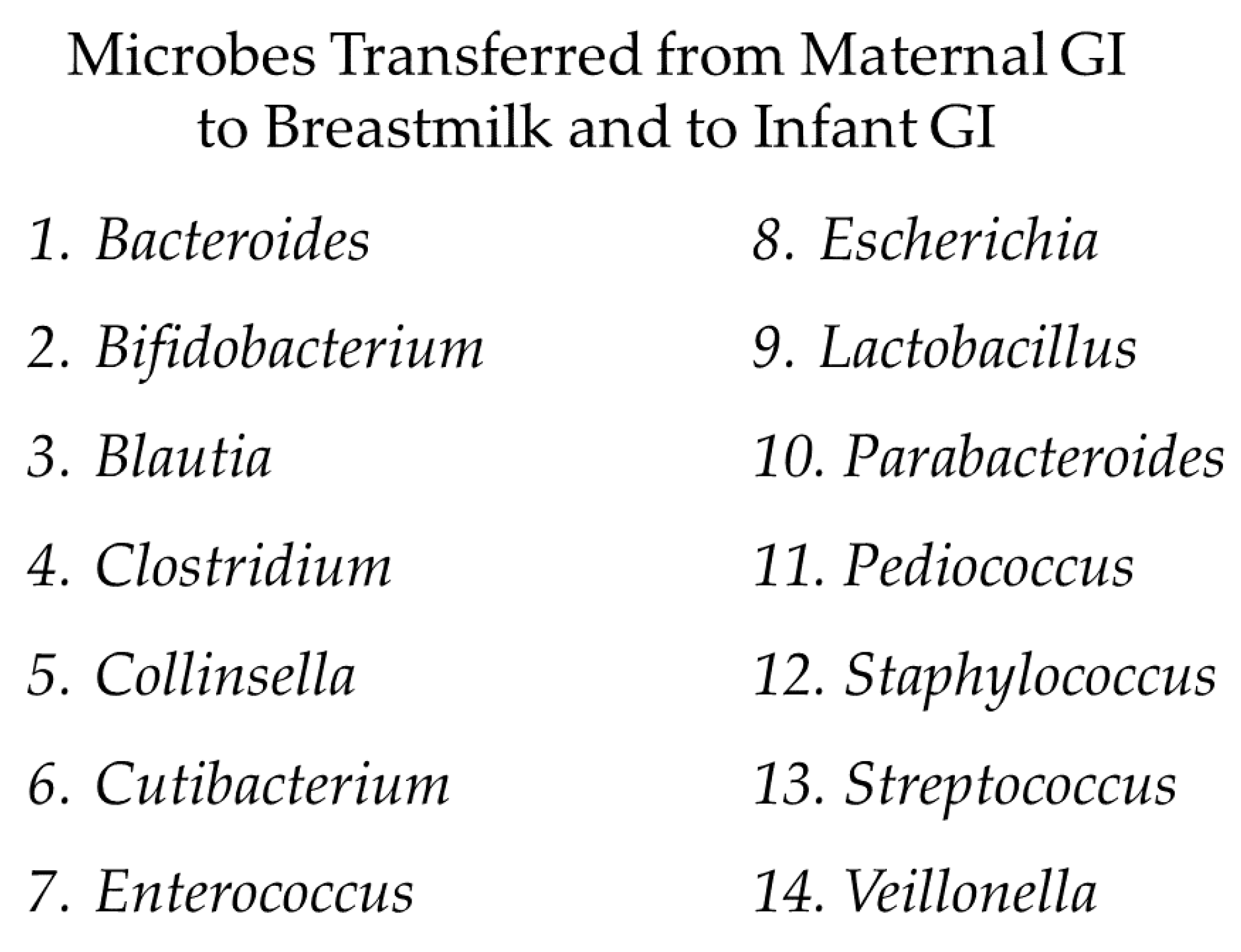
5.1. Breastmilk Microbiota

What is known about raw breastmilk and its utility in ‘managing our microbes’? Knowledge about breastmilk expanded from an assumption of sterility in past decades to more recent characterizations as a complex, living food which contains all the nutrients needed for offspring to grow and thrive, as well as microbes and other bioactive components [104,110,111]. A key component of the bioactivity of milks is the interdependent networks of microbial communities making up the microbiota of milks [104,112]. The dense and diverse milk microbiota and its components support colonization and maturation of the gut, immune, neural, and respiratory systems of infants, and increases protections against gut and respiratory pathogens by direct and indirect mechanisms of colonization resistance [9,104,110,111].
Recent reviews [104,110,111,113] depict highly variable but some common ‘core’ microbes typically present in breastmilk (often dominated by Staphylococcus and Streptococcus, with highly diverse minor taxa including low abundances of Lactobacillus, Pseudomonas, Bifidobacterium, Propionibacterium, Bacteroides, Corynebacterium, Enterococcus, Acinetobacter, Rothia, Cutibacterium, and Veillonella). These and other reviews note a paradigm shift from the now clearly outdated assumption of breastmilk as sterile to the current understanding of breastmilk as a complex ecosystem with dense and diverse natural microbiota. The recent hypothesis of an entero-mammary pathway is consistent with multiple lines of evidence demonstrating the internal physiologic transfer of gut microbes, including anaerobes Lactobacillus and Bifidobacterium that do not reside on human skin, to mammary tissue [110,113]. Zimmerman and Curtis [110] presented evidence of the transference of at least 14 microbes from the maternal gut to breastmilk and infant guts. Lyons and colleagues [113] note multiple studies that report the immunological enhancements of breastmilk composition for infants with infections, which is suggestive evidence for the retrograde backflow of microbes from nursing infants into mammary tissues and bi-directional microbial transfers.
Multiple studies reported higher bacterial diversity in breastmilk than in maternal or infant feces [104,110]. The maximal numbers of taxa per study in 44 breastmilk microbiota studies reported by Zimmerman and Curtis [110] are depicted in the taxonomic hierarchy in Figure 2, representing 22 to 260 species (203–512 strains) per breastmilk sample.
Despite extensive knowledge about the microbiota of breastmilk [104,110,111,113] and its protective effects on infant health, none of the studies available to date identified specific microbes or specific microbial consortia associated with the benefits or protections lost with pasteurization. Supplemental studies using in vitro and in vivo animal models document plausible mechanisms for a number of common microbes in breastmilk that are discussed in more detail in our companion paper [13].
Our companion manuscript [13] cited multiple recent reviews regarding hypotheses about the origins of such a diverse microbiota, including the ‘entero-mammary’ route, whereby gut microbes appear to be transferred from maternal gut to mammary tissue, breastmilk, and the gut of breastfeeding infants. Zimmerman and Curtis [110] documented 14 genera of gut microbes for which evidence of the transfer to the breastmilk and gut of breastfeeding infants was demonstrated (Figure 3).

5.2. Microbial Ecology of Breastmilk and Reductionist Dilemma

Uncertainties about which strains among the dense and diverse breastmilk microbiota drive benefits limit our ability to artificially ‘manage our microbes’ with specific supplements or probiotic strains at present. Differences were noted in a recent study comparing aspects of the microbiota of pooled donor breastmilk before and after Holder pasteurization [114]. Raw donor breastmilk pools had a significantly higher abundance of Staphylococcus, Kaistobacter, and Acinetobacter, while pasteurized donor breastmilk pools had a significantly higher abundance of Pseudomonas and Paracoccus. It is uncertain if any of these correlative changes cause any benefits and risks to infants. Similarly, the study reported that pasteurized donor breastmilk had similar or higher estimates of microbial diversity compared to raw breastmilk. However, no causal evidence is currently available for predicting what changes in the composition and functionality of the breastmilk microbiota are linked to the loss of benefits for pasteurized donor milk in the extensive body of evidence from systematic reviews of clinical studies [115,116,117,118,119,120].
Further, the developmental and preventive benefits of strategies to ‘manage our microbes’ merit attention. A recent clinical trial administered a single probiotic strain to 31 out of 60 infants who were exclusively fed breastmilk, complete with its dense and diverse microbiota [121]. The researchers administered a potential probiotic strain (Bifidobacterium longum subsp. infantis EVC001, optimized to utilize all known human milk oligosaccharides or prebiotics) to exclusively breastfed infants at daily doses of ~2 × 10¹⁰ colony-forming units from day 7 to day 28 postnatal. The probiotic treatment administered to breastfed infants was associated with significant differences in gut function and immunity compared to controls without supplementation, detecting a functional link between a potential probiotic strain, gut ecology, and immunoregulation during the first months of life. The probiotic strain enhanced metabolic partnerships with developing infant gut microbiota that were negatively correlated with pro-inflammatory cytokine production and positively correlated with interferon beta and Bifidobacteriaceae. In contrast, the gut microbiota of control infants were associated with higher pro-inflammatory cytokine production by three taxa (Clostridaceae, Enterobacteriaceae, and Staphylococcaceae). In this case, daily doses of a single optimized strain, plus a rich microbial diet (raw breastmilk), improved gut function and immune status compared with breastfed controls in this small population of healthy infants.
A related issue for the future applications of such studies to ‘managing our microbes’ applies to an active research area in the infant formula industry: supplementing formula with components that might mimic the composition and functionality of breastmilk [122]. Might future infant formulas successfully mimic breastmilk components and functions to restore the benefits for gut and immune system development associated with raw breastmilk? Although some health benefits have been observed in studies concerned with the feeding of infants with formula that is supplemented with specific probiotics, health benefits to infants are variable in efficacy by strain, disease, and study [122]. A recent systematic review by Almeida and colleagues [123] concludes that no consensus has developed around the bioactive supplements, including probiotics, to enhance infant formulas and restore the functional effects documented in breastmilk. Further research is needed before infant formulas can be designed to effectively ‘manage our microbes’.
As noted previously, supplementing infant formula with bioactive compounds, including beneficial bacteria or probiotics, represents a reductionist challenge. It seems unlikely that the single-strain EVC001 tested above in breastfed infants, or other single-bacterial strains with probiotic potential, would be sufficient to fully restore benefits to infants afforded by the natural breastmilk microbiota. While it is possible that a supplement containing a consortium of microbial strains plus prebiotic nutrients (termed synbiotics) might adequately represent the composition and functionality of the raw breastmilk and restore benefits, it is also possible that a microbiota more akin to FMT may be needed to provide daily doses of a dense and diverse microbiota or consortium of key strains needed by a pre-term or ill infant. In other words, akin to FMT, a dense and diverse microbiota from raw breastmilk may be needed, rather than one or a few microbial strains, to holistically ‘seed and feed’ dysbiotic infants.
Breastmilk, with its indigenous microbiota intact, confers plausible benefits to infants, supporting growth and the development of a healthy gut microbiota, as well as proper maturation and differentiation of the immune, neural, and respiratory systems. Breastmilk, particularly when combined with the vaginal delivery of infants, ‘seeds and feeds’ the infant gut [9], providing microbes that seed the naïve gastrointestinal system and nutritive components that feed both the infant and microbial cells. Without ‘seeding’ a dense and diverse microbiota, abnormal development of the immune system is more likely, potentially contributing to inflammatory diseases, asthma, and allergies [121,124].

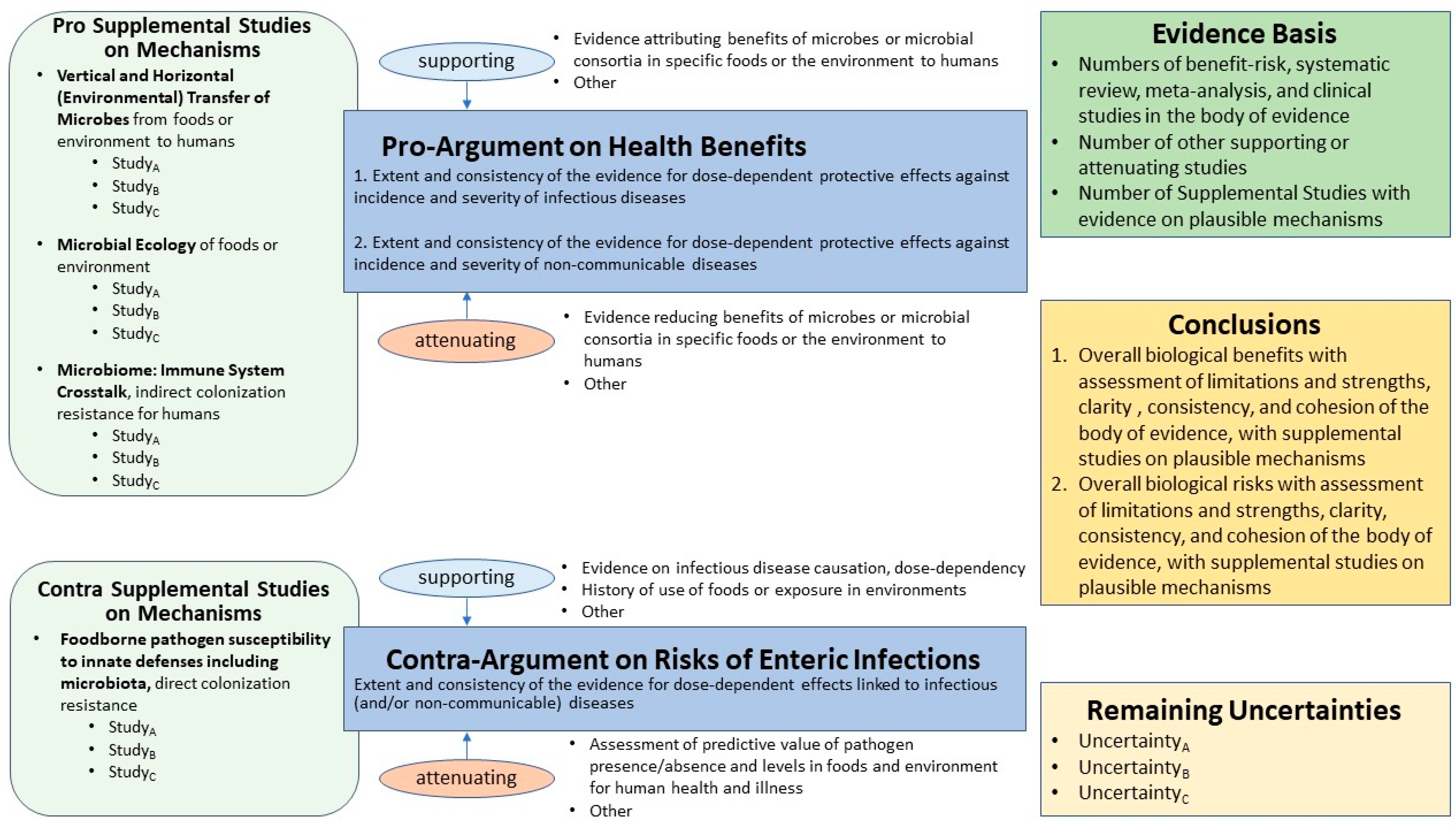
5.3. Benefit–Risk Methodology Applied to the Breastmilk Ecosystem

Multiple studies cited in the examples introduced herein observe that the microbiota is physically and functionally positioned at the interface between the environment and the genes of human ecosystems [18,96,100]. These observations are consistent with the need to insert the microbiota into our frameworks for assessing safety and risk to human superorganisms [7,8]. Much of the extensive literature documenting interactions of the gut microbiota with pathogens illustrates a major limitation for risk analysis for infectious and non-infectious diseases: results of small studies that are correlative in nature rather than causal are insufficient for building robust risk assessment models and for supporting decisions about ‘managing our microbes’ to optimize healthy colonization resistance and minimize acute and chronic disease [13].
A tool that balances the need for a coherent and transparent synthesis of large bodies of potentially conflicting evidence and the human tendency to succumb to confirmation bias, particularly for controversial issues involving fear and dread, is the technique of evidence mapping [125], which has been recently applied by us to the breastmilk ecosystem [13]. Notably, the evidence map approach is amenable to the inclusion of studies on both infectious and non-communicable diseases, unlike the microbial risk framework illustrated in Figure 1.
Fear and dread of microbes as germs that will kill us appear to factor strongly into a policy that is becoming more controversial with expanding knowledge of the natural microbiota of milks from -omics studies in this decade: the decision to require the pasteurization of breastmilk from donors in most human milk banks around the world [105,106,107,108,109].
In contrast, the policies of Germany, Japan, and Norway provide raw breastmilk (mother’s own milk or raw donor breastmilk) to neonatal intensive care unit (NICU) infants [126,127]. Policies for Germany and Norway are described on the website of the European Milk Bank Association (https://europeanmilkbanking.com/country/, accessed on 28 June 2021). Donor breastmilk is screened in Germany and Norway for potential pathogens, and screened donor milk is provided in raw form to pre-term and ill infants whose own mothers cannot provide sufficient breastmilk. Rather than pasteurize donor milk, most milk banks in Norway discard any donor milk that exceeds established microbial limits. In Oslo University Hospital, one of Norway’s major hospitals, 88% of donor milk passed screening in 2019 [128] and was administered to infants without causing any documented infections.
A rich and coherent body of evidence exists that shows both the health benefits of raw breastmilk to infants and the significant loss of those benefits with pasteurization [115,116,117,118,119,120]. These studies represent the ‘evidence basis‘ for an ‘evidence map’ of the raw (unpasteurized) breastmilk microbiota ecosystem [13] (see our submitted companion manuscript). The template for this evidence map (Figure 4) illustrates the components necessary to document the ‘state of the science’ and uncertainties for infant diets in order to ‘manage our microbes’.
The template (Figure 4) includes an upper right text box (darker-green fill) representing the evidence basis, documenting the numbers of studies in different categories of evidence illustrating the ‘state of the science’. Studies forming the evidence basis are structured under the text boxes in the middle and left columns of the figure. The middle text boxes (blue fill) represent the pro- and contra-arguments (benefits and risks respectively for this application). The bulleted text annotates evidence supporting and attenuating the arguments. The left-most text boxes with rounded corners (lighter-green fill) document the supplemental studies on mechanisms. Conclusions are provided in the middle-right text box (darker-yellow fill), with the remaining uncertainties provided in lower-right text box (light-yellow fill).
An evidence map provides a structured, visual depiction of the ‘state of the science’ and the remaining uncertainties for human health and disease. The evidence basis box provides a concise and transparent summary of the extent, quality, and consistency of the evidence (numbers of benefit–risk assessments, meta-analyses, systematic reviews, human cohort studies, randomized trials, quantitative microbial risk assessments, and reviews). The conclusion box provides insights from the full body of evidence, including strength and consistency (e.g., limited/convincing; inconclusive/conclusive). Perhaps the most important communication in the evidence map is remaining uncertainties, reflecting the nature of science that often provides indirect and ambiguous evidence that has limitations for predicting future benefits and risks to human populations other than those enrolled in the cited cohort studies.
Each component of the evidence map template is required to convey the ‘state of the science’ and the complexities of real-world scientific evidence that often includes ambiguous and conflicting studies. Without a visual map of the full body of evidence, inconsistencies within and a lack of coherence of the body of evidence may not be transparently communicated to decision makers or to other stakeholders responsible for decisions. Incomplete and misleading risk communications about the current ‘state of the science’ renders subsequent simulations of potential benefits and risks invalid, or results in biased predictions that grossly underestimate uncertainty about both the actual and relative benefits and risks.
For this example, the major studies documented in the evidence basis for benefits and risks of pasteurizing donor breastmilk in the companion manuscript [13] are briefly summarized herein. The evidence basis includes a benefit–risk assessment [115,116], a systematic review [117], a systematic review and meta-analysis [118], and two subsequent cohort studies [119,120].
Overall, studies on raw breastmilk provide clear, convincing, and conclusive evidence of benefits (dose-dependent protective effects) against incidence and severity of infectious diseases (ear and upper respiratory infections, diarrhea), obesity; and probable evidence for protection against asthma, celiac, Crohn’s, diabetes, eczema, high blood pressure, ulcerative colitis, and wheezing. Compared to raw breastmilk, formula and pasteurized donor breastmilk were associated with a significant loss of benefits (loss of protection against mortality, NEC, sepsis, and other disease endpoints for pre-term infants). Evidence for assessing the risks of pathogen infections in infants fed breastmilk versus pasteurized donor breastmilk is limited and inconclusive.

5.4. Future Evidence-Based Policies for Donor Breastmilk?

One source of controversy for the four examples is that assumptions rooted in 20th-century science and germ theory may effectively exclude evidence generated by -omics studies in the current decade. Broad consensus exists that the first choice for nutrition and development of healthy gut and immune systems is raw breastmilk (mother’s own milk), including low birth weight or pre-term infants at higher risk of morbidity and mortality than full-term infants [105,106,107,108,109].
The pasteurization policy of milk banks appears inconsistent with the available evidence and the ‘state of the science’ in the 21st century. The desire to provide pre-term and ill neonates with the best chances for survival and healthy development in challenging nosocomial environments may be influenced by aspects of human nature, including emotional, family, and social factors, as well as scientific evidence. Donor milk banks appear to require pasteurization because potential pathogens may be present in raw breastmilk. On the other hand, human cohort studies conducted around the world demonstrate significant decreases in health benefits to neonates fed pasteurized donor milk when compared with fresh raw breastmilk [115,116,117,118,119,120]. Further, pathogen presence in foods is insufficient to predict risk without additional data on the levels of pathogens, pathogen growth and survival in foods and GI ecosystems, and dose–response relationships [7,30,31].
The fear of microbes as germs appears to entrench well-meaning scientists and regulators in misconceptions of 20th-century science, and wall them off from consideration of advances in knowledge about the microbiota of milks, particularly the rich body of evidence for both the benefits and risks of raw breastmilk. In addition, germaphobia may be fueled by misinformation about formula milk to families around the world that discourages breastfeeding, despite the significant loss of benefits associated with infant formula compared to breastfeeding [129]. To date, only 25 countries have legislation aligned with the International Code of Marketing of Breastmilk Substitutes, designed to protect families against aggressive marketing of formula milk that may increase risks to infants. Certainly, the economic incentives of the global infant formula industry could misalign with practices that maximize the benefits to infants and families and actually put infants at higher risk without appropriate attention to ‘managing our microbes’.
Planning is underway for a series of international workshops to convene key stakeholders for decisions about the pasteurization of donor breastmilk. Our intent is to apply the evidence map for the breastmilk ecosystem [13] to build a common frame of reference for developing evidence-based decisions on pasteurized and raw milks.

5.5. Summary of Breastmilk Ecosystem Evidence Map

The breastmilk example illustrates the complexity of the natural breastmilk microbiota and the reductionist dilemma of limited knowledge for identifying the microbes driving the benefits associated with breastfeeding. The evidence map approach provides coherent and transparent communication of the ‘state-of-the-science’ and uncertainties for microbial benefits and risks associated with the breastmilk microbiota to assist in deeper deliberations of the evidence with decision makers and stakeholders.

6. Future for ‘Managing Our Microbes’

In all four examples introduced herein (CDI, Staph A, gut–brain axis effects on ASD, and breastmilk), the gut microbiota appears to strongly influence the extent to which the microbiota and microbiota-derived metabolites are involved in healthy and dysbiotic human superorganisms. However, limited knowledge of the complexity of gut microbiota structure, function, and driving mechanisms restricts present capacities to design microbiota-informed therapeutics to restore dysbiotic human superorganisms [1,2,26]. Similarly, limited knowledge also restricts present capacities to design microbiota-informed supplements to support healthy and resilient human superorganisms and enhance disease prevention.
A common ecological and technical challenge to effectively ‘manage our microbes’ is the difficulty of identifying and testing single probiotic candidate strains or practical numbers of strains to represent the consortia or networks of strains from the hugely complex, dense, and diverse gut and breastmilk microbiota that actually drive health and reduce the likelihood of disease. The utility of evidence mapping is particularly strong for communicating the ‘state of the science’ to diverse audiences: (1) researchers designing multi-omic studies testing causality for health benefits; and (2) stakeholders and the public when controversial, complex, and emotionally charged societal issues require greater care to ensure coherence and transparency.
Intermediate strategies merit consideration between the extremes: (i) treating or preventing dysbiosis with single probiotic strains; and (ii) treating with huge numbers of diverse undefined or minimally characterized microbes in FMT and raw breastmilk. In addition, designing and testing combination strategies with microbial ecology in mind (e.g., the benefits of combinations of dietary, probiotic, and prebiotic interventions of different doses and compositions) merits further research.
Dietary advice could be geared towards ‘seeding and feeding’ the gut microbes, with attention to whole unprocessed foods complete with natural microbiota, fermented foods, and other ‘functional foods’ that provide both beneficial microbes and prebiotic nutrients to promote their survival, growth, and engraftment as residents in the midst of 100 trillion potential competitors in the human superorganism gut.
Diet can significantly influence superorganism health, the gut microbiota, and attempts to ‘manage our microbes’ [130]. Simply put, the energy source(s) provided through dietary nutrients determine which microbes can establish residence and flourish, which microbial co-partners can remain a minor or transient part of the gut ecosystem, and which are likely to die off or be eliminated through peristalsis. Additional research is needed to make inferences about the effectiveness of ‘managing our microbes’ by supplementing with dietary prebiotics or increasing dietary diversity that may then increase gut microbiome richness and maintain a health-supportive, resilient gut microbiome [1]. ‘Westernized’ diets high in fat and sugar can rapidly degrade the human gut microbiome [131]. However, the complex interconnections across gut, immune, neural, and respiratory systems point to a potential error: imagining that diet alone is the solution to managing microbes in every scenario. This is the subject of an impending invited review article (Dietert, in preparation).
While diet can alter gut microbiota, established microbial residents of the gut are also able to exquisitely affect diet in the following numerous different ways. These include microbial control of food preferences [132,133,134], food intake (i.e., appetite-satiety regulation) [135], taste [136] and odor [137] receptor regulation, flavor-taste thresholds [138], taste perceptions [139], food addiction [140], eating disorders [141], and even early developmental programming of eating behaviors (via maternal microbiota) [142]. Because microbiota can also regulate pain [21,143], they can make abrupt withdrawals from specific foods and/or drugs an additional challenge. For these reasons, it can be useful to sync major changes in diet with changes in gut microbiota that prefer the new dietary components as their energy source.
In addition, researchers are now questioning whether dietary advice might expand the concept of recommended daily allowances from vitamins to include microbes [144,145]. Clearly, as noted by Dietert and Silbergeld [8], pharmaceuticals, particularly polypharmacy, are a significant stressor to both healthy and dysbiotic human superorganisms. The impacts of pharmaceuticals and polypharmacy on ecosystems which include our indigenous microbial partners-in-health must be considered as part of the evidence basis for conducting safety and risk assessments in the 21st century and beyond.
The examples discussed herein are all amenable to analysis via evidence mapping using the template (Figure 4). Some key advantages that evidence mapping can provide for structuring the extensive evidence from complex -omics studies of the gut–lung–brain axis in healthy and dysbiotic human superorganisms for diverse audiences include the following [125]. Evidence mapping:
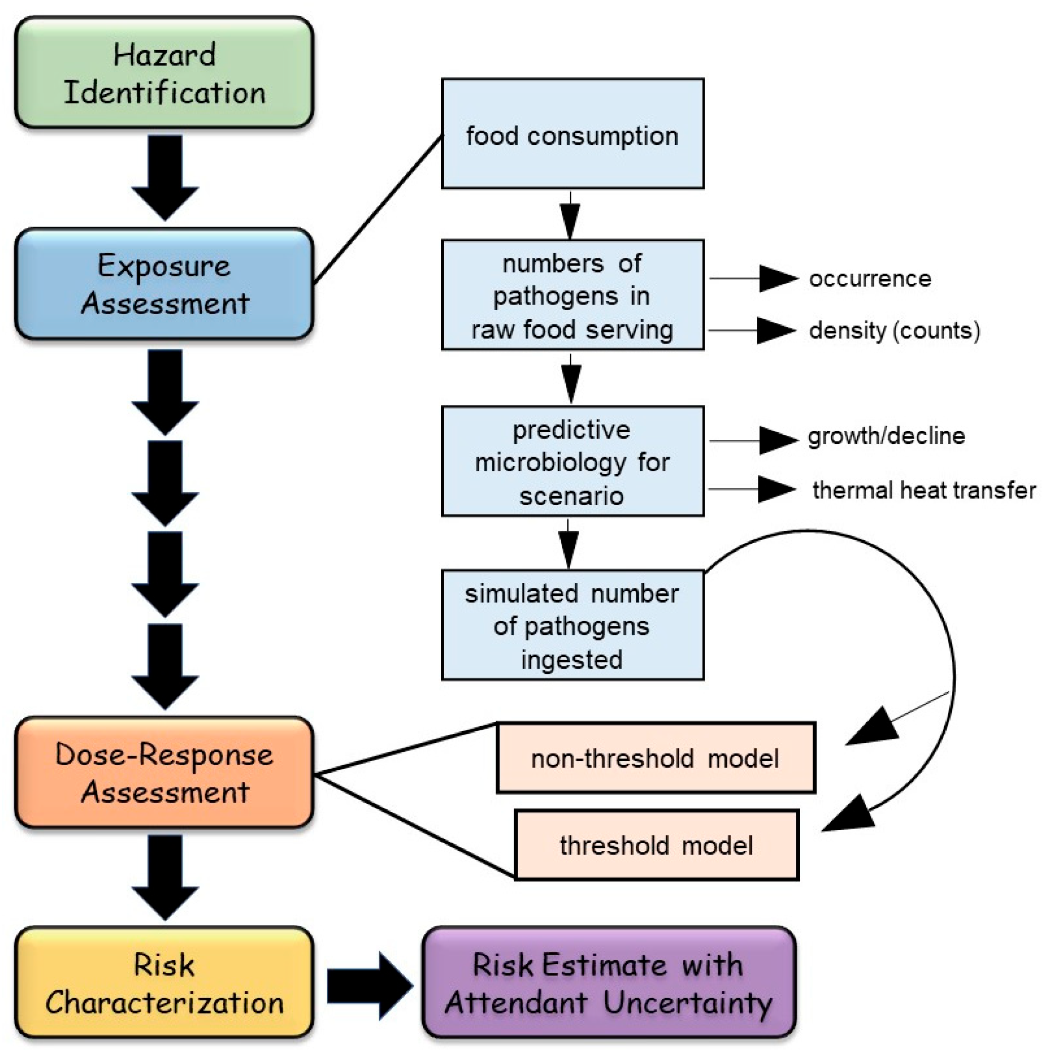
  • Does not require complicated quantitative modeling of exposure assessment and dose–response assessment typical of quantitative microbial risk assessment (QMRA) for data synthesis;
  • Presents simple qualitative narrative in a structured format, with a graphical representation of the evidence basis, drawing attention to evidence for both pro- and contra-arguments, with supporting and attenuating data;
  • Assists a diverse array of experts and non-experts in paying attention to the entire ‘state of the science’, a visual picture of the evidence basis, quality of evidence, and uncertainty;
  • Promotes openness and transparency for evaluating rarely unambiguous scientific evidence for applications in risk analysis;
  • Assists risk analysts in avoiding traps such as ‘confirmation bias’ that may distort judgments about weighing and synthesizing evidence from multiple disciplines; and
  • Facilitates constructive dialogue between diverse perspectives/opinions of all stakeholders, including decisions makers and the public.
In summary, clear, coherent descriptions of the ‘state of the science’ and uncertainties for the gut–lung–brain axis structured as evidence maps can contribute to the design of key studies testing for causality of effects that go beyond measuring correlations, but enable quantitative or qualitative predictions of the key consortia needed to support health. Such studies are essential for filling knowledge gaps which, at present, limit broader applications of correlative studies in a coherent and transparent manner that could support decision making, holistic risk management, and the design of microbiota-based preventative and therapeutic strategies to holistically ‘manage our microbes’.

Author Contributions

Conceptualization, M.E.C., R.R.D. and D.W.N.; methodology, M.E.C.; formal analysis, M.E.C.; investigation, M.E.C. and R.R.D.; resources, M.E.C.; writing—original draft preparation, M.E.C.; writing—review and editing, M.E.C., R.R.D. and D.W.N.; visualization, M.E.C. and M.M.S.; project administration, M.E.C.; funding acquisition, M.E.C. All authors have read and agreed to the published version of the manuscript.

Funding

This research received no external funding.

Institutional Review Board Statement

Not applicable.

Informed Consent Statement

Not applicable.

Data Availability Statement

Not applicable.

Acknowledgments

The authors appreciate helpful comments from journal reviewers and Ryan Bentz for graphical assistance.

Conflicts of Interest

Author have no potential conflict of interest to declare except R.R.D. who has consulted for a probiotics company on education.

References

  1. Heiman, M.L.; Greenway, F.L. A Healthy Gastrointestinal Microbiome Is Dependent on Dietary Diversity. Mol. Metab. 2016, 5, 317–320. [Google Scholar] [CrossRef] [PubMed]
  2. National Institutes of Health (NIH)—Human Microbiome Portfolio Analysis Team. A Review of 10 Years of Human Microbiome Research Activities at the US National Institutes of Health, Fiscal Years 2007–2016. Microbiome 2019, 7, 31. [Google Scholar] [CrossRef] [PubMed]
  3. Integrative HMP; Proctor, L.M.; Creasy, H.H.; Fettweis, J.M.; Lloyd-Price, J.; Mahurkar, A.; Huttenhower, C. The Integrative Human Microbiome Project. Nature 2019, 569, 641–648. [Google Scholar] [CrossRef] [Green Version]
  4. Dietert, R.R. The Human Superorganism: How the Microbiome Is Revolutionizing the Pursuit of a Healthy Life; Dutton: New York, NY, USA, 2016. [Google Scholar]
  5. Dietert, R.R. Safety and Risk Assessment for the Human Superorganism. Hum. Ecol. Risk Assess. 2017, 23, 1819–1829. [Google Scholar] [CrossRef] [Green Version]
  6. Dietert, R.R.; Dietert, J.M. Twentieth Century Dogmas Prevent Sustainable Healthcare. Am. J. Biomed. Sci. Res. 2021, 13, 409–417. [Google Scholar]
  7. Coleman, M.; Elkins, C.; Gutting, B.; Mongodin, E.; Solano-Aguilar, G.; Walls, I. Microbiota and Dose Response: Evolving Paradigm of Health Triangle. Risk Anal. 2018, 38, 2013–2028. [Google Scholar] [CrossRef]
  8. Dietert, R.R.; Silbergeld, E.K. Biomarkers for the 21st Century: Listening to the Microbiome. Toxicol. Sci. 2015, 144, 208–216. [Google Scholar] [CrossRef] [Green Version]
  9. Dietert, R.R. A Focus on Microbiome Completeness and Optimized Colonization Resistance in Neonatology. NeoReviews 2018, 19, 78–88. [Google Scholar] [CrossRef]
  10. Sleator, R.D. The Human Superorganism–of Microbes and Men. Med. Hypotheses 2010, 74, 214–215. [Google Scholar] [CrossRef]
  11. Blaser, M.J. The Microbiome Revolution. J. Clin. Investig. 2014, 124, 4162–4165. [Google Scholar] [CrossRef] [Green Version]
  12. Marks, H.; Coleman, M. Scientific Data and Theories for Salmonellosis Dose—Response Assessment. Hum. Ecol. Risk Assess. Int. J. 2017, 23, 1857–1876. [Google Scholar] [CrossRef]
  13. Coleman, M.E.; Dietert, R.R.; North, D.W.; Stephenson, M.M. Examining Evidence of Benefits and Risks for Pasteurizing Donor Breastmilk. Appl. Microbiol. 2021, 1, 408–425. [Google Scholar] [CrossRef]
  14. Endt, K.; Stecher, B.; Chaffron, S.; Slack, E.; Tchitchek, N.; Benecke, A.; Hardt, W.D. The Microbiota Mediates Pathogen Clearance from the Gut Lumen after Non-Typhoidal Salmonella Diarrhea. PLoS Pathog. 2010, 6, 1001097. [Google Scholar] [CrossRef] [PubMed] [Green Version]
  15. Brugiroux, S.; Beutler, M.; Pfann, C.; Garzetti, D.; Ruscheweyh, H.J.; Ring, D.; Stecher, B. Genome-Guided Design of a Defined Mouse Microbiota That Confers Colonization Resistance against Salmonella Enterica Serovar Typhimurium. Nat. Microbiol. 2016, 2, 16215. [Google Scholar] [CrossRef] [PubMed]
  16. Gazzaniga, F.S.; Camacho, D.M.; Wu, M.; Silva Palazzo, M.F.; Dinis, A.L.; Grafton, F.N.; Ingber, D.E. Harnessing Colon Chip Technology to Identify Commensal Bacteria That Promote Host Tolerance to Infection. Front. Cell. Infect. Microbiol. 2021, 11, 105. [Google Scholar] [CrossRef]
  17. Rogers, A.W.; Tsolis, R.M.; Bäumler, A.J. Salmonella versus the Microbiome. Microbiol. Mol. Biol. Rev. 2021, 85, e00027-19. [Google Scholar] [CrossRef]
  18. Sehgal, K.; Khanna, S. Gut Microbiome and Clostridioides Difficile Infection: A Closer Look at the Microscopic Interface. Ther. Adv. Gastroenterol. 2021, 14, 1756284821994736. [Google Scholar] [CrossRef]
  19. Kim, S.; Covington, A.; Pamer, E.G. The Intestinal Microbiota: Antibiotics, Colonization Resistance, and Enteric Pathogens. Immunol. Rev. 2017, 279, 90–105. [Google Scholar] [CrossRef]
  20. Palmer, A.D.; Slauch, J.M. Mechanisms of Salmonella Pathogenesis in Animal Models. Hum. Ecol. Risk Assess. Int. J. 2017, 23, 1877–1892. [Google Scholar] [CrossRef]
  21. Dietert, R.R.; Dietert, J.M. Microbiome First Approaches in Pain Prevention and Management. Am. J. Biomed. Sci. Res. Press 2021, 14, 182–192. [Google Scholar]
  22. Levy, E.I.; Hoang, D.M.; Vandenplas, Y. The Effects of Proton Pump Inhibitors on the Microbiome in Young Children. Acta Paediatr. 2020, 109, 1531–1538. [Google Scholar] [CrossRef]
  23. Abenavoli, L.; Scarpellini, E.; Colica, C.; Boccuto, L.; Salehi, B.; Sharifi-Rad, J.; Capasso, R. Gut Microbiota and Obesity: A Role for Probiotics. Nutrients 2019, 11, 2690. [Google Scholar] [CrossRef] [Green Version]
  24. King, C.H.; Desai, H.; Sylvetsky, A.C.; LoTempio, J.; Ayanyan, S.; Carrie, J.; Crandall, K.A.; Fochtman, B.C.; Gasparyan, L.; Gulzar, N.; et al. Baseline Human Gut Microbiota Profile in Healthy People and Standard Reporting Template. PLoS ONE 2019, 14, e0206484. [Google Scholar] [CrossRef] [Green Version]
  25. Almonacid, D.E.; Kraal, L.; Ossandon, F.J.; Budovskaya, Y.V.; Cardenas, J.P.; Bik, E.M.; Goddard, A.D.; Richman, J.; Apte, Z.S. 16S RRNA Gene Sequencing and Healthy Reference Ranges for 28 Clinically Relevant Microbial Taxa from the Human Gut Microbiome. PLoS ONE 2017, 12, e0176555. [Google Scholar] [CrossRef] [Green Version]
  26. Ducarmon, Q.R.; Kuijper, E.J.; Olle, B. Opportunities and Challenges in Development of Live Biotherapeutic Products to Fight Infections. J. Infect. Dis. 2021, 223, S283–S289. [Google Scholar] [CrossRef]
  27. Hromada, S.; Clark, R.L.; Qian, Y.; Watson, L.; Safdar, N.; Venturelli, O.S. Species Richness Determines, C. Difficile Invasion Outcome in Synthetic Human Gut Communities. bioRxiv 2021, 1–27. [Google Scholar] [CrossRef]
  28. Maki, J.J.; Klima, C.L.; Sylte, M.J.; Looft, T. The Microbial Pecking Order: Utilization of Intestinal Microbiota for Poultry Health. Microorganisms 2019, 7, 376. [Google Scholar] [CrossRef] [Green Version]
  29. National Research Council (US) Committee on the Institutional Means for Assessment of Risks to Public Health. Risk Assessment in the Federal Government: Managing the Process; National Academies Press (US): Washington, DC, USA, 1983; ISBN 978-0-309-03349-7. [Google Scholar]
  30. Marks, H.M.; Coleman, M.E.; Lin, C.T.J.; Roberts, T. Topics in Microbial Risk Assessment: Dynamic Flow Tree Process. Risk Anal. 1998, 18, 309–328. [Google Scholar] [CrossRef]
  31. Codex Alimentarius Commission (CAC). Principles and Guidelines for the Conduct of Microbiological Risk Assessment. Available online: http://www.fao.org/3/y1579e/y1579e05.htm (accessed on 8 March 2021).
  32. Smith, A.B.; Soto Ocana, J.; Zackular, J.P. From Nursery to Nursing Home: Emerging Concepts in Clostridioides Difficile Pathogenesis. Infect. Immun. 2020, 88, e00934-19. [Google Scholar] [CrossRef]
  33. Centers for Disease Control and Prevention (CDC). Healthcare-Associated Infections—Community Interface (HAIC). Clostridioides Difficile Infection (CDI) Tracking; CDC: Atlanta, GA, USA, 2020.
  34. Stein, R.R.; Bucci, V.; Toussaint, N.C.; Buffie, C.G.; Rätsch, G.; Pamer, E.G.; Sander, C.; Xavier, J.B. Ecological Modeling from Time-Series Inference: Insight into Dynamics and Stability of Intestinal Microbiota. PLoS Comput. Biol. 2013, 9, e1003388. [Google Scholar] [CrossRef]
  35. Buffie, C.G.; Bucci, V.; Stein, R.R.; McKenney, P.T.; Ling, L.; Gobourne, A.; Pamer, E.G. Precision Microbiome Reconstitution Restores Bile Acid Mediated Resistance to Clostridium Difficile. Nature 2015, 517, 205–208. [Google Scholar] [CrossRef] [Green Version]
  36. Yadav, D.; Khanna, S. Safety of Fecal Microbiota Transplantation for Clostridioides Difficile Infection Focusing on Pathobionts and SARS-CoV-2. Ther. Adv. Gastroenterol. 2021, 14, 17562848211009694. [Google Scholar] [CrossRef]
  37. Khanna, S. Microbiota Restoration for Recurrent Clostridioides Difficile: Getting One Step Closer Every Day! J. Intern. Med. 2021, 290, 294–309. [Google Scholar] [CrossRef]
  38. Langdon, A.; Schwartz, D.J.; Bulow, C.; Sun, X.; Hink, T.; Reske, K.A.; Jones, C.; Burnham, C.D.; Dubberke, E.R.; Dantas, G. Microbiota Restoration Reduces Antibiotic Resistant Bacteria Gut Colonization in Patients with Recurrent Clostridioides Difficile Infection from the Open-Label PUNCH CD Study. Genome Med. 2021, 13, 28. [Google Scholar] [CrossRef]
  39. Vakili, B.; Fateh, A.; Aghdaei, H.A.; Sotoodehnejadnematalahi, F.; Siadat, S.D. Intestinal Microbiota in Elderly Inpatients with Clostridioides Difficile Infection. Infect. Drug Resist. 2020, 13, 2723. [Google Scholar] [CrossRef]
  40. Rodriguez, C.; Taminiau, B.; Korsak, N.; Avesani, V.; Van Broeck, J.; Brach, P.; Daube, G. Longitudinal Survey of Clostridium Difficile Presence and Gut Microbiota Composition in a Belgian Nursing Home. BMC Microbiol. 2016, 16, 229. [Google Scholar] [CrossRef] [Green Version]
  41. Liang, G.; Bushman, F.D. The Human Virome: Assembly, Composition and Host Interactions. Nat. Rev. Microbiol. 2021, 19, 514–527. [Google Scholar] [CrossRef] [PubMed]
  42. Martínez, A.; Kuraji, R.; Kapila, Y.L. The Human Oral Virome: Shedding Light on the Dark Matter. Periodontol 2000 2021, 87, 282–298. [Google Scholar] [CrossRef] [PubMed]
  43. Łusiak-Szelachowska, M.; Weber-Dąbrowska, B.; Żaczek, M.; Borysowski, J.; Górski, A. The Presence of Bacteriophages in the Human Body: Good, Bad or Neutral? Microorganisms 2020, 8, 2012. [Google Scholar] [CrossRef] [PubMed]
  44. Nayfach, S.; Páez-Espino, D.; Call, L. Metagenomic Compendium of 189,680 DNA Viruses from the Human Gut Microbiome. Nat. Microbiol. 2021, 6, 960–970. [Google Scholar] [CrossRef] [PubMed]
  45. Śliwka, P.; Ochocka, M.; Skaradzińska, A. Applications of Bacteriophages against Intracellular Bacteria. Crit. Rev. Microbiol. 2021, 47, 1–18. [Google Scholar] [CrossRef]
  46. Fujimoto, K.; Kimura, Y.; Shimohigoshi, M.; Satoh, T.; Sato, S.; Tremmel, G.; Uematsu, M.; Kawaguchi, Y.; Usui, Y.; Nakano, Y.; et al. Metagenome Data on Intestinal Phage-Bacteria Associations Aids the Development of Phage Therapy against Pathobionts. Cell Host. Microbe 2020, 28, 353–355. [Google Scholar] [CrossRef]
  47. Adriaenssens, E.M. Phage Diversity in the Human Gut Microbiome: A Taxonomist’s Perspective. mSystems 2021, 6, e00799-21. [Google Scholar] [CrossRef]
  48. Li, T.; Zhang, Y.; Dong, K.; Kuo, C.J.; Li, C.; Zhu, Y.Q.; Qin, J.; Li, Q.T.; Chang, Y.F.; Guo, X.; et al. Isolation and Characterization of the Novel Phage JD032 and Global Transcriptomic Response during JD032 Infection of Clostridioides Difficile Ribotype 078. MSystems. mSystems 2020, 5, e00017-20. [Google Scholar] [CrossRef]
  49. Hinc, K.; Kabała, M.; Iwanicki, A.; Martirosian, G.; Negri, A.; Obuchowski, M. Complete Genome Sequence of the Newly Discovered Temperate Clostridioides Difficile Bacteriophage PhiCDKH01 of the Family Siphoviridae. Arch. Virol. 2021, 166, 2305–2310. [Google Scholar] [CrossRef]
  50. Heuler, J.; Fortier, L.C.; Sun, X. Clostridioides Difficile Phage Biology and Application. FEMS Microbiol. Rev. 2021, 45, 5. [Google Scholar] [CrossRef]
  51. Selle, K.; Fletcher, J.R.; Tuson, H. In Vivo Targeting of Clostridioides Difficile Using Phage-Delivered CRISPR-Cas3 Antimicrobials. mBio 2020, 11, e00019-20. [Google Scholar] [CrossRef] [Green Version]
  52. Wu, N.; Dai, J.; Guo, M.; Li, J.; Zhou, X.; Li, F.; Gao, Y.; Qu, H.; Lu, H.; Jin, J.; et al. Pre-Optimized Phage Therapy on Secondary Acinetobacter Baumannii Infection in Four Critical COVID-19 Patients. Emerg. Microbes Infect. 2021, 10, 612–618. [Google Scholar] [CrossRef]
  53. Cheung, G.Y.C.; Bae, J.S.; Otto, M. Pathogenicity and Virulence of Staphylococcus Aureus. Virulence 2021, 12, 547–569. [Google Scholar] [CrossRef]
  54. Tong, S.Y.; Davis, J.S.; Eichenberger, E. Staphylococcus Aureus Infections: Epidemiology, Pathophysiology, Clinical Manifestations, and Management. Clin. Microbiol. Rev. 2015, 28, 603–661. [Google Scholar] [CrossRef] [Green Version]
  55. Food and Drug Administration (FDA). Bad Bug Book; FDA: Washington, DC, USA, 2012.
  56. Strowd, L.C.; Feldman, S. Lead with Your Gut: Research-Implicated Infant Gut Staphylococcal Strains May Predict Development of Atopic Dermatitis. Br. J. Dermatol. 2019, 180, 1296–1297. [Google Scholar] [CrossRef]
  57. Kang, M.S.; Lim, H.S.; Oh, J.S.; Lim, Y.J.; Wuertz-Kozak, K.; Harro, J.M.; Achermann, Y. Antimicrobial Activity of Lactobacillus Salivarius and Lactobacillus Fermentum against Staphylococcus Aureus. Pathog. Dis. 2017, 75, ftx009. [Google Scholar] [CrossRef]
  58. Khamash, D.F.; Voskertchian, A.; Milstone, A.M. Manipulating the Microbiome: Evolution of a Strategy to Prevent, S. Aureus Disease in Children. J. Perinatol. 2018, 38, 105–109. [Google Scholar] [CrossRef]
  59. Rao, C.; Coyte, K.Z.; Bainter, W.; Geha, R.S.; Martin, C.R.; Rakoff-Nahoum, S. Multi-Kingdom Ecological Drivers of Microbiota Assembly in Preterm Infants. Nature 2021, 591, 633–638. [Google Scholar] [CrossRef]
  60. Nataraj, B.H.; Ramesh, C.; Mallappa, R.H. Extractable Surface Proteins of Indigenous Probiotic Strains Confer Anti-Adhesion Knack and Protect against Methicillin-Resistant Staphylococcus Aureus Induced Epithelial Hyperpermeability in HT-29 Cell Line. Microb. Pathog. 2021, 158, 104974. [Google Scholar] [CrossRef]
  61. Shi, B.; Leung, D.Y.; Taylor, P.A.; Li, H. MRSA Colonization Is Associated with Decreased Skin Commensal Bacteria in Atopic Dermatitis. J. Investig. Dermatol. 2018, 138, 1668. [Google Scholar] [CrossRef] [Green Version]
  62. Patrick, G.J.; Liu, H.; Alphonse, M.P.; Dikeman, D.A.; Youn, C.; Otterson, J.C.; Wang, Y.; Ravipati, A.; Mazhar, M.; Denny, G.; et al. Epicutaneous Staphylococcus Aureus Induces IL-36 to Enhance IgE Production and Ensuing Allergic Disease. J. Clin. Investig. 2021, 131, e143334. [Google Scholar] [CrossRef]
  63. Liu, J.N.; Shin, Y.S.; Yoo, H.S.; Nam, Y.H.; Jin, H.J.; Ye, Y.M.; Nahm, D.H.; Park, H.S. The Prevalence of Serum Specific IgE to Superantigens in Asthma and Allergic Rhinitis Patients. Allergy Asthma Immunol. Res. 2014, 6, 263–266. [Google Scholar] [CrossRef] [Green Version]
  64. Tomczak, H.; Wróbel, J.; Jenerowicz, D.; Sadowska-Przytocka, A.; Wachal, M.; Adamski, Z.; Czarnecka-Operacz, M.M. The Role of Staphylococcus Aureus in Atopic Dermatitis: Microbiological and Immunological Implications. Postepy Dermatol. Alergol. 2019, 36, 485–491. [Google Scholar] [CrossRef]
  65. Tsilochristou, O.; Toit, G.; Sayre, P.H.; Roberts, G.; Lawson, K.; Sever, M.L.; Bahnson, H.T.; Radulovic, S.; Basting, M.; Plaut, M.; et al. Immune Tolerance Network Learning Early About Peanut Allergy Study Team. Association of Staphylococcus Aureus Colonization with Food Allergy Occurs Independently of Eczema Severity. J. Allergy Clin. Immunol. 2019, 144, 494–503. [Google Scholar] [CrossRef]
  66. Kim, Y.C.; Won, H.K.; Lee, J.W.; Sohn, K.H.; Kim, M.H.; Kim, T.B.; Chang, Y.S.; Lee, B.J.; Cho, S.H.; Bachert, C.; et al. Staphylococcus Aureus Nasal Colonization and Asthma in Adults: Systematic Review and Meta-Analysis. J. Allergy Clin. Immunol. Pract. 2019, 615, 606–615. [Google Scholar] [CrossRef] [PubMed]
  67. Tang, H.H.F.; Lang, A.; Teo, S.M.; Judd, L.M.; Gangnon, R.; Evans, M.D.; Lee, K.E.; Vrtis, R.; Holt, P.G.; Lemanske, R.F., Jr.; et al. Developmental Patterns in the Nasopharyngeal Microbiome during Infancy Are Associated with Asthma Risk. J. Allergy Clin. Immunol. 2021, 147, 1683–1691. [Google Scholar] [CrossRef] [PubMed]
  68. Huang, R.; Ju, Z.; Zhou, P.K. A Gut Dysbiotic Microbiota-Based Hypothesis of Human-to-Human Transmission of Non-Communicable Diseases. Sci. Total Environ. 2020, 745, 141030. [Google Scholar] [CrossRef] [PubMed]
  69. Otto, M. Staphylococcus Aureus Toxins. Curr. Opin. Microbiol. 2014, 17, 32–37. [Google Scholar] [CrossRef] [PubMed] [Green Version]
  70. Oliveira, D.; Borges, A.; Simões, M. Staphylococcus Aureus Toxins and Their Molecular Activity in Infectious Diseases. Toxins 2018, 10, 252. [Google Scholar] [CrossRef] [PubMed] [Green Version]
  71. Spaulding, A.R.; Salgado-Pabón, W.; Kohler, P.L.; Horswill, A.R.; Leung, D.Y.; Schlievert, P.M. Staphylococcal and Streptococcal Superantigen Exotoxins. Clin. Microbiol. Rev. 2013, 26, 422–447. [Google Scholar] [CrossRef] [Green Version]
  72. Vickery, T.W.; Ramakrishnan, V.R.; Suh, J.D. The Role of Staphylococcus Aureus in Patients with Chronic Sinusitis and Nasal Polyposis. Curr. Allergy Asthma Rep. 2019, 11, 19–21. [Google Scholar] [CrossRef]
  73. Muluk, N.B.; Altın, F.; Cingi, C. Role of Superantigens in Allergic Inflammation: Their Relationship to Allergic Rhinitis, Chronic Rhinosinusitis, Asthma, and Atopic Dermatitis. Am. J. Rhinol. Allergy 2018, 32, 502–517. [Google Scholar] [CrossRef]
  74. Bachert, C.; Holtappels, G.; Merabishvili, M.; Meyer, T.; Murr, A.; Zhang, N.; Van Crombruggen, K.; Gevaert, E.; Völker, U.; Bröker, B.M.; et al. Staphylococcus Aureus Controls Interleukin-5 Release in Upper Airway Inflammation. J. Proteom. 2018, 180, 53–60. [Google Scholar] [CrossRef]
  75. Bachert, C.; Humbert, M.; Hanania, N.A.; Zhang, N.; Holgate, S.; Buhl, R.; Bröker, B.M. Staphylococcus Aureus and Its IgE-Inducing Enterotoxins in Asthma: Current Knowledge. Eur. Respir. J. 2020, 55, 1901592. [Google Scholar] [CrossRef]
  76. Ueki, S.; Miyabe, Y.; Yamamoto, Y. Charcot-Leyden Crystals in Eosinophilic Inflammation: Active Cytolysis Leads to Crystal Formation. Curr. Allergy Asthma Rep. 2019, 19, 35. [Google Scholar] [CrossRef]
  77. Tsang, M.S.; Sun, X.; Wong, C.K. The Role of New IL-1 Family Members (IL-36 and IL-38) in Atopic Dermatitis, Allergic Asthma, and Allergic Rhinitis. Curr. Allergy Asthma Rep. 2020, 20, 40. [Google Scholar] [CrossRef]
  78. Shirai, Y.; Arai, H.; Tamaki, K.; Konishi, H.; Kawase, Y.; Shimizu, N.; Tateda, K.; Yoda, H. Neonatal Methicillin-Resistant Staphylococcus Aureus Colonization and Infection. J. Neonatal. Perinat. Med. 2017, 10, 439–444. [Google Scholar] [CrossRef]
  79. Khamash, D.F.; Mongodin, E.F.; White, J.R.; Voskertchian, A.; Hittle, L.; Colantuoni, E.; Milstone, A.M. The Association Between the Developing Nasal Microbiota of Hospitalized Neonates and Staphylococcus Aureus Colonization. Open Forum Infect. Dis. 2019, 6, 62. [Google Scholar] [CrossRef] [Green Version]
  80. Bessesen, M.T.; Kotter, C.V.; Wagner, B.D.; Adams, J.C.; Kingery, S.; Benoit, J.B.; Frank, D.N. MRSA Colonization and the Nasal Microbiome in Adults at High Risk of Colonization and Infection. J. Infect. 2015, 71, 649–657. [Google Scholar] [CrossRef] [Green Version]
  81. Roghmann, M.C.; Lydecker, A.D.; Langenberg, P.; Mongodin, E.F.; Johnson, J.K. Microbiological Effect of Mupirocin and Chlorhexidine for Staphylococcus Aureus Decolonization in Community and Nursing Home Based Adults. Diagn. Microbiol. Infect. Dis. 2017, 88, 53–57. [Google Scholar] [CrossRef] [Green Version]
  82. Nordengrün, M.; Abdurrahman, G.; Treffon, J.; Wächter, H.; Kahl, B.C.; Bröker, B.M. Allergic Reactions to Serine Protease-Like Proteins of Staphylococcus Aureus. Front. Immunol. 2021, 12, 913. [Google Scholar] [CrossRef]
  83. Pollmann, M.; Nordhoff, M.; Pospischil, A.; Tedin, K.; Wieler, L.H. Effects of a Probiotic Strain of Enterococcus Faecium on the Rate of Natural Chlamydia Infection in Swine. Infect. Immun. 2005, 73, 4346–4353. [Google Scholar] [CrossRef] [Green Version]
  84. Van Hai, N. Research Findings from the Use of Probiotics in Tilapia Aquaculture: A Review. Fish Shellfish Immunol. 2015, 45, 592–597. [Google Scholar]
  85. Angelakis, E. Weight Gain by Gut Microbiota Manipulation in Productive Animals. Microb. Pathog. 2017, 106, 162–170. [Google Scholar] [CrossRef]
  86. Wu, Y.; Zhu, C.; Chen, Z.; Chen, Z.; Zhang, W.; Ma, X.; Jiang, Z. Protective Effects of Lactobacillus Plantarum on Epithelial Barrier Disruption Caused by Enterotoxigenic Escherichia Coli in Intestinal Porcine Epithelial Cells. Vet. Immunol. Immunopathol. 2016, 172, 55–63. [Google Scholar] [CrossRef]
  87. Becker, K.; Heilmann, C.; Peters, G. Coagulase-negative staphylococci. Clin. Microbiol. Rev. 2014, 27, 870–926. [Google Scholar] [CrossRef] [PubMed] [Green Version]
  88. Kosecka-Strojek, M.; Sabat, A.J.; Akkerboom, V.; Becker, K.; Zanten, E.; Wisselink, G.; Miedzobrodzki, J.; Koois-tra-Smid, A.; Friedrich, A.W. Development and Validation of a Reference Data Set for Assigning Staphylococcus Species Based on Next-Generation Sequencing of the 16S-23S RRNA Region. Front. Cell. Infect. Microbiol. 2019, 9, 278. [Google Scholar] [CrossRef] [PubMed] [Green Version]
  89. Chin, D.; Goncheva, M.I.; Flannagan, R.S.; Deecker, G.-O.; Guariglia-Oropeza, V.; Ensminger, A.W.; Heinrichs, D.E. Coagulase-Negative Staphylococci Release a Purine Analog That Inhibits Staphylococcus Aureus Virulence. Nat. Commun. 2021, 12, 1887. [Google Scholar] [CrossRef] [PubMed]
  90. Iwase, T.; Uehara, Y.; Shinji, H.; Tajima, A.; Seo, H.; Takada, K.; Agata, T.; Mizunoe, Y. Staphylococcus Epidermidis Esp Inhibits Staphylococcus Aureus Biofilm Formation and Nasal Colonization. Nature 2010, 465, 346–349. [Google Scholar] [CrossRef] [PubMed]
  91. Nakamura, K.; O’Neill, A.M.; Williams, M.R.; Cau, L.; Nakatsuji, T.; Horswill, A.R.; Gallo, R.L. Short Chain Fatty Acids Produced by Cutibacterium Acnes Inhibit Biofilm Formation by Staphylococcus Epidermidis. Sci. Rep. 2020, 10, 21237. [Google Scholar] [CrossRef] [PubMed]
  92. Glatthardt, T.; Campos, J.C.M.; Chamon, R.C.; Sá Coimbra, T.F.; Rocha, G.A.; Melo, M.A.F.; Parente, T.E.; Lobo, L.A.; Antunes, L.C.M.; Dos Santos, K.R.N.; et al. Small Molecules Produced by Commensal Staphylococcus Epidermidis Disrupt Formation of Biofilms by Staphylococcus Aureus. Appl. Environ. Microbiol. 2020, 86, e02539-19. [Google Scholar] [CrossRef]
  93. Cole, A.L.; Sundar, M.; Lopez, A.; Forsman, A.; Yooseph, S.; Cole, A.M. Identification of Nasal Gammaproteobacteria with Potent Activity against Staphylococcus Aureus: Novel Insights into the “Noncarrier” State. Msphere 2021, 6, e01015–e01020. [Google Scholar] [CrossRef]
  94. Vuong, H.E.; Hsiao, E.Y. Emerging Roles for the Gut Microbiome in Autism Spectrum Disorder. Biol. Psychiatry 2017, 81, 411–423. [Google Scholar] [CrossRef] [Green Version]
  95. Adams, J.B.; Johansen, L.J.; Powell, L.D.; Quig, D.; Rubin, R.A. Gastrointestinal Flora and Gastrointestinal Status in Children with Autism–Comparisons to Typical Children and Correlation with Autism Severity. BMC Gastroenterol. 2011, 11, 22. [Google Scholar] [CrossRef] [Green Version]
  96. Kang, D.W.; Adams, J.B.; Coleman, D.M.; Pollard, E.L.; Maldonado, J.; McDonough-Means, S.; Krajmalnik-Brown, R. Long-Term Benefit of Microbiota Transfer Therapy on Autism Symptoms and Gut Microbiota. Sci. Rep. 2019, 9, 5821. [Google Scholar] [CrossRef]
  97. Abdellatif, B.; McVeigh, C.; Bendriss, G.; Chaari, A. The Promising Role of Probiotics in Managing the Altered Gut in Autism Spectrum Disorders. Int. J. Mol. Sci. 2020, 21, 4159. [Google Scholar] [CrossRef]
  98. Johnson, D.; Letchumanan, V.; Thurairajasingam, S.; Lee, L.H. A Revolutionizing Approach to Autism Spectrum Disorder Using the Microbiome. Nutrients 2020, 12, 1983. [Google Scholar] [CrossRef]
  99. Salamone, D.; Rivellese, A.A.; Vetrani, C. The Relationship between Gut Microbiota, Short-Chain Fatty Acids and Type 2 Diabetes Mellitus: The Possible Role of Dietary Fibre. Acta Diabetol. 2021, 58, 1131–1138. [Google Scholar] [CrossRef]
  100. Ye, F.; Gao, X.; Wang, Z.; Cao, S.; Liang, G.; He, D.; Zhang, Q. Comparison of Gut Microbiota in Autism Spectrum Disorders and Neurotypical Boys in China: A Case-Control Study. Synth. Syst. Biotechnol. 2021, 6, 120–126. [Google Scholar] [CrossRef]
  101. Shaaban, S.Y.; El Gendy, Y.G.; Mehanna, N.S.; El-Senousy, W.M.; El-Feki, H.S.; Saad, K.; El-Asheer, O.M. The Role of Probiotics in Children with Autism Spectrum Disorder: A Prospective, Open-Label Study. Nutr. Neurosci. 2018, 21, 676–681. [Google Scholar] [CrossRef]
  102. Qureshi, F.; Adams, J.; Hanagan, K.; Kang, D.W.; Krajmalnik-Brown, R.; Hahn, J. Multivariate Analysis of Fecal Metabolites from Children with Autism Spectrum Disorder and Gastrointestinal Symptoms before and after Microbiota Transfer Therapy. J. Pers. Med. 2020, 10, 152. [Google Scholar] [CrossRef]
  103. Ho, L.; Tong, V.J.W.; Syn, N.; Nagarajan, N.; Tham, E.H.; Tay, S.K.; Shorey, S.; Tambyah, P.A.; Law, E.C.N. Gut Microbiota Changes in Children with Autism Spectrum Disorder: A Systematic Review. Gut Pathog. 2020, 12, 6. [Google Scholar] [CrossRef] [Green Version]
  104. Oikonomou, G.; Addis, M.F.; Chassard, C.; Nader-Macias, M.E.F.; Grant, I.; Delbès, C.; Even, S. Milk Microbiota: What Are We Exactly Talking About? Front. Microbiol. 2020, 11, 60. [Google Scholar] [CrossRef] [Green Version]
  105. American Academy of Pediatrics (AAP). Committee Donor Human Milk for the High-Risk Infant: Preparation, Safety, and Usage Options in the United States. Pediatrics 2017, 139, e20163440. [Google Scholar] [CrossRef] [Green Version]
  106. Klotz, D.; Jansen, S.; Gebauer, C.; Fuchs, H. Handling of Breast Milk by Neonatal Units: Large Differences in Current Practices and Beliefs. Front. Pediatr. 2018, 6, 235. [Google Scholar] [CrossRef]
  107. Picaud, J.C.; Buffin, R.; Gremmo-Feger, G.; Rigo, J.; Putet, G.; Casper, C.; Working group of the French Neonatal Society on fresh human milk use in preterm infants. Review Concludes That Specific Recommendations Are Needed to Harmonise the Provision of Fresh Mother’s Milk to Their Preterm Infants. Acta Paediatr. 2018, 107, 1145–1155. [Google Scholar] [CrossRef]
  108. Moro, G.E.; Billeaud, C.; Rachel, B.; Calvo, J.; Cavallarin, L.; Christen, L.; Picaud, J.C. Processing of Donor Human Milk: Update and Recommendations from the European Milk Bank Association (EMBA). Front. Pediatr. 2019, 7, 49. [Google Scholar] [CrossRef] [Green Version]
  109. World Health Organization (WHO). Donor Human Milk for Low-Birth-Weight Infants; Last Update; World Health Organization: Geneva, Switzerland, 2019. [Google Scholar]
  110. Zimmermann, P.; Curtis, N. Breast Milk Microbiota: A Review of the Factors That Influence Composition. J. Infect. 2020, 81, 17–47. [Google Scholar] [CrossRef] [PubMed]
  111. Carr, L.E.; Elolimy, A.; Rosa, F.; Virmani, M.D.; Munblit, D.; Yeruva, L. Role of Human Milk Bioactives on Infants’ Gut and Immune Health. Front. Immunol. 2021, 12, 290. [Google Scholar] [CrossRef] [PubMed]
  112. Witkowska-Zimny, M.; Kaminska-El-Hassan, E. Cells of Human Breast Milk. Cell. Mol. Biol. Lett. 2017, 22, 11. [Google Scholar] [CrossRef] [PubMed]
  113. Lyons, K.E.; Ryan, C.A.; Dempsey, E.M.; Ross, R.P.; Stanton, C. Breast Milk, a Source of Beneficial Microbes and Associated Benefits for Infant Health. Nutrients 2020, 12, 1039. [Google Scholar] [CrossRef] [PubMed]
  114. García-González, I.; Corona-Cervantes, K.; Hernández-Quiroz, F.; Villalobos-Flores, L.E.; Galván-Rodríguez, F.; Romano, M.C.; Miranda-Brito, C.; Piña-Escobedo, A.; Borquez-Arreortúa, F.G.; Rangel-Calvillo, M.N.; et al. The Effect of Holder Pasteurization on the Diversity of the Human Milk Bacterial Microbiota Using High-Throughput DNA Sequencing. J. Hum. Lact. 2021, 1–13. [Google Scholar] [CrossRef] [PubMed]
  115. Meltzer, H.M.; Brandtzæg, P.; Knutsen, H.K.; Løland, B.F.; Odland, J.Ø.; Skåre, J.U.; Torheim, L.E. Benefit and Risk Assessment of Breastmilk for Infant Health in Norway. Opinion of the Steering of the Norwegian Scientific Committee for Food Safety; Norwegian Scientific Committee for Food Safety (VKM): Nydalen, Norway, 2013. [Google Scholar]
  116. Meltzer, H.M.; Brandtzæg, P.; Knutsen, H.K.; Løland, B.F.; Odland, J.Ø.; Skåre, J.U.; Torheim, L.E. Benefit and Risk Assessment of Breastmilk for Infant Health in Norway. Eur. J. Nutr. Food Saf. 2016, 6, 101–110. [Google Scholar] [CrossRef] [Green Version]
  117. Miller, J.; Tonkin, E.; Damarell, R.; McPhee, A.; Suganuma, M.; Suganuma, H.; Collins, C. A Systematic Review and Meta-Analysis of Human Milk Feeding and Morbidity in Very Low Birth Weight Infants. Nutrients 2018, 10, 707. [Google Scholar] [CrossRef] [Green Version]
  118. Villamor-Martínez, E.; Pierro, M.; Cavallaro, G.; Mosca, F.; Kramer, B.W.; Villamor, E. Donor Human Milk Protects against Bronchopulmonary Dysplasia: A Systematic Review and Meta-Analysis. Nutrients 2018, 10, 238. [Google Scholar] [CrossRef] [Green Version]
  119. Sun, H.; Han, S.; Cheng, R.; Hei, M.; Kakulas, F.; Lee, S.K. Testing the Feasibility and Safety of Feeding Preterm Infants Fresh Mother’s Own Milk in the NICU: A Pilot Study. Sci. Rep. 2019, 9, 941. [Google Scholar] [CrossRef]
  120. Ford, S.L.; Lohmann, P.; Preidis, G.A.; Gordon, P.S.; O’Donnell, A.; Hagan, J.; Hair, A.B. Improved Feeding Tolerance and Growth Are Linked to Increased Gut Microbial Community Diversity in Very-Low-Birth-Weight Infants Fed Mother’s Own Milk Compared with Donor Breast Milk. Am. J. Clin. Nutr. 2019, 109, 1088–1097. [Google Scholar] [CrossRef]
  121. Henrick, B.M.; Rodriguez, L.; Lakshmikanth, T.; Pou, C.; Henckel, E.; Arzoomand, A.; Olin, A.; Wang, J.; Mikes, J.; Tan, Z.; et al. Bifidobacteria-Mediated Immune System Imprinting Early in Life. Cell 2021, 184, 3884–3898. [Google Scholar] [CrossRef]
  122. Salminen, S.; Stahl, B.; Vinderola, G.; Szajewska, H. Infant Formula Supplemented with Biotics: Current Knowledge and Future Perspectives. Nutrients 2020, 12, 1952. [Google Scholar] [CrossRef]
  123. Almeida, C.C.; Mendonça Pereira, B.F.; Leandro, K.C.; Costa, M.P.; Spisso, B.F.; Conte-Junior, C.A. Bioactive Compounds in Infant Formula and Their Effects on Infant Nutrition and Health: A Systematic Literature Review. Int. J. Food Sci. 2021, 2021, 8850080. [Google Scholar] [CrossRef]
  124. Hu, Y.; Chen, Y.; Liu, S.; Jiang, F.; Wu, M.; Yan, C.; Tong, S. Breastfeeding Duration Modified the Effects of Neonatal and Familial Risk Factors on Childhood Asthma and Allergy: A Population-Based Study. Respir. Res. 2021, 22, 41. [Google Scholar] [CrossRef]
  125. Wiedemann, P.; Schütz, H.; Spangenberg, A.; Krug, H.F. Evidence Maps: Communicating Risk Assessments in Societal Controversies: The Case of Engineered Nanoparticles. Risk Anal. Int. J. 2011, 31, 1770–1783. [Google Scholar] [CrossRef]
  126. Grøvslien, A.H.; Grønn, M. Donor Milk Banking and Breastfeeding in Norway. J. Hum. Lact. 2009, 25, 206–210. [Google Scholar] [CrossRef]
  127. Mizuno, K.; Sakurai, M.; Itabashi, K. Necessity of Human Milk Banking in Japan: Questionnaire Survey of Neonatologists. Pediatr. Int. 2015, 57, 639–644. [Google Scholar] [CrossRef]
  128. Grøvslien, A.H. Personal Communication. Human Milk Bank Monitoring for Norway; University of Oslo: Oslo, Norway, 2020. [Google Scholar]
  129. World Health Organization (WHO). WHO/UNICEF Statement on the 40th Anniversary of the International Code of Marketing Breastmilk Substitutes; WHO: Geneva, Switzerland, 2021. [Google Scholar]
  130. Beam, A.; Clinger, E.; Hao, L. Effect of Diet and Dietary Components on the Composition of the Gut Microbiota. Nutrients 2021, 13, 2795. [Google Scholar] [CrossRef]
  131. Barber, T.M.; Valsamakis, G.; Mastorakos, G.; Hanson, P.; Kyrou, I.; Randeva, H.S.; Weickert, M.O. Dietary Influences on the Microbiota-Gut-Brain Axis. Int. J. Mol. Sci. 2021, 22, 3502. [Google Scholar] [CrossRef]
  132. Alcock, J.; Maley, C.C.; Aktipis, C.A. Is Eating Behavior Manipulated by the Gastrointestinal Microbiota? Evolutionary Pressures and Potential Mechanisms. Bioessays 2014, 36, 940–949. [Google Scholar] [CrossRef]
  133. Esberg, A.; Haworth, S.; Hasslöf, P.; Lif Holgerson, P.; Johansson, I. Oral Microbiota Profile Associates with Sugar Intake and Taste Preference Genes. Nutrients 2020, 12, 681. [Google Scholar] [CrossRef] [Green Version]
  134. Khan, A.S.; Keast, R.; Khan, N.A. Preference for Dietary Fat: From Detection to Disease. Prog. Lipid. Res. 2020, 78, 101032. [Google Scholar] [CrossRef]
  135. Han, H.; Yi, B.; Zhong, R.; Wang, M.; Zhang, S.; Ma, J.; Yin, Y.; Yin, J.; Chen, L.; Zhang, H. From Gut Microbiota to Host Appetite: Gut Microbiota-Derived Metabolites as Key Regulators. Microbiome 2021, 9, 162. [Google Scholar] [CrossRef]
  136. Leung, R.; Covasa, M. Do Gut Microbes Taste? Nutrients 2021, 13, 2581. [Google Scholar] [CrossRef]
  137. Bienenstock, J.; Kunze, W.A.; Forsythe, P. Disruptive Physiology: Olfaction and the Microbiome-Gut-Brain Axis. Biol. Rev. Camb. Philos. Soc. 2018, 93, 390–403. [Google Scholar] [CrossRef]
  138. Schwartz, M.; Canon, F.; Feron, G.; Neiers, F.; Gamero, A. Impact of Oral Microbiota on Flavor Perception: From Food Processing to In-Mouth Metabolization. Foods 2021, 10, 2006. [Google Scholar] [CrossRef]
  139. Jurczak, A.; Jamka-Kasprzyk, M.; Bębenek, Z.; Staszczyk, M.; Jagielski, P.; Kościelniak, D.; Gregorczyk-Maga, I.; Kołodziej, I.; Kępisty, M.; Kukurba-Setkowicz, M.; et al. Differences in Sweet Taste Perception and Its Association with the Streptococcus Mutans Cariogenic Profile in Preschool Children with Caries. Nutrients 2020, 12, 2592. [Google Scholar] [CrossRef]
  140. Novelle, M.G. Decoding the Role of Gut-Microbiome in the Food Addiction Paradigm. Int. J. Environ. Res. Public Health 2021, 18, 6825. [Google Scholar] [CrossRef] [PubMed]
  141. Herman, A.; Bajaka, A. The Role of the Intestinal Microbiota in Eating Disorders—Bulimia Nervosa and Binge Eating Disorder. Psychiatry Res. 2021, 300, 113923. [Google Scholar] [CrossRef] [PubMed]
  142. Pocheron, A.L.; Le Dréan, G.; Billard, H.; Moyon, T.; Pagniez, A.; Heberden, C.; Le Chatelier, E.; Darmaun, D.; Michel, C.; Parnet, P. Maternal Microbiota Transfer Programs Offspring Eating Behavior. Front. Microbiol. 2021, 12, 672224. [Google Scholar] [CrossRef] [PubMed]
  143. Shiro, Y.; Arai, Y.C.; Ikemoto, T.; Ueda, W.; Ushida, T. Correlation Between Gut Microbiome Composition and Acute Pain Perception in Young Healthy Male Subjects. Pain Med. 2021, 22, 1522–1531. [Google Scholar] [CrossRef] [PubMed]
  144. Hill, C. RDA for Microbes—Are You Getting Your Daily Dose? Biochemist 2018, 40, 22–25. [Google Scholar] [CrossRef]
  145. Marco, M.L.; Hill, C.; Hutkins, R.; Slavin, J.; Tancredi, D.J.; Merenstein, D.; Sanders, M.E. Should There Be a Recommended Daily Intake of Microbes? J. Nutr. 2020, 150, 3061–3067. [Google Scholar] [CrossRef] [PubMed]
Figure 1. Traditional microbial risk assessment paradigm, redrawn from Marks et al. [30]. Transdisciplinary risk assessment teams incorporate available evidence from: (i) epidemiologic and clinical studies on hazard identification; (ii) microbiological surveys, microbial ecology, and predictive microbiology studies on exposure assessment; (iii) human and animal challenge studies on dose–response assessment; and (iv) data for characterizing risks and uncertainties for alternative risk management intervention scenarios on risk characterization to estimate risks with attendant uncertainty.
Applmicrobiol 01 00031 g001
Figure 2. Breastmilk microbiota diversity as summarized by Zimmerman and Curtis [110].
Applmicrobiol 01 00031 g002
Figure 3. Microbes transferred from the maternal GI to breastmilk and to the infant GI (Zimmerman and Curtis, 2020). GI = gastrointestinal tract or gut.
Applmicrobiol 01 00031 g003
Figure 4. Template for organizing the components of evidence maps for dietary and environmental exposures. The upper-right text box (darker-green fill) represents the evidence basis, documenting the numbers of studies in different categories of evidence (denoted by A, B, C subscripts) illustrating the ‘state of the science’. Studies forming the evidence basis are structured under the middle text boxes (blue fill) for pro- and contra-arguments, with bulleted text annotating the evidence of the supporting and attenuating studies, and the left-most text boxes with rounded corners (lighter-green fill) for supplemental studies on mechanisms. Conclusions are provided in the middle-right text box (darker-yellow fill), with the remaining uncertainties provided in the lower-right text box (light-yellow fill). The mechanisms for colonization resistance are described in-text.
Applmicrobiol 01 00031 g004
Table 1. Comparison of Human Gut Microbiota at Phylum Level from Recent Studies.
Predominant PhylaKing et al., 2019Abenavoli et al., 2019
Actinobacteria2%3%
Bacteriodetes73%23%
Firmicutes22%64%
Proteobacter2%8%
Table 2. Incidence rates for reported cases in 2018 from two US populations [33].
Age GroupIncidence Per 100,000 for Community-Associated CDIIncidence Per 100,000 for Healthcare-Associated CDI
1–17279
18–444218
45–647972
>65169262
Publisher’s Note: MDPI stays neutral with regard to jurisdictional claims in published maps and institutional affiliations.

