Article

Noninvasive Screening of Basal Cell Carcinomas: A Comparison of the Structure and Physical Properties of Large and Small Nodular Lesions Using Vibrational OCT and Histopathology

1
Department of Pathology and Laboratory Medicine, Robert Wood Johnson Medical School, Rutgers, The State University of New Jersey, Piscataway, NJ 08854, USA
2
OptoVibronex, LLC, Ben Franklin Tech Ventures, Bethlehem, PA 18105, USA
*
Author to whom correspondence should be addressed.
Submission received: 27 March 2025 / Revised: 14 April 2025 / Accepted: 23 April 2025 / Published: 1 May 2025

Simple Summary

Basal cell carcinoma (BCC) is a cancerous skin lesion characterized by groups or nests of basal-like cells with an outer layer of palisading cells. In this study, we use histopathology and vibrational optical coherence tomography (VOCT) to image and measure the physical properties of small and large nodular BCCs. Both small and large BCCs are characterized by nodules clearly seen by histopathology that can also be seen noninvasively by OCT imaging and characterized by measurements of the resonant frequencies of the tissue components. While the edges of the small lesion are free of cancer, those of the large lesion do not appear clean by both histopathology and VOCT measurements. It is concluded that VOCT is a noninvasive technique that can be used to screen for BCCs in conjunction with dermoscopy and visual inspection.

Abstract

Approximately 5.4 million nonmelanoma skin cancers are treated each year in the US. Of this number 80 to 90% are basal cell carcinomas. In this study, we compare optical coherence tomography (OCT) images and vibrational optical coherence tomography (VOCT) measurements made on small and large nodular basal cell carcinomas (BCCs) using histopathology, OCT images, and VOCT physical measurements. OCT images were broken into green, blue, and red subchannel images and compared to histopathology conducted on the excised tissue. While our results suggest that both small and large BCCs have similar morphologies that include circular nodules with palisading cells surrounding the lesions, no part of the excised lesions appeared like normal skin. In the small lesion, the nodule is surrounded by tissue that may be considered to have “clear” margins even though the rete ridges are missing, and the ECM does not resemble normal skin. The edges of the lesion are free of palisading cells with only some inflammatory cells being present. In contrast, the mechanovibrational spectrum of the large lesion appeared to have an increased amount of large fibrotic peaks with decreased vibrations for normal cells and cancer-associated fibroblasts compared to the smaller nodule. These results indicate that it is possible to identify the location of the BCC nodules noninvasively using VOCT. The ability to noninvasively identify nodular BCCs using this technique makes it a useful adjunct to visual inspection and dermoscopy to identify cancerous lesions seen in the clinic. Since VOCT can be operated remotely, it can serve as a noninvasive screening tool to be used by general practitioners in areas where dermatologists are in short supply.

1. Introduction

Approximately 5.4 M BCCs and squamous cell cancers (SCCs) are diagnosed in the US each year; about 8 out of 10 of these cancers are BCCs [1], of which 84.8% are reported to be the nodular type [2]. Diagnostic evaluation of BCC is done primarily by visual inspection and dermoscopy. Dermoscopy is the most widely used method to detect BCCs since they are characterized by several morphological characteristics. BCCs typically present clinically as pearly, shiny, smooth nodules with small dilated, branched blood vessels that can ulcerate or bleed [1]. Dermoscopic diagnostic criteria for pigmented BCCs have been well documented [3,4]. While dermoscopic diagnostic criteria are quite diverse, based on recent VOCT studies the physical properties of different types of BCCs are similar [5].
More advanced noninvasive diagnostic techniques are needed and are being developed, including reflectance confocal microscopy, elastography, optical coherence tomography (OCT), and vibrational optical coherence tomography [4,5]. OCT has clinical value by providing accurate tumor measurements for superficial lesions when compared to histopathology [6] and has good diagnostic accuracy for diagnosing superficial tumors with depths less than or equal to 0.4 mm [6].
In superficial BCCs, the tumor lesions are characterized by changes in both hyaluronan (HA) and collagen. Changes in HA staining and proliferating cell nuclear antigen (PCNA) immunoreactivity have been reported [7,8,9]. Tumor islands of infiltrative BCC stain weakly to moderately for HA and exhibit intense cellular proliferation [7].
In nodular BCCs, the collagen network produces a localized darkening when viewed using optical polarizing imaging [10], and advanced stages of several carcinomas are characterized by extensive deposition of fibrillar collagens in the microenvironment [11].
COL10A1 is expressed by specific cancer-associated fibroblast (CAF) populations associated with the infiltrative stroma of BCCs [12]. Col10A1 activates signaling pathways and regulates gene expression. It activates FAK signaling and MAPK pathways that can lead to collagen deposition via upregulation of mechanotransduction [13,14]. High-density collagen production, crosslinking, and orientation are associated with a poor prognosis for several cancers. Several collagen receptors are expressed by T cells and several integrins that bind to collagen are also expressed by T cells. High-density collagen drives macrophage M1 to M2 transition, affecting T cell activity. It also increases TGF-beta signaling, which leads to collagen deposition [15]. For this reason, dermal collage replacement with fibrotic tissue is an important aspect of BCC formation and growth.
Collagen is reported to be deposited in the tumor microenvironment to form a collagen wall along which tumor cells can infiltrate and prevent drugs from working on the tumor cells [16]. In mammary tumors, alignment of the collagen matrix is a prognostic factor for disease progression as well as in prostate tumors [17]. Intravital imaging studies of breast tumors have shown that during migration, cancer cells use collagen fibers as tracks to leave the primary tumor [18]. These studies suggest that collagen deposition and orientation are important aspects of tumor formation and growth.
The purpose of this paper is to compare OCT, histopathology images, and VOCT data to study the relationship between collagen and CAFs in different BCCs. The results of this pilot study suggest that collagen deposition and orientation may differ during growth of different nodular BCCs.

2. Methods

This study compares the morphology and physical properties of small and large nodular BCCs using histopathology, VOCT imaging, and physical data from vibrational studies as described previously [5,19,20]. In this study, we analyze the histopathology images of 2 BCCs collected as part of a larger 50-lesions study previously published [5].

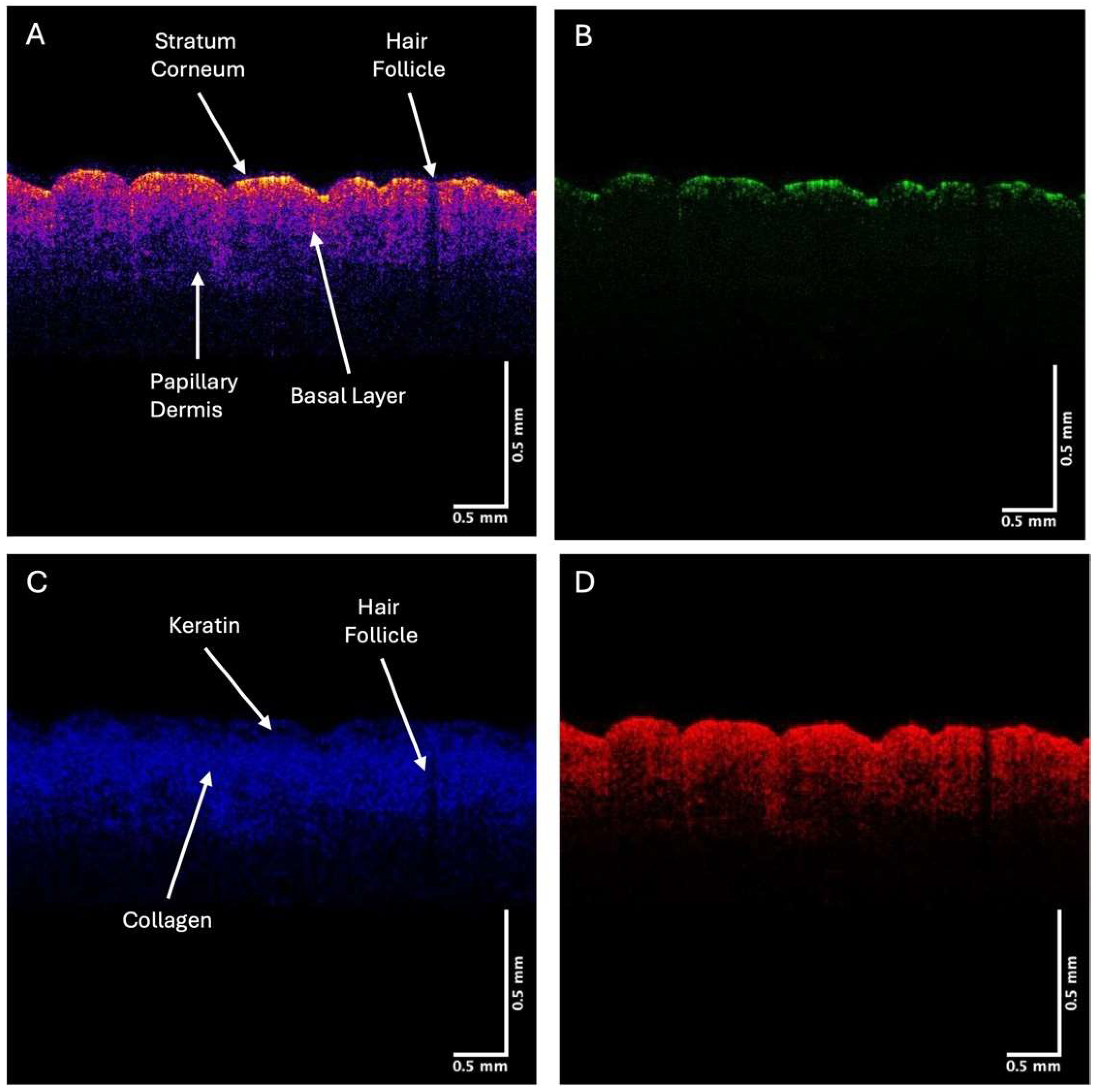
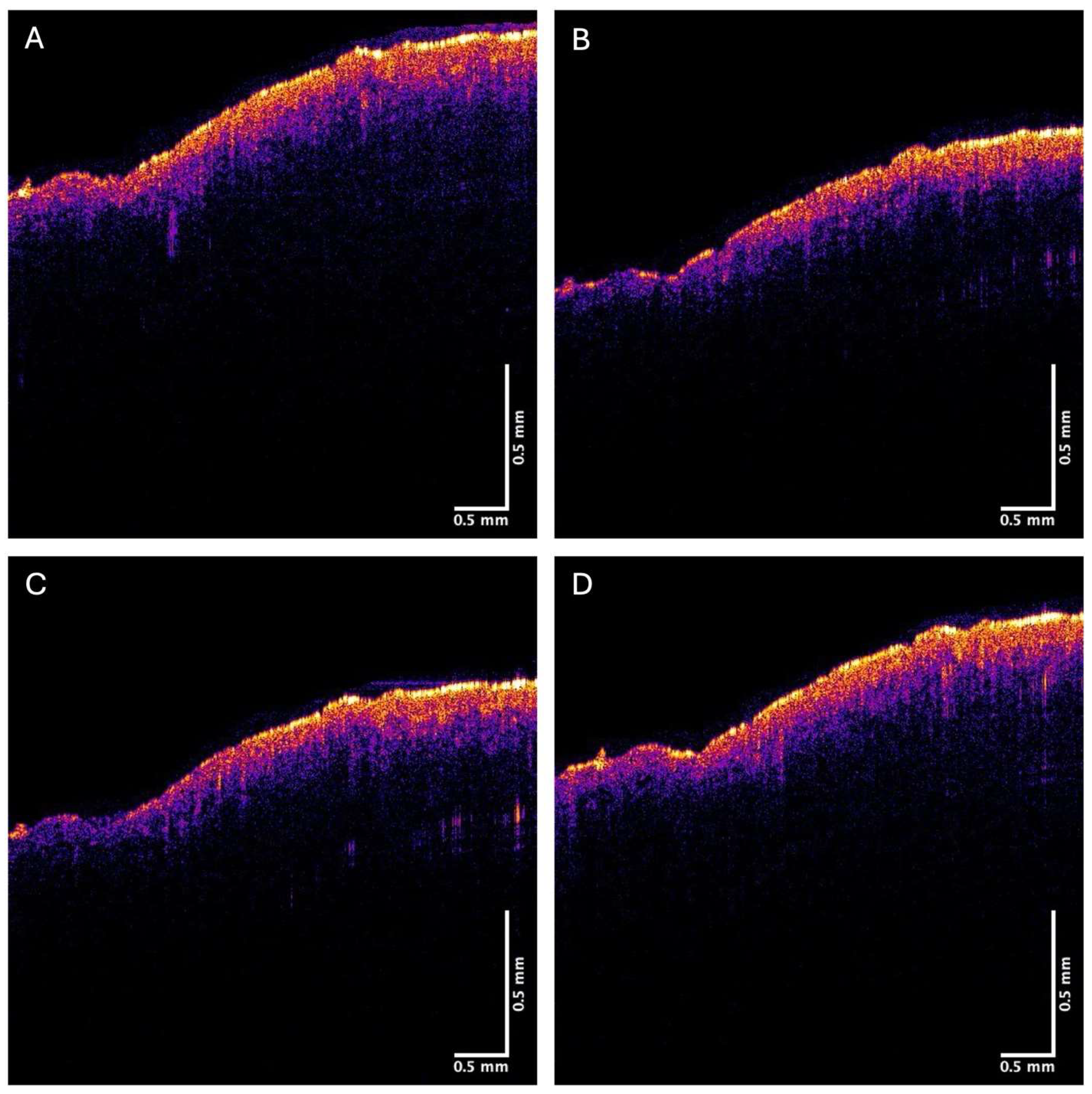
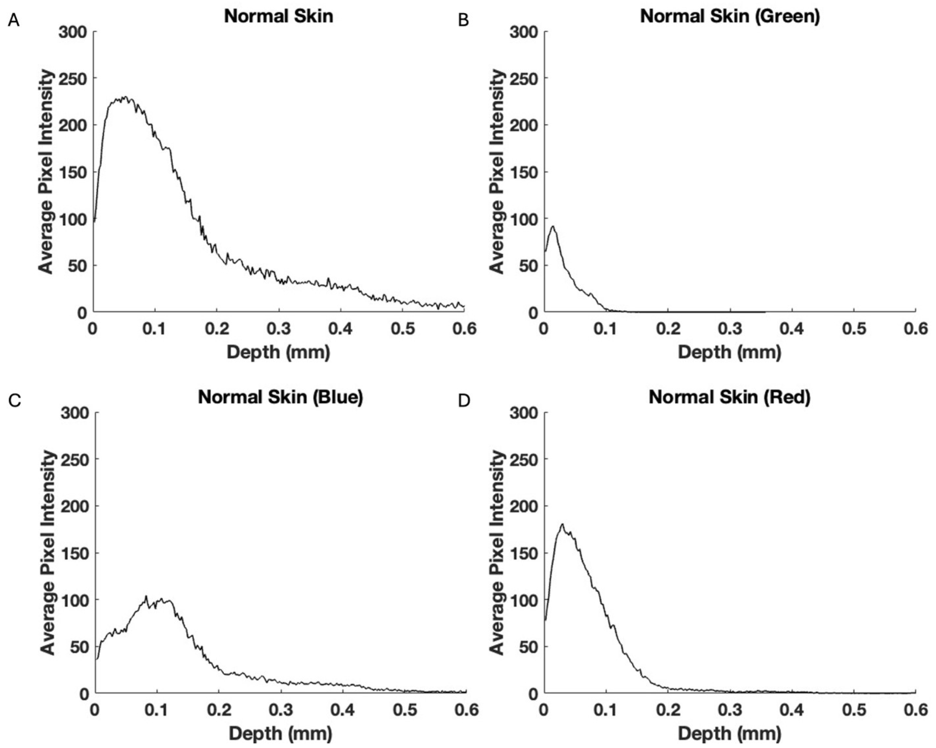
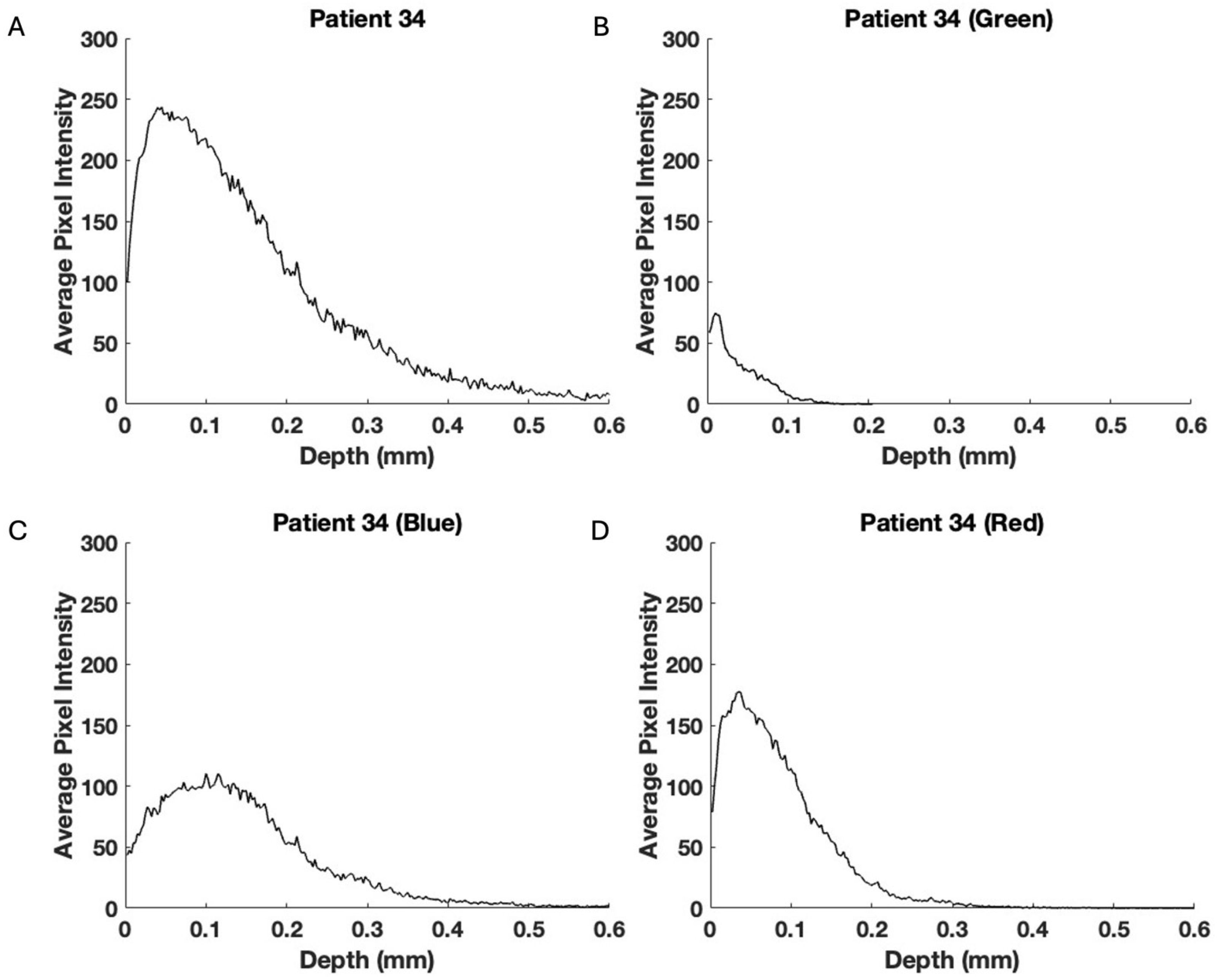
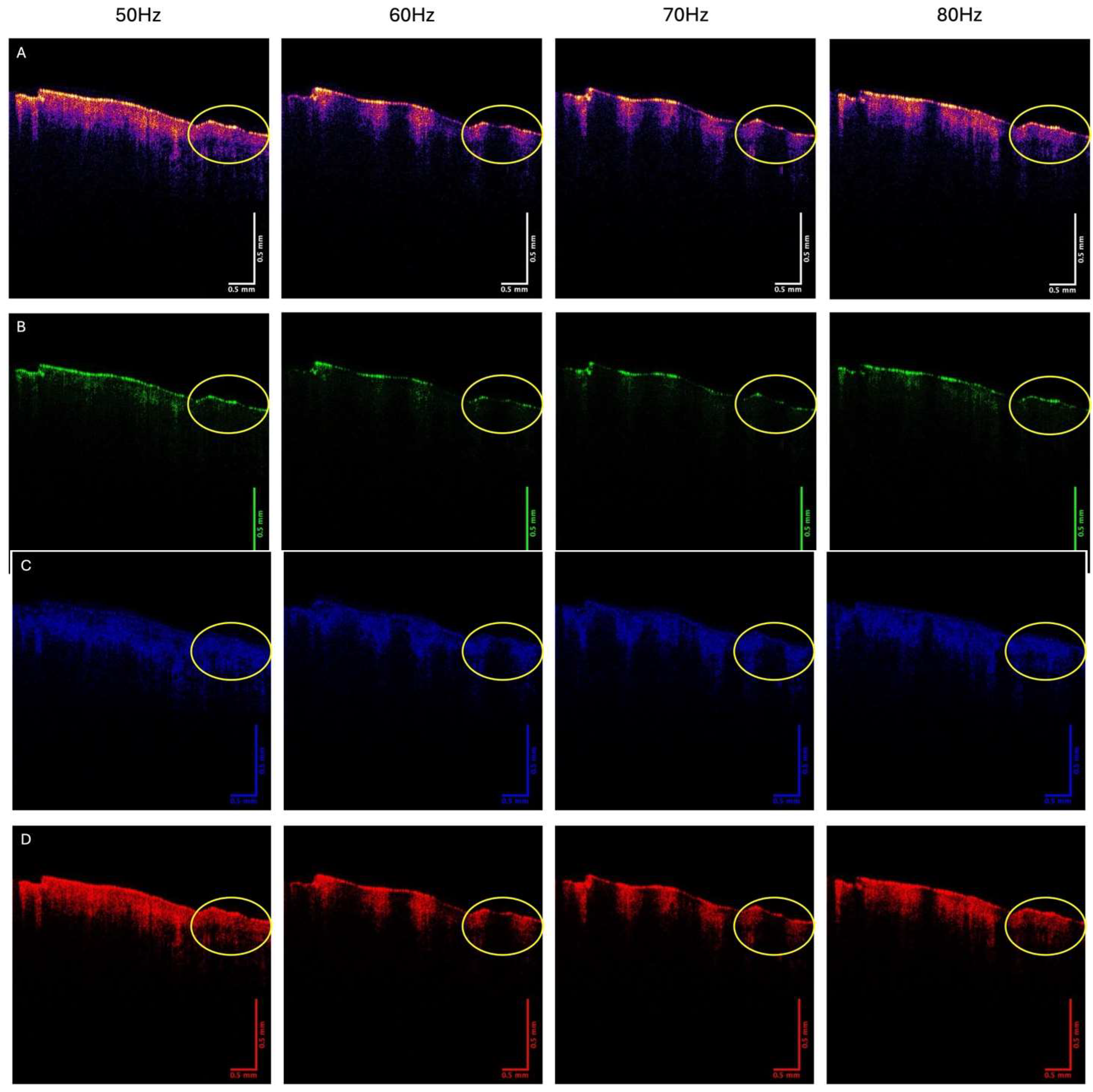
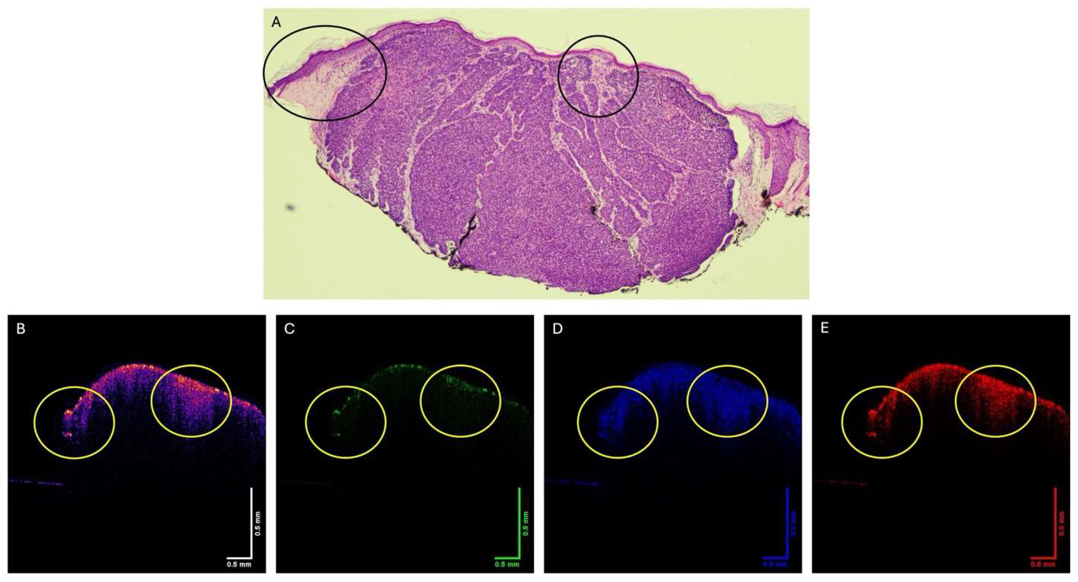
2.1. OCT Image Collection

Images of normal skin and suspected skin cancers were collected using a modified Lumedica OQ Labscope 2.0 (Lumedica, Inc. Durham, NC, USA) as described previously, and which operated at an infrared light wavelength of 840 nm [5]. The measurements were made in vivo on intact skin and in vitro on excised BCC lesion biopsies. All images were made as part of IRB-approved clinical studies on skin at Rutgers Center for Dermatology, as discussed previously [5,19,20]. Clinical diagnoses were made by a board-certified dermatopathologist after H&E staining of tissue sections as part of routine clinical skin excisional protocols. Raw image OCT data were collected in the scanning mode at a scan rate of 13,000 frames per second and processed using MATLAB R2024b and image J software 1.5 [5,19,20]. All sample OCT cross-sectional images were scanned horizontally across the skin or excised lesion. The OCT grayscale scans were color-coded using image J, as reported previously [5]. Pixel intensity versus depth plots were generated by scanning the grayscale OCT images parallel to the surface [5]. The OCT grayscale images were also broken into green, blue, and red subchannel images using a lookup table to break each pixel into low (green), medium (blue), and high (red) pixel intensities [19]. A combination of green, blue, and red colors in varying intensities produces all the colors in the color-coded image; the image processing algorithms map the green, blue, and red components of each pixel. By breaking up the total image into differences in the pixel intensity distribution at each point, it is possible to examine differences in the reflection of the different layers of skin and skin lesions. OCT subchannel images in conjunction with vibrational data were used to provide information on the tissue components present in each sample.

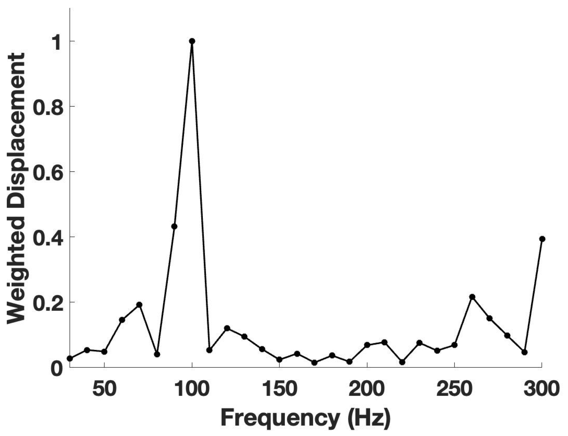
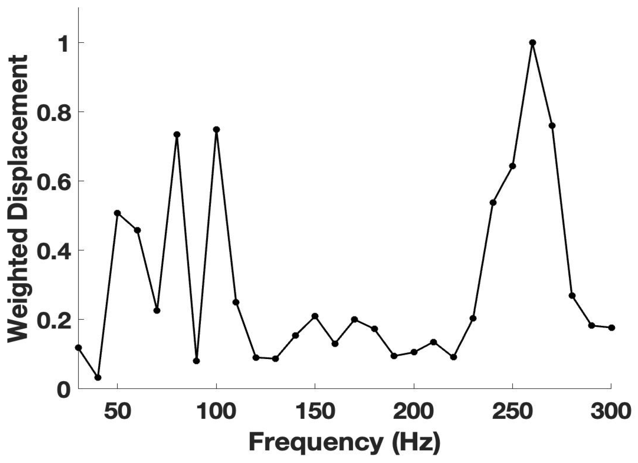
2.2. VOCT Measurements
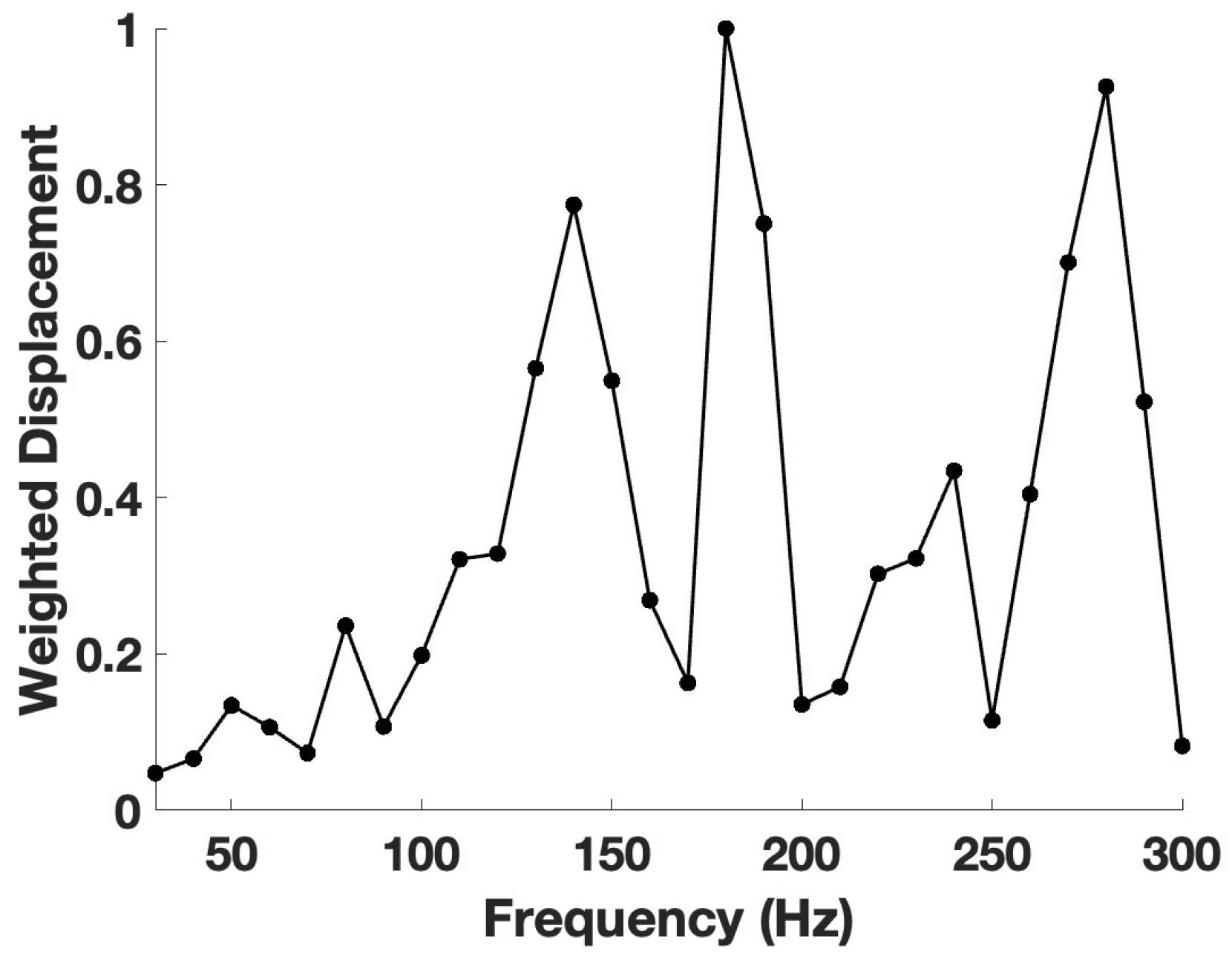
The OQ Labscope 2.0 was modified by adding a 2-inch-diameter acoustic speaker placed about 2.0 inches from the tissue that vibrated the tissue at frequencies between 30 and 300 Hz at 10 Hz intervals [20]. A sinusoidal sound wave was created by the speaker placed near the tissue to be studied. A power of 55 dB was produced by the speaker using an app supplied by the I5 computer contained in the OCT. The speaker was used to transversely vibrate the skin [20]. The sound wave deformation and light were applied to the surface along the axis of the light beam. The in-phase (elastic) sample deflection of the surface along the direction of the light beam was measured as a function of the sound frequency by the OCT. All weighted displacement measurements were made from sample deflection data at a single point based on the location of the lesion determined from images made using the visual camera contained in the OCT. Deflection data in the presence of the sample were divided by the deflection of the speaker in the absence of the sample to create a normalized displacement [20]. The data were processed using MATLAB software, as discussed previously [20]. The maximum displacement of the tissue was detected by measuring the frequency dependence of the tissue deformation based on the reflected infrared light. The reflected light was filtered to collect only vibrations that were in phase (elastic component) with the sound input signal. The amplitude of the displacement was plotted against the frequency of the vibrations to create a mechanovibrational spectrum for each sample. The result was a spectrum of displacements for specific tissue components as a function of the frequency of the applied sound; the resonant frequency of each tissue component, e.g., cells (50–80 Hz), dermal collagen (100–120 Hz), blood vessels (130–150 Hz), and fibrotic tissue (180–260 Hz), has been assigned previously based on studies on a variety of soft tissues and cancerous lesions (see Table 1) [20].

3. Results

Figure 1A shows a color-coded OCT image of normal skin, showing the surface undulations. The yellow color is generated by the stratum corneum, pink and red are reflections from the germinating cell layers, and the blue is generated by the reflections from the basal cell and papillary dermal layers. The image in Figure 1A is broken into low (Figure 1B, green), medium (Figure 1C, blue), and high (Figure 1C, red) subchannel pixel intensity images, providing details about the different layers found in the sample. The green layer (B) provides information on the corneocytes, which are filled with highly concentrated keratin; the blue layer (C) contains collagen in the papillary dermis and keratin in the granulating layers, while the red image (D) contains information on all layers except the stratum corneum. Note the presence of a hair follicle in Figure 1A,C,D (black vertical line). The hyporeflective regions parallel to the surface in the blue image putatively represent keratin in the intermediate filaments in the germinating cell layers, similar to the hyporeflective region seen in the hair follicles.
Figure 2 shows a weighted displacement versus frequency plot (mechanovibrational spectrum) for VOCT studies on normal skin. Note the cellular resonant frequencies seen at about 60 Hz +/−10 Hz (normal cells), 100 Hz (papillary dermal collagen), 150 Hz (normal blood vessels), and 250 Hz and above (reticular dermal collagen). Note the mechanovibrational spectrum of normal skin is dominated by the papillary collagen signal.
Figure 3 shows OCT images of normal skin during vibration at 50 (A), 60 (B), 70 (C), and 80 (D) Hz. Note the images are similar for the different frequencies for skin in vivo.
Typical plots of pixel intensity versus depth for normal skin (A), as well as green (B), blue (C), and red (D) subchannels are shown in Figure 4. The green subchannel (B) contributes to the image at a depth of about 0.01 mm, the blue channel (C) contributes maximally at the depths between 0.02 and 0.15 mm, and minimally through the whole lesion depth, and the red channel (D) is seen throughout the depth of the skin but does not appear to show the stratum corneum.
Figure 5 shows histopathology performed on a small nodular BCC lesion from patient 34 (A) and color-coded OCT images of the lesion (B) and the green (C), blue (D), and red (E) subchannels. The histopathology (A) shows a single nodular lesion that is circled, some inflammatory cells to the right of the nodule, and clear edges that lack rete ridges on both sides of the nodule. The reflection of lesion cells in the green subchannel appear to be sparse, while the blue and red subchannels show differences in the components in the circled BCC lesion and the neighboring regions of the tissue. Note that the BCC nodule is circled in the histopathology image (A), in the OCT image B, and in subchannel images C, D, and E. Note the decreased intensity of the green subchannel in the area in C where the nodule is circled. Also, the hyporeflective region near the surface of the lesion is disappears in the blue subchannel (circled nodular region) in D where the lesion is seen.
Figure 6 shows the pixel intensity versus depth plots for the images shown in Figure 5 for the nodular BCC from patient 34. Note the decreased height of the green subchannel (Figure 6B) compared to normal skin (Figure 4B).
Figure 7 is a plot of weighted displacement versus frequency (mechanovibrational spectrum) for the nodular BCC from patient 34 (Figure 5). Note the large resonant frequency peaks at about 60, 80, 100, and 260 Hz. Based on previous study results, these peaks represent normal cells (60 Hz), cancer-associated fibroblasts (80 Hz), dermal collagen (100 Hz), and fibrotic tissue (250–260 Hz) [20]. Note the increased peak sizes at 60, 80, and 260 Hz compared to normal skin (Figure 2), suggesting that increased cellular proliferation of both normal skin cells (60 Hz) and cancer-associated fibroblasts (80 Hz), as well as the deposition of fibrotic tissue (250–280 Hz), occur during nodular BCC formation.
Figure 8 shows OCT images of the circled nodular BCC from patient 34 at vibrational frequencies of 50, 60, 70, and 80 Hz for the color-coded image (A) and the green (B), blue (C), and red (D) subchannels. Note the disappearance of the green, blue, and red subchannel images of the circled lesion at 60 and 70 Hz. The images vibrated at 50 and 80 Hz appear similar.
Figure 9 shows histological (A) and OCT (B through E) images of a large nodular BCC from patient 39. The OCT images for the green (C), blue (D), and red (E) subchannels are shown. The regions circled in the histopathology appear to contain normal cells and dermal collagen. The OCT sub-images of these circled regions appear to be different compared to the bulk of the lesion. However, these circled regions appear suspicious when viewed by VOCT. The edges of this nodular BCC do not look similar to the edges of the small BCC and do not appear to be free of the cancerous tissue compared to the lesion seen in Figure 5A. The green subchannel image of both ends of this lesion is of low pixel intensity (Figure 9C).
Figure 10 is a plot of pixel intensity versus depth for the large nodular BCC from patient 39 seen in Figure 9A. Note the reduced pixel intensity for the green subchannel compared to normal skin (Figure 4B) and the small nodular BCC (Figure 6B). The green subchannel peak height is only a small fraction of that of normal skin.
Figure 11 shows a plot of weighted displacement versus frequency for the nodular BCC from patient 39. Note the absence of large 50 Hz and 80 Hz peaks. while the peaks starting at 130 to 280 Hz are very large. This suggests that even though the lesion is made of cancer-associated fibroblasts surrounded by palisading cells based on the histopathology, the mechanovibrational spectrum appears to be reflective of a large amount of fibrotic tissue. The peak at 130–140 Hz suggests that small blood vessels are present, indicative of the cancerous lesion.
Figure 12 shows the OCT images for the BCC from patient 39 when vibrated at 50, 60, 70, and 80 Hz. The color-coded OCT image is shown in (A), while the green subchannel (B), blue subchannel (C), and red subchannel (D) are also shown. Note that even the circled areas of the lesion become faint at vibrational frequencies of 60 and 70 Hz. The remainder of the lesion also is very faint when vibrated at 60 and 70 Hz, suggesting that even the circled edges of the lesion cannot be called “clean”.

4. Discussion

New techniques to noninvasively distinguish cancerous from benign lesions are needed as the number of skin cancers increase due to sun exposure. It has been recognized that the physical properties of cells and extracellular matrices play a large role in the function and pathophysiology of tissues [19,20,21,22,23,24,25,26]. BCCs have been reported to present clinically as pearly, shiny, smooth nodules with small dilated, branched blood vessels that can ulcerate or bleed [1]. Based on VOCT results, the resonant frequency and elastic moduli of nodular, superficial, sclerotic, and pigmented BCCs have been shown to all have 80 Hz, 130 Hz, and 250 Hz resonant frequency peaks not found in normal skin [27,28]. In all BCC types studied using VOCT, new 80 Hz, 130 Hz, and 260 Hz resonant frequency peaks were found, similar to those present in other carcinomas including squamous cell carcinoma (SCCs) and melanomas [28], suggesting that cancerous lesions all have similar biophysical properties that can be studied noninvasively using VOCT to understand the pathobiology of skin cancer formation and growth patterns.
In this study, we compared OCT images and VOCT measurements made on small and large nodular BCCs using histopathology, OCT images, and VOCT physical measurements. Our studies cannot be used to image HA reported to be altered in the stroma surrounding BCCs; however, the conversion of dermal collagen with a resonant frequency of 100 Hz into fibrotic tissue with resonant frequencies between 180 and 260 Hz can be studied. While our results suggest that both small and large nodular BCCs have similar morphologies that include nodules with palisading cells surrounding the lesions, no part of the excised lesions appeared like normal skin. In the small lesion, the nodule was surrounded by tissue that was considered to have “clean” margins based on histopathology even though the rete ridges were missing. The edges of the lesion (see Figure 5) were free of palisading cells with only some inflammatory cells being present. Based on the OCT images shown in Figure 5, the BCC nodule reflects less light where the lesion is present. This is the case even though the mechanovibrational spectrum of the lesion appeared to have an increased number of normal cells (50 Hz peak), CAFs (80 Hz peak) (see Figure 7), and large fibrotic peaks. Based on these observations, it is possible to identify the location of a BCC lesion noninvasively using OCT based on the green and blue subchannel images. Previously, we reported that the green subchannel represents reflections by the stratum corneum while the blue channel provides information on the collagen and fibrotic tissue [5]. The ability to noninvasively identify nodular BCCs using this technique makes it a useful adjunct to visual inspection and dermoscopy, which are used by dermatologists to identify cancerous lesions seen in the clinic. It is interesting that images found by vibrating the nodules of the biopsy at 60 and 70 Hz result in reduced light reflections. Light reflections are reduced by large particle aggregates of either cells or any structures present that approach the wavelength of the incident light in size [5]. This suggests that the nodules reflect less light due to their large size.
In contrast, in the large nodular BCC (Figure 9), the cells are more tightly packed and are separated by what appears to be dense collagen fibrosis. The edges of this lesion do not appear clean since the OCT images and subchannel images appear to lack cells in the green channel, and the green channel disappears in the sample vibrated at 60 and 70 Hz (see Figure 11). The low value of the cellular peaks at 50 and 80 Hz in Figure 10 and the large value of the peaks at 130–150 Hz and 180–260 Hz suggest that the cells are intimately linked with the collagen fibrotic tissue that converts the dermal collagen normally seen at 100 Hz into highly crosslinked collagen networks with increasing stiffnesses. The lack of 50 Hz and 80 Hz peaks characteristic of highly cellular lesions may indicate that cell–collagen interactions appear to bind the cells tightly to the fibrotic collagen so they vibrate as one unit and not as separate entities. This close association between CAFs and fibrotic collagen may serve to limit the migration of the cells in the nodule, although it may not limit the ultimate nodular dimensions. Further studies are needed to consider the relationship between collagen orientation and number of cells in the nodules in both BCCs and other cancerous lesions that are more likely to migrate.
Recently, we have reported the use of pixel intensity versus depth plot parameters such as maximum pixel height and width at half height along with machine learning to differentiate between BCCs and normal skin with a sensitivity and specificity of 93.7% and 100%, respectively [5]. These results indicate that noninvasive OCT images can be quantitatively used to screen lesions to identify BCCs and to identify the size and depth of a lesion [5]. Additional study results suggest that BCCs can be differentiated from other cancerous lesions based on the ratios of the mechanovibrational peaks, which differ for BCCs, SCCs, and melanomas [27,28].
Since OCT and VOCT measurements can be made remotely over the internet, they can be useful during telemedicine for the screening of lesions by practitioners in areas where dermatologists are in short supply. The images and mechanovibrational data can be stored in the cloud and reviewed by dermatologists remotely for detailed evaluations.

5. Conclusions

In this study, we compared histopathology, OCT images, and VOCT measurements made on small and large nodular BCCs. While our results are limited, they suggest that both small and large nodular BCCs have similar morphologies that include palisading cells surrounding the lesions; however, no part of the excised lesions appeared like normal skin. In the small lesion, the lesion edges were clean based on histopathology. The ability to noninvasively identify nodular BCCs using this technique makes it a useful adjunct to visual inspection and dermoscopy, which are used by dermatologists to identify cancerous lesions seen in the clinic. It is interesting to note that the vibration of the biopsy at 60 and 70 Hz results in the disappearance of the lesion, confirming the location of the nodule. Further studies are needed to indicate whether lesion size and the height of the cellular and collagen fibrotic mechanovibrational peaks are related.
OCT images can be quantitatively used to screen lesions to identify BCCs, and BCCs can be differentiated from other cancerous lesions based on the ratios of the different mechanovibrational peaks, as shown previously. OCT and VOCT measurements can be made remotely over the internet, and they can be useful in telemedicine for the screening of lesions by practitioners in areas where dermatologists are in short supply.

Author Contributions

Conceptualization, F.H.S. and G.K.; methodology, T.D., G.K. and A.P., formal analysis, F.H.S., T.D. and G.K.; investigation, G.K., A.P. and T.D.; data curation, T.D., A.P. and G.K.; writing—original draft preparation, F.H.S. and T.D.; writing—review and editing, F.H.S., T.D., G.K. and A.P. All authors have read and agreed to the published version of the manuscript.

Funding

Partial support for this project was provided by Ben Franklin Tech Partners during 2023–2025.

Institutional Review Board Statement

The protocol was approved by the I.R.B. at Robert Wood Johnson Medical School on 16 April 2024, IRB Number: Pro2023002455.

Informed Consent Statement

All subjects provided consent.

Data Availability Statement

Data available at optovibronex.com.

Conflicts of Interest

FHS is a stockholder, and TD is an employee of OptoViobronex.

References

  1. Available online: www.cancer.org/cancer/types/basal-and-squamous-cell-skin-cancer/about/key-statistics.html (accessed on 6 March 2025).
  2. Negrutiu, M.; Danescu, S.; Popa, T.; Focsan, M.; Vesa, S.C.; Baican, A. Advancements in Basal Cell Carcinoma Diagnosis: Non-Invasive Imaging and Multimodal Approach. J. Clin. Med. 2024, 13, 39. [Google Scholar] [CrossRef] [PubMed]
  3. Tabanlıoğlu Onan, D.; Şahin, S.; Gokoz, O.; Erkin, G.; Cakır, B.; Elcin, G.; Kayıkcıoğlu, A. Correlation between the dermatoscopic and histopathological features of pigmented basal cell carcinoma. J. Eur. Acad. Dermatol. Venereol. 2010, 24, 1317–1325. [Google Scholar] [CrossRef] [PubMed]
  4. Ferrante di Ruffano, L.; Dinnes, J.; Deeks, J.J.; Chuchu, N.; Bayliss, S.E.; Davenport, C.; Takwoingi, Y.; Godfrey, K.; O’Sullivan, C.; Matin, R.N.; et al. Optical coherence tomography for diagnosing skin cancer in adults. Cochrane Database Syst. Rev. 2018, 12, CD013189. [Google Scholar] [CrossRef]
  5. Silver, F.H.; Deshmukh, T.; Patel, A.; Dhillon, J.; Bobra, A.; Nadiminti, H. Rapid Noninvasive Skin Screening for Basal Cell Carcinomas using Vibrational Optical Coherence Tomography. Br. J. Cancer Res. 2025, 8, 747–755. [Google Scholar] [CrossRef]
  6. Coleman, A.J.; Richardson, T.J.; Orchard, G.; Uddin, A.; Choi, M.J.; Lacy, K.E. Histological correlates of optical coherence tomography in non-melanoma skin cancer. Ski. Res. Technol. 2013, 19, 10–19. [Google Scholar] [CrossRef]
  7. Bertheim, U.; Hofer, P.A.; Engström-Laurent, A.; Hellström, S. The stromal reaction in basal cell carcinomas. A prerequisite for tumour progression and treatment strategy. Br. J. Plast. Surg. 2004, 57, 429–439. [Google Scholar] [CrossRef] [PubMed]
  8. Tzellos, T.G.; Kyrgidis, A.; Vahtsevanos, K.; Triaridis, S.; Printza, A.; Klagas, I.; Zvintzou, E.; Kritis, A.; Karakiulakis, G.; Papakonstantinou, E. Nodular basal cell carcinoma is associated with increased hyaluronan homeostasis. J. Eur. Acad. Dermatol. Venereol. 2011, 25, 679–687. [Google Scholar] [CrossRef] [PubMed]
  9. Markowska, A.; Antoszczak, M.; Markowska, J.; Huczyński, A. Role of Hyaluronic Acid in Selected Malignant Neoplasms in Women. Biomedicines 2023, 11, 304. [Google Scholar] [CrossRef]
  10. Jermain, P.R.; Iorizzo, T.W.; Maloney, M.; Mahmoud, B.; Yaroslavsky, A.N. Design and Validation of a Handheld Optical Polarization Imager for Preoperative Delineation of Basal Cell Carcinoma. Cancers 2022, 14, 4049. [Google Scholar] [CrossRef]
  11. Bourgot, I.; Primac, I.; Louis, T.; Noël, A.; Maquoi, E. Reciprocal Interplay Between Fibrillar Collagens and Collagen-Binding Integrins: Implications in Cancer Progression and Metastasis. Front. Oncol. 2020, 10, 1488. [Google Scholar] [CrossRef]
  12. Esposito, M.; Yerly, L.; Shukla, P.; Herme, V.; Sella, F.; Balazs, Z.; Evelyn Lattmann, E. COL10A1 expression distinguishes a subset of cancer-associated fibroblasts present in the stroma of high-risk basal cell carcinoma. Br. J. Dermatol. 2024, 191, 775–790. [Google Scholar] [CrossRef] [PubMed]
  13. Yi, Q.; Zhu, G.; Zhu, W.; Wang, J.; Ouyang, X.; Yang, K.; Zhong, J. Oncogenic mechanisms of COL10A1 in cancer and clinical challenges (Review). Oncol. Rep. 2024, 52, 162. [Google Scholar] [CrossRef] [PubMed]
  14. Silver, F.H.; Deshmukh, T. Do tensile and shear forces exerted on cells influence mechanotransduction through stored energy considerations? BIOCELL 2024, 48, 525–540. [Google Scholar] [CrossRef]
  15. Rømer, A.M.A.; Thorseth, M.L.; Madsen, D.H. Immune Modulatory Properties of Collagen in Cancer. Front. Immunol. 2021, 12, 791453. [Google Scholar] [CrossRef]
  16. Zhang, Q.; An, Z.Y.; Jiang, W.; Jin, W.L.; He, X.Y. Collagen code in tumor microenvironment: Functions, molecular mechanisms, and therapeutic implications. Biomed. Pharmacother. 2023, 166, 115390. [Google Scholar] [CrossRef]
  17. De Martino, D.; Bravo-Cordero, J.J. Collagens in Cancer: Structural Regulators and Guardians of Cancer Progression. Cancer Res. 2023, 83, 1386–1392. [Google Scholar] [CrossRef]
  18. Sahai, E.; Wyckoff, J.; Philippar, U.; Segall, J.E.; Gertler, F.; Condeelis, J. Simultaneous imaging of GFP, CFP and collagen in tumors in vivo using multiphoton microscopy. BMC Biotechnol. 2005, 5, 14. [Google Scholar] [CrossRef]
  19. Baeyens, N.; Bandyopadhyay, C.; Coon, B.G.; Yun, S.; Schwartz, M.A. Endothelial fluid shear stress sensing in vascular health and disease. J. Clin. Investig. 2016, 26, 821–828. [Google Scholar] [CrossRef]
  20. Li, X.; Dao, M.; Lykotrafitis, G.; Karniadakis, G.E. Biomechanics and biorheology of red blood cells in sickle cell anemia. J. Biomech. 2017, 50, 34–41. [Google Scholar] [CrossRef]
  21. Bruckner, B.R.; Janshoff, A. Importance of integrity of cell-cell junctions for the mechanics of confluent MDCK II cell. Sci. Rep. 2018, 8, 14117. [Google Scholar] [CrossRef]
  22. Van Damme, T.; Colige, A.; Syx, D.; Giunta, C.; Lindert, U.; Rohrbach, M.; Aryani, O.; Alanay, Y.; Simsek-Kiper, P.Ö.; Kroes, H.Y.; et al. Expanding the clinical and mutational spectrum of the Ehlers–Danlos syndrome, dermatosparaxis type. Genet. Med. 2016, 18, 882–891. [Google Scholar] [CrossRef] [PubMed]
  23. Houdusse, A.; Sweeney, H.L. How Myosin Generates Force on Actin Filaments. Trends Biochem. Sci. 2016, 41, 989–997. [Google Scholar] [CrossRef] [PubMed]
  24. Garcia, M.A.; Nelson, W.J.; Chavez, N. Cell-Cell Junctions Organize Structural and Signaling Networks. Cold Spring Harb. Perspect. Biol. 2018, 10, a029181. [Google Scholar] [CrossRef] [PubMed]
  25. Rodriguez-Hernandez, I.; Cantelli, G.; Bruce, F.; Sanz-Moreno, V. Rho, ROCK and actomyosin contractility in metastasis as drug targets. F1000Research 2016, 5, 783. [Google Scholar] [CrossRef]
  26. Karantza, V. Keratins in health and cancer: More than mere epithelial cell markers. Oncogene 2011, 30, 127–138. [Google Scholar] [CrossRef]
  27. Silver, F.H.; Deshmukh, T.; Patel, A.; Nadaminti, H. Use of Optical Coherence Tomography Images to Differentiate Between Normal Skin, Skin Lesions, and Melanoma: A Pilot Study. J. Cancer Sci. Therapy 2024, 4, 1751. [Google Scholar]
  28. Silver, F.H.; Deshmukh, T.; Ryan, N.; Romm, A.; Nadiminti, H. “Fingerprinting” Benign and Cancerous Skin Lesions Using Vibrational Optical Coherence Tomography: Differentiation among Cancerous Lesion Types Based on the Presence of New Cells, Blood Vessels, and Fibrosis. Biomolecules 2022, 12, 1332. [Google Scholar] [CrossRef]
Figure 1. Color-coded OCT images of normal skin (A) and green (B), blue (C), and red (D) subchannel images. The low (B), medium (C), and high (D) pixel intensities of each pixel were determined from a look-up table [5]. The green subchannel shows the number and location of the corneocytes, while the blue subchannel has hyporeflective regions putatively containing keratin-synthesizing cells and collagen from the papillary dermis, and the red subchannel reflects the images seen in both the green and blue subchannels added together.
Onco 05 00023 g001
Figure 2. A typical plot of weighted displacement versus frequency (mechanovibrational spectrum) obtained from VOCT studies on normal skin. Cellular resonant frequencies are seen at about 60 Hz +/−10 Hz (normal cells), collagen at 100 Hz (papillary dermal collagen), and 260 Hz (reticular collagen). Note the mechanovibrational spectrum of normal skin is dominated by the dermal collagen signal.
Onco 05 00023 g002
Figure 3. OCT images of normal skin during vibration at 50 (A), 60 (B), 70 (C), and 80 (D) Hz. Note the images are similar for the different frequencies for skin in vivo.
Onco 05 00023 g003
Figure 4. Typical plots of pixel intensity versus depth for normal skin (A) and green (B), blue (C), and red (D) subchannels. The blue subchannel shows an inflection point at about 0.05 mm, which may be due to reflections from the interface between keratin-producing cells in the granulating region and the beginning of the papillary dermis. Note the red subchannel appears to provide information on reflections from the green and blue subchannels in the skin.
Onco 05 00023 g004
Figure 5. A comparison between the histopathology image of a section of a small nodular BCC stained with H&E from patient 34 (A), color-coded OCT images of the lesion (B), and green (C), blue (D), and red (E) subchannel images. Lesion cells in the green subchannel appear to disappear in the BCC nodule (see circle in (C)), while the hyporeflective regions near the surface disappear in the nodular region (D). The red subchannel appears to be a combination of the green and blue subchannels.
Onco 05 00023 g005
Figure 6. Plots of weighted displacement versus frequency for the nodular BCC from patient 34 for the color-coded image (A) and the green (B), blue (C), and red (D) subchannel images. Note the height of the green subchannel is reduced compared to that of normal skin (Figure 4B).
Onco 05 00023 g006
Figure 7. A plot of weighted displacement versus frequency for a small nodular BCC from patient 34. Note the large resonant frequency peaks at about 60, 80, 100, and 260 Hz. Based on previous study results, these peaks represent normal cells (60 Hz), cancer-associated fibroblasts (80 Hz), papillary dermal collagen (100 Hz), and fibrotic tissue (260–280 Hz) [5]. Note that the increased peak sizes at 80 and 260 Hz for this BCC compared to normal skin (Figure 2) suggest that cellular proliferation of both normal skin cells and of cancer-associated fibroblasts lead to the deposition of fibrotic tissue (260 Hz).
Onco 05 00023 g007
Figure 8. Color-coded OCT images of a nodular BCC from patient 34 (A) and green (B), blue (C), and red (D) subchannels at vibrational frequencies of 50, 60, 70, and 80 Hz. The location of the nodular BCC is circled in each image. Note that the image of the lesion appears to diminish when the sample is vibrated at 60 and 70 Hz.
Onco 05 00023 g008
Figure 9. Shows histological (A) and OCT (BE) subchannel images of a large nodular BCC from patient 39. The OCT images for the green (C), blue (D), and red (E) subchannels provide information about the cells and the fibrosis present in the lesion. Note how the circled regions appear different from the rest of the cancerous nodules; however, they do not look like normal skin since they disappear when vibrated. The blue channel lacks hyporeflective regions seen in normal skin.
Onco 05 00023 g009
Figure 10. A plot of pixel intensity versus depth for the large nodular BCC from patient 39 seen in Figure 9A. The plots are shown for the color-coded (A), green (B), blue (C) and red (D) subchannel OCT images. Note the reduced pixel intensity for the green subchannel compared to normal skin (Figure 4B) and the small nodular BCC (Figure 6B).
Onco 05 00023 g010
Figure 11. A plot of weighted displacement versus frequency for the large nodular BCC from patient 39. Note the increased peak heights at 130–150 Hz (thin and normal blood vessels) and 180 through 280 Hz (fibrotic tissue), based on previous publications [20]. Note the reduced peak heights at 50 and 80 Hz compared to Figure 7 for the small nodular BCC.
Onco 05 00023 g011
Figure 12. OCT images for the large nodular BCC from patient 39 vibrated at 50 (A), 60 (B), 70 (C), and 80 Hz (D). The intensities of all the subchannel images are diminished at frequencies of 60 and 70 Hz, suggesting that even the circled lesion edges are not “clean”.
Onco 05 00023 g012
Table 1. Resonant frequencies of skin components based on VOCT studies [5,19,20].
Tissue ComponentResonant Frequency (Hz)
Epithelial Cells50–60
Cancer Associated Fibroblasts70–80
Dermal Collagen100
New Thin Blood Vessels130
Normal Blood Vessels150
Fibrotic Tissue250–260
Disclaimer/Publisher’s Note: The statements, opinions and data contained in all publications are solely those of the individual author(s) and contributor(s) and not of MDPI and/or the editor(s). MDPI and/or the editor(s) disclaim responsibility for any injury to people or property resulting from any ideas, methods, instructions or products referred to in the content.

