Article

AlphaGlue: A Novel Conceptual Delivery Method for α Therapy

1
School of Physics, University of Bristol, Bristol BS7 1TL, UK
2
Centre for Medical and Radiation Physics, University of Wollongong, Wollongong 2500, Australia
3
Division of Health Science, University of Osaka, Osaka 565-0871, Japan
*
Author to whom correspondence should be addressed.
These authors contributed equally to this work.
Current address: Muotech, Ul. Juliusza Slowackiego 24, 35-069 Rzeszow, Poland.
BioMedInformatics 2025, 5(4), 58; https://doi.org/10.3390/biomedinformatics5040058
Submission received: 18 June 2025 / Revised: 12 September 2025 / Accepted: 9 October 2025 / Published: 13 October 2025

Abstract

Extensive research is being carried out on the application of α particles for cancer treatment. A key challenge in α therapy is how to deliver the α emitters to the tumour. In AlphaGlue, a novel treatment delivery concept, the α emitters are suspended in a thin layer of glue that is put on top of the tumour. In principle, this should be an easy and safe way to apply α therapy. In this study, the effectiveness of AlphaGlue is evaluated using GEANT4 and GEANT4-DNA simulations to calculate the DNA damage as a function of depth. Two radionuclides are considered in this work, 211At and 224Ra. The results indicate that, as a concept, the method offers a promising hypothesis for treating superficial tumours, such as skin cancer, when 224Ra is applied directly on the tissue and stabilized with a glue layer. This results in 2 × 10 5 complex double strand breaks and 5 × 10 5 double strand breaks at 5 mm depth per applied 224Ra atom. When applying a 224Ra atom concentration of ( 4.35 ± 0.2 ) × 10 11 / cm2 corresponding to an activity of ( 21.8 ± 1 ) μ Ci/cm2 on the skin surface, the RBE weighted dose exceeds 20 Gy at 5 mm depth. Hence, there is significant cell death at 5 mm into the tissue; a depth matching clinical requirements for skin cancer treatment. Given the rapidly falling weighted dose versus depth curve, the treatment depth can be tuned with good precision. The results of this study show that AlphaGlue is a promosing treatment and open the pathway towards the next stage of the research, which includes in-vitro studies.

1. Introduction

Radiotherapy is a standard treatment for many types of cancer. Alpha particle radiation therapy is a radiotherapy technique that involves applying α -emitting isotopes to treat tumours or metastatic cancer. Due to the high linear energy transfer (LET) of α particles, they deposit their energy over a very short distance (≲100 μ m), resulting in a highly localized dose. This localization is a critical factor for improving cancer treatment by minimizing damage to surrounding healthy tissue [1].
Several therapeutic approaches rely on α particles, including Targeted Alpha Therapy (TAT) [1,2], which uses isotopes such as 255Ac [3,4,5,6,7,8,9] and 211At [10,11,12,13,14], to precisely deliver α radiation doses to tumours. Clinical studies have demonstrated the effectiveness of TAT in treating various malignancies, including leukemia, brain tumours, ovarian, and prostate cancers, underscoring the significant clinical potential of targeted α -emitter therapies.
In addition to TAT, 224Ra has been utilized in Diffusing α -emitters Radiation Therapy (DaRT) [9,15,16,17,18], a novel therapeutic approach in which a small seeds containing 224Ra are implanted into the tumour. As 224Ra decays, it releases 220Rn gas, which diffuses into the surrounding tissue and extends the range of alpha particle delivery, thereby achieving a greater treatment depth. Initial clinical results have demonstrated high efficacy in treating skin cancers, with complete response rates ranging from 89% to 100% [8,16,18,19]. While targeted α therapies have demonstrated clinical success, and DaRT has expanded the potential of α -emitter for treatments of solid tumours, it is still relatively cumbersome to apply as multiple seeds loaded with 224Ra need to be inserted with less than 5 mm spacing [19]. The combination of the clinical success and the cumbersome application process, supports the relevance of exploring alternative delivery concepts such as AlphaGlue. Here we evaluate a novel concept of α therapy with α emitters suspended in glue. This is then applied to the skin to treat the tumour. In addition, AlphaGlue may offer a less invasive and potentially easier-to-use alternative, which could be particularly advantageous for sensitive areas such as facial skin cancers, but also anal, vaginal cancer or endometriosis. Similar approaches exploiting β -particles exist. Here radioactive patches are applied or loaded creams are used, for example 90Y [20,21], 188Re [22,23,24,25], 32P [26] and 106Ru [27]. The ranges of α ’s and β ’s and their DNA damage spectra are very different. A comparison study is out of the scope of this paper.

1.1. AlphaGlue

AlphaGlue is a novel delivery method for α therapy that involves applying a layer of glue loaded with α emitters to the surface of the skin. An unloaded second layer of glue may be applied to shield any potential other skin from the radiation, as discussed later in Section 2.1. Figure 1 shows a sketch of the concept.
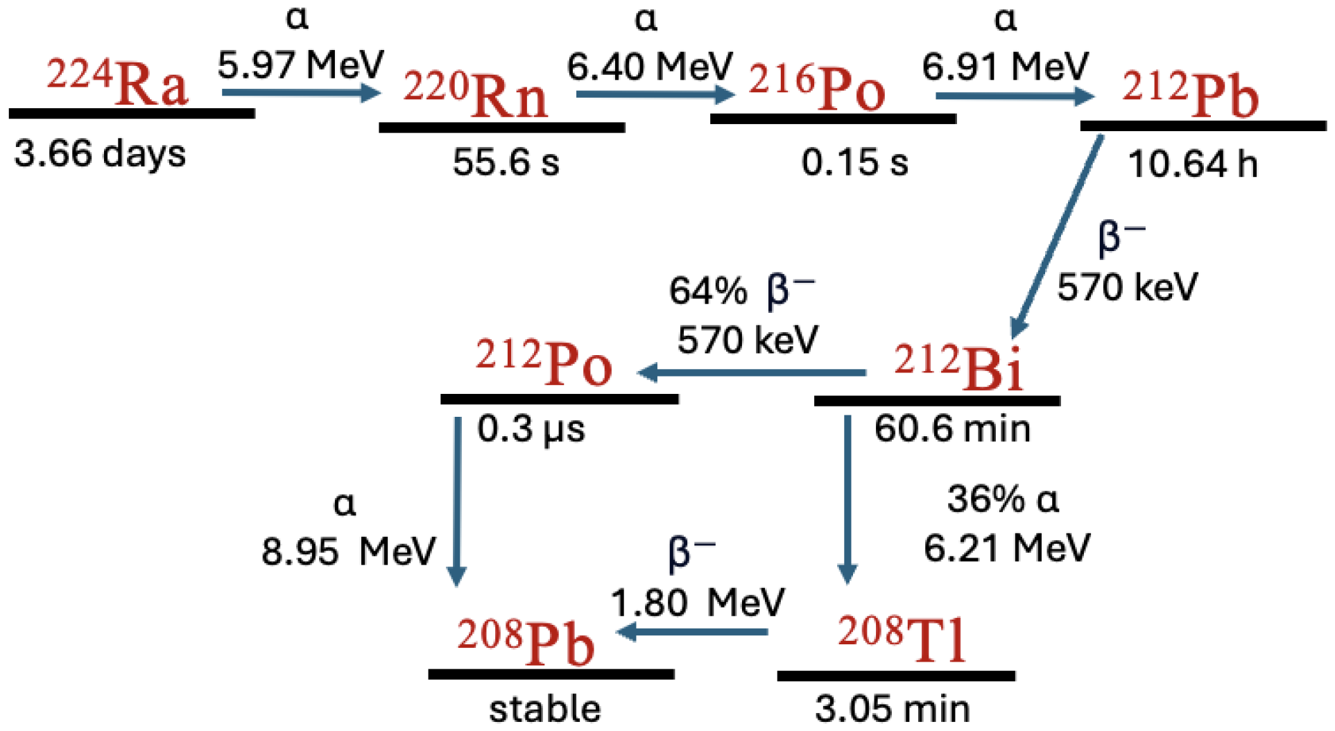
In this study, the loaded glue contains either 211At or 224Ra. 211At has two decay paths, and in either of them a single α is produced, as shown in Figure 2. In contrast, 224Ra undergoes four α decays in its decay chain, as shown in Figure 3, resulting in multiple emissions that deliver a highly localized dose. To evaluate and compare the therapeutic potential of 211At and 224Ra within the AlphaGlue framework, this study investigates the induced DNA damage through detailed Monte Carlo simulations. Melanoma invasion depth is typically between 1 and 4 mm, with thicker tumors exceeding 4 mm [28,29]. Therefore, we evaluate the effectiveness at these depths.

1.2. DNA Damage

In this study, the focus is on investigating DNA damage caused by the isotopes 224Ra and 211At when applied to the AlphaGlue, and comparing their effects. DNA damage, which can lead to cell death if not repaired by the cell, occurs through two primary mechanisms: direct and indirect. Direct damage takes place when particles interact with the DNA molecule and deposit their energy directly into it. Indirect damage occurs when particles interact with water molecules surrounding the DNA, producing free radicals that subsequently damage the DNA [30].

2. Materials and Methods

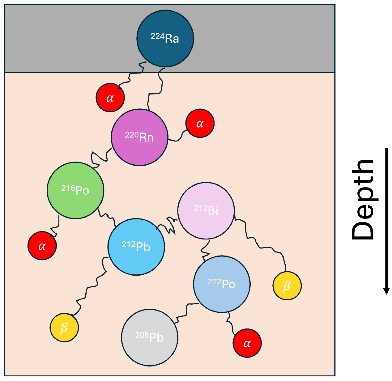
The DNA damage induced by 211At and 224Ra was simulated using Geant4 and Geant4-DNA, modeling both particle transport and nanoscale interactions within a cellular environment. The simulation process consists of two steps. First, the geometry of AlphaGlue, including radioactive decay and the generation of secondary particles, is modeled to calculate the particle spectrum at various distances from the source. This process is illustrated in Figure 4. Second, the DNA damage within a simplified cell nucleus is assessed based on this particle spectrum. Both stages of the simulation are performed using Geant4 (version 11.1) [31,32,33] and Geant4-DNA [34,35,36,37,38].

2.1. Decay Simulation

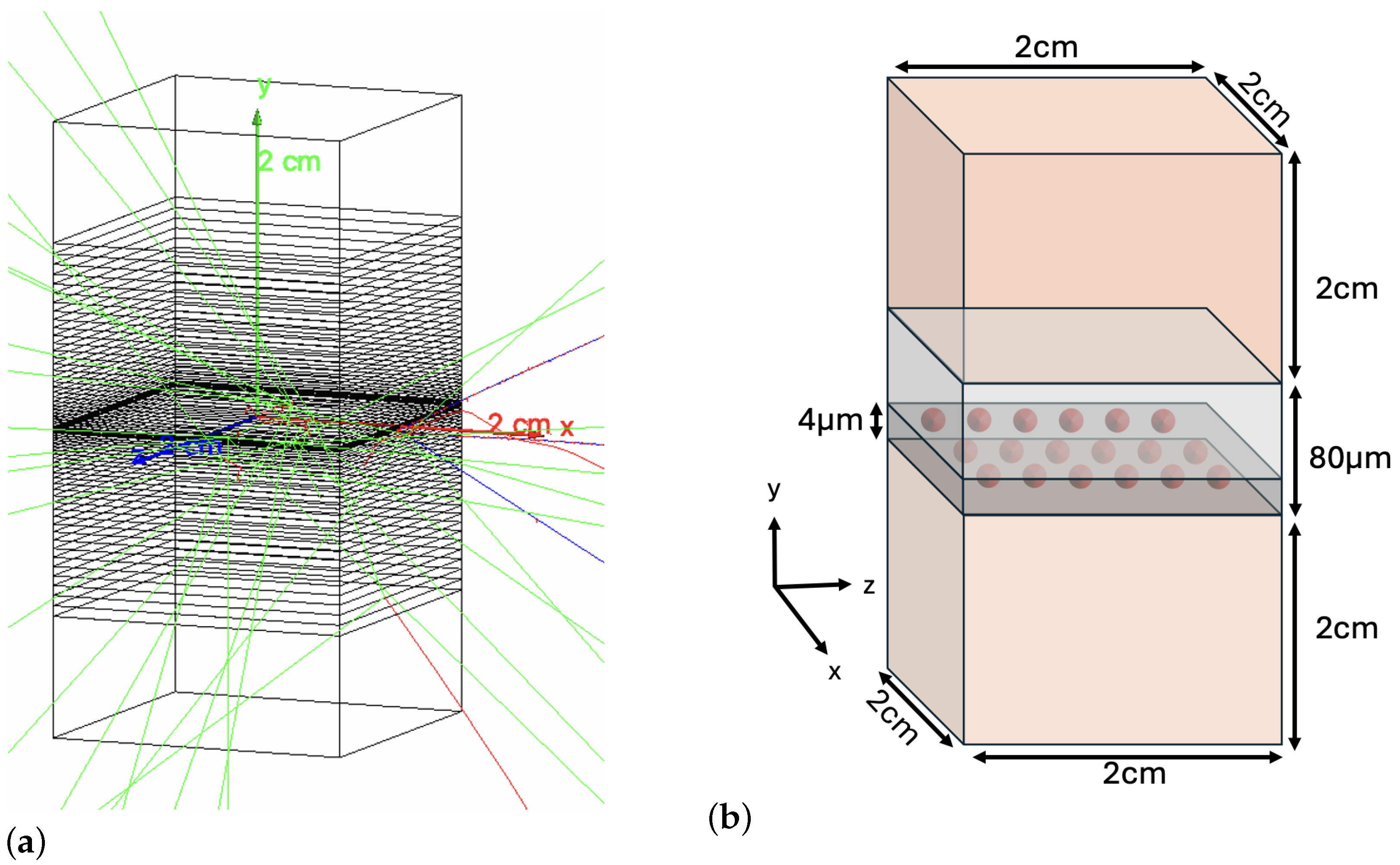
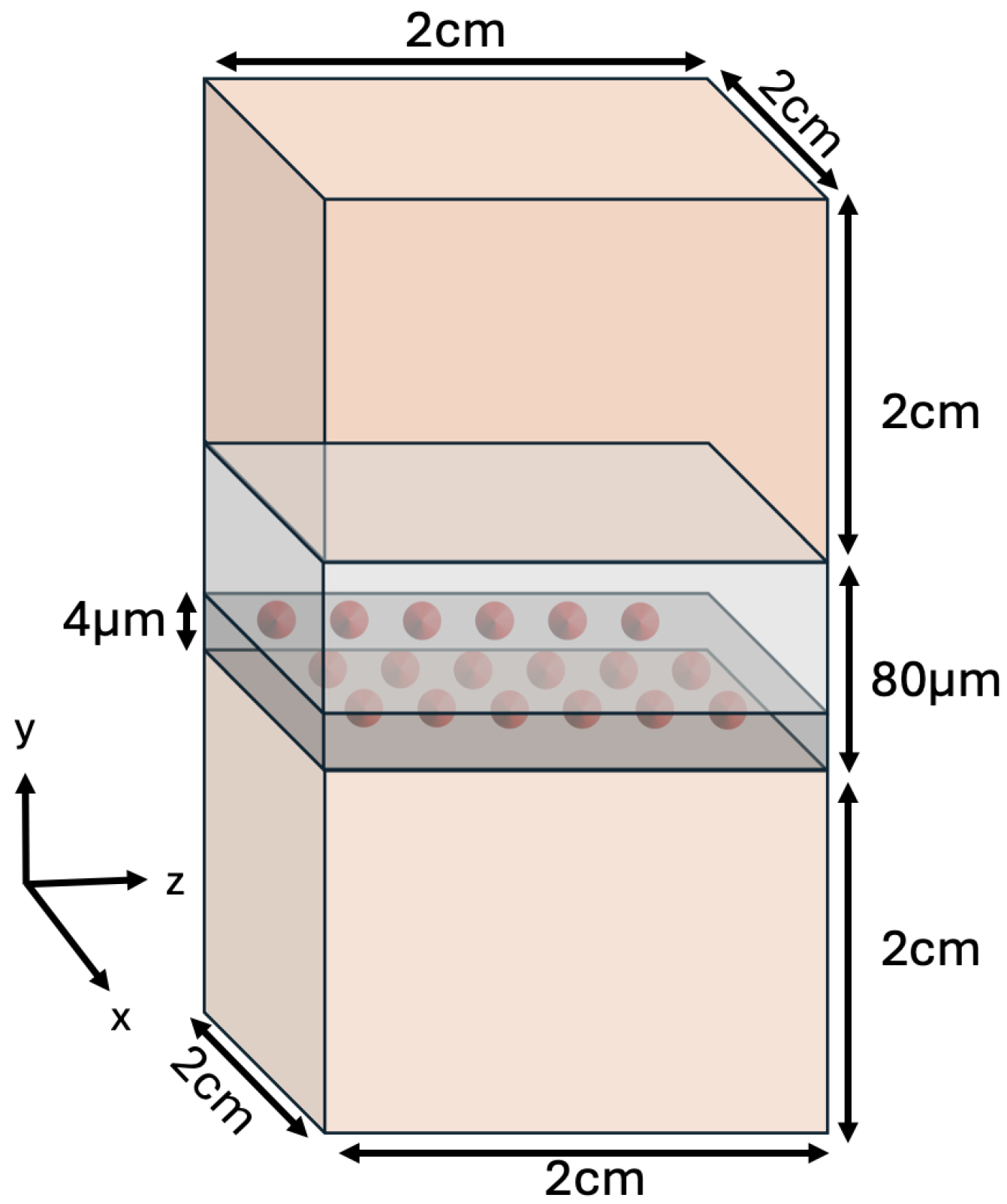
The AlphaGlue model shown in Figure 1, was simulated using Geant4, see Figure 5. The glue model was represented as a cuboid structure with a thickness of 80 μ m and dimensions of 20 mm in length and width. It was surrounded by two water boxes, the first positioned above and the second below, also measuring 20 × 20 × 20 mm3. These water boxes contained 300 nm-thick thin shell layers divided into a series of voxels, which acted as detectors to record all decay products and secondary particles from the initial decay at specified distances for use in the DNA simulation component (see Section 2.2). A 4 μ m layer of the glue was loaded, followed by a 76 μ m layer of unloaded glue on top to limit radiation exposure in cases where healthy tissue may be adjacent to the treated area. The DNA damage in the tumour was calculated in the bottom water box. The top water box is used to calculate the damage coming from radiation leaking through the unloaded glue.
As Geant4-DNA supports DNA damage calculations only in water, this medium was chosen [36,38,39,40]. Water is also widely used as the reference medium in skin dosimetry, for example VARSKIN [41,42] treats skin as a homogeneous block and uses absorbed energy distributions calculated in water. The same approach is applied and in ion-beam treatment planning, where soft tissues differ from water by less than 1% in stopping power ratio [41,43]. In addition, the diffusion constants for the isotopes are measured in water.
The G 4 E m P e n e l o p e P h y s i c s physics constructor was employed to describe electromagnetic interactions, with a low-energy production cutoff of 100 eV, corresponding to the model’s low-energy limit [44]. To simulate the radioactive decays in the decay chain, the G 4 R a d i o a c t i v e D e c a y P h y s i c s constructor was activated. The daughter nuclei from the decay of 224Ra which enter the tumour tissue diffuse. This diffusion is modeled as in [45]. The decay products are moved using a random walk using the diffusion constants D 220 R n = 1.12 × 10 3 mm2 s 1 , D 212 P b = 1.22 × 10 5 mm212 s 1 and D 212 B i = 6.78 × 10 7 mm2 s 1 from [46]. The 216Po and 212Po ions have relatively short half lives and therefore are assumed to decay at the same location as the parent nuclei. 212Pb nuclei leak out of the tumour via vascular routes through interaction with surrounding molecules such as proteins. 212Pb nuclei are removed at random, resulting in a leakage of 50%. The information for each particle entering the layer, including particle type, kinetic energy, initial position, and momentum, is calculated and saved in a phase space file. This file is used as input for the DNA damage simulation. Each particle is recorded in the phase space file upon entering the x or z face. This process is repeated for each particle track until the particle either exits the layer or loses all its kinetic energy. Secondary particles that exit one voxel and enter a neighboring voxel are saved to account for lateral scattering. Particles reentering the same voxel are not saved to avoid double counting in the DNA simulation. This is because scattered particles are explicitly tracked and accounted for within the DNA simulation.

2.2. DNA Damage Simulation

The DNA molecule is simulated within a 300 nm voxel, as shown in Figure 6. The DNA molecule structure was created using the “fractaldna” Python package (v0.6.0) [47], which generates short building blocks that can be repeated to create a continuous DNA structure.
Each straight chromatin fiber segment is 75 nm long, has a radius of 11 nm, and includes 33 nucleosomes. The positions of the deoxyribose-phosphate backbone (0.29 nm in diameter), histones (2.5 nm in diameter), and nucleotide base pairs are extracted from this model and represented as spheres, with nucleotide base pairs placed randomly with equal probability for each pair, achieving a density of 0.011 base pairs per cubic nm (bp/nm3), which is representative of a human cell nucleus [48].
The simulation tracks particles within the cell nucleus at the nanoscale using the Geant4-DNA low-energy extension cut of 100 eV. The G 4 E m D N A P h y s i c s _ o p t i o n 7 physics constructor was used, as in [11,45], as G 4 E m D N A P h y s i c s _ o p t i o n 7 combines low energy electron models with default Geant4-DNA electron models [37]. DNA damage yield is computed by simulating both direct and indirect DNA damage using a previously developed tool [49].
Direct and indirect damage can affect various components of DNA, including strand breaks in the double-helix backbone and damage to the bases. Strand breaks can result from both direct and indirect damage and are classified into three types: single-strand breaks (SSBs), double-strand breaks (DSBs), and complex double-strand breaks (cDSBs). We follow the classification of strand break clusters from [50]. As shown in Figure 7, SSBs occur when one strand of the DNA backbone is broken. These are typically non-lethal, as they are relatively easy for the cell to repair [51]. DSBs occur when both strands of the DNA backbone experience at least one break within 10 base pairs. Some DSBs are more complex, known as cDSBs, which involve additional strand breaks on either strand within 10 base pairs of the initial DSB. These cDSBs are the most challenging type of damage for the cell to repair [52].
For indirect damage caused by particle interactions with water molecules and the resulting radicals, the radicals are tracked for up to 5 ns. Radicals located more than 9 nm away from the deoxyribose-phosphate molecules are removed from the simulation [54,55,56].
To calculate the direct DNA damage, the energy deposited within a radius of 0.35 nm from the deoxyribose-phosphate molecule is summed, and the linear damage model is applied [57]. In this model, energy deposits below 5 eV are assumed to cause no damage, while for deposits above 37.5 eV the damage probability is 100%. For energy depositions between these values a linear probability of damage occurring is applied. Indirect damage strand breaks are assumed to only occur due to reactions between the hydroxyl radical and deoxyribose-phosphate backbone. A 40.5% probability of such a reaction resulting in a strand break is applied [45,54,55,58].

3. Results

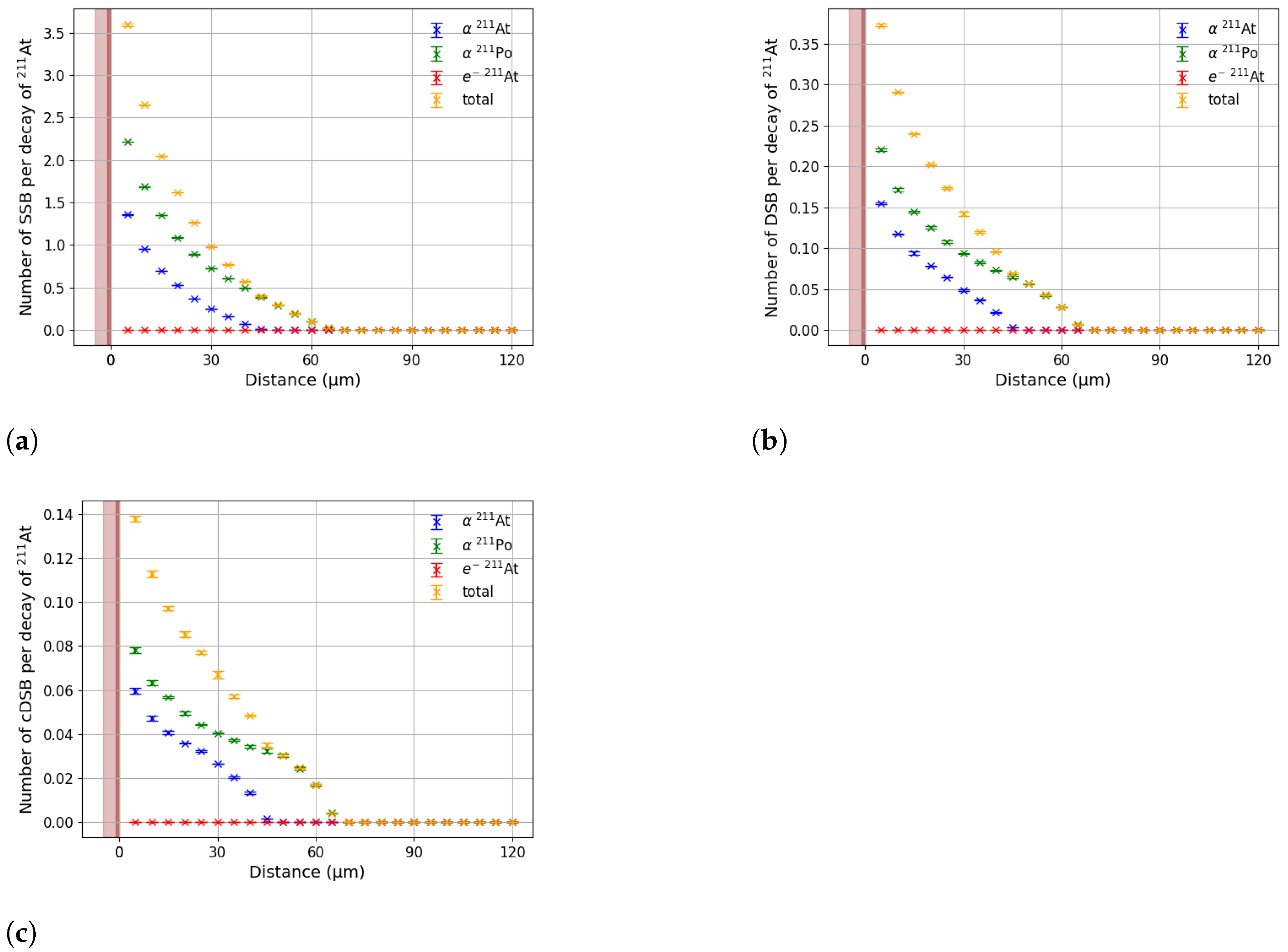
First the DNA damage when loading the 4 μ m thick interface layer with 211At was calculated. To load the glue, random locations were assigned for each of the 211At atoms based on a uniform distribution such that the 211At atoms were evenly spread throughout the 2 cm × 2 cm × 4 μ m volume. Figure 8 shows the number of SSBs, DSBs, and cDSBs as a function of depth for the 211At loaded layer. The shaded region represents the loaded glue, while the lighter region corresponds to the unloaded glue layer, which is applied to limit radiation exposure to any potential adjacent healthy tissue. DNA damage contributions from 207Bi were neglected due to its long half-life of approximately 38 years.
The results show that DNA damage is caused by α particles including all secondary delta electrons; the contribution of other electrons to breaks is negligible. 211At has two possible decay paths, as illustrated in Figure 2. The probability of decaying via 211Po is 58%, while the probability of decaying via 211At is 42%. This is the main reason why there is more damage coming from the α from 211Po compared to the α coming from 211At. In addition, the α produced in the 211Po decay has a higher energy of 7.45 MeV while the α originating in the 211At decay has an energy of 5.87 MeV, leading to a larger range for the α coming from the 211Po decay. The α from the 211At has a range of only just over 40 μ m in water, while the α from the 211Po has a range of just over 65 μ m. The slight bump in breaks towards the end of the range of each α is due to the Bragg peak.
On the opposite side of the glue, corresponding to the healthy tissue, no DNA damage is observed. This result confirms the treatment’s ability to confine the radiation effects to a short, localized range while minimizing exposure to healthy tissue. This is important when the treatment is used inside a body cavity.
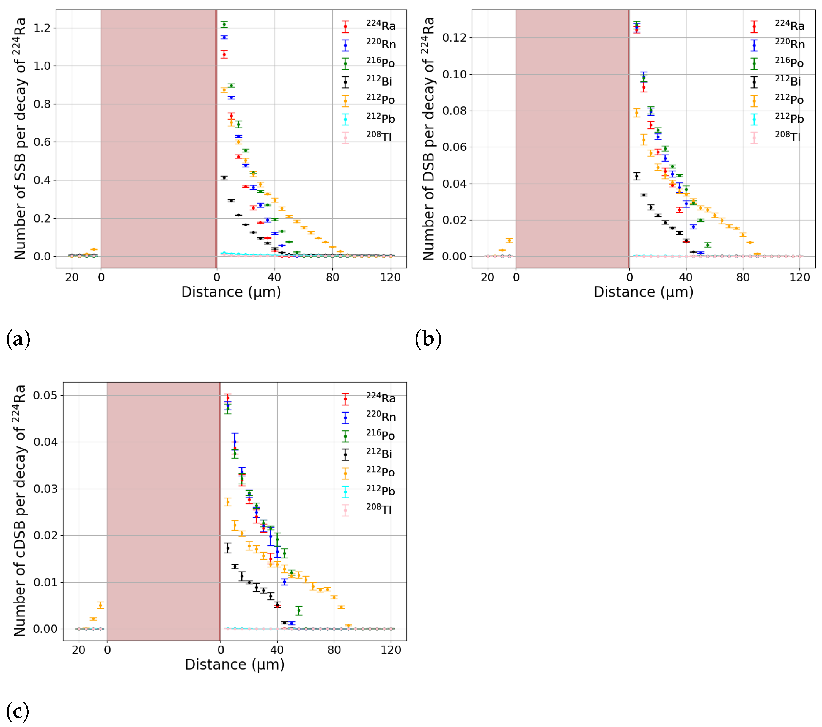
The results show that this treatment with 211At works, but the range of the damage in the tissue is too limited. An alternative would be loading the glue with another isotope like for example 224Ra. As mentioned before 224Ra has a decay cascade, as illustrated in Figure 3, which includes 220Rn. If the 220Rn enters the tissue, it will diffuse leading to a much deeper treatment range [49]. The results when loading the glue with 224Ra are presented in Figure 9. It shows the contribution to DNA damage from each isotope in the 224Ra decay chain as a function of distance, for single-strand breaks (SSBs), double-strand breaks (DSBs), and complex double-strand breaks (cDSBs). Most of the α ’s emitted have an energy of around 6–7 MeV and thus a similar range for their damage. The α emitted in the 211Po decay has an energy of 8.95 MeV. The range for DNA damage of this α stretches to 90 μ m. 212Pb and 208Tl only emit a β . Due to the sparse ionization along a β track compared to an α track, β s only contribute to the generation of SSBs. When looking at the unloaded side of the glue, only very little damage is observed, demonstrating that shielding with a thin layer of glue is sufficient to protect other tissue. Application of a thicker layer of glue is possible to increase the shielding.
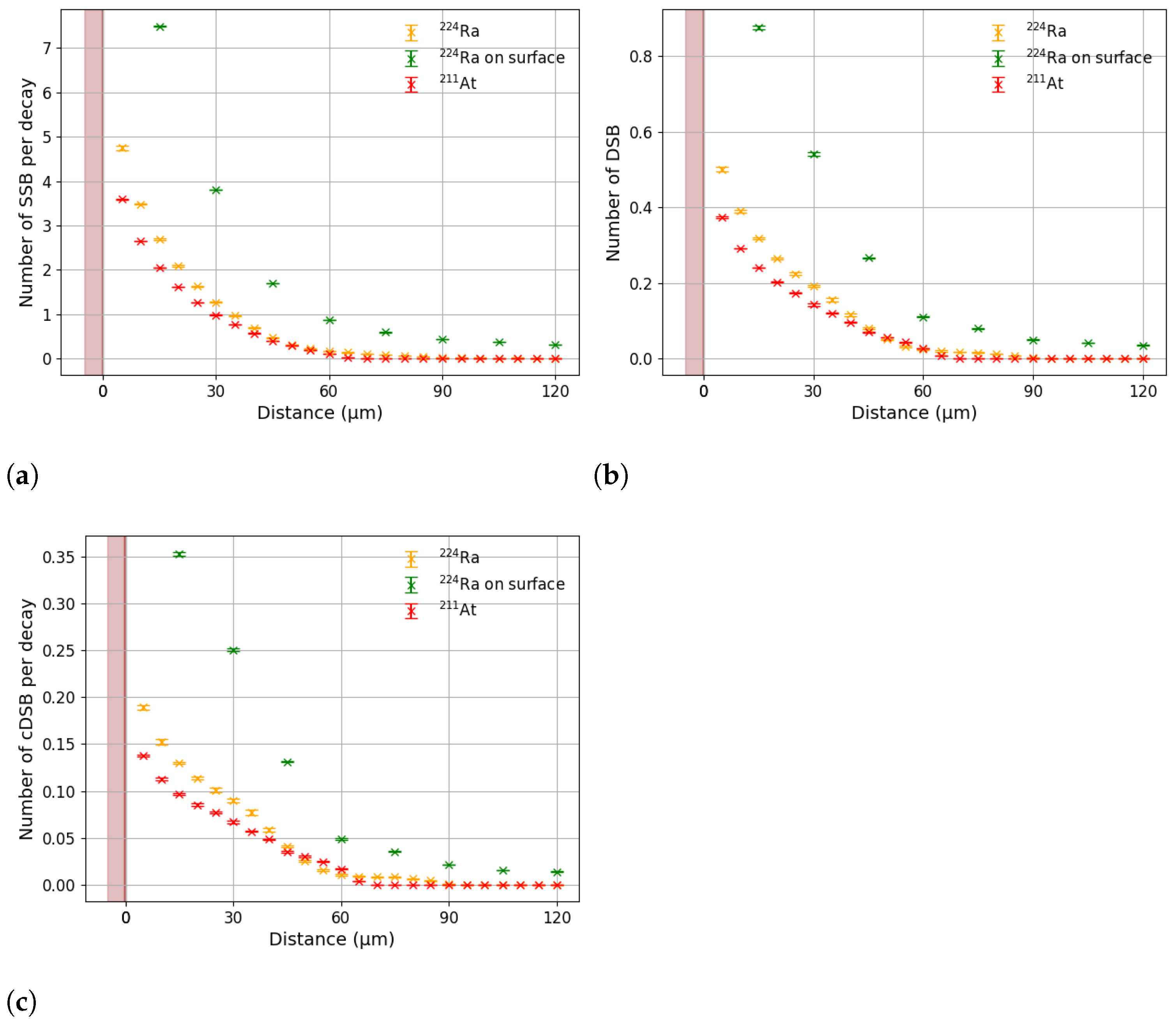
The results show that 224Ra induces a higher number of DNA breaks compared to 211At, as shown in Figure 10 as well, particularly at greater distances from the source. However, the maximum treatment depth using 224Ra in this configuration is still too shallow, ≲90 μ m, for clinical applications where a greater treatment depth is required. This is because only very few 220Rn isotopes can escape from the glue into the tissue. In DaRT, which uses a stainless steel seed, only isotopes that are located in the first ∼30 nm can escape into the tissue [59]. Given that a 4 μ m thick glue layer with the 224Ra uniformly spread throughout is used, a negligible number of 220Rn isotopes makes it into the tissue, limiting the treatment depth.
To enhance the effectiveness of the AlphaGlue treatment, in principle the 224Ra can be applied directly on the skin after which glue is applied on top to keep it in place and shield potential healthy tissue. Despite potential practical issues with this as a treatment, it is nevertheless worthwhile exploring the difference in performance, as it provides a natural limiting case for treatment and there are ways to achieve this or something similar in the clinic.
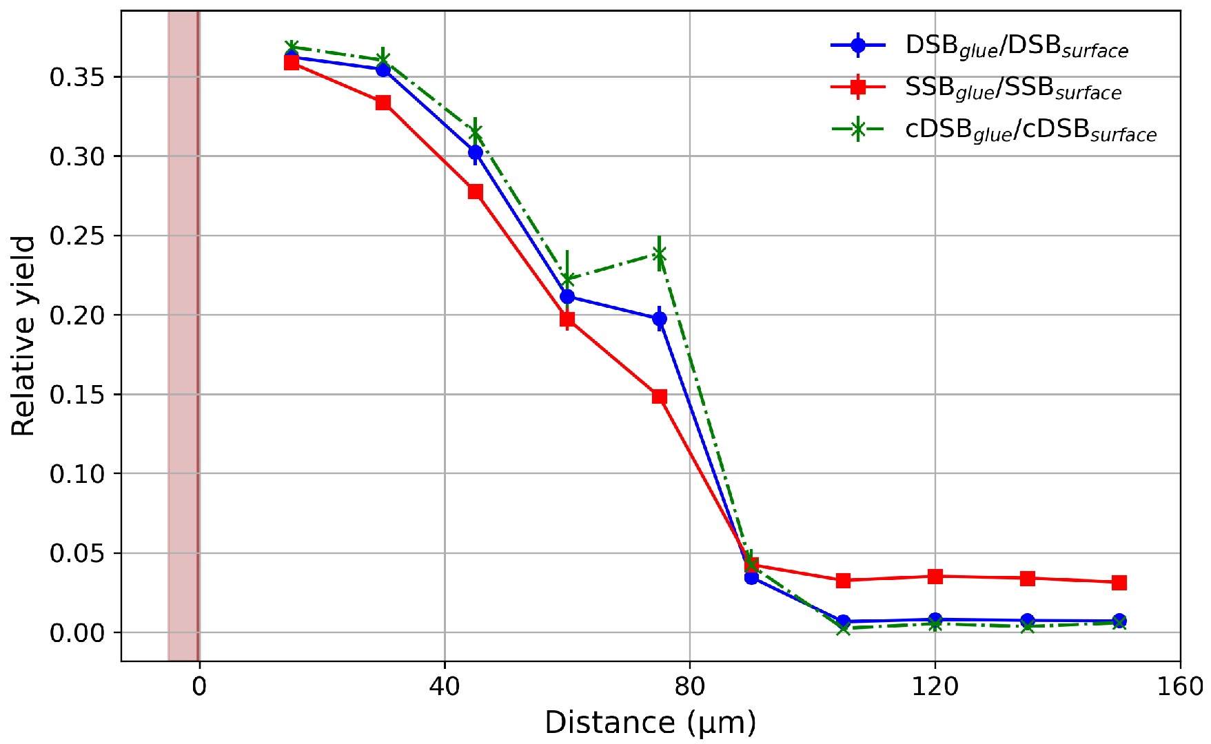
Figure 10 shows the DNA damage distributions for three cases: 224Ra-loaded glue, 211At-loaded glue and 224Ra directly applied on the surface. For the 224Ra-loaded glue and the 211At-loaded glue, the isotopes were uniformly loaded within the glue and evenly spread over the 2 × 2 cm2 area. For the 224Ra directly applied on the surface, the isotopes were uniformly spread over the 2 × 2 cm2 area. Surface application of 224Ra significantly increases DNA damage at greater distances compared to both embedded 224Ra and 211At by a factor 3–4 at small distances. The increase in damage when applying the 224Ra on the surface is not just by having more α particles entering the tissue. This can be seen in Figure 11, which shows the ratio of DNA damage occurring for the loaded glue and the DNA damage occurring for the case of applying 224Ra directly on the surface. For all three types of damage, this ratio is not a constant with depth as it would be if it is just an effect of the number of α ’s, but it decreases rapidly, demonstrating that applying 224Ra directly on the surface yields more damage at depth. The increase in damage at depth for 224Ra on the surface originates from more 220Rn entering the tissue and diffusing before decaying. This enhances the depth profile of the treatment significantly. Figure 12 shows the number of DSBs and cDSBs as a function of depth for surface-applied 224Ra over a much larger depth range. The results show that the DNA damage can extend beyond 2–4 mm beneath the skin [60] making it feasible as a skin cancer treatment.

3.1. Effective Treatment Depth

224Ra applied on the glue surface produced the most extended DNA damage profile. The actual amount of DNA damage at a certain depth depends on the number of 224Ra isotopes used; enough 224Ra isotopes need to be provided to get enough DNA damage at depth to eradicate the tumour. When using photon therapy, tumours can usually be eradicated by a single fraction of photons amounting to a typical dose of 20–25 Gy [61]. The damage per dose for α particles is much higher than for photons. This is expressed in the relative biological effectiveness (RBE). RBE is defined as
RBE = D end point / photon D end point / quantity
where the endpoint is a biological quantity of interest. In this study two end points DSBs and cDSBs have been used. 60Co was used as the photon reference [45]. The RBE-weighted dose, D R B E is the product of the RBE and the absorbed dose [62]. The D R B E allows dose thresholds to be compared between treatment types.
D RBE = RBE × D
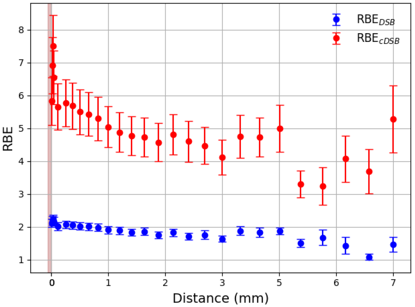
Figure 13 shows the RBE as a function of distance from the surface, calculated using both DSB and cDSB yields relative to a 60Co reference. The 60Co simulation was performed by irradiating a spherical water volume with photons of 1.17 and 1.33 MeV energies at 5 mm from the centre of the DNA target ensuring charged particle equilibrium [63]. This simplified spherical isotropic setup was chosen to provide a reproducible photon reference, consistent with previous Geant4-DNA studies [11,45]. In contrast, α -particles deposit their dose over sub-mm ranges without reaching equilibrium, which necessitates a different geometry for the AlphaGlue calculations. R B E D S B and R B E c D S B were then obtained by comparing the DSB and complex DSB yields from 224Ra on the surface to those from 60Co. Near the source, R B E c D S B reaches up to 7, drops to 5 at 1 mm and then falls slowly at larger distances but remains between 4 and 5. At small distances, the R B E D S B is just over 2 and drops to 2 around 1 mm. After 1 mm, the value changes but remains between 1 and 2.
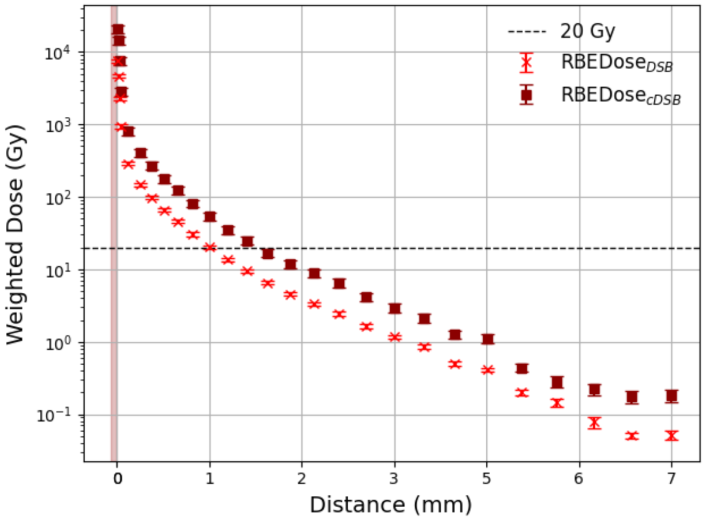
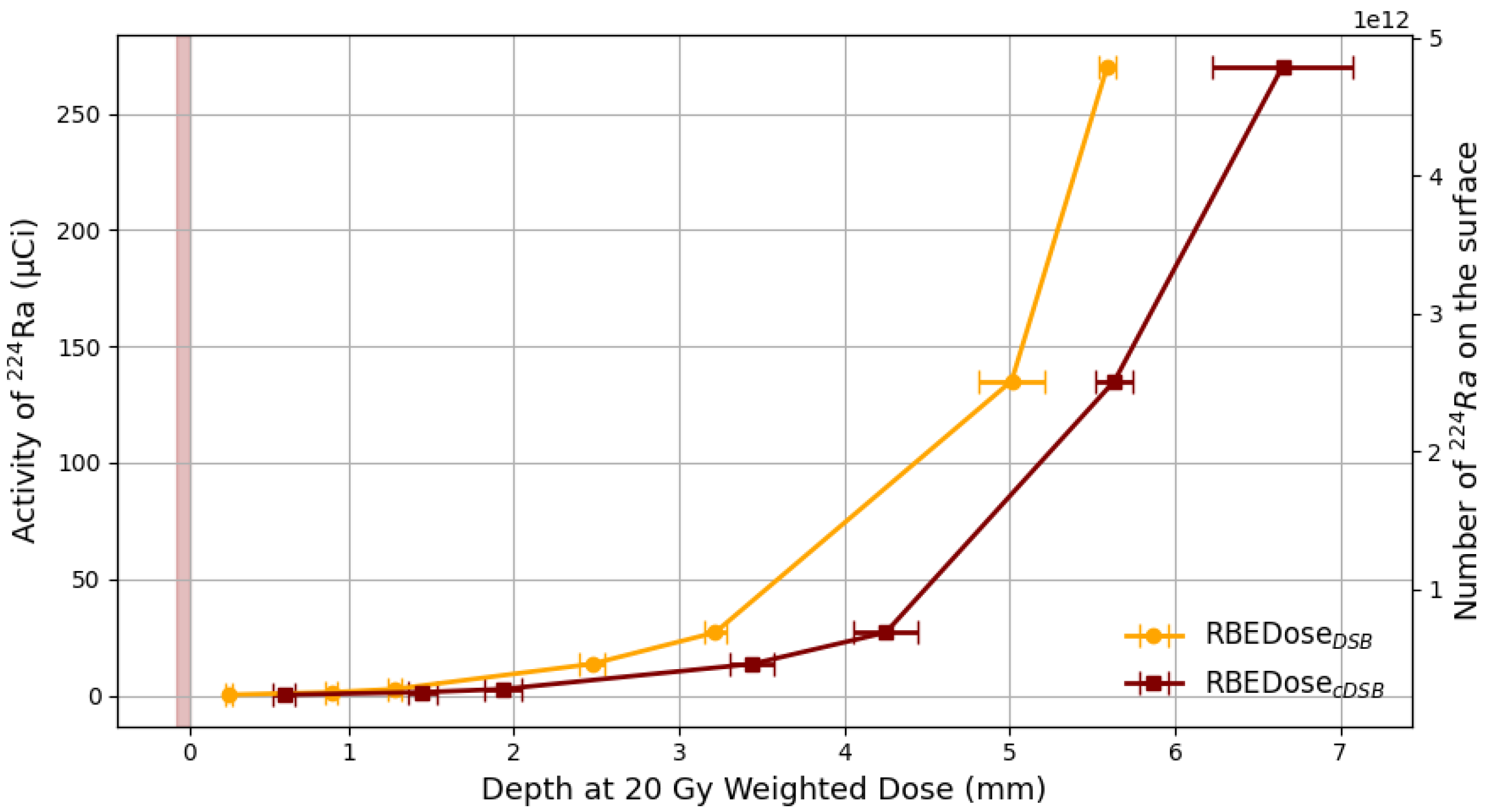
Figure 14 shows the D R B E for AlphaGlue loaded with 3 μ Ci of 224Ra, which corresponds to 5.06 × 10 10 224Ra isotopes, at the surface. It is clear that the weighted dose decreases sharply with depth and reaches the 20 Gy threshold at approximately 1 mm for the end point using DSBs and at approximately 1.6 mm for cDSBs. This depth can be tuned by selecting a different number of 224Ra isotopes. This is shown in Figure 15, where the 20 Gy depth is shown for different numbers of 224Ra isotopes. The results show that tumour eradication can happen at clinically relevant depths of up to 5 mm by using ( 1.74 ± 0.08 ) × 10 12  224Ra atoms evenly spread over the 2 × 2 cm2 area on the skin surface corresponding to an activity of ( 87 ± 5 )   μ Ci when using the cDSBs for the RBE calculation and ( 2.6 ± 0.2 ) × 10 12  224Ra atoms evenly spread over the 2 × 2 cm2 area on the skin surface corresponding to an activity of ( 135 ± 13 )   μ Ci when using the DSBs for the RBE calculation. This is equivalent to a 224Ra atom concentration of ( 4.35 ± 0.2 ) × 10 11 / cm2 or an activity of ( 21.8 ± 1 ) μ Ci/cm2 when using the cDSBs for the RBE calculation and ( 6.50 ± 0.5 ) × 10 11 / cm2 or an activity of ( 34 ± 3 ) μ Ci/cm2 when using the DSBs for the RBE calculation.
Given the rapidly falling weighted dose versus depth curve, the treatment depth can be tuned with good precision by decreasing the number of 224Ra isotopes. The number of required 224Ra isotopes to achieve cell death at clinically relevant depths is not excessive; a vial of Xofigo [64], which is an approved α therapy exploiting 223Ra, that is used to treat adults with cancer of the prostate when the cancer has spread to the bones [65,66], contains 9.4 × 10 12 223Ra atoms.

3.2. Comparison with DaRT

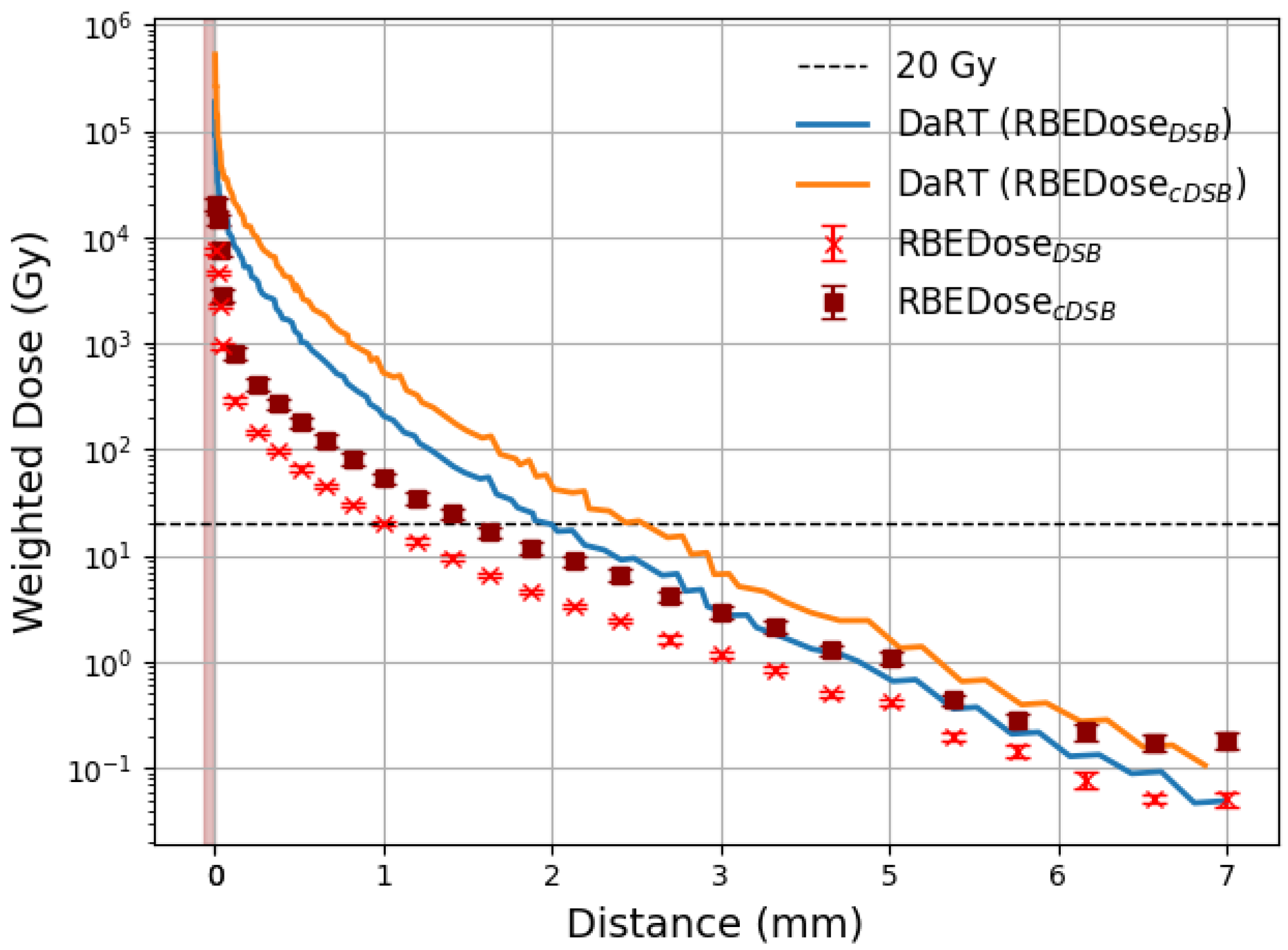
Both DaRT and AlphaGlue use 224Ra but the delivered radiation pattern is different due to the different source geometry. Figure 16 shows a comparison of the RBE weighted dose as a function of depth. The RBE weighted dose for DaRT was calculated in [45]. For AlphaGlue the 224Ra activity is spread over a larger area while in DaRT the 224Ra activity is spread over the small surface of the seed. The initial drop in weighted dose is due to the α ’s produced in the 224Ra decay and subsequent decays due to 220Rn isotopes and their daughter isotopes that do not enter the tissue and thus not diffuse. This is limited to a depth ≲100 μ m. Subsequently, the weighted dose drops faster for DaRT than for AlphaGlue. This is because the initial emission for the cylindrical seed leads to a particle density decrease following ∼ 1 R in addition to the decrease due to particle decay. The RBE weighted dose in AlphaGlue decreases mainly due to particle decay. Hence, it drops slower than for DaRT and the RBE weighted dose graphs become parallel when the decrease is dominated by the decay of the diffused particles. At a radial distance of 5 mm from the seed centre, the RBE cDSB -weighted dose decreases to approximately 1 Gy. In our AlphaGlue surface model, the RBE cDSB -weighted dose at 5 mm is likewise about 1 Gy. This agreement at depth, despite the different geometries (interstitial seed versus surface application), provides further confidence in the conceptual validity of the AlphaGlue approach and opens the pathway towards subsequent in-vitro and, eventually, in-vivo investigations that will allow the proposed solution to be assessed in a more realistic context.

4. Discussion

This study focused on the characterisation of a novel α -therapy delivery concept, called AlphaGlue. In AlphaGlue, α -emitters are suspended in glue which is directly applied to the skin-tissue. A layer of unloaded glue can be applied on top to shield the radiation. A Monte Carlo simulation study using GEANT4 and GEANT4-DNA was done to calculate the DNA damage as a function of depth. A configuration with a 4 μ m thick loaded glue layer covered with a 76 μ m layer of unloaded glue was explored. Two isotopes, 211At and 224Ra, were studied. The results showed that, in the standard loaded glue configuration, DNA damage induced by 211At was confined to approximately 70 μ m, while using 224Ra extended the damage range slightly further to around 90 μ m from the glue layer. The key reason for the limited range is that there is only damage due to the emission of α particles that enter the tissue. Applying 224Ra on the surface of the tissue and using a glue layer to keep it in place and shield potential tissue on the far side, significantly enhanced the treatment depth, with DSB and cDSB damage produced beyond 5 mm into the tissue; a depth exceeding the clinical requirements for nasal carcinoma treatment.
The calculated activity provides a reference for how much material would be required to achieve a clinically relevant dose at depth. For instance, an activity of (34 ± 3) μ Ci/cm2 was needed to reach 20 Gy at 5 mm in our model, which is comparable to the activity levels of existing clinical treatments such as Xofigo [64], which exploits 223Ra dichloride, administered at approximately 30 μ Ci (1100 kBq/mL) for prostate cancer. This supports the plausibility of the AlphaGlue concept. Specifically, to achieve cell death at 5 mm depth, a 224Ra atom concentration of (4.35 ± 0.2) × 1011/cm2, corresponding to (21.8 ± 1) μ Ci/cm2 using cDSB-based RBE, or (6.50 ± 0.5) × 1011/cm2, corresponding to (34 ± 3) μ Ci/cm2 using DSB-based RBE, is required.

Looking Towards the Future

The presented study is based solely on Monte Carlo simulations and designed to explore the feasibility of the AlphaGlue concept in ideal circumstances. A direct comparison with DaRT [45], see Section 3.2, further shows that both approaches can deliver an R B E c D S B -weighted dose of approximately 1 Gy at 5 mm, despite their different geometries (interstitial seed vs. surface application). This shows that the AlphaGlue concept can yield similar clinical outcomes as a successful alternative α -particle based treatment for superficial tumours, highlighting the clinical relevance of the concept explored here.
In-vitro experimental studies are being planned, and improvements to the models will include the implementation of more realistic skin geometries and the incorporation of heterogeneous tissue layers.
Towards clinical deployment, many safety and engineering aspects need to be addressed. However, comparable approaches such as β -emitting skin patches have already demonstrated that radionuclide delivery to superficial tumours is clinically feasible and safe [20,21,22,23,24,25]. Clinical deployment of DaRT, which also relies on 224Ra, has already demonstrated that this isotope can be used safely and effectively for skin cancer [8,18]. Similarly, Xofigo, a 223Ra based α -therapy, is widely used in routine practice [65,66]. A practical approach for AlphaGlue could involve a two-component glue system, with an additional unloaded protective layer on top. One of the glue components would be pre-mixed with the 224Ra. The contents of the vial with the pre-mixed component can be sucked into a syringe which already has the second glue component in it. Simulations show that an 76 μ m unloaded glue layer on top of the loaded glue is sufficient to contain α -radiation, while a thin gas-tight foil can prevent 220Rn escape. Such a procedure would be conceptually similar to existing deployment of Xofigo, and the short range of α -radiation makes it straightforward to ensure safety for both patients and medical staff. Furthermore, the decay of 224Ra (half-life 3.6 days) means that if the glue remains in place for a few weeks, residual activity falls to negligible levels, avoiding long-term radioactive waste.

5. Conclusions

A key challenge in α therapy is how to deliver the α emitters to the tumour. In this study, the effectiveness of a novel treatment delivery concept, AlphaGlue, is evaluated. In AlphaGlue the α emitters are delivered using a thin layer of glue that is put on top of the tumour. This may offer a less invasive and potentially easier-to-use alternative, which could be particularly advantageous for sensitive areas such as facial skin cancers, but also anal, vaginal cancer or endometriosis. The DNA damage as a function of depth is calculated using GEANT4 and GEANT4-DNA simulations using 221At and 224Ra. It is shown that significant cell death at 5 mm into the tissue can be achieved when applying a 224Ra atom concentration of ( 4.35 ± 0.2 ) × 10 11 / cm2, which corresponds to a local activity of ( 21.8 ± 1 ) μ Ci/cm2, when using the cDSBs for the RBE calculation and ( 6.50 ± 0.5 ) × 10 11 / cm2, which corresponds to a local activity of ( 34 ± 3 ) μ Ci/cm2, on the skin surface when using the DSBs for the RBE calculation. This study has shown that there is good potential to develop the AlphaGlue style treatment further and opens the pathway to in-vitro experiments.

Author Contributions

Conceptualization, L.A.S., M.D. and J.V.; methodology, L.A.S., L.B. and C.D.S.; software, L.A.S., L.B., C.D.S., Y.S. and J.D.; validation, L.A.S., L.B., C.D.S., M.D., Y.S. and J.D.; formal analysis, L.A.S.; investigation, L.A.S., M.D. and J.V.; resources, J.V.; data curation, L.A.S.; writing—original draft preparation, L.A.S., M.D. and J.V.; writing—review and editing, A.C., S.G. and J.V.; visualization, L.A.S.; supervision, S.G., D.S., J.V. and A.R.; project administration, J.V.; funding acquisition, L.A.S. and J.V. All authors have read and agreed to the published version of the manuscript.

Funding

This work was supported by the Custodian of the Two Holy Mosques Scholarship Program, provided by the Saudi government.

Data Availability Statement

The data presented in this study are available on request from the corresponding author.

Acknowledgments

The authors gratefully acknowledge Hormuud Telecom and Innovii Digital Service for granting access to supplementary computing resources at their data center in Mogadishu, which enabled the completion of the simulation work presented in this study. Special thanks are also extended to Transparency Solutions for their ongoing efforts in facilitating and supporting the collaboration between the University of Bristol and both Hormuud Telecom and Innovii Digital Service. This research also benefited from the high-performance computing and data storage infrastructure of the Advanced Computing Research Centre at the University of Bristol.

Conflicts of Interest

The authors declare no conflicts of interest.

Abbreviations

The following abbreviations are used in this manuscript:
LETlinear energy transfer
TATTargeted Alpha Therapy
DaRTDiffusing α -emitters Radiation Therapy
SSBSingle Strand Break
DSBDouble Strand Break
cDSBComplex Double Strand Break
RBErelative biological effectiveness

References

  1. Zhang, J.; Qin, S.; Yang, M.; Zhang, X.; Zhang, S.; Yu, F. Alpha-emitters and targeted alpha therapy in cancer treatment. iRADIOLOGY 2023, 1, 245–261. [Google Scholar] [CrossRef]
  2. Jang, A.; Kendi, A.T.; Johnson, G.B.; Halfdanarson, T.R.; Sartor, O. Targeted alpha-particle therapy: A review of current trials. Int. J. Mol. Sci. 2023, 24, 11626. [Google Scholar] [CrossRef]
  3. Jalloul, W.; Ghizdovat, V.; Stolniceanu, C.; Ionescu, T.; Grierosu, I.; Pavaleanu, I.; Moscalu, M.; Stefanescu, C. Targeted Alpha Therapy: All We Need to Know about 225Ac’s Physical Characteristics and Production as a Potential Theranostic Radionuclide. Pharmaceuticals 2023, 16, 1679. [Google Scholar] [CrossRef] [PubMed]
  4. Ma, J.; Li, L.; Liao, T.; Gong, W.; Zhang, C. Efficacy and Safety of 225Ac-PSMA-617-Targeted Alpha Therapy in Metastatic Castration-Resistant Prostate Cancer: A Systematic Review and Meta-Analysis. Front. Oncol. 2022, 12, 796657. [Google Scholar] [CrossRef] [PubMed]
  5. Alvarez, N.H.; Bauer, D.; Michel, A.L.; Carter, L.M.; Lewis, J.S. Targeted Alpha Therapy with 225Ac-Macropa-Isatuximab for CD38-positive Hematological Malignancies. bioRxiv 2024. [Google Scholar]
  6. Schatz, C.; Zitzmann-Kolbe, S.; Moen, I.; Klotz, M.; Nair, S.; Stargard, S.; Bjerke, R.M.; Biseth, K.W.; Feng, Y.Z.; Indrevoll, B.; et al. Preclinical Efficacy of a PSMA-Targeted Actinium-225 Conjugate (225Ac-Macropa-Pelgifatamab): A Targeted Alpha Therapy for Prostate Cancer. Clin. Cancer Res. 2024, 30, 2531–2544. [Google Scholar] [CrossRef]
  7. Li, L.; Rousseau, J.; Jaraquemada-Peláez, M.d.G.; Wang, X.; Robertson, A.; Radchenko, V.; Schaffer, P.; Lin, K.S.; Bénard, F.; Orvig, C. 225Ac-H4py4pa for Targeted Alpha Therapy. Bioconjug. Chem. 2021, 32, 1348–1363. [Google Scholar] [CrossRef]
  8. D’Andrea, M.A.; VanderWalde, N.A.; Ballo, M.T.; Patra, P.; Cohen, G.N.; Damato, A.L.; Barker, C.A. Feasibility and Safety of Diffusing Alpha-Emitter Radiation Therapy for Recurrent or Unresectable Skin Cancers. JAMA Netw. Open 2023, 6, e2312824. [Google Scholar] [CrossRef] [PubMed]
  9. Miller, S.; Lecavalier-Barsoum, M.; Ma, K.; Santos Dutra, M.; Kaitoukov, Y.; Bahoric, B.; Tomic, N.; Dinelle, F.; Enger, S.; Batist, G.; et al. Feasibility and safety of endoscopic ultrasound-guided diffusing alpha emitter radiation therapy for advanced pancreatic cancer: Preliminary data. Endosc. Int. Open 2024, 12, E1085–E1091. [Google Scholar] [CrossRef]
  10. Albertsson, P.; Bäck, T.; Bergmark, K.; Hallqvist, A.; Johansson, M.; Aneheim, E.; Lindegren, S.; Timperanza, C.; Smerud, K.; Palm, S. Astatine-211 based radionuclide therapy: Current clinical trial landscape. Front. Med. 2023, 9, 1076210. [Google Scholar] [CrossRef] [PubMed]
  11. De Sio, C.; Ballisat, L.; Beck, L.; Guatelli, S.; Sakata, D.; Shi, Y.; Duan, J.; Sabah, L.A.; Velthuis, J.; Rosenfeld, A. Targeted alpha therapies using 211At: A Geant4 simulation of dose and DNA damage. Phys. Medica 2025, 129, 104860. [Google Scholar] [CrossRef]
  12. Ghosh, S.; Banerjee, D.; Guleria, A.; Chakravarty, R. Production, purification and formulation of nanoradiopharmaceutical with 211At: An emerging candidate for targeted alpha therapy. Nucl. Med. Biol. 2024, 138–139, 108947. [Google Scholar] [CrossRef]
  13. Batra, V.; Samanta, M.; Makvandi, M.; Groff, D.; Martorano, P.; Elias, J.; Ranieri, P.; Tsang, M.; Hou, C.; Li, Y.; et al. Preclinical Development of 211At meta-astatobenzylguanidine (211At-MABG) as an Alpha Particle Radiopharmaceutical Therapy for Neuroblastoma. Clin. Cancer Res. 2022, 28, 4146–4157. [Google Scholar] [CrossRef] [PubMed]
  14. Kashiyama, R.; Watanabe, H.; Akasaka, T.; Fujimoto, H.; Murakami, M.; Ooe, K.; Toyoshima, A.; Nakashima, K.; Ono, M. Feasibility of Targeted Alpha Therapy for Alzheimer’s Disease Using 211At-labeled Agent Targeting Amyloid-β Aggregates. ChemRxiv 2025. [Google Scholar] [CrossRef] [PubMed]
  15. Bellia, S.; Feliciani, G.; Del Duca, M.; Monti, M.; Turri, V.; Sarnelli, A.; Romeo, A.; Kelson, I.; Keisari, Y.; Popovtzer, A.; et al. Clinical evidence of abscopal effect in cutaneous squamous cell carcinoma treated with diffusing alpha emitters radiation therapy: A case report. J. Contemp. Brachyther. 2019, 11, 449–457. [Google Scholar] [CrossRef]
  16. Popovtzer, A.; Rosenfeld, E.; Mizrachi, A.; Bellia, S.R.; Ben-Hur, R.; Feliciani, G.; Sarnelli, A.; Arazi, L.; Deutsch, L.; Kelson, I.; et al. Initial Safety and Tumor Control Results From a “First-in-Human” Multicenter Prospective Trial Evaluating a Novel Alpha-Emitting Radionuclide for the Treatment of Locally Advanced Recurrent Squamous Cell Carcinomas of the Skin and Head and Neck. Int. J. Radiat. Oncol. Biol. Phys. 2020, 106, 571–578. [Google Scholar] [CrossRef]
  17. Yang, G.; Harrison, L. A Hard Target Needs a Sharper DaRT. Int. J. Radiat. Oncol. Biol. Phys. 2020, 107, 152–153. [Google Scholar] [CrossRef]
  18. Popovtzer, A.; Mizrachi, A.; D’Andrea, M.; VanderWalde, N.A.; Kurman, N.; Rosenfeld, E.; Ben-Hur, R.; Bellia, S.R.; Feliciani, G.; Silvern, D.; et al. Extended Follow-Up Outcomes from Pooled Prospective Studies Evaluating Efficacy of Interstitial Alpha Radionuclide Treatment for Skin and Head and Neck Cancers. Cancers 2024, 16, 2312. [Google Scholar] [CrossRef]
  19. Yoshimura, R.; Toda, K.; Watanabe, H.; Miura, M.; Notake, R.; Murakami, N.; Igaki, H.; Nakamura, S.; Umezawa, R.; Kadoya, N.; et al. Efficacy and safety of diffusing alpha-emitter radiation therapy (DaRT) for head and neck cancer recurrence after radiotherapy. Int. J. Clin. Oncol. 2025, 30, 893–903. [Google Scholar] [CrossRef]
  20. Pashazadeh, A.; Landes, R.; Boese, A.; Kreissl, M.; Klopfleisch, M.; Friebe, M. Superficial skin cancer therapy with Y-90 microspheres: A feasibility study on patch preparation. Skin Res. Technol. 2020, 26, 25–29. [Google Scholar] [CrossRef]
  21. Finger, P. Yttrium-90 Episcleral Plaque Brachytherapy for Choroidal Melanoma. J. Vitreoretin. Dis. 2024, 8, 210–214. [Google Scholar] [CrossRef]
  22. Baxi, S.; Vohra, S.; Hong, A.; Mulholland, N.; Heuschkel, M.; Dahlhoff, G.; Cardaci, G.; Mirzaei, S.; Sathekge, M. Effectiveness and Patient Experiences of Rhenium Skin Cancer Therapy for Nonmelanoma Skin Cancer: Interim Results from the EPIC-Skin Study. J. Nucl. Med. 2024, 65, 1450–1455. [Google Scholar] [CrossRef]
  23. Cipriani, C.; Desantis, M.; Dahlhoff, G.; Brown, S.D., III; Wendler, T.; Olmeda, M.; Pietsch, G.; Eberlein, B. Personalized irradiation therapy for NMSC by rhenium-188 skin cancer therapy: A long-term retrospective study. J. Dermatol. Treat. 2022, 33, 969–975. [Google Scholar] [CrossRef]
  24. Cardaci, G.; Baxi, S.; Vohra, S.; Allison, C.; Hong, A.; Mulholland, N.; Sathekge, M.; Mokoala, K.; Heuschkel, M.; Tietze, J.; et al. Efficacy, Safety, and Patient Reported Outcomes of Rhenium-Skin Cancer Therapy for Non-Melanoma Skin Cancer: 1-Year Results from the EPIC-Skin Study. Adv. Radiat. Oncol. 2025, 10, 101802. [Google Scholar] [CrossRef] [PubMed]
  25. Krönert, M.; Schwarzenböck, S.; Kurth, J.; Heuschkel, M.; Krause, B.J.; Emmert, S.; Tietze, J.K. Patient-Orientated Evaluation of Treatment of Non-Melanoma Skin Cancer with Rhenium-188 Compared to Surgery. Healthcare 2024, 12, 921. [Google Scholar] [CrossRef] [PubMed]
  26. Hari Hara Sudhan, N.; Pandit, N.; Kumari, R.; Sunitha, V. Non-invasive treatment of recalcitrant keloids with contact brachytherapy using Phosphorus 32 skin patch and its effectiveness: A pilot study. J. Nucl. Med. 2023, 64 (Suppl. 1), 579. [Google Scholar]
  27. Jiang, P.; Purtskhvanidze, K.; Kandzia, G.; Neumann, D.; Luetzen, U.; Siebert, F.; Roider, J.; Dunst, J. 106Ruthenium eye plaque brachytherapy in the management of medium sized uveal melanoma. Radiat. Oncol. 2020, 15, 183. [Google Scholar] [CrossRef]
  28. Antohe, M.; Coman, A.; Turcu, G.; Nedelcu, R.I.; Brinzea, A.; Balaban, M.; Moroianu, A.; Manea, L.; Hulea, I.; Balasescu, E.; et al. The prognostic significance of the clinical and histological parameters in primary cutaneous melanoma patients. Med. Pharm. Rep. 2022, 95, 229–235. [Google Scholar] [CrossRef]
  29. Chen, M.L.; de Vere Hunt, I.J.; John, E.M.; Weinstock, M.A.; Swetter, S.M.; Linos, E. Differences in Thickness-Specific Incidence and Factors Associated With Cutaneous Melanoma in the US From 2010 to 2018. JAMA Oncol. 2022, 8, 755–759. [Google Scholar] [CrossRef]
  30. Buxton, G.V.; Greenstock, C.L.; Phillips Helman, W.; Ross, A.B. Critical review of rate constants for reacitons of hydrated electrons. J. Phys. Chem. Ref. Data 1988, 17. [Google Scholar] [CrossRef]
  31. Ivanchenko, V.; Jones, F.; Jun, S.; Kaitaniemi, P.; Karakatsanis, N. Recent developments in Geant4. Nucl. Instruments Methods Phys. Res. A 2016, 835, 186–225. [Google Scholar]
  32. Allison, J.; Amako, K.; Apostolakis, J.; Araujo, H.; Dubois, P.A.; Asai, M.; Barrand, G.; Capra, R.; Chauvie, S.; Chytracek, R.; et al. Geant4 developments and applications. IEEE Trans. Nucl. Sci. 2006, 53, 270–278. [Google Scholar] [CrossRef]
  33. Agostinelli, S.; Allison, J.; Amako, K.; Apostolakis, J.; Araujo, H.; Arce, P.; Asai, M.; Axen, D.; Banerjee, S.; Barrand, G.; et al. Geant4–a simulation toolkit. Nucl. Instruments Methods Phys. Res. Sect. A 2003, 506, 250–303. [Google Scholar] [CrossRef]
  34. Incerti, S.; Baldacchino, G.; Bernal, M.; Capra, R.; Champion, C.; Francis, Z.; Guèye, P.; Mantero, A.; Mascialino, B.; Moretto, P.; et al. The Geant4-DNA project. Int. J. Model. Simul. Sci. Comput. 2010, 1, 157–178. [Google Scholar] [CrossRef]
  35. Incerti, S.; Ivanchenko, A.; Karamitros, M.; Mantero, A.; Moretto, P.; Tran, H.; Mascialino, B.; Champion, C.; Ivanchenko, V.; Bernal, M.; et al. Comparison of Geant4 very low energy cross section models with experimental data in water. Med. Phys. 2010, 37, 4692–4708. [Google Scholar] [CrossRef]
  36. Bernal, M.A.; Bordage, M.C.; Brown, J.M.C.; Davídková, M.; Delage, E.; El Bitar, Z.; Enger, S.A.; Francis, Z.; Guatelli, S.; Ivanchenko, V.N.; et al. Track structure modeling in liquid water: A review of the Geant4-DNA very low energy extension of the Geant4 Monte Carlo simulation toolkit. Phys. Medica 2015, 31, 861–874. [Google Scholar] [CrossRef] [PubMed]
  37. Incerti, S.; Kyriakou, I.; Bernal, M.; Bordage, M.; Francis, Z.; Guatelli, S.; Ivanchenko, V.; Karamitros, M.; Lampe, N.; Lee, S.B.; et al. Geant4-DNA example applications for track structure simulations in liquid water: A report from the Geant4-DNA Project. Med. Phys. 2018, 45, e722–e739. [Google Scholar] [CrossRef] [PubMed]
  38. Tran, H.N.; Archer, J.; Baldacchino, G.; Brown, J.M.; Chappuis, F.; Cirrone, G.A.P.; Desorgher, L.; Dominguez, N.; Fattori, S.; Guatelli, S.; et al. Review of chemical models and applications in Geant4-DNA: Report from the ESA BioRad III Project. Med. Phys. 2024, 51, 5873–5889. [Google Scholar] [CrossRef]
  39. Kyriakou, I.; Sakata, D.; Tran, H.N.; Perrot, Y.; Shin, W.G.; Lampe, N.; Zein, S.; Bordage, M.C.; Guatelli, S.; Villagrasa, C.; et al. Review of the Geant4-DNA simulation toolkit for radiobiological applications at the cellular and DNA level. Cancers 2021, 14, 35. [Google Scholar] [CrossRef]
  40. Adjei, D.; Trinh, N.D.; Mostafavi, M. Application of Geant4-DNA for simulating water radiolysis induced by Auger electron-emitting radionuclides. J. Radiat. Res. 2023, 64, 369–378. [Google Scholar] [CrossRef]
  41. Thomson, W. Using VARSKIN+v1.2 to estimate dose from direct skin contamination with radionuclides 223Ra, 212 Pb and 225 Ac; considerations for Nuclear Medicine staff and associated Personal Protective Equipment (PPE). Nucl. Med. Commun. 2024, 45, 159–168. [Google Scholar]
  42. Hamby, D.; Mangini, C.; Luitjens, J.; Boozer, D.; Tucker, Z.; Rose, C.; Flora, R. VARSKIN+ 1.0 A Computer Code for Skin Contamination and Dosimetry Assessments. Available online: https://www.nrc.gov/docs/ML2120/ML21208A020.pdf (accessed on 6 September 2025).
  43. Yagi, M.; Wakisaka, Y.; Takeno, J.; Kanada, S.; Tsubouchi, T.; Hamatani, N.; Maruo, H.; Takashina, M.; Ishii, T.; Kanai, T.; et al. Dosimetric impact of stopping power for human bone porosity with dual-energy computed tomography in scanned carbon-ion therapy treatment planning. Sci. Rep. 2024, 14, 17440. [Google Scholar] [CrossRef]
  44. Arce, P.; Bolst, D.; Bordage, M.C.; Brown, J.; Cirrone, P.; Cortés-Giraldo, M.A.; Cutajar, D.; Cuttone, G.; Desorgher, L.; Dondero, P.; et al. Report on G4-Med, a Geant4 benchmarking system for medical physics applications developed by the Geant4 Medical Simulation Benchmarking Group. Med. Phys. 2021, 48, 19–56. [Google Scholar] [CrossRef]
  45. Ballisat, L.; De Sio, C.; Beck, L.; Guatelli, S.; Sakata, D.; Shi, Y.; Duan, J.; Velthuis, J.; Rosenfeld, A. Dose and DNA damage modelling of diffusing alpha-emitters radiation therapy using Geant4. Phys. Medica 2024, 121, 103367. [Google Scholar] [CrossRef]
  46. Arazi, L. Diffusing alpha-emitters radiation therapy: Approximate modeling of the macroscopic alpha particle dose of a point source. Phys. Med. Biol. 2020, 65, 015015. [Google Scholar] [CrossRef]
  47. Chatzipapas, K.; Dordevic, M.; Zivkovic, S.; Tran, N.H.; Lampe, N.; Sakata, D.; Petrovic, I.; Ristic-Fira, A.; Shin, W.G.; Zein, S.; et al. Geant4-DNA simulation of human cancer cells irradiation with helium ion beams. Phys. Medica 2023, 112, 102613. [Google Scholar] [CrossRef] [PubMed]
  48. Ghosh, S.K.; Jost, D. How epigenome drives chromatin folding and dynamics, insights from efficient coarse-grained models of chromosomes. PLoS Comput. Biol. 2018, 14, e1006159. [Google Scholar] [CrossRef] [PubMed]
  49. Ballisat, L.; Beck, L.; De Sio, C.; Guatelli, S.; Sakata, D.; Incerti, S.; Tran, H.N.; Duan, J.; Maclean, K.; Shi, Y.; et al. In-silico calculations of DNA damage induced by α-particles in the 224Ra DaRT decay chain for a better understanding of the radiobiological effectiveness of this treatment. Phys. Medica 2023, 112, 102626. [Google Scholar] [CrossRef]
  50. Nikjoo, H.; O’neoll, P.; Goodhead, D.T.; Terrissol, M. Computational modelling of low-energy electron-induced DNA damage by early physical and chemical events. Int. J. Radiat. Biol. 1997, 71, 467–483. [Google Scholar] [CrossRef]
  51. Abbotts, R.; Wilson, D.M., III. Coordination of DNA single strand break repair. Free Radic. Biol. Med. 2017, 107, 228–244. [Google Scholar] [CrossRef] [PubMed]
  52. Schipler, A.; Iliakis, G. DNA double-strand–break complexity levels and their possible contributions to the probability for error-prone processing and repair pathway choice. Nucleic Acids Res. 2013, 41, 7589–7605. [Google Scholar] [CrossRef] [PubMed]
  53. Cannan, W.J.; Pederson, D.S. Mechanisms and consequences of double-strand DNA break formation in chromatin. J. Cell. Physiol. 2016, 231, 3–14. [Google Scholar] [CrossRef] [PubMed]
  54. Shin, W.G.; Sakata, D.; Lampe, N.; Belov, O.; Tran, N.H.; Petrovic, I.; Ristic-Fira, A.; Dordevic, M.; Bernal, M.A.; Bordage, M.C.; et al. A Geant4-DNA evaluation of radiation-induced DNA damage on a human fibroblast. Cancers 2021, 13, 4940. [Google Scholar] [CrossRef]
  55. Sakata, D.; Suzuki, M.; Hirayama, R.; Abe, Y.; Muramatsu, M.; Sato, S.; Belov, O.; Kyriakou, I.; Emfietzoglou, D.; Guatelli, S.; et al. Performance evaluation for repair of HSGc-C5 carcinoma cell using Geant4-DNA. Cancers 2021, 13, 6046. [Google Scholar] [CrossRef]
  56. Mady, M.M.; Fathy, M.M.; Youssef, T.; Khalil, W.M. Biophysical characterization of gold nanoparticles-loaded liposomes. Phys. Medica 2012, 28, 288–295. [Google Scholar] [CrossRef]
  57. Friedland, W.; Jacob, P.; Bernhardt, P.; Paretzke, H.G.; Dingfelder, M. Simulation of DNA damage after proton irradiation. Radiat. Res. 2003, 159, 401–410. [Google Scholar] [CrossRef] [PubMed]
  58. Sakata, D.; Hirayama, R.; Shin, W.G.; Belli, M.; Tabocchini, M.A.; Stewart, R.D.; Belov, O.; Bernal, M.A.; Brown, J.M.; et al. Prediction of DNA rejoining kinetics and cell survival after proton irradiation for V79cells using Geant4-DNA. Phys. Medica 2023, 105, 102508. [Google Scholar] [CrossRef]
  59. Fedorchenko, D.; Alani, S. Simulation of particle release for Diffusing Alpha-Emitters Radiation Therapy. Appl. Radiat. Isot. 2023, 197, 110825. [Google Scholar] [CrossRef]
  60. Jomah, J.; Elsafi, R.A.; Ali, K.S.A.E.; Abdullah, R.; Gelidan, A.G. Nasal skin thickness measurements using computed tomography in an adult Saudi population. Plast. Reconstr. Surg.–Glob. Open 2019, 7, e2450. [Google Scholar] [CrossRef]
  61. Arazi, L.; Cooks, T.; Schmidt, M.; Keisari, Y.; Kelson, I. Treatment of solid tumors by interstitial release of recoiling short-lived alpha emitters. Phys. Med. Biol. 2007, 52, 5025. [Google Scholar] [CrossRef]
  62. ICRU. ICRU Report 93: Prescribing, Recording, and Reporting Light Ion Beam Therapy. J. ICRU 2016, 16, 37–58. [Google Scholar]
  63. Mathuthu, M.; Mdziniso, N.W.; Asres, Y.H. Dosimetric evaluation of cobalt-60 teletherapy in advanced radiation oncology. J. Radiother. Pract. 2019, 18, 88–92. [Google Scholar] [CrossRef]
  64. European Medicines Agency. Xofigo, radium 223Ra dichloride. Available online: https://www.ema.europa.eu/en/medicines/human/EPAR/xofigo (accessed on 4 September 2025).
  65. Deshayes, E.; Roumiguie, M.; Thibault, C.; Beuzeboc, P.; Cachin, F.; Hennequin, C.; Huglo, D.; Rozet, F.; Kassab-Chahmi, D.; Rebillard, X.; et al. Radium 223 dichloride for prostate cancer treatment. Drug Des. Dev. Ther. 2017, 11, 2643–2651. [Google Scholar] [CrossRef] [PubMed]
  66. European Medicines Agency. Xofigo: EPAR-Product Information. 2013. Available online: https://www.ema.europa.eu/en/medicines/human/EPAR/xofigo (accessed on 4 September 2025).
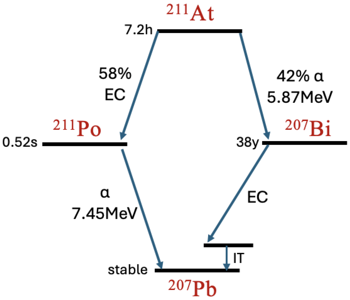
Figure 1. The glue layer loaded with 211At or 224Ra covers healthy tissue above and the tumour below.
Biomedinformatics 05 00058 g001
Figure 2. The decay chain of 211At.
Biomedinformatics 05 00058 g002
Figure 3. The decay chain of 224Ra.
Biomedinformatics 05 00058 g003
Figure 4. Schematic overview of the AlphaGlue decay calculation: 224Ra isotopes decay producing an α and a 220Rn. The 220Rn will then decay to 216Po producing another α . The 216Po decays with the emission of another α to 212Pb. 212Pb undergoes β decay producing 212Bi which also undergoes β decay to 212Po. The 212Po decay produces a final α . The random walk paths of the α ’s, β ’s and isotopes and the energy deposition along the paths is calculated using Geant4 to give the energy deposition as a function of depth.
Biomedinformatics 05 00058 g004
Figure 5. Visualization of the simulation geometry in Geant4 (a) for the AlphaGlue configuration (b). The two 2 × 2 × 2 cm3 water volumes contain 300 nm-thick detector shells.
Biomedinformatics 05 00058 g005
Figure 6. Simplified cell nucleus. An α -particle (blue) enters from the bottom and secondary electrons (red) are tracked.
Biomedinformatics 05 00058 g006
Figure 7. We follow the classification of strand break clusters from [50]. A SSB (Single-Strand Break) involves a break in only one strand of the DNA backbone. A DSB (Double-Strand Break) occurs when both strands experience breaks within ≤10 base pairs. A cDSB (Complex Double-Strand Break) represents a more complex form of DSB, involving additional strand breaks within≤10 base pairs, making it more challenging to repair [53].
Biomedinformatics 05 00058 g007
Figure 8. DNA damage caused by each particle in the 211At chain: α particles from 211At, α particles from 211Po, and electrons from 211At, along with the total of all these, as a function of distance: single strand breaks (SSBs) (a), double strand breaks (DSBs) (b) and complex double strand breaks (cDSBs) (c). Results are normalized per decay.
Biomedinformatics 05 00058 g008
Figure 9. DNA damage caused by each particle in the 224Ra chain as a function of distance: single strand breaks (SSBs) (a), double strand breaks (DSBs) (b) and complex double strand breaks (cDSBs) (c). Results are normalized per decay.
Biomedinformatics 05 00058 g009
Figure 10. Comparison of the DNA damage caused by each particle in the 224Ra-loaded and 211At-loaded 4 μ m thick glue layer and when 224Ra is administered directly onto the skin, as a function of distance: single strand breaks (SSBs) (a), double strand breaks (DSBs) (b) and complex double strand breaks (cDSBs) (c). Results are normalized per decay.
Biomedinformatics 05 00058 g010
Figure 11. Ratio of DNA damage yield (DSB, SSB, and cDSB) from 224Ra embedded in glue relative to 224Ra applied on the surface, as a function of depth.
Biomedinformatics 05 00058 g011
Figure 12. Double-strand breaks (DSBs), and complex double-strand breaks (cDSBs) as a function of depth per decay.
Biomedinformatics 05 00058 g012
Figure 13. RBE as a function of distance, based on DSB and cDSB yields relative to 60Co.
Biomedinformatics 05 00058 g013
Figure 14. RBE-weighted dose as a function of distance for AlphaGlue loaded with 3 μ Ci of 224Ra.
Biomedinformatics 05 00058 g014
Figure 15. Required activity of 224Ra (in μ Ci) as a function of depth to reach a 20 Gy weighted dose.
Biomedinformatics 05 00058 g015
Figure 16. RBE-weighted dose as a function of distance for AlphaGlue and DaRT loaded with 3 μ Ci of 224Ra. The DaRT data are taken from [45].
Biomedinformatics 05 00058 g016
Disclaimer/Publisher’s Note: The statements, opinions and data contained in all publications are solely those of the individual author(s) and contributor(s) and not of MDPI and/or the editor(s). MDPI and/or the editor(s) disclaim responsibility for any injury to people or property resulting from any ideas, methods, instructions or products referred to in the content.