Review

Mechanical Characterization of Natural Polymers Using Brillouin Spectroscopy: A Comprehensive Review

by
José A. Rodrigues
1,2,*,
Bruno Esteves
1,2,
Patrícia Costa
1,2 and
José H. Correia
1,2
1
CMEMS-UMinho, University of Minho, 4800-058 Guimarães, Portugal
2
LABBELS-Associate Laboratory, Braga/Guimarães, Portugal
*
Author to whom correspondence should be addressed.
Photochem 2025, 5(4), 34; https://doi.org/10.3390/photochem5040034
Submission received: 18 September 2025 / Revised: 17 October 2025 / Accepted: 20 October 2025 / Published: 23 October 2025

Abstract

Experimental analysis of the viscoelastic properties of natural polymers over different testing durations and response time scales yields complementary insights into their static and dynamic mechanical behavior. Within this context, Brillouin spectroscopy has emerged as a contactless, non-invasive and label-free tool for the mechanical characterization of materials. In this review article, we provide a comprehensive overview of recent advances in Brillouin spectroscopy techniques applied to various natural polymers, including proteins, carbohydrates, and polysaccharides. We discuss the principles of Brillouin scattering and their application in investigating the mechanical properties of natural polymers. Additionally, we explore future perspectives and challenges. This review aims to provide researchers and practitioners with a comprehensive understanding of the capabilities and limitations of Brillouin spectroscopy for the mechanical characterization of natural polymers, promoting new advances in this interdisciplinary field.

1. Introduction

Biomaterials have attracted significant attention due to their extensive applications in medicine. The biocompatibility of biomaterials is a very important issue, and extensive research efforts have been dedicated to this field over the past decades [1]. The term “biomaterial” was defined in 1986 by Prof. David Franklyn Williams as “a non-viable material used in a medical device, intended to interact with biological systems” [1,2,3]. The necessity to redefine biomaterials emerged because the characterization as ‘non-viable’ was no longer applicable. Additionally, biomaterials found applications beyond implanted devices, encompassing roles in drug delivery systems, imaging contrast agents, and tissue engineering constructs [1]. The most widely accepted definition of a biomaterial is that employed by the American National Institutes of Health, which describes it as “any substance or combination of substances, other than drugs, synthetic or natural in origin, which can be used for any period of time, which augments or replaces partially or totally any tissue, organ or function of the body, in order to maintain or improve the quality of life of the individual” [2,4].
The primary and crucial attribute of any biomaterial is biocompatibility. Additionally, qualities such as non-toxicity, resistance to corrosion, durability, toughness, and a low modulus of elasticity are among the essential characteristics that define a high-quality biomaterial [5]. Biomaterials are classified into four main categories: polymers, ceramics, metals and composites (Table 1) [6,7]. Polymeric biomaterials stand out as the most extensively employed materials in biomedical applications among various biomaterials. Their widespread usage is attributed to their flexibility, biocompatibility, and mechanical, chemical, thermal and electrical characteristics [5]. Polymers consist of repetitive units of a single monomer, classified into three types: natural, biodegradable synthetic and non-biodegradable synthetic polymers. Natural polymers, derived from plant and animal sources, are abundant, and their greater biocompatibility is attributed to the presence of binding sites for cells and adhesion molecules. Synthetic biodegradable polymers mainly contribute to repair and regeneration processes. In contrast, non-biodegradable synthetic polymers find applications as fillers, bone fixing agents and supporting materials for bone structures [5,6].
Natural polymers will be the focus of this review. These materials have undergone extensive investigation for diverse biomedical uses such as drug delivery and regenerative medicine. Due to their biochemical resemblance to components of the human extracellular matrix, these molecules are readily embraced by the body. Moreover, these polymers offer numerous benefits, including their natural abundance, ease of isolation, and the potential for chemical modification to fulfill various technological requirements [8].
The mechanical characteristics of materials play a crucial role in biomedical applications [6]. With rare exceptions, polymers fall into the category of substances called viscoelastic bodies. As the name suggests, these materials exhibit an intermediate response to external forces, presenting characteristics between those of an elastic solid and a viscous liquid [9]. Understanding the mechanical properties of a material is an important challenge to predict its behavior under different loading conditions and design products that withstand the desired level of stress. Conducting experimental analyses of a biomaterial’s viscoelastic properties across different testing durations and response time scales reveals complementary insights into both its static and dynamic characteristics. Within this context, Brillouin spectroscopy is increasingly recognized as a non-contact, non-invasive, and label-free technique which is well-suited for precisely measuring the speed and attenuation of sound on a micrometer/picosecond spatial/temporal scale [10].
This review focuses on the viscoelastic characterization of natural polymers by Brillouin spectroscopy. Briefly, this spectroscopy is based on inelastic scattering of light in the GHz range (0.1 to 100 GHz) by thermally excited acoustical phonons. With changes in the frequency of the scattered light, it is possible to obtain the viscoelastic properties of the sample without contact and in a non-destructive manner [11,12].

2. History of Brillouin Spectroscopy

The first theoretical prediction of the inelastic light scattering by thermally induced acoustic waves was reported by the French physicist Léon Brillouin in 1922. This inelastic light scattering was called Brillouin scattering [12,13]. In 1918, Leonid Mandelshtam initially described true spontaneous Brillouin scattering, which arises from dissipative fluctuations within a medium. However, he focused on experimentally validating his theoretical predictions, and it was only in 1926 that he officially published these finding [14,15]. In 1930, Eugenii Gross conducted the first experimental study of the Brillouin effect, offering empirical confirmation. He successfully observed the inelastic scattering of light from acoustic oscillations of various liquids (aniline, toluene, benzene, water, ethyl alcohol and ethyl ether), revealing distinctive frequency shifts [12,16,17]. Thus, the Brillouin shift serves as a characteristic reflection of the underlying intermolecular interactions that distinguish materials [17]. Interferometry can be employed to discern the small frequency shifts in Brillouin light scattering. While the Fabry–Perot interferometer was already a well-established tool [18], one of the initial demonstrations of its use as a dispersive element for high-resolution frequency analysis in Brillouin scattering was presented in 1942 by C. S. Venkateswaran [17,19].
Early Brillouin light scattering studies faced limitations due to the availability of an appropriate light source. Like any spontaneous and weak scattering technique, it necessitates a substantial photon flux through the scattering volume. Initial studies were restricted to using light sources like Hg or Zn vapor lamps [17]. The invention of the laser in the 1960s [20] marked a significant advancement in light sources, providing the necessary power to reduce acquisition times and enhance resolution. This breakthrough led to a proliferation of experimental studies in the field of condensed matter, solidifying the laser as a key tool in this area [21]. The Brillouin spectrometer developed by Raymond Chiao in 1964, which incorporates a laser, Fabry–Perot interferometer, and photomultiplier tube detection, aligns with the design of modern-day Brillouin spectrometers. Nevertheless, advancements in each component of the spectrometer, especially in interferometry, have significantly improved the ability to accurately measure hypersonic sound frequencies [17,22]. In 1970, John Sandercock introduced an actively stabilized multi-passed interferometer. The advantage of using multi-passing, which involves reusing the transmitted light through multiple passes in the interferometer, is the exponential increase in contrast with the number of passes. This ultra-high contrast capability enables the differentiation of extremely weak signals (such as the Brillouin scattering resonances) even when a very strong signal (like the Rayleigh peak) is present [17,23]. In 1982, John Sandercock proposed the tandem multi-pass Fabry–Perot interferometer [17,24,25]. This spectrometer design stands as one of the foremost scanning interferometers for Brillouin light scattering and continues to be widely used in contemporary research and applications [17]. Although such designs produce high spectral resolution and contrast, they have low throughput (high loss) and long spectral scan times. This was overcome in 2008 with the development of a high-resolution spectrometer based on virtual imaged phased array (VIPA) proposed by Shirasaki in 1996 [26,27,28]. The VIPA can be seen as an adapted form of a Fabry–Perot etalon. It has the ability to spatially disperse the spectrum without requiring scanning of the interferometer’s optical cavity, allowing for convenient and efficient spectrum acquisition using a scientific camera [26]. Table 2 summarizes the timeline events related to the history of Brillouin spectroscopy.

3. Brillouin Scattering

3.1. Physical Principles

When a light beam interacts with matter it can cause different effects, namely the absorption and scattering of light. These effects can be used to examine material properties [11,29]. The atoms and molecules of matter absorb the energy of light, with some being dissipated as heat and the remainder being re-emitted, resulting in an exponential decay in the intensity of the light with propagation distance [29]. Scattering effect depends on the density of the physical medium and the relation between particle size and wavelength of the incident beam. The scattered light can be categorized into elastic scattering (Rayleigh), where the direction of the beam changes while its frequency (and therefore the energy) remains constant, and inelastic scattering (Brillouin and Raman), involving changes in frequency and beam direction during propagation [21,29]. Brillouin scattering is based on the interaction between photons and thermally excited acoustic phonons [26,29]. Since phonons are density perturbations that travel at the speed of sound, this interaction leads to a change in the frequency of the scattered light due to the Doppler shift [11,29]. Brillouin spectroscopy measures these density fluctuations that probe microscale viscoelastic properties of a material [11]. The typical Brillouin spectrum (Figure 1) presents an intense central Rayleigh peak, with the same frequency as the illumination, and two equally shifted peaks, known as Stokes and anti-Stokes Brillouin peaks [11,26,29]. The Brillouin spectrum contains two important data for the viscoelasticity of a material: frequency shift (wB) and full width at half maximum (FWHM) of the Stokes and anti-Stokes peaks (Γ) [29].
The Brillouin frequency shift is given by:
w B = ± V . q
where V is the acoustic wave velocity in the material, and q is the momentum exchanged in the scattering process [11,29]. The sign ± suggests that the energy can either be transferred from phonon to photon (+) or from photon to phonon (−) [29]. The momentum exchanged is given by:
q = 2 . n . k i . sin θ 2
where n is the refractive index of the material, θ the scattering angle, and ki the wave vector of the incident light [11,29]. This vector is given by:
k i = 2 π λ i
where λi is the incident light wavelength in vacuum. For a backscattering geometry, i.e., θ = 180°, the frequency shift of Brillouin peaks is equal to [11]:
w B = ± 4 . π . n λ i . V
The acoustic velocity is determined as a function of the material density ( ρ ) and the longitudinal modulus (M) by the equation [29]:
V = M ρ
Thus, Equations (4) and (5) suggest that the longitudinal elastic modulus can be evaluated using data collected by Brillouin spectroscopy [11,29]. The longitudinal elastic modulus is given by:
M = ρ . V 2 = ρ . w B 2 4 . n 2 . k i 2
Brillouin spectrum analysis provides a distinct characterization of the mechanical properties of a material (for a known density and refractive index). This uniqueness arises from the fact that the characteristics of sound waves, including their speed and attenuation, inherently rely on the viscoelastic properties of the material [26]. The equation that governs the propagation of longitudinal acoustic waves in a viscoelastic medium, the complex longitudinal modulus (M*), is given by [11,26]:
M * w = M w + i M w  
where M′ is the storage modulus and M″ the loss modulus. The first provides information about the elastic properties of a material (energy storage), while the second is related to longitudinal viscosity (energy dissipation) [26,29]. When density fluctuations occur at frequencies below the structural relaxation rate, the value of M′ is lower, because the molecules do not respond elastically; instead, there is a possibility of partial diffusion under compression, indicating a viscous regime. Conversely, for density fluctuations at frequencies higher than the relaxation rate, molecules lack the time to diffuse and respond elastically to the disturbance, resulting in an increase in the elastic modulus, indicating an elastic regime [11]. In viscoelastic materials, analyzing Brillouin spectra provides easy determination of the M* at the specific frequency of Brillouin peaks [11]. Brillouin peaks can be reproduced by a damped harmonic oscillator (DHO) function, which is the theoretical expression derived from the Brillouin line shape of transparent viscoelastic materials acquired at a single q [10,11,30,31,32]:
I w = I 0 π Γ B . w B 2 w 2 w B 2 2 + Γ B . w 2  
It involves three fitting parameters: frequency position (wB), FWHM (ΓB), and the integrated intensity (I0) of the Brillouin peaks [10,31]. The wB and ΓB derived from the Brillouin peak fitting analysis allow determining M′ and M″ by the equations [11,32,33]:
M w B = ρ q 2 . w B 2  
M w B = ρ q 2 . w B . Γ B  
where λ is the excitation wavelength, and ρ and n are the mass density and refractive index of the medium, respectively.

3.2. Scattering Geometries ρ

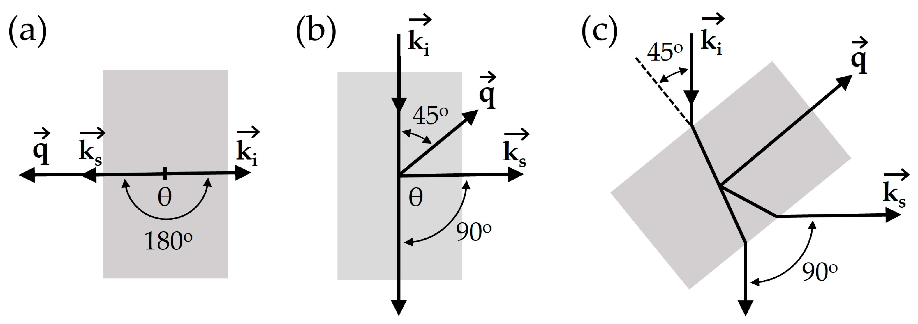
Scattering geometries refer to the geometric relationships between the sample orientation and the incident and scattered light beams. Furthermore, these geometries play a crucial role in determining the direction and magnitude of the acoustic scattering wave vector (q). This factor is essential for the accurate interpretation of Brillouin spectroscopy experiments. The most commonly used scattering geometries include 180 (backscattering geometry), 90N, and 90A (Figure 2) [21].
The backscattering (Figure 2a) and 90N (Figure 2b) geometries are commonly used due to its easy alignment, i.e., the angle between the incident and scattered light beam outside the sample is 180° and 90°, respectively. In the backscattering geometry, it is important to note that in elastically isotropic and homogeneous samples, only the longitudinal phonons can be measured due to symmetry considerations [34,35]. The magnitude of the scattering acoustic wave vector is:
q = 2 . n λ 0  
where n is the index of refraction and λ0 is the wavelength of the incident light in vacuum. With respect to the 90N geometry, the wave vector of the scattering vibration is positioned at 45° from both the incident and scattered wave vectors. The magnitude of the scattering acoustic wave vector is [34]:
q = 2 . n λ 0  
For the 90A scattering geometry (Figure 2c), the angle of incident laser beam on the sample surface is 45°. The scattered light is collected perpendicularly to the incident beam. This geometry enables the determination of the acoustic velocity without requiring knowledge of the refractive index value [35,36]. The magnitude of the scattering sound wave vector is:
q = 2 λ 0  

3.3. Mechanical Considerations in Brillouin Scattering of Natural Polymers

The mechanical interpretation of Brillouin spectroscopy in natural polymers requires a careful consideration of their intrinsic heterogeneity. Natural polymers such as collagen, keratin, silk fibroin, or polysaccharide-based hydrogels exhibit complex architectures, where factors such as anisotropy, hydration state, mass density, significantly affect the measured Brillouin frequency shift and linewidth [11].
In an ideal isotropic elastic solid, the mechanical response is fully described by the elastic stiffness tensor, which reduces to two independent constants: the bulk modulus and the shear modulus. In such a case, Brillouin scattering primarily probes the longitudinal acoustic mode, associated with the longitudinal modulus [11]. However, many natural polymers deviate from isotropy due to fibrillar orientation, hierarchical organization, or directional crosslinking. In anisotropic materials, different tensor components contribute depending on the direction of the scattering vector, and the Brillouin frequency shift reflects only a projection of the full elastic tensor [37,38].
Although conventional Brillouin setups do not directly measure transverse modes, complementary geometries or multi-angle measurements can be used to infer viscoelastic anisotropy and shear behavior [11]. Since the shear modulus governs resistance to shape deformation, its evaluation is fundamental in characterizing the viscoelasticity of biological tissues and hydrated polymer networks.
Hydration effects introduce an additional level of complexity. Water induces plasticization, increases molecular mobility, and alters both density and refractive index, thereby modulating the Brillouin shift. Highly hydrated states typically reduce M′ while increasing M″, reflecting a shift toward viscous relaxation regimes. Therefore, quantitative comparison between dry and hydrated samples requires independent or simultaneous measurement of density and refractive index, to avoid misinterpretation of apparent modulus variations [39].
Finally, the hierarchical organization of natural polymers, often combining crystalline and amorphous domains, results in spatially varying Brillouin signatures. In such cases, multiplexed or axial scanning Brillouin microscopy may be necessary to resolve mechanical gradients, especially in tissues or biomimetic scaffolds. When complemented with structural or optical imaging (e.g., polarization microscopy, OCT), Brillouin spectroscopy enables a more comprehensive reconstruction of the material’s anisotropic and frequency-dependent mechanical response [11,40].

4. Brillouin Spectroscopy Experimental Setup

Brillouin frequency shifts typically fall within the range of several gigahertz with linewidth in the range of 0.1–1 GHz. Achieving precise measurements of these shifts requires two crucial elements: a monochromatic light source (laser) and a high-resolution spectrometer [14,21,29]. Figure 3 shows a schematic of a typical Brillouin spectroscopy setup (backscattering geometry) [35].

4.1. Laser

The light source for Brillouin scattering experiments must be monochromatic to avoid ambiguities in the spectrum that could make it difficult to assign the observed peaks [34]. The laser emission spectral width must be lower than the width of the Brillouin peak to avoid their spectral broadening and maximize inelastic scattering. The noise caused by the laser and the stability of its frequency are important factors in obtaining more accurate data [29]. Due to the λ−4 dependence of the intensity of the scattered light, green and blue lasers with narrow spectral band width are the most commonly used. The power of the laser rarely exceeds 100 mW (continuous wave laser) [29,34,35].

4.2. Spectrometer

The most significant challenge has consistently been obtaining high-resolution spectrometer capable of discerning a relatively weak Brillouin signal located just a few GHz away from the laser frequency [14,29,35]. Several spectrometer designs have been suggested to address this challenge, namely the tandem Fabry–Perot interferometer and the VIPA.

4.2.1. Tandem Fabry–Perot Interferometer

The main optical component of a Fabry–Perot interferometer is a pair of parallel mirrors separated by a free space of defined length, commonly referred to as a Fabry–Perot etalon [29,34,35]. Depending on the distance between the mirrors, light either passes through or is reflected by the instrument. By scanning the distance between the mirrors, spectral selectivity is achieved within the free spectral range of the instrument, defined as the spectral distance between neighboring maxima in the transmission spectrum. Since for this interferometer, a greater resolution implies greater mirror spacing and lower free spectral range, a single etalon may not always be sufficient to detect Brillouin signals in semi-opaque and turbid samples [29,35]. In 1970, John Sandercock showed that contrast could be significantly improved by a multipassed arrangement [23].
In the standard Brillouin scattering experiment, the Fabry–Perot interferometer is used as a variable narrow band-pass filter, sweeping the chosen free-spectral range. This can be achieved by scanning the space between the mirrors [34,35]. In tandem interferometry, the light goes through two Fabry–Perot in series. The use of two synchronized Fabry–Perot interferometers not only improves contrast but also broadens the free-spectral range of the instrument, allowing for the use of larger mirror gaps [24,29,35]. The multi-pass tandem Fabry–Perot spectrometer, conceptualized by John Sandercock four decades ago, has still demonstrated remarkable success as an instrument to study weak inelastically scattered light signals [14]. In these spectrometers, the two sets of parallel mirrors are scanned using piezoelectric transducers. The free-spectral range is defined by the overlap between the two sets of mirrors (individually dictated by mirror distance), which is adjustable) and high-contrast is achieved by triple-passing (3 + 3) the transmitted peaks through each of the two Fabry–Perot interferometers in a tandem configuration (Figure 4) [11,24,34].
The multi-pass Fabry–Perot spectrometer in tandem configuration is the Brillouin spectrometer with the highest contrast (>1015 or 150 dB), being limited only by the dark current of the detection photomultiplier [11,14]. However, this spectrometer is limited by the relatively long signal acquisition time (>1 s) [14,21,29]. The minimum acquisition time is limited by the scanning speed of the mirrors and cannot be significantly improved [29]. A large number of sampling points (>10,000) can take many hours, which is not suitable for biological samples [21,29].

4.2.2. Virtually Imaged Phased Array

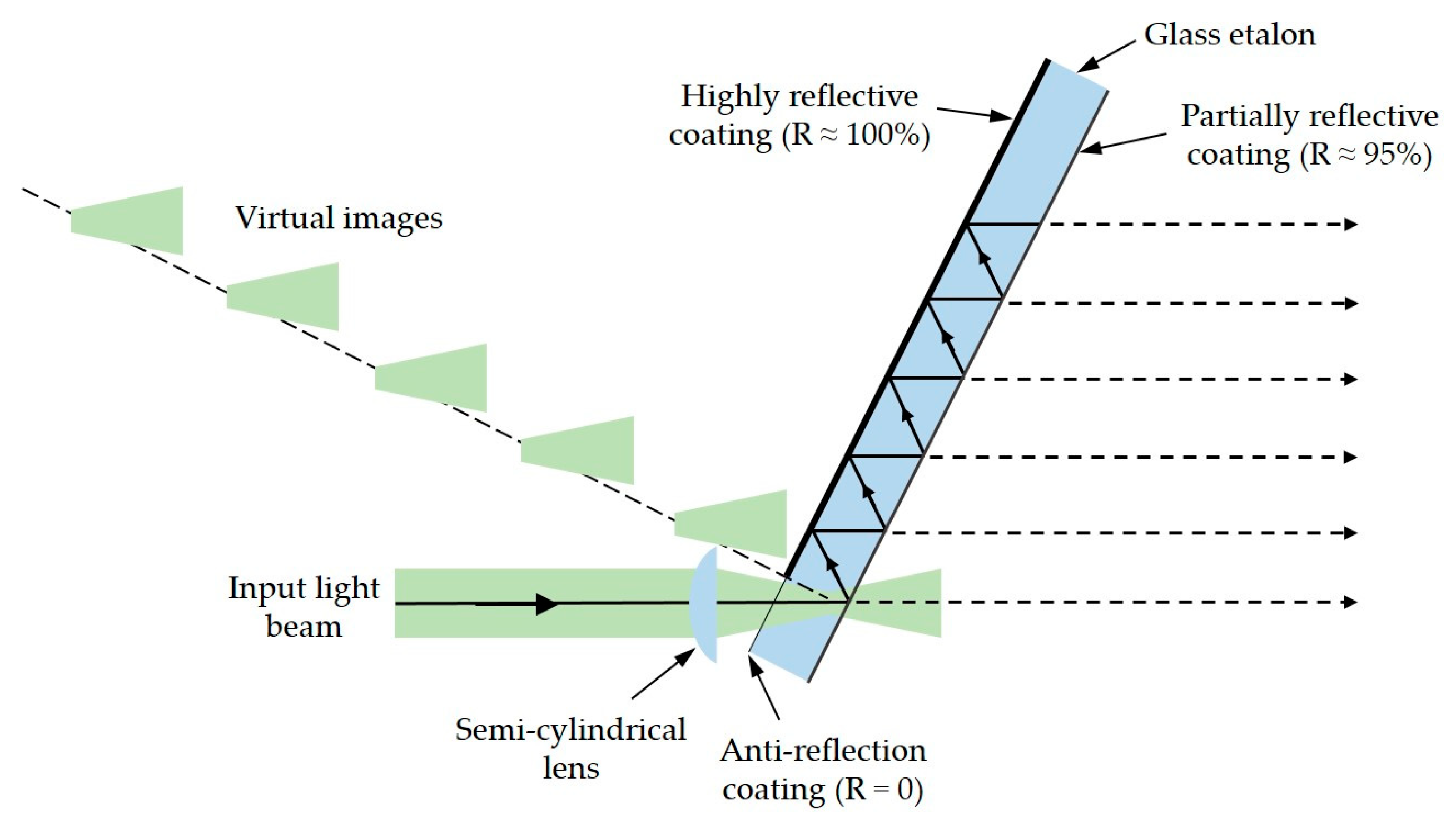
The main disadvantage of spectrometers based on the Fabry–Perot principle, i.e., long signal acquisition time, has been overcome by the introduction of a new spectrometer based on a VIPA [21,41]. For faster acquisition speeds, both single-stage and multi-stage VIPA spectrometers are widely used for Brillouin measurements [29]. A simple VIPA design consists of a semi-cylindrical lens that focuses a ray of monochromatic light onto a glass etalon with reflective coatings through a slit opening in the coating, offering sufficiently large angular dispersion to separate Brillouin Stokes and anti-Stokes peaks from the Rayleigh signal [27,29,41]. One side of the glass etalon is coated with a high-reflection film (about 95%). The other side is coated with a 100-reflection film except in the slit opening area, which is coated with anti-reflection film [27,28,41,42,43]. As the incident light beam undergoes multiple reflections inside the plate, the transmitted light generates a “virtual” array of beams aligned along the surface normal of the plate. At the VIPA exit, the beams diverge and interfere, similar to a diffraction grating, so that different wavelengths form constructive interference in different angular directions (Figure 5) [41,43,44]. In the case of light comprising multiple wavelengths, the various spectral components are diffracted at different angles and the VIPA disperses all wavelengths simultaneously. In contrast, a typical angle-dispersive etalon transmits a narrow band at a specific incident angle. Therefore, the VIPA enables highly efficient spectral separation with significantly higher throughput compared to a typical etalon [41].
The acquisition time of VIPA spectrometers typically ranges around 100 milliseconds, depending on sample transparency and laser power. This speed is at least an order of magnitude faster than tandem Fabry–Perot spectrometers, enabling the extension of this technique to biological materials, which are generally more sensitive to damage by light. However, single-stage VIPA spectrometers offer only 30 dB of contrast, rendering them inadequate for measuring turbid samples [11,28,29,45]. To improve contrast to 50–60 dB, two VIPA spectrometers can be orthogonally placed in tandem to one another, albeit with a decrease in signal strength [29,45,46]. Additional approaches have been adopted to enhance the contrast of this type of spectrometer, including the incorporation of a third VIPA spectrometer stage [43], employing a Fabry–Perot etalon as a narrowband filter [47,48], and optimizing signal collection efficiency [49], among other methods [46].

4.3. Detector

The choice of detector for a Brillouin scattering spectrometer involves a trade-off between high efficiency in the energy range of interest and good signal-to-noise ratio. In experiments with large samples generating strong signals, photomultiplier tubes offer the best signal-to-noise ratio, thanks to their remarkably low dark count (as low as less than 5 counts/s) and wide dynamic range, despite their quantum efficiency being around 20%. Conversely, for small and weakly scattering samples, avalanche photodiodes with their high quantum efficiency (up to 70%) and moderately low dark count (approximately 25 counts/s) can significantly reduce collection times, helping to address issues related to the long-term stability of interferometer alignment and natural frequency drift of the source. However, in cases where materials produce extremely weak Brillouin signals in the presence of a strong elastic scattering from surface imperfections, such as in opaque materials, photomultiplier tubes may still be the preferred choice due to their lower dark current [29,34]. Charge-Coupled Devices (CCDs) are also used as detector in Brillouin spectroscopy, as they can be useful for capturing spatially resolved Brillouin spectra, although they may not offer the same sensitivity as photomultiplier tubes or avalanche photodiodes [29].

5. Natural Polymers

Natural polymers constitute a class of polymers sourced directly from nature. Predominantly composed of carbohydrates and proteins, these polymers serve as intrinsic components of plants and animals, primarily fulfilling structural roles [50]. With desirable attributes such as abundant availability, biodegradability, and renewability, natural polymers have found diverse applications. They can be categorized into two main groups based on their sources: non-mammalian (including marine algae, crustaceans, insects, plants, and microorganisms) and mammalian-based (including proteins and glycosaminoglycans) [51]. Figure 6 shows some natural polymers (and an example of their origin) covered in this review.

5.1. Collagen

Collagen constitutes one of the most important natural biopolymers [52]. Collagen is an abundant insoluble fibrous protein that makes up the majority of the extracellular matrix in animals [53,54]. Collagen can be sourced from various natural origins, including mammals such as porcine and bovine, as well as marine organisms like fish [53,54]. The collagen networks play a crucial role in regulating cellular behavior by maintaining the extracellular microenvironment [54,55]. This biological polymer is the main component of connective tissues, comprising over 30% by weight of the total proteins found in the animal body. It works as an intracellular adhesive, preventing tissues from stretching or suffering damage [52,53,55]. Typically, collagens exhibit a highly organized triple helix structure composed of polypeptide chains and are categorized into up to 29 different types [52,54,55,56]. In fibrous forms, of which type I collagen is the best characterized and most ubiquitous representative, molecules are regularly assembled both laterally and longitudinally. These molecules are extensively cross-linked to create hierarchical structures of fibrils and fibers [57]. Collagen has wide application in the pharmaceutical, cosmetic and food industries [52].
The first application of Brillouin spectroscopy in biopolymers was carried out by R. Harley et al. in 1977 [58]. Brillouin spectroscopy was used to determine the elastic moduli of type I collagen extracted from rat tail tendons and estimate the hydrogen bond force constants. R. Harley also studied the effects of hydration on Brillouin spectra to obtain information about the association of water with collagen in the native state. The authors noted that drying the samples increased the stiffness [10,21,58]. This study was later extended to include measurements of the anisotropic nature of the elastic properties in collagen, obtained by measuring the velocity of propagation of both longitudinally and transversely polarized elastic waves traveling at various angles to the fiber axis. Assuming a transversely isotropic model, the five elastic constants were determined for dry collagen [21,59]. The shear modulus is relatively low and there is considerable anisotropy in Young’s modulus for strains at different angles to the fiber axis. Upon rehydration, the tendon becomes markedly less stiff and the anisotropy in Young’s modulus increases. These measurements represent the earliest documented instances of these parameters for collagen fibers. They hold significance in two aspects: firstly, they showcase the utility of Brillouin scattering in examining biological materials, and secondly, they contribute to a more comprehensive comprehension of collagen elasticity, which is pivotal in understanding the characteristics of collagen-based composite materials like bone, cartilage, and skin [59]. It should be noted that the elastic longitudinal modulus M (on the order of GPa) of biological systems measured by Brillouin light scattering at GHz frequencies may not be related to Young’s modulus (on the order of kPa) measured by rheology obtained at much low (kHz) frequencies: the former reflects the high-frequency elastic response probed by acoustic phonons, while Young’s modulus represents the quasi-static mechanical response measured under macroscopic deformation.
In 1979, J. Randall and J. Vaughan measured Brillouin scattering spectra for wet unstretched rat tail collagen, with the scattering vector parallel and perpendicular to the fiber axis [60]. Only twenty years later new Brillouin spectroscopy studies on collagen appeared, with S. Lees et al. in 1990 examining the axial velocity of wet rat tail tendon collagen [61].
In 2014, F. Palombo et al. [10,57] investigated Brillouin light scattering on type I collagen in rat tail tendon, type II collagen in articular cartilage, and nuchal ligament elastin. They used a backscattering geometry and a reflective substrate to achieve the complete characterization of fiber biomechanics. Two distinct peaks were observed: one associated with a bulk mode arising from phonon propagation along a quasi-radial direction to the fiber axis, and another corresponding to a mode parallel to the surface, depending on the orientation of the sample relative to the fiber axis. The last peak was fitted to a model that describes wave propagation through a hexagonally symmetric elastic solid. This fitting process allowed for the determination of the five components of the elasticity tensor, which were then combined to derive the axial and transverse Young’s moduli, as well as the shear and bulk moduli of the fibers. The moduli of collagen and elastin were significantly higher than those observed at lower frequencies with macroscopic strains, and the discrepancy between them was relatively small, suggesting that molecular-scale viscoelastic effects are responsible for the frequency dependence of fiber biomechanics [10,57].
In 2018, R. Edginton et al. [21,62] used Brillouin spectroscopy to investigate the mechanics and structure of purified collagen and elastic fibers, focusing on the impact of the fibrillar structure and hydration level. This study resulted in a comprehensive characterization of the mechanical tensor and elastic moduli. By comparing this data with quasi-static measurements obtained at varying hydration levels, they achieved a comprehensive understanding of the viscoelastic behavior of the fibers [21,62].
More recently, in 2020, M. Bailey et al. [33] aimed to deepen the informative content of the Brillouin spectrum of gelatin gels (denatured type I collagen), comparing them with real tissue samples. These gels serve as simple model systems derived from the most prevalent structural protein. Through adjustments in polymer concentration, a broad spectrum of static and dynamic macroscopic mechanical moduli can be covered, mirroring those observed in many biological tissues. The results indicate that the viscoelastic parameters derived from Brillouin spectroscopy in gelatin hydrogels, serving as model systems for protein networks, are primarily influenced by the interaction between the solute and solvent relaxation dynamics [33].

5.2. Cellulose

Cellulose is the most abundant biopolymer and the main structural component of plant cell walls [63]. Cellulose provides rigidity and strength to plant cells and tissues, serving as structuring element in the complex architecture of their cell walls [63,64]. It is a long-chain polysaccharide composed of 7000 to 15,000 glucose monomer units, which are alternately rotated 180° [64]. In plants, cellulose forms complex hierarchical fibers with other biopolymers, such as hemicelluloses, pectins, and lignins [63,64,65]. Similar to many fibrous materials in biological matter, cellulosic fibers display directional mechanical properties. These fibers may consist of several cell wall layers that have varying chemical composition, thickness, and cellulose fibril orientation [64,65]. The commercial purification of cellulose primarily focuses on cotton linters and wood pulp. Cotton linters are preferred for their high cellulose content, while wood pulp is chosen for its relative abundance and ease of harvesting from wood and straw. Purifying cellulose in its natural state presents challenges due to its insolubility in conventional solvents [66].
The first application of Brillouin spectroscopy on cellulose dates back to 1976, when G. Patterson used this technique to obtain the Brillouin scattering spectrum of a commercial cellulose acetate film containing additives [67].
In 2010, L. Sui et al. used Brillouin light scattering to determine the in-plane and out-of-plane elastic constants of layer-by-layer assembled cellulose nanocrystal films with different thicknesses and concentrations [68].
Daniel R. Williams et al. [69] in 2019, employed Brillouin light scattering to determine the full elastic stiffness tensor of six species of bamboo fiber variants, providing a comprehensive mechanical profile that includes Young’s moduli, shear moduli, bulk modulus, and Poisson’s ratios. The fibers, composed of sclerenchyma cells, exhibit hierarchical architectures in which crystalline and amorphous cellulose nanograins are embedded within a lignin and hemicellulose matrix. By modeling bamboo fibers as transversely isotropic composites, the study highlights the dominant role of cellulose nanograins in governing axial stiffness. The results reveal a linear scaling of elastic properties with fiber density. Additionally, alkali-treated commercial fibers exhibited reduced moduli, indicating that cellulose crystallinity is preserved while matrix components are more affected, reinforcing cellulose’s critical mechanical role. Overall, the work demonstrates the capacity of Brillouin light scattering to non-invasively quantify anisotropic elastic behavior in cellulose-rich fibers, validating its application for probing the mechanics of semi-crystalline biopolymers in natural composites [69].
In 2020, K. Elsayad et al. [70] used Brillouin light scattering microspectroscopy for assessment of the mechanical properties of viscose and softwood pulp fibers. The elastic modulus (storage modulus) representing the elastic behavior and the loss modulus (dampening coefficient) representing the viscous behavior were evaluated. The results reveal that while softwood pulp has relatively uniform moduli, viscous fibers exhibit significant spatial heterogeneity in moduli [70]. Viscose fibers are produced through the viscose process, where wood cellulose is dissolved in a solvent and then extruded into yarn form, resulting in a highly anisotropic material. Consequently, viscose fibers are distinguished from wood fibers by being composed entirely of cellulose, which has a different crystalline structure and lacks the hierarchical structural complexity of natural wood fibers [71,72].
More recently, in 2024, C. Czibula et al. [73] applied Brillouin light scattering spectroscopy to evaluate the directional elastic stiffness and mechanical characteristics of a viscose fiber. The results reveal a distinct contrast between the material stiffness, determined by direct measurement of the elastic stiffness tensor with Brillouin light scattering, and engineering material parameters such as Young’s modulus, which are derived from mechanical testing. The authors concluded that his method to measure all stiffness properties of transversely isotropic fibers provides more comprehensive and distinct results than the collection of different micromechanical testing methods required to test Young’s moduli, shear moduli, and Poisson’s ratios in the axial and radial direction of a fiber [73].
In the same year, M. Samalova et al. examined the role of expansins—proteins that regulate cell wall mechanics—in Arabidopsis thaliana roots, specifically their impact on the viscoelastic properties of the cell wall, which is composed of cellulose microfibrils embedded in a matrix of hemicellulose and pectin. Using Brillouin light scattering microscopy together with atomic force microscopy (AFM), the authors demonstrate that overexpression of EXPANSIN1 (EXPA1) results in a measurable increase in cell wall stiffness, accompanied by changes in wall composition and gene expression. Brillouin light scattering detected an increased Brillouin frequency shift upon EXPA1 overexpression, indicating enhanced stiffness likely linked to altered cellulose-matrix interactions. Complementary Fourier-transform infrared analysis revealed rapid pectin demethylesterification, which may affect cellulose accessibility and mechanical coupling. Overall, this work validates Brillouin light scattering as a non-invasive method for probing cell wall mechanics in vivo and highlights its sensitivity to changes in cellulose-associated architecture, reinforcing its potential for studying cellulose mechanics within complex biological structures [74].
More recently, L. Pachernegg-Mair et al. studied the effects of ionic liquid 1-ethyl-3-methylimidazolium acrylate treatment on the structural and mechanical properties of flax fibers, which are highly enriched in cellulose. Through a combination of Brillouin spectroscopy, AFM, wide-angle X-ray scattering (WAXS), and tensile testing, the authors evaluate how the selective dissolution of non-cellulosic components alters fiber stiffness and morphology. Flax fibers, composed of more than 70% cellulose—primarily located in the S2 layer of the cell wall—were analyzed before and after treatment. Brillouin measurements revealed that axial stiffness remained relatively stable, indicating the preservation of cellulose microfibril integrity, whereas the shear modulus decreased markedly, reflecting degradation of the hemicellulose–pectin matrix. Complementary AFM and WAXS analyses confirmed a low microfibril angle (~5°), validating Brillouin spectroscopy’s sensitivity to the anisotropic architecture of cellulose. Overall, the study demonstrates that Brillouin spectroscopy can effectively differentiate between stiffness governed by cellulose microfibrils and shear properties dependent on the surrounding matrix, highlighting its value as a non-invasive technique for directional mechanical characterization of cellulose within natural fiber composites [75].

5.3. Chitin and Chitosan

Chitin is the second most prevalent natural biopolymer, sourced from the exoskeletons of crustaceans, as well as from the cell walls of fungi and insects [76,77]. Chitin is a nitrogenous polysaccharide insoluble in water, essentially composed of N-acetyl-d-glucosamine linked by β-1,4-glycosidic bonds, with a chemical structure highly similar to cellulose [77,78]. The presence of hydrogen bonds in the structure of chitin facilitates the formation of crystalline states and their transformation into new derivatives [77]. Chitosan, a copolymer of N-acetyl-d-glucosamine and d-glucosamine units, is a product derived from de-N-acetylation of chitin in the presence of hot alkali. Chitosan is, in fact, a collective name representing a family of de-N-acetylated chitins, deacetylated to different degrees [76,78].
The only study reported in the literature of using Brillouin light scattering to measure the mechanical properties of chitin/chitosan was carried out by A. Yoshihara et al. [79] in 2012. They performed Brillouin light scattering studies on the wings of six dragonfly species common in Japan, which are mainly composed of chitin. The authors effectively detected Brillouin light scattering from longitudinal acoustic phonons propagating perpendicular to the wing plane, with a thickness of a few micrometers. The Brillouin shift in these phonons in dragonfly wing chitin was 19.5 ± 0.4 GHz in all six species of dragonflies, in conventional backscattering geometry at 488 nm excitation. Dragonfly cuticles are found to be elastically harder material than other organic amorphous materials [79].

5.4. Silk

In this review, the term silk refers to materials formed from protein-based fibers spun by living organisms. The essential components of silk biopolymer are amino acids. These amino acids, with their specific sequences, dictate the function of the protein by forming primary, secondary, tertiary, and quaternary structures. Across nature, numerous organisms, including spiders, silkworms, scorpions, bees, and ants, produce silk and silk-like proteins. However, silk fibroins and silk-like proteins from each organism exhibit different physical and biological characteristics due to their different amino acid sequences, spinning conditions, and hierarchical structures [80,81].
In 2013, K. Koski et al. [82] used Brillouin light scattering to obtain the entire stiffness tensors (revealing negative Poisson’s ratios), refractive indices, and longitudinal and transverse sound velocities for several spider silks. These findings provide a comprehensive quantification of the linear elastic response across all potential deformation modes, a level of detail inaccessible through conventional stress-strain testing methods. They also employed Brillouin imaging principles to spatially map the elastic stiffness of a spider web. This approach allowed variations in individual fibers, junctions and adhesive points to be mapped without causing deformation or disturbance, performing measurements in a non-invasive and non-contact manner [82]. Spider silk has viscoelastic, anisotropic, and highly nonlinear characteristics, causing changes in measured phonon frequencies upon mechanical deformation. This phenomenon complicates attempts to isolate the purely linear elastic contribution [10,82].
Brillouin light scattering analysis performed by D. Schneider et al. [10,83] in 2016 to spider dragline silk revealed a hypersonic phononic bandgap and a negatively dispersive region. These features are thought to emerge from the interaction between the uniaxial symmetry of the nanofibrils and the non-local nonlinear mechanical behavior of the surrounding inhomogeneous amorphous polymer matrix [10,83]. In the same year, B. Lee et al. [84] studied the elastic properties of fibroin silk under varying temperature or pressure using Brillouin spectroscopy. The Brillouin frequency shift decreased with heating. The Brillouin frequency shift, the sound velocity, and the refractive index of the silk film increased significantly with compression [84].
In 2020, Z. Wang et al. [85] used micro-Brillouin light spectroscopy to study the elastic properties of the Nephila pilipes spider silk. The authors claim to be the first to perform Brillouin light scattering measurements on a single spider fiber. Through extensive multi-angle measurements and meticulous attention to optical birefringence, coupled with accurate phonon mode assignments, they successfully determined the complete elastic tensor and characteristic mechanical attributes of spider silk [85].
In 2025, A. Aluculesei et al. [38] used Brillouin light spectroscopy to determine the complete elastic tensor and mechanical properties of Bombyx mori silkworm silk fibers and to compare their anisotropic elasticity with spider silk. Silkworm silk possesses notable elastic anisotropy rooted in its hierarchical structure, with mechanical properties that remain stable under strain. This contrasts with spider silk’s strain-hardening behavior, highlighting how differences in protein composition and supramolecular assembly lead to distinct mechanical responses [38].

5.5. Keratin

Keratin represents the most abundant structural proteins in epithelial cells and, together with collagen, is the most important biopolymer in animals [86]. Keratin is a fibrous structural protein that forms the main structural component of hair, nails, feathers, horns, claws, wool, and hooves in vertebrates [86,87,88,89,90]. It is a tough and insoluble protein that provides strength, weatherproofing, structural integrity, and impact protection to various biological structures [86,88]. Keratin is characterized by its high content of sulfur-containing amino acids, particularly cysteine, which form disulfide bonds between protein chains, contributing to its stability and resilience. Additionally, keratin can undergo cross-linking and polymerization to create durable and resilient structures [88,89]. These biopolymers can be categorized based on their sulfur content into hard and soft keratins. Hard keratins, characterized by a higher sulfur content, are primarily responsible for the resilient epidermal structure. They consist of intermediate filaments organized in orderly arrays embedded within a crosslinked matrix. On the other hand, soft keratins, containing a lower sulfur content, are composed of bundles of cytoplasmic filaments that are loosely packed, imparting resilience to epithelial tissues like the epidermis [87,90]. The molecular structure of keratin can be classified as α-keratin or β-keratin, based on the hydrogen bonding of the protein’s primary structure. Most mammalian keratin is composed of α helices, while the feathers, beaks, and scales of avian reptiles are composed primarily of the β-keratin variant common among sauropsids [86,87,88]. Similar to other polymers, water content influences the mechanical and physical properties of keratin by acting as a plasticizer and altering hydrogen bonding within its structure [88].
In 1979, J. Randall and J. Vaughan [60] not only investigated Brillouin light scattering in type I collagen extracted from rat tail tendons but also explored the impact of horsehair keratin fiber orientation through Brillouin scattering measurements. Their observations on keratin suggested that Brillouin frequency shifts could be related to the fiber’s water content and the degree of helical extension [60].
In 2021, N. Correa et al. [91] used Brillouin microscopy to map the viscoelastic properties of human hair and to evaluate the mechanical effects of cosmetic bleaching treatments. Through longitudinal Brillouin measurements, distinct frequency shifts were observed between the cuticle and cortex, enabling spatially resolved stiffness profiling across hair microstructures. Given that the cuticle and cortex are primarily composed of keratin in β-sheet and α-helical conformations, respectively, the Brillouin frequency shifts detected after bleaching—rising from 18.7 to 19.7 GHz in the cuticle and from 20.7 to 21.0 GHz in the cortex—indicate an increase in longitudinal modulus. The technique successfully captured micromechanical alterations induced by chemical treatment, confirming its sensitivity to keratin structural changes. Furthermore, mapping of Brillouin frequency and linewidth provided insight into both the elastic (storage) and viscous (loss) components of the material’s response. Overall, the study demonstrates that Brillouin microscopy offers a non-destructive, contactless method to quantify directional stiffness in keratinous fibers and effectively detect treatment-induced modifications, reinforcing its value in characterizing keratin mechanics at the microscale [91].

5.6. Starch

Starch, the major reserve polysaccharide of green plants, is a biopolymer that constitutes two-thirds of the carbohydrate caloric intake of most humans [92]. It is semi-crystalline in nature and has a structure composed of glucose units linked by glycosidic bonds [93]. The two major polysaccharides of starch are amylose and amylopectin. Amylose and amylopectin are high molecular weight biopolymers that possess many of the general properties of chemically synthesized polymers. Amylose is a slightly branched polymer composed of α (1,4) glucopyranose with a degree of polymerization of 6000. Amylopectin is a highly branched polymer consisting of α (1,4) glucopyranose linked by α (1,6) bonds and a degree of polymerization of about 2 million. Amylose is amorphous in nature while amylopectin is crystalline [92,93]. Commercial starches are obtained from seeds, particularly corn, waxy corn, high-amylose corn, wheat and rice, and from tubers or roots, particularly potatoes, sweet potatoes and tapioca [92].
A. Rakymzhan et al. [94,95] used Raman and Brillouin spectroscopies for the time-resolved analysis of chemical and elastic properties of Populus and Geranium leaves. The results showed that the Brillouin frequency shift in dry plant leaves increased progressively over time, indicating an increase in stiffness. Simultaneous examination using Brillouin and Raman spectroscopy throughout the time-resolved drying process of Populus leaves revealed a 25% increase in elastic modulus over 12 h. Furthermore, several Raman peak area ratios experienced up to a sevenfold increase over 36 h, potentially linked to enhanced cohesion of the starch structure through additional hydrogen bonds present in amylose and amylopectin [94,95].

5.7. Gelatin

Gelatin is one of the most versatile biopolymers and has numerous applications in food, confectionery, pharmaceutical/medical, cosmetic, and technical products [96,97]. Gelatin is a heterogeneous mixture of peptides derived from the parent protein collagen by processes that break down the secondary and higher structures with varying degrees of hydrolysis of the polypeptide structure [96,98]. The amino acids that make up collagen and gelatin have very similar relative proportions and sequences, but the physical characteristics of these proteins differ significantly. As mentioned before, collagen is the main component of all white fibrous connective tissues in animal bodies. Unlike gelatin, collagen remains insoluble in water. However, when heated above the denaturation temperature of native collagen, gelatin dissolves easily in water. Under the same conditions, collagen contracts and loses its ability to retain water [96]. Any tissue rich in collagen can serve as raw material in the gelatin production process. Preferred sources include leathers, skins, and bones from mammals such as pigs and cows, although gelatin can also be derived from the skins of cold- and warm-water fish species. The fabrication process involves cleaning the source tissues, followed by pre-treatment, gelatin extraction, filtration/purification/sterilization, concentration, drying, and milling [96,99].
D. Bedborough and D. Jackson [100] reported in 1976 the first study of Brillouin scattering in gelatin gel using a double-pass Fabry–Perot spectrometer. They recorded the Brillouin spectrum of gelatin gels at room temperature as a function of gelatin concentration. They found that both Brillouin shifts and linewidths increase linearly with gelatin concentration [100].
A. Bot et al. [101] in 1995conducted Brillouin light scattering experiments in gelatin gels to determine the speed of sound and sound attenuation relative to gelatin concentration. The results revealed a strong coupling in the gel between the network and fluid dynamics at high frequencies. Moreover, sound attenuation increases with increasing gelatin concentration [101].
In 1997, P. Zhao and J. Vanderwal [102] published findings on Brillouin spectra as a function of temperature at various gelatin gel concentrations. The temperature-dependent results revealed that for dilute solutions, the Brillouin shift increases consistently with temperature, whereas for concentrated solutions, the Brillouin shift decreases. Regardless of the gelatin concentration, there was a reduction in the Brillouin linewidth with increasing temperature. The temperature dependencies of the storage modulus and loss modulus exhibit similar behaviors to the Brillouin shift and linewidth, respectively. Regarding the gelatin concentration, it was evident that both the Brillouin shift and the linewidth increase with higher gelatin concentrations [102].
In 2017, Z. Meng et al. [103] used dual Brillouin/Raman spectroscopy to evaluate the mechanical and chemical properties of nanostructured hydrogel networks. They synthesized covalently cross-linked hydrogels from gelatin to mimic different tissue stiffness. Photo-cross-linkable gelatin methacrylate (GelMA) was obtained by modifying the gelatin’s amine groups with methacrylic anhydride. A positive correlation between the Brillouin shift and the polymer concentrations was observed. However, the Brillouin linewidth, indicative of the hydrogel network’s viscous properties, did not consistently correlate with GelMA concentration. Additionally, they created a nanocomposite hydrogels loaded with hydroxyapatite nanoparticles (nHAp) and covalently cross-linked GelMA hydrogels to mimic mineralized tissue. A negative relationship between nHAp concentration and Brillouin shift was observed, suggesting a reduction in hydrogel network elasticity on a microscopic scale with the addition of nHAp to GelMA. Moreover, the Brillouin peak’s linewidth was significantly broader compared to its pure GelMA counterparts [103].
N. Correa et al. [104] in 2019 presented an effective workflow for Brillouin data collection and image analysis on gelatin hydrogels with various levels of stiffness, achieved by adjusting cross-linker and formalin concentrations. The results showed that the Brillouin frequency shift increases with increasing gelatin or formalin content, which suggests an increase in stiffness as water content is reduced. This methodology proves invaluable for examining tissue analogs that closely mimic biological tissues while maintaining physiological hydration levels [104]. In 2020, M. Bailey et al. [33] used gelatin gels as model systems to evaluate the biophysical significance of the information provided by Brillouin spectroscopy. The data indicated that at lower polymer concentrations, the spectrum is affected by changes in the effective viscosity of the solvent water due to its interaction with the polymer, as well as the extent of cross-linking within the polymer matrix. As the polymer concentration increased, spectral modifications occurred, reflecting longer timescale dynamics, such as the sol–gel transition, structural relaxation of the fluid phase, and eventual transition to the glass phase, which is accompanied by an increase of four times in longitudinal Brillouin modulus [33].
More recently, in 2023, M. Ahart and R. Hemley [97] presented a detailed study of gelatin water solution in diamond anvil cells in conjunction with Brillouin scattering from ambient pressure to 12 GPa. A typical set of Brillouin spectra of 20% gelatin measured at room temperature has two pairs of Brillouin peaks in each spectrum and a Rayleigh peak at zero frequency. One pair of peaks corresponds to the longitudinal acoustic mode and the other to the transverse acoustic mode. Pressure dependence results for 4%, 10%, and 20% gelatin solutions revealed that longitudinal and transverse acoustic modes increase with increasing pressure [97]. In the same year, A. Laktionova et al. [105] applied Brillouin spectroscopy to medical gelatin hydrogels with different water contents and glutaraldehyde treatment to describe the behavior of the longitudinal elastic modulus. They found that the longitudinal modulus increases with increasing protein concentration. Within the protein concentration range of 0 to 60–70%, the increase in elastic modulus aligns well with an additive model, combining contributions from bulk water and hydrated protein. In the concentration range of 60–70% to 100%, where water is absent, changes in protein hydration determine the modulus of elasticity. They further found that glutaraldehyde treatment had a smaller effect on elastic modulus compared to hydration [105].

5.8. Agar-Agar

Agar-agar, commonly known as agar, was the first phycocolloid used as a food additive, with a history of over 300 years in the Far East. Phycocolloids are gel-forming substances extracted from seaweed, valued for their colloidal properties and versatile applications [106]. Agar is defined as a strong gelling hydrocolloid sourced from marine algae [106,107]. It is a mixture of agarose and agaropectin fractions in variable proportions depending on the original raw material and the manufacturing process used [106]. Although high-purity agar remains insoluble in cold water, it disperses colloidally in water above 90 °C [107].
In microbiology, agar is used as a solid medium for culturing microorganisms due to its ability to maintain a stable gel state at temperatures suitable for microbial growth. It provides a solid surface for bacteria, fungi, and other microorganisms to grow on and allows for easy observation and manipulation of microbial colonies [106]. In a recent study from 2023, B. Esteves et al. explored the potential of Brillouin spectroscopy as a non-contact, real-time method to evaluate the viscoelastic characteristics of agar-based culture media. These properties help to measure the concentration of individual components within the gel and assess its suitability for cell cultivation. Four types of agar gel samples were examined, including agar alone and agar combined with three common components found in cell growth media (tryptone, yeast extract, and malt extract). The results revealed that lower concentrations of additives in the agar gels resulted in a decrease in frequency shift compared to gels composed of only agar and water at equivalent solute concentrations. As additive concentrations increased, the Brillouin shift also increased, although at varying rates depending on the type of additive. Regarding linewidth, minimal effects were observed at low additive concentrations. However, at higher concentrations, the increase in linewidth was slower compared to agar gels containing only water and agar. This research provides valuable insights into the development of culture media with optimal mechanical properties for cell growth, as well as the advancement of non-invasive and real-time monitoring tools to evaluate culture media during cell growth and propagation [108].

5.9. Hyaluronic Acid

Hyaluronic acid (also known as sodium hyaluronate or hyaluronan) is a naturally occurring biopolymer, which has important biological functions in bacteria and higher animals including humans [109,110]. In the human body, hyaluronic acid takes the form of a sodium salt (hyaluronate) that is negatively charged and highly hydrophilic, being found in most connective tissues and particularly concentrated in synovial fluid, skin, umbilical cords, and vitreous humor of the eye [109,110]. It is a type of glycosaminoglycan, which is a long, unbranched polysaccharide composed of repeating units of d-glucuronic acid and n-acetyl-d-glucosamine [109]. Among its functions in the body, hyaluronic acid serves to retain water and provide lubrication to moving parts of the body, such as joints and muscles. Its texture and compatibility with body tissues make it an effective moisturizer, often used in skin care products. Hyaluronic acid is one of nature’s most hydrophilic molecules and can be described as nature’s moisturizer [110]. In clinical settings, hyaluronic acid serves as a diagnostic indicator for numerous conditions such as cancer, rheumatoid arthritis, and liver diseases. Additionally, it is utilized to supplement deficient synovial fluid in arthritic patients through intra-articular injections. Moreover, hyaluronic acid finds application in specific ophthalmological and otological surgeries, as well as in cosmetic procedures aimed at soft tissue regeneration and reconstruction [109].
In 1992, S. Lee et al. [111] reported the first study of the effects of water content of wet-spun Na-hyaluronate films on mechanical properties via Brillouin spectroscopy. Brillouin shift is directly related to the speed of sound. Brillouin spectroscopy revealed a phase transition occurring between 84% and 88% relative humidity in wet spun films of Na-hyaluronate. A substantial decrease of approximately 40% in the speed of sound was observed at this transition. This decline in sound velocity appears linked to alterations in the intermolecular bonding among hyaluronate molecules. Below the transition, the speed of sound was remarkably elevated, suggesting robust bonding [111]. One year later, the same author measured Brillouin spectra of wet-spun films of Li- and Na-hyaluronate between 0% and 93% of relative humidity. The speed of sound is very high in Li- and Na-hyaluronate films at low relative humidity values. Brillouin spectra show substantial coupling between longitudinal acoustic phonons and a relaxation mode of the water of hydration. Between 84% and 88% relative humidity, the uncoupled phonon frequency for both Li- and Na-hyaluronate is found to drop by 40% and 25% for phonons propagating in the parallel and perpendicular directions, respectively, indicative of a phase transition [112].
More recently, in 2018, N. Hauck et al. [113] reported Brillouin shifts in microgels based on hyaluronic acid. Their investigation revealed that crosslinked microgels using a longer chain length of the bifunctional crosslinker produced a larger Brillouin shift. This difference mirrored trends in Young’s modulus observed through indentation of identical samples [113].

6. Conclusions

Biomaterials have demonstrated their immense potential in multifaceted applications. Furthermore, they open up many interesting avenues for developing new strategies and improving existing applications. Natural polymers constitute a class of biomaterials originating directly from nature. Composed predominantly of carbohydrates and proteins, these polymers serve as intrinsic components of plants and animals, fulfilling mainly structural functions. Understanding the mechanical properties of these materials is an important challenge to predict their behavior under different loading conditions and design products that withstand the desired stress level. Performing experimental analyses of the viscoelastic properties of natural polymers over different testing durations and response time scales provides complementary information on their static and dynamic mechanical properties. Within this context, Brillouin spectroscopy is increasingly recognized as a non-contact, non-invasive, and label-free technique suitable for characterizing natural polymers in terms of their viscoelastic behavior. This technique is based on the analysis of inelastic scattering of light in the GHz range by interaction with thermally excited acoustic phonons. By quantifying the frequency shifts in the scattered light, it is possible to obtain the viscoelastic properties of the sample without contact and in a non-destructive manner. In this review, we presented the principles of Brillouin scattering and a comprehensive overview of its application in probing the mechanical properties of various natural polymers, namely collagen, cellulose, chitin and chitosan, silk, keratin, starch, gelatin, agar-agar, and hyaluronic acid. Table 3 summarizes representative Brillouin frequency shifts reported in the literature for these natural polymers. It should be noted that frequency shifts may vary depending on hydration, measurement geometry, and sample preparation.
In summary, these studies have demonstrated that while water’s role in determining spectroscopic viscoelastic properties is crucial, the Brillouin signal is equally influenced by the concentration and extent of cross-linking in the polymer phase. Brillouin spectroscopy proves particularly effective in analyzing glass transitions, in situ polymerization, and various phase transitions of polymers and oligomers. Utilizing multiple geometries enables precise access to the material’s optical index and its evolution with temperature.
To further advance the use of Brillouin spectroscopy in the study of natural polymers, several key challenges must be addressed. Future developments should focus on improving the interpretation of Brillouin signals in complex, heterogeneous biopolymer systems such as collagen, cellulose, silk, and keratin, where hydration, molecular orientation, and hierarchical structure strongly influence the measured viscoelastic response. Standardized protocols for sample preparation and environmental control—particularly regarding water content and temperature—are essential to enable meaningful comparisons across different studies.
In addition, strategies to correlate Brillouin frequency shifts with conventional mechanical parameters (e.g., elastic modulus, relaxation behavior) will be crucial for integrating Brillouin data with established rheological and nanoindentation techniques. Advancements in multimodal approaches combining Brillouin spectroscopy with Raman, OCT, or AFM could provide more comprehensive insights into structure-property relationships in natural polymers.
With targeted methodological improvements and a deeper physicochemical understanding of natural polymer systems, Brillouin spectroscopy has the potential to become a key tool for elucidating the mechanical behavior of these biopolymers in both fundamental research and biomedical applications.

Author Contributions

Conceptualization, J.A.R.; methodology, J.A.R.; investigation, J.A.R., B.E. and P.C.; writing—original draft preparation, J.A.R.; writing—review and editing, B.E., P.C. and J.H.C.; supervision, J.H.C. All authors have read and agreed to the published version of the manuscript.

Funding

This research received no external funding.

Data Availability Statement

No new data were created or analyzed in this study. Data sharing is not applicable to this article.

Acknowledgments

This work is supported by CMEMS-UMinho Strategic Project UIDB/04436. J. A. Rodrigues thanks FCT (Fundação para a Ciência e a Tecnologia) for his contract with reference 2023.06261.CEECIND/CP2841/CT0011 (https://doi.org/10.54499/2023.06261.CEECIND/CP2841/CT0011). B. Esteves thanks FCT for the PhD grant 2021.07239.BD (https://doi.org/10.54499/2021.07239.BD).

Conflicts of Interest

The authors declare no conflicts of interest.

References

  1. Ghasemi-Mobarakeh, L.; Kolahreez, D.; Ramakrishna, S.; Williams, D. Key terminology in biomaterials and biocompatibility. Curr. Opin. Biomed. Eng. 2019, 10, 45–50. [Google Scholar] [CrossRef]
  2. Marin, E.; Boschetto, F.; Pezzotti, G. Biomaterials and biocompatibility: An historical overview. J. Biomed. Mater. Res. Part A 2020, 108, 1617–1633. [Google Scholar] [CrossRef]
  3. Williams, D.F. Definitions in Biomaterials. In Proceedings of a Consensus Conference of the European Society for Biomaterials, Chester, England, 3–5 March 1986; Williams, D.F., Ed.; Elsevier: Amsterdam, The Netherlands, 1987; Volume 4. [Google Scholar]
  4. Bergmann, C.P.; Stumpf, A. Dental Ceramics, 1st ed.; Springer: Berlin/Heidelberg, Germany, 2013. [Google Scholar]
  5. Marjanović-Balaban, Ž.; Jelić, D. Polymeric Biomaterials in Clinical Practice. In Biomaterials in Clinical Practice, 1st ed.; Zivic, F., Affatato, S., Trajanovic, M., Schnabelrauch, M., Grujovic, N., Choy, K.L., Eds.; Springer: Cham, Switzerland, 2017; Chapter 4; pp. 101–117. [Google Scholar]
  6. Paul, S. Biomedical Engineering and Its Applications in Healthcare, 1st ed.; Springer: Singapore, 2019. [Google Scholar]
  7. Bharadwaj, A. An Overview on Biomaterials and Its Applications in Medical Science. In International Conference on Futuristic and Sustainable Aspects in Engineering and Technology, Mathura, India, 18–19 December 2020; IOP Conference Series: Materials Science and Engineering; IOP Publishing: Bristol, UK, 2021; Volume 1116, p. 012178. [Google Scholar]
  8. Bhatia, S. Natural Polymers vs. Synthetic Polymer. In Natural Polymer Drug Delivery Systems, 1st ed.; Springer: Cham, Switzerland, 2016; pp. 95–118. [Google Scholar]
  9. Shaw, M.T.; MacKnight, W.J. Introduction to Polymer Viscoelasticity, 3rd ed.; Wiley: Hoboken, NJ, USA, 2005. [Google Scholar]
  10. Bottani, C.E.; Fioretto, D. Brillouin scattering of phonons in complex materials. Adv. Physics X 2018, 3, 1467281. [Google Scholar] [CrossRef]
  11. Palombo, F.; Fioretto, D. Brillouin Light Scattering: Applications in Biomedical Sciences. Chem. Rev. 2019, 119, 7833–7847. [Google Scholar] [CrossRef]
  12. Kojima, S. 100th Anniversary of Brillouin Scattering: Impact on Materials Science. Materials 2022, 15, 3518. [Google Scholar] [CrossRef]
  13. Brillouin, L. Diffusion de la lumière et des rayons X par un corps transparent homogène. Ann. Phys. 1922, 9, 88–122. [Google Scholar] [CrossRef]
  14. Meng, Z.; Traverso, A.J.; Ballmann, C.W.; Troyanova-Wood, M.A.; Yakovlev, V.V. Seeing cells in a new light: A renaissance of Brillouin spectroscopy. Adv. Opt. Photon. 2016, 8, 300–327. [Google Scholar] [CrossRef]
  15. Mandelstam, L.I. Light scattering by inhomogeneous media. Zh. Russ. Fiz. Khim. Ova. 1926, 58, 381–391. (In Russian) [Google Scholar]
  16. Gross, E. Change of Wave-length of Light due to Elastic Heat Waves at Scattering in Liquids. Nature 1930, 126, 201–202. [Google Scholar] [CrossRef]
  17. Singaraju, A.B.; Bahl, D.; Stevens, L.L. Brillouin Light Scattering: Development of a Near Century-Old Technique for Characterizing the Mechanical Properties of Materials. AAPS PharmSciTech 2019, 20, 109. [Google Scholar] [CrossRef] [PubMed]
  18. Perot, A.; Fabry, C. On the Application of Interference Phenomena to the Solution of Various Problems of Spectroscopy and Metrology. Astrophys. J. 1899, 9, 87. [Google Scholar] [CrossRef]
  19. Venkateswaran, C.S. Interferometric studies of light scattering in mobile liquids. Proc. Indian Acad. Sci. Sect. A 1942, 15, 322–337. [Google Scholar] [CrossRef]
  20. Maiman, T.H. Stimulated Optical Radiation in Ruby. Nature 1960, 187, 493–494. [Google Scholar] [CrossRef]
  21. Rioboó, R.J.J.; Gontán, N.; Sanderson, D.; Desco, M.; Gómez-Gaviro, M.V. Brillouin Spectroscopy: From Biomedical Research to New Generation Pathology Diagnosis. Int. J. Mol. Sci. 2021, 22, 8055. [Google Scholar] [CrossRef] [PubMed]
  22. Chiao, R.Y.; Stoicheff, B.P. Brillouin Scattering in Liquids Excited by the He–Ne Maser. J. Opt. Soc. Am. 1964, 54, 1286–1287. [Google Scholar] [CrossRef]
  23. Sandercock, J.R. Brillouin scattering study of SbSI using a double-passed, stabilised scanning interferometer. Opt. Commun. 1970, 2, 73–76. [Google Scholar] [CrossRef]
  24. Sandercock, J.R. Trends in brillouin scattering: Studies of opaque materials, supported films, and central modes. In Light Scattering in Solids III, 1st ed.; Cardona, M., Güntherodt, G., Eds.; Springer: Berlin/Heidelberg, Germany, 1982; pp. 173–206. [Google Scholar]
  25. Dil, J.G.; van Hijningen, N.C.J.A.; van Dorst, F.; Aarts, R.M. Tandem multipass Fabry-Perot interferometer for Brillouin scattering. Appl. Opt. 1981, 20, 1374–1381. [Google Scholar] [CrossRef]
  26. Prevedel, R.; Diz-Muñoz, A.; Ruocco, G.; Antonacci, G. Brillouin microscopy: An emerging tool for mechanobiology. Nat. Methods 2019, 16, 969–977. [Google Scholar] [CrossRef] [PubMed]
  27. Shirasaki, M. Large angular dispersion by a virtually imaged phased array and its application to a wavelength demultiplexer. Opt. Lett. 1996, 21, 366–368. [Google Scholar] [CrossRef] [PubMed]
  28. Scarcelli, G.; Yun, S.H. Confocal Brillouin microscopy for three-dimensional mechanical imaging. Nat. Photon. 2007, 2, 39–43. [Google Scholar] [CrossRef]
  29. Poon, C.; Chou, J.; Cortie, M.; Kabakova, I. Brillouin imaging for studies of micromechanics in biology and biomedicine: From current state-of-the-art to future clinical translation. J. Phys. Photon. 2020, 3, 012002. [Google Scholar] [CrossRef]
  30. Mattana, S.; Mattarelli, M.; Urbanelli, L.; Sagini, K.; Emiliani, C.; Serra, M.D.; Fioretto, D.; Caponi, S. Non-contact mechanical and chemical analysis of single living cells by microspectroscopic techniques. Light Sci. Appl. 2017, 7, 17139. [Google Scholar] [CrossRef]
  31. Mattarelli, M.; Capponi, G.; Passeri, A.A.; Fioretto, D.; Caponi, S. Disentanglement of Multiple Scattering Contribution in Brillouin Microscopy. ACS Photon. 2022, 9, 2087–2091. [Google Scholar] [CrossRef]
  32. Filippidi, E.; Dhiman, A.K.; Li, B.; Athanasiou, T.; Vlassopoulos, D.; Fytas, G. Multiscale Elasticity of Epoxy Networks by Rheology and Brillouin Light Spectroscopy. J. Phys. Chem. B 2024, 128, 12628–12637. [Google Scholar] [CrossRef]
  33. Bailey, M.; Alunni-Cardinali, M.; Correa, N.; Caponi, S.; Holsgrove, T.; Barr, H.; Stone, N.; Winlove, C.P.; Fioretto, D.; Palombo, F. Viscoelastic properties of biopolymer hydrogels determined by Brillouin spectroscopy: A probe of tissue micromechanics. Sci. Adv. 2020, 6, eabc1937. [Google Scholar] [CrossRef] [PubMed]
  34. Speziale, S.; Marquardt, H.; Duffy, T.S. Brillouin Scattering and its Application in Geosciences. Rev. Miner. Geochem. 2014, 78, 543–603. [Google Scholar] [CrossRef]
  35. Rouxel, D.; Thevenot, C.; Nguyen, V.S.; Vincent, B. Brillouin spectroscopy of polymer nanocomposites. In Spectroscopy of Polymer Nanocomposites, 1st ed.; Thomas, S., Rouxel, D., Ponnamma, D., Eds.; William Andrew: Norwich, NY, USA, 2016; pp. 362–392. [Google Scholar]
  36. Krüger, J.K.; Müller, U.; Bactavatchalou, R.; Liebschner, D.; Sander, M.; Possart, W.; Wehlack, C.; Baller, J.; Rouxel, D. Mechanical Interphases in Epoxies as seen by Nondestructive High-Performance Brillouin Microscopy. In Adhesion; Possart, W., Ed.; Wiley-VCH: New York, NY, USA, 2005; pp. 125–142. [Google Scholar]
  37. Hammoud, B.; Schumacher, J.S.; Zhang, H.; Dutra, B.A.; Susanna, B.N.; Scarcelli, G.; Randleman, J.B. Brillouin microscopy for focal biomechanical measurements in normal and keratoconic corneas: A narrative review. Surv. Ophthalmol. 2025. [CrossRef]
  38. Aluculesei, A.; Zhang, Y.; Huang, S.; Wang, Z.; Cang, Y.; Min, Y.; Fytas, G. Elasticity Anisotropy of Bombyx mori Silkworm Silk Fiber by Brillouin Light Spectroscopy. Biomacromolecules 2025, 26, 2479–2486. [Google Scholar] [CrossRef] [PubMed]
  39. Antonacci, G.; Beck, T.; Bilenca, A.; Czarske, J.; Elsayad, K.; Guck, J.; Kim, K.; Krug, B.; Palombo, F.; Prevedel, R.; et al. Recent progress and current opinions in Brillouin microscopy for life science applications. Biophys. Rev. 2020, 12, 615–624. [Google Scholar] [CrossRef] [PubMed]
  40. Webb, J.N.; Zhang, H.; Roy, A.S.; Randleman, J.B.; Scarcelli, G. Detecting Mechanical Anisotropy of the Cornea Using Brillouin Microscopy. Transl. Vis. Sci. Technol. 2020, 9, 26. [Google Scholar] [CrossRef]
  41. So, P. Brillouin bioimaging. Nat. Photon. 2008, 2, 13–14. [Google Scholar] [CrossRef]
  42. Zhang, J.; Scarcelli, G. Mapping mechanical properties of biological materials via an add-on Brillouin module to confocal microscopes. Nat. Protoc. 2021, 16, 1251–1275. [Google Scholar] [CrossRef]
  43. Scarcelli, G.; Yun, S.H. Multistage VIPA etalons for high-extinction parallel Brillouin spectroscopy. Opt. Express 2011, 19, 10913–10922. [Google Scholar] [CrossRef]
  44. Zhu, X.; Lin, D.; Zhang, Z.; Xie, X.; He, J. Dispersion Characteristics of the Multi-mode Fiber-fed VIPA Spectrograph. Astron J. 2023, 165, 228. [Google Scholar] [CrossRef]
  45. Ng, R.C.; El Sachat, A.; Cespedes, F.; Poblet, M.; Madiot, G.; Jaramillo-Fernandez, J.; Florez, O.; Xiao, P.; Sledzinska, M.; Sotomayor-Torres, C.M.; et al. Excitation and detection of acoustic phonons in nanoscale systems. Nanoscale 2022, 14, 13428–13451. [Google Scholar] [CrossRef] [PubMed]
  46. Edrei, E.; Gather, M.C.; Scarcelli, G. Integration of spectral coronagraphy within VIPA-based spectrometers for high extinction Brillouin imaging. Opt. Express 2017, 25, 6895–6903. [Google Scholar] [CrossRef]
  47. Fiore, A.; Zhang, J.; Shao, P.; Yun, S.H.; Scarcelli, G. High-extinction virtually imaged phased array-based Brillouin spectroscopy of turbid biological media. Appl. Phys. Lett. 2016, 108, 203701. [Google Scholar] [CrossRef] [PubMed]
  48. Shao, P.; Besner, S.; Zhang, J.; Scarcelli, G.; Yun, S.-H. Etalon filters for Brillouin microscopy of highly scattering tissues. Opt. Express 2016, 24, 22232–22238. [Google Scholar] [CrossRef]
  49. Meng, Z.; Yakovlev, V.V. Optimizing signal collection efficiency of the VIPA-based Brillouin spectrometer. J. Innov. Opt. Health Sci. 2015, 8, 1550021. [Google Scholar] [CrossRef]
  50. Doppalapudi, S.; Katiyar, S.; Domb, A.J.; Khan, W. Biodegradable Natural Polymers. In Advanced Polymers in Medicine, 1st ed.; Puoci, F., Ed.; Springer: Cham, Switzerland, 2014; pp. 33–66. [Google Scholar]
  51. Fan, J.; Abedi-Dorcheh, K.; Vaziri, A.S.; Kazemi-Aghdam, F.; Rafieyan, S.; Sohrabinejad, M.; Ghorbani, M.; Adib, F.R.; Ghasemi, Z.; Klavins, K.; et al. A Review of Recent Advances in Natural Polymer-Based Scaffolds for Musculoskeletal Tissue Engineering. Polymers 2022, 14, 2097. [Google Scholar] [CrossRef]
  52. Adamiak, K.; Sionkowska, A. Current methods of collagen cross-linking: Review. Int. J. Biol. Macromol. 2020, 161, 550–560. [Google Scholar] [CrossRef]
  53. Ghomi, E.R.; Nourbakhsh, N.; Kenari, M.A.; Zare, M.; Ramakrishna, S. Collagen-based biomaterials for biomedical applications. J. Biomed. Mater. Res. Part B Appl. Biomater. 2021, 109, 1986–1999. [Google Scholar] [CrossRef]
  54. Wang, Y.; Wang, Z.; Dong, Y. Collagen-Based Biomaterials for Tissue Engineering. ACS Biomater. Sci. Eng. 2023, 9, 1132–1150. [Google Scholar] [CrossRef] [PubMed]
  55. Chattopadhyay, S.; Raines, R.T. Collagen-based biomaterials for wound healing. Biopolymers 2014, 101, 821–833. [Google Scholar] [CrossRef]
  56. Chen, H.; Xue, L.; Gong, G.; Pan, J.; Wang, X.; Zhang, Y.; Guo, J.; Qin, L. Collagen-based materials in reproductive medicine and engineered reproductive tissues. J. Leather Sci. Eng. 2022, 4, 3. [Google Scholar] [CrossRef]
  57. Palombo, F.; Winlove, C.P.; Edginton, R.S.; Green, E.; Stone, N.; Caponi, S.; Madami, M.; Fioretto, D. Biomechanics of fibrous proteins of the extracellular matrix studied by Brillouin scattering. J. R. Soc. Interface 2014, 11, 20140739. [Google Scholar] [CrossRef]
  58. Harley, R.; James, D.; Miller, A.; White, J.W. Phonons and the elastic moduli of collagen and muscle. Nature 1977, 267, 285–287. [Google Scholar] [CrossRef]
  59. Cusack, S.; Miller, A. Determination of the elastic constants of collagen by Brillouin light scattering. J. Mol. Biol. 1979, 135, 39–51. [Google Scholar] [CrossRef] [PubMed]
  60. Randall, J.T.; Vaughan, J.M. Brillouin scattering in systems of biological significance. Philos. Trans. R. Soc. Lond. Ser. A Math. Phys. Sci. 1979, 293, 341–348. [Google Scholar] [CrossRef]
  61. Lees, S.; Tao, N.-J.; Lindsay, S.M. Studies of Compact Hard Tissues and Collagen by Means of Brillouin Light Scattering. Connect. Tissue Res. 1990, 24, 187–205. [Google Scholar] [CrossRef]
  62. Edginton, R.S.; Green, E.M.; Winlove, C.P.; Fioretto, D.; Palombo, F. Dual scale biomechanics of extracellular matrix proteins probed by Brillouin scattering and quasistatic tensile testing. In Biophysics, Biology and Biophotonics III: The Crossroads, SPIE BiOS, San Francisco, CA, USA, 27–28 January 2018; Wax, A., Backman, V., Eds.; SPIE: San Francisco, CA, USA, 2018; Volume 10504, pp. 33–39. [Google Scholar]
  63. Heinze, T. Cellulose: Structure and Properties. In Cellulose Chemistry and Properties: Fibers, Nanocelluloses and Advanced Materials, 1st ed.; Rojas, O.J., Ed.; Springer: Cham, Switzerland, 2015; pp. 1–52. [Google Scholar]
  64. Gibson, L.J. The hierarchical structure and mechanics of plant materials. J. R. Soc. Interface 2012, 9, 2749–2766. [Google Scholar] [CrossRef]
  65. Salmén, L. Wood Cell Wall Structure and Organisation in Relation to Mechanics. In Plant Biomechanics, 1st ed.; Geitmann, A., Gril, J., Eds.; Springer: Cham, Switzerland, 2018; Chapter 1; pp. 3–19. [Google Scholar]
  66. Wüstenberg, T. Cellulose. In Cellulose and Cellulose Derivatives in the Food Industry; Wüstenberg, T., Ed.; Wiley-VCH: New York, NY, USA, 2014; Volume 24, pp. 91–142. [Google Scholar]
  67. Patterson, G.D. Brillouin scattering from polymer films. J. Polym. Sci. Polym. Phys. Ed. 1976, 14, 143–149. [Google Scholar] [CrossRef]
  68. Sui, L.; Huang, L.; Podsiadlo, P.; Kotov, N.A.; Kieffer, J. Brillouin Light Scattering Investigation of the Mechanical Properties of Layer-by-Layer Assembled Cellulose Nanocrystal Films. Macromolecules 2010, 43, 9541–9548. [Google Scholar] [CrossRef]
  69. Williams, D.R.; Nurco, D.J.; Rahbar, N.; Koski, K.J. Elasticity of bamboo fiber variants from Brillouin spectroscopy. Materialia 2019, 5, 100240. [Google Scholar] [CrossRef]
  70. Elsayad, K.; Urstöger, G.; Czibula, C.; Teichert, C.; Gumulec, J.; Balvan, J.; Pohlt, M.; Hirn, U. Mechanical Properties of cellulose fibers measured by Brillouin spectroscopy. Cellulose 2020, 27, 4209–4220. [Google Scholar] [CrossRef]
  71. Klemm, D.; Heublein, B.; Fink, H.-P.; Bohn, A. Cellulose: Fascinating Biopolymer and Sustainable Raw Material. Angew. Chem. Int. Ed. 2005, 44, 3358–3393. [Google Scholar] [CrossRef] [PubMed]
  72. Ganser, C.; Weber, F.; Czibula, C.; Bernt, I.; Schennach, R.; Teichert, C. Tuning hardness of swollen viscose fibers. Bioinspired Biomim. Nanobiomaterials 2014, 3, 131–138. [Google Scholar] [CrossRef]
  73. Czibula, C.; Ulz, M.H.; Wagner, A.; Elsayad, K.; Hirn, U.; Koski, K.J. The elastic stiffness tensor of cellulo-sic viscose fibers measured with Brillouin spectroscopy. J. Phys. Phot. 2024, 6, 035012. [Google Scholar] [CrossRef]
  74. Samalova, M.; Melnikava, A.; Elsayad, K.; Peaucelle, A.; Gahurova, E.; Gumulec, J.; Spyroglou, I.; Zemlyanskaya, E.V.; Ubogoeva, E.V.; Balkova, D.; et al. Hormone-regulated expansins: Expression, localization, and cell wall biomechanics in Arabidopsis root growth. Plant Physiol. 2023, 194, 209–228. [Google Scholar] [CrossRef]
  75. Pachernegg-Mair, L.; Schaubeder, J.B.; Brandberg, A.; Thoman, M.; Urstöger, G.; Rüggeberg, M.; Koski, K.J.; Plank, H.; Hirn, U.; Spirk, S.; et al. Ionic liquid treatment of flax fibers and the effects on morphology and mechanical properties. Mater. Sci. Eng. A 2025, 942, 148675. [Google Scholar] [CrossRef]
  76. Tharanathan, R.N.; Kittur, F.S. Chitin—The Undisputed Biomolecule of Great Potential. Crit. Rev. Food Sci. Nutr. 2003, 43, 61–87. [Google Scholar] [CrossRef]
  77. Lv, J.; Lv, X.; Ma, M.; Oh, D.-H.; Jiang, Z.; Fu, X. Chitin and chitin-based biomaterials: A review of advances in processing and food applications. Carbohydr. Polym. 2023, 299, 120142. [Google Scholar] [CrossRef] [PubMed]
  78. Giraldo, J.D.; García, Y.; Vera, M.; Garrido-Miranda, K.A.; Andrade-Acuña, D.; Marrugo, K.P.; Rivas, B.L.; Schoebitz, M. Alternative processes to produce chitin, chitosan, and their oligomers. Carbohydr. Polym. 2024, 332, 121924. [Google Scholar] [CrossRef]
  79. Yoshihara, A.; Miyazaki, A.; Maeda, T.; Imai, Y.; Itoh, T. Spectroscopic characterization of dragonfly wings common in Japan. Vib. Spectrosc. 2012, 61, 85–93. [Google Scholar] [CrossRef]
  80. Holland, C.; Numata, K.; Rnjak-Kovacina, J.; Seib, F.P. The Biomedical Use of Silk: Past, Present, Future. Adv. Healthc. Mater. 2018, 8, 1800465. [Google Scholar] [CrossRef]
  81. Laity, P.R.; Holland, C. Native Silk Feedstock as a Model Biopolymer: A Rheological Perspective. Biomacromolecules 2016, 17, 2662–2671. [Google Scholar] [CrossRef] [PubMed]
  82. Koski, K.J.; Akhenblit, P.; McKiernan, K.; Yarger, J.L. Non-invasive determination of the complete elastic moduli of spider silks. Nat. Mater. 2013, 12, 262–267. [Google Scholar] [CrossRef]
  83. Schneider, D.; Gomopoulos, N.; Koh, C.Y.; Papadopoulos, P.; Kremer, F.; Thomas, E.L.; Fytas, G. Nonlinear control of high-frequency phonons in spider silk. Nat. Mater. 2016, 15, 1079–1083. [Google Scholar] [CrossRef]
  84. Lee, B.W.; Ryeom, J.; Ko, J.-H.; Kim, D.W.; Park, C.H.; Park, J.; Ko, Y.H.; Kim, K.J. Pressure and temperature dependences of the acoustic behaviors of biocompatible silk studied by using Brillouin spectroscopy. J. Korean Phys. Soc. 2016, 69, 213–219. [Google Scholar] [CrossRef]
  85. Wang, Z.; Cang, Y.; Kremer, F.; Thomas, E.L.; Fytas, G. Determination of the Complete Elasticity of Nephila pilipes Spider Silk. Biomacromolecules 2020, 21, 1179–1185. [Google Scholar] [CrossRef]
  86. Wang, B.; Yang, W.; McKittrick, J.; Meyers, M.A. Keratin: Structure, mechanical properties, occurrence in biological organisms, and efforts at bioinspiration. Prog. Mater. Sci. 2016, 76, 229–318. [Google Scholar] [CrossRef]
  87. Feroz, S.; Muhammad, N.; Ratnayake, J.; Dias, G. Keratin-Based materials for biomedical applications. Bioact. Mater. 2020, 5, 496–509. [Google Scholar] [CrossRef] [PubMed]
  88. McLellan, J.; Thornhill, S.G.; Shelton, S.; Kumar, M. Keratin-Based Biofilms, Hydrogels, and Biofibers. In Keratin as a Protein Biopolymer, 1st ed.; Sharma, S., Kumar, A., Eds.; Springer: Cham, Switzerland, 2019; Chapter 7; pp. 187–200. [Google Scholar]
  89. Shavandi, A.; Silva, T.H.; Bekhit, A.A.; Bekhit, A.E.-D.A. Keratin: Dissolution, extraction and biomedical application. Biomater. Sci. 2017, 5, 1699–1735. [Google Scholar] [CrossRef] [PubMed]
  90. Wang, L.; Shang, Y.; Zhang, J.; Yuan, J.; Shen, J. Recent advances in keratin for biomedical applications. Adv. Colloid Interface Sci. 2023, 321, 103012. [Google Scholar] [CrossRef]
  91. Correa, N.; Cardinali, M.A.; Bailey, M.; Fioretto, D.; Pudney, P.D.A.; Palombo, F. Brillouin microscopy for the evaluation of hair micromechanics and effect of bleaching. J. Biophotonics 2021, 14, e202000483. [Google Scholar] [CrossRef]
  92. Morrison, W.R.; Karkalas, J.; Starch. Methods in Plant Biochemistry; Dey, P.M., Ed.; Academic Press: San Diego, CA, USA, 1990; pp. 323–352. [Google Scholar]
  93. Hemamalini, T.; Dev, V.R.G. Comprehensive review on electrospinning of starch polymer for biomedical applications. Int. J. Biol. Macromol. 2018, 106, 712–718. [Google Scholar] [CrossRef] [PubMed]
  94. Rakymzhan, A.; Yakupov, T.; Yelemessova, Z.; Bukasov, R.; Yakovlev, V.V.; Utegulov, Z.N. Monitoring of vegetation drying by Brillouin and Raman spectroscopies. In Sensing for Agriculture and Food Quality and Safety IX, SPIE Commercial + Scientific Sensing and Imaging, Baltimore, MD, USA, 17–21 April 2016; Kim, M.S., Chao, K., Chin, B.A., Cho, B.-K., Eds.; SPIE: Anaheim, CA, USA, 2017; Volume 10217, pp. 88–93. [Google Scholar]
  95. Rakymzhan, A.; Yakupov, T.; Yelemessova, Z.; Bukasov, R.; Yakovlev, V.V.; Utegulov, Z.N. Time-resolved assessment of drying plants by Brillouin and Raman spectroscopies. J Raman Spectrosc. 2019, 50, 1881–1889. [Google Scholar] [CrossRef]
  96. Haug, I.J.; Draget, K.I. Gelatin. In Handbook of Hydrocolloids, 2nd ed.; Phillips, G.O., Williams, P.A., Eds.; Woodhead Publishing: Cambridge, England, 2009; pp. 142–163. [Google Scholar]
  97. Ahart, M.; Hemley, R.J. Sound Velocity and Equation of State of Ballistic Gelatin by Brillouin Scattering. Materials 2023, 16, 1279. [Google Scholar] [CrossRef]
  98. Liu, D.; Nikoo, M.; Boran, G.; Zhou, P.; Regenstein, J.M. Collagen and Gelatin. Annu. Rev. Food Sci. Technol. 2015, 6, 527–557. [Google Scholar] [CrossRef]
  99. Alipal, J.; Pu’AD, N.M.; Lee, T.; Nayan, N.; Sahari, N.; Basri, H.; Idris, M.; Abdullah, H. A review of gelatin: Properties, sources, process, applications, and commercialisation. Mater. Today Proc. 2021, 42, 240–250. [Google Scholar] [CrossRef]
  100. Bedborough, D.; Jackson, D. Brillouin scattering study of gelatin gel using a double passed Fabry-Perot spectrometer. Polymer 1976, 17, 573–576. [Google Scholar] [CrossRef]
  101. Bot, A.; Schram, R.P.C.; Wegdam, G.H. Brillouin light scattering from a biopolymer gel: Hypersonic sound waves in gelatin. Colloid Polym. Sci. 1995, 273, 252–256. [Google Scholar] [CrossRef]
  102. Zhao, P.; Vanderwal, J. Brillouin scattering study of gelatin gel. Polym. Gels Networks 1997, 5, 23–36. [Google Scholar] [CrossRef]
  103. Meng, Z.; Thakur, T.; Chitrakar, C.; Jaiswal, M.K.; Gaharwar, A.K.; Yakovlev, V.V. Assessment of Local Heterogeneity in Mechanical Properties of Nanostructured Hydrogel Networks. ACS Nano 2017, 11, 7690–7696. [Google Scholar] [CrossRef] [PubMed]
  104. Correa, N.; Harding, S.; Bailey, M.; Brasselet, S.; Palombo, F. Image analysis applied to Brillouin images of tissue-mimicking collagen gelatins. Biomed. Opt. Express 2019, 10, 1329–1338. [Google Scholar] [CrossRef]
  105. Laktionova, A.V.; Dobrynina, E.A.; Zykova, V.A.; Surovtsev, N.V. Brillouin Scattering Study of Gelatin Films with Different Water Concentrations. Bull. Russ. Acad. Sci. Phys. 2023, 87, S66–S70. [Google Scholar] [CrossRef]
  106. Armisén, R.; Gaiatas, F. Agar. In Handbook of Hydrocolloids, 2nd ed.; Phillips, G.O., Williams, P.A., Eds.; Woodhead Publishing: Cambridge, England, 2009; pp. 82–107. [Google Scholar]
  107. Selby, H.H.; Whistler, R.L. Agar. In Industrial Gums, 3rd ed.; Whistler, R.L., Bemiller, J.N., Eds.; Academic Press: San Diego, CA, USA, 1993; pp. 87–103. [Google Scholar]
  108. Esteves, B.; Rocha, A.; Silva, M.; Correia, J.; Rodrigues, J. Non-contact determination of the viscoelastic properties of agar culture media by Brillouin spectroscopy. Materialia 2023, 30, 101858. [Google Scholar] [CrossRef]
  109. Kogan, G.; Šoltés, L.; Stern, R.; Gemeiner, P. Hyaluronic acid: A natural biopolymer with a broad range of biomedical and industrial applications. Biotechnol. Lett. 2006, 29, 17–25. [Google Scholar] [CrossRef]
  110. Necas, J.; Bartosikova, L.; Brauner, P.; Kolar, J. Hyaluronic acid (hyaluronan): A review. Vet. Med. 2008, 53, 397–411. [Google Scholar] [CrossRef]
  111. Lee, S.A.; Oliver, W.F.; Rupprecht, A.; Song, Z.; Lindsay, S.M. Observation of a phase transition in wet-spun films of Na-hyaluronate. Biopolymers 1992, 32, 303–306. [Google Scholar] [CrossRef] [PubMed]
  112. Lee, S.A.; Flowers, M.R.; Oliver, W.F.; Rupprecht, A.; Lindsay, S.M. Brillouin-scattering study of hyaluronic acid: Dynamic coupling with the water of hydration and phase transitions. Phys. Rev. E 1993, 47, 677–683. [Google Scholar] [CrossRef] [PubMed]
  113. Hauck, N.; Seixas, N.; Centeno, S.P.; Schlüßler, R.; Cojoc, G.; Müller, P.; Guck, J.; Wöll, D.; Wessjohann, L.A.; Thiele, J. Droplet-Assisted Microfluidic Fabrication and Characterization of Multifunctional Polysaccharide Microgels Formed by Multicomponent Reactions. Polymers 2018, 10, 1055. [Google Scholar] [CrossRef] [PubMed]
Figure 1. Typical Brillouin spectrum with Rayleigh peak at the center and Stokes and anti-Stokes Brillouin peaks shifted in frequency (wB) and with FWHM (ΓB).
Photochem 05 00034 g001
Figure 2. Illustration of the most commonly used scattering geometries for Brillouin spectroscopy: (a) backscattering; (b) 90N; (c) 90A. The wave vector of the acoustic wave is presented by q and the wave vectors of the incident and scattered light are presented by ki and ks, respectively.
Photochem 05 00034 g002
Figure 3. Schematic of a typical Brillouin spectroscopy setup (backscattering geometry). Laser light is directed to the sample and the scattering light is collected according to the target geometry (see Section 3.2). In the depicted setup the laser light is directed to the sample using a small prism. The scattered light is then collected and collimated by L1, going around the prism, and finally being focused on the spectrometer input by L2. The optoelectronic conversion of the spectrometer output is performed using a photomultiplier tube (PMT).
Photochem 05 00034 g003
Figure 4. Schematic of the multi-pass Fabry–Perot spectrometer (3 + 3) in a tandem configuration conceptualized by John Sandercock. It includes two Fabry–Perot interferometer (FP 1 and FP2), a narrow band-pass filter (NBF), and a single-photon avalanche photodiode (SPAD). Adapted from [11].
Photochem 05 00034 g004
Figure 5. Schematic of a VIPA. The right surface of the glass plate is coated with a high-reflection film (about 95%) and the left surface is coated with a 100%-reflection film, except the window area, which is coated with anti-reflection film. The glass plate produces many beams diverging from individual virtual images of the light source. These beams interfere and form an output spatially dispersed by wavelength. Adapted from [44].
Photochem 05 00034 g005
Figure 6. Natural mammalian- and non-mammalian-based polymers covered in this review.
Photochem 05 00034 g006
Table 1. Classification of the biomaterials.
BiomaterialsPolymersNatural (e.g., collagen, agar)
Biodegradable synthetic (e.g., polylactic acid)
Non-biodegradable synthetic (e.g., polyamide)
CeramicsBioinert (e.g., alumina)
Bioactive (e.g., apatite-wollastonite glass ceramics)
Bioresorbable (e.g., calcium phosphate)
MetalBiodegradable (e.g., magnesium alloys)
Nonbiodegradable (e.g., titanium)
CompositesPolymeric (e.g., polyamide 6/poly(l-lactic acid))
Metallic (e.g., CoCrWNi)
Ceramic (e.g., Ca7MgSi4O16)
Table 2. Timeline events related to the history of the Brillouin spectroscopy.
YearEventRef.
1899A. Perot and C. Fabry developed the Fabry–Perot interferometer[18]
1922Léon Brillouin predicted inelastic light scattering by thermally induced acoustic waves[13]
1926L. Mandelstam independently predicted light scattering from thermally excited acoustic waves[15]
1930Eugenii Gross empirically confirmed the Brillouin effect with the first experimental study[16]
1942Venkateswaran used the Fabry–Perot interferometer as a dispersive element for high-resolution frequency analysis in Brillouin scattering[19]
1960Theodore H. Maiman presents the world’s first operating laser[20]
1964Raymond Chiao developed a Brillouin spectrometer which incorporates a laser, Fabry–Perot interferometer, and photomultiplier tube detection[22]
1970John Sandercock showed for the first time that contrast can be significantly improved by the multipass Fabry–Perot interferometer[23]
1982John Sandercock proposed the tandem multi-pass Fabry–Perot interferometer[24]
1996Shirasaki proposed the virtual imaged phased array (VIPA)[27]
2008First high-resolution spectrometer based on VIPA[28]
Table 3. Representative Brillouin frequency shifts for selected natural polymers.
Natural PolymerSample TypeBrillouin Frequency Shift (GHz)Excitation Light (nm)Ref.
CollagenRat tail tendon (dried)10.65–18.9514 or 488[58,62]
Rat tail tendon (hydrated)9–10.29
CelluloseFibers8.4–9.4532[70]
ChitinDragonfly wing19.5488[79]
SilkFibers15–17.5532[83]
KeratinHorsehair (dried)11.2488[60]
Horsehair (hydrated)7.7–8.7
StarchGeranium leaf (dried)7.7532[95]
Geranium leaf (live)7.4
Gelatin4–18% (w/w) gel7.4–8.4532[105]
Agar-agar0.5–3.7 (w/w) gel7.55–7.61532[108]
Hyaluronic acid75% hydrated film12514[111]
Disclaimer/Publisher’s Note: The statements, opinions and data contained in all publications are solely those of the individual author(s) and contributor(s) and not of MDPI and/or the editor(s). MDPI and/or the editor(s) disclaim responsibility for any injury to people or property resulting from any ideas, methods, instructions or products referred to in the content.