Article

Upcycled Carbon Dots as Multifunctional Boosters for Broad-Spectrum Photostable Sunscreens

by
Gustavo Teixeira Machado
1,
Caio Rui Chiabai
1,2,
Isaac dos Santos Orgino
3,
Leticia Neves Ferraz
1,
Flavia Dayrell França
1,
Fábio Luiz Partelli
4,
Paulo Eduardo Narcizo de Souza
5,
Ana Sofia Fernandes
6,
Ana Luísa Gomes Júlio
6,
André Rolim Baby
2,
George Ricardo Santana Andrade
7,* and
Fabiana Vieira Lima Solino Pessoa
1,*
1
Department of Health Sciences, Federal University of Espírito Santo, São Mateus 29932-540, Brazil
2
Department of Pharmacy, Faculty of Pharmaceutical Sciences, University of São Paulo, São Paulo 05508-900, Brazil
3
Department of Natural Science, Federal University of Espírito Santo, São Mateus 29932-540, Brazil
4
Department of Agricultural and Biological Sciences, Federal University of Espírito Santo, São Mateus 29932-540, Brazil
5
Institute of Physics, University of Brasília, Brasilia 70910-900, Brazil
6
CBIOS, Universidade Lusófona’s Research Center for Biosciences & Health Technologies, 1749-024 Lisboa, Portugal
7
Postgraduate Program in Natural Science, Federal University of Espírito Santo, São Mateus 29932-540, Brazil
*
Authors to whom correspondence should be addressed.
Photochem 2025, 5(4), 32; https://doi.org/10.3390/photochem5040032
Submission received: 25 August 2025 / Revised: 23 September 2025 / Accepted: 6 October 2025 / Published: 13 October 2025

Abstract

Sustainable ultraviolet (UV) filters that couple photoprotection with antioxidant activity are needed. Carbon dots (CDots) derived from agro-industrial waste have emerged as promising candidates. CDots were prepared from Coffea canephora (coffee leaf) residues by a one-pot microwave route and characterized by UV–Vis, FTIR, and TEM. Antioxidant capacity was determined by CUPRAC and DPPH-EPR. The photoprotective efficacy was assessed in vitro by diffuse reflectance spectrophotometry before and after solar-simulator exposure. Nearly spherical CDots (3.3 ± 0.7 nm) displayed a 4.16 eV optical bandgap and broad absorption from 200 to 400 nm. At 10 μg mL−1, CDots exhibited 24.62 ± 0.19% antioxidant activity relative to Trolox by CUPRAC, while by DPPH-EPR, they showed 99.9 ± 12.5% of radical quenching at 240 µg mL−1. Addition of 4.5% w/w (dry basis) CDots to the sunscreen system increased the in vitro SPF from 26 ± 13 to 161 ± 8 (p < 0.05) while maintaining the critical wavelength at 380 ± 0.64 nm. After 30 min of irradiation, the SPF dropped only 10%, versus 44% for the CDots-free sample (control), indicating superior photostability. Coffee leaf CDots acted as an efficient broadband UV absorber and antioxidant that markedly enhanced and stabilized a conventional sunscreen formulation. The work positions waste-derived CDots as an eco-friendly, next-generation multifunctional ingredient, aligning with circular economy principles.

1. Introduction

Prolonged exposure of human skin to sunlight has significant potential to cause both short- and long-term molecular and structural alterations. In the short term, effects include erythema (skin redness), which activates melanocytes and enhances melanin production, leading to skin darkening [1]. Prolonged exposure, on the other hand, can trigger photoaging, characterized by the irreversible loss of skin elasticity, and contribute to the development of skin cancer [2,3]. UVA rays penetrate deeply into the dermis, causing functional and structural alterations, while UVB rays are more energetic and are associated with sunburns and damage to the epidermis and superficial dermis [4]. Prolonged exposure of the skin to ultraviolet (UV-A and UV-B) rays induces immunosuppression, generating oxidative stress and inflammation through the production of reactive oxygen species (ROS), such as hydroperoxyl (HO2•), superoxide (O2), and hydroxyl (•OH) radicals. These radicals can alter the structure and/or function of proteins, damage DNA, and promote genetic mutations, which, combined with immune system suppression, increase the likelihood of cancer cell development [5]. Although the body’s innate antioxidant defense can neutralize ROS, this system may become overwhelmed, leading to a state of oxidative stress or immunosuppression, which can even trigger carcinogenesis [6].
Over the years, sunscreens have been widely used to reduce damage caused by sun exposure, preventing burns, edema, and skin cancer. Sunscreen efficacy is defined by its ability to protect the skin against UV-induced burns, with its performance level indicated by the sun protection factor (SPF) [7]. They are divided into inorganic and organic filters. Conventional sunscreens typically contain organic filters, such as derivatives of salicylates, cinnamates, benzophenones, and dibenzoylmethanes, while inorganic filters are mostly metal oxides, primarily titanium dioxide (TiO2) and zinc oxide (ZnO) [8,9].
Although most sunscreens are considered safe for topical use, some individuals experience sensitivity, skin irritation, and allergic reactions [10]. In addition, environmental concerns have been raised, as significant concentrations of chemical filters such as oxybenzone and octinoxate have been detected in coastal and ocean waters, linked to the use of sunscreens. Studies indicated that these compounds can have toxic effects on corals, contributing to bleaching and reef degradation [11].
A material that has not been extensively explored in this field is Carbon Dots (CDots). CDots are a versatile class of nanomaterials composed of small carbon particles, typically smaller than 10 nanometers, characterized by an internal structure comprising sp2/sp3-hybridized carbon and surface groups, such as hydroxyl (-OH), carboxyl (-COOH), and amine (-NH2), that influence their dispersibility, stability, and reactivity. These particles have been the subject of intense multidisciplinary research due to their promising luminescent properties, good chemical stability, high biocompatibility, biodegradability, high dispersibility in different solvents, ease of synthesis, and low cost [12], making them versatile for a wide range of applications [13].
There are various methodologies for synthesizing carbon nanoparticles depending on the desired properties. These include top-down approaches such as arc discharge, laser ablation, and electrochemical oxidation, and bottom-up strategies, such as combustion, hydrothermal treatment, and microwave-assisted solvothermal processes [14]. CDots can be obtained from a wide range of carbon sources, including agro-industrial residues, like rice husk, white pepper, sugarcane bagasse, coconut husk, corn oil, among others [15]. Their chemical structure and surface functionalities depend directly on the synthesis method and the precursor material employed [16].
Plant extracts can serve as a complex carbon source, naturally enriched with a variety of organic functional groups that facilitate the surface functionalization of the nanoparticles. For instance, studies using HPLC-DAD have identified numerous organic compounds in alcoholic extracts of Coffea canephora leaves, including chlorogenic acids, caffeine, trigonelline, and various other secondary phenolic compounds [17]. Beyond their rich chemical composition, the use of coffee leaf extracts represents a sustainable strategy to valorize agricultural residues. Given that coffee is one of the most widely consumed beverages worldwide, large quantities of leaves are generated as waste during cultivation and processing. By employing these leaves as a precursor for carbon dots, it is possible to convert this abundant biomass into high-value nanomaterials while reducing environmental impact and promoting circular economy practices.
Plant-derived CDots stand out for their antioxidant and anti-inflammatory activities, as well as their ability to protect against UV radiation—properties that originate from the bioactive compounds present in the plant precursors. These characteristics allow tailoring their properties for specific applications, including cosmetic formulations for application in the skin aging control, treatment of skin inflammations, and photoprotection [18]. Moreover, the possibility of synthesizing CDots from agro-industrial waste aligns with the growing demand for sustainable and environmentally friendly products, offering an innovative alternative to conventional UV filters. In the agro-industrial context, CDots can be employed not only as a strategy for waste valorization but also as nanofertilizers, contributing to light conversion and nutrient delivery [19]. In addition, they can provide solar photoprotection by mitigating UV-B stress in plants [20], as excessive sunlight exposure may impair photosynthetic activity and overall plant development through both direct and indirect mechanisms [21].
Recent research has explored the potential of CDots synthesized from carrot juice via a continuous microflow method as green and effective sunscreen agents. Their efficacy as a UV filter agent was assessed by incorporating them into an emulsion and measuring the sample’s UV transmittance. A cream containing 5% by mass of these CDots showed excellent photoprotective performance, transmitting only 2.7% of UVB and 4.9% of UVA radiation, which was reported to be superior to some commercial agents [22].
In a study by Campalani et al. 2022 it was observed that CDots obtained from fish scales, when incorporated at 5% by weight into a gelatin film, with negligible impact on the film’s transparency, blocked almost 70% of UV radiation, as determined by UV transmittance spectroscopy [23].
Chen et al. [24] synthesized L-cysteine-derived CDots that filtered 99% UVC, 97% UVB, and 86% UVA at 0.5 mg mL−1. These nanodots mitigated oxidative damage and delayed UVB-induced aging in zebrafish, suggesting dual physical and biological photoprotection.
Chatzimitakos et al. (2020) produced CDots from the micro-alga Dunaliella salina; dry powders displayed in vitro SPF values of 13–18, and, when dispersed in model emulsions, the nanodots significantly boosted overall SPF without cytotoxic effects, demonstrating their potential as natural primary filters [25].
Given the above, the development of photoprotective alternatives that not only reduce UV-induced damage but also provide antioxidant action and minimize environmental impacts becomes essential. In this context, the incorporation of nanomaterials such as CDots into photoprotective formulations emerges as a promising approach. Due to their chemical properties and potential upcycling from agro-industrial waste, plant-derived CDots are being investigated as sustainable adjuncts to complement conventional UV filters. Here, we evaluate coffee leaf CDots as multifunctional boosters and assess their in vitro antioxidant and broadband UV-shielding performance in a model sunscreen.

2. Experimental Section

2.1. Reagents

Glycerin and ethanol (70%) were provided by Dinâmica (Indaiatuba, Brazil). Butyl methoxydibenzoylmethane (BMDBM) and ethylhexyl p-methoxycinnamate (EHMC) were donated by Symrise (São Paulo, Brazil). Crodafos® CES [cetearyl alcohol (and) dicetyl phosphate (and) ceteth-10 phosphate] and Optiphen® [phenoxyethanol (and) caprylyl glycol] were sourced from Croda (Barcelona, Spain) and Ashland (São Paulo, Brazil), respectively. Caprylic/capric glyceride was acquired from AQIA (São Paulo, Brazil). Dulbecco’s modified Eagle’s medium with high glucose (DMEM) was purchased from Biowest (Nuaillé, France). Trypsin, penicillin–streptomycin solution, fetal bovine serum, dimethyl sulfoxide (DMSO), and thiazolyl blue tetrazolium bromide (MTT) were acquired from Sigma–Aldrich (Saint Louis, MO, USA). Ultrapure water was used, and all ingredients and reagents were utilized as received, without further purification.

2.2. Methods

2.2.1. Synthesis of Carbon Dots

The preparation of CDots was carried out using an adaptation of methodologies reported in the literature [26,27]. Agricultural waste, specifically coffee plantation leaves (Coffea canephora Pierre ex A Froehne), commonly known as Conilon/Robusta coffee, was used as the carbon source. The leaves were donated by the experimental farm of UFES, São Mateus campus (18°40′23″ S, 39°51′22″ W, 36 m a.s.l.), Espírito Santo state, Brazil.
Initially, 60 g of coffee leaves were macerated and immersed in 600 mL of 70% w/w ethanol, followed by magnetic stirring for 4 h at room temperature. The mixture was then filtered, and the solvent was evaporated on a heating plate at a controlled temperature (70–80 °C). The resulting solid was suspended in 150 mL of distilled water and heated in a household microwave oven (PMO23BB, 900W, Philco, São Paulo, Brazil) until complete evaporation. The residue was then resuspended in 200 mL of distilled water, centrifuged at 15,000 rpm, and filtered through 0.22 μm cellulose acetate membranes. The dry residue of the CDots aqueous suspension was determined by a gravimetric method adapted from the standard “Loss on Drying” procedure [28]. known volume of the suspension was placed in a pre-weighed glass container and dried in an oven at 105 °C until a constant weight was achieved. This was confirmed by successive weighings after cooling in a desiccator. The dry residue was calculated by subtracting the initial weight of the empty container from the final weight, and the concentration was expressed in mg/mL.

2.2.2. Nanoparticle Characterization

The nanoparticles were characterized using UV/Vis Absorption Spectroscopy, Transmission Electron Microscopy (TEM), and Fourier-transform infrared (FTIR) spectroscopy. The characterization via UV/Vis absorption spectroscopy was performed using a GENESYS 10S UV/Vis Spectrophotometer (Thermo Scientific, Waltham, MA USA) in the wavelength range of 200 to 800 nm. The sample suspensions containing CDots were diluted in water at a 1:10 ratio and transferred to a quartz cuvette, suitable for ultraviolet range analysis. TEM images were acquired using a Jeol 2100 TEM-MSC (Tokyo, Japan) microscope operating at 200 kV. The samples were sonicated in isopropyl alcohol (1:10, v/v) and deposited onto copper grids with an ultrathin carbon film (Ted pella, Inc., Redding, CA, USA) FTIR analyses were performed using a Shimadzu IRPrestige-21 spectrometer (Kyoto, Japan), employing the KBr pellet method. Photoluminescence study was performed using a Jasco FP8600 spectrofluorophotometer (Tokyo, Japan).

2.2.3. Cytotoxicity Assessment

Cell Viability Assay
Human epidermal immortalized keratinocyte cells (HaCat) were purchased from CLS Cell Lines Service (Eppelheim, Germany) and routinely cultured in high-glucose Dulbecco’s Modified Eagle Medium (DMEM highglucose), supplemented with 10% fetal bovine serum and 1.0% penicillin-streptomycin solution, under air with 5% CO2 at 37 °C. Approximately 6 × 10³ cells were cultured per well in 96-well plates. After seeding, cells were treated for 48 h with different concentrations of the CDots (10, 100, 250, 500, and 1000 μg mL−1). CDots were dispersed in phosphate-buffered saline (PBS), with PBS used as a negative control and dimethyl sulfoxide (DMSO) as a positive control. Afterwards, 200 μL of MTT solution (0.5 mg mL−1) in culture medium was added to each well and incubated for 4 h at 37 °C. After this incubation, the wells were washed with PBS, and 200 μL of DMSO was added to solubilize the formed crystals, which were then detected by colorimetric assay at 570 nm using a microplate reader (Synergy™ HTX Multimode Reader from Agilent BioTek Instruments (Santa Clara, CA, USA). The colorimetric assay was based on the ability of viable cells to metabolize MTT into dark blue formazan crystals. Three independent experiments were performed, each with four replicates [29].
Membrane Stabilization Assay—Hemolysis Test
The hemolytic activity of CDots was assessed using a spectrophotometric method with adaptations [30,31]. The samples were dispersed in phosphate-buffered saline (PBS) with glucose (100 μg mL−1) at several concentrations (5; 10; 25; 75; 100; 150; 300; 500; 800; 1000 μg mL−1). Fresh sheep blood was obtained from Ebefarma® (Itaborai, RJ, Brazil) and the red blood cells (RBC) were subsequently separated. A volume of 50 μL of the erythrocyte suspension was added to each tube containing 950 μL of the sample solution; followed by incubation for 30 min. Distilled water was used as a positive control and PBS with glucose served as a negative control. After the incubation period, the tubes were centrifuged at 3000 rpm for 5 min (FANEM®, Guarulhos, Brazil). A volume of 200 μL of the supernatants was transferred to 96-well plates and the liberated hemoglobin in the supernatant was measured in the microplate reader Kasuaki (Wuxi Hiwell-Diatek Instruments Co., Wuxi, China) at a wavelength of 540 nm. The results were compared with the negative control and thus established the percentage of erythrocyte membrane stabilization.

2.2.4. Evaluation of Antioxidant Activity

Free Radical Scavenging Capacity by the Copper Reduction Method
The antiradical assay was performed based on the methodology proposed by [32], with several modifications. The CUPRAC (Cupric Reducing Antioxidant Capacity) method relies on the electron transfer between the antioxidant substance and the cupric ion (Cu2+), resulting in its reduction to the cuprous ion (Cu+) by reducing agents, such as antioxidants. The cuprous ion, in turn, forms a stable complex with neocuproine, thereby increasing the absorbance at 450 nm [32,33,34].
In microcentrifuge tubes, 250 μL of the following solutions were added: copper (II) chloride (10−2 M, aqueous solution), neocuproine (7.5 × 10⁻³ M, ethanolic solution), NH4Ac buffer (pH 7.0), and either the sample or a freshly prepared Trolox solution. The CDots samples were tested at concentrations of 75, 150, 300, and 600 µg mL−1. After incubation for 1 h at 28 °C, the absorbance was measured at 450 nm using a UV-Vis spectrophotometer (model 5100, Shanghai Metash Instruments, Shanghai, China) with 1.4 mL quartz cuvettes and a 1 cm optical path.
The cupric ion reducing capacity was expressed in Trolox equivalent antioxidant capacities (TEAC) and calculated from a calibration curve obtained by linear regression, constructed using different concentrations of Trolox (0.7821, 1.5643, 3.1285, 6.2570, 12.5150, 18.7720, and 25.029 µg mL−1) in an aqueous solution containing 1% Tween 20 (r2 = 0.9952). TEAC was determined by the equation:
TEAC = ABS ÷ 0.012 − 0.833
where ABS represents the absorbance of the sample at a wavelength of 450 nm.
Antioxidant Activity by Electron Paramagnetic Resonance
Antioxidant activity was also determined by electron paramagnetic resonance (EPR) using a Bruker spectrometer (EMXplus, Ettlingen, Germany) equipped with a cylindrical cavity operating in the X-band (9.8 GHz) high sensitivity cavity (Bruker ER 4119HS, Ettlingen, Germany). The experimental parameters were set to a modulation amplitude/frequency of 1 G/100 kHz and a microwave power of 20.00 mW. The EPR spectra were recorded at room temperature over a magnetic field range of 336 to 366 mT under ambient conditions. Free radical content was determined by spectra double integration [35].
For the analysis of the samples, the DPPH radical was dissolved in ethanol (250 µM) and used as a reference [36]. Aliquots of the CDots samples were added to the DPPH solution, yielding final concentrations of 6, 12, 24, 30, 60, 72, and 240 µg mL−1. The samples were kept at room temperature for 60 min, protected from light, and subsequently frozen in liquid nitrogen until measurement. For analysis, 50 μL of each sample was deposited into Blauband microcapillaries (Intramark, Wertheim, Germany). The mean inhibitory concentration (IC50), corresponding to the concentration required to scavenge 50% of the DPPH free radicals, was determined from a plot of the relative DPPH concentration (Crel) as a function of the CDots concentrations (μg mL−1). The Crel values were calculated according to Equation (2):
Crel (%) = (DIEPR/DIref) × 100
where DIEPR and DIref represent the double integral of the CDots/DPPH solution and the reference sample, respectively. DIEPR represents the reference concentration of DPPH, that is, at the onset of the reaction (time zero) [37].

2.2.5. Sunscreen Samples

Sunscreens were prepared as oil-in-water emulsions according to Neto et al., 2023, with some modifications [38]. The controls (CF) were the formulations prepared without adding CDots. CDots concentration is expressed on a dry-extract basis. Formulations were prepared by incorporating 10% (w/w) of the CDot-containing extract. The extract’s dry residue, determined gravimetrically, was 45% (w/w). The qualitative and quantitative (%, w/w) compositions of the sunscreens are described in Table 1.

2.2.6. In Vitro Photoprotective Efficacy and Photostability Assay

The samples were evaluated by diffuse reflectance spectrophotometry using an integrated sphere (Labsphere UV-2000S® Ultraviolet Transmittance Analyzer, North Sutton, NH, USA), following the method of Marcelino et al. [39]. Aliquots of 1.3 mg cm−2 were weighed and applied uniformly to the surface of 25 cm2 polymethyl methacrylate plates (PMMA). After, the plates were allowed to dry for 30 min in the dark before measurements. Five different points were measured on each plate in triplicate, employing a freshly prepared glycerin-coated plate as the blank. The data were processed using the UV-2000® software over a wavelength range of 290–400 nm with a scan rate of 1.0 nm. The results were converted into in vitro sun protection factor (SPF) and critical wavelength (λc) values.
The formulations’ functional photostability was assessed by comparing in vitro SPF values obtained before and after exposure to simulated solar radiation, in accordance with reports [40,41].
Samples were exposed to a UV irradiance of 55 W m−2 for 90 min (total dose of 297 kJ m−2) in an Atlas Suntest® CPS+ (Atlas Material Testing Technology, Mt Prospect, IL, USA) solar simulator equipped with a xenon lamp and a set of coated and special UV glass filters, reproducing outdoor global solar radiation in the 300–400 nm range. After irradiation, efficacy parameters were recalculated and compared with pre-stress values [42,43].

2.2.7. Statistical Treatment

The statistical analysis was performed using Minitab® software (Version 21.1.0), adopting a significance level of 5% (α = 0.05). All assays were conducted at least in triplicate (n ≥ 3). Results were expressed as mean ± standard deviation. Normality was assessed using graphical methods (histograms) and the Kolmogorov–Smirnov test. Homoscedasticity was confirmed with Levene’s test. Differences in effects among multiple samples will be evaluated by one-way analysis of variance (ANOVA) followed by the Tukey post-test.

3. Results and Discussion

3.1. Synthesis and Characterization

In this work, we employed a green, cost-effective, and rapid approach to synthesize carbon dots using agro-industrial waste and a household microwave. This strategy enhances process scalability while promoting waste recycling and adding value to an otherwise discarded byproduct. Thus, an alcoholic extract of Coffea canephora leaves was employed as the carbon precursor for the synthesis of CDots. A previous study using HPLC-DAD reported a detailed chromatographic profile of Coffea canephora leaf extracts, which identified a variety of organic compounds, including chlorogenic acids, caffeine, trigonelline, and secondary phenolic compounds [17]. Such compounds are rich in carbon, nitrogen, and oxygen, providing an excellent feedstock for carbon dot formation. During the microwave-assisted synthesis, these molecules undergo carbonization, leading to the nucleation and growth of carbonaceous nanodomains. Simultaneously, the presence of oxygenated and nitrogen-containing species in the extract promotes the in situ functionalization of the CDots’ surface with organic groups.
FTIR spectroscopy was employed to identify the components, and functional groups present on the surface of the samples. Figure 1 displays the FTIR spectra of the CDots, showing bands centered at 2896 and 2976 cm−1, which are attributed to the C–H stretching vibrations [44]. A broad and intense peak at 3427 cm−1 is observed, corresponding to the symmetric and asymmetric stretching vibrations of –OH and N–H groups, with the broadening attributed to hydrogen bonding. Additionally, a peak at 1643 cm−1 is assigned to the C=O stretching vibration, likely from carboxylic acid groups. The band at 1383 cm−1 may correspond to the asymmetric stretching of C–O–C or to C–H bending, possibly associated with methyl groups. Peaks at 1047 and 1086 cm−1 are assigned to symmetric and asymmetric stretching vibrations of C–O–C bonds [45,46]. Meanwhile, the peak at 668 cm−1 indicates an out-of-plane bending vibration of aromatic C–H bonds [47]. These results suggest that the surface of the CDots derived from coffee leaves can contain carboxyl, hydroxyl, and amine functional groups.
The presence of hydroxyl, carboxyl, and amine groups, as revealed by FTIR analysis, can significantly enhance the hydrophilicity of the carbon nanoparticles, which improves their dispersion and uniform distribution within cosmetic formulations, such as sunscreens. In practical terms, this enhanced solubility can enable lighter formulations and more pleasant skin feel by reducing the need for large amounts of oily components typically used in conventional sunscreens [48]. Furthermore, recent studies have demonstrated that the presence of such functional groups imparts polymer-like characteristics to CDots, enabling them to effectively retain water molecules through absorption and swelling processes [18,49]. These surface moieties are also capable of participating in hydrogen bonding or electrostatic interactions with organic UV filters, leading to enhanced photostability and reduced photodegradation upon UV exposure. Additionally, surface functional groups such as carboxyl and hydroxyl moieties can act as hydrogen donors, reacting with unstable free radicals. Overall, the radical scavenging capacity of CDots is generally attributed to both the hydrogen-donating ability of their surface edge groups and the electron transfer processes occurring within their sp2-conjugated core [50].
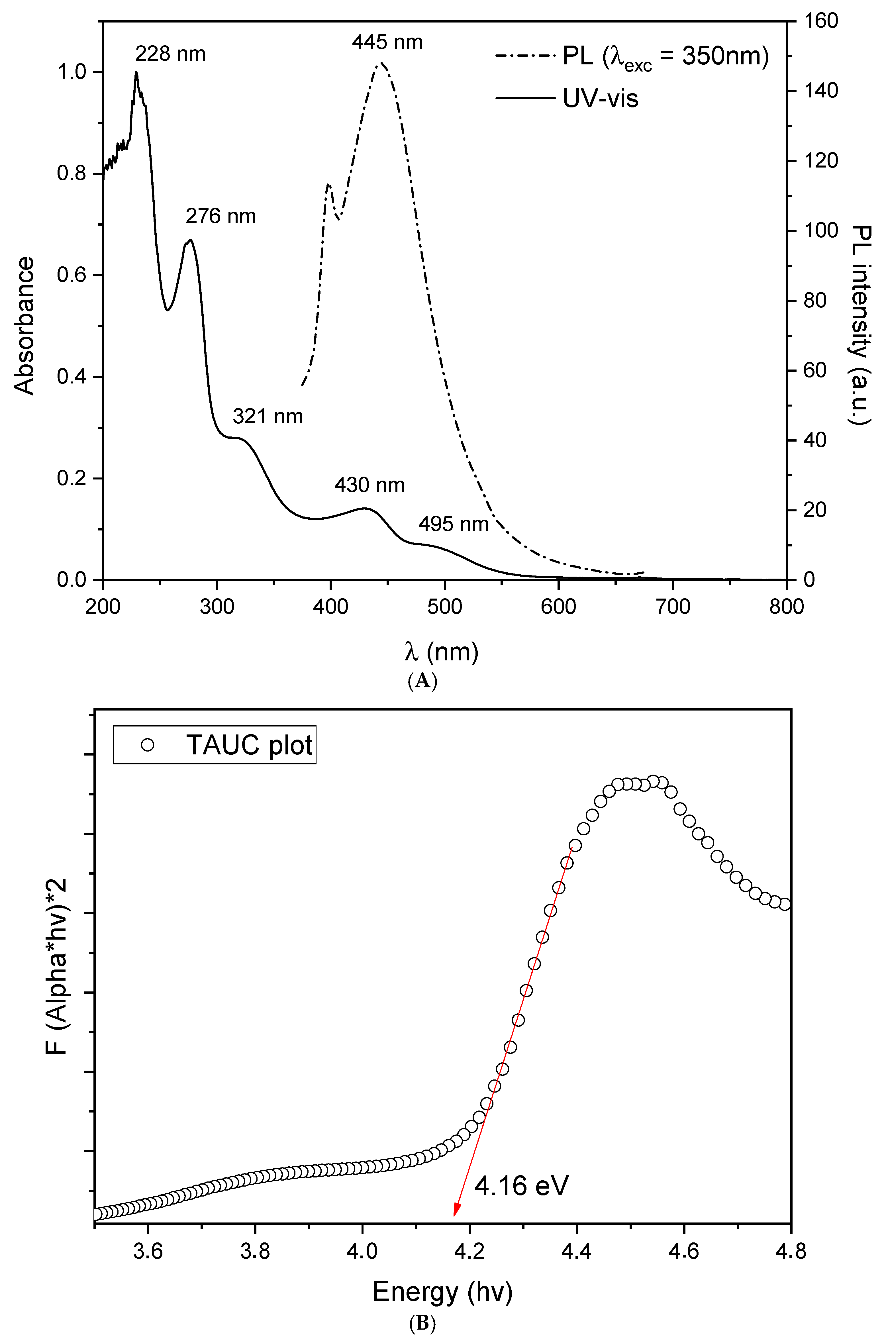
The aqueous suspension of CDots was also analyzed using a UV–Vis spectrophotometer. As shown in Figure 2A, the CDots exhibited multimodal absorption spanning the 200–800 nm range. A high-intensity band centered at 228 nm was observed, which can be attributed to the π–π transition of sp2 domains in the carbon core [51]. The band centered at 276 nm may be associated with the n → π transition in the C=O bond [52], which can arise from surface functional groups such as carboxyl and carboxylate groups [53]. The absorption bands at longer wavelengths are typically related to surface states and nanoparticle functionalization with nitrogen- or oxygen-containing groups [54], which originate from the carbon source used during the synthesis.
The optical bandgap of a material represents the minimum energy required to promote an electron from the ground state to an excited state. Therefore, materials with a larger optical bandgap require higher-energy photons to induce electronic transitions. The bandgap energy can be estimated from the absorption spectrum of the material using the Tauc equation [55].
α   ν = A ν E g n
where α is the absorption coefficient, is Planck’s constant, ν is the frequency of the incident light, A is a material-dependent constant, Eg is the optical bandgap energy, and n is an exponent that depends on the nature of the electronic transition—n = 1/2 for direct bandgap and n = 2 for indirect bandgap. In this study, the bandgap value was estimated by constructing a Tauc plot (see Figure 2B). In this plot, the linear portion of the curve is extrapolated to intersect the x-axis, and the corresponding energy value gives the bandgap, which in this case was found to be 4.16 eV. A material with such a wide optical bandgap requires high-energy photons to trigger electronic transitions, meaning it absorbs less low-energy radiation, such as visible light and most UVA and UVB radiation, making it a promising candidate for photoprotection applications [56,57].
To further characterize the optical properties of the synthesized CDots, their photoluminescence (PL) emission spectrum was measured under excitation at 350 nm. As seen in Figure 2A, a broad PL band ranging ~400–600 nm with a maximum at 445 nm was observed. This characteristic and widely reported blue emission is commonly attributed to radiative recombination through surface defect states and functional moieties containing oxygen and nitrogen [58,59]. Such groups introduce mid-gap energy levels, allowing radiative recombination. The UV–vis absorption profile and the surface-state-dominated PL are in good agreement with the FTIR results, which suggested the presence of abundant oxygenated and nitrogen-containing groups on the nanoparticle surface.
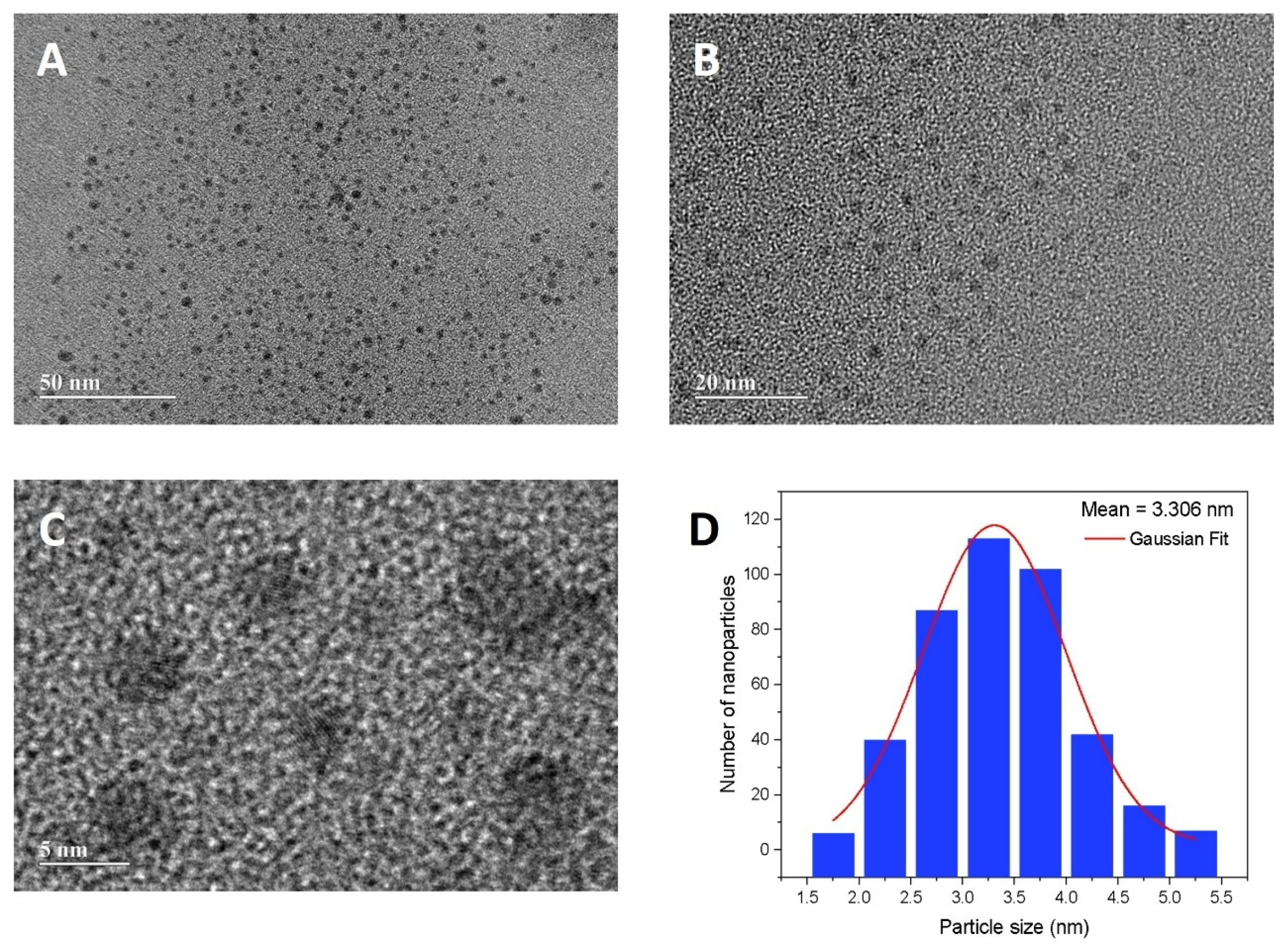
The morphology and size of the synthesized carbon dots (CDots) were investigated using high-resolution transmission electron microscopy (HRTEM). Figure 3A–C show images of the nanoparticles (in high-contrast), which exhibit an approximately spherical shape and a monodisperse distribution. The size distribution histogram presented in Figure 3D reveals that the particle diameters range from 1.8 to 5.3 nm, with an average diameter of 3.3 ± 0.7 nm, based on statistical analysis of a representative number of particles. These characterization results confirm the successful formation of nearly spherical nanoparticles via a bottom-up microwave-assisted synthesis route. This approach stands out as a more environmentally friendly, practical, and cost-effective alternative when compared to conventional synthetic methods, which often require high temperatures, toxic solvents, and prolonged reaction times [26].

3.2. Cytotoxicity Detection and Hemocompatibility

Cell viability was quantified by the MTT colorimetric assay on HaCaT cells following 48 h exposure to a dry-weight CDots stock (874.6 µg mL−1) serially diluted to 10–1000 µg mL−1. A clear, concentration-dependent decline in metabolic activity was observed, with viability decreasing progressively as the CDots concentration increased (Figure 4A). These findings demonstrate that the CDots exerted a concentration-dependent cytotoxicity effect on keratinocytes.
To further investigate the observed cytotoxicity, membrane-stabilization studies were conducted using erythrocytes (RBCs). The hemolysis assay aimed to evaluate the mechanical stability of the RBC membrane in the presence of the test compounds. In our study, the CDots sample (dry residue 103.55 mg mL−1) was tested at concentrations ranging from 5 to 1000 μg mL−1 (Figure 4B).
The cell viability by MTT revealed that varying the concentrations of CDots induced progressively significant reductions in cell viability, yielding viability values of 70.37 ± 8.0, 56.8 ± 2.5, 48.4 ± 3.6, 29.5 ± 1.6 and 6.2 ± 0.8% for 10, 100, 250, 500, and 1000 µg·mL−1, respectively (mean ± SD; n = 3). Based on linear regression of percent viability versus CDots concentration within the 100–500 µg·mL−1 range, the IC50 was 206.9 ± 36.5 µg·mL−1 (mean ± SD; n = 3).
Chatzimitakos et al. [25] evaluated the cytotoxicity of CDots obtained by Dunaliella salina microalgae derivatives in HaCaT cells at concentrations ranging from 100 to 800 μg mL−1 using the crystal violet assay. After 24 h of incubation, cell viability remained near 100% for concentrations up to 600 μg mL−1, and only at 800 μg mL−1 was a significant reduction in viability observed. Ozdemir et al. [60] reported a selective cytotoxic effect of CDots derived from Echinophora tenuifolia on different cell lines. Exposures from 0 to 100 μg mL−1 over 24, 48, and 72 h showed that, in L929 mouse fibroblasts, viability remained high (≈ 80.31%) even at the highest concentration (100 μg mL−1) after 72 h, with no statistically significant differences versus control at concentrations ≤ 25 μg mL−1. In HepG2 human hepatocellular carcinoma cells, however, CDots induced a pronounced, dose-dependent cytotoxicity, i.e., after 72 h, viability dropped to 27.87% at 100 μg mL−1.
A review by Nagoc et al. (2023) likewise noted that plant-derived CDots exhibit low cytotoxicity at concentrations up to 200 μg mL−1 [18].
CDots were evaluated in human dermal fibroblasts, where cell viability remained above 85% at concentrations up to 1000 µg·mL−1 [54]. Similarly, in normal rat myoblast L6 cells, cell viability (MTT assay) was not affected even at high concentrations of CDots (200 µg·mL−1) after 48 h of exposure [61].
Although several reports indicated relatively low cytotoxicity of CDots across diverse cell types, the studies demonstrated that higher concentrations may be substantially harmful, particularly to epithelial cells, such as HaCaT. This variability likely reflects differences in experimental conditions and CDots composition, which can affect their cellular uptake and intracellular interactions [18].
HaCaT studies with near-infrared CDs from plant sources showed no significant difference until 200 µg·mL−1 [62] and broader tolerability (viability ~60% at 1 mg·mL−1; projected LC50 ≈ 1660 µg·mL−1) [63]. Together with our IC50 in the 102–103 µg·mL−1 range, these reports suggest that cytotoxicity is strongly influenced by precursor and surface functionalities. Because HaCaT-specific literature is still scarce, we restrict cross-cell-line comparisons to qualitative context and indicate when synthesis routes or surface chemistries differ from those used in our study.
To further assess the CDots’ compatibility, their hemolytic activity against RBCs was investigated. This assay is a key indicator of membrane-damaging potential, as RBC lysis is analogous to lysosomal leakage and can induce tissue inflammation [64]. The assay revealed that even at the highest concentration tested (1000 µg/mL), the CDots induced less than 7.8% hemolysis, indicating minimal membrane-damaging potential (Figure 4B). Although a statistically significant difference (p < 0.05) compared to the negative control was observed at concentrations from 300 µg/mL upwards, the low overall percentage of lysis confirmed that the CDots exhibited excellent hemocompatibility. This finding agrees with previous reports, such as that of Wang et al. [51], who observed significant hemolysis (>5%) only at much higher concentrations (above 2500 µg/mL).

4. Antioxidant Activity

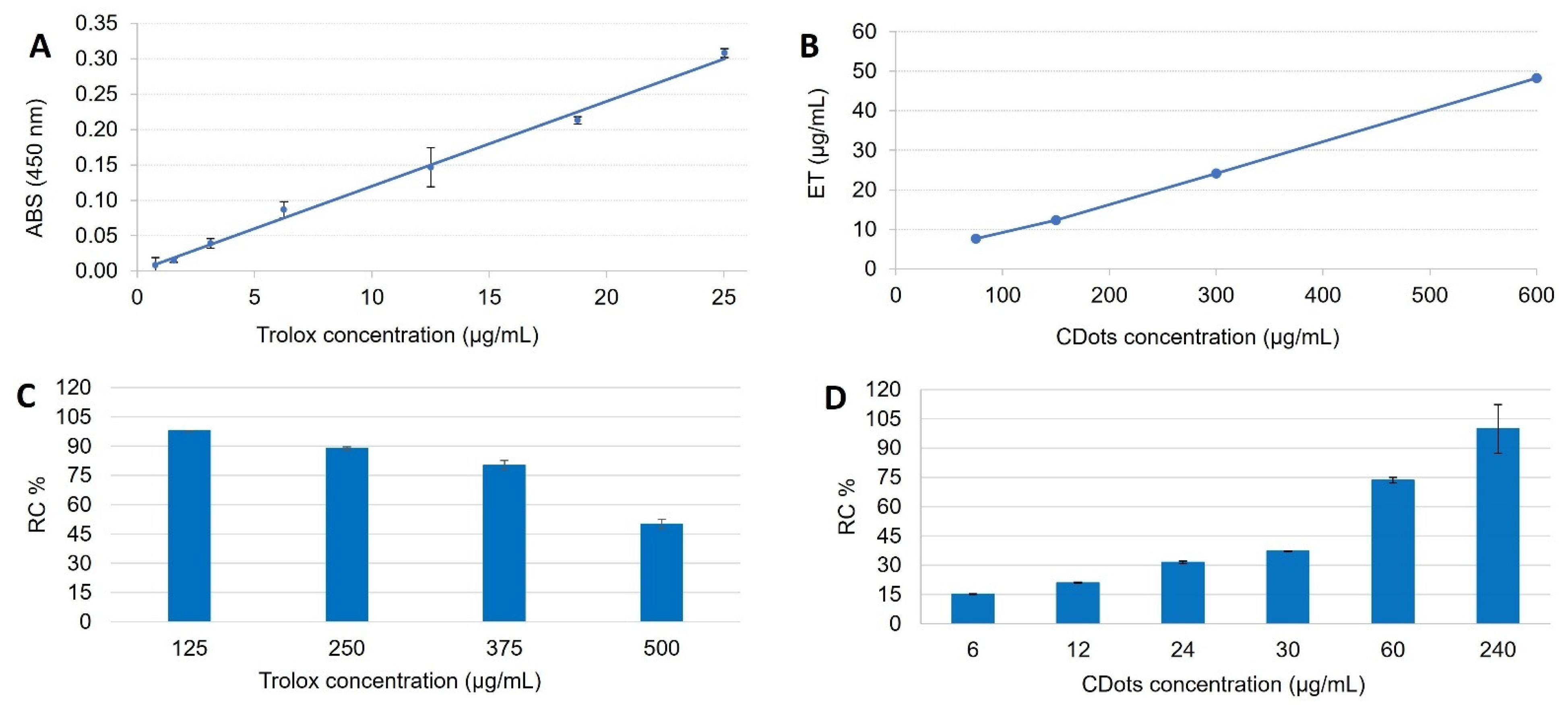
The antioxidant potential of the CDots was assessed by its cupric ion (Cu2+) reducing capacity and free-radical scavenging activity, measured by electron paramagnetic resonance (EPR), with Trolox used as the reference. Trolox, a water-soluble form of vitamin E, is an antioxidant widely employed as a reference standard due to its well-established activity [65]. In the linear regression analysis using the least-squares method, the linear correlation coefficient obtained from the Trolox curve was r = 0.9999 (Figure 5A), which is considered highly acceptable (r > 0.99) and indicative of adequate linearity profile. Additionally, the Pearson coefficient of determination (r2) was 0.9952 for the evaluated concentrations, confirming a strong linear correlation between absorbance and the concentration of the antioxidant sample.
Antioxidant activity was also evaluated by electron paramagnetic resonance (EPR), employing the DPPH radical as a marker. The CDots sample were systematically dispersed, and different concentrations were tested until reaching a range in which the samples exhibited a significant distinction in antioxidant activity. As illustrated in Figure 5B, the double integral of the EPR spectra represents the intensity of the DPPH signal, which is proportional to the remaining free radical population in the sample comparable with standard Trolox.
The CDots reducing capacity (6% w/v, dry mass) was evaluated at concentrations of 75, 150, 300, and 600 μg mL−1, corresponding to 7.64, 12.28, 24.14, and 48.31 μg mL−1 (r2 = 0.999) of TEAC, respectively (Figure 5A,B), as calculated by the TEAC equation. At an isolated concentration of 10 μg mL−1, the samples exhibited 24.62% antioxidant activity relative to Trolox, in addition to displaying dose-dependent antioxidant activity.
The results in Figure 5C,D indicated that increasing the concentration of Trolox and CDots progressively reduced the double integral of the DPPH-EPR signal, evidencing their antioxidant action through the consumption of free radicals, a dose-dependent effect was observed, and CDots showed greater capacity than Trolox to scavenge DPPH radicals. With a 99.89% reduction in the initial DPPH concentration (250 µM) in the presence of 240 μg mL−1 of CDots solution (equivalent to 14.4 μg mL−1 of CDots dry mass). This finding reinforces the material’s high antioxidant capacity, corroborating the results obtained from the CUPRAC assay and highlighting the potential of CDots as effective free radical scavengers [18,22].
The combined results from antioxidant activity methods confirmed that CDots exhibited significant antioxidant activity, both via the reduction in cupric ions (CUPRAC) and the inactivation of free radicals (EPR-DPPH). This performance suggested that CDots may serve as a potent antioxidant, with potential biomedical and technological applications. Further studies are required to elucidate the underlying mechanisms of this activity and to assess their efficacy in more complex biological matrices.
Ozdemir et al. [60] produced red-emissive carbon quantum dots from the medicinal herb Echinophora tenuifolia by a rapid microwave route. Antioxidant performance assessed with the CUPRAC assay revealed a reducing power comparable to, and in some dilutions exceeding, that of the crude plant extract, while the dots retained high photostability and biocompatibility, underscoring their dual imaging/antioxidant promise.
Kasif et al. [66] reported a one-step hydrothermal synthesis of N,S-codoped carbon dots. Electron paramagnetic resonance(EPR) spectroscopy showed these dots scavenged >80% of model free radicals at sub-milligram-per-milliliter levels, combined with negligible cytotoxicity toward mammalian cells, the data highlight their suitability as potent antioxidant nanocarriers for biomedical and cosmetic applications.
Li et al. [22] investigated carbon dots synthesized via a green microfluidic process using carrot juice as a feedstock. The authors applied the DPPH• radical-scavenging assay to quantify the antioxidant capacity of the CDs. Their results demonstrated a high level of free-radical removal, with even low concentrations of CDs causing a significant decrease in DPPH• absorbance, indicative of efficient radical neutralization. Moreover, the study reported low cytotoxicity of these nanomaterials, highlighting their potential as multifunctional agents in sunscreen formulations: beyond UV-blocking, they serve as powerful free-radical scavengers capable of mitigating oxidative processes associated with cutaneous photoaging.

5. In Vitro SPF

The in vitro functional evaluation of SPF is used as a preliminary assessment in sunscreen development. The in vitro SPF of the samples containing CDots, together with well-established organic UV filters (BMDBM and EHMC) at their maximum concentrations the European cosmetic regulation (Regulation (EC) No. 1223/2009) [67], was determined using the standardized method proposed by (COLIPA, 2009) [40]. The critical wavelength (cλ) is another key parameter in efficacy evaluation, as it indicates whether the sunscreen achieves broad-spectrum protection. It corresponds to the longest UV wavelength that a sunscreen can effectively absorb/filter and is calculated as the point in the absorption spectrum at which the area under the curve reaches 90% of the total sample absorption within the 290–400 nm range [68]. The U.S. Food and Drug Administration (FDA) defines 370 nm as the minimum cλ for broad-spectrum sunscreens, which must also exhibit an SPF of at least 15 [69].
The formulation containing 4.5% CDots (dry residue) achieved a 619% increase in SPF value (from 26 ± 13.0 to 161 ± 7.8). With respect to critical wavelength (λc), both the control and CDots containing formulations exceeded 380 nm (Table 2).
With respect to photostability, the CDots-enriched sample retained 90.2% of its initial SPF after 30 min of solar-simulator exposure, whereas the control preserved only 56% under the same conditions. After 90 min, the CDots sample provided measurable protection (14.3 ± 2.2; 9% of the initial SPF), a value comparable to that of the control after merely 30 min. A decrease in SPF after irradiation is expected, especially in the control sample containing BMDBM/EHMC, as BMDBM, a widely used UVA filter, is known to undergo photodegradation due to multiple isomerization pathways upon UV exposure [70]. However, following 30 min of exposure, the control formulation fell below the SPF threshold, while the CDots-enriched sunscreen continued to satisfy the requirement.
As shown in Table 2, both the CDots sample and the control were classified as effective for broad-spectrum photoprotection (cλ above 370 nm and SPF higher than 15). The UVA/UVB absorbance ratio was 0.766 for the CDots formulation versus 0.717 for the control (pre-irradiation), indicating a modest shift toward UVA attenuation that may contribute to more balanced broad-spectrum coverage.
Recent literature indicated that CDots are emerging as multifunctional cosmetic ingredients capable of broad-spectrum UV absorption, ROS suppression, and formulation transparency. A 2023 review on plant extract-derived CDots emphasized their inherent antioxidant, anti-inflammatory, and UV-filtering properties, positioning them as sustainable alternatives for next-generation sunscreens [18].
Although some studies have suggested photoprotective activity, to the best of our knowledge, none of them have evaluated the functional in vitro SPF using diffuse reflectance spectrophotometry with an integrating sphere, as performed in our investigation. The photoprotective performance of modified CDots, synthesized from citric acid and nitrogen compounds, was confirmed through both in vivo human trials and theoretical analysis. The material prevented skin erythema at radiation doses 5x the minimal required (5 MED) and showed a theoretically calculated SPF of approximately, 22 [71].
The synergistic effect of CDots with traditional inorganic UV filters has also been investigated. Researchers developed transparent composite films by combining CDots with nanocellulose and ZnO nanostructures. The results indicated that the addition of CDots significantly enhanced the UV-filtering capability of the films compared to those containing only nanocellulose and ZnO. This demonstrated that CDots can act as effective enhancers for inorganic filters, improving the overall photoprotective performance of the final material [16].

6. Limitations and Challenges

Despite the promising results, this study has limitations that pave the way for future research. While CDots are widely reported as biocompatible, their environmental safety depends on dose, surface chemistry, environmental concentration, and transformation. The evaluation of photoprotective efficacy was conducted exclusively through in vitro methods. Therefore, the UVA Protection Factor (UVA-PF) was not quantified, as determination is ordinarily paired with in vivo SPF testing, which we deferred pending completion of our safety assessment.
Although the antioxidant activity and SPF boost are clear, the exact photophysical mechanisms by which CDots interact with organic UV filters to enhance photostability remain to be elucidated. As for future perspectives, conducting cytotoxicity assays is suggested to ensure safety for cosmetic application, and environmental risks to large scale is a point raised for future exploration
Optimizing the CDots concentration in the formulation is another important avenue, aiming to balance maximum efficacy, cost, and sensory attributes. Finally, the long-term physicochemical stability of the final formulation should be investigated to ensure its commercial viability.
Accordingly, we report in vitro SPF as a comparative screening outcome on PMMA plates that mimic skin microrelief. Therefore, the absolute values reported should not equated with in vivo SPF. Performance should be validated using methods compliant with current regulatory requirements.

7. Conclusions

Coffee leaf-derived CDots were prepared via a rapid, low-cost microwave process combining nanometric size, broad UV absorption, and potent antioxidant activity. When incorporated at 4.5% (w/w, dry basis) into a sunscreen system, they increased the in vitro SPF by more than six-fold while preserving a critical wavelength ≥ 380 nm and limiting SPF loss to 10% after 30 min of intense artificial UV exposure. These findings highlighted CDots as sustainable, multifunctional boosters that enhance efficacy and photostability of conventional formulations, providing a credible pathway toward greener, high-performance sun-care products. Further in vivo efficacy, safety, and stability studies are warranted to translate this laboratory’s evidence into commercial and regulatory success.

Author Contributions

G.T.M. took part of writing—original draft, investigation and experimental. C.R.C. contributed with writing—original draft and participated of data analysis. C.R.C., I.d.S.O., L.N.F., F.D.F., F.L.P., P.E.N.d.S., A.S.F. and A.L.G.J. were involved in investigation and experimental analysis. A.R.B. provided the laboratory, conceptualization, methodology to measure SPF and photostability. G.R.S.A. took part of conceptualization, writing—original draft, review and editing, supervision of the obtained data of CDots synthesis and characterization. F.V.L.S.P. involved of conceptualization, supervision, project administration and funding acquisition. All authors have read and agreed to the published version of the manuscript.

Funding

This work was funded by Fundação de Amparo à Pesquisa do Espírito Santo, FAPES (grant number 952/2022 and 658/2024). G.T.M is highly thankful to Universidade Federal do Espírito Santo (UFES) and to Conselho Nacional de Desenvolvimento Científico e Tecnológico (CNPq) for the undergraduate research scholarship. A.R.B. thanks CNPq for the Research Productivity Scholarship (process 303862/2022-0); and the Fundação de Amparo à Pesquisa do Estado de São Paulo, FAPESP (grant number 2019/16169-0). A.S.F. and A.L.G.J. thank the CBIOS funding from FCT Foundation for Science and Technology, I.P., under the UIDB/04567/2020 and UIDP/04567/2020 projects.

Institutional Review Board Statement

Not applicable.

Informed Consent Statement

Not applicable.

Data Availability Statement

The original contributions presented in this study are included in the article. Further inquiries can be directed to the corresponding authors.

Acknowledgments

The authors are also grateful to Multi-User High-Resolution Microscopy Laboratory (LabMic/UFG) for the HRTEM images.

Conflicts of Interest

The authors declare no conflict of interest.

References

  1. Marionnet, C.; Tran, C.; Bastien, P.; Bielicki, A.; Golebiewski, C.; Vieu, D.-L.; Suida-Batista, A.; Candau, D.; Bernerd, F. A broader filtration of UVA1 wavelengths improves skin photoprotection. J. Dermatol. Sci. 2018, 91, 337–340. [Google Scholar] [CrossRef]
  2. Chhabra, G.; Garvey, D.R.; Singh, C.K.; Mintie, C.A.; Ahmad, N. Effects and Mechanism of Nicotinamide Against UVA- and/or UVB-mediated DNA Damages in Normal Melanocytes. Photochem. Photobiol. 2019, 95, 331–337. [Google Scholar] [CrossRef]
  3. Cohen, G.; Jakus, J.; Portillo, M.; Gvirtz, R.; Ogen-Shtern, N.; Silberstein, E.; Ayzenberg, T.; Rozenblat, S. In vitro, ex vivo, and clinical evaluation of anti-aging gel containing EPA and CBD. J. Cosmet. Dermatol. 2023, 22, 3047–3057. [Google Scholar] [CrossRef] [PubMed]
  4. Yang, J.-W.; Fan, G.-B.; Tan, F.; Kong, H.-M.; Liu, Q.; Zou, Y.; Tan, Y.-M. The role and safety of UVA and UVB in UV-induced skin erythema. Front. Med. 2023, 10, 1163697. [Google Scholar] [CrossRef] [PubMed]
  5. Cadet, J.; Douki, T.; Ravanat, J.-L. Oxidatively Generated Damage to Cellular DNA by UVB and UVA Radiation. Photochem. Photobiol. 2015, 91, 140–155. [Google Scholar] [CrossRef]
  6. Chen, L.; Hu, J.Y.; Wang, S.Q. The role of antioxidants in photoprotection: A critical review. J. Am. Acad. Dermatol. 2012, 67, 1013–1024. [Google Scholar] [CrossRef]
  7. Khan, M.A. Sun protection factor determination studies of some sunscreen formulations used in cosmetics for their selection. J. Drug Deliv. Ther. 2018, 8, 149–151. [Google Scholar] [CrossRef]
  8. Jallad, K.N. Chemical characterization of sunscreens composition and its related potential adverse health effects. J. Cosmet. Dermatol. 2017, 16, 353–357. [Google Scholar] [CrossRef]
  9. Nery, É.M.; Martinez, R.M.; Velasco, M.V.R.; Baby, A.R. A short review of alternative ingredients and technologies of inorganic UV filters. J. Cosmet. Dermatol. 2021, 20, 1061–1065. [Google Scholar] [CrossRef]
  10. Nash, J.F.; Tanner, P.R. Relevance of UV filter/sunscreen product photostability to human safety. Photodermatol. Photoimmunol. Photomed. 2014, 30, 88–95. [Google Scholar] [CrossRef]
  11. Downs, C.A.; Bishop, E.; Diaz-Cruz, M.S.; Haghshenas, S.A.; Stien, D.; Rodrigues, A.M.S.; Woodley, C.M.; Sunyer-Caldú, A.; Doust, S.N.; Espero, W.; et al. Oxybenzone contamination from sunscreen pollution and its ecological threat to Hanauma Bay, Oahu, Hawaii, U.S.A. Chemosphere 2022, 291, 132880. [Google Scholar] [CrossRef] [PubMed]
  12. Perumal, S.; Atchudan, R.; Edison, T.N.J.I.; Lee, Y.R. Sustainable synthesis of multifunctional carbon dots using biomass and their applications: A mini-review. J. Environ. Chem. Eng. 2021, 9, 105802. [Google Scholar] [CrossRef]
  13. Rabe, D.I.A.; Awak, M.M.A.; Yang, F.; Okonjo, P.A.; Dong, X.; Teisl, L.R.; Wang, P.; Tang, Y.; Pan, N.; Sun, Y.-P.; et al. The dominant role of surface functionalization in carbon dots’ photo-activated antibacterial activity. Int. J. Nanomed. 2019, 14, 2655–2665. [Google Scholar] [CrossRef]
  14. Robein, Y.; Ambrusi, R.E.; Pronsato, M.E.; Di Nezio, M.S.; Brizuela, G. Brizuela A theoretical study of the functionalized carbon dots surfaces binding with silver nanostructures. Comput. Theor. Chem. 2023, 1223, 114087. [Google Scholar] [CrossRef]
  15. Fang, M.; Wang, B.; Qu, X.; Li, S.; Huang, J.; Li, J.; Lu, S.; Zhou, N. State-of-the-art of biomass-derived carbon dots: Preparation, properties, and applications. Chin. Chem. Lett. 2024, 35, 108423. [Google Scholar] [CrossRef]
  16. Feng, X.; Zhao, Y.; Jiang, Y.; Miao, M.; Cao, S.; Fang, J. Use of carbon dots to enhance UV-blocking of transparent nanocellulose films. Carbohydr. Polym. 2017, 161, 253–260. [Google Scholar] [CrossRef]
  17. DePaula, J.; Partelli, F.L.; Batista, A.M.; Calado, V.; Farah, A. Major Bioactive Compounds in Seeds, Husks, and Leaves of Selected Genotypes of Coffea canephora cv. Conilon from Three Consecutive Crops. Plants 2025, 14, 1040. [Google Scholar] [CrossRef]
  18. Ngoc, L.T.N.; Moon, J.-Y.; Lee, Y.-C. Plant Extract-Derived Carbon Dots as Cosmetic Ingredients. Nanomaterials 2023, 13, 2654. [Google Scholar] [CrossRef]
  19. Cheng, B.; Yang, Z.; Chen, F.; Yue, L.; Cao, X.; Li, J.; Qian, H.-L.; Yan, X.-P.; Wang, C.; Wang, Z. Biomass-derived carbon dots with light conversion and nutrient provisioning capabilities facilitate plant photosynthesis. Sci. Total Environ. 2023, 901, 165973. [Google Scholar] [CrossRef] [PubMed]
  20. Wang, H.; Kang, Y.; Yang, N.; Li, H.; Huang, S.; Liang, Z.; Zeng, G.; Huang, Y.; Li, W.; Zheng, M.; et al. Inhibition of UV-B stress in lettuce through enzyme-like Scutellaria baicalensis carbon dots. Ecotoxicol. Environ. Saf. 2022, 246, 114177. [Google Scholar] [CrossRef] [PubMed]
  21. de Paula Bernado, W.; Santos, A.R.; Vale, E.M.; Pireda, S.; Correia, L.Z.; Desouza, G.A.R.; de Abreu, D.P.; Carvalho, L.K.O.; Almeida, F.A.; Baroni, D.F. UV-B reduction and excess: Management strategies regarding Coffea sp. crop. Sci. Hortic. 2024, 323, 112499. [Google Scholar] [CrossRef]
  22. Li, Z.; Miao, Z.; Shen, J.; Wang, J.; Lin, L. Green synthesis of carbon dots by microflow method and their application as sunscreen agent. Chin. J. Chem. Eng. 2025, 77, 135. [Google Scholar] [CrossRef]
  23. Campalani, C.; Causin, V.; Selva, M.; Perosa, A. Fish-Waste-Derived Gelatin and Carbon Dots for Biobased UV-Blocking Films. ACS Appl. Mater. Interfaces 2022, 14, 35148–35156. [Google Scholar] [CrossRef]
  24. Chen, C.; Yu, L.; Li, X.; Yu, Z.; Song, D.; Wang, S.; Li, F.; Jiang, S.; Chen, Y.; Xu, J.; et al. Reducing Oxidative Stress Levels and Inhibiting Aging by l-Cysteine-Derived Carbon Dots with Highly Efficient Broad-Spectrum UV Absorption. ACS Appl. Mater. Interfaces 2024, 16, 43189–43198. [Google Scholar] [CrossRef]
  25. Chatzimitakos, T.G.; Kasouni, A.; Troganis, A.; Leonardos, I.; Tzovenis, I.; Ntzouvaras, A.; Stalikas, C. Carbon Nanodots Synthesized from Dunaliella salina as Sun Protection Filters. C 2020, 6, 69. [Google Scholar] [CrossRef]
  26. Davi, L.B.O.; Silva, M.S.; Ferreira, R.L.; Muniz, W.; Ribeiro, A.S.; Lima, D.J.P.; de Oliveira, I.N.; Barbosa, C.D.A.E.S. Multifunctional carbon dots derived from dansyl chloride for ratiometric thermal sensor and reactive oxygen generation. Dye. Pigment. 2021, 194, 109549. [Google Scholar] [CrossRef]
  27. Kumawat, M.; Thakur, M.; Gurung, R.; Srivastava, R. Graphene Quantum Dots from Mangifera indica: Application in Near-Infrared Bioimaging and Intracellular Nano-thermometry. ACS Sustain. Chem. Eng. 2016, 5, 1382–1391. [Google Scholar] [CrossRef]
  28. Agência Nacional de Vigilância Sanitária (ANVISA). Farmacopeia Brasileira, 6th ed.; Method 5.2.9 Loss on Drying; Agência Nacional de Vigilância Sanitária: Brasília, Brazil, 2019; Volume 1.
  29. Flórido, A.; Saraiva, N.; Cerqueira, S.; Almeida, N.; Parsons, M.; Batinic-Haberle, I.; Miranda, J.P.; Costa, J.G.; Carrara, G.; Castro, M.; et al. The manganese(III) porphyrin MnTnHex-2-PyP5+ modulates intracellular ROS and breast cancer cell migration: Impact on doxorubicin-treated cells. Redox Biol. 2019, 20, 367–378. [Google Scholar] [CrossRef]
  30. Labu, Z.K.; Laboni, F.R.; Tarafdar, M.; Howlader, M.S.I.; Rashid, M.H. Membrane Stabilization as a Mechanism of Anti-Inflammatory and Thrombolytic Activities of Ethanolic Extract of Arial Parts of Spondiasis pinanata (Family: Anacardiaceae). Pharmacologyonline 2015, 2, 44–51. [Google Scholar]
  31. Sarveswaran, R.; Jayasuriya, W.J.A.B.; Thusharie, S.N.M.T.S.S. In vitro assays to investigate the anti-inflammatory activity of herbal extracts: A Review. World J. Pharm. Res. 2017, 6, 131–141. [Google Scholar]
  32. Apak, R.; Güçlü, K.; Özyürek, M.; Karademir, S.E. Novel Total Antioxidant Capacity Index for Dietary Polyphenols and Vitamins C and E, Using Their Cupric Ion Reducing Capability in the Presence of Neocuproine:  CUPRAC Method. J. Agric. Food Chem. 2004, 52, 7970–7981. [Google Scholar] [CrossRef]
  33. Munteanu, I.G.; Apetrei, C. Analytical Methods Used in Determining Antioxidant Activity: A Review. Int. J. Mol. Sci. 2021, 22, 3380. [Google Scholar] [CrossRef]
  34. Özyürek, M.; Güçlü, K.; Tütem, E.; Başkan, K.S.; Erçağ, E.; Çelik, S.E.; Baki, S.; Yıldız, L.; Karaman, Ş.; Apak, R. A comprehensive review of CUPRAC methodology. Anal. Methods 2011, 3, 2439–2453. [Google Scholar] [CrossRef]
  35. Ferreira-Nunes, R.; da Silva, S.M.M.; de Souza, P.E.N.; de Magalhães, P.O.; Cunha-Filho, M.; Gratieri, T.; Gelfuso, G.M. Incorporation of Eugenia dysenterica extract in microemulsions preserves stability, antioxidant effect and provides enhanced cutaneous permeation. J. Mol. Liq. 2018, 265, 408–415. [Google Scholar] [CrossRef]
  36. Ordoñez Lozada, M.I.; Rodrigues Maldonade, I.; Bobrowski Rodrigues, D.; Silva Santos, D.; Ortega Sanchez, B.A.; Narcizo de Souza, P.E.; Longo, J.P.; Bernardo Amaro, G.; de Lacerda de Oliveira, L. Physicochemical characterization and nano-emulsification of three species of pumpkin seed oils with focus on their physical stability. Food Chem. 2021, 343, 128512. [Google Scholar] [CrossRef]
  37. Alves, T.E.P.; Kolodziej, C.; Burda, C.; Franco, A. Effect of particle shape and size on the morphology and optical properties of zinc oxide synthesized by the polyol method. Mater. Des. 2018, 146, 125–133. [Google Scholar] [CrossRef]
  38. Souza Neto, A.V.; Balla, D.Q.; Candido, T.M.; Rosado, C.; Baby, A.R.; Pessoa, F.V.L.S. Effect of an Emollient Emulsion Containing 15.0% of Caprylic/Capric Triglyceride on the Urocanic Acid of the Stratum Corneum. Life 2023, 13, 876. [Google Scholar] [CrossRef]
  39. Silva Marcelino, P.; Martinez, R.M.; Daneluti, A.L.M.; Morocho-Jácome, A.L.; Pessoa, F.V.L.S.; Rijo, P.; Rosado, C.; Velasco, M.V.R.; Baby, A.R. In Vitro Photoprotection and Functional Photostability of Sunscreen Lipsticks Containing Inorganic Active Compounds. Cosmetics 2023, 10, 46. [Google Scholar] [CrossRef]
  40. COLIPA. Method for the In Vitro Determination of UVA Protection Provided by Sunscreen Products; European Cosmetics Association: Brussels, Belgium, 2009. [Google Scholar]
  41. Matts, P.J.; Alard, V.; Brown, M.W.; Ferrero, L.; Gers-Barlag, H.; Issachar, N.; Moyal, D.; Wolber, R. The COLIPA in vitro UVA method: A standard and reproducible measure of sunscreen UVA protection. Int. J. Cosmet. Sci. 2010, 32, 35–46. [Google Scholar] [CrossRef] [PubMed]
  42. Popiół, J.; Gunia-Krzyżak, A.; Piska, K.; Żelaszczyk, D.; Koczurkiewicz, P.; Słoczyńska, K.; Wójcik-Pszczoła, K.; Krupa, A.; Kryczyk-Poprawa, A.; Żesławska, E.; et al. Discovery of Novel UV-Filters with Favorable Safety Profiles in the 5-Arylideneimidazolidine-2,4-dione Derivatives Group. Molecules 2019, 24, 2321. [Google Scholar] [CrossRef] [PubMed]
  43. Scalia, S.; Mezzena, M. Photostabilization Effect of Quercetin on the UV Filter Combination, Butyl Methoxydibenzoylmethane–Octyl Methoxycinnamate. Photochem. Photobiol. 2010, 86, 273–278. [Google Scholar] [CrossRef] [PubMed]
  44. Egorova, M.; Tomskaya, A.; Smagulova, S.A. Optical Properties of Carbon Dots Synthesized by the Hydrothermal Method. Materials 2023, 16, 4018. [Google Scholar] [CrossRef]
  45. Hijazi, S.; Ganie, A.S.; Rahman, M.M.; Shah, W.A. Facile synthesis of surface-functionalized fluorescent carbon quantum dots for the selective detection of ferric ions. Environ. Sci. Nano 2023, 10, 3281–3294. [Google Scholar] [CrossRef]
  46. Singaravelu, C.M.; Deschanels, X.; Rey, C.; Causse, J. Investigation on Fluorescence Origin and Spectral Heterogeneity in Carbon Dots: A Dynamic Perspective. ChemPhotoChem 2024, 8, e202400044. [Google Scholar] [CrossRef]
  47. Nandiyanto, A.B.D.; Oktiani, R.; Ragadhita, R. How to Read and Interpret FTIR Spectroscope of Organic Material. Indones. J. Sci. Technol. 2019, 4, 97–118. [Google Scholar] [CrossRef]
  48. Macedo, T.M.; Paulo, Í.S.; Dusi, R.G.; Magalhães, A.O.; Ferreira, E.S.; de Freitas Espeleta, A.; Lucchese, A.M. Innovations in dermocosmetics: Carbon Dot applications on human skin. MRS Commun. 2025; in press. [Google Scholar] [CrossRef]
  49. Dong, C.; Xu, M.; Wang, S.; Ma, M.; Akakuru, O.U.; Ding, H.; Wu, A.; Zha, Z.; Wang, X.; Bi, H. Fluorescent carbon dots with excellent moisture retention capability for moisturizing lipstick. J. Nanobiotechnol. 2021, 19, 299. [Google Scholar] [CrossRef]
  50. Li, Q.; Shen, X.; Xing, D. Carbon quantum dots as ROS-generator and -scavenger: A comprehensive review. Dye. Pigment. 2023, 208, 110784. [Google Scholar] [CrossRef]
  51. Wang, W.; Chen, J.; Wang, D.; Shen, Y.; Yang, L.; Zhang, T.; Ge, J. Facile synthesis of biomass waste-derived fluorescent N, S, P co-doped carbon dots for detection of Fe3+ ions in solutions and living cells. Anal. Methods 2021, 13, 789–795. [Google Scholar] [CrossRef]
  52. Liu, Y.; Huang, H.; Cao, W.; Mao, B.; Liu, Y.; Kang, Z. Advances in carbon dots: From the perspective of traditional quantum dots. Mater. Chem. Front. 2020, 4, 1586–1613. [Google Scholar] [CrossRef]
  53. Piasek, A.; Pulit-Prociak, J.; Zielina, M.; Banach, M. Fluorescence of D-Glucose-Derived Carbon Dots: Effect of Process Parameters. J. Fluoresc. 2024, 34, 1693–1705. [Google Scholar] [CrossRef]
  54. Silva, L.E.; de Calado, O.L.L.; de Oliveira Silva, S.F.; da Silva, K.R.M.; Henrique Almeida, J.; de Oliveira Silva, M.; da Viana, R.S.; de Souza Ferro, J.N.; de Almeida Xavier, J.; Barbosa, C.D.A.E.S. Lemon-derived carbon dots as antioxidant and light emitter in fluorescent films applied to nanothermometry. J. Colloid Interface Sci. 2023, 651, 678–685. [Google Scholar] [CrossRef]
  55. Darwesh, A.H.A.; Mohammed, P.A.; Mamand, S.M.; Hussen, S.A.; Aziz, S.B.; Brza, M.A.; Abdullah, R.M.; Karim, W.O. Investigation of Structural and Optical Characteristics of Biopolymer Composites Based on Polyvinyl Alcohol Inserted with PbS Nanoparticles. Coatings 2023, 13, 578. [Google Scholar] [CrossRef]
  56. Al-Attafi, K.; Al-Keisy, A.; Alsherbiny, M.A.; Kim, J.H. Zn2SnO4 ternary metal oxide for ultraviolet radiation filter application: A comparative study with TiO2 and ZnO. Sci. Technol. Adv. Mater. 2023, 24, 2277678. [Google Scholar] [CrossRef] [PubMed]
  57. Silva, M.R.F.; Alves, M.F.R.P.; Cunha, J.P.G.Q.; Costa, J.L.; Silva, C.A.; Fernandes, M.H.V.; Vilarinho, P.M.; Ferreira, P. Nanostructured transparent solutions for UV-shielding: Recent developments and future challenges. Mater. Today Phys. 2023, 35, 101131. [Google Scholar] [CrossRef]
  58. Lei, M.; Xie, Y.; Chen, L.; Liu, X.; Yang, Y.; Zheng, J.; Li, Q. Surface state modulation of blue-emitting carbon dots with high quantum yield and high product yield. RSC Adv. 2022, 12, 27431–27441. [Google Scholar] [CrossRef]
  59. Yan, F.; Jiang, Y.; Sun, X.; Bai, Z.; Zhang, Y.; Zhou, X. Surface modification and chemical functionalization of carbon dots: A review. Microchim. Acta 2018, 185, 424. [Google Scholar] [CrossRef]
  60. Ozdemir, N.; Tan, G.; Tevlek, A.; Arslan, G.; Zengin, G.; Sargin, I. Dead Cell Discrimination with Red Emissive Carbon Quantum Dots from the Medicinal and Edible Herb Echinophora tenuifolia. J. Fluoresc. 2025; in press. [Google Scholar] [CrossRef]
  61. Jhonsi, M.A.; Ananth, D.A.; Nambirajan, G.; Sivasudha, T.; Yamini, R.; Bera, S.; Kathiravan, A. Antimicrobial activity, cytotoxicity and DNA binding studies of carbon dots. Spectrochim. Acta Part A Mol. Biomol. Spectrosc. 2018, 196, 295–302. [Google Scholar] [CrossRef] [PubMed]
  62. Shi, W.; Li, J.; Pu, J.; Cheng, G.; Liu, Y.; Xiao, S.; Cao, J. Epigynum auritum-Derived Near-Infrared Carbon Dots for Bioimaging and Antimicrobial Applications. Molecules 2025, 30, 422. [Google Scholar] [CrossRef]
  63. Yalshetti, S.; Thokchom, B.; Bhavi, S.M.; Singh, S.R.; Patil, S.R.; Harini, B.P.; Sillanpää, M.; Manjunatha, J.G.; Srinath, B.S.; Yarajarla, R.B. Microwave-assisted synthesis, characterization and in vitro biomedical applications of Hibiscus rosa-sinensis Linn.-mediated carbon quantum dots. Sci. Rep. 2024, 14, 9915. [Google Scholar] [CrossRef]
  64. Alghareeb, S.A.; Alsughayyir, J.; Alfhili, M.A. Stimulation of Hemolysis and Eryptosis by α-Mangostin through Rac1 GTPase and Oxidative Injury in Human Red Blood Cells. Molecules 2023, 28, 6495. [Google Scholar] [CrossRef] [PubMed]
  65. Silvestrini, A.; Meucci, E.; Ricerca, B.M.; Mancini, A. Total Antioxidant Capacity: Biochemical Aspects and Clinical Significance. Int. J. Mol. Sci. 2023, 24, 10978. [Google Scholar] [CrossRef] [PubMed]
  66. Kasif, M.; Alarifi, A.; Afzal, M.; Thirugnanasambandam, A. N, S-codoped carbon dots for antioxidants and their nanovehicle potential as molecular cargoes. RSC Adv. 2024, 14, 32041–32052. [Google Scholar] [CrossRef] [PubMed]
  67. European Union. Regulation (EC) No. 1223/2009 of the European Parliament and of the Council of 30 November 2009 on Cosmetic Products. Official Journal of the European Union, 1223/2009. 2009, pp. 59–209. Available online: https://eur-lex.europa.eu/legal-content/EN/ALL/?uri=celex%3A32009R1223 (accessed on 10 August 2025).
  68. Stevanato, R.; Bertelle, M.; Fabris, S. Photoprotective characteristics of natural antioxidant polyphenols. Regul. Toxicol. Pharmacol. 2014, 69, 71–77. [Google Scholar] [CrossRef]
  69. FDA. Sunscreen Drug Products for Over-the-Counter Human Use. Fed. Regist. 2019, 84, 98–355. [Google Scholar]
  70. Berenbeim, J.A.; Wong, N.G.K.; Cockett, M.C.R.; Berden, G.; Oomens, J.; Rijs, A.M.; Dessent, C.E.H. Unravelling the Keto–Enol Tautomer Dependent Photochemistry and Degradation Pathways of the Protonated UVA Filter Avobenzone. J. Phys. Chem. A 2020, 124, 2919–2930. [Google Scholar] [CrossRef]
  71. Hu, G.; Lei, B.; Jiao, X.; Wu, S.; Zhang, X.; Zhuang, J.; Liu, X.; Hu, C.; Liu, Y. Synthesis of modified carbon dots with performance of ultraviolet absorption used in sunscreen. Opt. Express 2019, 27, 7629–7641. [Google Scholar] [CrossRef]
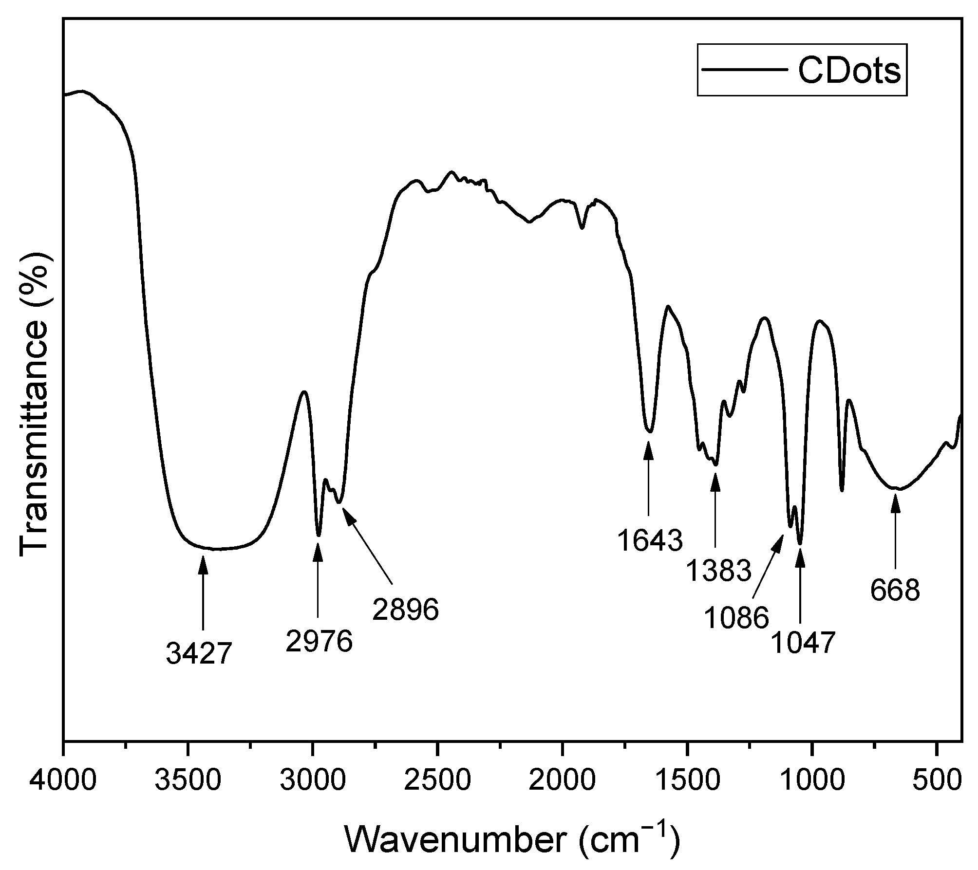
Figure 1. FTIR Spectrum of the Carbon Dots.
Photochem 05 00032 g001
Figure 2. (A) UV–Vis absorption spectrum and PL emission and (B) Tauc plot for bandgap determination of the CDots sample. The red arrow indicates the linear extrapolation to the x-axis, where the intercept gives the Eg value.
Photochem 05 00032 g002
Figure 3. (AC) TEM images at different magnification and (D) size distribution of the as-prepared CDots.
Photochem 05 00032 g003
Figure 4. Cytocompatiblity: (A) HaCat cells cytotoxic effects at different concentrations after 48 h of treatment; (B) Hemolytic assay of CDots at different concentrations. Legend: C-: negative control. Bars represent mean ± SD of (A) quadruplicates of three independent experiments and (B) sextuplicates from two independent experiments. One-way ANOVA, followed by Tukey’s post hoc; * p < 0.05 vs. control. Error bars represent SD.
Photochem 05 00032 g004
Figure 5. Reducing capacity of Trolox (A) and Trolox equivalent (ET) by CDots (B) evaluated using the CUPRAC method, and percentage decay (RC%) of the DPPH free radical at different Trolox concentrations (C) compared with CDots (D). Data are presented as mean ± standard deviation (n = 3), with error bars denoting SD, except for panel B, where they are absent due to the standard deviation being lower than 2%.
Photochem 05 00032 g005
Table 1. Qualitative and quantitative (% w/w) compositions of the sunscreens.
Ingredients (INCI)FunctionControl (%)CDots (%)
Phenoxyethanol (and) caprylyl glycol (Optiphen®)Preservative1.01.0
Cetearyl alcohol (and) dicetyl phosphate (and) ceteth—10 phosphate (Crodafos® CES)Self-emulsifying wax6.06.0
Butyl MethoxydibenzoymethaneUVA filter5.05.0
Ethylhexyl MethoxycinnamateUVB filter10.010.0
CDots (dry-mass)Active-4.5
Purified waterVehicle**
* Sufficient to complete 100.0%.
Table 2. In vitro sun protection factor (SPF) and critical wavelength (cλ) values of the control cream and the same formulation enriched with CDots before and after solar simulation exposure.
Before
Irradiation
After Irradiation (Min)
306090
SPFcλ (nm)SPFcλ (nm)SPFcλ (nm)SPFcλ (nm)
Control26 ± 13.0381 ± 0.714.58 ± 2.2379.2 ± 0.8____
CDots161 ± 7.8 A380 ± 0.6145.22 ± 10.1 A378 ± 0.526.28 ± 5.2 B377 ± 1.014.26 ± 2.2 B375
Legend: SPF—sun protection factor; cλ—critical wavelength. Values are mean ± SD (n = 3 plates). Different superscript letters within a same line denote significant differences (one-way ANOVA, Tukey, p < 0.05, n = 3 plates). Formulations: Control—BMDBM 5% and EHMC 10%; CDots—control formulation + CDots 4.5% (w/w, dry basis).
Disclaimer/Publisher’s Note: The statements, opinions and data contained in all publications are solely those of the individual author(s) and contributor(s) and not of MDPI and/or the editor(s). MDPI and/or the editor(s) disclaim responsibility for any injury to people or property resulting from any ideas, methods, instructions or products referred to in the content.