Article

Fluorescence of 8-Acyl-1-Pyrrolidinylnaphthalenes

Department of Chemistry, College of William and Mary, Williamsburg, VA 23185, USA
*
Author to whom correspondence should be addressed.
Photochem 2025, 5(3), 27; https://doi.org/10.3390/photochem5030027
Submission received: 14 August 2025 / Revised: 11 September 2025 / Accepted: 17 September 2025 / Published: 19 September 2025

Abstract

Four 8-acyl-1-pyrrolidinylnaphalenes are prepared where the acyl group is pivaloyl (6), benzoyl (7), benzyloxycarbonyl (8), and ethyloxycarbonyl (9). Crystal structures for 68 show that both the carbonyl and pyrrolidinyl groups are nearly perpendicular to the naphthalene ring. Esters 8 and 9 fluoresce more strongly than ketones 6 and 7. All show some solvatofluoro-chromic emission from a charge-transfer excited state. Calculations suggest that both the acyl and amino groups twist back toward planarity with the naphthalene in the relaxed first singlet excited state. With 8 and 9, co-planarity is within 20°, while with 6 and 7, the carbonyl approaches no closer than 30°. With 6 and 7, the charge-transfer emission is replaced with a shorter wavelength band with more polar solvents. Despite the twisted geometries and steric interference toward planarization, these systems do not show emission from a twisted intramolecular charge-transfer (TICT) state.

1. Introduction

Peri-disubstituted naphthalenes (Figure 1) have unusual structures and properties because the substituents are so close that they interfere with each other, typically by steric repulsion [1,2]. The non-bonded strain manifests as distortions in the naphthalene structure. The substituents will bend away from each other (splay angle), move to opposite sides of the naphthalene plane, and the naphthalene will twist about the fused center bond [3]. The electronic nature of the substituents can perturb the steric interaction. Two electron-donating groups will give rise to strong repulsion, as seen in proton sponge (bis-1,8-(dimethylamino)naphthalene) [4]. An electron-donating group paired with an electron-accepting group typically gives the “windshield wiper phenomenon” where the donating group moves toward the electron-accepting group while the electron-accepting group moves away from the electron-donating group [3]. This unusual distortion has been taken as evidence of a bonding interaction [5,6], but this interpretation has been questioned by Schiemenz [7]. With formyl and acetyl groups and a dimethyl amino donating group, protonation occurs at the carbonyl oxygen and gives rise to a long C-N bond between the peri substituents. With pivaloyl and benzoyl groups, protonation occurs at the nitrogen [8,9]. Another consequence of the peri-interaction is that the rotation about the single bonds to the naphthalene may be restricted and result in atropisomerism [6,10,11]. When the atropisomers can be separated by chiral chromatography, the rates of rotation are conveniently measured by their racemization [6]. Otherwise, dynamic NMR has been used [10,11].
In this paper, we are interested in 1-dialkylamino-8-acyl naphthalenes where the steric interaction forces these otherwise nominally planar groups to rotate out of the naphthalene plane. For the acetyl and pivaloyl structures 1 and 2 (Figure 1), crystal structures (CIF 7217128 and 7217130, respectively) show that the carbonyl is rotated out-of-plane by 74° for 1 and 80° for 2 [9]. While the dimethylamino group is pyramidal, one of the two methyl groups is rotated by 80° in 1 and 84° in 2. Both 1 and 2 exhibit axial chirality due to hindered rotations about the bonds to the peri-substituents [10]. As a result, the two methyl groups are not equivalent in the 1H NMR spectra. Rotation about either bond will interconvert the methyl groups. For 1, the rate of interconversion is ~230 Hz at room temperature, which is also the coalescence temperature, and the ΔG is 14.2 kcal/mol. For 2, coalescence of the methyl group resonances occurs at 109 °C, and the rate of interconversion is calculated to be 0.06 Hz at room temperature based on a ΔG of 19.2 kcal/mol [10].
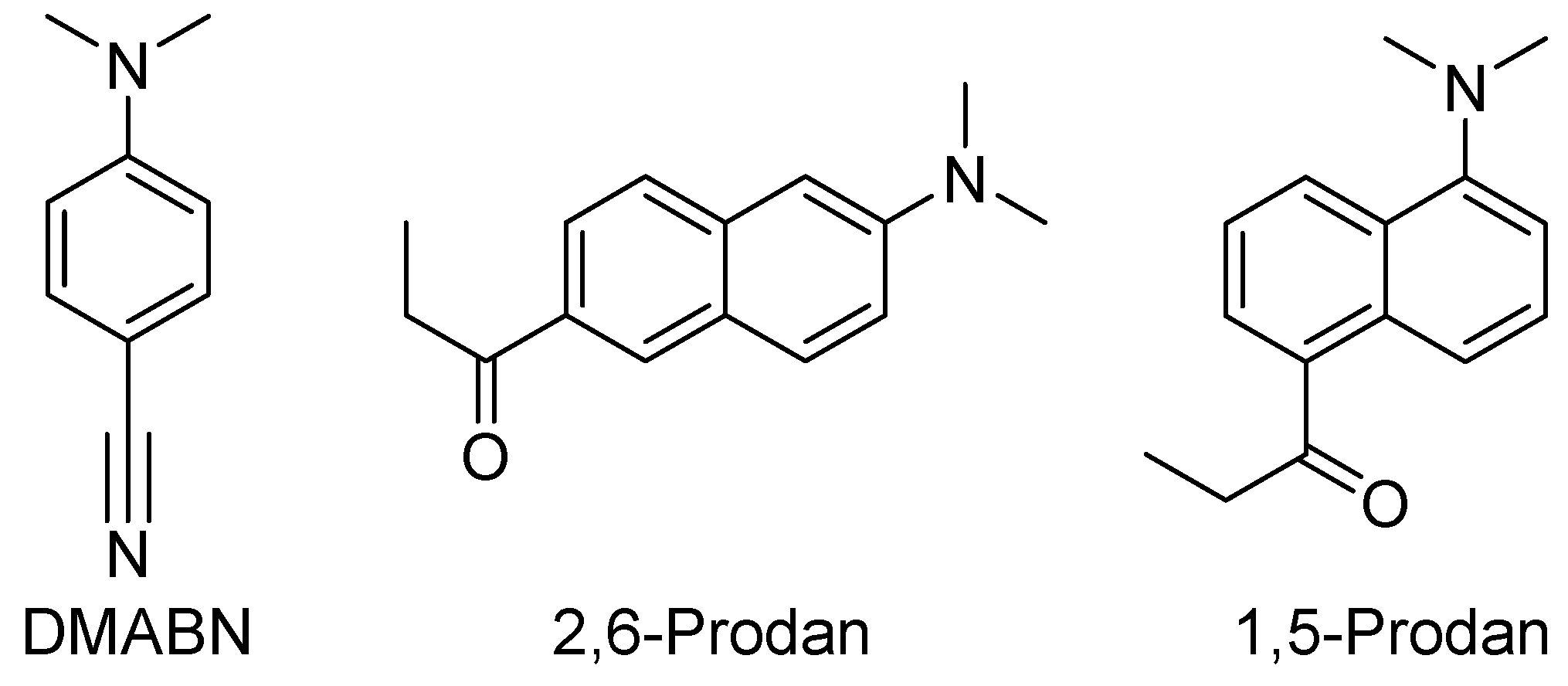
Twisting of donor or acceptor groups on aromatic scaffolds upon photoexcitation can give rise to fluorescent excited states with high degrees of charge transfer. The increase in the dipole moment of the excited state results in strong, positive solvatofluoro-chromism. Such molecules can be useful as chemosensors of micropolarity [12,13]. The best-known example of a small organic compound with a twisted intramolecular charge-transfer (TICT) excited state is 4-dimethylaminobenzonitrile (DMABN, Figure 2). This compound shows dual emission: a higher energy component from a locally excited (LE) state and a lower energy component from the TICT excited state. Corroboration for the TICT structure comes from model compounds where a twist is either enforced or prevented. Enforced twisting gives rise to TICT-like emission, whereas enforced planarity results in LE-like emission [14,15,16,17,18].
Prodan (6-propionyl-2-(dimethylamino)naphthalene Figure 2) is another fluorophore that shows strong intramolecular charge-transfer emission [19]. Like DMABN it has a dimethylamino electron-donating group, but its electron-accepting group is a ketone, not a nitrile. Theoretical studies pointed to emission from a TICT state [20,21], but some experimental results did not support this assignment [22,23,24]. The photophysical behavior of two model compounds, one where the amino group is co-planar and the other where it is twisted, suggested that the intramolecular charge-transfer emissive state is planar (PICT) [25,26]. The 1,5- and 1,8-regioisomers of Prodan also show ICT emission [10,27,28,29,30,31,32]. Planar derivatives of these regioisomers show similar emission properties, thus supporting emission from a PICT state. Thus far, model compounds that force the amino group to remain twisted have eluded preparation. In these and other systems, computational results indicate that the amino group twists towards co-planarity in the relaxed first singlet excited state. The 1,8-systems seem ideal for probing the possibility of a TICT state since planarization would result in high steric strain. When the amino group has two alkyl groups (Figure 1, 12) or when one of the two alkyl groups of the amine is constrained in a ring (Figure 1, 35), experimental and computational results still indicate the tendency for PICT emission [28,33]. In this paper we have tied the two alkyl groups together in a pyrrolidine ring (Figure 1, 69) hoping we might see the emissive behavior of the excited state where the donor and acceptor groups remain twisted. Because these ICT systems are used as molecular fluorescent sensors for probing biological and material systems, understanding the underlying sensing mechanism is critical for properly interpreting their outputs.

2. Materials and Methods

Reagents were obtained from Acros Organics (Tewksbury, MA, USA)or Sigma-Aldrich (Saint Louis, MO, USA). Proton and carbon nuclear magnetic resonance spectra were obtained with an Agilent DD2-400 spectrometer (Santa Clara, CA, USA). Single crystal X-ray diffraction measurements were made at 100 K using Mo Kα radiation on a Bruker-AXS D8 Venture four-circle diffractometer (Billerica, MA, USA), equipped with a microfocus tube and a Photon 3 CPAD detector (Madison, WI, USA). All solvents used for absorption and fluorescence were spectrophotometric grade. Absorption and fluorescence data were collected using a fiber optic system with an Ocean Optics Maya CCD detector (Orlando, FL, USA) using a miniature deuterium/tungsten lamp and a 365 nm LED light source, respectively. Cuvettes were thermostated at 23 °C for fluorescence studies. Emission intensities were processed by subtracting the electronic noise, converting wavelengths to wavenumbers (Jacobian transformation), multiplying by λ2/λmax2 to account for the effect of the abscissa-scale transformation [34,35], and dividing by the spectral response of the Hamamatsu S10420 CCD (Saitama, Japan). Relative quantum yields were determined using anthracene as the reference (Φ = 0.30) using the method of standard additions. Excitation spectra were obtained with an Agilent Cary Fluorescence Spectrophotometer (Cary, NC, USA).
Electronic structure calculations were carried out using Gaussian 16 [36]. Ground-state geometries were optimized using the DFT CAM-B3YLP method with the 6-311G + (2d,p) basis set. Excited states were optimized using the TD-SCF DFT CAM-B3LYP method with the 6-311G + (2d,p) basis set. Calculations did not incorporate a solvent model.
The syntheses of 69 are shown in Scheme 1. Compound 10 was prepared in two steps from 1,8-diaminonaphthalene, as described previously [33,37]. The pyrrolidine ring was installed by sequential nucleophilic substitution with 1,4-dibromobutane. Compounds 69 are prepared from the lithium salt of 11 by nucleophilic substitution with various acyl chlorides. Crystals for X-ray diffraction were grown by dissolving the compounds in a minimum amount of ethyl acetate (~1 mL), diluting with hexanes (~10 mL), and allowing the solvents to evaporate slowly.

2.1. 1-(8-Bromonaphthalen-1-yl)pyrrolidine (11)

8-Bromonaphthalen-1-amine 10 (1.11 g, 5.0 mmol) was combined with 1,4-dibromobutane (2.0 g, 9.3 mmol), K2CO3 (0.76 g, 5.5 mmol), KI (0.20 g, 1.2 mmol) and DMF (5 mL) in an Ace-Thred glass pressure tube. The tube was sealed with a PTFE plug with an FETFE o-ring and heated to 100 °C overnight. After cooling to room temperature, the contents were transferred to a separatory funnel with a 20% ethyl acetate/hexane mixture (150 mL). The organic phase was washed with water (3 × 200 mL), dried over Na2SO4, and concentrated in vacuo. The reaction and workup were repeated on the same scale. Simple distillation of the combined products under vacuum (0.05 torr) gave 11 as an oily semi-solid (1.92 g, 7.0 mmol, 70%). NMR 1H (400 MHz, CDCl3): 7.76–7.70 (m, 2H), 7.45 (d, J = 8.3, 1H), 7.38 (dd, J = 7.9, 7.6 Hz, 1H), 7.20 (dd, J = 7.9, 7.8 Hz, 1H), 7.16 (d, J = 7.6 Hz, 1H), 3.52 (br s, 2H), 2.71 (br s, 2H), 2.11 (br s, 2H), 1.91 (br s, 2H); 13C (400 MHz, CDCl3): δ 147.3, 137.2, 132.6, 128.4, 126.8, 126.3, 125.6, 122.5, 118.6, 115.3, 53.2, 24.0. HRMS (ESI): calcd. for C14H14BrNH+ [M + H]+ 276.03824; found 276.03811.

2.2. Acylation of (8-(Pyrrolidin-1-yl)naphthalen-1-yl)lithium

1-(8-Bromonaphthalen-1-yl)pyrrolidine 11 (0.46 g, 1.7 mmol) was dissolved in THF (10 mL) and cooled to −78 °C under Ar. A solution of n-BuLi in hexanes (1.1 mL, 1.6 M, 1.8 mmol) was added dropwise and the reaction was stirred for 20 min. The acyl chloride (1.8 mmol) was added in one portion quickly. The reaction was allowed to warm to room temperature and then quenched with sat. aq. NH4Cl (5 mL). It was transferred to a separatory funnel with ethyl acetate/hexanes (15%, 150 mL). The organic phase was washed with water (2 × 100 mL), dried over Na2SO4, and concentrated in vacuo. The acylated product was purified by column chromatography on silica gel.

2.2.1. 2,2-Dimethyl-1-(8-(pyrrolidin-1-yl)naphthalen-1-yl)propan-1-one (6)

Chromatography with a gradient elution from 4 to 8% ethyl acetate in hexanes gave 6 as a white solid (250 mg, 52%), m.p. 77–80 °C. UV (EtOH): λmax = 307 nm, log ε = 4.1; NMR 1H (400 MHz, CDCl3): 7.79 (d, J = 8.1 Hz, 1H), 7.56 (d, J = 8.1 Hz, 1H), 7.43 (dd, J = 8.1, 7.1, 1H), 7.41 (dd, J = 8.1, 7.5 Hz, 1H), 7.21 (d, J = 7.5, 1H), 7.09 (d, J = 7.1, 1H), 3.40–3.26 (m, 2H), 3.23–3.15 (m, 1H), 2.17–2.09 (m, 1H), 2.06–1.91 (m, 2H), 1.88–1.78 (m, 1H), 1.78–1.66 (m, 1H), 1.04 (s, 9H); 13C (400 MHz, CDCl3): δ 212.64, 148.28, 137.58, 134.42, 129.25, 128.43, 126.32, 125.14, 124.46, 124.10, 115.5, 58.31, 50.27, 45.62, 27.24, 23.63, 23.04.

2.2.2. Phenyl(8-(pyrrolidin-1-yl)naphthalen-1-yl)methanone (7)

Chromatography with a gradient elution from 0 to 4% ethyl acetate in hexanes gave 7 as a white solid (290 mg, 58%), m.p. 93–94 °C. UV (EtOH): λmax = 284 nm, log ε = 4.1; NMR 1H (400 MHz, CDCl3): 7.91 (d, J = 8.3 Hz, 1H), 7.72 (d, J = 8.2 Hz, 1H), 7.56–7.46 (m, 4H), 7.40–7.36 (m, 2H), 7.63–7.28 (m, 3H), 3.33–3.12 (m, 1H), 2.95–2.82 (m, 1H), 2.51–2.38 (m, 1H), 2.00–1.91 (m, 1H), 1.88–1.68 (m, 2H), 1.68–1.59 (m, 1H), 1.28–1.13 (m, 1H); 13C (400 MHz, CDCl3): δ 193.17, 147.38, 139.01, 137.40, 134.58, 132.05, 131.14, 128.67, 127.99, 127.81, 126.91, 125.92, 125.28, 125.24, 120.67, 56.49, 51.65, 24.17.

2.2.3. Benzyl 8-(Pyrrolidin-1-yl)-1-naphthoate (8)

Chromatography with a gradient elution from 0 to 4% ethyl acetate in hexanes gave 8 as a white solid (340 mg, 62%), m.p. 78–79 °C. UV (EtOH): λmax = 274 nm, log ε = 4.3; NMR 1H (400 MHz, CDCl3): 7.86 (d, J = 7.2 Hz, 1H), 7.65 (d, J = 8.1 Hz, 1H), 7.48 (dd, J = 8.1, 7.5 Hz, 1H), 7.46–7.29 (m, 8H), 5.32 (s, 2H), 3.5–2.6 (m, 4H), 1.97 (br s, 4H); 13C (400 MHz, CDCl3): δ 171.09, 147.93, 136.24, 134.83, 131.04, 129.46, 128.97, 128.50, 127.98, 127.83, 126.78, 125.39, 125.14, 124.83, 119.80, 66.66, 54.77, 24.63.

2.2.4. Ethyl 8-(Pyrrolidin-1-yl)-1-naphthoate (9)

Chromatography with a gradient elution from 1 to 4% ethyl acetate in hexanes gave 9 as an oil (100 mg, 22%). The prior fraction (270 mg) was contaminated with 31% 1-(pyrrolidine-1-yl)naphthalene, giving an aggregate yield of 68%. UV (EtOH): λmax = 282 nm, log ε = 3.9; NMR 1H (400 MHz, CDCl3): 7.85 (d, J = 7.8 Hz, 1H), 7.64 (d, J = 8.1 Hz, 1H), 7.51–7.39 (m, 4H), 4.30 (q, J = 7.1 Hz, 2H), 3.6–2.6 (m, 4H), 1.95 (br s, 4H), 1.34 (t, J = 7.1 Hz, 3H); 13C (400 MHz, CDCl3): δ 171.3, 148.0, 134.9, 131.4, 129.3, 128.9, 126.7, 125.3, 125.1, 124.8, 119.9, 60.8, 54.7, 24.6, 14.2. HRMS (ESI): calcd. for C17H19NO2H+ [M + H]+ 270.14886; found 270.14874.

3. Results

3.1. Structural Studies

3.1.1. X-Ray Structures

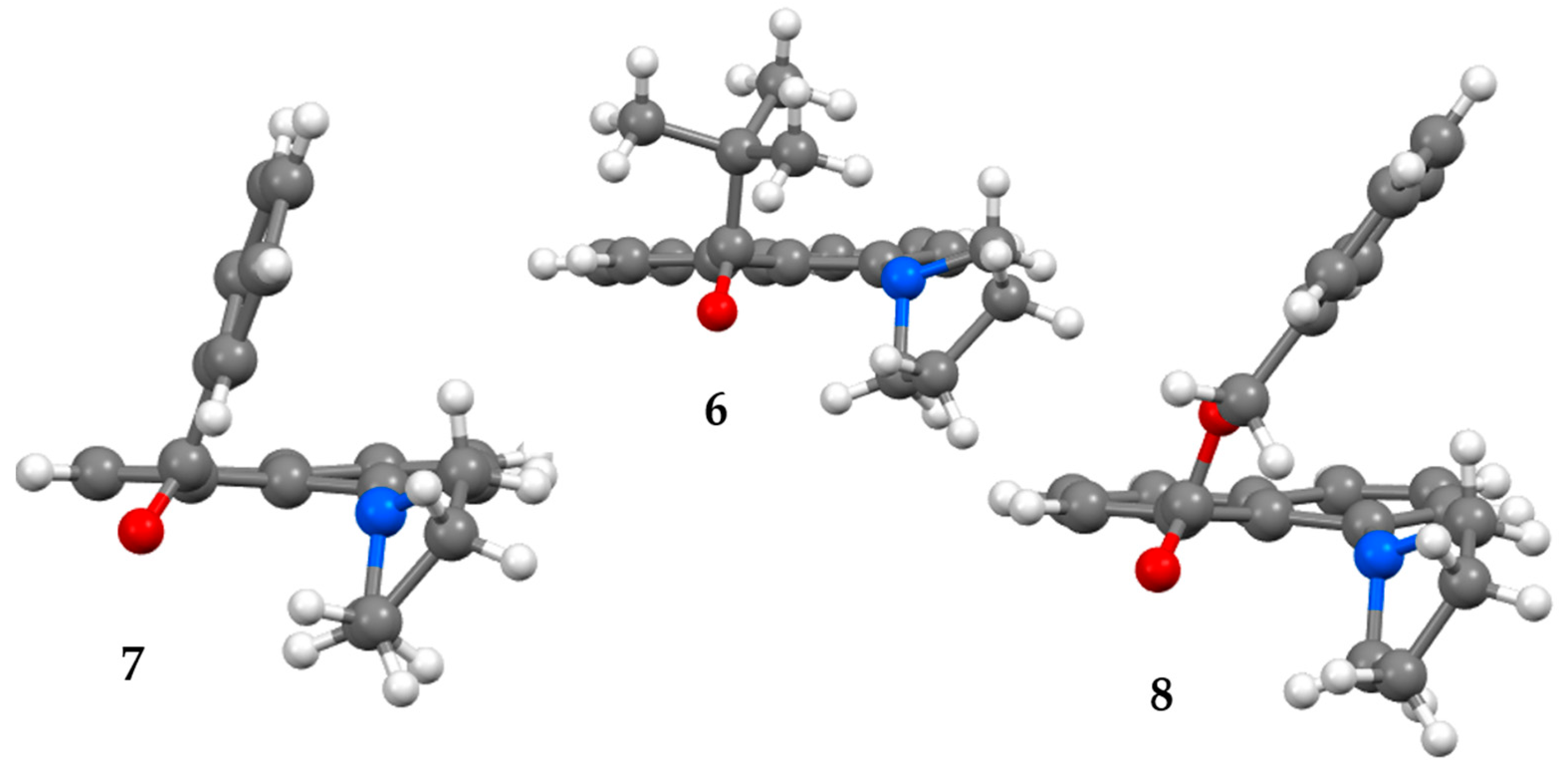
Compounds 68 readily formed crystals through slow evaporation of a 10% ethyl acetate/hexane solution, but 9 remained an oil. X-ray diffraction structures of 68 are shown in Figure 3, and the crystallographic information files are available in the Supplementary Information. These structures reveal that both the carbonyl and pyrrolidine groups are significantly twisted out of the plane of the naphthalene ring. The amount of twist can be characterized by the dihedral angles shown in Table 1. The carbonyl group twists beyond 90° and points slightly away from the pyrrolidine. Of the three carbonyls, the pivaloyl group is closest to being perpendicular (108°). For all three compounds, one of the two pyrrolidine methylene groups attached to the nitrogen atom is even closer to being perpendicular (≤98°). The other methylene group is nearly co-planar with the naphthalene reflecting the pyramidal nature of the nitrogen atom. The nitrogen lone pair is nearly coplanar as well. Thus, these ground-state structures are predisposed to forming a TICT excited state.
The structural features of 68 are similar to those of related dimethylamino derivative like 1 and 2 [5,9]. Table 2 shows several of the distortions that are unique to the peri-substituted naphthalenes. Among these are the distance between the atoms directly connected to the peri-positions (C(O) ···N) and the splay angle, which is defined as (θ1 + θ2 + θ3) 357.2° [38]. All derivatives display the “windshield wiper phenomenon” wherein the amino group tilts toward the carbonyl group while the latter tilts away from the former. Both the carbonyl and amino groups can be characterized by their deviation from planarity. For the carbonyl group, this deviation is measured by ΔC: the distance between the carbonyl carbon and the plane defined by the three atoms attached to the carbonyl carbon (O, C8, C(R)) [5]. For the amino group the percentage planarity (%PL) of the amino group is calculated as (Σ(C-N-C) −328.4) × 3.16 [7]. Not surprisingly, the two t-butyl derivatives, 2 and 6, show the largest distortions. The peri-substituents are the farthest apart in 6, while the splay angle in 2 is very slightly greater. Both have the least planar amino and carbonyl groups, although the distortion in the latter is still small. The amino groups in 7 and 8 are more planar than the others.

3.1.2. NMR Studies

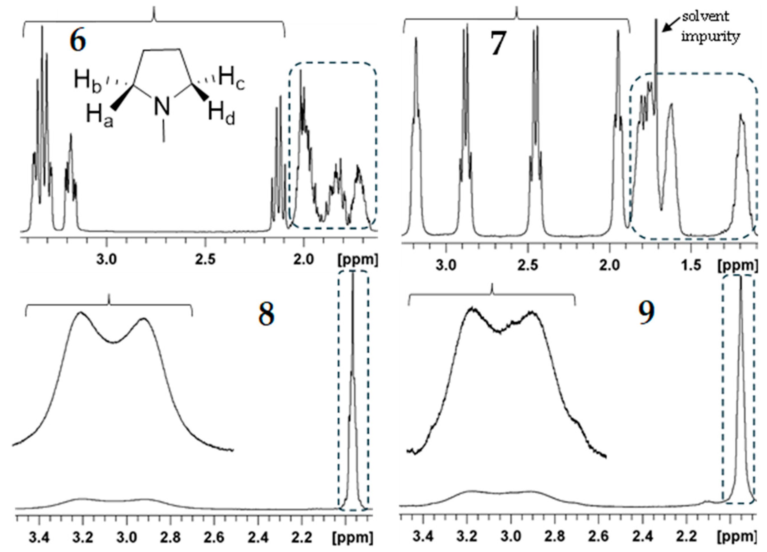
The proton NMR spectra of 68 indicate that there is restricted rotation about both the C(1)-N and C(8)-CO single bonds. While the pyrrolidine ring itself has C2v symmetry, its twisted conformation in these compounds negates the horizontal mirror plane of symmetry, and the inward-pointing (toward the carbonyl group) methylene H-atoms (e.g., Ha and Hd, Figure 4) become diastereotopic with their outward-pointing partners (Hb and Hc). Rotation about the C(1)-N bond will interconvert Ha with Hc and Hb with Hd. Unlike the pyrrolidine, the carbonyl group has neither a vertical mirror plane nor an axis of symmetry. The two twisted conformations of the carbonyl group are axially chiral, and these compounds will show atropisomerism [10]. The carbonyl group negates the vertical plane of symmetry of the adjacent, twisted pyrrolidine and makes Ha diastereotopic with Hd and Hb diastereotopic with Hc. In the case of slow rotation about both peri-bonds, the four H-atoms on the two methylenes attached to the N-atom become distinct in the proton NMR spectrum. Ketones 6 and 7 display this non-equivalence (Figure 4). In contrast, esters 8 and 9 show near coalescence of two sets of two H-atoms. Rotation about the peri-bonds is much faster with the two esters than with the two ketones.

3.2. Photophysical Studies

3.2.1. Absorption

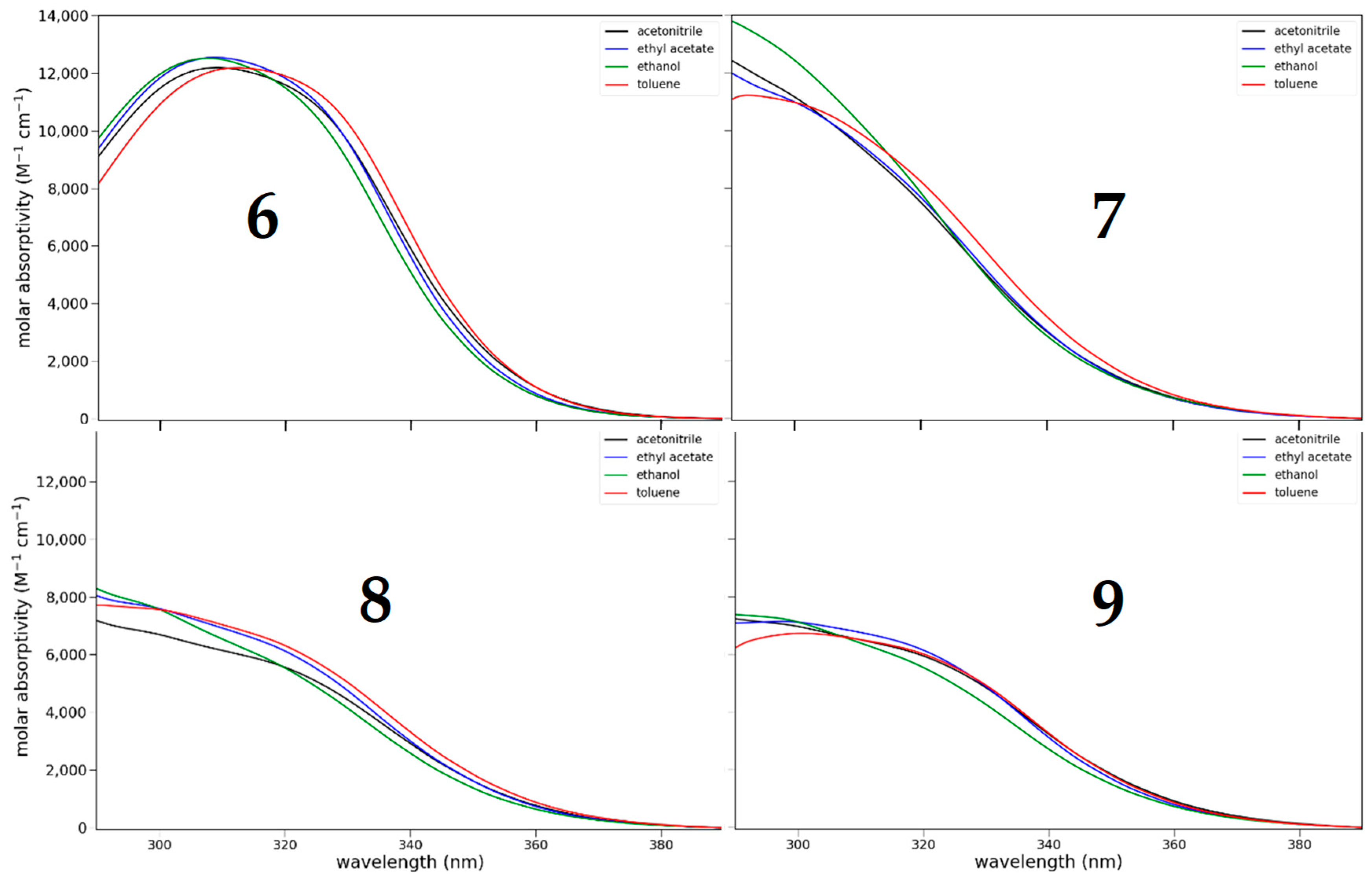
Compounds 69 show moderate absorption (log ε = 4) around 310 nm (Figure 5). Only ketone 6 shows a discrete maximum, while for the others the band is a shoulder. The absorption was determined in a range of solvents of different polarity and proticity: acetonitrile (dielectric constant (εr) = 37.5, aprotic), ethyl acetate (εr = 6.0, aprotic), ethanol (εr = 24.5, protic), toluene (εr = 2.4, aprotic). The absorption shows little dependence on the nature of the solvent.

3.2.2. Fluorescence

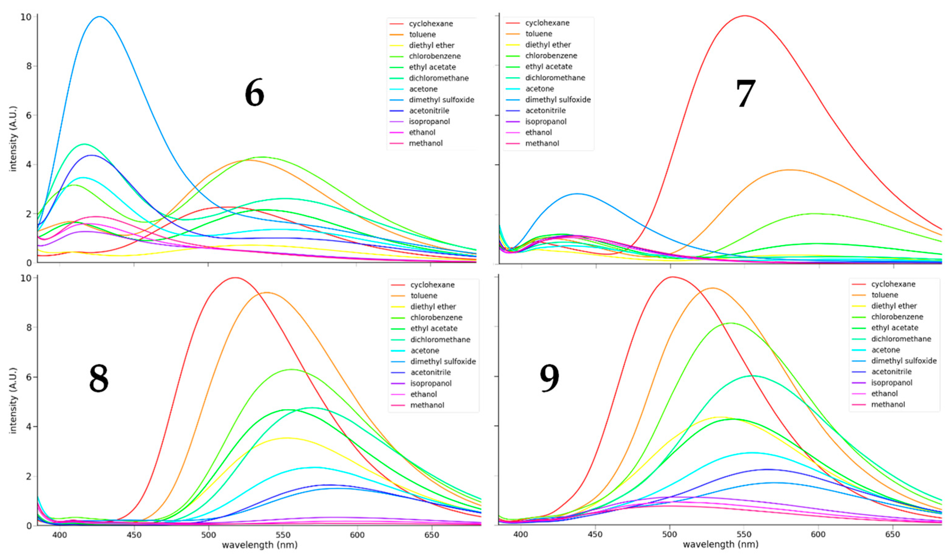
The fluorescence behavior of compounds 69 is varied (Figure 6 and Table 3). The relative fluorescence quantum yields were determined in toluene using anthracene as a reference (Φ = 0.30). Their quantum yields are 0.062 ± 0.019, 0.007 ± 0.0005, 0.24 ± 0.01, and 0.34 ± 0.05, respectively. The fluorescence intensity shows the same ordering in all solvents studied: 9 > 8 >> 6 >>> 7. The two ketones were much weaker fluorophores than the esters. The benzoyl ketone 7 fluoresces weakly only in a few nonpolar solvents. Otherwise, it was essentially not fluorescent. Ketone 6 shows dual emission. With nonpolar, aprotic solvents, a long wavelength band dominates. With more polar solvents, a short wavelength band grows in at the expense of the long wavelength band. Both 6 and 7 show an unusually strong short-wavelength emission in DMSO. This increased intensity is likely due to the greater viscosity of DMSO (1.99 mPa·s). In a separate experiment, the fluorescence intensity of the short-wavelength emission of 6 increases by 45% in going from dimethoxymethane (0.42 mPa·s) to tetraethylene glycol dimethyl ether (3.38 mPa·s). The esters fluoresce most strongly in nonpolar solvents. The emission intensity diminishes as the solvent polarity increases. The fluorescence is greatly reduced in protic solvents, a trend that applies to all four compounds.
The long-wavelength emission band for all four compounds shows solvatofluoro-chromism. The degree of solvatofluoro-chromism can be measured through plots of the emission maxima vs. some measure of the solvent’s polarity. The solvent polarity parameter ET(30) is generally useful in this regard [39]. Plots of the long-wavelength emission maxima vs. the ET(30) parameter are shown in Figure 7. The slope of the best-fit line is negative in all cases. A negative slope indicates that polar solvents better solvate the excited state leading to lower energy and longer wavelength emission, and implicates an intramolecular charge-transfer excited state. Both ketones 6 and 7 have truncated plots. For 6, the ICT band is a shoulder with polar and protic solvents. For 7, quenching is nearly complete with solvents more polar than ethyl acetate. Benzyl ester 8 shows ICT emission for all solvents, including the protic ones. For the simple ester 9 the protic solvents give emission bands at shorter wavelengths. A short wavelength band appears in the emission spectra of 6 and 7. The band is most noticeable in 6. The emission maxima of this band do not show appreciable solvatofluoro-chromism (Figure 7). The slopes of the best-fit lines are much smaller than for the ICT emission. Because these are at high energy and show little solvatofluoro-chromism, they likely arise from the locally excited (LE) state.
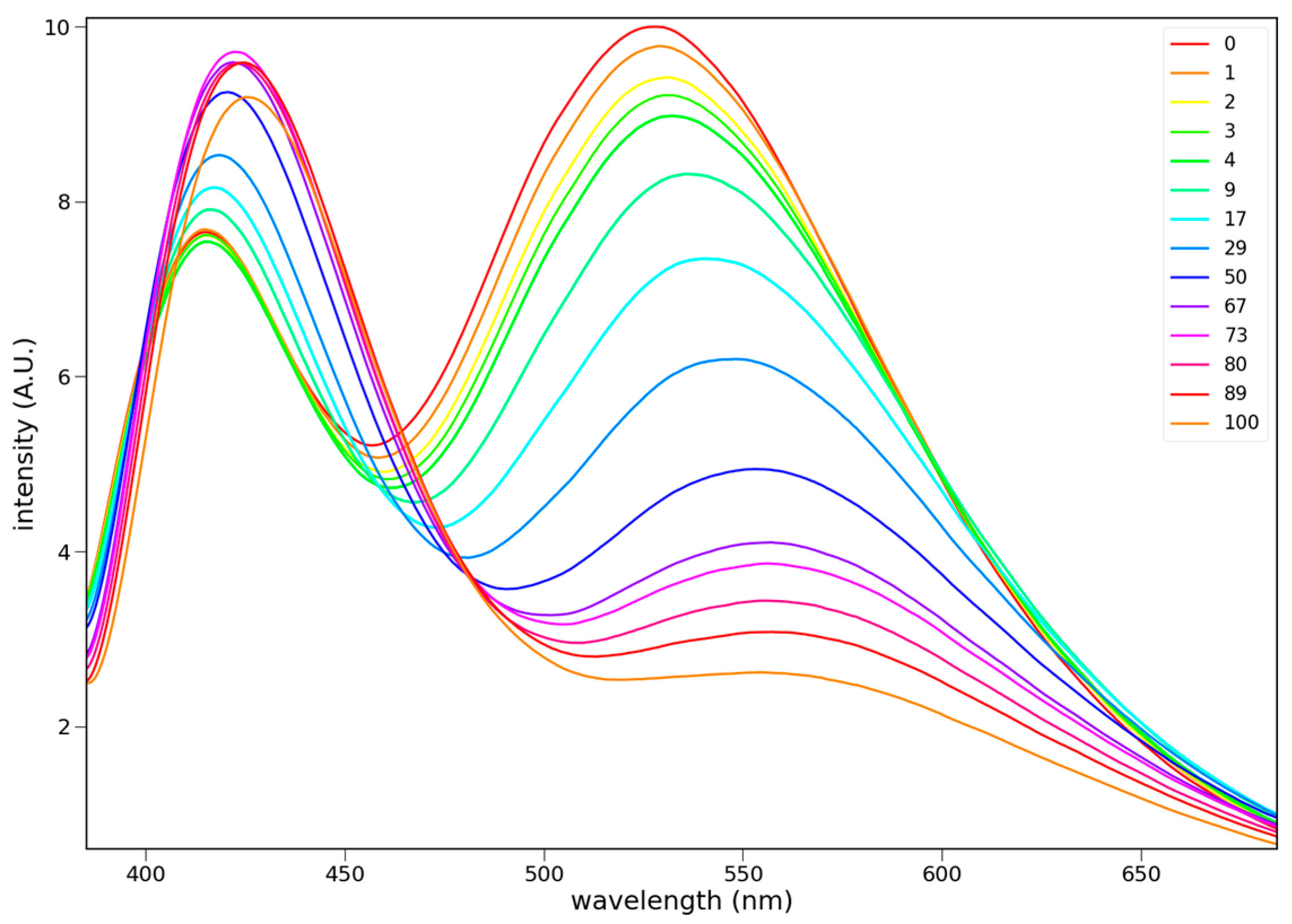
The fluorescence behavior of 6 is unusual. It shows significant emission from a higher energy state in addition to a lower energy ICT excited state. The ICT emission shifts to lower energy as the solvent polarity increases. The higher energy emission becomes more dominant with increasing solvent polarity, as shown through a preferential solvation study (Figure 8) with toluene and acetonitrile. The ICT band prevails in pure toluene, while the higher energy band dominates in acetonitrile. Excitation spectra (Figure 9) show that the two states arise from different ground states. The isoemissive points in Figure 8 are consistent with a solvent-dependent equilibrium process.

3.3. Computational Studies

3.3.1. Ground State Structures

The optimized structures for 69 were calculated using Gaussian 16 using the DFT CAM-B3YLP method with the 6-311G + (2d,p) basis set [35]. The computed structures for 68 compare favorably with the crystal structures (Table 4). The dihedral angles for the pyrrolidine component agree within 1.8°. The dihedral angle for the carbonyl groups is within 0.6° for 6, but deviates by 7.6° and 11° for 7 and 8. Calculations predict that the carbonyl group is more perpendicular than is indicated by the crystal structures.

3.3.2. Excited State Structures

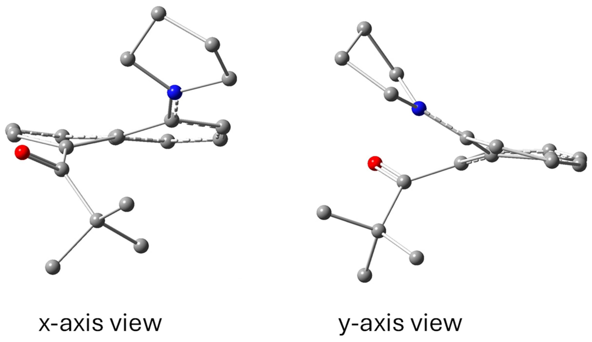
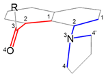
The structures of the relaxed, first singlet excited states of 69 were calculated using Gaussian 16 using the TD-SCF DFT CAM-B3LYP method with the 6-311G + (2d,p) basis set. As with the previous computational studies on 1-dialkylamino-8-acylnaphthalenes, both substituents are predicted to twist toward planarity with the naphthalene ring [28,33]. While the N-atom is mostly pyramidal in the ground state, it becomes significantly planar in the excited state. The degree of twisting of the two substituents (red for the carbonyl and blue for the amino group) is approximated by the average of the dihedral angles (labelled 1 and 2) indicated by the figure in Table 5. The naphthalene also twists about the fused bond in the excited state (green). If the inner two atoms of each dihedral angle pair were perfectly trigonal planar, then the individual angles of each pair would be identical. The deviation values report the difference between the individual values and result from the fact that the inner atoms are slightly pyramidal.
The carbonyl group could have twisted to point toward or away from the pyrrolidine. Optimization gives the latter configuration. The carbonyl groups of esters 8 and 9 are predicted to be nearly co-planar with the naphthalene (~10°). Both ketones 6 and 7 retain some twisting (~30–40°). The pyrrolidinyl group is also nearly co-planar with the naphthalene (~15°) for all compounds. Finally, the naphthalene becomes significantly distorted from planarity by twisting 15° about the fused bond (C9–C10). This structural change results in the two substituents moving to opposite sides of the naphthalene (Figure 10) thereby reducing the steric strain that results from their twisting toward planarity.

3.3.3. Molecular Orbitals and Electronic Transitions

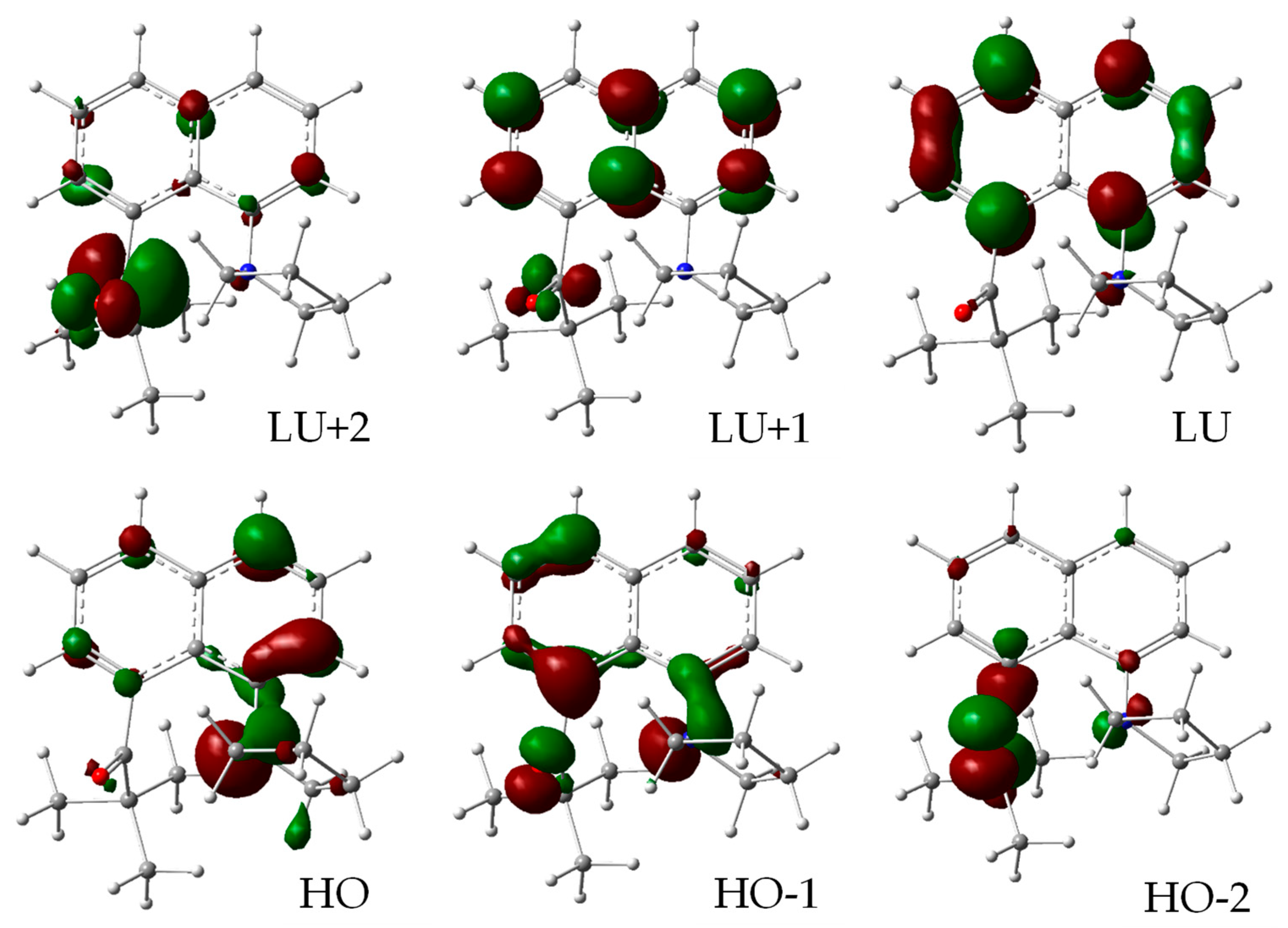
Both the absorption and fluorescence are predicted to occur primarily from a HO-LU transition. The frontier molecular orbitals for the ground state of 6 are shown in Figure 11. Those for 79 are shown in Figures S6–S8. For all four compounds, the HOMO is primarily the N lone pair orbital. For all but 7 the LUMO is the naphthalene B2g LUMO. For 7 the LUMO involves the carbonyl π* orbital, and the naphthalene B2g orbital is LU + 1.
For the relaxed, first singlet excited state, the LU becomes the in-phase combination of the carbonyl π* orbital and the naphthalene B2g orbital (Figures S9–S12 in the Supplementary Materials). The predicted UV absorption and fluorescence emission wavelengths, oscillator strengths, and transition configurations are shown in Table 6. The calculations capture the characteristic short wavelength absorptions for these compounds but underestimate bathochromic shifts for the fluorescence.
Ketones 6 and 7 have lower fluorescence quantum yields than esters 8 and 9. Simple ketones do not fluoresce because of efficient intersystem crossing to a triplet state enabled by spin–orbit coupling according to the El-Sayed rules [40]. The first excited singlet states are mostly π → π* for these systems. Intersystem crossing (ISC) would require an n → π* triplet state of similar energy. The relevant n → π* triplet states are shown in Table 7 for 69. Ethyl ester 9 does not have a close lying n → π* triplet state, and it fluoresces the strongest. For the remaining three compounds, the unrelaxed singlet states have an n → π* triplet state that is close in energy. For 68, their fluorescence intensity follows the singlet-triplet gap—negative with 7, slightly positive with 6 and 8, but smaller with the former than the latter. In ketones 6 and 7, the n-orbital is relatively high in energetic ordering (HO-2 vs. HO-4), and this factor may be decisive in their physical behavior.

4. Conclusions

The present compounds have donor and acceptor groups on the peri-positions of naphthalene. Their close proximity results in them twisting to be nearly perpendicular with respect to the naphthalene plane. All show some charge-transfer fluorescence and this band displays solvatofluoro-chromism that is seen in related systems. Calculations suggest that both the donor and acceptor group twist toward planarity in the relaxed first excited state. While the pyrrolidinyl group becomes nearly co-planar with the naphthalene, the degree of co-planarity of the carbonyl depends on the structure. Whereas the ester carbonyls also become nearly co-planar, the pivaloyl and benzoyl carbonyls remain about one-third twisted. The fluorescence of esters 8 and 9 gets weaker as the solvent polarity increases due to the smaller S0–S1 energy gap. Protic solvents give rise to H-bond quenching seen in other twisted Prodan compounds [27,28]. The fluorescence of 6 and 7 is much weaker than the two esters. Calculations point to the possibility of intersystem crossing to a triplet state as an explanation.
The most remarkable result here is the photophysical behavior of 6 and 7. While the charge transfer band appears with low polarity solvents, it is replaced quickly with a higher energy band as the solvent polarity increases. The high-energy band arises from a species that absorbs at slightly higher wavelengths. Proton NMR shows that rotation about the peri-bonds is much slower for 6 and 7 than 8 and 9. If the higher-energy-emitting species is one that twists too slowly in the excited state, then emission would occur from a locally excited state. The viscosity-dependent increase in emission intensity is consistent with restricted rotation being a critical factor. Such an interpretation result would corroborate the PICT model for emission in these Prodan systems.

Supplementary Materials

The following supporting information can be downloaded at: https://www.mdpi.com/article/10.3390/photochem5030027/s1. Crystallographic information files (CIF) for compounds 68; checkCIF report from the Cambridge Crystallographic Data Centre (CCDC); Bond lengths [Å] and angles [°] for Compounds 68; Proton and carbon NMR spectra for 610; Frontier molecular orbitals for the ground states and relaxed first singlet excited states of 69.

Author Contributions

Conceptualization, C.A.; methodology, C.A., A.L., L.T. and R.P.; formal analysis, C.A.; investigation, C.A., A.L., L.T. and R.P.; data curation, C.A., A.L., L.T. and R.P.; writing—original draft preparation, C.A.; writing—review and editing, C.A., A.L., L.T. and R.P.; supervision, C.A. All authors have read and agreed to the published version of the manuscript.

Funding

This research received no external funding.

Institutional Review Board Statement

No applicable.

Informed Consent Statement

Not applicable.

Data Availability Statement

The data presented in this study are available either in this article or in its Supplementary Materials.

Acknowledgments

The authors acknowledge William & Mary Research Computing for providing computational resources and/or technical support that have contributed to the results reported within this paper.

Conflicts of Interest

The authors declare no conflicts of interest.

Abbreviations

The following abbreviations are used in this manuscript:
DMABNDimethylaminobenzonitrile
ICTIntramolecular charge transfer
ProdanPropionyldimethylaminonaphthalene
PICTPlanar intramolecular charge transfer
TICTTwisted intramolecular charge transfer

References

  1. Balasubramaniyan, V. Peri Interaction in Naphthalene Derivatives. Chem. Rev. 1966, 66, 567–641. [Google Scholar] [CrossRef]
  2. Reza, A.I.; Iwai, K.; Nishiwaki, N. A Study of the Correlation between the Bulkiness of Peri-Substituents and the Distortion of a Naphthalene Ring. Molecules 2023, 28, 5343. [Google Scholar] [CrossRef]
  3. Schiemenz, G.P.; Schiemenz, B.; Petersen, S.; Wolff, C. Hexacoordinated Carbon or Tetracovalent Silicon? Chirality 1998, 10, 180–189. [Google Scholar] [CrossRef]
  4. Einspahr, H.; Robert, J.B.; Marsh, R.E.; Roberts, J.D. Peri Interactions: An X-Ray Crystallographic Study of the Structure of 1,8-Bis(Dimethylaminonaphthalene). Acta Crystallogr. B Struct. Crystallogr. Cryst. Chem. 1973, 29, 1611–1617. [Google Scholar] [CrossRef]
  5. Schweizer, W.B.; Procter, G.; Kaftory, M.; Dunitz, J.D. Structural Studies of 1,8-Disubstituted Naphthalenes as Probes for Nucleophile-electrophile Interactions. Helv. Chim. Acta 1978, 61, 2783–2808. [Google Scholar] [CrossRef]
  6. Clayden, J.; McCarthy, C.; Helliwell, M. Bonded Peri-Interactions Govern the Rate of Racemisation of Atropisomeric 8-Substituted 1-Naphthamides. Chem. Commun. 1999, 2059–2060. [Google Scholar] [CrossRef]
  7. Schiemenz, G.P. Peri-Interactions in Naphthalenes, 14 [1]. Pyramidalization versus Planarization at Nitrogen in 8-Dialkylamino-Naphth-1-Yl Compounds as a Measure of Peri Bond Formation. Z. Naturforsch. B 2006, 61, 535–554. [Google Scholar] [CrossRef]
  8. Lari, A.; Pitak, M.B.; Coles, S.J.; Rees, G.J.; Day, S.P.; Smith, M.E.; Hanna, J.V.; Wallis, J.D. Models for Incomplete Nucleophilic Attack on a Protonated Carbonyl Group and Electron-Deficient Alkenes: Salts and Zwitterions from 1-Dimethylamino-Naphthalene-8-Carbaldehyde. Org. Biomol. Chem. 2012, 10, 7763. [Google Scholar] [CrossRef]
  9. Mercadal, N.; Day, S.P.; Jarmyn, A.; Pitak, M.B.; Coles, S.J.; Wilson, C.; Rees, G.J.; Hanna, J.V.; Wallis, J.D. O- vs. N-Protonation of 1-Dimethylaminonaphthalene-8-Ketones: Formation of a Peri N-C Bond or a Hydrogen Bond to the Pi-Electron Density of a Carbonyl Group. CrystEngComm 2014, 16, 8363–8374. [Google Scholar] [CrossRef]
  10. Kiefl, C. Correlated Rotations and Unusual Fluorescence Properties of peri-Substituted, Axially Chiral Naphthyl Ketones. Eur. J. Org. Chem. 2000, 2000, 3279–3286. [Google Scholar] [CrossRef]
  11. Kiefl, C.; Zinner, H.; Burgemeister, T.; Mannschreck, A. Axially Chiral 2-(Dialkylamino)Benzamides. Barriers to Rotations, Determined by NMR and by Racemization (Neighboring Rotor Substituents on Arene Systems, Part 3). Recl. Trav. Chim. Pays-Bas 2010, 115, 125–132. [Google Scholar] [CrossRef]
  12. Rettig, W. Charge Separation in Excited States of Decoupled Systems: Twisted Intramolecular Charge Transfer (TICT) Compounds and Implications for the Development of New Laser Dyes and for the Primary Process of Vision and Photosynthesis. Angew. Chem. 1986, 98, 969–986. [Google Scholar] [CrossRef]
  13. Rettig, W.; Lapouyade, R. Fluorescence Probes Based on Twisted Intramolecular Charge Transfer (TICT) States and Other Adiabatic Photoreactions. In Topics in Fluorescence Spectroscopy; Springer: Berlin/Heidelberg, Germany, 2002; pp. 109–149. [Google Scholar]
  14. Okada, T.; Uesugi, M.; Köhler, G.; Rechthaler, K.; Rotkiewicz, K.; Rettig, W.; Grabner, G. Time-Resolved Spectroscopy of DMABN and Its Cage Derivatives 6-Cyanobenzquinuclidine (CBQ) and Benzquinuclidine (BQ). Chem. Phys. 1999, 241, 327–337. [Google Scholar] [CrossRef]
  15. Köhler, G.; Rechthaler, K.; Grabner, G.; Luboradzki, R.; Suwińska, K.; Rotkiewicz, K. Structure of Cage Amines as Models for Twisted Intramolecular Charge-Transfer States. J. Phys. Chem. A 1997, 101, 8518–8525. [Google Scholar] [CrossRef]
  16. Wermuth, G.; Rettig, W. The Interaction of Close-Lying Excited States: Solvent Influence on Fluorescence Rate and Polarization in Substituted Indolines. J. Phys. Chem. 1984, 88, 2729–2735. [Google Scholar] [CrossRef]
  17. Grabowski, Z.R.; Rotkiewicz, K.; Siemiarczuk, A. Dual Fluorescence of Donor-Acceptor Molecules and the Twisted Intramolecular Charge Transfer (TICT) States. J. Lumin. 1979, 18, 420–424. [Google Scholar] [CrossRef]
  18. Kochman, M.A.; Durbeej, B. Simulating the Nonadiabatic Relaxation Dynamics of 4-(N, N-Dimethylamino) Benzonitrile (DMABN) in Polar Solution. J. Phys. Chem. A 2020, 124, 2193–2206. [Google Scholar] [CrossRef]
  19. Weber, G.; Farris, F.J. Synthesis and Spectral Properties of a Hydrophobic Fluorescent Probe: 6-Propionyl-2-(Dimethylamino) Naphthalene. Biochemistry 1979, 18, 3075–3078. [Google Scholar] [CrossRef] [PubMed]
  20. Parusel, A.B.J.; Nowak, W.; Grimme, S.; Kohler, G. Comparative Theoretical Study on Charge-Transfer Fluorescence Probes: 6-Propanoyl-2-(N, N-Dimethylamino) Naphthalene and Derivatives. J. Phys. Chem. A 1998, 102, 7149–7156. [Google Scholar] [CrossRef]
  21. Parusel, A.B.J.; Schneider, F.W.; Köhler, G. An Ab Initio Study on Excited and Ground State Properties of the Organic Fluorescence Probe PRODAN. J. Mol. Struct. THEOCHEM 1997, 398, 341–346. [Google Scholar] [CrossRef]
  22. Catalan, J.; Perez, P.; Laynez, J.; Blanco, F.G. Analysis of the Solvent Effect on the Photophysics Properties of 6-Propionyl-2-(Dimethylamino)Naphthalene (PRODAN). J. Fluoresc. 1991, 1, 215–223. [Google Scholar] [CrossRef] [PubMed]
  23. Balter, A.; Nowak, W.; Pawekiewicz, W.; Kowalczyk, A. Some Remarks on the Interpretation of the Spectral Properties of Prodan. Chem. Phys. Lett. 1988, 143, 565–570. [Google Scholar] [CrossRef]
  24. Kawski, A. Ground-and Excited-State Dipole Moments of 6-Propionyl-2-(Dimethylamino) Naphthalene Determined from Solvatochromic Shifts. Z. Naturforsch. A 1999, 54, 379–381. [Google Scholar] [CrossRef]
  25. Lobo, B.C.; Abelt, C.J. Does PRODAN Possess a Planar or Twisted Charge-Transfer Excited State? Photophysical Properties of Two PRODAN Derivatives. J. Phys. Chem. A 2003, 107, 10938–10943. [Google Scholar] [CrossRef]
  26. Davis, B.N.; Abelt, C.J. Synthesis and Photophysical Properties of Models for Twisted PRODAN and Dimethylaminonaphthonitrile. J. Phys. Chem. A 2005, 109, 1295–1298. [Google Scholar] [CrossRef]
  27. Chen, T.; Lee, S.W.; Abelt, C.J. 1,5-Prodan Emits from a Planar Intramolecular Charge-Transfer Excited State. ACS Omega 2018, 3, 4816–4823. [Google Scholar] [CrossRef]
  28. Abelt, C.; Sweigart, K. Twisted 8-Acyl-1-Dialkyl-Amino-Naphthalenes Emit from a Planar Intramolecular Charge Transfer Excited State. Photochem 2024, 4, 1–13. [Google Scholar] [CrossRef]
  29. Benedetti, E.; Veliz, A.B.; Charpenay, M.; Kocsis, L.S.; Brummond, K.M. Attachable Solvatochromic Fluorophores and Bioconjugation Studies. Org. Lett. 2013, 15, 2578–2581. [Google Scholar] [CrossRef]
  30. Benedetti, E.; Kocsis, L.S.; Brummond, K.M. Synthesis and Photophysical Properties of a Series of Cyclopenta [b] Naphthalene Solvatochromic Fluorophores. J. Am. Chem. Soc. 2012, 134, 12418–12421. [Google Scholar] [CrossRef]
  31. Brummond, K.M.; Kocsis, L.S. Intramolecular Didehydro-Diels–Alder Reaction and Its Impact on the Structure–Function Properties of Environmentally Sensitive Fluorophores. Acc. Chem. Res. 2015, 48, 2320–2329. [Google Scholar] [CrossRef]
  32. Jockusch, S.; Zheng, Q.; He, G.S.; Pudavar, H.E.; Yee, D.J.; Balsanek, V.; Halim, M.; Sames, D.; Prasad, P.N.; Turro, N.J. Two-Photon Excitation of Fluorogenic Probes for Redox Metabolism: Dramatic Enhancement of Optical Contrast Ratio by Two-Photon Excitation. J. Phys. Chem. C 2007, 111, 8872–8877. [Google Scholar] [CrossRef]
  33. Abelt, C.; Day, I.; Zhao, J.; Pike, R. Fluorescence of Half-Twisted 10-Acyl-1-Methyltetrahydrobenzoquinolines. Molecules 2024, 29, 3016. [Google Scholar] [CrossRef]
  34. Lakowicz, J.R. Principles of Fluorescence Spectroscopy; Springer: Boston, MA, USA, 1999; ISBN 978-1-4757-3063-0. [Google Scholar]
  35. Mooney, J.; Kambhampati, P. Get the Basics Right: Jacobian Conversion of Wavelength and Energy Scales for Quantitative Analysis of Emission Spectra. J. Phys. Chem. Lett. 2013, 4, 3316–3318. [Google Scholar] [CrossRef]
  36. Frisch, M.J.; Trucks, G.W.; Schlegel, H.B.; Scuseria, G.E.; Robb, M.A.; Cheeseman, J.R.; Scalmani, G.; Barone, V.; Petersson, G.A.; Nakatsuji, H.; et al. Gaussian 16, Version 1.1; Gaussian, Inc.: Wallingford, CT, USA, 2016. [Google Scholar]
  37. Jurok, R.; Cibulka, R.; Dvořáková, H.; Hampl, F.; Hodačová, J. Planar Chiral Flavinium Salts—Prospective Catalysts for Enantioselective Sulfoxidation Reactions. Eur. J. Org. Chem. 2010, 2010, 5217–5224. [Google Scholar] [CrossRef]
  38. Knight, F.R.; Fuller, A.L.; Bühl, M.; Slawin, A.M.Z.; Woollins, J.D. Synthetic and Structural Studies of 1,8-Chalcogen Naphthalene Derivatives. Chem. Eur. J. 2010, 16, 7503–7516. [Google Scholar] [CrossRef] [PubMed]
  39. Reichardt, C.; Welton, T. Solvents and Solvent Effects in Organic Chemistry; Wiley-VCH Verlag GmbH: Hoboken, NJ, USA, 2011. [Google Scholar]
  40. El-Sayed, M.A. Triplet State. Its Radiative and Nonradiative Properties. Acc. Chem. Res. 1968, 1, 8–16. [Google Scholar] [CrossRef]
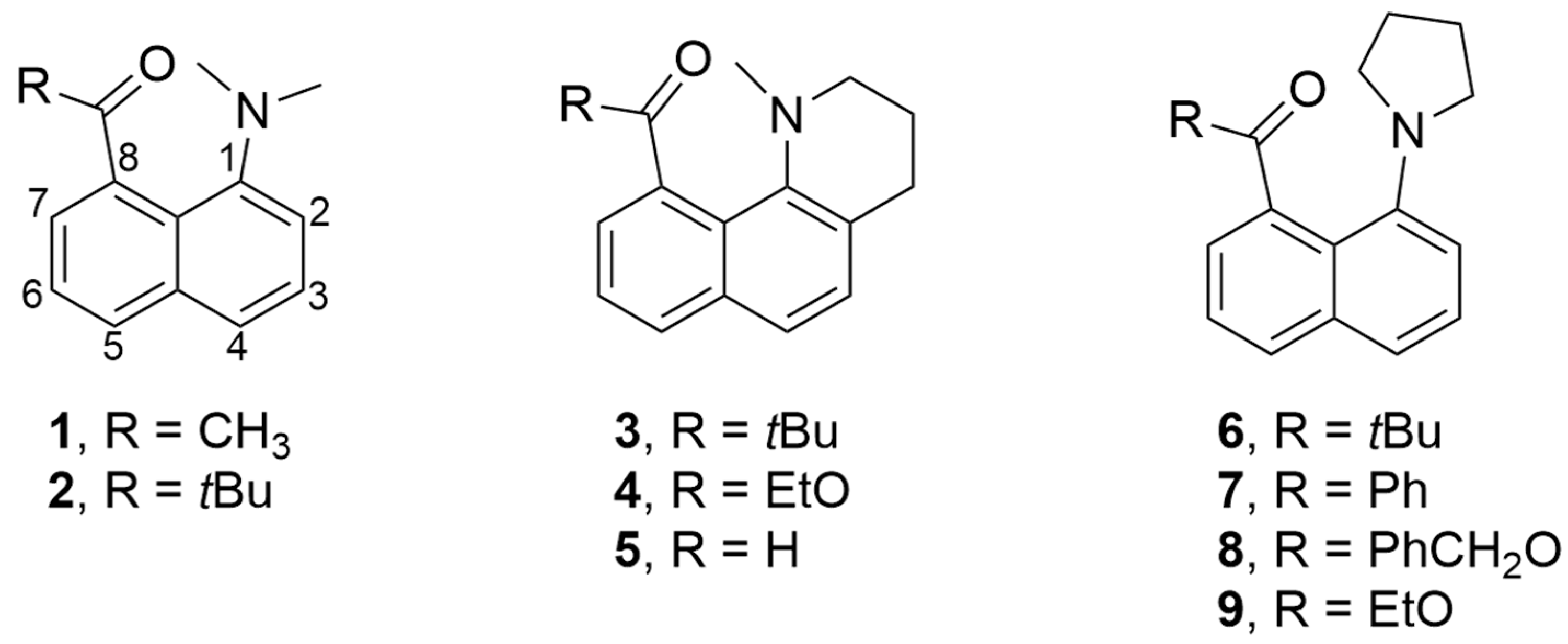
Figure 1. Structures of various 1-dialkylamino-8-acylnaphthalenes.
Photochem 05 00027 g001
Figure 2. Structures of DMABN, 2,6-Prodan and 1,5-Prodan.
Photochem 05 00027 g002
Scheme 1. Synthetic pathways for the preparation of 69 and 11.
Photochem 05 00027 sch001
Figure 3. X-ray structures of 6 (top middle), 7 (bottom left), and 8 (bottom right). Atom, color: H, white, C, gray; N, blue; O, red.
Photochem 05 00027 g003
Figure 4. Partial 1H NMR spectra of 6 (top left), 7 (top right), 8 (bottom left), and 9 (bottom right) highlighting the pyrrolidine hydrogen resonances Ha–Hd under the brackets. The other four pyrrolidine hydrogen resonances are indicated in the dashed boxes.
Photochem 05 00027 g004
Figure 5. UV-VIS spectra of 6 (top left), 7 (top right), 8 (bottom left), and 9 (bottom right) in acetonitrile (), ethyl acetate (), ethanol (), and toluene ().
Photochem 05 00027 g005
Figure 6. Fluorescence spectra of 1.1 × 10⁻⁴ M 6 (top left), 2.1 × 10⁻⁴ M 7 (top right), 8.0 × 10⁻⁵ M 8 (bottom left), and 1.3 × 10⁻⁴ M 9 (bottom right) in cyclohexane (), toluene (), diethyl ether (), chlorobenzene (), ethyl acetate (), dichloromethane (), acetone (), dimethylsulfoxide (), acetonitrile (), isopropanol (), ethanol (), and methanol (). Excitation at 366 nm.
Photochem 05 00027 g006
Figure 7. Lower left: solvatofluoro-chromism plots of the charge-transfer emission maximum (cm−1) vs. ET(30) for 6 (◊, solid line), 7 (□, short dashed line), 8 (Δ, long dashed line), and 9 (ο, dotted line). The slopes of the best-fit lines are −107, −151, −99, and −110, respectively. Upper right: Solvatofluoro-chromism plots of the emission maxima (cm−1) vs. ET(30) for 6 (◊, solid line) and 7 (□, dashed line) with polar solvents. The slopes of the best-fit lines are −25 and −18, respectively.
Photochem 05 00027 g007
Figure 8. Fluorescence spectra of 1.1 × 10⁻⁴ M 6 in mixtures of toluene and acetonitrile. The acetonitrile mole % is indicated in the upper right.
Photochem 05 00027 g008
Figure 9. Excitation spectra of 1.1 × 10⁻⁴ M 6 in toluene (λem = 520 nm, λem = 420 nm) and acetonitrile (λem = 520 nm, λem = 420 nm). For reference, the relative absorption is shown as dashed lines: toluene (— —), acetonitrile (– – –).
Photochem 05 00027 g009
Figure 10. Perspective views (defined in Table 5) of the optimized excited state structure of 6. Atom, color: H, white, C, gray; N, blue; O, red.
Photochem 05 00027 g010
Figure 11. Frontier molecular orbitals (isovalue 0.06) for the ground state of 6.
Photochem 05 00027 g011
Table 1. Select dihedral angles (°) from X-ray structures of 68.
CarbonylPyrrolidinePyrrolidine’Photochem 05 00027 i001
6−108.1−98.123.2
7−120.5−96.331.3
8−117.3−94.531.1
Table 2. Various geometrical values from X-ray structures of 1, 2, and 68.
12678Photochem 05 00027 i002
C(O)····N (Å)2.562.672.692.602.65
C1····C8 (Å)2.492.522.512.502.51
Splay (°)3.88.38.24.55.5
θ1 (°)123.0124.3125.0123.1122.0
θ3 (°)116.1117.7116.8116.6117.4
%PL29.421.419.837.541.8
ΔC (Å)0.090.090.100.060.05
Table 3. Emission maxima (nm) and relative quantum yields (Φ) for 69 in various solvents.
6789
Solventλmax (nm)Φλmax (nm)Φλmax (nm)Φλmax (nm)Φ
(CH2)65240.0315600.0165250.2365130.382
PhCH35390.0625910.0075480.2425360.390
Et2O5420.0135980.0015640.1015470.206
PhCl5480.0766130.0055640.1795500.360
EtOAc5500.0396130.0035630.1305540.198
CH2Cl24200.0704330.0025780.1435650.290
(CH3)2CO4200.0424400.0015780.0725660.149
DMSO4320.0964430.0035970.0545820.102
CH3CN4260.0464410.0015940.0535780.120
iPrOH4240.0154410.0016060.0155340.065
EtOH4240.0164390.0016150.0095280.051
CH3OH4300.0194430.0016250.0065220.039
Table 4. Select dihedral angles (°) for computed structures of 68 a.
CarbonylPyrrolidinePyrrolidine’Photochem 05 00027 i003
6−107.5−97.823.3
(−108.1)(−98.1)(23.2)
7−113.4−94.532.5
(−120.5)(−96.3)(31.3)
8−106.3−93.829.6
(−117.3)(−94.5)(31.1)
a X-ray values from Table 1 are shown in parentheses.
Table 5. Select average dihedral angles a (°) and deviations b for the computed, relaxed first excited-singlet state structures of 69.
redbluegreenPhotochem 05 00027 i004
6−39.2−15.717.5
(7.8)(13.7)(5.6)
7−30.9−15.416.4
(1.0)(7.3)(3.6)
8−11.0−16.315.5
(7.0)(4.8)(4.0)
9−12.0−14.715.7
(6.4)(6.1)(4.5)
a red: (r1 + r2)/2, blue: (b1 + b2)/2, green: (g1 + g2)/2. b red: |r1–r2|, blue: |b1–b2|, green: |g1–g2|.
Table 6. Calculated UV absorption and fluorescence emission wavelengths, oscillator strengths, and transition configurations for 69.
67 a89
S0→S1
λmax (nm)291292294294
f0.1280.1420.1270.126
Transition,HO → LU, 68HO → LU + 1, 62HO → LU, 69HO → LU (0.69)
coeff. × 100HO-2 → LU + 1, −12HO → LU, 27HO-4 → LU + 1, 11HO-2 → LU + 1, 11
HO-2 → LU + 2, 10
S0←S1
λmax (nm)465490480472
f0.0800.0520.0820.084
Transition,HO → LU, 70HO → LU, 70HO → LU, 70HO → LU, 70
coeff. × 100
a This transition is S0→S2. There is a weak (f = 0.002) S0→S1 transition at 308 nm.
Table 7. Characterization of the n → π* triplet states for 69.
6789
n → π* TnT5T6T5T11
E(Tn) (eV) a4.304.214.285.65
(4.26)(4.25)(4.21)(4.21)
Transition, bHO-2 → LU, 68HO-2 → LU, 33HO-4 → LU, 64HO-4 → LU + 6, 14
coeff. × 100HO-2 → LU + 1, 19HO-2 → LU + 1, 41HO-4 → LU + 1, −13HO-4 → LU + 12, −27
HO → LU + 1, 13HO-2 → LU + 2, 15HO-3 → LU, −12HO-4 → LU + 14, 11
HO → LU, 10HO-1 → LU, −16HO → LU + 1, −16HO-1 → LU + 1, −24
HO → LU, 34 HO → LU + 6, 24
a The value in parentheses below is the energy of the unrelaxed first singlet excited state (S1). b The n-orbital is highlighted in bold and italics.
Disclaimer/Publisher’s Note: The statements, opinions and data contained in all publications are solely those of the individual author(s) and contributor(s) and not of MDPI and/or the editor(s). MDPI and/or the editor(s) disclaim responsibility for any injury to people or property resulting from any ideas, methods, instructions or products referred to in the content.