Perspective

Photoproducts of Porphyrins with a Focus on Protoporphyrin IX

IZKF Core Unit Proteomics, Medical Faculty, University of Münster, 48149 Münster, Germany
Photochem 2025, 5(2), 10; https://doi.org/10.3390/photochem5020010
Submission received: 18 March 2025 / Revised: 9 April 2025 / Accepted: 10 April 2025 / Published: 14 April 2025

Abstract

:
Porphyrins play important roles in biological systems including oxygen transport and catalysis. Due to their tetrapyrrole core structure, they exhibit exceptional photophysical and electrochemical properties and find many applications in both technical and life science fields, including photodynamic therapy and neurosurgery. The irradiation of porphyrins may cause modifications to their molecular structure or their degradation. Such photobleaching processes potentially affect the success and sensitivity of photosensitizer applications. While there have been many studies using fluorescence spectroscopy to investigate this phenomenon, reports about analytically validated structures of photoproducts are scarce. It is, however, necessary to know the individual contributions of different molecules to the fluorescence signal in order to evaluate it correctly. This review provides a summary of the current state of knowledge in this respect, discussing especially the validated hydroxyaldehyde and formyl photo-oxidation products of protoporphyrin IX.

1. Introduction

Porphyrins are heterocyclic organic compounds consisting of modified pyrrole subunits, with porphin being the core structure (Figure 1). They play important roles in biological systems including oxygen transport and light harvesting; well-known representatives include heme and chlorophyll [1]. Due to their conjugated system of 26 π-electrons, 18 of which form a continuous cycle, porphyrins typically absorb in the visible region of the electromagnetic spectrum and are colored [2]. Additionally, the conformational flexibility of the tetrapyrrole complex enables the same molecule to act as a cofactor for different biochemical reactions [3]. For an extensive introduction to the porphyrin field, see textbook [4].
Porphyrins exhibit exceptional photophysical and electrochemical properties and find applications in different fields, e.g., as electroactive agents in analytical chemistry [5], as catalysts for CO2 reduction [6] and in wastewater treatment [7], as well as sensitizers in solar cells [8], and in the medical field in antimicrobial and photodynamic therapy (PDT) and cancer treatment [1,9,10,11]. When such a molecule absorbs the energy of an incident photon, its electrons move from the ground state to an excited state [12]. For porphyrins, the Soret band is defined as the wavelength at ~400 nm where absorption is at its maximum. The electrons return to their equilibrium state by either the emission of lower-energy photons (fluorescence), non-radiative release of energy, or intersystem crossing [13]. Photoproducts can be generated in the process, which vary depending on the type of porphyrin, the properties of the light, and the surrounding conditions, namely the biological matrix (see sections below). In this context, the term “bleaching” is often used, which refers to the loss of the ability of a porphyrin to absorb light due to changes in its structure or its degradation.
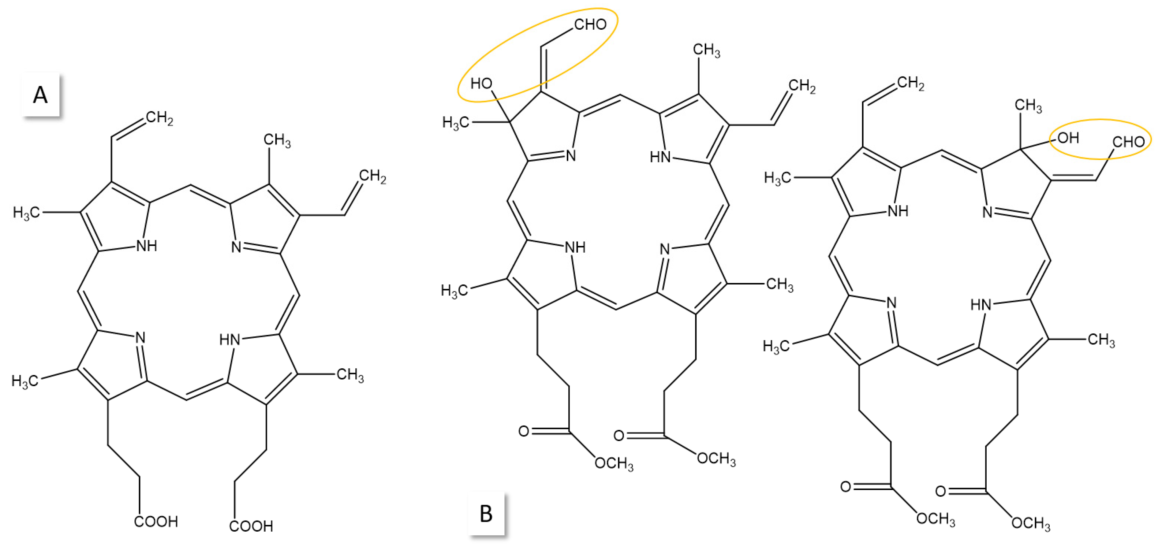
It has become very important to study porphyrin photostability in order to find the best treatment conditions for PDT [14]. There have also been concerns about the photobleaching of protoporphyrin (PPIX, Figure 2A) during the fluorescence-guided resection (FGR) of glioblastoma potentially impairing sensitivity. With this method, malignant brain tumors are visualized during surgery based on their PPIX fluorescence (for an introduction and review, see [9,15,16]). The high PPIX levels result from its increased synthesis in tumor compared to normal tissue following the exogenous administration of 5-aminolevulinic acid (5-ALA), which is converted into PPIX during heme biosynthesis [17]. This surgical method improves tumor resection considerably [9], which is a major factor determining progression and survival prognosis [16]. With regard to photobleaching, neurosurgeons observed that under operating light, fluorescence decayed to 36% in 25 min for violet blue and in 87 min for white light excitation [16]. Researchers including ourselves are working on several topics for improvement in FGR such as the development of more sensitive hardware and better software. In this context, multiple open questions need to be addressed, which require specific measurements of individual porphyrins. They include the following: (1) What is the contribution of hexahydroporphyrin intermediates of heme biosynthesis (porphyrinogens) or that of endogenous porphyrins (e.g., coproporhyrin, uroporphyrin) to the PPIX fluorescence spectrum? (2) How accurately can the PPIX concentration in tumor tissue be determined with surgical microscopes and imaging devices monitoring the fluorescence signal? (3) What is the impact of the photobleaching of individual compounds on fluorescence sensitivity?
Despite the fact that different fluorophores exhibit different excitation and emission energies, spectra from biological matrices such as tumor tissue are sum spectra and thus not very specific. The measured fluorescence is highly dependent on the microenvironment and pH [18], as well as the autofluorescence from endogenous fluorophores such as nicotinamide adenine dinucleotide hydrate (NADH), flavin derivatives, and collagen [12,15,19,20].
In contrast, mass spectrometry (MS) can target individual porphyrins and porphyrinogens based on their molecular mass and ensure their identity using specific gas-phase fragment ions (for the few available studies, see [21,22,23,24]). Thus, our research combines the advantages of substance-specific MS detection with traditional spectroscopic fluorescence measurement in order to better evaluate the quantification of individual porphyrins [19,22,25,26]. We compared fluorescence and MS data of high-grade gliomas and detected the overestimation of the PPIX concentration using fluorescence information [19]. We also used MS to demonstrate that PPIX is a new target for liquid biopsy in patients with glioma [22]. Here, the current, and surprisingly not very extensive, state of knowledge about validated porphyrin photoproduct structures is reviewed with a focus on PPIX in order to set the stage for future MS-based analytical investigations, which will aim for the simultaneous quantification in blood and brain tissue of several porphyrins important in the heme biosynthesis pathway [17]. To this end, it is necessary to know their molecular mass, core structures, and side-chain composition.

2. Definitions of Photoreactions

The International Union of Pure and Applied Chemistry (IUPAC) defines photoprocesses, i.e., reactions under the influence of light, as follows [27]:
(1)
Oxidation reactions:
(a)
Loss of one or more electrons from a chemical species as a result of the photoexcitation of this species;
(b)
Reaction of a substance with oxygen (called photo-oxygenation, when oxygen remains in the product).
(2)
Photoreduction:
(a)
Addition of one or more electrons to a photoexcited species;
(b)
Photochemical hydrogenation of a substance.
(3)
Photoinitiated reactions: neither the substrate nor the oxygen is electronically excited.
For photosensitized reactions involving oxygen, type I and type II mechanisms were defined (for more information, see [28,29] and textbooks such as [30]). They apply to photoreactions including initial electron or hydrogen atom abstraction as an oxidizing step. Type I sensitizers undergo photoinduced electron transfer. This leads to the formation of the reactive oxygen ion O2∙− [31] and HO2. The superoxide anion radical O2∙− is formed after the sensitizer∙− donates an electron to O2 (filling one of the two degenerate orbitals of diradical molecular oxygen, leading to a charged ion with a singly unpaired electron) or by charge transfer to O2. Type II is described as the sensitized formation of singlet oxygen 1O2 (O=O); it is a sensitizer energy-transfer process to oxygen. Singlet oxygen is in a quantum state where all electrons are spin-paired; it is much more reactive that the triplet ground state of O2 [32].
Different wavelengths of light may activate different chromophores in a single molecule and induce different reactive excited states and, thus, may result in different photoproducts (for more information, see Reference [33]). It is also important to note that photoreactions of porphyrins may differ depending on their side-chain structures, because these groups determine parameters such as pH dependence, hydrophobicity, and charge state. Photoproducts differ in their molecular structure from the original photosensitizer and may thus exhibit a shifted wavelength of their absorption maximum [34].

3. Photostability of PPIX

Upon photonic excitation, the PPIX molecule is raised to a higher singlet excited state, from which it can populate the triplet state due to intersystem crossing [35]. In the presence of molecular oxygen, this triplet state can be quenched efficiently as a result of a near-resonant energy transfer from the triplet manifold to that of oxygen, resulting in the formation of singlet oxygen. This singlet oxygen can either attack biomolecules in the direct vicinity, initiating the photodynamic effect, or degrade the photosensitizer molecules (bleaching) [35].
It has been known since the work of Fischer and Herrle [36] that porphyrins are photo-oxidized in the presence of oxygen, but the nature of the products was not determined until much later. In 1966, Inhoffen et al. [37] elucidated two isomeric photoproducts of PPIX-dimethyester (Figure 2) using an extended analytical toolkit (thin-layer chromatography (TLC); electron, infrared, and nuclear magnetic resonance (NMR) spectroscopy; MS) and proved the incorporation of two additional oxygen atoms. White light irradiation of the original compound in neutral solvent with a steady oxygen flow yielded 80% of the so-called photoprotoporphyrin (PPP)-dimethylester, the isomers of which were separated and validated.
Figure 2. (A) PPIX; (B) isomeric photoproducts (photoprotoporphyrin (PPP)-dimethylester) of PPIX-dimethylester as elucidated by Inhoffen et al. [37]. Modified sites are circled.
Photochem 05 00010 g002
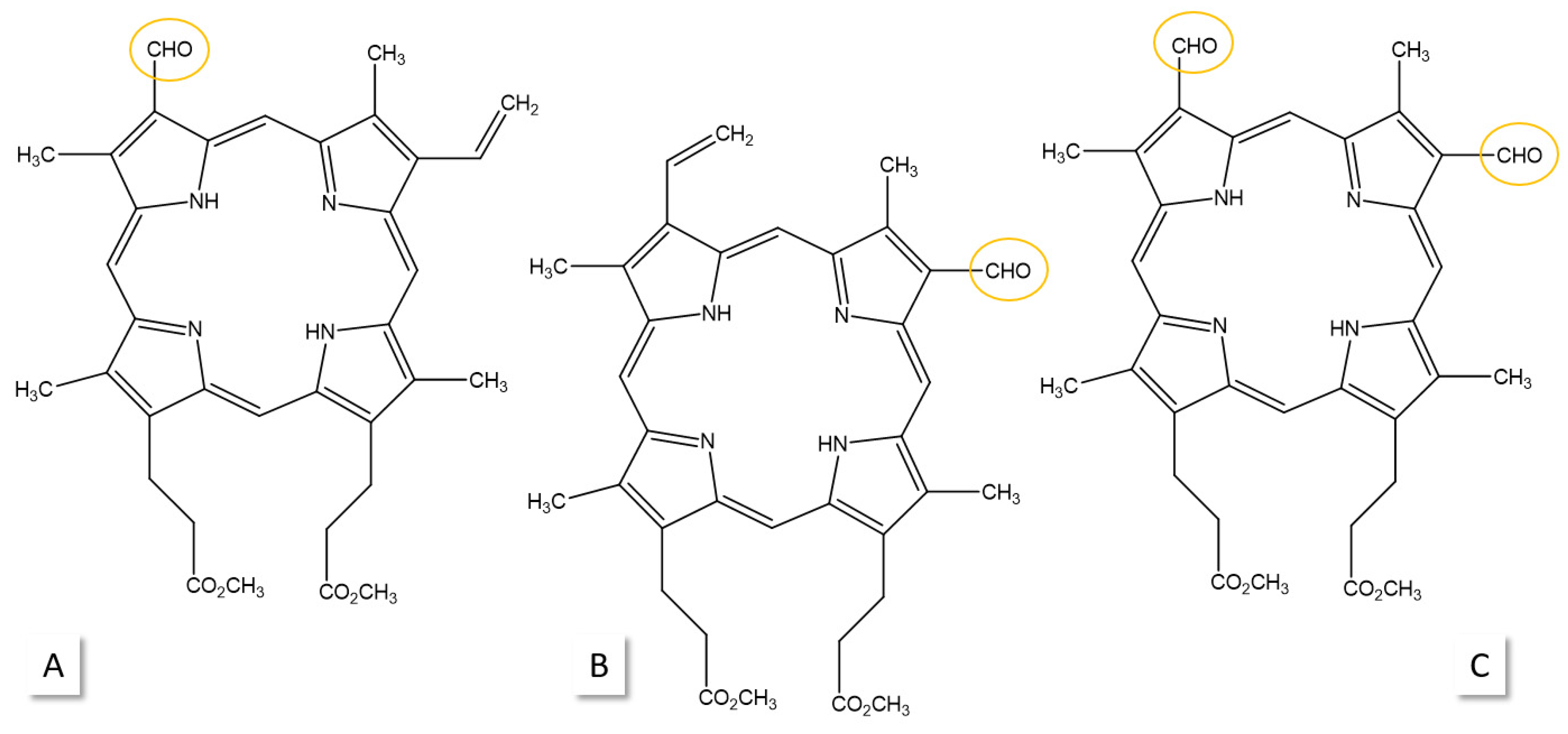
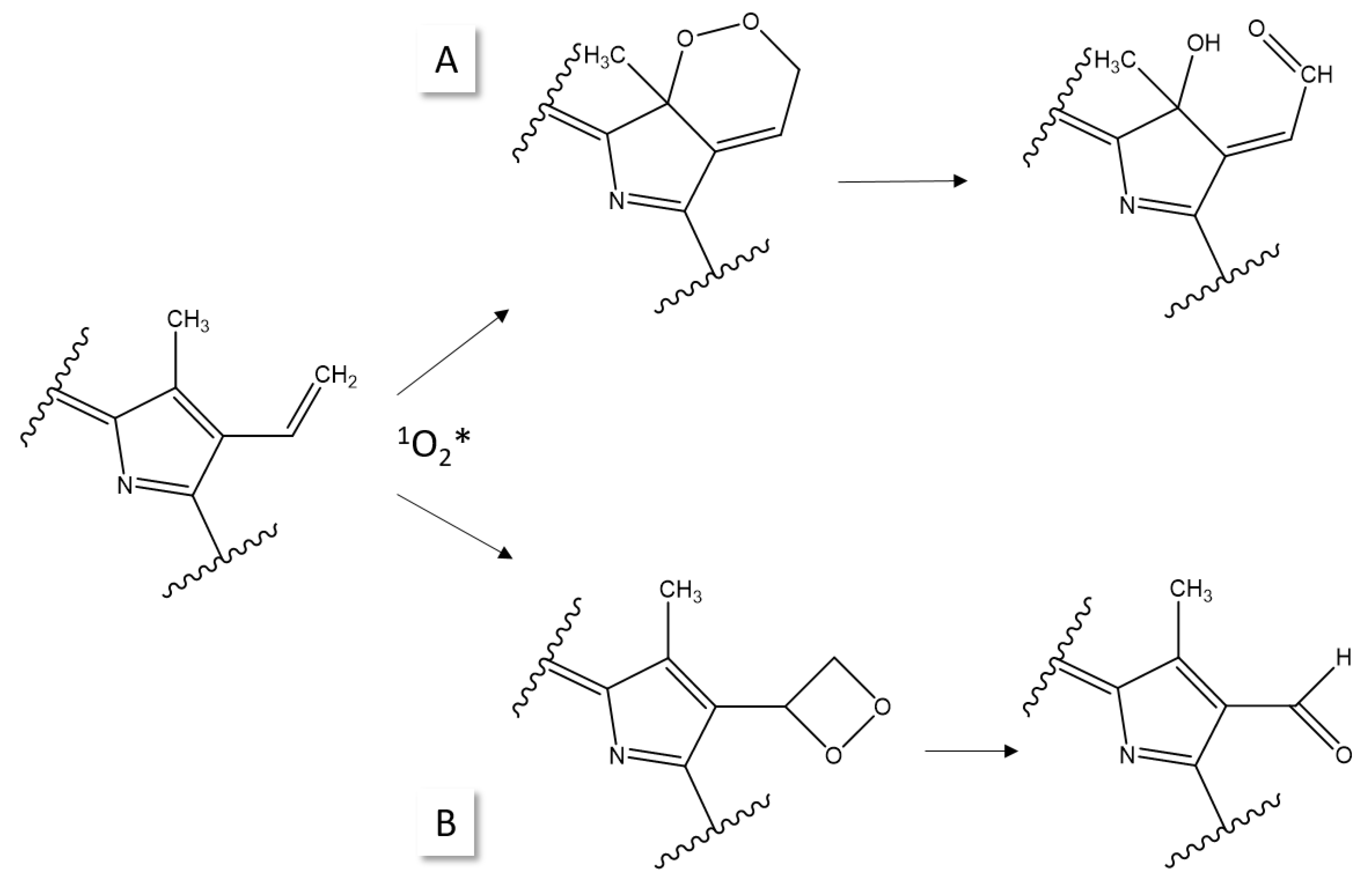
These hydroxyaldehyde products were confirmed by Cox and Whitten in 1982 [38] in addition to mono- and diformyl products using the liquid chromatography (LC) separation of photoproducts in comparison to synthetic reference substances (Figure 3). The authors found that PPPs (Figure 2) predominated when the protoporphyrin derivatives were irradiated in most organic solvents, while the formyl products became dominant when the irradiation experiments were carried out in aqueous micelles, in monolayer films at an air–water interface, or supported multilayer assemblies. The oxidation processes were discussed based on familiar photo-oxidation pathways (see Section 2) [28]. For the PPPs, the cycloaddition of oxygen to a diene unit was suggested, while the formyl products were explained with a dioxetane intermediate (Figure 4).
The interaction of excited porphyrin with molecular oxygen in solution can give rise to both singlet oxygen formation (Figure 5A) and electron transfer to generate a superoxide (Figure 5B), with the first process being more efficient than the latter. Cox and Witten [38,39] demonstrated with the use of singlet oxygen quenchers that all discussed photoproducts were formed directly from singlet oxygen attack on ground-state protoporphyrin and that they did not arise primarily from interconversion to one another. Quenchers slowed down the rate of photo-oxidation but simultaneously increased the yield of the products. The primary photoproducts may decompose, presumably by photo-oxidation, under the reaction conditions, because the products can absorb both in the same region as the original compound and at slightly longer wavelengths. In addition, the primary products may react with the activated forms of oxygen generated by PPIX-dimethylester. Appreciable amounts of formyl products (Figure 3), but not PPP, were still formed where most of the porphyrins in excited states were quenched by an electron acceptor. Thus, a small amount of interaction of porphyrin in an excited state with molecular oxygen may result in electron transfer to give oxidized porphyrin (cation radicals) and superoxide anion, leading to formyl products through the reaction of the superoxide with the porphyrin π cation [38,39].
Wessles et al. [35] noted that cycloaddition to one of the two vinyl substituents of PPIX-dimethylester leading to PPPs includes the removal of the adjacent double bond at the macrocycle, causing a pronounced change in the spectral properties and in the fluorescence decay kinetics (new absorption band ~670 nm; new emission band ~676 nm). In contrast, when oxygen is added directly to the vinyl group yielding formyl photoproducts, the main ring structure remains unaltered. Furthermore, in the presence of oxidizable amino acids, the photo-oxidation process can lead to oxygen intermediates that might attack the macrocycle, leading to ring opening and thus complete degradation. For instance, for liposomes and cells, it was shown that the PPP yield dropped and photo-oxidation resulted in the attack of the macrocycle [35].
When experiments were carried out under oxygen depletion in dimethylformamide, the PPP fluorescence signal was not detected, suggesting that oxygen was required for photobleaching [40]. In contrast, fluorescence photobleaching was observed during 5-ALA-induced PPIX PDT of MLL cells in vitro under both oxygenated and hypoxic conditions [41]. PPP was the main photoproduct and accumulated at higher levels in the absence of oxygen, likely as a result of its reduced photobleaching under hypoxia. Two more photoproducts were reported with no further structure description or validation, called product II (peak at 655 nm) and product III (618 nm). The authors suggested that product II was a photoproduct of PPP and not of PPIX, while product III was thought to be a contribution from uroporphyrin or coproporphyrin [40].
Notably, the literature is confusing with regard to the assignment and naming of photoproducts. In a 2024 report on the analysis of photoproduct formation from PPIX dissolved in dimethyl sulfoxide, the formyl photoproduct was referred to as product II without MS or other validation [42]. PPP formation was dominant under 635 nm irradiation of PPIX, with higher photoproduct formation at a low fluence rate, while irradiation with 405 nm yielded more product II with a fluorescence peak at 654 nm, with higher photoproduct formation at a higher fluence rate [42].
Interestingly, in a comparative study of the photoactivity toward murine B16F-10 melanoma cells of synthetic PPIX and PPIX extracted from the Harderian gland of rats, it was found that the synthetic PPIX demonstrated a photocytotoxicity ten times lower than that of the isolated PPIX material and that the rate of cell internalization for both endogenous PPIX and its photoproduct was eightfold greater than that obtained for the synthetic porphyrin [43]. Obviously, the isolation of PPIX from the gland did not result in a pure compound but rather in PPIX accompanied by residual biological material, which amplified its behavior in the experiments.

4. Photoreduction

Studies on the photoreduction of porphyrins are less common. In 1962, Mauzerall [44] demonstrated that water-soluble porphyrins were readily photoreduced to dihydro- and tetrahydroporphyrin (Figure 6) with high quantum yield by mild reducing agents such as bi-tertiary amines in dependence of pH. A third stage, porphyrinogen, was only reached with special reducing agents. The investigation was based on spectroscopic methods without subsequent orthogonal validation of the proposed structures.
Other authors performed illumination experiments of hematoporphyrin derivatives in phosphate-buffered saline and assigned reduced porphyrin structures (chlorin-, bacteriochlorin-type molecules), as shown in Figure 6, to their products based on the fluorescence spectroscopy of TLC-separated substances [45]. Considering that no reducing agent was present in these experiments, the question arises of why no PPP was formed. Clearly, an extended analytical workup is necessary to validate and prove such hypothetical structures.

5. Photobleaching of PDT Agents

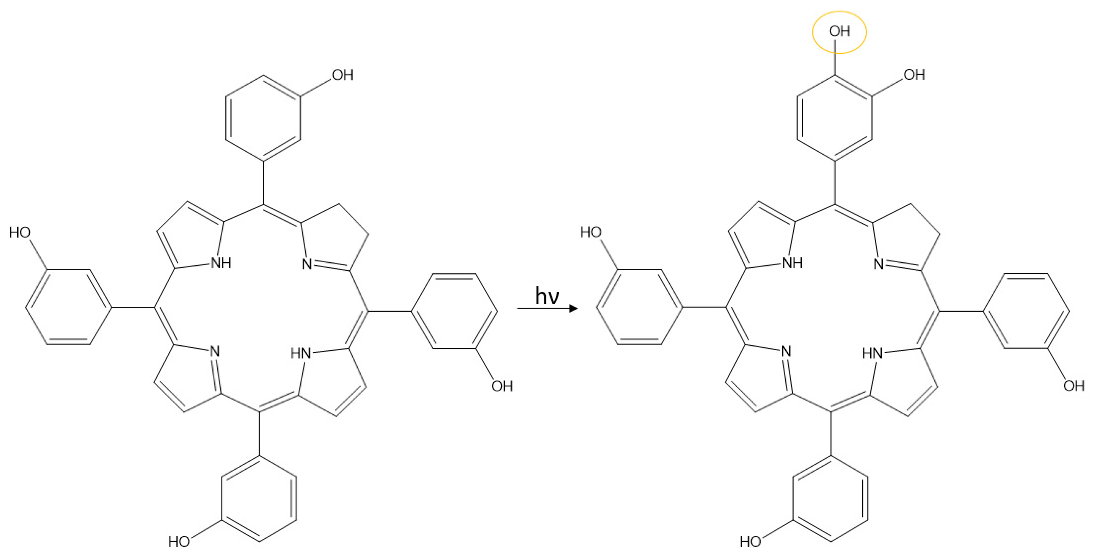
Temoporfin, or Foscan (5, 10, 15, 20 tetrakis (meso-hydroxyphenyl) clorin; m-THPC), is a photosensitizer used in PDT [14] and has thus often been studied. Analyses based on TLC, LC-MS, NMR, and ultraviolet–vis detection suggested isomeric photoproducts under the incorporation of one oxygen atom (Figure 7) [46,47]. The reduction of quinones was also proposed but eventually assigned as an artifact from MS analysis [47]. MALDI-TOF analyses detected mono-, di-, and trihydroxy derivatives of m-THPC as well as a photoproduct resulting from the opening of the reduced pyrrole ring [48]. Similar results (incorporation of up to two oxygen atoms) were obtained for m-THPP (5, 10, 15, 20 tetrakis (meso-hydroxyphenyl) porphyrin) with MALDI-FTICR-MS [49] and for m-THPBC (5, 10, 15, 20 tetrakis (meso-hydroxyphenyl) bacteriochlorin) with MALDI-TOF experiments [49].
The addition of two nominal mass units of 32 to the molecule was also described for 5, 10, 15, 20-tetraphenyl porphyrin; 5, 10, 15-tri (p-tolyl) porphyrin; and their zinc analogs using LC-MS. The proposed product was assigned, using ring opening, to a dehydrated zinc biladienone structure without further validation [50].

6. Conclusions

In this review, the current knowledge on validated chemical structures of photoproducts of porphyrins in general, and PPIX in particular, was assembled. The majority of the analyses of porphyrins are based on optical spectroscopy, which cannot unequivocally deduce a molecular structure. True structural analysis of photoproducts is sparse and increasingly performed with MS-based methods. Notably, the most comprehensive chemical analysis of the products of porphyrin irradiation experiments is also the oldest, and it was carried out to find a new synthesis method for spirographisporphin, starting with PPIX-dimethylester [37]. From this 60-year-old work, we now know the structure of PPP, which is the main photoproduct and is formed by the incorporation of two extra oxygen atoms into the molecule. Photo-oxidation is the dominant process in photobleaching, although photoreduction should be kept in mind for reactions in complex biological matrices. Total photodecomposition under ring opening generates a variety of products which are hard to follow analytically, because photobleaching is strongly dependent on the biological matrix and irradiation conditions. Thus, scientific experiments conducted under well-defined conditions only allow us to extrapolate to the true processes occurring in the matrix of interest. In order to gain the best insights into photobleaching in glioma or other tissues, surrogate test matrices need to be found, which come as close to the human sample as possible. We have been using pig brain tissue for such purposes [19], but even then, brain substructures may differ in composition and need to be considered separately for the best results. It is next to impossible to properly define the contributions of all fluorescing molecules in tissue. However, we can at least try to elucidate those molecules resulting from (pro)drugs which we administer, in our case, for FGR. These include intermediates from the 5-ALA-induced PPIX synthesis pathway and their photobleaching products.

Funding

This research received funding from Deutsche Krebshilfe (70116073, 70116288).

Data Availability Statement

No new data were created in this work.

Acknowledgments

The author is grateful for the collaboration with E. Suero-Molina (Neurosurgery, University Clinic Münster) in the Deutsche Krebshilfe project.

Conflicts of Interest

The author declares no conflicts of interest.

Abbreviations

The following abbreviations are used in this manuscript:
PPIXProtoporphyrin IX
PDTPhotodynamic therapy
MSMass spectrometry
FGRFluorescence-guided resection
5-ALA5-Aminolevulinic acid
PPPPhotoprotoporphyrin
m-THPC5, 10, 15, 20 tetrakis (meso-hydroxyphenyl) clorin
LCLiquid chromatography
MALDI-TOFMatrix-assisted laser desorption/ionization time-of-flight mass spectrometry
NMRNuclear magnetic resonance
FTICRFourier-transform ion cyclotron resonance

References

  1. Nikolaou, V.; Nikoloudakis, E.; Ladomenou, K.; Charalambidis, G.; Coutsolelos, A. Porphyrins—Valuable pigments of life. Front. Chem. Biol. 2024, 2, 1346465. [Google Scholar] [CrossRef]
  2. Lash, T.D. Origin of aromatic character in porphyrinoid systems. J. Porphyr. Phthalocyanines 2011, 15, 1093–1115. [Google Scholar] [CrossRef]
  3. Senge, M.O.; MacGowan, S.A.; O′Brien, J.M. Conformational control of cofactors in nature—The influence of protein-induced macrocycle distortion on the biological function of tetrapyrroles. Chem. Commun. 2015, 51, 17031–17063. [Google Scholar] [CrossRef]
  4. Brothers, P.J.; Senge, M.O. Fundamentals of Porphyrin Chemistry; Wiley: Hoboken, NJ, USA, 2022. [Google Scholar]
  5. Biesaga, M.; Pyrzynska, K.; Trojanowicz, M. Porphyrins in analytical chemistry. A review. Talanta 2000, 51, 209–224. [Google Scholar] [CrossRef] [PubMed]
  6. Gu, S.; Marianov, A.N.; Lu, T.; Zhong, J. A review of the development of porphyrin-based catalysts for electrochemical CO2 reduction. Chem. Eng. J. 2023, 470, 144249. [Google Scholar] [CrossRef]
  7. Gamelas, S.R.D.; Tomé, J.P.C.; Tomé, A.C.; Lourenço, L.M.O. Porphyrin-containing materials for photodegradation of organic pollutants in wastewaters: A review. Catal. Sci. Technol. 2024, 14, 2352–2389. [Google Scholar] [CrossRef]
  8. Song, H.; Liu, Q.; Xie, Y. Porphyrin-sensitized solar cells: Systematic molecular optimization, coadsorption and cosensitization. Chem. Commun. 2018, 54, 1811–1824. [Google Scholar] [CrossRef]
  9. Stummer, W.; Müther, M.; Spille, D. Beyond fluorescence-guided resection: 5-ALA-based glioblastoma therapies. Acta Neurochir. 2024, 166, 163. [Google Scholar] [CrossRef]
  10. Akbar, A.; Khan, S.; Chatterjee, T.; Ghosh, M. Unleashing the power of porphyrin photosensitizers: Illuminating breakthroughs in photodynamic therapy. J. Photochem. Photobiol. B 2023, 248, 112796. [Google Scholar] [CrossRef]
  11. Dar, U.A.; Shahnawaz, M.; Taneja, P.; Dar, M.A. Recent advances in main group coordination driven porphyrins: A comprehensive review. ChemistrySelect 2024, 9, e202304817. [Google Scholar] [CrossRef]
  12. Zanetti-Polzi, L.; Amadei, A.; Djemili, R.; Durot, S.; Schoepff, L.; Heitz, V.; Ventura, B.; Daidone, I. Interpretation of experimental Soret bands of porphyrins in flexible covalent cages and in their related Ag(I) fixed complexes. J. Phys. Chem. C 2019, 123, 13094–13103. [Google Scholar] [CrossRef]
  13. Pettersson, K.; Kilså, K.; Mårtensson, J.; Albinsson, B. Intersystem crossing versus electron transfer in porphyrin-based donor-bridge-acceptor systems: Influence of a paramagnetic species. J. Am. Chem. Soc. 2004, 126, 6710–6719. [Google Scholar] [CrossRef]
  14. Allamyradov, Y.; ben Yosef, J.; Annamuradov, B.; Ateyeh, M.; Street, C.; Whipple, H.; Er, A.O. Photodynamic therapy review: Past, present, future, opportunities and challenges. Photochem 2024, 4, 434–461. [Google Scholar] [CrossRef]
  15. Gautheron, A.; Bernstock, J.D.; Picart, T.; Guyotat, J.; Valdés, P.A.; Montcel, B. 5-ALA induced PpIX fluorescence spectroscopy in neurosurgery: A review. Front. Neurosci. 2024, 18, 1310282. [Google Scholar] [CrossRef]
  16. Stummer, W.; Baumgartner, R. ALA-PDT and ALA-FD in the brain: New prospects for treating malignant gliomas. Compr. Ser. Photochem. Photobiol. Sci. 2006, 7, 155–175. [Google Scholar]
  17. Yien, Y.Y.; Perfetto, M. Regulation of heme synthesis by mitochondrial homeostasis proteins. Front. Cell Dev. Biol. 2022, 10, 895521. [Google Scholar] [CrossRef] [PubMed]
  18. Uehlinger, P.; Zellweger, M.; Wagnières, G.; Juillerat-Jeanneret, L.; van den Bergh, H.; Lange, N. 5-Aminolevulinic acid and its derivatives: Physical chemical properties and protoporphyrin IX formation in cultured cells. J. Photochem. Photobiol. B 2000, 54, 72–80. [Google Scholar] [CrossRef]
  19. Walke, A.; Black, D.; Valdes, P.A.; Stummer, W.; König, S.; Suero-Molina, E. Challenges in, and recommendations for, hyperspectral imaging in ex vivo malignant glioma biopsy measurements. Sci. Rep. 2023, 13, 3829. [Google Scholar] [CrossRef]
  20. Spikes, J.D. Quantum yields and kinetics of the photobleaching of hematoporphyrin, Photofrin II, tetra(4-sulfonatophenyl)-porphine and uroporphyrin. Photochem. Photobiol. 1992, 55, 797–808. [Google Scholar] [CrossRef]
  21. Fyrestam, J.; Bjurshammar, N.; Paulsson, E.; Johannsen, A.; Oestman, C. Determination of porphyrins in oral bacteria by liquid chromatography electrospray ionization tandem mass spectrometry. Anal. Bioanal. Chem. 2015, 407, 7013–7023. [Google Scholar] [CrossRef]
  22. Walke, A.; Krone, C.; Stummer, W.; König, S.; Suero Molina, E. Protoporphyrin IX in serum of high-grade glioma patients: A novel target for disease monitoring via liquid biopsy. Sci. Rep. 2024, 14, 4297. [Google Scholar] [CrossRef] [PubMed]
  23. Benton, C.M.; Lim, C.K.; Moniz, C.; Jones, D.J.L. Porphyrinogen fragmentation profiles by ultra-high-performance liquid chromatography/electrospray ionisation tandem mass spectrometry. Rapid Commun. Mass Spectrom. RCM 2011, 25, 3749–3757. [Google Scholar] [CrossRef] [PubMed]
  24. Fateen, E.; Abd-Elfattah, A.; Gouda, A.; Ragab, L.; Nazim, W. Porphyrins profile by high performance liquid chromatography/electrospray ionization tandem mass spectrometry for the diagnosis of porphyria. Egypt. J. Med. Hum. Genet. 2011, 12, 49–58. [Google Scholar] [CrossRef]
  25. Walke, A.; Molina, E.S.; Stummer, W.; Koenig, S. Protoporphyrin IX analysis from blood and serum in the context of neurosurgery of glioblastoma. In Mass Spectrometry in Life Sciences and Clinical Laboratory; IntechOpen Ltd.: London, UK, 2021; p. 1. [Google Scholar]
  26. Suero Molina, E.; Black, D.; Walke, A.; Azemi, G.; D′Alessandro, F.; König, S.; Stummer, W. Unraveling the blue shift in porphyrin fluorescence in glioma: The 620 nm peak and its potential significance in tumor biology. Front. Neurosci. 2023, 17, 1261679. [Google Scholar] [CrossRef]
  27. The International Union of Pure and Applied Chemistry. IUPAC—Photooxidation (P04640). Available online: https://goldbook.iupac.org/terms/view/P04640 (accessed on 29 January 2025).
  28. Baptista, M.S.; Cadet, J.; Di Mascio, P.; Ghogare, A.A.; Greer, A.; Hamblin, M.R.; Lorente, C.; Nunez, S.C.; Ribeiro, M.S.; Thomas, A.H.; et al. Type I and type II photosensitized oxidation reactions: Guidelines and mechanistic pathways. Photochem. Photobiol. 2017, 93, 912–919. [Google Scholar] [CrossRef] [PubMed]
  29. Park, J.; Tang, H.; Zhang, P. Differentiation of Superoxide Radical Anion and Singlet Oxygen and Their Concurrent Quantifications by Nuclear Magnetic Resonance. Anal. Chem. 2023, 95, 5293–5299. [Google Scholar] [CrossRef]
  30. Klan, P.; Wirz, J. Photochemistry of Organic Compounds; Wiley & Sons Ltd.: Hoboken, NJ, USA, 2009; ISBN 978-1-4051-6173-2. [Google Scholar]
  31. Hayyan, M.; Hashim, M.A.; AlNashef, I.M. Superoxide ion: Generation and chemical implications. Chem. Rev. 2016, 116, 3029–3085. [Google Scholar] [CrossRef]
  32. Schweitzer, C.; Schmidt, R. Physical mechanisms of generation and deactivation of singlet oxygen. Chem. Rev. 2003, 103, 1685–1757. [Google Scholar] [CrossRef]
  33. Protti, S.; Ravelli, D.; Fagnoni, M. Wavelength dependence and wavelength selectivity in photochemical reactions. Photochem. Photobiol. Sci. 2019, 18, 2094–2101. [Google Scholar] [CrossRef]
  34. Rotomskis, R.; Bagdonas, S.; Streckyte, G. Spectroscopic studies of photobleaching and photoproduct formation of porphyrins used in tumor therapy. J. Photochem. Photobiol. B 1996, 33, 61. [Google Scholar] [CrossRef]
  35. Wessels, J.M.; Sroka, R.; Heil, P.; Seidlitz, H.K. Photodegradation of protoporphyrin-dimethylester in solution and in organized environments. Int. J. Radiat. Biol. 1993, 64, 475–484. [Google Scholar] [CrossRef]
  36. Fischer, H.; Herrle, K. Einwirkung von Licht auf Porphyrine. (I. Mitteilung.) Überführung von Ätio-porphyrin I in bilirubinoide Farbstoffe. Hoppe-Seyler´S Z. Physiol. Chem. 1938, 251, 85–96. [Google Scholar] [CrossRef]
  37. Inhoffen, H.H.; Bliesener, C.; Brockmann, H. Zur weiteren Kenntnis des Chlorophylls und des Hämins, VIII. Tetrahedron Lett. 1966, 7, 3779–3783. [Google Scholar] [CrossRef]
  38. Cox, G.S.; Whitten, D.G. Mechanisms for the photooxidation of protoporphyrin IX in solution. J. Am. Chem. Soc. 1982, 104, 516–521. [Google Scholar] [CrossRef]
  39. Cox, G.S.; Whitten, D.G. Excited state interactions of protoporphyrin IX and related porphyrins with molecular oxygen in solutions and in organized assemblies. Adv. Exp. Med. Biol. 1983, 160, 279–292. [Google Scholar] [CrossRef]
  40. Ericson, M.B.; Grapengiesser, S.; Gudmundson, F.; Wennberg, A.-M.; Larkö, O.; Moan, J.; Rosén, A. A spectroscopic study of the photobleaching of protoporphyrin IX in solution. Lasers Med. Sci. 2003, 18, 56–62. [Google Scholar] [CrossRef]
  41. Dysart, J.S.; Patterson, M.S. Photobleaching kinetics, photoproduct formation, and dose estimation during ALA induced PpIX PDT of MLL cells under well oxygenated and hypoxic conditions. Photochem. Photobiol. Sci. 2006, 5, 73–81. [Google Scholar] [CrossRef]
  42. Ogbonna, S.J.; Masuda, K.; Hazama, H. The effect of fluence rate and wavelength on the formation of protoporphyrin IX photoproducts. Photochem. Photobiol. Sci. 2024, 23, 1627–1639. [Google Scholar] [CrossRef]
  43. Reis, E.R.; Ferreira, L.P.; Nicola, E.M.D.; Borissevitch, I. Comparative study of phototoxicity of protoporphyrin IX synthetic and extracted from ssp Rattus novergicus albinus rats toward murine melanoma cells. Eur. Biophys. J. 2018, 47, 601–609. [Google Scholar] [CrossRef]
  44. Mauzerall, D. The photoreduction of porphyrins: Structure of the products. J. Am. Chem. Soc. 1962, 84, 2437–2445. [Google Scholar] [CrossRef]
  45. Rotomskis, R.; Streckyte, G.; Bagdonas, S. Phototransformations of sensitizers. 2. Photoproducts formed in aqueous solutions of porphyrins. J. Photochem. Photobiol. B 1997, 39, 172–175. [Google Scholar] [CrossRef]
  46. Jones, R.M.; Wang, Q.; Lamb, J.H.; Djelal, B.D.; Bonnett, R.; Lim, C.K. Identification of photochemical oxidation products of 5,10,15,20-tetra(m-hydroxyphenyl)chlorin by on-line high-performance liquid chromatography-electrospray ionization tandem mass spectrometry. J. Chromatogr. 1996, 722, 257–265. [Google Scholar] [CrossRef]
  47. Bonnett, R.; Martínez, G. Photobleaching of compounds of the 5,10,15,20-Tetrakis(m-hydroxyphenyl)porphyrin Series (m-THPP, m-THPC, and m-THPBC). Org. Lett. 2002, 4, 2013–2016. [Google Scholar] [CrossRef]
  48. Kasselouri, A.; Bourdon, O.; Demore, D.; Blais, J.C.; Prognon, P.; Bourg-Heckly, G.; Blais, J. Fluorescence and mass spectrometry studies of meta-tetra(hydroxyphenyl)chlorin photoproducts. Photochem. Photobiol. 1999, 70, 275–279. [Google Scholar] [CrossRef]
  49. Lassalle, H.-P.; Lourette, N.; Maunit, B.; Muller, J.-F.; Guillemin, F.; Bezdetnaya-Bolotine, L. MALDI-TOF mass spectrometric analysis for the characterization of the 5,10,15,20-tetrakis(m-hydroxyphenyl)bacteriochlorin (m-THPBC) photoproducts in biological environment. J. Mass Spectrom. 2005, 40, 1149–1156. [Google Scholar] [CrossRef]
  50. Golec, B.; Buczynska, J.; Nawara, K.; Gorski, A.; Waluk, J. Photodegradation of free base and zinc porphyrins in the presence and absence of oxygen. Photochem. Photobiol. Sci. 2023, 22, 2725–2734. [Google Scholar] [CrossRef]
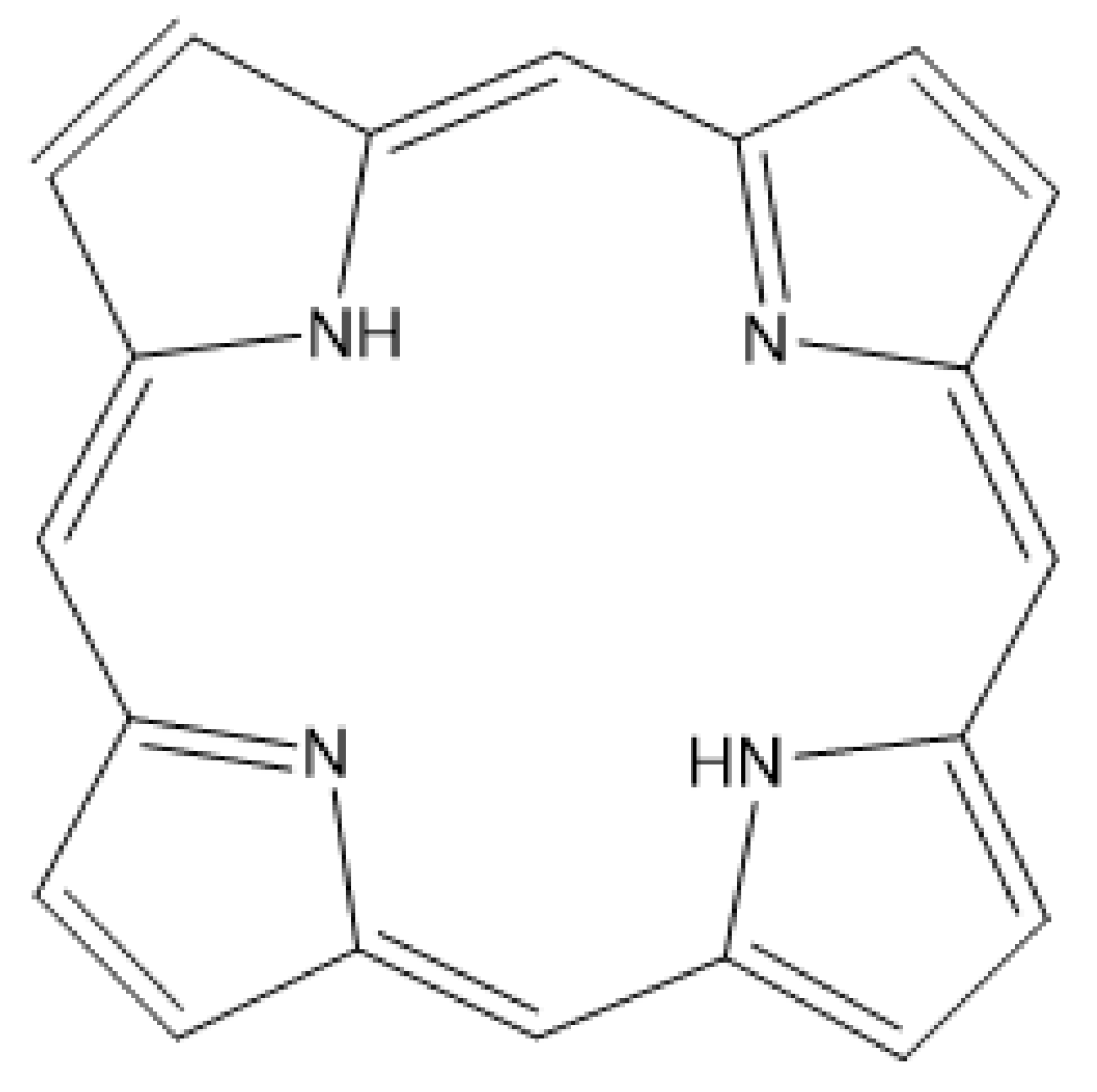
Figure 1. Porphin: the core structure of porphyrins.
Photochem 05 00010 g001
Figure 3. Mono- (A,B) and diformyl (C) photoproducts of PPIX-dimethylester as suggested by Cox and Whitten [38]. Modified sites are circled.
Photochem 05 00010 g003
Figure 4. Suggested mechanisms for the photo-oxidation of PPIX-diester (A) via the cycloaddition of oxygen to a diene unit or (B) via a dioxetane intermediate [38].
Photochem 05 00010 g004
Figure 5. Interaction of excited porphyrin with molecular oxygen in solution can give rise to both (A) singlet oxygen formation and (B) electron transfer to generate superoxide (for more information, see Ref. [38]).
Photochem 05 00010 g005
Figure 6. Structures of photoreduced porphyrins according to Mauzerall [44]. From the left: dihydroporphyrin/phlorin, tetrahydroporphyrin/porphomethene, and hexahydroporphyrin/porphyrinogen. For clarity, alkyl substituents on the ring carbons and hydrogens on the methine bridges were omitted. The yellow circles indicate modified positions (compared to porphin, Figure 1).
Photochem 05 00010 g006
Figure 7. Photobleaching of m-THPC leading to isomeric products based on the incorporation of an extra oxygen atom [46,47]. The yellow circle indicates the modified site.
Photochem 05 00010 g007
Disclaimer/Publisher’s Note: The statements, opinions and data contained in all publications are solely those of the individual author(s) and contributor(s) and not of MDPI and/or the editor(s). MDPI and/or the editor(s) disclaim responsibility for any injury to people or property resulting from any ideas, methods, instructions or products referred to in the content.