Article

Effect of Organic Acid Mixtures on the Extraction Efficiency, Physicochemical, and Thermal Properties of Pigskin Gelatin and Resulting Films

by
Diego Ezequiel Velazquez
1,2,3 and
María Emilia Latorre
3,4,*
1
Instituto de Física de Materiales Tandil (IFIMAT), Facultad de Ciencias Exactas, Universidad nacional del centro de la Provincia de Buenos Aires (UNCPBA), Tandil B7000, Argentina
2
Centro de Investigaciones en Física e Ingeniería (CIFICEN), Tandil B7000, Argentina
3
Consejo de Investigaciones Científicas y Técnicas (CONICET), Ciudad Autónoma de Buenos Aires C1425, Argentina
4
Centro de Investigación Veterinaria de Tandil (CIVETAN), Tandil B7000, Argentina
*
Author to whom correspondence should be addressed.
Physchem 2025, 5(3), 38; https://doi.org/10.3390/physchem5030038
Submission received: 10 June 2025 / Revised: 14 August 2025 / Accepted: 4 September 2025 / Published: 11 September 2025
(This article belongs to the Section Biophysical Chemistry)

Abstract

Animal tissue by-products, rich in collagen, represent a valuable source of biomaterials. Understanding their physicochemical and thermal behavior is essential for expanding their applications. In this study, pigskin gelatin was extracted through acid hydrolysis using a combination of acetic acid (AH) and either lactic, citric, or ascorbic acid (75:25, v:v, [0.5 M]), followed by thermal denaturation. We evaluated the physicochemical properties of the gelatin solutions (pH, hydroxyproline content, and extraction yield), as well as the macroscopic gel characteristics. Gelatin films were then prepared and analyzed for moisture content, color, and thermal properties. One-way ANOVA was applied to compare treatments, and Pearson’s correlation was used to assess the relationship between the solution pH and physicochemical parameters. Significant differences in the final pH of the solutions were observed among the acid mixture treatments, though the hydroxyproline content and extraction yield were not significantly affected. All gelatin solutions formed stable gels, and the resulting films exhibited similar moisture content. Thermal analysis revealed treatment-dependent variations. Specifically, a significant negative correlation (p < 0.005) was found between the gelatin solution pH and the melting temperature. These results suggest that the use of organic acid mixtures can effectively modulate gelatin properties, offering a versatile approach for tailoring biomaterials for both food and non-food applications.

1. Introduction

Gelatin is a protein-based biopolymer primarily derived from animal by-products such as hides, bones, and tendons, mainly from porcine and bovine sources. These collagen-rich tissues are low-cost and widely used in the food, pharmaceutical, and biomedical industries for the production of value-added materials, including collagen, gelatin, and bioactive peptides [1]. Hides, in particular, are abundant in type I collagen, which is composed of a triple helix structure consisting of two α1-chains and one α2-chain. Gelatin is obtained through the partial hydrolysis and thermal denaturation of collagen, a process that disrupts its tertiary and secondary structures and partially cleaves the non-covalent interactions within the primary structure [2].
The extraction process, hydrolysis and denaturation, plays a crucial role in determining the physicochemical characteristics of gelatin. Acidic hydrolysis is commonly used for porcine skins and leads to the production of type A gelatin, whereas alkaline pretreatment—typically applied to bovine hides—yields type B gelatin [3]. The fundamental structural unit of collagen, tropocollagen, is approximately 300 nm in length and 1.5 nm in diameter. During the extraction process, the collagen triple helix results in a heterogeneous mixture of polypeptide chains with a random coil conformation [4,5]. Gelatin typically exhibits a molecular weight distribution consisting of α-chains (~110 kDa), β-chains (~210 kDa), and γ-chains (~310 kDa), although remnants of native triple helices may remain after extraction [6,7].
When temperatures exceed 40–45 °C, gelatin’s polypeptide chains assume a disordered configuration in solution. A gel network is formed when concentrations exceed 5 mg/mL, as a result of partial renaturation that occurs upon cooling [5]. Biocompatibility, biodegradability, low antigenicity, and a variety of physical properties, such as gelling, thickening, emulsifying, foaming, and film-forming capabilities, are the primary factors contributing to its functional versatility [6,7].
Type A gelatin is obtained by acid extraction, presenting an isoelectric point in 6~9, and is the most used among the covalently bound collagens with a lesser cross-linking degree recognized by pigskin [3]. In recent years, there has been a growing interest in applying green technologies for gelatin extraction to improve efficiency while minimizing environmental impact. Among these, acid-assisted extractions using organic acids—such as citric or lactic acid—have emerged as sustainable alternatives to strong mineral acids [8].
Although acidic gelatin extraction from pigskin is well documented [2,9,10], all previous studies evaluated only single-acid pretreatments (e.g., acetic, sulfuric acid, phosphoric acid, citric). None have investigated the application of food-grade organic acid mixtures without subsequent neutralization. Table 1 summarizes these prior studies and highlights the methodological novelty of our approach.
Acetic acid (AH) is widely used for gelatin extraction due to its efficiency and the favorable properties it imparts to the final product [10,11,12,13,14]. However, the distinct properties of other acids—such as citric, lactic, ascorbic, hydrochloric, and sulfuric—may complement or enhance the functional effects of AH when used in combination [10,13].
To preserve the beneficial characteristics of AH while exploring potential synergistic effects, this study proposes evaluating the impact of a binary acid system consisting of 75% AH and 25% of a secondary food-grade organic acid, both applied at the same molar concentration (0.5 M), during the acid hydrolysis pretreatment step.
The effects of this acid combination were assessed by analyzing the PS characteristics, hydroxyproline content, gelatin extraction yield, and the color and thermal properties of the resulting gelatin-based films.

2. Materials and Methods

2.1. Raw Material Preparation

Pigskin (PS) used for gelatin extraction was obtained from male pigs (Duroc × Yorkshire; 6-month-old, animal weight approximately 200–220 kg). The frozen (Wilpool S.A., São Paulo, Brazil) PS was sourced from a local slaughterhouse. The PS, free from fat and hair and cut into approximately 5 × 5 mm, was weighed and washed with distilled water at a ratio of 1:5 (w:v; g:mL) for 2 h at room temperature to remove soluble globular proteins. The cleaned PS was then filtered, vacuum-packed, and stored at −18 °C until further use.

2.2. Gelatin Extraction by Acid Treatment

Gelatin (G) was extracted from PS following the method described by Velázquez et al. [13], with minor modifications. The PS samples underwent primary acid hydrolysis (0.5 M) using mixtures containing 75% acetic acid (AH) and 25% of one of three food-grade organic acids: lactic acid (AL), citric acid (AC), or ascorbic acid (AA). The evaluated treatments were AH:AL, AH:AC, and AH:AA (75:25, v:v), while a 0.5 M AH solution (100:0) was used as the control.
Gelatin and gelatin films obtained using only AH have been previously characterized [13]. For the hydrolysis step, the pigskin samples were immersed in the acid mixtures using a tissue-to-solution ratio of 1:5 (w:v) and incubated at 4 °C for 24 h with periodic manual stirring. After incubation, the tissues were filtered at room temperature, and the acid solutions were discarded. The soaked tissues were weighed to determine their swelling capacity, defined as the amount of acid solution absorbed.
Gelatin extraction was then performed through thermal denaturation and solubilization. Distilled water was added to the pretreated tissue at a ratio of 1:5 (w:v), and the mixture was heated at 90 °C for 90 min with intermittent stirring. The gelatin-rich solution was separated from the residual tissue by filtration at 45 °C. The resulting gelatin solution was stored at 4 °C for 10–12 h to allow for gelation and to determine its pH.
Prior to film formation, the total hydroxyproline content ([Hyp]) and total soluble protein content were quantified in each gelatin solution. Figure 1 represents the acid thermal extraction process employed to obtain gelatin from the different treatments. All extractions were conducted in duplicate (n = 2), and all subsequent physicochemical and thermal analyses were performed in triplicate (n = 3).

2.2.1. pH Analysis

The pH of the acid solutions and G solutions was measured using a pH meter (HANNA Edge® HI5222, manufactured in Romania; Woonsocket, RI 02895, USA). All measurements were performed in triplicate.

2.2.2. Degree of Pigskin Swelling

The degree of swelling of the tissue matrix was determined as the ratio between the mass of the acid solution retained and the initial mass of the tissue, both on a wet basis. Specifically, the wet mass of the tissue after acid hydrolysis and filtration (mS, g) was compared with the initial wet mass of the tissue (mi, g). Results are expressed as grams of acid solution per gram of wet tissue [g·g−1 (w.b.)].
P S S w e l l i n g = m S m i m i

2.2.3. Quantification of Total Hydroxyproline [Hyp]

The hydroxyproline [Hyp] content was quantified in the gelatin solutions and in the washed raw PS tissue. Samples were hydrolyzed in 6 N hydrochloric acid (HCl) at 110 °C for 16 h, using a sample-to-solvent ratio of 1:10 (m:v; g:ml). After hydrolysis, the samples were neutralized, and the [Hyp] content was determined using the colorimetric method described by Bergman and Loxley [15]. Colorimetric reactions were performed in duplicate for each sample. Results are expressed as milligrams of hydroxyproline per gram of dry base of PS [mg Hyp·g−1 (d.b.)].

2.2.4. Extraction Performance

The yield of the by-product G was evaluated as the quotient of the total collagen protein content in the G solution [Hyp-G] and the total collagen protein content present in PS [Hyp-T]. Both concentrations were determined according to the previously described method. The protein recovery yield from gelatin was calculated as follows:
% Y G = [   H y p G ] [ H y p T ] × 100

2.3. Preparation of Gelatin Films

The G solutions obtained from each treatment were used to prepare the films. Initially, the gelled G solutions were solubilized at 45 °C for 20 min, followed by the addition of glycerol (40% w/w) as a plasticizer. The mixtures were maintained at 45 °C for 30 min under constant stirring.
Subsequently, 15 mL of each solution was poured into silicone molds (per well), and the films were obtained by dehydration in an oven at 37.5 °C for 24 h. The resulting films were stored under vacuum in polyethylene bags at 4 °C.
The moisture content, thermal properties, and color of the films were evaluated for each gelatin film

2.3.1. Moisture Content of Gelatin Films

The gelatin films were oven-dried at 37 °C for 48 h until reaching a constant weight. The initial weight (m0) and final weight (mf) of each film were recorded. The moisture content (m.c.) was calculated using the following equation:
% m . c . = ( m 0   m f ) m 0 × 100

2.3.2. Differential Scanning Calorimetry (DSC)

The thermal properties of the gelatin films (G-films) were evaluated using a DSC Setaram® Evo 131, manufactured in Lyon, France. Disc-shaped samples (~10 mg) were oven-dried at 37 °C for 24 h prior to analysis and placed in sealed aluminum crucibles.
DSC measurements were performed using a heating rate of 10 °C·min−1 over a temperature range of 25 to 300 °C, with argon used as the purge gas. An empty crucible was used as the reference.
A third-degree polynomial function was applied for baseline correction. The thermal parameters analyzed included the enthalpy of denaturation (ΔH), glass transition temperature (Tg), and melting temperature (Tm). All measurements were conducted in triplicate (n = 3).

2.3.3. Color Analysis of Gelatin Films

The color of the G-films was evaluated using a Minolta Chroma CR-400 colorimeter (Minolta Co., Ltd., Osaka, Japan). Samples were placed on a white calibration tile, and the chromaticity coordinates a* and b*, as well as lightness L*, were measured according to the CIELAB color space, using a D65 illuminant and a standard observer angle of 2°. Color measurements were taken at five different points on each film (n = 5).

2.4. Statistical Analysis

Results are presented as mean values, standard deviation (±sd), and standard error of the mean (SE). Comparisons between treatments were conducted using one-way analysis of variance (ANOVA), followed by Tukey’s post hoc test (α = 0.05). Additionally, a Pearson correlation matrix (α = 0.05) was generated to assess correlations between all analyzed parameters and the pH of the gelatin solutions. All statistical analyses were performed using GraphPad Prism version 5.00 for Windows (GraphPad Software, San Diego, CA, USA; http://www.graphpad.com).

3. Results and Discussion

The pH values of the acid mixtures used in the hydrolysis stage were consistent with the theoretical estimates calculated for mixed-acid solutions (Table 2). PS swelling differed significantly between the control (AH 100:0) and the mixed-acid treatments. In particular, hydrolysis with AH:AL produced greater swelling than AH:AA. These differences were not directly related to the pH of the acid solutions. Acid effects on swelling are well known; in particular, the acid dissociation constant (pKa) plays a critical role in determining the swelling degree of a material such as a polymer or hydrogel [16,17]. The swelling behavior of polyelectrolytes, including those present in food matrices and biological tissues, is strongly pH-dependent. In the present study, the pKa values of the acids, together with their concentrations, determined the final pH of the mixed-acid solutions. The pH, in turn, dictates the charge state of the surrounding biopolymers and therefore their swelling degree. As the pH moves away from the protein isoelectric point (pI), the number of charged groups on the polymer increases, leading to greater electrostatic repulsion. This repulsion causes the polymer chains to expand, thus increasing swelling. According to Wang and Hu [18], the pI of raw hide or skin is approximately 7.7–7.9. All acids evaluated in this work produced solution pH values lower than the pI of skin tissue, indicating that swelling differences were only slightly affected by the pH of the solutions (pKa of the mixed acids).
We hypothesize that this discrepancy arises from the intrinsic complexity of PS tissue compared with simpler hydrogel systems. Pigskin is a composite biological material composed predominantly of collagen, but it also contains substantial amounts of elastin and proteoglycans. Moreover, the collagen in pigskin is arranged in a three-dimensional woven fiber network [19], which may modulate its swelling behavior in ways that differ from those observed in isolated proteins or synthetic hydrogels.
Hydrolysis is a critical step in gelatin production, as the interaction between acid and collagen, along with the pH, temperature, and the concentrations of both acid and substrate, governs the physicochemical properties of the final product [1]. Chemical hydrolysis destabilizes intra- and intermolecular cross-links in collagen fibers (tropocollagen) [2] and removes non-collagenous compounds. During acid hydrolysis, part of the collagen in PS becomes solubilized (soluble collagen—not assessed in this study), while a larger fraction remains insoluble. The pretreatment serves two main purposes: (i) to disrupt the collagen structure by breaking covalent and hydrogen bonds, and (ii) to remove non-collagenous material, as reported in similar extraction processes [20]. In this study, acid hydrolysis cleaved peptide bonds within collagen chains, partially denaturing the triple helix and enabling gelatin solubilization during subsequent hot-water extraction (>60 °C).
The resulting gelatin solution pH values are shown in Table 2. The gelatin pH solution followed the same trend as that of the initial acid treatments: pH_G: AH:AC < AH:AL < AH:AA < AH (control). Table 3 presents the results for the total hydroxyproline content ([Hyp], mg·g−1, d.b.) and gelatin yield (%YG). No significant differences were observed among treatments for either variable.
Pearson correlation analysis (Table 4) revealed no statistically significant associations between the evaluated variables and the pH of the gelatin solutions (pH-G) within the tested range (3.3–4.0). The lack of correlation indicates that the pH did not significantly influence the hydroxyproline concentration ([Hyp]) or gelatin yield (%YG) under the experimental conditions. These results suggest that the slight pH variations among the organic acid mixtures used in this study were not a determining factor for either parameter.
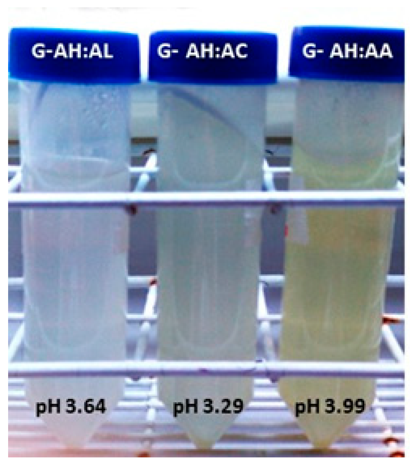
The G solutions obtained were stored at 4 °C for 24 h, and their gelling properties were evaluated prior to film formation. Visual inspection showed that all treatments exhibited good gelling capacity, with slight differences in opacity and color tone (Figure 2). All gels remained stable at room temperature and underwent solubilization only at temperatures above 40–45 °C. It is well established that attractive forces such as hydrogen bonding and hydrophobic interactions predominate under conditions of a slight positive net charge, facilitating the formation of a three-dimensional network and a stable gel [21]. According to Goudie et al. [22], gels formed at optimal pH values (pH < isoelectric point, pI) exhibit enhanced gel-forming ability. For PS gelatin, this optimal pH is slightly acidic, approximately 4–6 [23].
The G solutions were used to produce films following the procedure described in Section 2.2, and their physicochemical and thermal properties were subsequently evaluated. The moisture content of the resulting G-based films showed minor variations among treatments; however, these differences were not statistically significant (Table 5).
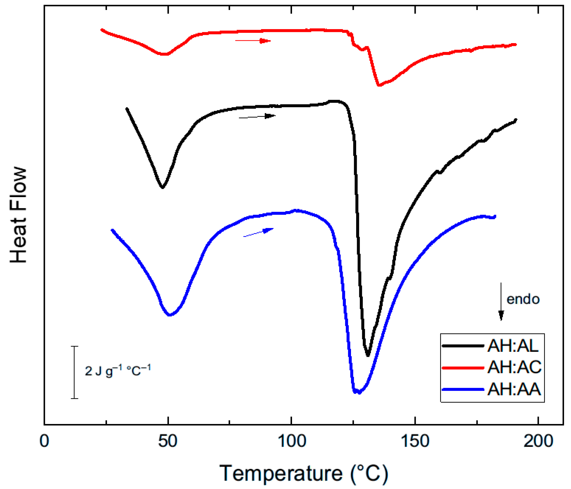
The results of the thermal properties of the G-films (Table 5) showed significant differences in the glass transition temperature (Tg), melting temperature (Tm), and enthalpy change (ΔH). Within the studied temperature range, the DSC curves displayed two well-defined endothermic signals during heating (see Figure 3), consistent with observations for low-moisture gelatin films [24,25,26]. The first signal, relatively smaller and at lower temperatures (<60 °C), corresponds to a glass transition, associated with the mobility of amorphous regions (Tg). This transition should theoretically manifest itself as a step in the heat capacity. However, in general, it appears (as here) as a peak, since it develops in superposition with other phenomena. In this case, it is very likely that the endothermic signal detected also contains the heat of the denaturation of triple helices renatured upon cooling the gelatin and matured during drying of the films. This hypothesis is consistent with the fact that this first signal is less intense in the AH:AC films, which contain higher [H+], and therefore offer greater electrostatic resistance to helix reformation.
The second endothermic signal, more intense and occurring at higher temperatures (>120 °C), is due to a first-order transition linked to the melting of the crystalline junction regions (Tm), and to the loss of strongly bound water, according to thermogravimetric measurements carried out in non-hermetic crucibles, such as those used in this work [26].
It is known that the Tg and Tm values of gelatin films depend strongly on their moisture content [24,25]. The values found in this study are within the range expected for films with a moisture content (m.c.) of approximately 10%, in coincidence with the m.c. determined for our samples (Table 5).
A significant negative correlation was observed between Tm and pH-G (Table 3), indicating that the melting temperature of crystalline junction zones is affected by the pH-G within the studied pH range (3.23–3.99). As expected, the pH affects the charges of the gelatin chains, influencing intermolecular electrostatic interactions and, therefore, the thermal stability of the films. The correlation found shows that G-films with pH levels farther from the pI have a lower Tm, due to greater electrostatic repulsion. On the other hand, the Tg and ΔH did not show a significant correlation with the pH of the G solutions used to obtain the G-films (Table 4). However, the significant differences found between acid mixtures suggest that each organic acid may uniquely influence this parameter. It would be interesting to further investigate this analysis over a wider range of pHs and compositions of the acid mixtures used.
Previous results by Velázquez et al. [13] showed that the presence of AL, AC, and AA in mixtures with AH altered the thermodynamic parameters of gelatin derived from PS. The acid intrinsic characteristics appear to reflect alterations in hydrophobic interactions and hydrogen bonding within the protein matrix, as well as in chain mobility, thus influencing the thermal transitions of G-films. These differences suggest that the type of organic acid used in combination with AH may affect the number of hydrophobic and/or hydrogen bond interactions in the protein matrix and/or the mobility of the chains, thus modifying the thermal properties of the G-films.
The color parameters of the G-films showed clear differences between the AH:AA treatment and the AH mixtures containing AL or AC (Table 6). The color changes observed with the addition of ascorbic acid (AA) during gelatin extraction are consistent with previous findings by Velázquez et al. [13], who examined the individual effects of each acid. The presence of AA in the acid mixture (AH:AA 75:25) had a pronounced impact on the G-film color. The resulting brown-yellow hue, characterized by low L* values and strong red (+a*) and yellow (+b*) tones, suggests that AA underwent oxidation during film dehydration. Ascorbic acid is a labile, water-soluble compound that degrades with heat and in the presence of air at room temperature. Furthermore, AA solutions degrade readily when stored at ambient conditions [27]. According to Yin et al. [28], AA is unstable in aqueous solutions, and its degradation is a major cause of quality and color changes during food processing and storage. These authors emphasize that stability analysis of AA is critical for its proper application. Therefore, to better understand AA oxidation and its interaction with proteins, further studies are necessary to evaluate the influence of external factors and the effect of the AA concentration during the PS acid hydrolysis step.

4. Conclusions

The incorporation of secondary organic acids in combination with acetic acid (AH) induced measurable changes in the properties of the resulting gelatin solutions and gelatin-based films.
The pH of the acid solution significantly influenced the final pH of the gelatin solution following pigskin (PS) hydrolysis and the thermal treatment. Although the tissue exhibited differences in swelling after acid hydrolysis, this response was not correlated with the pH of the acid mixture. Importantly, neither the gelatin yield nor the total hydroxyproline (Hyp) content in the gelatin solution was significantly affected by the acid treatment. The films produced from these gelatin solutions displayed notable differences in their thermal properties and color. Specifically, a significant negative correlation was observed between the pH of the gelatin solution (pH-G) and the melting temperature (Tm) of the gels, indicating that the melting of the crystalline junction zones is influenced by pH-G within the studied range (3.23–3.99). Finally, the presence of ascorbic acid during acid hydrolysis caused marked changes in the color of the resulting gelatin films. Further investigations are required to elucidate the molecular mechanisms underlying these differences. In particular, the chemical nature of the acids and their interactions with collagen during hydrolysis and film formation appear to play a critical role in determining the structural and thermal behavior of the resulting gelatin materials.

Author Contributions

D.E.V.: Conceptualization; investigation of part of the study; writing—original draft and review; validation. M.E.L.: Conceptualization; investigation; methodology; formal analysis of part the study; software; supervision; validation; writing—original draft; review and editing. All authors have read and agreed to the published version of the manuscript.

Funding

The authors declare that no financial support was received for the research, authorship, or publication of this article.

Data Availability Statement

The raw data supporting the conclusions of this article will be made available by the authors on request.

Acknowledgments

The authors are grateful for the support provided by the National Council for Scientific and Technical Research (CONICET), the National University of the Center of the Province of Buenos Aires (UNCPBA), and the National Agency for Scientific and Technical Promotion (ANPCyT). During the preparation of this manuscript, the authors used open access Gemini and chat GTP- Free version for the purposes of superficial text editing. The authors have reviewed and edited the output and take full responsibility for the content of this publication.

Conflicts of Interest

The authors declare no conflicts of interest.

References

  1. Ahmad, M.; Benjakul, S.; Nalinanon, S.; Sumpavapol, P. Gelatin from Lizardfish (Saurida tumbil) Skin: Properties as Affected by Extraction Conditions. Food Hydrocoll. 2010, 24, 616–622. [Google Scholar]
  2. See, S.F.; Ghassem, M.; Mamot, S.; Babji, A.S. Effect of Different Pretreatment on Functional Properties of African Catfish (Clarias gariepinus) Skin Gelatin. J. Food Sci. Technol. 2015, 52, 753–760. [Google Scholar] [CrossRef]
  3. Lu, Y.; Luo, Q.; Chu, Y.; Tao, N.; Deng, S.; Wang, L.; Li, L. Application of Gelatin in Food Packaging: A Review. Polymers 2022, 14, 436. [Google Scholar] [CrossRef]
  4. Ahmad, M.; Benjakul, S. Characteristics of Gelatin from the Skin of Unicorn Leatherjacket (Aluterus monoceros) as Influenced by Extraction Conditions. Food Hydrocoll. 2011, 25, 381–388. [Google Scholar] [CrossRef]
  5. Roussenova, M.; MacNab, M.; Murray, B.S. Effect of Gelatin Type and Bloom Strength on Gelation Behavior and Rheology. Food Hydrocoll. 2014, 35, 575–584. [Google Scholar]
  6. Duconseille, A.; Astruc, T.; Quintana, N.; Meersman, F.; Sante-Lhoutellier, V. Gelatin Structure and Composition Linked to Functional Properties: A Review. Crit. Rev. Food Sci. Nutr. 2017, 57, 1133–1143. [Google Scholar]
  7. Alipal, J.; Jamilah, B.; Hashim, D.M.; Sahilah, A.M. Effects of Different Drying Methods on the Properties of Fish Gelatin Films. Food Hydrocoll. 2021, 112, 106267. [Google Scholar]
  8. Noor, N.Q.I.M.; Razali, R.S.; Ismail, N.K.; Ramli, R.A.; Razali, U.H.M.; Bahauddin, A.R.; Zaharudin, N.; Rozzamri, A.; Bakar, J.; Shaarani, S.M. Application of Green Technology in Gelatin Extraction: A Review. Processes 2021, 9, 2227. [Google Scholar] [CrossRef]
  9. Cao, Y.; Zhang, Y.; Xia, W. Preparation and Characterization of Gelatin from Tilapia Skin Using Combined Pretreatments. Food Biosci. 2020, 35, 100580. [Google Scholar]
  10. Yang, Z.-X.; Sha, X.-M.; Wang, H.; Fang, T.; Shu, S.; Tu, Z.-C. Effect of acid pretreatments with various acid types on gelling properties and identification characteristics of pigskin gelatin. Food Chem. X 2025, 26, 102211. [Google Scholar] [CrossRef] [PubMed]
  11. Sompie, M.; Surtijono, S.E.; Pontoh, J.H.W.; Lontaan, N.N. The Effects of Acetic Acid Concentration and Extraction Temperature on Physical and Chemical Properties of Pigskin Gelatin. Procedia Food Sci. 2015, 3, 383–388. [Google Scholar] [CrossRef]
  12. Mishra, B.P.; Maheswarappa, N.B.; Rao, B.E.; Banerjee, R.; Mallika, E.N.; Rao, T.S.; Nath, D.N.; Prasad, M.G.; Belore, B.M.; Dasoja, S.; et al. Extraction And Characterization Of Pig Skin Gelatin Compared To Commercial Porcine Gelatin. J. Meat Sci. 2023, 18, 36–42. [Google Scholar] [CrossRef]
  13. Velázquez, L.; Sanchez, D.; Latorre, M.E. Pigskin Treatment Using Different Food-Grade-Acids: Effects on The Physicochemical Characteristics of The By-Products. Food Sci. J. 2023, 5, 103–115. [Google Scholar] [CrossRef]
  14. Lestari, N.; Manalu, L.P.; Hidayat, T.; Junaidi, L.; Hartanto, E.S.; Rienoviar; Saputra, S.H.; Zulham, A.; Mala, D.M.; Dewi, C.; et al. The effect of citric and acetic acid treatment on gelatin production from catfish skin. BIO Web Conf. 2024, 87, 03004. [Google Scholar] [CrossRef]
  15. Bergman, M.; Loxley, R. Two improved and simplified methods for the spectrophotometric determination of hydroxyproline. Anal. Chem. 1963, 35, 1961–1965. [Google Scholar] [CrossRef]
  16. Gupta, N.V.; Shivakumar, H.G. Investigation of Swelling Behavior and Mechanical Properties of a pH-Sensitive Superporous Hydrogel Composite. Iran. J. Pharm. Res. 2012, 11, 481–493. [Google Scholar] [PubMed] [PubMed Central]
  17. Liu, Z.; Basem, A.; Mostafa, L.; Jasim, D.J.; Al-Rubaye, A.H.; Salahshour, S.; Hekmatifar, M.; Esmaeili, S. Investigating the effect of pH on the swelling process, mechanical and thermal attributes of polyacrylamide hydrogel structure: A molecular dynamics study. Case Stud. Therm. Eng. 2024, 55, 104148. [Google Scholar] [CrossRef]
  18. Wang, Y.; Hu, L. Essential role of isoelectric point of skin/leather in leather processing. J. Leather Sci. Eng. 2022, 4, 25. [Google Scholar] [CrossRef]
  19. Andrei, W.; Quan, H.; Brown, K.A.; Williams, A.; Proud, W.G.; Meyers, M.A. Tensile behavior and structural characterization of pig dermis. Acta Biomater. 2019, 86, 77–95. [Google Scholar] [CrossRef] [PubMed]
  20. Joy, J.M.; Padmaprakashan, A.; Pradeep, A.; Paul, P.T.; Mannuthy, R.J.; Mathew, S. A Review on Fish Skin-Derived Gelatin: Elucidating the Gelatin Peptides—Preparation, Bioactivity, Mechanistic Insights, and Strategies for Stability Improvement. Foods 2024, 13, 2793. [Google Scholar] [CrossRef]
  21. Yang, Y.L.; Leone, L.M.; Kaufman, L.J. Elastic moduli of collagen gels can be predicted from two-dimensional confocal microscopy. Biophys. J. 2009, 97, 2051–2060. [Google Scholar] [CrossRef] [PubMed]
  22. Goudie, K.J.; McCreath, S.J.; Parkinson, J.A.; Davidson, C.M.; Liggat, J.J. Investigation of the influence of pH on the properties and morphology of gelatin hydrogels. J. Polym. Sci. 2023, 61, 2316. [Google Scholar] [CrossRef]
  23. Koli, J.; Basu, S.; Nayak, B.; Patange, S.B.; Pagarkar, A.; Gudipati, V. Effect of pH and ionic strength on functional properties of fish gelatin in comparison to mammalian gelatin. Fish. Technol. 2013, 50, 126–132. [Google Scholar]
  24. Slade, L.; Levine, H. Polymer-chemical properties of gelatin in foods. In Advances in Meat Research, Vol. 4: Collagen as a Food; Pearson, T.R., Dutson, A.M., Bailey, A., Eds.; AVI Publishing: Westport, CT, USA, 1987; pp. 283–311. [Google Scholar]
  25. Sobral, P.J.D.A.; Habitante, A.M.Q.B. Phase transitions of pigskin gelatin. Food Hydrocoll. 2001, 15, 377–382. [Google Scholar] [CrossRef]
  26. Mukherjee, I.; Rosolen, M. Thermal transitions of gelatin evaluated using DSC sample pans of various seal integrities. J. Therm. Anal. Calorim. 2013, 114, 1161–1166. [Google Scholar] [CrossRef]
  27. Nicolaï, B.; Barrio, M.; Tamarit, J.; Céolin, R.; Rietveld, I. Thermal expansion of L-ascorbic acid. Eur. Phys. J. Spec. Top. 2017, 226, 905–912. [Google Scholar] [CrossRef]
  28. Yin, X.; Chen, K.; Cheng, H.; Chen, X.; Feng, S.; Song, Y.; Liang, L. Chemical Stability of Ascorbic Acid Integrated into Commercial Products: A Review on Bioactivity and Delivery Technology. Antioxidants 2022, 11, 153. [Google Scholar] [CrossRef] [PubMed] [PubMed Central]
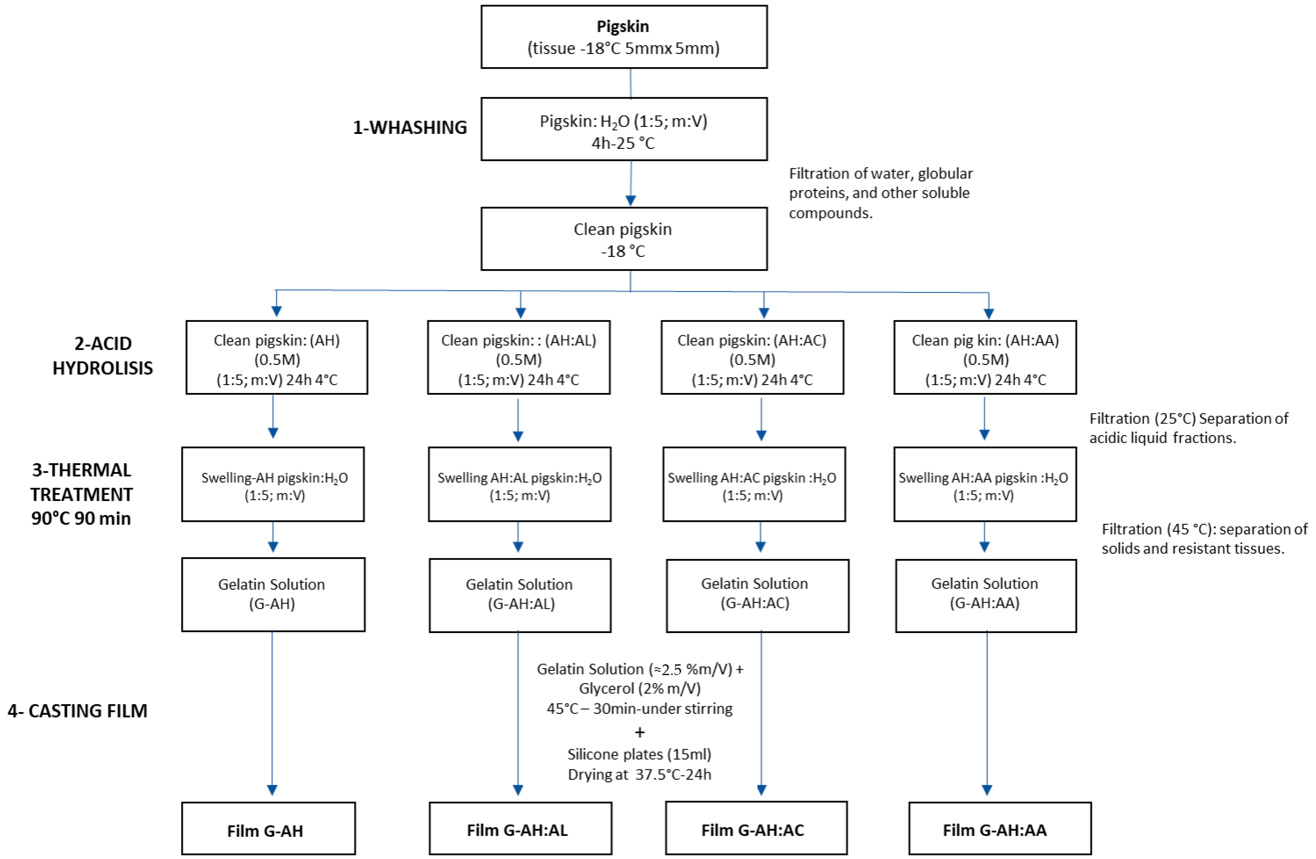
Figure 1. Schematic representation of the extraction process of gelatin (G) from pigskin (PS) using different acid pretreatments.
Physchem 05 00038 g001
Figure 2. Gels formed from G solutions obtained through AH:AL, AH:AC, and AH:AA treatments (75:25; v:v, 0.5 M).
Physchem 05 00038 g002
Figure 3. Representative DSC curves of the G-films. The color arrows indicate the way of the experiments (heating).
Physchem 05 00038 g003
Table 1. Summary of acid pretreatment-based gelatin extraction from pigskin in the literature.
PaperAcid TreatmentThermal TreatmentResults ReportedNotes
Sompie et al. [11] Acetic acid (0.5 M)
cc. 2, 4, and 6% (v/v) 24 h
3 h—50, 55, and 60 °CYield, protein gelatin. pH;
viscosity strength.
Acid solution neutralized (pH 6) before thermal treatment.
Mishra et al. [12]Acetic acid
(0.5 M) 24 h
6 h—60 °CYield, hydroxyproline, pH,
proximate composition;
sensory; water activity.
Acid solution neutralized (pH 7) before thermal treatment.
Velázquez et al. [13]Acetic, citric, lactic,
ascorbic acid (0.5 M)
24 h 4 °C
90 min
85–90 °C
Yields, Hyp content, film color, and thermal properties. Thermal treatment without solution neutralization.
Yang et al. [10] Acetic acid, citric acid
HCl, H2SO4 (0.2 M) 4 h—60 °C
12 h—60 °C Gel strength; MW
distribution, peptides analysis.
Acid solution neutralized before thermal treatment.
Table 2. Hydrogen ion concentration [H+] calculated from the acid dissociation constant and acid concentration, measured pH of the 0.5 M acid solution, PS swelling after acid hydrolysis, pH of the gelatin solution after thermal extraction, and hydroxyproline content of the gelatin solution (mg Hyp/mL). Values are expressed as mean ± standard deviation. One-way ANOVA (p-values) followed by Tukey’s post hoc test (α = 0.05). Different superscript letters within the same column indicate significant differences (p < 0.05).
Treatment [H+] *Acid Solution (0.5 M) pHPSSwellingGelatin Solution pHmg Hyp.ml−1 G Solution
AH (100:0)2.96 × 10−32.55 ± 0.07 a3.02 ± 0.07 c4.13 ± 0.04 a2.81 ± 0.57
AH:AL (75:25)8.82 × 10−32.12 ± 0.01 b4.06 ± 0.16 b3.64 ± 0.02 b2.50 ± 0.15
AH:AC(75:25)3.05 × 10−21.74 ± 0.01 c3.55 ± 0.32 ab3.29 ± 0.02 c2.61 ± 0.16
AH:AA(75:25)6.52 × 10−32.31 ± 0.01 d3.40 ± 0.13 a3.99 ± 0.01 d2.63 ± 0.27
* [H+] = √((Ka1×C1] + Ka2 × [C2])); PSSwelling = (g PSS. g−1 PS0 (w.b.)].
Table 3. Hydroxyproline content in gelatin (G) [mg of Hyp. g−1 PS, dry basis (d.b.)], and G yield (YG%) calculated as the ratio between [Hyp] in the G fraction and [Hyp] in the PS tissue. Mean values and standard deviations (±). One-way ANOVA was applied (p-values).
Treatmentmg HypG. g−1 PS (d.b.)%YG
AH (100:0)57.11 ± 8.2475.1 ± 10.8
AH:AL (75:25)55.70 ± 3.7973.2 ± 4.89
AH:AC (75:25)55.63 ± 4.4973.1 ± 5.90
AH:AA (75:25)55.75 ± 1.3673.3 ± 1.70
p-value0.97040.7081
Table 4. Pearson correlation coefficients between gelatin solution pH and pH of acid solution used for PS pretreatment, gelatin yield (YG), hydroxyproline content in the gelatin solution [Hyp G], film thermal properties (Tg, Tm; ΔH), moisture content (m.o.) of gelatin films, and color parameters of gelatin film.
Pearson
Parameters
pH Acid Solutions%YG[Hyp]TgTm∆Hm.o.L*a*b*
r0.9800.1000.0820.621−0.780−0.0230.510−0.2800.3300.340
95% confidence interval0.90 to 1.0−0.50 to 0.64−0.37 to 0.51−0.074 to 0.91−0.94 to −0.34−0.64 to 0.62−0.52 to 0.93−0.64 to 0.18−0.13 to 0.68−0.12 to 0.68
R squared0.960.010.00680.390.610.000530.260.080.110.11
p-value
P-(two-tailed)<0.00010.75150.73020.07440.00450.94990.30340.22690.14910.1472
P-summary valuesnsnsnssnsnsnsnsns
(α = 0.05)YesNoNoNoYesNoNoNoNoNo
s: significant; ns: no significant.
Table 5. Hydrogen ion concentration [H+] calculated from the gelatin solution pH, relative moisture content (%m.c.), and DSC results of G-films. [H+]G = 10−pHG; % m.c.: (g H2O·100 g−1 G-film); Tg: glass transition temperature (°C); Tm: melting temperature (°C); ΔH: thermal transition enthalpy (J·g−1 G-film).
Treatment[H+]G%m.c. *
(g H2O.100 g−1)
Tg *
(°C)
Tm *
(°C)
ΔH *
(J.g−1)
AH:AL (75:25)2.29 × 10−410.47 ± 0.60 48.6 ± 0.5 a131.1 ± 0.9 ab180 ± 14.6 b
AH:AC (75:25)5.12 × 10−411.79 ± 0.56 49.1 ± 1.5 ab139.0 ± 3.2 a56.7 ± 23.4 a
AH:AA (75:25)1.02 × 10−413.29 ± 0.13 51.0 ± 0.2 b123.9 ± 5.0 b130 ± 31.1 ab
p-value<0.0010.08220.03670.02390.0038
* Mean values and standard deviations (±). One-way ANOVA and Tukey’s post hoc test (α = 0.05). Different letters indicate statistically significant differences between treatments.
Table 6. Color variables in G-films: lightness (L*); red–green coordinate (a*); yellow–blue coordinate (b*), and G-film images.
TreatmentL*a*b*Films
AH:AL (75:25)84.7 ± 0.43 a−1.64 ± 0.02 a11.6 ± 0.83 aPhyschem 05 00038 i001
AH:AC (75:25)85.0 ± 1.33 a−1.29 ± 0.28 a9.58 ± 2.50 aPhyschem 05 00038 i002
AH:AA (75:25)74,7 ± 1.74 b3.59 ± 1.27 b45.2 ± 3.79 cPhyschem 05 00038 i003
p-value<0.001<0.001<0.001
Mean values and standard deviations (±). One-way ANOVA and Tukey’s post hoc test (α = 0.05). Different letters indicate statistically significant differences among treatments.
Disclaimer/Publisher’s Note: The statements, opinions and data contained in all publications are solely those of the individual author(s) and contributor(s) and not of MDPI and/or the editor(s). MDPI and/or the editor(s) disclaim responsibility for any injury to people or property resulting from any ideas, methods, instructions or products referred to in the content.